영남의대병리학교실 최준혁 - 영남대학교 의과대학...
TRANSCRIPT

Gastrointestinal stromal tumor
영남의대 병리학교실
최준혁

Classification of gastrointestinal mesenchymal tumor
• Gastrointestinal stromal tumor(GIST)• Smooth muscle tumors : leiomyoma, leiomyosarcoma• Neurogenic tumors : schwannoma, neurofibroma,
granular cell tumor, ganglioneuroma,MPNST
• Vascular tumors : hemangioma, lymphangioma, glomustumor, angiosarcoma, Kaposi sarcoma
• Lipomatous tumors : lipoma, liposarcoma

Historical overview :Gastrointestinal mesenchymal tumors
1941 Golden,et al Smooth muscle tumor (leiomyoma, leiomyosarcoma)
1983 Mazur,et al Gastric stromal tumor- lack of smooth muscle differentiation
1984 Herrera,et al Plexosarcoma(or GANT) - autonomic nerve differentiation
1998 Hirota,et al Gastrointestinal stromal tumor(GIST) - interstitial cells of Cajal phenotype differentiation- c-kit gene mutation
1998 Kindblom,et al Gastrointestinal pacemaker tumor

Interstitial cells of Cajal (ICC)
Origin Multipotential mesenchymal stem cells Function Control of gut motilitySite Between autonomic nerve and muscle
wall of GITMorphology Fusiform, stellateProto-oncogene c-kit gene expression Immunostain c-kit(CD117) protein

Interstitial cell of Cajal, small intestine
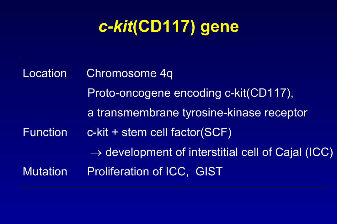
c-kit(CD117) gene
Location Chromosome 4qProto-oncogene encoding c-kit(CD117), a transmembrane tyrosine-kinase receptor
Function c-kit + stem cell factor(SCF)
→ development of interstitial cell of Cajal (ICC) Mutation Proliferation of ICC, GIST

Gastrointestinal stromal tumor(GIST)Definition• Neoplasm showing differentiation toward ( or being
derived from) the phenotype of interstitial cells of Cajal(ICC)
C-kit(CD117)• Diagnostic immunohistochemical marker Anatomic sites• 50 - 60% stomach• 20 - 30% small bowel • 10% large bowel• 5% esophagus • 5 % mesentery, omentum, retroperitoneum

Gross features of GIST
• 3 gross types 1. polypoid submucosal growth 2. exophytic subserosal lesion3. dumbbell growth
• Smooth or bosselated outer surface• Gray to pink in color with rubbery consistency• Hemorrhage, necrosis, cystic degeneration


Microscopic features of GIST
Spindle cell(70%) spindle, elongated nuclei paler eosinophilic cytoplasm sheets, fascicles, storiform, palisading
Epithelioid cell(20%) round or polygonal cells abundant eosinophilic or clear cytoplasm
Mixed(10%)


Skenoid fiber in GIST
• 10 –20 % of cases
• Hyaline or fibrillary eosinophilic structures,
seemly composed of nodular tangles of collagen fiber
• No histogenetic significance

Skenoid fiber

Cytologic and growth patterns of GIST
Cytologic types Growth patternsSpindle cell Fascicular Epithelioid StoriformPlasmacytoid cell PalisadingSignet ring cell Diffuse, sheet-likeGranular cell Organoid(nested)Multinucleated Myxoid
Alveolar
(Semin Diag Pathol 13:297-313,1996)

Immunophenotypes of GIST
Immunostain Positivity
c-kit(CD117) 85 - 100%
CD34 60 - 70%
SMA 30 - 40%
Desmin 1 - 2%
S-100 protein 5%

CD 117

Parameters used in evaluation of GIST malignancy reported in literatures
Site Size Mitosis Histologic type Cytologic atypia NecrosisAmin - + + - - -Kindblom - - + - + + Panizo - + + - - -Trupiano - + + + + -Miettinen + + + - - -Fletcher - - + + + -
Amin, et al: Am J Clin Pathol 100:428-32,1993.Kindblom, et al : Am J Clin Pathol 152:1259-69,1998.Panizo, et al: Int J Surg Pathol 8:133-44, 2000Trupiano, et al : AJSP 26:705-14, 2002.Miettinen, et al : Hum Pathol 33:478-83, 2002.Fletcher, et al : Sarcoma 2:133-41,1998.

Criteria of GIST malignancy by Miettinen
Probably benign Uncertain or Probably malignant
LMP
Stomach ≤5 cm 5-10 cm >10 cm
<5/50HPF <5/50HPF >5/50HPF
Intestine ≤2 cm 2-5 cm >5 cm
<5/50HPF <5/50HPF >5/50HPF
(Hum Pathol 33:478-83,2002)

Criteria of GIST malignancy by Fletcher
epithelioid cell≥ 2
spindle cells, frank pleomorphism/hyperchromasia≥ 3
spindle cells, no atypia≥ 5
Malignant
epithelioid cell1spindle cells, no atypia3 – 4
spindle cells, mild pleorphism/hyperchromasia2 – 3
Borderline
epithelioid cell0spindle cells, no atypia0 – 2
Benign
Histologic typeMitosis/30HPF
(Sarcoma 2 :133-41,1998)

Pathology report form of GIST in Fletcher’s Institute
Dx: Stomach, antrum, wedge resection :Gastrointestinal stromal sarcomawith 1) Cell type : spindle
2) Tumor size : 7 cm3) Mitosis : 11/30 HPF4) Necrosis : present5) No vascular invasion6) c-kit(CD117) : diffuse cytoplasmic strong positive7) Clear resection margin

Histologic grading is not correlated strongly with metastatic risk or survival
• At least 10% of GIST (<5 cm and <5/50HPF) in more than 500 GISTs reviewed by Dr. Fletcher
→ metastasis
• Any GIST can not be definitely regarded as benign.
• Distinction between benign and malignant appears to be a practically impossible at current time.

Diagnosis of Gastrointestinal Stromal Tumors:A Consensus Approach
Fletcher CDM, Miettinen M, Weiss S, et al(Hum Pathol 33:459-65,2002)
• develop a scheme based on risk assessment rather than try to define strict criteria to separate benign and malignant

Proposed Approach for Defining Risk of Aggressive Behavior in GIST
Risk Size(cm) Mitotic count(/50HPF) Very low <2 <5Low 2-5 <5
Intermediate < 5 6-10 5-10 <5
High >5 >5 >10 any mitosesAny size >10
(Hum Pathol 33:459-65,2002)

Approximate risk of metastasis/death
Risk Metastasis/death
Very low <5%
Low 5 - 20%
Intermediate 20 - 50%
High >50%
(Fletcher CDM, letter communication, 2002)

Diagnostic term recommended by Fletcher
Risk Size(cm) Mitotic count(/50HPF) Pathology diagnosis
Very low <2 cm <5 Gastrointestinal stromal tumor
Low 2-5 <5 Gastrointestinal stromal sarcoma
Intermediate < 5 6-10 Gastrointestinal stromal sarcoma
5-10 <5
High >5 >5 Gastrointestinal stromal sarcoma
>10 any mitoses
Any size >10
(Fletcher CDM, letter communication, 2002)

Suggested pathology report form of GIST
Dx: Stomach, antrum, wedge resection :Gastrointestinal stromal sarcomawith 1) Tumor size : 7 cm
2) Mitosis : 9/50 HPF3) cell type : spindle 4) necrosis : present5) no vascular invasion6) c-kit(CD117) : diffuse cytoplasmic strong positive7) Clear resection margin
Note : According to risk assessment (Hum Pathol,33:459-65, 2002), this tumor is considered as high risk

Frozen section of GIST
• Is the lesion stromal tumor ?
• Frozen diagnosis “Stromal tumor; determination of specific type and whether it is malignant must await permanent sections”
(AFIP, Fascicle 18,1996)

Procedure for pathologic examination
1. Measure greatest diameter2. Note location, color, necrosis, hemorrhage on cross section3. Check if tumor appears to obliterate mucosal-submucosal interface4. Sample1) area where most closely approaches the mucosa, including ulcers2) areas of different consistency and color3) both intramural and extramural component of large tumor4) about one block per centimeter of maximum diameter
5. Micro. examination1) count mitoses in contiguous fields in the highest mitotic activity area
6. Immunostain1) routine : CD117, CD34, SMA, desmin, S-100 protein
(AFIP, Fascicle 18,1996)

Pathology report of c-kit(CD117) negative GIST
Dx: Spindle cell(or epithelioid) stromal neoplasm, most consistent with gastrointestinal stromaltumor(or gastrointestinal stromal sarcoma)
(Hum Pathol 33:459-65, 2002)

Gastrointestinal autonomic nerve tumor(GANT)
Definition :Stromal tumor showing autonomic nerve differentiationultrastructurally
Site • GI tract : small intestine• Mesentery, retropritoneum, pelvisMicroscopic features• Similar to GISTImmunostain• CD117, CD34, S-100 protein, NSE, PGP 9.5• Synaptophysin, chromogranin

GANT, mesentery(77/F) (Courtesy of Dr. Fletcher)

GANT(AJSP 17: 887-97, 1993)

EM study for diagnosis of GANT is not required now
• Exact same KIT mutation and same biological
behavior as other GISTs
• No longer worth diagnosing

No longer talk about divergent differentiation in GIST
Differentiation Neural No meaning at all
GANT no longer exists as separate entityMyoid
SMA (+) Nothing Present focally in most GIST
Desmin (+) Extremely rare(< 2% of cases)

Leiomyoma in GIT
• Esophagus : most common
• Well circumscribed, non-encapsulated
• Mature smooth muscle cells with eosinophilic cytoplasm
• Low cellular
• No tumor necrosis
• Immunostain : SMA, desmin

Leiomyoma, esophagus

Leiomyosarcoma in GIT
• Large bowel
• Spindle, cigar-shaped nuclei
• Epithelioid cells
• Large size : >5 cm
• Immunostain : SMA, desmin

Leiomyosarcoma

Epithelioid leiomyosarcoma, stomach(17/F) (Courtesy of Dr. Fletcher)

Epithelioid leiomyosarcoma vs Carcinoma
Epithelioid leiomyosa. Carcinoma
Cytokeratin + +
EMA – +
Desmin + –
Caldesmon + –

Criteria of leiomyosarcoma in GIT
Leiomyoma Leiomyosarcoma Ranchod ≥ 5/10HPFEvans ≥ 1/10 HPFCunningham <5 cm and <5/10HPF ≥ 10/10 HPF Watanabe ≥ 10/50HPF
Ranchod, et al. Cancer 39:255-62,1977.Evans. Cancer 56:2243-50, 1985.Cunningham, et al. AJSP 17:588-94,1993.Watanabe, et al : WHO, 2nd ed,1990.

No any really meaningful criteria until now between leiomyoma vs leiomyosarcoma in GIT
smooth muscle tumor
Esophagus most are benign
Large bowel submucosal small lesions are benign
others malignant

Schwannoma in GIT
Schwannoma in GIT Schwannoma in soft tissue
Capsule - +
Nuclear palisading - +
Vascular hyalin. - +
Verocay body - +
Xanthoma cells -/+ +
Lymphoid cuff + -
Antoni A and B - +
S-100 protein + +

Schwannoma, stomach

Soft tissue tumors histologically resembling GIST
• Intra-abdominal fibromatosis
• Solitary fibrous tumor
• Inflammatory myofibroblastic tumor
• Follicular dendritic cell tumor
• True histiocytic lymphoma

Intra-abdominal fibromatosis
• Pelvic, mesenteric, retroperitoneum• Circumscribed or infiltrative, firm, homogenous• Long fascicles of bland-looking spindle cells • Collagenous or myxoid stroma• Infiltrative margin• Immunostain :
Positive : CD117(focal weak), SMA, desminNegative : CD34, S-100 protein

Intra-abdominal fibromatosis

Solitary fibrous tumor(SFT)• Pleura, intraabdomen, pelvis, retroperitoneum, peritoneum• Well circumscribed • Alternating hypercellular and hypocellular area• Bland-looking short spindle or oval cells• Haphazard, storiform or fascicular pattern of spindle cells• Intimate intertwining of thin or thick collagen fibrils with
spindle cells• Hemangiopericytoma-like vascular pattern• Immunostain : CD34, CD99, bcl-2• Malignant SFT: > 4/10 HPF, hypercellularity, pleomorphism

Solitary fibrous tumor, retroperitoneum

Solitary fibrous tumor

Inflammatory myofibroblastic tumor
• Abdominal cavity, gastrointestinal• 10-25% local recur, 5% metastasis• ALK(anaplastic lymphoma kinase) gene rearrangements• Myofibroblastic proliferation with atypical nuclei• 3 patterns : hypocelluar fibrous, cellular, myxoid/vascular• Inflammatory cells, plasma cell, lymphocytes• Immunostain :
Vimentin(100%), SMA(78%), desmin(50%), ALK(36%), CD34(18%), EMA(16%)

IMT, mesentery (49/M) (Courtesy of Dr. Fletcher)

IMT, retroperitoneum (46/M)

IMT, retroperitoneum (46/M)

Follicular dendritic cell tumor
• Gastrointestinal tract, intra-abdominal
• Tumor cells are spindle, oval or polygonal shape
• Eosinophilic cytoplasm, poorly defined cell border
• Syncytial appearance
• Whorls, fascicles, storiform pattern, diffuse sheets
• Diffusely small lymphocytes
• Immunostain : CD21, CD35

FDRCT, small intestine (33/F) (Courtesy of Dr. Fletcher)

FDRCT

True histiocytic lymphoma
• Lymphoma showing true histiocytic differentiation :
0.5% of NHL
• Gastrointestinal tract, skin, soft tissue
• Spindle, round, irregular or grooved nuclei
• Abundant eosinophilic cytoplasm
• Diffuse, non-cohesive, sarcomatoid appearance
• Immunostain : CD68, lysozyme, CD4, CD11c, CD14,

True histiocytic lymphoma, mesentery (58/M) (Courtesy of Dr. Fletcher)