document14
DESCRIPTION
14. The Autonomic Nervous System. Autonomic Nervous System (ANS). The ANS consists of motor neurons that: Innervate smooth and cardiac muscle and glands Make adjustments to ensure optimal support for body activities Operate via subconscious control. Autonomic Nervous System (ANS). - PowerPoint PPT PresentationTRANSCRIPT
PowerPoint® Lecture Slides prepared by Janice Meeking, Mount Royal College
C H A P T E R
Copyright © 2010 Pearson Education, Inc.
14
The Autonomic Nervous System
Copyright © 2010 Pearson Education, Inc.
Autonomic Nervous System (ANS)
• The ANS consists of motor neurons that:
• Innervate smooth and cardiac muscle and glands
• Make adjustments to ensure optimal support for body activities
• Operate via subconscious control
Copyright © 2010 Pearson Education, Inc.
Autonomic Nervous System (ANS)
• Other names
• Involuntary nervous system
• General visceral motor system

Copyright © 2010 Pearson Education, Inc.
Central nervous system (CNS) Peripheral nervous system (PNS)
Motor (efferent) divisionSensory (afferent)division
Somatic nervoussystem
Autonomic nervoussystem (ANS)
Sympatheticdivision
Parasympatheticdivision
Figure 14.1
Copyright © 2010 Pearson Education, Inc.
Somatic and Autonomic Nervous Systems
• The two systems differ in
• Effectors
• Efferent pathways (and their neurotransmitters)
• Target organ responses to neurotransmitters
Copyright © 2010 Pearson Education, Inc.
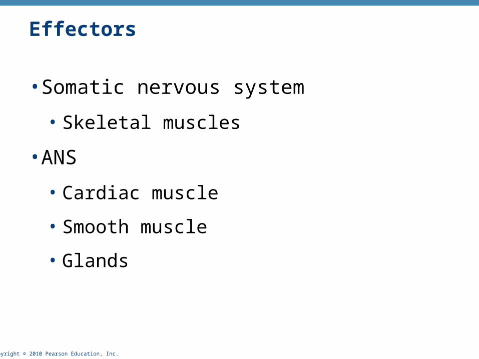
Effectors
• Somatic nervous system
• Skeletal muscles
• ANS
• Cardiac muscle
• Smooth muscle
• Glands
Copyright © 2010 Pearson Education, Inc.
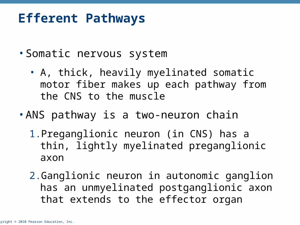
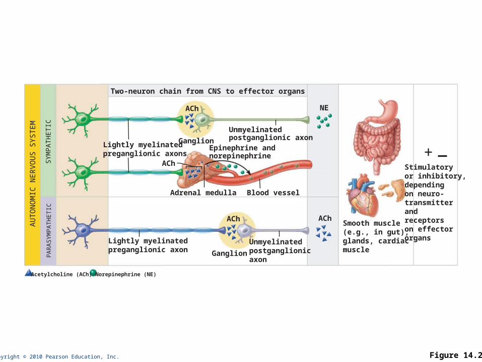
Efferent Pathways
• Somatic nervous system
• A, thick, heavily myelinated somatic motor fiber makes up each pathway from the CNS to the muscle
• ANS pathway is a two-neuron chain
1. Preganglionic neuron (in CNS) has a thin, lightly myelinated preganglionic axon
2. Ganglionic neuron in autonomic ganglion has an unmyelinated postganglionic axon that extends to the effector organ
Copyright © 2010 Pearson Education, Inc.
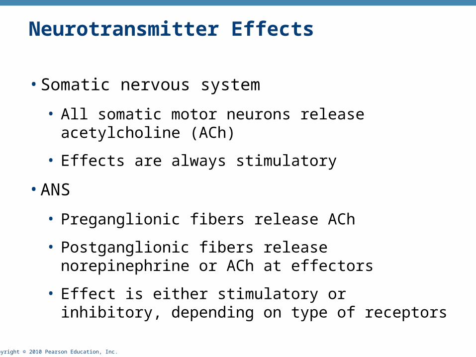
Neurotransmitter Effects
• Somatic nervous system
• All somatic motor neurons release acetylcholine (ACh)
• Effects are always stimulatory
• ANS
• Preganglionic fibers release ACh
• Postganglionic fibers release norepinephrine or ACh at effectors
• Effect is either stimulatory or inhibitory, depending on type of receptors
Copyright © 2010 Pearson Education, Inc.
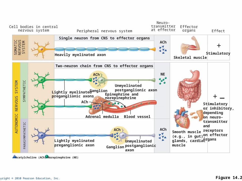
Skeletal muscle
Cell bodies in centralnervous system Peripheral nervous system Effect
+
+
Effectororgans
ACh
AChSmooth muscle(e.g., in gut),glands, cardiacmuscle
Ganglion
Adrenal medulla Blood vessel
ACh
ACh
ACh
NE
Epinephrine andnorepinephrine
Acetylcholine (ACh) Norepinephrine (NE)
Ganglion
Heavily myelinated axon
Lightly myelinatedpreganglionic axon
Lightly myelinatedpreganglionic axons
Neuro-transmitterat effector
Unmyelinatedpostganglionicaxon
Unmyelinatedpostganglionic axon
Stimulatory
Stimulatoryor inhibitory,dependingon neuro-transmitterandreceptorson effectororgans
Single neuron from CNS to effector organs
Two-neuron chain from CNS to effector organs
SO
MA
TIC
NER
VO
US
SYSTE
M
AU
TO
NO
MIC
NER
VO
US
SYS
TEM
PA
RA
SYM
PA
TH
ETIC
SYM
PA
TH
ETIC
Figure 14.2
Copyright © 2010 Pearson Education, Inc.
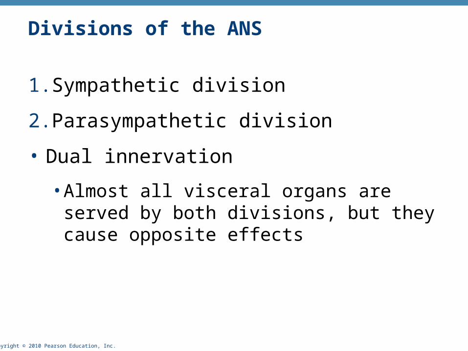
Divisions of the ANS
1.Sympathetic division
2.Parasympathetic division
• Dual innervation
• Almost all visceral organs are served by both divisions, but they cause opposite effects

Copyright © 2010 Pearson Education, Inc.
Role of the Parasympathetic Division
• Promotes maintenance activities and conserves body energy
• Its activity is illustrated in a person who relaxes, reading, after a meal
• Blood pressure, heart rate, and respiratory rates are low
• Gastrointestinal tract activity is high
• Pupils are constricted and lenses are accommodated for close vision
Copyright © 2010 Pearson Education, Inc.
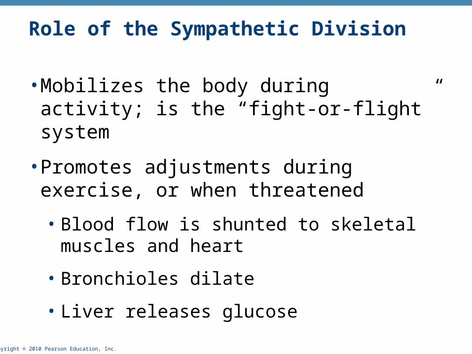
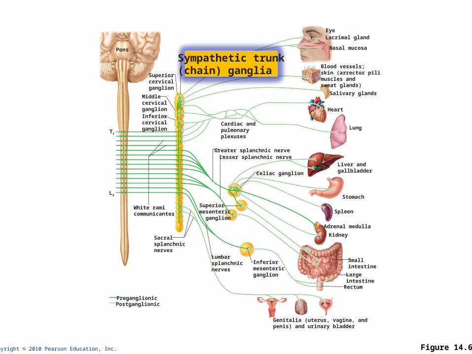
Role of the Sympathetic Division
• Mobilizes the body during activity; is the “fight-or-flight” system
• Promotes adjustments during exercise, or when threatened
• Blood flow is shunted to skeletal muscles and heart
• Bronchioles dilate
• Liver releases glucose
Copyright © 2010 Pearson Education, Inc.
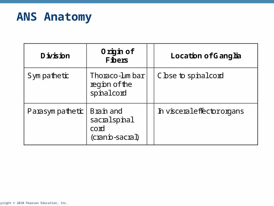
Division Origin of
Fibers Location of Ganglia
Sympathetic Thoraco-lumbar region of the spinal cord
Close to spinal cord
Parasympathetic Brain and sacral spinal cord (cranio-sacral)
In visceral effector organs
ANS Anatomy
Copyright © 2010 Pearson Education, Inc.
Salivaryglands
Eye
Skin*
Heart
Lungs
Liverand gall-bladder
Genitals
Pancreas
Eye
Lungs
Bladder
Liver andgall-bladder
Pancreas
Stomach
Cervical
Sympatheticganglia
Cranial
Lumbar
Thoracic
Genitals
Heart
Salivaryglands
Stomach
Bladder
Adrenalgland
Parasympathetic Sympathetic
Sacral
Brainstem
L1
T1
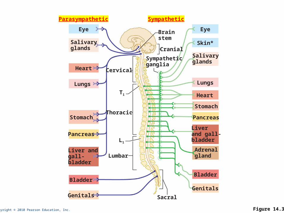
Figure 14.3

Copyright © 2010 Pearson Education, Inc.
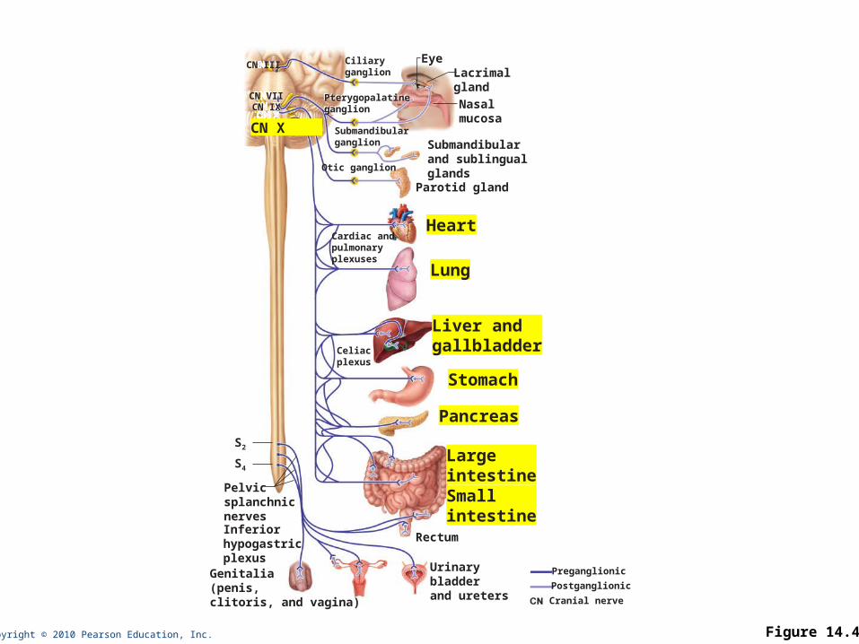
Cranial Outflow
Cranial Nerve Ganglia (Terminal Ganglia)
Effector Organ(s)
Oculomotor (III) Ciliary Eye
Facial (VII) Pterygopalatine Submandibular
Salivary, nasal, and lacrimal glands
Glossopharyngeal (IX)
Otic Parotid salivary glands
Vagus (X) Within the walls of target organs
Heart, lungs, and most visceral organs
Sacral
Outflow
S2-S4
Within the walls of target organs
Large intestine, urinary bladder, ureters, and reproductive organs
Parasympathetic (Craniosacral) Division Outflow
Copyright © 2010 Pearson Education, Inc.
Pterygopalatineganglion
EyeLacrimalgland
Nasalmucosa
Ciliaryganglion
Pterygopalatineganglion
Submandibularganglion Submandibular
and sublingualglands
CN III
CN VIICN IX
CN X
Otic ganglion
Parotid gland
Heart
Lung
Liver andgallbladder
Stomach
Pancreas
Urinarybladderand ureters
Smallintestine
Largeintestine
S2
Pelvicsplanchnicnerves
Genitalia(penis,clitoris, and vagina)
Rectum
Celiacplexus
Inferiorhypogastricplexus
Cardiac andpulmonaryplexuses
S4
Preganglionic
Postganglionic
Cranial nerve
Figure 14.4
Copyright © 2010 Pearson Education, Inc. Figure 14.6
Superiorcervicalganglion
MiddlecervicalganglionInferiorcervicalganglion
Sympathetic trunk(chain) ganglia
Pons
L2
T1
White ramicommunicantes
Liver andgallbladder
Stomach
Spleen
Kidney
Adrenal medulla
Smallintestine
Largeintestine
Genitalia (uterus, vagina, andpenis) and urinary bladder
Celiac ganglion
Inferiormesenteric ganglion
Lesser splanchnic nerveGreater splanchnic nerve
Superior mesenteric ganglion
Lumbarsplanchnic nerves
EyeLacrimal gland
Nasal mucosa
Blood vessels;skin (arrector pilimuscles andsweat glands)
Salivary glands
Heart
Lung
Rectum
Cardiac andpulmonaryplexuses
PreganglionicPostganglionic
Sacralsplanchnicnerves
Copyright © 2010 Pearson Education, Inc.
Pathways with Synapses in the Adrenal Medulla
• Some preganglionic fibers pass directly to the adrenal medulla without synapsing
• Upon stimulation, medullary cells secrete norepinephrine and epinephrine into the blood

Copyright © 2010 Pearson Education, Inc.
Visceral Reflexes
• Visceral reflex arcs have the same components as somatic reflexes
• Main difference: visceral reflex arc has two neurons in the motor pathway

Copyright © 2010 Pearson Education, Inc.
Referred Pain
• Visceral pain afferents travel along the same pathways as somatic pain fibers, contributing to the phenomenon of referred pain
• Pain stimuli arising in the viscera are perceived as somatic in origin

Copyright © 2010 Pearson Education, Inc. Figure 14.8
Heart
Lungs anddiaphragmLiver
Stomach
Kidneys
OvariesSmall intestine
Ureters
Urinarybladder
Colon
Pancreas
Liver
Heart
Appendix
Gallbladder
Copyright © 2010 Pearson Education, Inc.
Neurotransmitters
• Cholinergic fibers release the neurotransmitter ACh
• All ANS preganglionic axons
• All parasympathetic postganglionic axons
• Adrenergic fibers release the neurotransmitter NE
• Most sympathetic postganglionic axons
• Exceptions: sympathetic postganglionic fibers secrete ACh at sweat glands and some blood vessels in skeletal muscles
Copyright © 2010 Pearson Education, Inc. Figure 14.2
+
AChSmooth muscle(e.g., in gut),glands, cardiacmuscle
Ganglion
Adrenal medulla Blood vessel
ACh
ACh
ACh
NE
Epinephrine andnorepinephrine
Acetylcholine (ACh) Norepinephrine (NE)
Ganglion
Lightly myelinatedpreganglionic axon
Lightly myelinatedpreganglionic axons
Unmyelinatedpostganglionicaxon
Unmyelinatedpostganglionic axon
Stimulatoryor inhibitory,dependingon neuro-transmitterandreceptorson effectororgans
Two-neuron chain from CNS to effector organs
AU
TO
NO
MIC
NER
VO
US S
YS
TEM
PA
RA
SYM
PA
TH
ETIC
SYM
PA
TH
ETIC
Copyright © 2010 Pearson Education, Inc.
Receptors for Neurotransmitters
1.Cholinergic receptors for ACh
2.Adrenergic receptors for NE
Copyright © 2010 Pearson Education, Inc.
Cholinergic Receptors
• Two types of receptors bind ACh
1.Nicotinic
2.Muscarinic
• Named after drugs that bind to them and mimic ACh effects
Copyright © 2010 Pearson Education, Inc.
Nicotinic Receptors
• Found on
• Motor end plates of skeletal muscle cells (Chapter 9)
• All ganglionic neurons (sympathetic and parasympathetic)
• Hormone-producing cells of the adrenal medulla
• Effect of ACh at nicotinic receptors is always stimulatory
Copyright © 2010 Pearson Education, Inc.
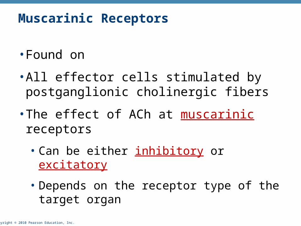
Muscarinic Receptors
• Found on
• All effector cells stimulated by postganglionic cholinergic fibers
• The effect of ACh at muscarinic receptors
• Can be either inhibitory or excitatory
• Depends on the receptor type of the target organ

Copyright © 2010 Pearson Education, Inc. Table 14.2

Copyright © 2010 Pearson Education, Inc.
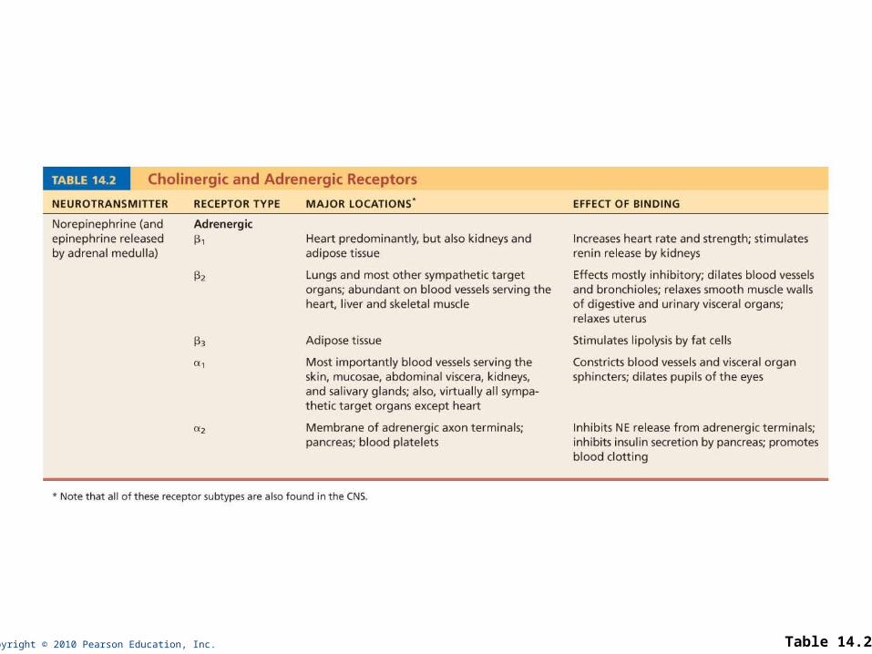
Adrenergic Receptors
• Two types
• Alpha () (subtypes 1, 2)
• Beta () (subtypes 1, 2 , 3)
• Effects of NE depend on which subclass of receptor predominates on the target organ
Copyright © 2010 Pearson Education, Inc. Table 14.2
Copyright © 2010 Pearson Education, Inc.
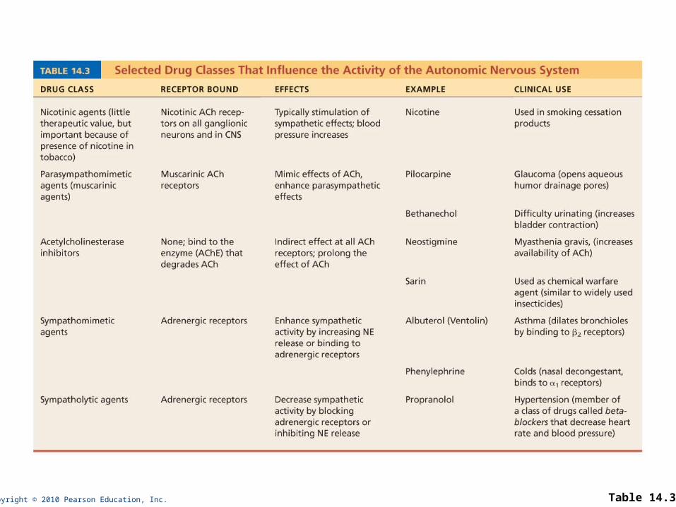
Effects of Drugs
• Atropine
• Anticholinergic; blocks muscarinic receptors
• Used to prevent salivation during surgery, and to dilate the pupils for examination
• Neostigmine
• Inhibits acetylcholinesterase
• Used to treat myasthenia gravis
Copyright © 2010 Pearson Education, Inc.
Effects of Drugs
• Over-the-counter drugs for colds, allergies, and nasal congestion
• Stimulate -adrenergic receptors
• Beta-blockers
• Drugs that attach to 2 receptors to dilate lung bronchioles in asthmatics; other uses
Copyright © 2010 Pearson Education, Inc. Table 14.3
Copyright © 2010 Pearson Education, Inc.
Interactions of the Autonomic Divisions
• Most visceral organs have dual innervation
• Dynamic antagonism allows for precise control of visceral activity
• Sympathetic division increases heart and respiratory rates, and inhibits digestion and elimination
• Parasympathetic division decreases heart and respiratory rates, and allows for digestion and the discarding of wastes

Copyright © 2010 Pearson Education, Inc.
Sympathetic Tone
• Sympathetic division controls blood pressure, even at rest
• Sympathetic tone (vasomotor tone)
• Keeps the blood vessels in a continual state of partial constriction
Copyright © 2010 Pearson Education, Inc.
Sympathetic Tone
• Sympathetic fibers fire more rapidly to constrict blood vessels and cause blood pressure to rise
• Sympathetic fibers fire less rapidly to prompt vessels to dilate to decrease blood pressure
• Alpha-blocker drugs interfere with vasomotor fibers and are used to treat hypertension

Copyright © 2010 Pearson Education, Inc.
Parasympathetic Tone
• Parasympathetic division normally dominates the heart and smooth muscle of digestive and urinary tract organs
• Slows the heart
• Dictates normal activity levels of the digestive and urinary tracts
• The sympathetic division can override these effects during times of stress
• Drugs that block parasympathetic responses increase heart rate and block fecal and urinary retention
Copyright © 2010 Pearson Education, Inc.
Cooperative Effects
• Best seen in control of the external genitalia
• Parasympathetic fibers cause vasodilation; are responsible for erection of the penis or clitoris
• Sympathetic fibers cause ejaculation of semen in males and reflex contraction of a female’s vagina

Copyright © 2010 Pearson Education, Inc.
Unique Roles of the Sympathetic Division
• The adrenal medulla, sweat glands, arrector pili muscles, kidneys, and most blood vessels receive only sympathetic fibers
• The sympathetic division controls
• Thermoregulatory responses to heat
• Release of renin from the kidneys
• Metabolic effects
• Increases metabolic rates of cells
• Raises blood glucose levels
• Mobilizes fats for use as fuels
Copyright © 2010 Pearson Education, Inc.
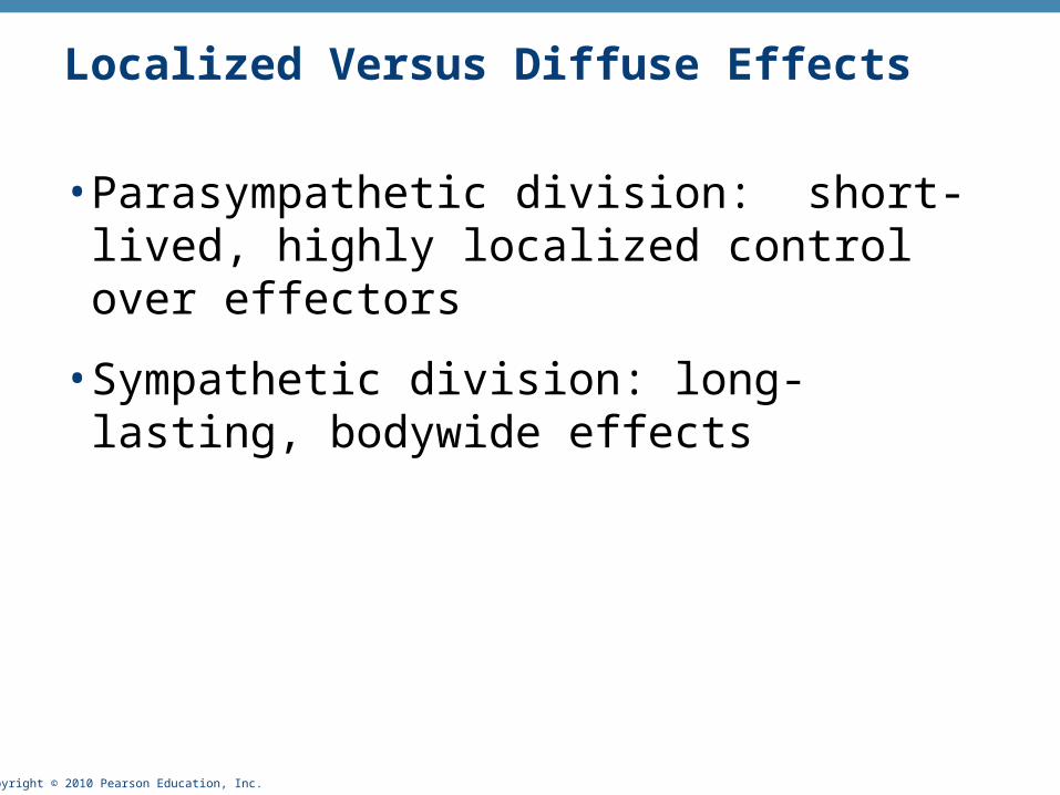
Localized Versus Diffuse Effects
• Parasympathetic division: short-lived, highly localized control over effectors
• Sympathetic division: long-lasting, bodywide effects
Copyright © 2010 Pearson Education, Inc.
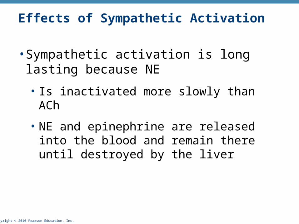
Effects of Sympathetic Activation
• Sympathetic activation is long lasting because NE
• Is inactivated more slowly than ACh
• NE and epinephrine are released into the blood and remain there until destroyed by the liver

Copyright © 2010 Pearson Education, Inc.
Control of ANS Functioning
• Hypothalamus—main integrative center of ANS activity
• Subconscious cerebral input via limbic lobe connections influences hypothalamic function
• Other controls come from the cerebral cortex, the reticular formation, and the spinal cord
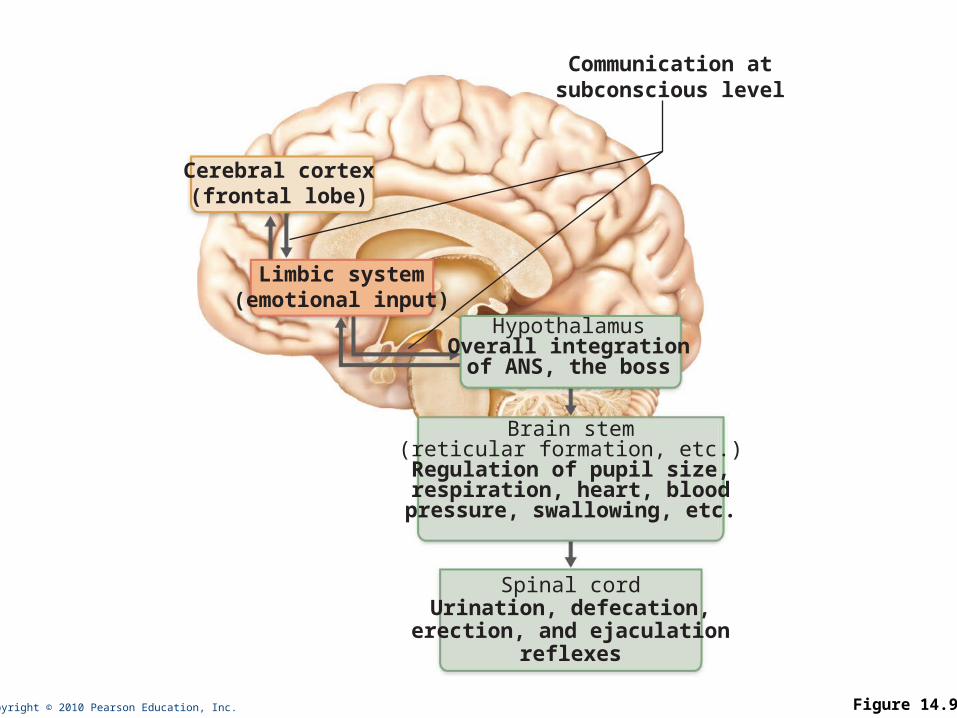
Copyright © 2010 Pearson Education, Inc. Figure 14.9
Cerebral cortex(frontal lobe)
Limbic system(emotional input)
Communication atsubconscious level
HypothalamusOverall integrationof ANS, the boss
Spinal cordUrination, defecation,
erection, and ejaculationreflexes
Brain stem(reticular formation, etc.)
Regulation of pupil size,respiration, heart, blood
pressure, swallowing, etc.
Copyright © 2010 Pearson Education, Inc.
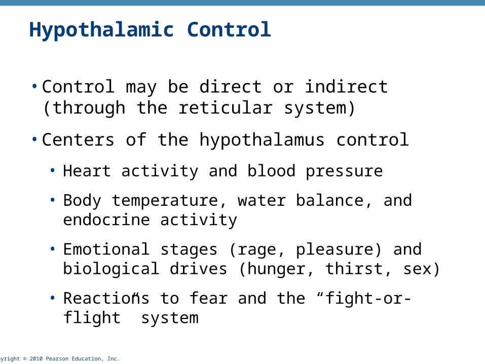
Hypothalamic Control
• Control may be direct or indirect (through the reticular system)
• Centers of the hypothalamus control
• Heart activity and blood pressure
• Body temperature, water balance, and endocrine activity
• Emotional stages (rage, pleasure) and biological drives (hunger, thirst, sex)
• Reactions to fear and the “fight-or-flight” system

Copyright © 2010 Pearson Education, Inc.
Developmental Aspects of the ANS
• Effects of age on ANS
• Constipation
• Dry eyes
• Frequent eye infections
• Orthostatic hypotension
• Low blood pressure occurs because aging pressure receptors respond less to changes in blood pressure with changes in body position and because of slowed responses by sympathetic vasoconstrictor centers