2011_amyloid-beta stability, oligomerization, and aggregation
TRANSCRIPT
Distinct Effects of Zn2�, Cu2�, Fe3�, and Al3� on Amyloid-�Stability, Oligomerization, and AggregationAMYLOID-� DESTABILIZATION PROMOTES ANNULAR PROTOFIBRIL FORMATION*□S
Received for publication, August 22, 2010, and in revised form, December 8, 2010 Published, JBC Papers in Press, January 7, 2011, DOI 10.1074/jbc.M110.177246
Wei-Ting Chen‡§, Yi-Hung Liao‡¶�, Hui-Ming Yu‡, Irene H. Cheng§, and Yun-Ru Chen‡1
From the ‡Genomics Research Center and the ¶Taiwan International Graduate Program, Chemical Biology and MolecularBiophysics Program, Genomics Research Center, Academia Sinica, 11574 Taipei, the §Institute of Brain Science, NationalYang-Ming University, 11221 Taipei, and the �Institute of Biochemical Sciences, National Taiwan University, 10617 Taipei, Taiwan
Abnormally high concentrations of Zn2�, Cu2�, and Fe3� arepresent along with amyloid-� (A�) in the senile plaques inAlzheimer disease, where Al3� is also detected. A� aggregation isthe key pathogenic event in Alzheimer disease, where A� olig-omers are the major culprits. The fundamental mechanism ofthese metal ions on A� remains elusive. Here, we employ 4,4�-Bis(1-anilinonaphthalene 8-sulfonate) and tyrosine fluores-cence, CD, stopped flow fluorescence, guanidine hydrochloridedenaturation, and photo-induced cross-linking to elucidate theeffect of Zn2�, Cu2�, Fe3�, and Al3� on A� at the early stage ofthe aggregation. Furthermore, thioflavin T assay, dot blotting,and transmission electron microscopy are utilized to examineA� aggregation. Our results show that Al3� and Zn2�, but notCu2� andFe3�, induce larger hydrophobic exposures ofA� con-formation, resulting in its significant destabilization at the earlystage. The metal ion binding induces A� conformationalchanges with micromolar binding affinities and millisecondbinding kinetics. Cu2� and Zn2� induce similar assembly oftransiently appearing A� oligomers at the early state. Duringthe aggregation, we found that Zn2� exclusively promotes theannular protofibril formation without undergoing a nucleationprocess, whereas Cu2� and Fe3� inhibit fibril formation by pro-longing the nucleation phases. Al3� also inhibits fibril forma-tion; however, the annular oligomers co-exist in the aggregationpathway. In conclusion, Zn2�, Cu2�, Fe3�, and Al3� adopt dis-tinct folding and aggregation mechanisms to affect A�, whereA�destabilization promotes annular protofibril formation.Ourstudy facilitates the understanding of annular A� oligomer for-mation upon metal ion binding.
The brain deposition of amyloid plaques composed of A�2 isthe pathological hallmark of AD (1, 2). A� is generated from
sequential cleavages of amyloid precursor protein by �- and�-secretases (3, 4). The predominantA� isoforms areA�40 andA�42, which differ in two residues at the C terminus, whereA�42 is less abundant but more neurotoxic (5–8). A� is anatively unfolded protein prone to aggregating into cross-�-amyloid fibrils through a nucleation-dependent polymerizationpathway (9). A� aggregation is considered the major culprit inAD, in which the A� oligomers, but not fibrils, better correlatewith cognitive impairment and synaptic dysfunction (10). A�oligomers are referred to various different metastable interme-diates found in the aggregation, including lowmolecularweightoligomers, spherical oligomers, A�-derived diffusible ligands,globulomers, annular protofibrils, A�56*, and curvilinearprotofibrils (11–18).Specific metal ions have been observed in the lesions of the
disease. Analysis of the autopsy of AD patients shows abnor-mally high levels of specific metal ions present in the senileplaques (Cu2�, 25 �g/g, �393 �M; Zn2�, 69 �g/g, �1055 �M;and Fe3�, 52�g/g,�940�M) (19, 20). The levels of Cu2�, Zn2�,and Fe3� in the AD neuropil are also significantly elevated (19),where Zn2� is elevated from 346 to 786 �M, Cu2� from 69 to304 �M, and Fe3� from 338 to 695 �M (19, 20). Al3� has alsobeen detected in amyloid fibers in cores of the senile plaques(21). Moreover, the imbalance of cellular Zn2� and/or Cu2�
homeostasis modulates AD pathology (22, 23), and dietaryCu2� and Al3� are risk factors for AD (24–25). These factsindicate that the elevation of the metal ions is relevant to ADpathology.A� is able to bind to the metal ions (22, 23). A� ion coordi-
nation, binding affinity, and induced aggregation have beenstudied intensively in various conditions; however, the resultsand mechanisms remain inconclusive. A�-Cu2� coordinationinvolves three intramolecular histidines (i.e.His-6, His-13, andHis-14) in A� (26–28), with the fourth coordinate being eitherthe amino group of theN terminus (29), an oxygen fromTyr-10(30), or an oxygen from Glu-3 (29). The A�-Zn2� complexis reportedly more complicated. A similar coordination withCu2�has been proposed forZn2�using the three histidines andthe N terminus (31, 32). Both intermolecular A�-Cu2� andA�-Zn2� coordination have been reported via histidine bridges(28, 30–33). Fe3� has also been shown to interact with histi-dines (37). Dissociation constants for the binding affinity of
* This work was supported by Grants 98-2320-B-001-020-MY3, 96-2113-M-001-030-MY2, and 97-2320-B-010-027-MY3 from the Genomics ResearchCenter (Academia Sinica, Taiwan, National Science Council, Taiwan) andNational Health Research Institute Grant NHRI-EX98-9816NC.
□S The on-line version of this article (available at http://www.jbc.org) containssupplemental text, Tables S1 and S2, and Figs. S1–S7.
1 To whom correspondence should be addressed: Genomics Research Cen-ter, Academia Sinica, Taiwan, 128, Academia Rd., Sec. 2, Nankang Dist.,Taipei 115, Taiwan. Tel.: 886-2-2787-1275; Fax: 886-2-2789-8771; E-mail:[email protected].
2 The abbreviations used are: A�, amyloid-�; AD, Alzheimer disease; Bis-ANS,4,4�-Bis(1-anilinonaphthalene 8-sulfonate); GdnHCl, guanidine hydrochlo-ride; PICUP, photo-induced cross-linking of unmodified proteins; ThT, thio-
flavin T; TEM, transmission electron microscopy; Tricine, N-[2-hydroxy-1,1-bis(hydroxymethyl)ethyl]glycine.
THE JOURNAL OF BIOLOGICAL CHEMISTRY VOL. 286, NO. 11, pp. 9646 –9656, March 18, 2011© 2011 by The American Society for Biochemistry and Molecular Biology, Inc. Printed in the U.S.A.
9646 JOURNAL OF BIOLOGICAL CHEMISTRY VOLUME 286 • NUMBER 11 • MARCH 18, 2011
at National T
aiwan U
niv Medical Library, on M
arch 13, 2011w
ww
.jbc.orgD
ownloaded from
http://www.jbc.org/content/suppl/2011/01/07/M110.177246.DC1.html Supplemental Material can be found at:

ions and A� ranging from attomolar to 11 �M for Cu2� and2–300 �M for Zn2� have been reported (38–40).Zn2� andCu2�have been shown to accelerateA�deposition
(41) but form amorphous aggregates (42–48). Al3� and Fe3�
promote A� fibrils (43, 49, 50) or oligomer formation (23).Zn2�, especially at lower concentrations, and Cu2� show pro-tective effects towardA�mediated toxicity (42, 51). In contrast,histidine-bridgedA�-Cu2�dimers are neurotoxic (33). In addi-tion, the metal chelators, clioquinol CQ and its analogousPBT2, are able to reverse ion-induced A� aggregation, reduceplaque load, and reverse cognition deficits in the transgenic ADmice (52, 53). The PBT2 is currently under phase II clinicaltrials (54).The involvement of the metal ions with A� in AD and the
potential development of metal chelating therapy indicate theimportance of elucidating fundamental mechanisms of theireffect on A�. Here, we systematically examine the metal ioneffects, especially of Zn2�, Cu2�, Fe3�, and Al3�, on early andaggregated stages of full-length A�40. By employing differentspectroscopic methods, including far-UV CD, tyrosine fluores-cence, and 4,4�-Bis(1-anilinonaphthalene 8-sulfonate) (Bis-ANS) fluorescence, we monitor the conformational changes ofA� and its ion binding affinity at the early stage. The bindingkinetics and conformational stability are further examined bystopped flowmachinery and guanidine hydrochloride denatur-ation. Photo-induced cross-linking of unmodified proteins(PICUP), thioflavin T (ThT) assay, dot blotting, and transmis-sion electron microscopy (TEM) are also employed to monitorthe oligomerization and fibrillization during aggregation. Amechanism for the metal ion effects on A� stability, oligomer-ization, and aggregation is then proposed.
EXPERIMENTAL PROCEDURES
Materials—GdnHCl, ThT, ammonium persulfate, and Tris(2,2�-bipyridyl)dichlororuthenium (II)) were purchased fromSigma-Aldrich (St. Louis, MO). Tris and NaCl were fromAmresco (Solon, OH). CaCl2�2H2O was from J. T. Backer(Phillipsburg, NJ). AlCl3�6H2O, CuCl2, FeCl3�6H2O, andZnCl2 were from Riedel-de Haeen Inc. (Sigma-Aldrich, St.Louis, MO). All metal ions were prepared in double-distilledMilli-Q water.A� Preparation—A�40 peptide was synthesized using Fmoc
(N-(9-fluorenyl)methoxycarbonyl) chemistry and purified byreversed phase high performance liquid chromatography, asdescribed previously (7). The molecular mass was identified byMALDI-TOF mass spectrometry (UltraFlex II; Bruker BioSci-ences, Billerica,MA). To prepare theA� stock, lyophilized pep-tide was freshly dissolved in Buffer A (10 mM Tris-HCl, pH 7.4)containing 8 MGdnHCl and refolded into Buffer A at a concen-tration of �1 mg/ml. Then the stock was then centrifuged at17,000 � g at 4 °C for 15 min. The supernatant was collectedand quantified by absorbance at 280 nm (� � 1,280 cm�1 M�1)(55) and used as a stock solution to prepare A� at 25 �M for allexperiments.Fluorescence Spectroscopy—Fluorescence emission spectra
were obtained using a FluoroMax-3 spectrofluorometer(Horiba Jobin Yvon). The emission spectra of Bis-ANS at 5 �M
were collected from 450 to 550 nm with excitation at 400 nm.
The emission spectra of tyrosinewere collected from290 to 360nm with excitation at 270 nm. Both fluorescent experimentswere performed at 25 °C. Temperature was controlled by a cir-culating water bath. The buffer backgrounds were subtracted.Far-UV CD—Far-UV CD spectra were collected from 250 to
202 nm at 25 °C using a Jasco J-815 spectropolarimeter (JascoInc., Easton, MD). A circular quartz cell with a path length of 1mm was used. Six scans were performed and averaged for eachcondition.Equilibrium Binding of Metal Ion and A� by Titration—The
25 �M A� solution was titrated with different metal ion stocksolutions: ZnCl2, CuCl2, and FeCl3 at 2 mM or AlCl3 at 20 mM.Less than 10% of the solution volume was increased after titra-tion. Three different signals were collected, and the dilutionfactor was corrected. The monitored signals were 490 nm forBis-ANS fluorescence, 305 nm for tyrosine fluorescence, and216 nm for CD. The final titration signal was used as unity fornormalization. The normalized data were plotted against themetal ion concentration. The amount of aluminum atom in thesolution was confirmed by atomic absorption spectroscopy.Data fitting was performed by using an equation describingsingle protein ligand binding (56),
P � L7 PL (Eq. 1)
r � �2Pt��1
*�Kd � Lt � Pt� � ��Kd � Lt � Pt�2 � 4PtLt�
1/ 2
where � is the fraction of the observed signal changes repre-senting the bound protein fraction, Pt is the total A� concen-tration, Lt is the total ligand concentration, and Kd is the disso-ciation constant. In addition, the datawere fitted to an equationdescribing one-protein and two-ligand binding,
P � L ¢O¡K1
PL (Eq. 2)
PL � L ¢O¡K2
PL2
r � �K1L � K1K2L2�/�1 � K1L � K1K2L2�
where � is the fraction of the observed signal change,K1 andK2are the association constants for the first and second ligandbinding, and L is the free ligand concentration. Because the freeligand concentration cannot be faithfully determined in ourexperimental conditions, we used the total ligand concentra-tion as L for the fitting.Stopped Flow Experiments for A� and Metal Ion Binding
Kinetics—The kinetics of A� and metal ion binding was exam-ined using a stopped flow module (Bio-Logic, Claix, France)attached to a Jasco J-815 spectropolarimeter. The stopped flowmodule was composed of a multimixer SFM-400, a motorpower supply MPS-250, and a photomultiplier system PMS-250. A fluorescence cuvette, FC-15, with an optical path of 1.5mm was used. Excitation at 270 nm was used to monitor thetyrosine fluorescence ofA� at 12.5�M.A singlemixing reaction
Distinct Metal Ion Effects on A� Folding and Aggregation
MARCH 18, 2011 • VOLUME 286 • NUMBER 11 JOURNAL OF BIOLOGICAL CHEMISTRY 9647
at National T
aiwan U
niv Medical Library, on M
arch 13, 2011w
ww
.jbc.orgD
ownloaded from
using a volume ratio of 9:1 for the metal ions and A� was per-formed at 25 °C. The final metal ion concentrations were 1.25mM; hence the metal-to-A� ratio was 100 to 1. Here, A� wasprepared in 100 mM Tris-HCl, pH 7.4, to avoid metal ion-in-duced acidity. In this buffer system, the reactions were in neu-tral pH. The flow rate was fixed at 11 ml/s, resulting in a deadtime of 4.7ms. The data were collected every 0.5ms for the first2 s, every 20 ms in the range of 2–60 s, and every 0.5 s for60–100 s. The data were fitted to multi-exponential equationsusing Bio-kine 32 V4.51 (Bio-Logic, Claix, France) with eithersingle or double exponential equations with a linear base line,
Y � ax � b � c1exp�k1t (Eq. 3)
Y � ax � b � c1exp�k1t � c2exp�k2t (Eq. 4)
where a and b are the slope and offset for the linear base line,and c and k are the amplitudes and rate constants for the expo-nential phases, respectively.GdnHCl Denaturation—The denaturation study was per-
formed, as described previously (58), at 25 °C. Briefly, the titra-tion was performed by titrating an unfolded A� solution inBuffer A containing �6.2 M GdnHCl into A� solution in BufferA containing �0.1 M GdnHCl. The desired metal ion concen-trations, 25 �M of A�, and 5 �M of Bis-ANS were present inboth solutions. The duration for each titration was �30 s, andeach set of denaturation experiment was less than 1 h. TheBis-ANS fluorescence emission at 490 nm was collected, aver-aged, and normalized. The normalized emission intensity ver-sus GdnHCl concentration was plotted.PICUP—The experiment was performed as described previ-
ously (59). Here, we prepared the A� stock by urea instead ofGdnHCl to facilitate running of SDS-PAGE. Briefly, A� sam-ples at 25 �M prepared in Buffer A with different ion concen-trations, as indicated, were immediately subjected to photo-induced cross-linking. A 90% A� solution was mixed with 5%each of 1 mM Tris(2,2�-bipyridyl)dichlororuthenium(II) and 20mM ammonium persulfate. After mixing, the samples wereexposed to a blue light LED in a closed chamber with a manualswitch for 10 s. The cross-linking reaction was stopped by add-ing the SDS-PAGE sample buffer, and the samples were run onTris-Tricine SDS-PAGE. All of the actions were performedwithout delay. The gel was subjected to Western blotting withanti-A� antibody 6E10 (Chemicon Inc., Billerica, MA) recog-nizing A� residues 1–17.ThT Assay—A� (25 �M) in Buffer A with different metal ion
concentrations and 25 �M ThT were incubated in an ELISAplate and monitored by a microplate reader (SpectraMax M5;Molecule Devices) at 25 °C. The samples were in quiescenceduring the incubation, except for 10 s of mixing prior to themeasurement. The ThT emission was measured at 485 nm,whereas excitation was at 442 nm. The signals were collectedautomatically every 1 h for the first 100 h, after which they werecollected in a longer intervals.Dot Blotting—To detect the oligomers during the aggrega-
tion, 2 �l of the samples from the ThT assay were dottedonto a nitrocellulose membrane at the indicated incubationtime and recognized using an anti-A� oligomer antibody,A11 (Invitrogen).
TEM—The samples were placed on glow-discharged, 400-mesh Formvar carbon-coated copper grids (EMS Inc., Hatfield,PA) for 3 min, rinsed, and negatively stained with 2% uranylacetate. The samples were examined with a Tecnai G2 SpiritTWIN TEM (FEI, Hillsboro, OR) with an accelerating voltageof 75 kV.Sedimentation Assay—The aggregated products of 25 �M
Zn2� or 100�MAl3� and 25�MA�were incubated in Buffer Aand subjected to centrifugation at 10,000� g at 4 °C for 20min.The supernatant and pellet were collected and analyzed by dotblotting using A11 and 6E10 antibodies and TEM. The pelletwas resuspended in the original volume of Buffer A.
RESULTS
Al3� andZn2� IncreaseHydrophobic Exposure of A�Confor-mation, whereas Cu2� Decreases—To reveal the effect of metalions on A�, the ion-induced A� structural changes were mon-itored by three spectroscopic techniques: Bis-ANS fluores-cence, tyrosine fluorescence, and far-UV CD spectra (Fig. 1).Bis-ANS is able to report the hydrophobic clusters exposed onprotein surfaces and to probe A� conformation at the earlystage, where the binding sites are at the flanking regions of theprotease-resistant segment of A� (58, 60, 61). The experimentswere performed with A� at 25 �M in the presence of Zn2�,Cu2�, Fe3�, and Al3� above their saturated concentrations, asdetermined by the metal ion binding experiment describedbelow. TheA� spectra with andwithout 200�Mof Zn2�, Cu2�,and Fe3� and 500 �M of Al3� are shown in Fig. 1A and supple-mental Fig. S1. In the presence of Al3� and Zn2�, the Bis-ANSemission of A�40 showed �30- and 15-fold enhancement,respectively, but decreased �2.5-fold in the presence of Cu2�.The enhancement of the emission intensity indicates a largerextent of hydrophobic clusters exposed on the protein sur-face. Fe3� did not induce significant changes on the hydro-phobic surfaces of A�. Apart from the intensity difference,all of the spectra were blue-shifted from 500 nm to a range of�480–495 nm, indicating Bis-ANS encountering the hydro-phobic environment.In addition to extrinsic fluorescence, we used intrinsic tyro-
sine fluorescence at residue 10 of A� to report local conforma-tional changes. In the absence of metal ions, the tyrosine emis-sion had a maximum at �302 nm while excited at 270 nm. Wefound that the tyrosine emissions were significantly quenchedin the presence of Fe3� and Cu2� but enhanced in the presenceof Al3� and Zn2� (Fig. 1B). The base lines of the spectra movedupward with concentration dependence to Al3� and Zn2� butnot to Fe3� and Cu2� (supplemental Fig. S2). The increasein base line may be due to the formation of other unknownfluorescence species. Furthermore, far-UV CD spectra wereemployed to examine the secondary structure changes of A�(Fig. 1C). In the absence of the metal ions, a random coil-dom-inant spectrum was observed, as expected (62). However, allfour metal ions further decreased ellipticity, especially withZn2� and Al3�, revealing that the metal ions reduced the con-tent of residual secondary structures existing in A� confor-mation. Together, our results showed that Zn2� and Al3�
increased the hydrophobic exposed protein surfaces, inducedhigher tyrosine emission, and reducedmost residual secondary
Distinct Metal Ion Effects on A� Folding and Aggregation
9648 JOURNAL OF BIOLOGICAL CHEMISTRY VOLUME 286 • NUMBER 11 • MARCH 18, 2011
at National T
aiwan U
niv Medical Library, on M
arch 13, 2011w
ww
.jbc.orgD
ownloaded from
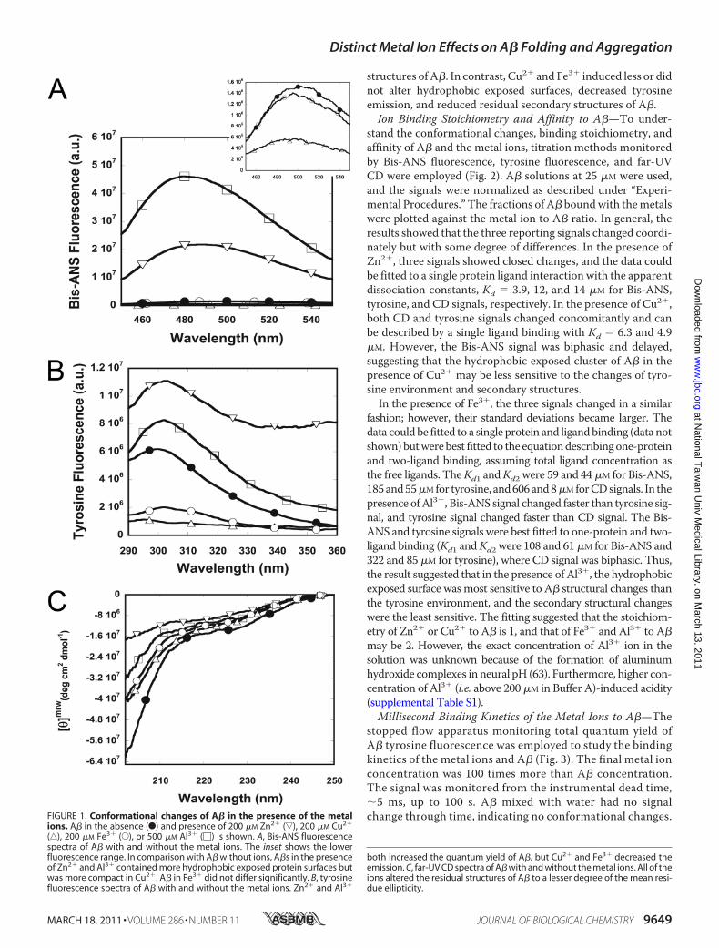
structures of A�. In contrast, Cu2� and Fe3� induced less or didnot alter hydrophobic exposed surfaces, decreased tyrosineemission, and reduced residual secondary structures of A�.Ion Binding Stoichiometry and Affinity to A�—To under-
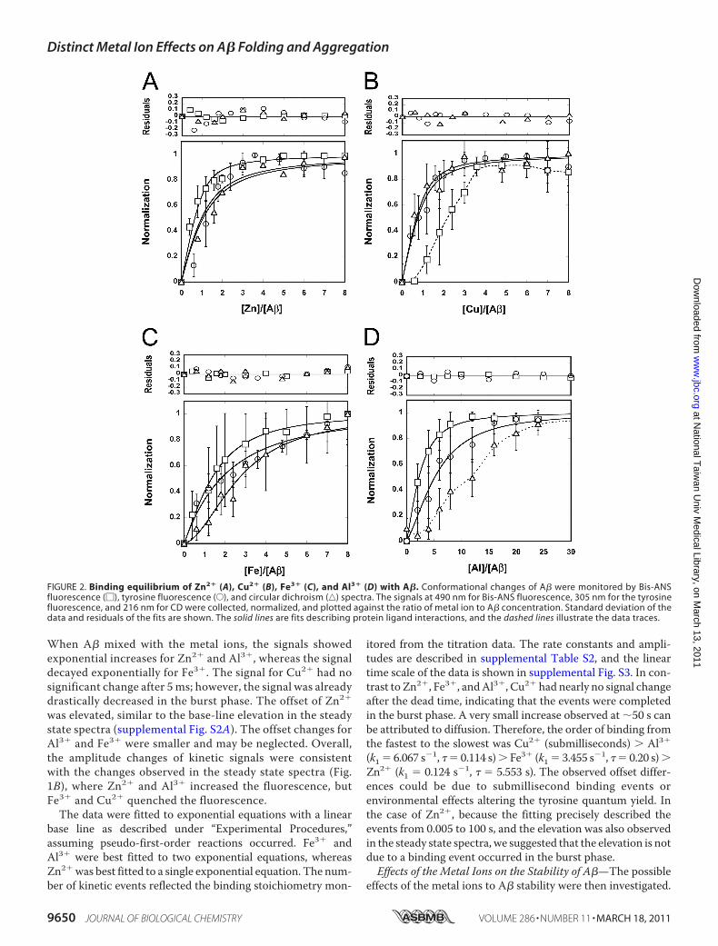
stand the conformational changes, binding stoichiometry, andaffinity of A� and the metal ions, titration methods monitoredby Bis-ANS fluorescence, tyrosine fluorescence, and far-UVCD were employed (Fig. 2). A� solutions at 25 �M were used,and the signals were normalized as described under “Experi-mental Procedures.” The fractions of A� boundwith themetalswere plotted against the metal ion to A� ratio. In general, theresults showed that the three reporting signals changed coordi-nately but with some degree of differences. In the presence ofZn2�, three signals showed closed changes, and the data couldbe fitted to a single protein ligand interaction with the apparentdissociation constants, Kd � 3.9, 12, and 14 �M for Bis-ANS,tyrosine, and CD signals, respectively. In the presence of Cu2�,both CD and tyrosine signals changed concomitantly and canbe described by a single ligand binding with Kd � 6.3 and 4.9�M. However, the Bis-ANS signal was biphasic and delayed,suggesting that the hydrophobic exposed cluster of A� in thepresence of Cu2� may be less sensitive to the changes of tyro-sine environment and secondary structures.In the presence of Fe3�, the three signals changed in a similar
fashion; however, their standard deviations became larger. Thedata could be fitted to a single protein and ligandbinding (data notshown)butwerebest fitted to the equationdescribingone-proteinand two-ligand binding, assuming total ligand concentration asthe free ligands. TheKd1 andKd2 were 59 and 44�M for Bis-ANS,185and55�Mfor tyrosine, and606and8�MforCDsignals. In thepresence ofAl3�, Bis-ANS signal changed faster than tyrosine sig-nal, and tyrosine signal changed faster than CD signal. The Bis-ANS and tyrosine signals were best fitted to one-protein and two-ligand binding (Kd1 andKd2 were 108 and 61�M for Bis-ANS and322 and 85 �M for tyrosine), where CD signal was biphasic. Thus,the result suggested that in the presence of Al3�, the hydrophobicexposed surface wasmost sensitive to A� structural changes thanthe tyrosine environment, and the secondary structural changeswere the least sensitive. The fitting suggested that the stoichiom-etry of Zn2� or Cu2� to A� is 1, and that of Fe3� and Al3� to A�may be 2. However, the exact concentration of Al3� ion in thesolution was unknown because of the formation of aluminumhydroxide complexes in neural pH (63). Furthermore, higher con-centration of Al3� (i.e. above 200 �M in Buffer A)-induced acidity(supplemental Table S1).Millisecond Binding Kinetics of the Metal Ions to A�—The
stopped flow apparatus monitoring total quantum yield ofA� tyrosine fluorescence was employed to study the bindingkinetics of the metal ions and A� (Fig. 3). The final metal ionconcentration was 100 times more than A� concentration.The signal was monitored from the instrumental dead time,�5 ms, up to 100 s. A� mixed with water had no signalchange through time, indicating no conformational changes.FIGURE 1. Conformational changes of A� in the presence of the metal
ions. A� in the absence (F) and presence of 200 �M Zn2� (ƒ), 200 �M Cu2�
(‚), 200 �M Fe3� (E), or 500 �M Al3� (�) is shown. A, Bis-ANS fluorescencespectra of A� with and without the metal ions. The inset shows the lowerfluorescence range. In comparison with A� without ions, A�s in the presenceof Zn2� and Al3� contained more hydrophobic exposed protein surfaces butwas more compact in Cu2�. A� in Fe3� did not differ significantly. B, tyrosinefluorescence spectra of A� with and without the metal ions. Zn2� and Al3�
both increased the quantum yield of A�, but Cu2� and Fe3� decreased theemission. C, far-UV CD spectra of A� with and without the metal ions. All of theions altered the residual structures of A� to a lesser degree of the mean resi-due ellipticity.
Distinct Metal Ion Effects on A� Folding and Aggregation
MARCH 18, 2011 • VOLUME 286 • NUMBER 11 JOURNAL OF BIOLOGICAL CHEMISTRY 9649
at National T
aiwan U
niv Medical Library, on M
arch 13, 2011w
ww
.jbc.orgD
ownloaded from
When A� mixed with the metal ions, the signals showedexponential increases for Zn2� and Al3�, whereas the signaldecayed exponentially for Fe3�. The signal for Cu2� had nosignificant change after 5 ms; however, the signal was alreadydrastically decreased in the burst phase. The offset of Zn2�
was elevated, similar to the base-line elevation in the steadystate spectra (supplemental Fig. S2A). The offset changes forAl3� and Fe3� were smaller and may be neglected. Overall,the amplitude changes of kinetic signals were consistentwith the changes observed in the steady state spectra (Fig.1B), where Zn2� and Al3� increased the fluorescence, butFe3� and Cu2� quenched the fluorescence.The data were fitted to exponential equations with a linear
base line as described under “Experimental Procedures,”assuming pseudo-first-order reactions occurred. Fe3� andAl3� were best fitted to two exponential equations, whereasZn2�was best fitted to a single exponential equation. The num-ber of kinetic events reflected the binding stoichiometry mon-
itored from the titration data. The rate constants and ampli-tudes are described in supplemental Table S2, and the lineartime scale of the data is shown in supplemental Fig. S3. In con-trast to Zn2�, Fe3�, andAl3�, Cu2� had nearly no signal changeafter the dead time, indicating that the events were completedin the burst phase. A very small increase observed at �50 s canbe attributed to diffusion. Therefore, the order of binding fromthe fastest to the slowest was Cu2� (submilliseconds) � Al3�
(k1 � 6.067 s�1, � � 0.114 s)� Fe3� (k1 � 3.455 s�1, � � 0.20 s)�Zn2� (k1 � 0.124 s�1, � � 5.553 s). The observed offset differ-ences could be due to submillisecond binding events orenvironmental effects altering the tyrosine quantum yield. Inthe case of Zn2�, because the fitting precisely described theevents from 0.005 to 100 s, and the elevation was also observedin the steady state spectra, we suggested that the elevation is notdue to a binding event occurred in the burst phase.Effects of the Metal Ions on the Stability of A�—The possible
effects of the metal ions to A� stability were then investigated.
FIGURE 2. Binding equilibrium of Zn2� (A), Cu2� (B), Fe3� (C), and Al3� (D) with A�. Conformational changes of A� were monitored by Bis-ANSfluorescence (�), tyrosine fluorescence (E), and circular dichroism (‚) spectra. The signals at 490 nm for Bis-ANS fluorescence, 305 nm for the tyrosinefluorescence, and 216 nm for CD were collected, normalized, and plotted against the ratio of metal ion to A� concentration. Standard deviation of thedata and residuals of the fits are shown. The solid lines are fits describing protein ligand interactions, and the dashed lines illustrate the data traces.
Distinct Metal Ion Effects on A� Folding and Aggregation
9650 JOURNAL OF BIOLOGICAL CHEMISTRY VOLUME 286 • NUMBER 11 • MARCH 18, 2011
at National T
aiwan U
niv Medical Library, on M
arch 13, 2011w
ww
.jbc.orgD
ownloaded from
Although A� is considered to be intrinsically disordered (62),solution NMR and limited proteolysis studies have shown theexistence of residual A� structures (64, 65). Previously, we haveshown that Bis-ANS fluorescence can distinguish “native” A�,A� in native buffer, unfolded A�, and A� in high concentra-tions of denaturant. The chemical denaturation coupled withdata fitting is able to provide equilibrium folding mechanismsfor A�40 and A�42 (58). Here, GdnHCl denaturation wasemployed to examine the stability changes of A� in the pres-
ence of the metal ions (Fig. 4). In the native buffer, Bis-ANSbinding to A� showed a significantly enhanced emission andblue-shifted spectrum in comparison with that in the presenceof unfolded A� in 3 M GdnHCl (supplemental Fig. S4A). Tyro-sine and CD spectra also showed different spectra between A�in native and unfolded conditions (supplemental Fig. S4, B andC). The Bis-ANS signals of A� in the absence and presence ofthemetal ions weremonitored in different GdnHCl concentra-tions, and the data were averaged and normalized. In theabsence of the metal ions, a tilted pretransition, �0.5 M Gdn-HCl, followed by a single transition between 0.5 and 1.5 MGdn-HCl, and a post-transition�1.5MGdnHClwere observed, indi-cating A� adopted an apparent two-state equilibriummechanism (N%U), where a “native” and an unfolded ensem-ble were present at the equilibrium (58). In the presence of 25�M Zn2�, 25 �M Cu2�, 50 �M Fe3�, or 100 �M Al3�, the con-centrations at the transitionmidpoints from the titration study,we found that Cu2� and Fe3� did not significantly alter themidpoint of the transition [GdnHCl]1⁄2 at �1 M. On the con-trary, Zn2� and Al3� shifted [GdnHCl]1⁄2 to �0.4 and �0.6 M,respectively. The pretransitions were lost in the presence ofZn2� and Al3�. The results demonstrated that Zn2� and Al3�
drastically destabilized A�. In addition, we performed reversedtitration to titrate unfoldedA�withA� in the native buffer. Thedenaturation was reversible in all conditions (supplemental Fig.S5A). Alternatively, we monitored the denaturation by tyrosinefluorescence (supplemental Fig. S5B). The results also showedthat A�s in the presence of Zn2� and Al3� shifted the mid-points to lower GdnHCl concentrations. Other ions that do notbind toA�, including Fe2�, Ca2�,Mg2�, andNa�, had no effecton A� stability (data not shown).Effect of Metal Ions on A� Oligomerization—We employed
PICUP assay to detect transiently appearing A� oligomers inthe presence of the metal ions at the early stage (Fig. 5). PICUP
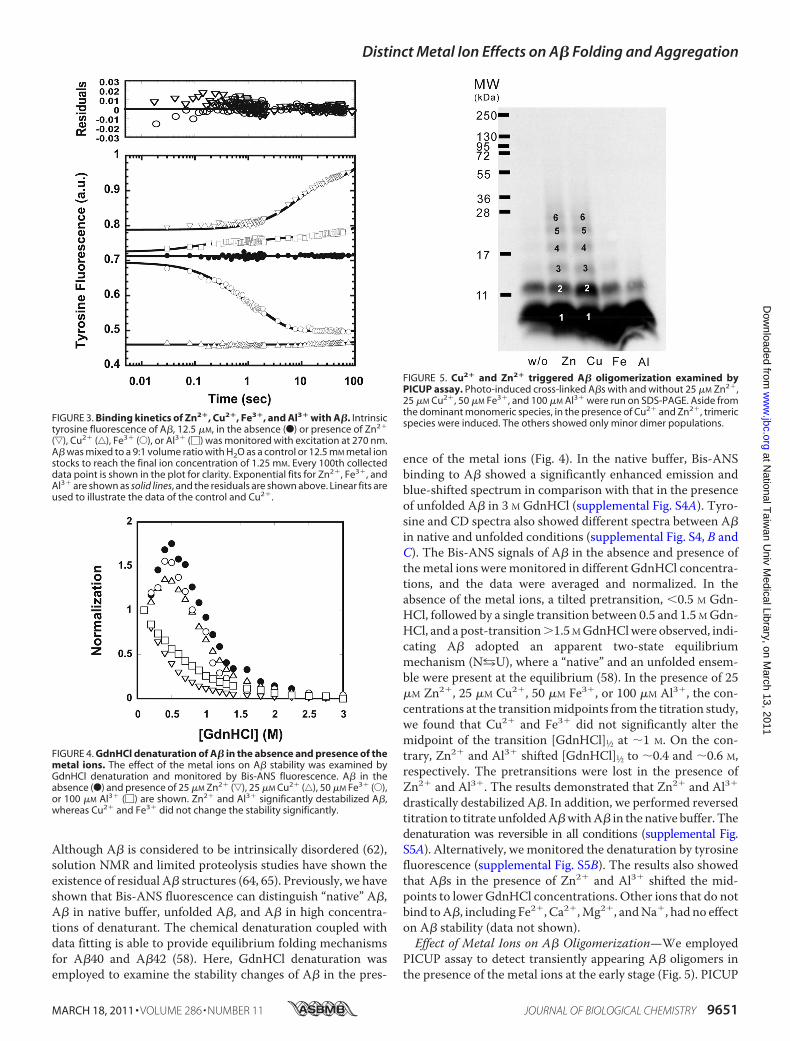
FIGURE 3. Binding kinetics of Zn2�, Cu2�, Fe3�, and Al3� with A�. Intrinsictyrosine fluorescence of A�, 12.5 �M, in the absence (F) or presence of Zn2�
(ƒ), Cu2� (‚), Fe3� (E), or Al3� (�) was monitored with excitation at 270 nm.A� was mixed to a 9:1 volume ratio with H2O as a control or 12.5 mM metal ionstocks to reach the final ion concentration of 1.25 mM. Every 100th collecteddata point is shown in the plot for clarity. Exponential fits for Zn2�, Fe3�, andAl3� are shown as solid lines, and the residuals are shown above. Linear fits areused to illustrate the data of the control and Cu2�.
FIGURE 4. GdnHCl denaturation of A� in the absence and presence of themetal ions. The effect of the metal ions on A� stability was examined byGdnHCl denaturation and monitored by Bis-ANS fluorescence. A� in theabsence (F) and presence of 25 �M Zn2� (ƒ), 25 �M Cu2� (‚), 50 �M Fe3� (E),or 100 �M Al3� (�) are shown. Zn2� and Al3� significantly destabilized A�,whereas Cu2� and Fe3� did not change the stability significantly.
FIGURE 5. Cu2� and Zn2� triggered A� oligomerization examined byPICUP assay. Photo-induced cross-linked A�s with and without 25 �M Zn2�,25 �M Cu2�, 50 �M Fe3�, and 100 �M Al3� were run on SDS-PAGE. Aside fromthe dominant monomeric species, in the presence of Cu2� and Zn2�, trimericspecies were induced. The others showed only minor dimer populations.
Distinct Metal Ion Effects on A� Folding and Aggregation
MARCH 18, 2011 • VOLUME 286 • NUMBER 11 JOURNAL OF BIOLOGICAL CHEMISTRY 9651
at National T
aiwan U
niv Medical Library, on M
arch 13, 2011w
ww
.jbc.orgD
ownloaded from
was developed to photo-cross-link the short-lived metastableassembly in the fast equilibrium at the early stage (59). Here, weexamined the effect in the presence of 25�MZn2�, 25�MCu2�,50 �M Fe3�, and 100 �M Al3�. Before PICUP, A�s were pre-dominantly monomers, as examined by SDS-PAGE (data notshown). After PICUP, A� in the absence of the metal ionsshowed a dominant monomeric species and a minor dimericspecies. No additional oligomer was induced in the presence of
Fe3� and Al3�. However, in the presence of Zn2� and Cu2�,trimeric species were induced. Therefore, our results showedthat Zn2� and Cu2�, but not Fe3� and Al3�, are capable ofinducing higher assembly of A� oligomers that transientlyappear at the early stage.Effect of the Metal Ions on A� Oligomerization and
Fibrillization—To investigate the kinetics of A� oligomeriza-tion and fibrillization, ThT assay was first applied to monitor
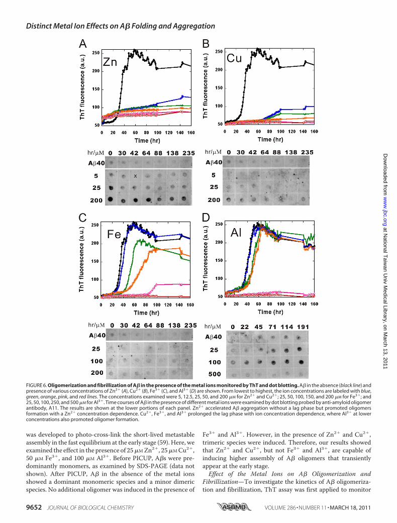
FIGURE 6. Oligomerization and fibrillization of A� in the presence of the metal ions monitored by ThT and dot blotting. A� in the absence (black line) andpresence of various concentrations of Zn2� (A), Cu2� (B), Fe3� (C), and Al3� (D) are shown. From lowest to highest, the ion concentrations are labeled with blue,green, orange, pink, and red lines. The concentrations examined were 5, 12.5, 25, 50, and 200 �M for Zn2� and Cu2�; 25, 50, 100, 150, and 200 �M for Fe3�; and25, 50, 100, 250, and 500 �M for Al3�. Time courses of A� in the presence of different metal ions were examined by dot blotting probed by anti-amyloid oligomerantibody, A11. The results are shown at the lower portions of each panel. Zn2� accelerated A� aggregation without a lag phase but promoted oligomersformation with a Zn2� concentration dependence. Cu2�, Fe3�, and Al3� prolonged the lag phase with ion concentration dependence, where Al3� at lowerconcentrations also promoted oligomer formation.
Distinct Metal Ion Effects on A� Folding and Aggregation
9652 JOURNAL OF BIOLOGICAL CHEMISTRY VOLUME 286 • NUMBER 11 • MARCH 18, 2011
at National T
aiwan U
niv Medical Library, on M
arch 13, 2011w
ww
.jbc.orgD
ownloaded from
the cross-�-sheet formation during aggregation (Fig. 6). ThT iscommonly used as a fluorescence probe in reporting thecross-� structures in amyloid fibrils (66). A� fibrillization isbelieved to undergo a nucleation-dependent pathway, where anoligomer nucleus is formed followed by an elongation event tomature fibrils (9). Five concentrations of the metal ions in dif-ferent ranges were used, as indicated, for a better visualizationof the effect. Without metal ions, A� followed the classic pat-tern of amyloid fibril formation to nucleate at a lag time of 20 hand elongated to reach a steady state after 60 h. In the presenceof metal ions, concentration-dependent effects on A� fibrilli-zation were observed. In the presence of Zn2�, the lag time wascompletely diminished, even in the lowest ion concentration (5�M), and ThT intensity increased readily without lag time (Fig.6A). A plateau was seen after 20 h with a lower ThT intensity incomparison with A� alone. Apart from Zn2�, the other threemetal ions had similar inhibition effects, albeit occurring at dif-ferent concentrations of ions. At 5 �M, Cu2� prolonged the lagphase significantly to more than 60 h and decreased ThT fluo-rescence (Fig. 6B). At 50 �M, Cu2� completely abolished theaggregation. Fe3� andAl3� above 50�Mboth prolonged the lagphase to�40 h, where Al3� has a strong inhibition effect above250 �M (Fig. 6, C and D). A weaker inhibition effect from Al3�
was observed when the pH was neutralized by performing theexperiment in Tris buffer at pH 7.85 (supplemental Fig. S6A).Thus, the strong inhibition may be primarily due to the acidicpH.The corresponding oligomer appearance was further exam-
ined by dot blotting probed by anti-amyloid oligomer antibody,A11. A11 is an amyloid oligomer-specific antibody recognizingcommon epitopes in various amyloid proteins (13). A� incu-bated with different metal ion concentrations were spotted onthe nitrocellulose membrane at different times during theaggregation. In the absence of metal ions, no significant A11-positive oligomer appeared. In the presence of Zn2�, startingfrom 25 to 200 �M, A11-positive signal was significantlyenhanced and appeared at the first time point (Fig. 6A). ForCu2� and Fe3�, no obvious A11-positive signal was detected(Fig. 6, B and C). Al3� induced A11-positive signals in a time-dependent manner; however, the signals disappeared in thepresence of 500 �M Al3� (Fig. 6D). The disappearance of A11signals was not caused by the acidic pH because the signal wasalso significantly weakened at higher concentrations of Al3�
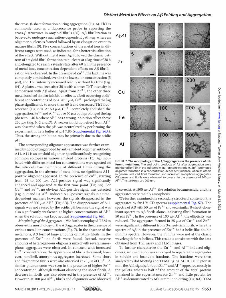
when the solution was kept neutral (supplemental Fig. 6B).Morphology of theAggregates—We further employedTEM to
observe themorphology of theA� aggregates in the presence ofvarious metal ion concentrations (Fig. 7). In the absence of themetal ions, A� formed large amounts of mature fibrils. In thepresence of Zn2�, no fibrils were found. Instead, massiveamounts of heterogeneous oligomersmixed with several amor-phous aggregates were observed. In contrast, with increasedCu2� concentration, the appearance of fibrils decreased; how-ever, nonfibril, amorphous aggregates increased. Some shortand fragmented fibrils were also observed at 25 �M of Cu2�. Asimilar phenomenon was seen in the presence of higher Fe3�
concentration, although without observing the short fibrils. Adecrease in fibrils was also observed in the presence of Al3�.However, at 100 �M Al3�, fibrils and oligomers were observed
to co-exist. At 500�MAl3�, the solution became acidic, and theaggregates were mainly amorphous.We further examined the secondary structural content of the
aggregates by far-UV CD spectra (supplemental Fig. S7). Thespectra of A�with 50�M of Fe3� showed similar �-sheet-dom-inant spectra to A� fibrils alone, indicating fibril formation in50 �M Fe3�. In the presence of 100 �M Al3�, the ellipticity wasreduced. The aggregates formed in 25 �M of Cu2� and Zn2�
were significantly different from �-sheet-rich fibrils, where thespectra of A� in the presence of Zn2� had a helix-like doubleminima spectra. However, the minima were not at the classicwavelength for -helices. This result is consistent with the dataobtained from ThT assay and TEM images.To further characterize the Zn2�- and Al3�-induced olig-
omers, sedimentation was employed to separate the aggregatesin soluble and insoluble fractions. The fractions were thenanalyzed by dot blotting and TEM (Fig. 8). At 10,000 � g for 20min, theA11 signals for bothZn2� andAl3� appearedmostly inthe pellets, whereas half of the amount of the total proteinremained in the supernatants for Zn2� and little protein forAl3� as demonstrated by 6E10 immunoblotting (Fig. 8A). TEM
FIGURE 7. The morphology of the A� aggregates in the presence of dif-ferent metal ions. The end point products of A� after aggregation weremonitored by TEM in the indicated metal ion concentrations. Zn2� promotedoligomer formation in a concentration-dependent manner, whereas othersin general reduced fibril formation and increased amorphous aggregates.Oligomers and fibrils were observed to co-exist in the presence of 100 �M
Al3�. The scale bars are 200 nm.
Distinct Metal Ion Effects on A� Folding and Aggregation
MARCH 18, 2011 • VOLUME 286 • NUMBER 11 JOURNAL OF BIOLOGICAL CHEMISTRY 9653
at National T
aiwan U
niv Medical Library, on M
arch 13, 2011w
ww
.jbc.orgD
ownloaded from
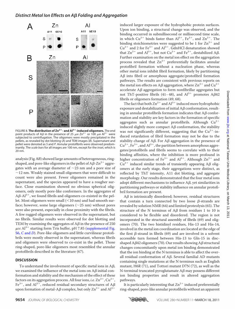
analysis (Fig. 8B) showed large amounts of heterogeneous, ring-shaped, and pore-like oligomers in the pellet of A�-Zn2� aggre-gates with an average diameter of �23 nm and a pore size of�12 nm. Weakly stained small oligomers that were difficult tocount were also present. Fewer oligomers remained in thesupernatant, and the species appeared to have a rougher sur-face. Close examination showed no obvious spherical olig-omers, only mostly pore-like conformers. In the aggregates ofA�-Al3�, we found fibrils and oligomers co-existed in the pel-let. Most oligomers were small (�10 nm) and had smooth sur-face; however, some large oligomers (�25 nm) without poreswere also present, especially in close proximity with the fibrils.A few rugged oligomers were observed in the supernatant, butno fibrils. Similar results were observed for dot blotting andTEM by examining the aggregates of A� in the presence of 250�M Al3� starting form Tris buffer, pH 7.85 (supplemental Fig.S6, C and D). Pore-like oligomers and little curvilinear protofi-brils were mostly observed in the supernatant, whereas fibrilsand oligomers were observed to co-exist in the pellet. Thosering-shaped, pore-like oligomers most resembled the annularprotofibrils described in the literature (67).
DISCUSSION
To understand the involvement of specific metal ions in A�,we examined the influence of the metal ions on A� initial con-formation and stability and themechanismof the effect of thesefactors on its aggregation process. All four ions, i.e.Zn2�, Cu2�,Fe3�, and Al3�, reduced residual secondary structures of A�upon formation of metal-A� complex, but only Zn2� and Al3�
induced larger exposure of the hydrophobic protein surfaces.Upon ion binding, a structural change was observed, and thebinding occurred in submillisecond or millisecond time scale,in which Cu2� binds faster than Al3�, Fe3�, and Zn2�. Thebinding stoichiometries were suggested to be 1 for Zn2� andCu2� and 2 for Fe3� and Al3�. GdnHCl denaturation showedthat Zn2� and Al3�, but not Cu2� and Fe3�, destabilized A�.Further examination on the metal ion effect on the aggregationprocess revealed that Zn2� preferentially facilitates annularprotofibril formation without a nucleation phase, whereasother metal ions inhibit fibril formation, likely by partitioningA� into fibril or amorphous aggregate/protofibril formationpathways. The results are consistent with previous reports onthe metal ion effects on A� aggregation, where Zn2� and Cu2�
accelerate A� aggregation to form nonfibrillar aggregates butnot ThT-positive fibrils (41–48), and Al3� promotes A�42fibrils or oligomers formation (49, 68).The fact that bothZn2� andAl3� inducedmore hydrophobic
exposure and destabilization of initial A� conformation, result-ing in annular protofibrils formation indicates that A� confor-mation and stability are key factors in the formation of specificaggregates such as annular protofibrils. Although Cu2�
induced slightly more compact A� conformation, the stabilitywas not significantly different, suggesting that the Cu2�-in-duced retardation of fibril formation may not be due to thestability change of A�. For A� aggregation in the presence ofCu2�, Fe3�, and Al3�, the partition between amorphous aggre-gates/protofibrils and fibrils seems to correlate with to theirbinding affinities, where the inhibition is more profound inhigher concentration of Fe3� and Al3�. Although Zn2� andCu2� induced similar trends of transiently appearing A� olig-omers at the early stage, their aggregations were distinct, asreflected by ThT intensity, A11 dot blotting, and aggregatemorphology. Our results demonstrated that the fourmetal ionsadopt different mechanisms to influence A�, yet similarities inpartitioning pathways or stability influence on annular protofi-bril formation are present.A� is intrinsically disordered; however, residual structures
that contain a turn connected by two loose �-strands arerevealed by solutionNMR (64) and limited proteolysis (65). Thestructure of the N terminus of A� from residues 1 to 10 isconsidered to be flexible and disordered. The region is notincorporated in the structural assembly of fibrils (69) and olig-omers (70). The two histidinyl residues, His-13 and His-14,involved in themetal ion coordination are located at the edge ofthe first �-strand in fibrils (69) and are involved in a solventaccessible turn formed between His-13 to Gln-15 in disc-shapedA�42 oligomers (70).Our results showingA� structuralchanges concomitantly upon metal ion binding demonstratedthat the ion binding at the N terminus is able to affect the over-all residual conformation of A�. Several familial AD mutantscontaining single mutations at the N terminus such as Englishmutant, H6R (71), and Tottori mutant D7N (72), as well as theN-terminal truncated pyroglutamate A� may possess differention binding properties and result in altered aggregationpathways.It is particularly interesting that Zn2� induced preferentially
ring-shaped, pore-like annular protofibrils without an apparent
FIGURE 8. The distribution of Zn2�- and Al3�-induced oligomers. The endpoint products of A� in the presence of 25 �M Zn2� or 100 �M Al3� weresubjected to centrifugation. The oligomers were mostly precipitated in thepellets, as revealed by dot blotting (A) and TEM images (B). Supernatant andpellet were denoted as S and P. Annular protofibrils were observed predom-inantly. The scale bars for all images are 100 nm, except for the inset, which is20 nm.
Distinct Metal Ion Effects on A� Folding and Aggregation
9654 JOURNAL OF BIOLOGICAL CHEMISTRY VOLUME 286 • NUMBER 11 • MARCH 18, 2011
at National T
aiwan U
niv Medical Library, on M
arch 13, 2011w
ww
.jbc.orgD
ownloaded from
nucleation process and that Al3� induced similar oligomers.Recently, heterogeneousA� oligomers have been found to existin immunological distinct structural states (67, 73). These A�oligomers have been characterized as prefibrillar oligomers,fibrillar oligomers, and annular protofibrils. The annular proto-fibrils are considered to be assembled from the prefibrillar oli-gomers, where the conversion is accelerated in the presence oflipid vesicles and other artificial conditions. The annular proto-fibrils are less toxic and membrane-permeable in comparisonwith prefibrillar oligomers, but they share common structuralepitopes with the bacterial toxin formed by �-barrel structures(67). In this study, the Zn2�- or Al3�-induced A� oligomerswere morphologically similar to the annular protofibrils andalso shared a common epitope recognized by A11 antibody.The extent to which the Zn2�- or Al3�-induced A� oligomersresemble those annular protofibrils or others remains unooknown. The structural conversion, membrane permeability,and toxicity of those annular protofibrils need further struc-tural and functional characterizations. However, our far-UVCD spectra of Zn2�-induced oligomers showed no enrichmentof the �-conformation, which suggests that the oligomers arenot fibrillar oligomers and should be different from the�-barreltoxins.In physiological conditions, less than 0.5 �M of extracellular
Zn2� is present in brain; however, a high concentration of Zn2�
was found in the glutamergic neurons during neuronal activityespecially in cerebrocortex or amygdala (22, 74). The presyn-apic Zn2� is concentrated by the zinc transporter protein,ZnT3, to achieve �300 �M in the cleft (22, 75). The in vivoevidence shows that the ZnT3 knock-out transgenic amyloidprecursor protein mice with a reduction of Zn2� in the hip-pocampus did not develop obvious amyloid plaques in compar-ison with those with normal ZnT3 level (34). In AD patients, adecrease of metallothionein 3 that regulates the uptake of syn-aptic Zn2� has been observed (35), and the release of synapticZn2� during activity is critical for A� oligomer synaptic target-ing (36). These studies show that synaptic Zn2� and Zn2�
homeostasis play crucial roles in AD pathology. According toour results, we suspect that the Zn2�-induced oligomer forma-tion resulting in heterogeneous A� annular protofibrils mayoccur at presynapses during synaptic activities, thereby trigger-ing oligomer formation and leading to concentrated A� olig-omers at the synaptic clefts. This hypothesis may also contrib-ute to the mechanism of rapid cognition restoration in themetal chelating therapy (53). In summary, our fundamentalfolding and aggregation studies facilitate the understanding ofthe clinically related metal ions in AD to A� and provide a ratio-nalized mechanism for annular protofibril formation. The studymay contribute to potential pathogenic implication in AD.
Acknowledgments—We thank the specialists of Academia Sinica,Tai-Lang Lin (Institute of Cellular andOrganismic Biology) for assist-ing TEM imaging, and Cheng-Ying Yu (Institute of Chemistry) forassisting atomic absorption operation.
REFERENCES1. Masters, C. L., Simms, G., Weinman, N. A., Multhaup, G., McDonald,
B. L., and Beyreuther, K. (1985) Proc. Natl. Acad. Sci. U.S.A. 82,
4245–42492. Mucke, L. (2009) Nature 461, 895–8973. Kang, J., Lemaire, H. G., Unterbeck, A., Salbaum, J. M., Masters, C. L.,
Grzeschik, K. H., Multhaup, G., Beyreuther, K., andMuller-Hill, B. (1987)Nature 325, 733–736
4. Thinakaran, G., and Koo, E. H. (2008) J. Biol. Chem. 283, 29615–296195. Bibl, M., Esselmann, H., Mollenhauer, B., Weniger, G., Welge, V., Liess,
M., Lewczuk, P., Otto, M., Schulz, J. B., Trenkwalder, C., Kornhuber, J.,and Wiltfang, J. (2007) J. Neurochem. 103, 467–474
6. Schoonenboom, N. S., Mulder, C., Van Kamp, G. J., Mehta, S. P., Schel-tens, P., Blankenstein, M. A., and Mehta, P. D. (2005) Ann. Neurol. 58,139–142
7. Burdick, D., Soreghan, B., Kwon, M., Kosmoski, J., Knauer, M., Henschen,A., Yates, J., Cotman, C., and Glabe, C. (1992) J. Biol. Chem. 267, 546–554
8. Jarrett, J. T., Berger, E. P., and Lansbury, P. T., Jr. (1993) Biochemistry 32,4693–4697
9. Roychaudhuri, R., Yang,M., Hoshi,M.M., andTeplow,D. B. (2009) J. Biol.Chem. 284, 4749–4753
10. Haass, C., and Selkoe, D. J. (2007) Nat. Rev. Mol. Cell Biol. 8, 101–11211. Walsh, D. M., Klyubin, I., Fadeeva, J. V., Cullen, W. K., Anwyl, R., Wolfe,
M. S., Rowan, M. J., and Selkoe, D. J. (2002) Nature 416, 535–53912. Lesne, S., Koh, M. T., Kotilinek, L., Kayed, R., Glabe, C. G., Yang, A.,
Gallagher, M., and Ashe, K. H. (2006) Nature 440, 352–35713. Kayed, R., Head, E., Thompson, J. L., McIntire, T. M., Milton, S. C., Cot-
man, C. W., and Glabe, C. G. (2003) Science 300, 486–48914. Lambert, M. P., Barlow, A. K., Chromy, B. A., Edwards, C., Freed, R.,
Liosatos, M., Morgan, T. E., Rozovsky, I., Trommer, B., Viola, K. L., Wals,P., Zhang, C., Finch, C. E., Krafft, G. A., and Klein,W. L. (1998) Proc. Natl.Acad. Sci. U.S.A. 95, 6448–6453
15. Hoshi, M., Sato, M., Matsumoto, S., Noguchi, A., Yasutake, K., Yoshida,N., and Sato, K. (2003) Proc. Natl. Acad. Sci. U.S.A. 100, 6370–6375
16. Barghorn, S., Nimmrich, V., Striebinger, A., Krantz, C., Keller, P., Janson,B., Bahr,M., Schmidt,M., Bitner, R. S., Harlan, J., Barlow, E., Ebert, U., andHillen, H. (2005) J. Neurochem. 95, 834–847
17. Harper, J. D., Wong, S. S., Lieber, C. M., and Lansbury, P. T. (1997) Chem.Biol. 4, 119–125
18. Lashuel, H. A., and Lansbury, P. T., Jr. (2006)Q. Rev. Biophys. 39, 167–20119. Lovell,M.A., Robertson, J. D., Teesdale,W. J., Campbell, J. L., andMarkes-
bery, W. R. (1998) J. Neurol. Sci. 158, 47–5220. Frederickson, C. J., Koh, J. Y., and Bush, A. I. (2005) Nat. Rev. Neurosci. 6,
449–46221. Yumoto, S., Kakimi, S., Ohsaki, A., and Ishikawa, A. (2009) J. Inorg.
Biochem. 103, 1579–158422. Bush, A. I. (2003) Trends Neurosci. 26, 207–21423. Zatta, P., Drago, D., Bolognin, S., and Sensi, S. L. (2009)Trends Pharmacol.
Sci. 30, 346–35524. Sparks, D. L., and Schreurs, B. G. (2003) Proc. Natl. Acad. Sci. U.S.A. 100,
11065–1106925. Frisardi, V., Solfrizzi, V., Capurso, C., Kehoe, P. G., Imbimbo, B. P., San-
tamato,A., Dellegrazie, F., Seripa,D., Pilotto, A., Capurso, A., and Panza, F.(2010) J. Alzheimers Dis. 20, 17–30
26. Ma, Q. F., Hu, J., Wu,W. H., Liu, H. D., Du, J. T., Fu, Y., Wu, Y.W., Lei, P.,Zhao, Y. F., and Li, Y. M. (2006) Biopolymers 83, 20–31
27. Syme, C. D., Nadal, R. C., Rigby, S. E., and Viles, J. H. (2004) J. Biol. Chem.279, 18169–18177
28. Minicozzi, V., Stellato, F., Comai, M., Serra, M. D., Potrich, C., Meyer-Klaucke, W., and Morante, S. (2008) J. Biol. Chem. 283, 10784–10792
29. Karr, J.W., Akintoye, H., Kaupp, L. J., and Szalai, V. A. (2005)Biochemistry44, 5478–5487
30. Stellato, F., Menestrina, G., Serra, M. D., Potrich, C., Tomazzolli, R.,Meyer-Klaucke, W., and Morante, S. (2006) Eur. Biophys. J. 35, 340–351
31. Danielsson, J., Pierattelli, R., Banci, L., andGraslund,A. (2007) FEBS J.274,46–59
32. Syme, C. D., and Viles, J. H. (2006) Biochim. Biophys. Acta 1764, 246–25633. Smith, D. P., Smith, D. G., Curtain, C. C., Boas, J. F., Pilbrow, J. R., Ciccoto-
sto, G. D., Lau, T. L., Tew, D. J., Perez, K., Wade, J. D., Bush, A. I., Drew,S. C., Separovic, F., Masters, C. L., Cappai, R., and Barnham, K. J. (2006)J. Biol. Chem. 281, 15145–15154
Distinct Metal Ion Effects on A� Folding and Aggregation
MARCH 18, 2011 • VOLUME 286 • NUMBER 11 JOURNAL OF BIOLOGICAL CHEMISTRY 9655
at National T
aiwan U
niv Medical Library, on M
arch 13, 2011w
ww
.jbc.orgD
ownloaded from
34. Lee, J. Y., Cole, T. B., Palmiter, R. D., Suh, S.W., and Koh, J. Y. (2002) Proc.Natl. Acad. Sci. U.S.A. 99, 7705–7710
35. Yu,W.H., Lukiw,W. J., Bergeron, C., Niznik, H. B., and Fraser, P. E. (2001)Brain Res. 894, 37–45
36. Deshpande, A., Kawai, H., Metherate, R., Glabe, C. G., and Busciglio, J.(2009) J. Neurosci. 29, 4004–4015
37. Nair, N. G., Perry, G., Smith, M. A., and Reddy, V. P. (2010) J. AlzheimersDis. 20, 57–66
38. Garzon-Rodriguez,W., Yatsimirsky, A. K., and Glabe, C. G. (1999) Bioorg.Med. Chem. Lett. 9, 2243–2248
39. Atwood, C. S., Scarpa, R. C., Huang, X., Moir, R. D., Jones, W. D., Fairlie,D. P., Tanzi, R. E., and Bush, A. I. (2000) J. Neurochem. 75, 1219–1233
40. Tougu, V., Karafin, A., and Palumaa, P. (2008) J. Neurochem. 104,1249–1259
41. Bush, A. I., Pettingell, W. H., Multhaup, G. d., Paradis, M., Vonsattel, J. P.,Gusella, J. F., Beyreuther, K., Masters, C. L., and Tanzi, R. E. (1994) Science265, 1464–1467
42. Yoshiike, Y., Tanemura, K.,Murayama,O., Akagi, T.,Murayama,M., Sato,S., Sun, X., Tanaka, N., and Takashima, A. (2001) J. Biol. Chem. 276,32293–32299
43. Ha, C., Ryu, J., and Park, C. B. (2007) Biochemistry 46, 6118–612544. Noy,D., Solomonov, I., Sinkevich,O., Arad, T., Kjaer, K., and Sagi, I. (2008)
J. Am. Chem. Soc. 130, 1376–138345. Ryu, J., Girigoswami, K., Ha, C., Ku, S. H., and Park, C. B. (2008) Biochem-
istry 47, 5328–533546. Smith, D. P., Ciccotosto, G. D., Tew,D. J., Fodero-Tavoletti,M. T., Johans-
sen, T., Masters, C. L., Barnham, K. J., and Cappai, R. (2007) Biochemistry46, 2881–2891
47. Garai, K., Sahoo, B., Kaushalya, S. K., Desai, R., and Maiti, S. (2007) Bio-chemistry 46, 10655–10663
48. Tougu, V., Karafin, A., Zovo, K., Chung, R. S., Howells, C.,West, A. K., andPalumaa, P. (2009) J. Neurochem. 110, 1784–1795
49. Ricchelli, F., Drago, D., Filippi, B., Tognon, G., and Zatta, P. (2005) CellMol. Life Sci. 62, 1724–1733
50. Kawahara, M., Kato, M., and Kuroda, Y. (2001) Brain Res. Bull. 55,211–217
51. Lovell,M. A., Xie, C., andMarkesbery,W. R. (1999)Brain Res. 823, 88–9552. Cherny, R. A., Atwood, C. S., Xilinas, M. E., Gray, D. N., Jones, W. D.,
McLean, C. A., Barnham, K. J., Volitakis, I., Fraser, F. W., Kim, Y., Huang,X., Goldstein, L. E.,Moir, R. D., Lim, J. T., Beyreuther, K., Zheng,H., Tanzi,R. E., Masters, C. L., and Bush, A. I. (2001) Neuron 30, 665–676
53. Adlard, P. A., Cherny, R. A., Finkelstein, D. I., Gautier, E., Robb, E., Cortes,M., Volitakis, I., Liu, X., Smith, J. P., Perez, K., Laughton, K., Li, Q. X.,Charman, S. A., Nicolazzo, J. A.,Wilkins, S., Deleva, K., Lynch, T., Kok, G.,Ritchie, C. W., Tanzi, R. E., Cappai, R., Masters, C. L., Barnham, K. J., and
Bush, A. I. (2008) Neuron 59, 43–5554. Faux, N. G., Ritchie, C.W., Gunn, A., Rembach, A., Tsatsanis, A., Bedo, J.,
Harrison, J., Lannfelt, L., Blennow, K., Zetterberg, H., Ingelsson, M., Mas-ters, C. L., Tanzi, R. E., Cummings, J. L., Herd, C.M., and Bush, A. I. (2010)J. Alzheimers Dis. 20, 509–516
55. Edelhoch, H. (1967) Biochemistry 6, 1948–195456. Chen, Y. R., and Clark, A. C. (2004) Protein Sci. 13, 2196–220657. Deleted in proof58. Chen, Y. R., and Glabe, C. G. (2006) J. Biol. Chem. 281, 24414–2442259. Bitan, G., Lomakin, A., and Teplow, D. B. (2001) J. Biol. Chem. 276,
35176–3518460. Brand, L., and Gohlke, J. R. (1972) Annu. Rev. Biochem. 41, 843–86861. LeVine, H., 3rd. (2002) Arch. Biochem. Biophys. 404, 106–11562. Riek, R., Guntert, P., Dobeli, H., Wipf, B., and Wuthrich, K. (2001) Eur.
J. Biochem. 268, 5930–593663. Vasudevaraju, P., Govindaraju,M., Palanisamy, A. P., Sambamurti, K., and
Rao, K. S. (2008) Indian J. Med. Res. 128, 545–55664. Hou, L., Shao, H., Zhang, Y., Li, H., Menon, N. K., Neuhaus, E. B., Brewer,
J. M., Byeon, I. J., Ray, D. G., Vitek, M. P., Iwashita, T., Makula, R. A.,Przybyla, A. B., and Zagorski, M. G. (2004) J. Am. Chem. Soc. 126,1992–2005
65. Lazo, N. D., Grant, M. A., Condron, M. C., Rigby, A. C., and Teplow, D. B.(2005) Protein Sci. 14, 1581–1596
66. LeVine, H., 3rd. (1993) Protein Sci. 2, 404–41067. Kayed, R., Pensalfini, A.,Margol, L., Sokolov, Y., Sarsoza, F., Head, E., Hall,
J., and Glabe, C. (2009) J. Biol. Chem. 284, 4230–423768. Drago, D., Bettella, M., Bolognin, S., Cendron, L., Scancar, J., Milacic, R.,
Ricchelli, F., Casini, A., Messori, L., Tognon, G., and Zatta, P. (2008) Int.J. Biochem. Cell Biol. 40, 731–746
69. Tycko, R. (2006) Q. Rev. Biophys. 39, 1–5570. Ahmed,M., Davis, J., Aucoin, D., Sato, T., Ahuja, S., Aimoto, S., Elliott, J. I.,
Van Nostrand, W. E., and Smith, S. O. (2010) Nat. Struct. Mol. Biol. 17,561–567
71. Janssen, J. C., Beck, J. A., Campbell, T. A., Dickinson, A., Fox, N. C., Har-vey, R. J., Houlden, H., Rossor,M. N., and Collinge, J. (2003)Neurology 60,235–239
72. Wakutani, Y., Watanabe, K., Adachi, Y., Wada-Isoe, K., Urakami, K., Ni-nomiya, H., Saido, T. C., Hashimoto, T., Iwatsubo, T., and Nakashima, K.(2004) J. Neurol. Neurosurg. Psychiatry 75, 1039–1042
73. Wu, J. W., Breydo, L., Isas, J. M., Lee, J., Kuznetsov, Y. G., Langen, R., andGlabe, C. (2010) J. Biol. Chem. 285, 6071–6079
74. Frederickson, C. J., and Bush, A. I. (2001) BioMetals 14, 353–36675. Palmiter, R. D., Cole, T. B., Quaife, C. J., and Findley, S. D. (1996) Proc.
Natl. Acad. Sci. U.S.A. 93, 14934–14939
Distinct Metal Ion Effects on A� Folding and Aggregation
9656 JOURNAL OF BIOLOGICAL CHEMISTRY VOLUME 286 • NUMBER 11 • MARCH 18, 2011
at National T
aiwan U
niv Medical Library, on M
arch 13, 2011w
ww
.jbc.orgD
ownloaded from