26 non-odontogenic cysts
TRANSCRIPT
7/21/2019 26 Non-Odontogenic Cysts
http://slidepdf.com/reader/full/26-non-odontogenic-cysts 1/7
1
Non-odontogenic Cysts
Dr. Ioannis G. Koutlas
Division of Oral Pathology
All pictures are intellectual property of the Division of Oral andMaxillofacia l Pathology or its Faculty. Duplication or any unauthorized
use is prohibited.
Developmental Cysts
• a.k.a. fissural cysts
• Exact pathogenesis of some of them uncertain
• Generally, slow increase
• May be identified incidentally
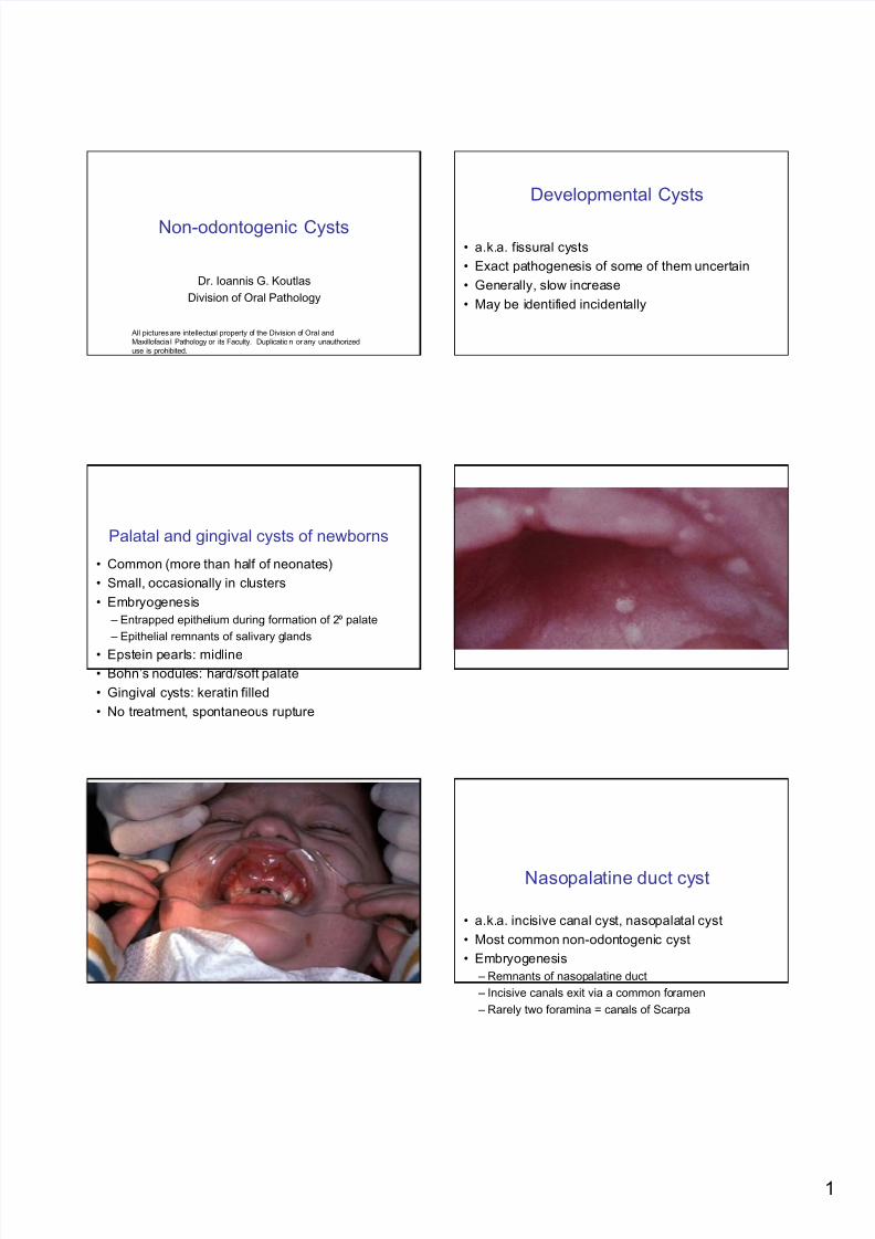
Palatal and gingival cysts of newborns
• Common (more than half of neonates)
• Small, occasionally in clusters
• Embryogenesis
– Entrapped epithelium during formation of 2º palate
– Epithelial remnants of salivary glands
• Epstein pearls: midline
• Bohn’s nodules: hard/soft palate
• Gingival cysts: keratin filled
• No treatment, spontaneous rupture
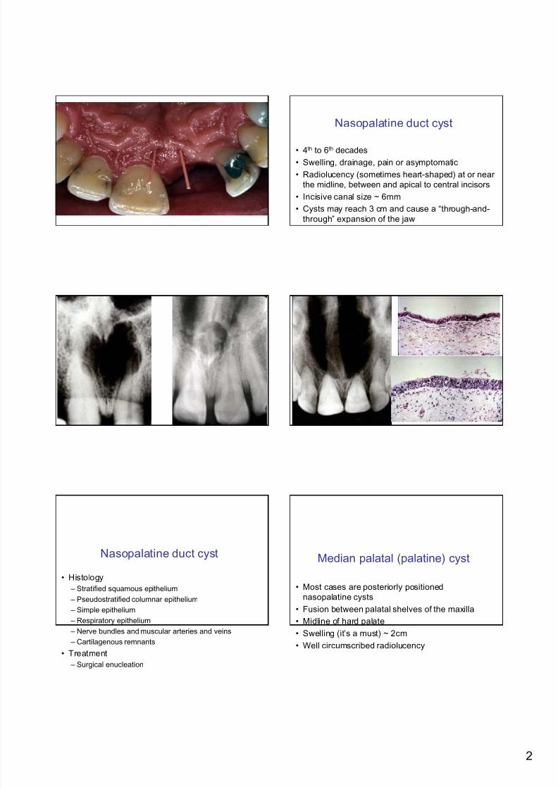
Nasopalatine duct cyst
• a.k.a. incisive canal cyst, nasopalatal cyst
• Most common non-odontogenic cyst
• Embryogenesis
– Remnants of nasopalatine duct
– Incisive canals exit via a common foramen
– Rarely two foramina = canals of Scarpa
7/21/2019 26 Non-Odontogenic Cysts
http://slidepdf.com/reader/full/26-non-odontogenic-cysts 2/7
2
Nasopalatine duct cyst
• 4th to 6th decades
• Swelling, drainage, pain or asymptomatic
• Radiolucency (sometimes heart-shaped) at or near
the midline, between and apical to central incisors
• Incisive canal size ~ 6mm
• Cysts may reach 3 cm and cause a “through-and-
through” expansion of the jaw
Nasopalatine duct cyst
• Histology
– Stratified squamous epithelium
– Pseudostratified columnar epithelium
– Simple epithelium
– Respiratory epithelium
– Nerve bundles and muscular arteries and veins
– Cartilagenous remnants
• Treatment
– Surgical enucleation
Median palatal (palatine) cyst
• Most cases are posteriorly positioned
nasopalatine cysts
• Fusion between palatal shelves of the maxilla
• Midline of hard palate
• Swelling (it’s a must) ~ 2cm
• Well circumscribed radiolucency
7/21/2019 26 Non-Odontogenic Cysts
http://slidepdf.com/reader/full/26-non-odontogenic-cysts 3/7
3
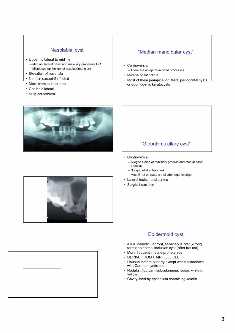
Nasolabial cyst
• Upper lip lateral to midline
– Medial - lateral nasal and maxillary processes OR
– Misplaced epithelium of nasolacrimal gland
• Elevation of nasal ala
• No pain except if infected
• More women than men
• Can be bilateral
• Surgical removal
“Median mandibular cyst”
• Controversial
– There are no epithelial lined processes
• Midline of mandible
• Most of them periapical or lateral periodontal cysts
or odontogenic keratocysts
“Globulomaxillary cyst”
• Controversial
– Alleged fusion of maxillary process and medial nasal
process
– No epithelial entrapment
– Most if not all cysts are of odontogenic origin• Lateral incisor and canine
• Surgical excision
Epidermoid cyst
• a.k.a. infundibular cyst, sebaceous cyst (wrongterm); epidermal inclusion cyst (after trauma)
• More frequent in acne-prone areas
• DERIVE FROM HAIR FOLLICLE
• Unusual before puberty except when associatedwith Gardner syndrome
• Nodular, fluctuant subcutaneous lesion, white oryellow
• Cavity lined by epithelium containing keratin
7/21/2019 26 Non-Odontogenic Cysts
http://slidepdf.com/reader/full/26-non-odontogenic-cysts 4/7
4
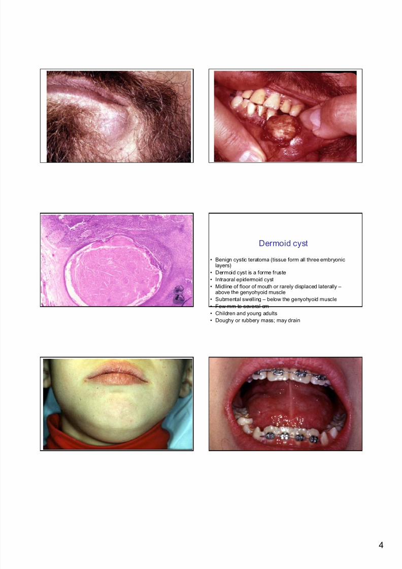
Dermoid cyst
• Benign cystic teratoma (tissue form all three embryoniclayers)
• Dermoid cyst is a forme fruste
• Intraoral epidermoid cyst
• Midline of floor of mouth or rarely displaced laterally – above the genyohyoid muscle
• Submental swelling – below the genyohyoid muscle• Few mm to several cm
• Children and young adults
• Doughy or rubbery mass; may drain
7/21/2019 26 Non-Odontogenic Cysts
http://slidepdf.com/reader/full/26-non-odontogenic-cysts 5/7
5
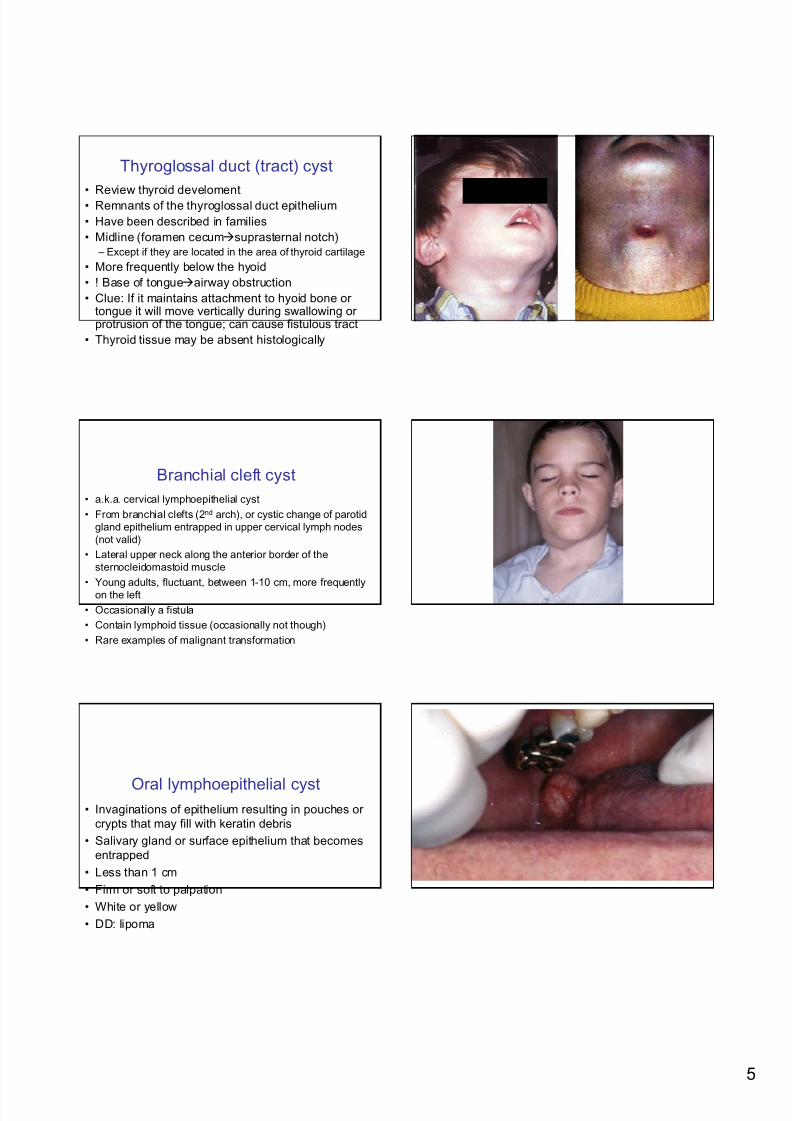
Thyroglossal duct (tract) cyst
• Review thyroid develoment• Remnants of the thyroglossal duct epithelium
• Have been described in families
• Midline (foramen cecumsuprasternal notch)
– Except if they are located in the area of thyroid cartilage
• More frequently below the hyoid
• ! Base of tongueairway obstruction
• Clue: If it maintains attachment to hyoid bone ortongue it will move vertically during swallowing orprotrusion of the tongue; can cause fistulous tract
• Thyroid tissue may be absent histologically
Branchial cleft cyst
• a.k.a. cervical lymphoepithelial cyst
• From branchial clefts (2nd arch), or cystic change of parotid
gland epithelium entrapped in upper cervical lymph nodes
(not valid)
• Lateral upper neck along the anterior border of the
sternocleidomastoid muscle
• Young adults, fluctuant, between 1-10 cm, more frequently
on the left
• Occasionally a fistula
• Contain lymphoid tissue (occasionally not though)
• Rare examples of malignant transformation
Oral lymphoepithelial cyst• Invaginations of epithelium resulting in pouches or
crypts that may fill with keratin debris
• Salivary gland or surface epithelium that becomes
entrapped
• Less than 1 cm
• Firm or soft to palpation
• White or yellow
• DD: lipoma
7/21/2019 26 Non-Odontogenic Cysts
http://slidepdf.com/reader/full/26-non-odontogenic-cysts 6/7
6
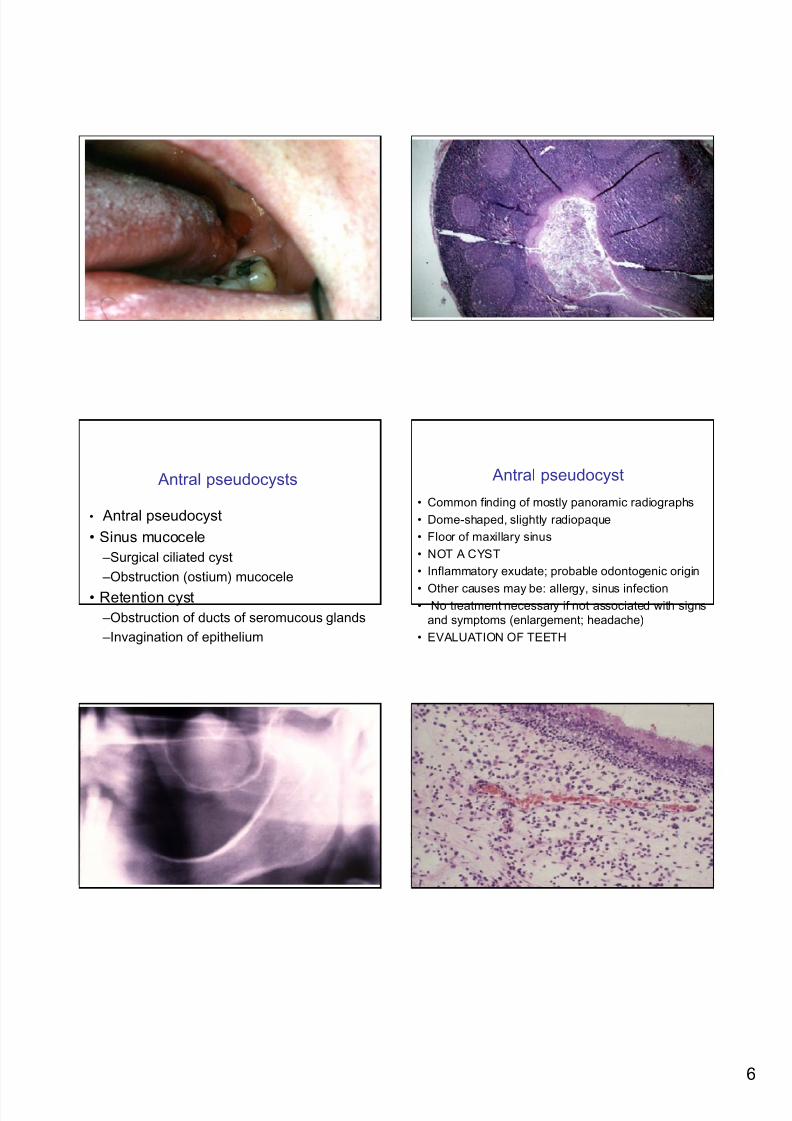
Antral pseudocysts
• Antral pseudocyst
• Sinus mucocele
–Surgical ciliated cyst
–Obstruction (ostium) mucocele
• Retention cyst
–Obstruction of ducts of seromucous glands
–Invagination of epithelium
Antral pseudocyst
• Common finding of mostly panoramic radiographs
• Dome-shaped, slightly radiopaque
• Floor of maxillary sinus
• NOT A CYST
• Inflammatory exudate; probable odontogenic origin
• Other causes may be: allergy, sinus infection• No treatment necessary if not associated with signs
and symptoms (enlargement; headache)
• EVALUATION OF TEETH
7/21/2019 26 Non-Odontogenic Cysts
http://slidepdf.com/reader/full/26-non-odontogenic-cysts 7/7
7
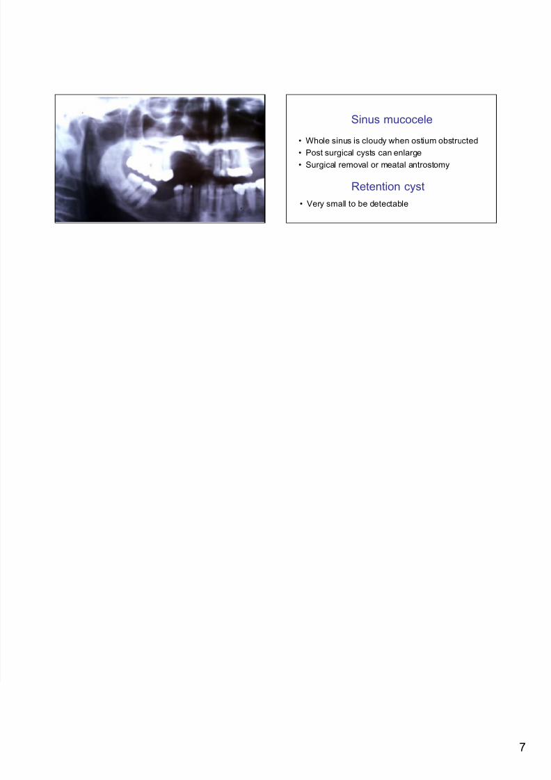
Sinus mucocele
• Whole sinus is cloudy when ostium obstructed
• Post surgical cysts can enlarge
• Surgical removal or meatal antrostomy
Retention cyst
• Very small to be detectable