511-2018-09-05-anatomy-ii - github pages
TRANSCRIPT
511-2018-09-05-anatomy-IIRick Gilmore
2018-09-05 13:24:30

Prelude
2/63

Today's Topics
Web resources
Wrap up on the forebrain
White matter tracts
The peripheral nervous system
·
·
·
·
3/63
Web resources
Harvard Whole Brain Atlas
Normal Anatomy
·
Whole Brain Atlas Top 100 Brain Structures List-
·
Linked list of structures-
4/63

Organization of the brain
Major division Ventricular Landmark Embryonic Division Structure
Forebrain Lateral Telencephalon Cerebral cortex
Basal ganglia
Hippocampus, amygdala
Third Diencephalon Thalamus
Hypothalamus
Midbrain Cerebral Aqueduct Mesencephalon Tectum, tegmentum
5/63

Organization of the brain
Major division Ventricular Landmark Embryonic Division Structure
Hindbrain 4th Metencephalon Cerebellum, pons
– Mylencephalon Medulla oblongata
6/63

Cerebral Cortex
Cerebral hemispheres
Groove (sulcus or sulci)
Bumps (gyrus or gyri)
Grey vs.white matter
Lobes
7/63

Cortical Gyri – Lateral
https://upload.wikimedia.org/wikipedia/commons/3/35/Gray726.png
8/63

Cortical Gyri – Medial
https://upload.wikimedia.org/wikipedia/commons/thumb/f/fe/Gray727.svg/1025px-Gray727.svg.png
9/63

Gray vs. White Matter
https://upload.wikimedia.org/wikipedia/commons/9/9a/Brainmaps-macaque-hippocampus.jpg
10/63
Lobes of the cerebral cortex
Frontal
Temporal
Parietal
Occipital
11/63


Lobes
http://40.media.tumblr.com/tumblr_m1kpkr7Wsq1rn6pqko1_500.jpg
13/63

Landmarks of the cortex
Longitudinal fissure
https://upload.wikimedia.org/wikipedia/commons/0/04/Human_brain_long
14/63
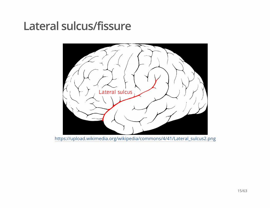
Lateral sulcus/fissure
https://upload.wikimedia.org/wikipedia/commons/4/41/Lateral_sulcus2.png
15/63

Central sulcus
https://upload.wikimedia.org/wikipedia/commons/8/88/Central_sulcus_diagram.png
16/63

Frontal lobe
Where is it?Anterior to central sulcus
Superior to lateral fissure
Dorsal to temporal lobe
·
·
·
17/63
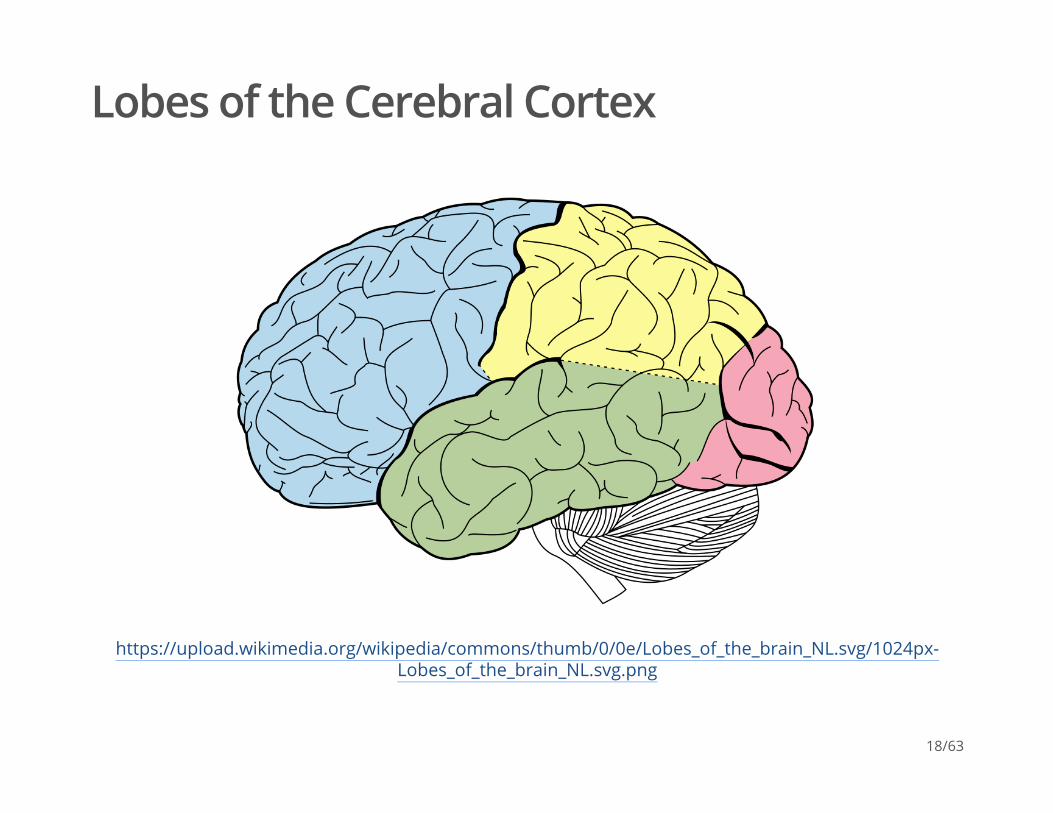
Lobes of the Cerebral Cortex
https://upload.wikimedia.org/wikipedia/commons/thumb/0/0e/Lobes_of_the_brain_NL.svg/1024px-Lobes_of_the_brain_NL.svg.png
18/63
Frontal lobe
What does it do?Primary motor cortex (M1)
Prefrontal cortex
·
Supplementary motor cortex
Frontal eye fields (FEF)
-
-
·
Planning, problem solving, working memory…?-
19/63

Frontal lobe
What does it do?Basal forebrain
Anterior cingulate cortex (ACC)
Primary olfactory cortex
·
Nucleus accumbens (NAcc), part of ventral striatum-
·
·
20/63

Cingulate Gyrus
http://cis.jhu.edu/data.sets/cortical_segmentation_validation/photos/cinggyrus75.jpg
21/63

Inferior Frontal Gyrus (IFG)
https://upload.wikimedia.org/wikipedia/commons/b/b2/Gray726_inferior_frontal_gyrus.png
22/63

Middle Frontal Gyrus (MFG)
https://upload.wikimedia.org/wikipedia/commons/7/7f/Gray726_middle_frontal_gyrus.png
23/63

Temporal lobe
Where is it?Ventral to frontal, parietal lobes
Inferior to lateral fissure
·
·
24/63

Temporal lobe
What does it do?Primary auditory cortex
Object, face recognition
Storage of memories about events, objects
Amygdala, hippocampus
·
·
·
·
25/63

Inferior Temporal Gyrus (ITG)
https://upload.wikimedia.org/wikipedia/commons/1/18/Gray726_inferior_temporal_gyrus.png
26/63

Entorhinal Cortex (ER)
https://upload.wikimedia.org/wikipedia/commons/1/15/Medial_surface_of_cerebral_cortex_-_entorhinal_cortex.png
27/63
Parietal lobe
Where is it?
What does it do?
Caudal to frontal lobe
Dorsal to temporal lobe
Posterior to central sulcus
·
·
·
Primary somatosensory cortex
Perception of spatial relations, action planning
·
·
28/63

Inferior Parietal Lobule
https://upload.wikimedia.org/wikipedia/commons/e/e3/Gray726_inferior_parietal_lobule.png
29/63

Superior Parietal Lobule
https://upload.wikimedia.org/wikipedia/commons/9/9d/Gray726_superior_parietal_lobule.png
30/63
Occipital lobe
Where is it?
What does it do?
Caudal to parietal & temporal lobes·
Primary visual cortex (V1)·
31/63
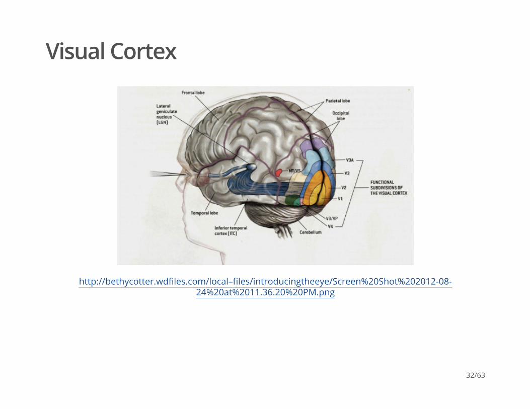
Visual Cortex
http://bethycotter.wdfiles.com/local–files/introducingtheeye/Screen%20Shot%202012-08-24%20at%2011.36.20%20PM.png
32/63

Insular cortex (insula)
Where is it?medial to temporal lobe
deep inside lateral fissure
·
·
33/63

Insula
https://upload.wikimedia.org/wikipedia/commons/b/b4/Sobo_1909_633.png
34/63

Insula
What does it do?Primary gustatory cortex
self-awareness, interpersonal experiences, motor control
·
·
35/63
Brodmann Areas
Korbinian Brodmann
http://www.spektrum.de/lexika/images/bio/fff1209_w.jpg
Cytoarchitectonic differences in cerebral cortex·
36/63

Brodmann Areas
https://upload.wikimedia.org/wikipedia/commons/0/09/Brodmann-areas.png
37/63
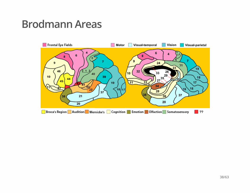
Brodmann Areas
38/63

Spinal cord
Rostral/Caudal axisSpinal column w/ vertebrae
Cervical (8), thoracic (12), lumbar (5), sacral (5), coccygeal (1)
Spinal segments & 31 nerve pairs
Cauda equina
·
·
·
·
39/63

Spinal cord
http://www.fauxpress.com/kimball/med/sensory/spinaldivisions.jpg
40/63
Spinal cord
Organization of the spinal cordDorsal/Ventral
Grey (interior) vs. white matter (exterior)
·
Dorsal root (sensory)
Ventral root (mostly motor)
-
-
·
41/63

42/63
Cross section of the spinal cord.

Organization of the PNS
Somatic division
Autonomic
Cranial nerves
Spinal nerves
43/63

Cranial nerves
Afferents (input), efferents (output), or mixed
Innervate head and neck
Olfactory (I), optic (II), (VIII) auditory, vagus (X), etc.
Spinal nerves
·
·
·
·
44/63

Cranial nerves
http://media-1.web.britannica.com/eb-media/44/54244-004-892C5169.jpg
45/63

Major white matter pathways
Brainstem
Projection fibers
Association fibers
Commissural fibers
46/63

(Oishi, Faria, Zijl, & Mori, 2010), Chapter 3, Figure 1.
47/63

Brainstem projections
Corticospinal tract (descending/efferent)
Dorsal column/medial lemniscus (ascending/afferent)
Superior/inferior cerebellar peduncles (from/to cerebellum)
·
·
·
48/63

(Oishi et al., 2010), Chapter 3, Figure 8.
49/63

Projection fiber tracts
Internal capsule·
Thalamic radiation
Cortico-{pontine, bulbar, reticular} tracts
-
-
50/63

(Oishi et al., 2010), Chapter 3, Figure 11.
51/63

(Oishi et al., 2010), Chapter 3, Figure 11.
52/63

Cortical white matter tracts
Superior/inferior longitudinal fasciculus
Superior/inferior fronto-occipital fasciculus
Cingulum, fornix (hyp-hip), stria terminalis (hyp-amyg)
·
Arcuate fasciculus part of sup. long. f.-
·
·
53/63

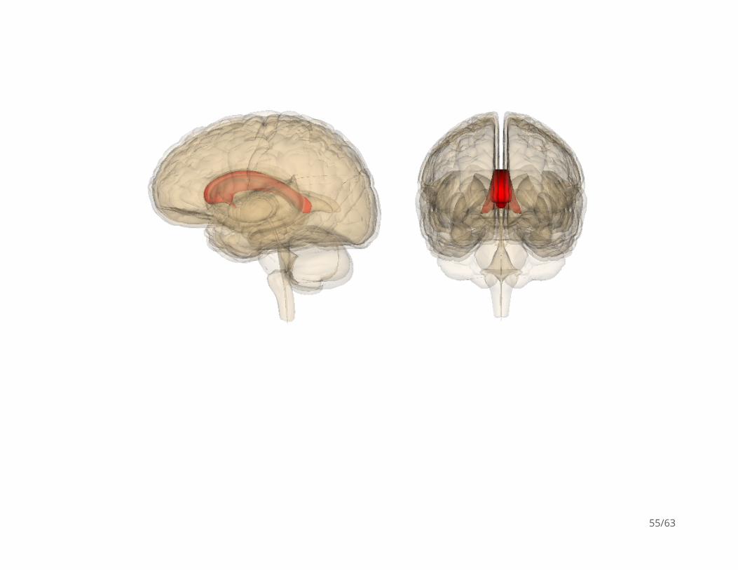
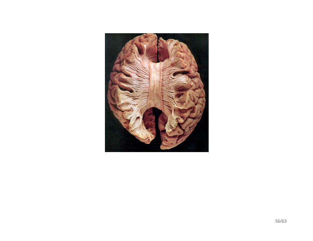
Commissural fibers
Corpus callosum
Anterior commissure (AC)
Posterior commissure (PC)
·
·
·
54/63
55/63
56/63

Anterior, Posterior Commissures
https://https://upload.wikimedia.org/wikipedia/commons/2/22/Gray720.png
57/63
Autonomic nervous system
CNS & PNS components
Controls “vegetative functions”
Two divisions
·
·
Limited voluntary control-
·
Sympathetic
Parasympathetic
-
-
58/63

ANS
https://4.bp.blogspot.com/_FBNLGBBprSE/TB5b9zkM11I/AAAAAAAAAHA/LBCT2HkOzvI/s400/PNS.GIF
59/63

Sympathetic division
Prepares body for action
“Fight or flight”
Spinal cord
NTs
·
·
·
ganglion chain along spinal column to End organs-
·
Preganglionic: ACh
Post: NE
-
-
60/63
Parasympathetic division
“Around” sympathetic
Restorative function
“Rest & digest”
Spinal cord (or Vagus n.) -> ganglia near end organs -> end organ
·
·
·
·
NT: ACh-
61/63

Next time
Neuroanatomy lab·
62/63

References
Oishi, K., Faria, A. V., Zijl, P. C. van, & Mori, S. (2010). MRI atlas of human white matter. Academic Press.
63/63