a codon-optimized bacterial antibiotic gene used as
TRANSCRIPT

Instructions for use
Title A Codon-Optimized Bacterial Antibiotic Gene Used as Selection Marker for Stable Nuclear Transformation in theMarine Red Alga Pyropia yezoensis
Author(s) Uji, Toshiki; Hirata, Ryo; Fukuda, Satoru; Mizuta, Hiroyuki; Saga, Naotsune
Citation Marine Biotechnology, 16(3), 251-255https://doi.org/10.1007/s10126-013-9549-5
Issue Date 2014-06
Doc URL http://hdl.handle.net/2115/57285
Rights The final publication is available at link.springer.com
Type article
Additional Information There are other files related to this item in HUSCAP. Check the above URL.
File Information Stable transformation(marine)revised 2014.6.pdf
Hokkaido University Collection of Scholarly and Academic Papers : HUSCAP

1
Short communication (Marine biotechnology)
A codon-optimized bacterial antibiotic gene used as selection marker for stable nuclear
transformation in the marine red alga Pyropia yezoensis
Toshiki Uji1,2, Ryo Hirata1,2, Satoru Fukuda1, Hiroyuki Mizuta1 and Naotsune Saga1
1Faculty of Fisheries Sciences, Hokkaido University, Hakodate 041-8611, Japan
2These authors contributed equally to this work.
Corresponding author: Naotsune Saga
Faculty of Fisheries Sciences, Hokkaido University, Hakodate 041-8611, Japan
Tel/Fax: +81-138-40-5533
E-mail: [email protected]
Running title: Stable nuclear transformation in Pyropia

2
Abstract
Marine macroalgae play an important role in marine coastal ecosystems and are widely used
as sea vegetation foodstuffs and for industrial purposes. Therefore, there have been increased
demands for useful species and varieties of these macroalgae. However, genetic
transformation in macroalgae has not yet been established. We have developed a dominant
selection marker for stable nuclear transformation in the red macroalga Pyropia yezoensis.
We engineered the coding region of the aminoglycoside phosphotransferase gene aph7’’ from
Streptomyces hygroscopicus to adapt codon usage of the nuclear genes of Pyropia yezoensis.
We designated this codon-optimized aph7’’ gene PyAph7. After bombarding P. yezoensis cells
with plasmids containing PyAph7 under the control of their endogenous promoter, 1.9 thalli
(or individuals) of hygromycin-resistant strains were isolated from a 10 mm square piece of
the bombarded thallus. These transformants were stably maintained throughout the asexual
life cycle. Stable expression of PyAph7was verified using Southern blot analysis, genomic
PCR and RT-PCR analyses. PyAph7 proved to be a new, versatile tool for stable nuclear
transformation in P. yezoensis.
Key words: Pyropia yezoensis; red alga; selection marker; stable transformation
3
Introduction
The marine red macroalga Pyropia yezoensis (nori in Japanese) is one of the most important
marine crops. It is widely cultivated in eastern Asian countries, including Japan, Korea and
China, and generates US $1.3 billion per year (Blouin et al. 2010). In addition, P. yezoensis
has attracted considerable interest as a model for physiological and genetic studies of marine
red algae (Saga and Kitade 2002, Waaland et al. 2004). To date, several studies have been
performed to make this alga a sophisticated model organism. For example, a laboratory
culturing system in which the life cycle of P. yezoensis could be completed within a few
months was established (Kuwano et al. 1996). In addition, a database for expressed
sequenced tags (EST) analysis is now available (Nikaido et al. 2000, Asamizu et al. 2003),
and recently the draft data of whole genome sequence has been analysed by next generation
sequencing (Nakamura et al. 2013). However, a stable transformation system, a powerful tool
both for elucidating gene functions and conferring valuable characteristics to an organism,
has not yet been established for P. yezoensis or other marine macroalgae.
As an initial step in establishing stable transformation, we previously developed a
transient gene expression system to monitor gene expression in P. yezoensis cells using
particle bombardment. Because P. yezoensis genes have a strong GC bias in the third
nucleotide of their codons, it is important to adapt codon usage of foreign genes to the
nuclear genes of P. yezoensis for their efficient expression. In fact, codon-optimized β-
glucuronidase (PyGUS) and GC-rich fluorescent proteins, such as AmCFP and sGFP(S65T),
have been expressed in P. yezoensis cells under the control of an endogenous promoter
(Fukuda et al. 2008, Mikami et al. 2009, Uji et al. 2010).
In addition to developing an efficient expression system, a reliable method to select

4
and isolate transformed cells is required to establish stable transformation in macroalgae.
Recently, we have revealed that P. yezoensis cells are sensitive to several aminoglycoside
antibiotics, including hygromycin B, paromomycin and geneticin (Takahashi et al. 2011).
Thus, these antibiotics are possible candidate selection agents for stable P. yezoensis
transformation.
Regarding a selection marker, the aminoglycoside phosphotransferase gene aph7’’
from Streptomyces hygroscopicus, which confers resistance against hygromycin B, should be
available for hygromycin-based stable transformant selection because the GC content in its
coding region is as high as 70.94% (Zalacain et al. 1986). The aph7’’ gene has been
successfully used for the transformation of green microalgae, such as Chlamydomonas
reinhardtii (Berthold et al. 2002), whose codons are also rich in GC residues. However,
several codons that are rarely used in P. yezoensis nuclear genes are found in the aph7” gene,
especially in its N-terminal region. These mismatches in codon usage would be predicted to
inhibit efficient translation in P. yezoensis cells.
Thus, in the present study, we synthesized a codon-optimized aph7” gene and
examined its utility as a selection marker for stable nuclear transformation in P. yezoensis.
Materials and methods
Culturing of P. yezoensis
Gametophytes of P. yezoensis strain TU-1 and transformants were cultured in enriched sea
life (ESL) medium under conditions described by Fukuda et al. (2008).
Plasmid construction

5
To construct a pEA7 plasmid, a fragment containing the ORF of PyAph7 and 3′ UTR of
CrRbcS2 was amplified using pHyg4 as a template and a pair of primers, XbaI-PyAph7-F (5′-
GCTCTAGAATGACGCAGGAGTCCCTGCTGCTGCTC-3′) and EcoRI-CrRbcS2-R (5′-
GGAATTCTTCCATGGGATGACGGGCCCGG-3′). The amplified PCR product was
digested with XbaI and EcoRI and subsequently inserted into XbaI–EcoRI-digested p35S-
PyGUS (Fukuda et al. 2008), which was designated p35S-PyAph7. To replace the CaMV 35S
promoter with an endogenous promoter, the 5′ upstream region of PyElf1 was amplified using
pPyElf1-PyGUS (Mikami et al. 2011) as a template and the following primers: HindIII-
PyElf1-F/XbaI-PyElf1-R (5′-CCCAAGCTTCCAGACCCGTGGAAAGTACCATC-3′
/5′ GCTCTAGACTTGCCCATGGTGGGGGGG-3′). The PCR product was digested with
HindIII and XbaI and subsequently inserted into the HindIII–XbaI site of p35S-PyAph7. This
resulted in pEA7 construction.
Particle bombardment
Expression plasmids were purified from 100 mL of E. coli culture using a NucleoBond Xtra
Midi (MACHEREY-NAGEL, Germany). For particle bombardment, a gametophytic thallus
with monosporangia covering a wide range of the thallus (>10 mm in width) was cut into 10-
mm-square pieces, which were subsequently placed on a filter paper. After removing excess
fluid, the expression plasmids were introduced into the gametophytic cells using PDS-
1000/He particle bombardment under the conditions described previously (Hirata et al. 2011).
Isolation of hygromycin-resistant transformants
The bombarded algal pieces were cultured in a 100-mL glass flask (Iwaki Sci Tech Div.,
Asahi Techno Glass, Japan) in 50 mL of ESL medium under non-selective conditions for 1
week. Subsequently, the medium was replaced with the ESL medium containing hygromycin
6
B (final concentration of 1 mg mL−1), and the medium was renewed weekly. After incubation
for 6–8 weeks in the antibiotic-containing medium, visible hygromycin-resistant
transformants regenerated from the pieces of bombarded thalli were individually isolated in
another culture flask and continuously cultured in ESL medium with or without 1 mg mL−1
hygromycin B.
Assay for hygromycin resistance
To prepare individuals for hygromycin resistance assay, gametophytes isolated as
hygromycin-resistant transformants were clonally propagated for 3 weeks in different culture
flasks containing ESL medium via monospores. Gametophytes of transformants or wild type
strains (ca. 20 mm in length) cultured were respectively transferred into a 6-well plate (3
individuals/well) (Iwaki Sci Tech Div., Asahi Techno Glass, Japan) containing 5 mL of ESL
medium with 0, 1.0, 2.5, 5.0, 7.5 or 10.0 mg mL−1 hygromycin B and incubated under
shaking culture for 2 weeks at 15°C. The medium was renewed weekly. After culture,
gametophyte viability was estimated by staining using 0.01% erythrosine (Wako Pure
Chemical Industries, Japan) in ESL medium according to a previous report (Takahashi et al.
2011).
Genomic PCR and RT-PCR
Genomic DNA was extracted from gametophytes of transformants cultured for 4 weeks in
ESL medium without hygromycin B or wild type strains for genomic PCR as described by
Hwang et al. (2010) and purified using a phenol–chloroform extraction and ethanol
precipitation. The precipitate was resuspended in 50 µL of TE buffer, and 2 µL of this
suspension was used as a template for genomic PCR. RNA extraction and cDNA synthesis
for RT-PCR were performed as described by Uji et al. (2012). Genomic PCR and RT-PCR
7
analyses were conducted using TaKaRa LA Taq with GC Buffer (TaKaRa-Bio). The primer
pairs PyAph7-RT-F/R (5′-CATTGACTCGGACGACTCCTACGCGAG-3′/5′-
AAGTCGTGCAGGAAGGTGAAG-3′) and PyElf1-RT-F/R (5′-
AAGGCCAAGGCACCCAAGCTG-3′/5′-ACCACACCAAGAGCGTCCAATC-3′) were
used to amplify the fragments of PyAph7 (864 bp) and PyElf1 (734 bp), respectively. The
amplified PCR products were examined on a 1.3% agarose gel.
Southern blotting
Genomic DNA was extracted using the cetyl trimethyl ammonium bromide (CTAB) method
from 1.0 g (FW) of wild-type and transformants that had been cultured for more than three
months after isolation. Extracted DNA was further purified by ultracentrifugation as follows:
3.1 g of cesium chloride (CsCl) and 15 µL of ethidium bromide (EtBr) were added to 3.0 mL
of the DNA solution and centrifuged at 400,000 g for 24 h at 20°C. The DNA band was
visualised under UV light and collected. EtBr was removed by three extractions with an equal
volume of 1-butanol. CsCl was removed by ethanol precipitation three times. Purified DNA
(2.0 µg) was digested with PstI, run on agarose gel and transferred to a nylon membrane. The
pEA7 plasmid was digested with XbaI and SalI and a 730 bp fragment of PyAph7 was
collected. This fragment was labelled by random 32P priming and used as a probe (see Fig.
1a).
Results and discussion
To optimize the codon usage of the aph7’’ coding region to that of P. yezoensis, we
employed site-directed mutagenesis using a pHyg4 plasmid that contained the aph7’’ gene
(Berthold et al. 2002) and a KOD-Plus-Mutagenesis Kit (TOYOBO, Japan) with the
oligonucleotides shown in Table S1. The synthetic aph7’’ gene was designated PyAph7 (Fig.

8
S1). Subsequently, the protein coding region of PyAph7 was fused with the endogenous
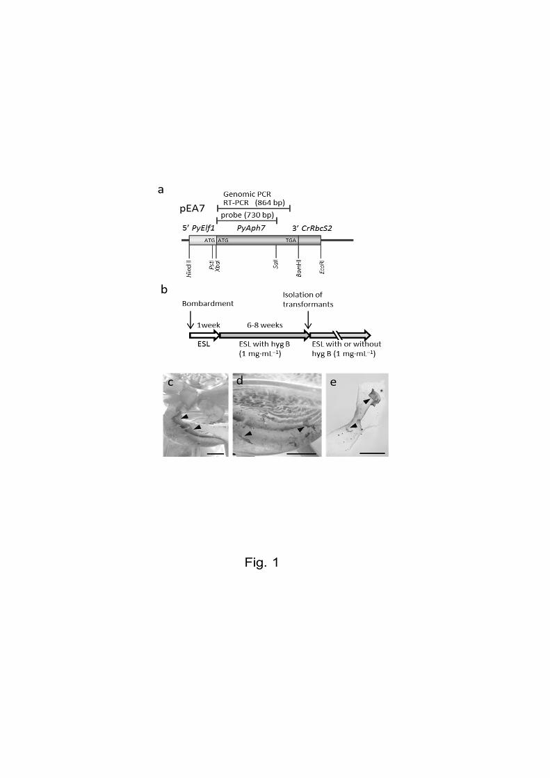
PyElf1 promoter (Mikami et al. 2011). The resulting plasmid was designed pEA7 (Fig. 1a).
The pEA7 expression plasmids containing PyAph7 were introduced into P. yezoensis
gametophytes by particle bombardment. When gametophytic thalli with the introduced pEA7
were cultured in a non-selective ESL medium for 1 week (Fig. 1b), some monospores
(asexual spores) were released from the bombarded thalli and adhered to the bottom of the
culture flask (Fig. 1c). The medium was subsequently replaced with selective ESL medium
containing 1.0 mg mL−1 hygromycin B, which effectively kills wild-type cells or, at least,
completely inhibits their growth (Takahashi et al. 2011). After 6–8 weeks of culture in the
selective medium, hygromycin-resistant thalli from them released monospores or from
vegetative cells of bombarded blades were approximately 5–10 mm long (Fig. 1d, e). These
transformants were transferred into separate culture vessels to identify the homogeneous lines
and cultured further in a non-selective ESL medium (Fig 1b). Consequently, an average of 1.9
thalli (or individuals) of the hygromycin-resistant strains were obtained from a piece of the
bombarded thallus (29 transformants per 15 bombardments). These transformants were
successfully maintained over more than 5 generations as independent lines through the
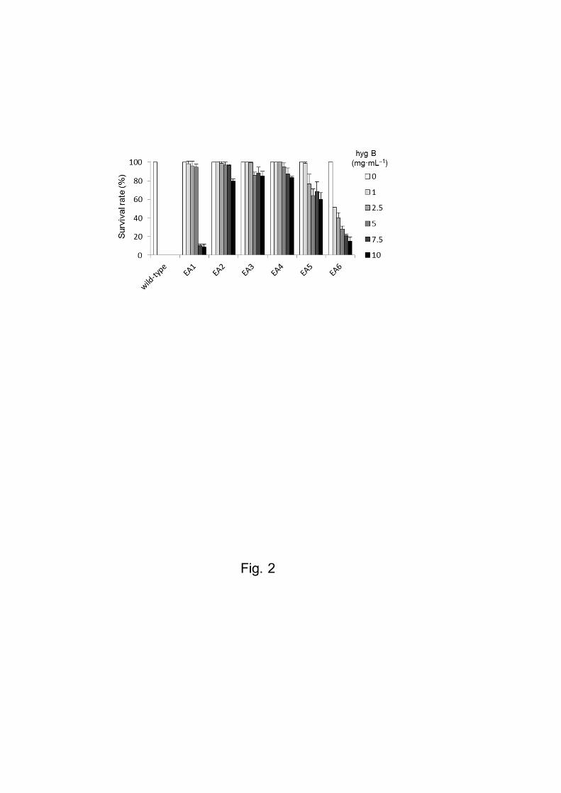
asexual life cycle via monospores. Subsequently, we examined the hygromycin B tolerance of
six isolated transformants: EA1–EA6. As shown in Figs. 2 and S2, when the gametophytes
were cultured in an ESL medium containing 1.0 mg mL−1 hygromycin B, all wild-type
gametophytes were dead after 2 weeks of culture. In contrast, all transformants survived and
grew in this medium. Transformants EA2, EA3 and EA4 survived even in the presence of
2.5–10.0 mg mL−1 hygromycin B (Figs. 2 and S2).
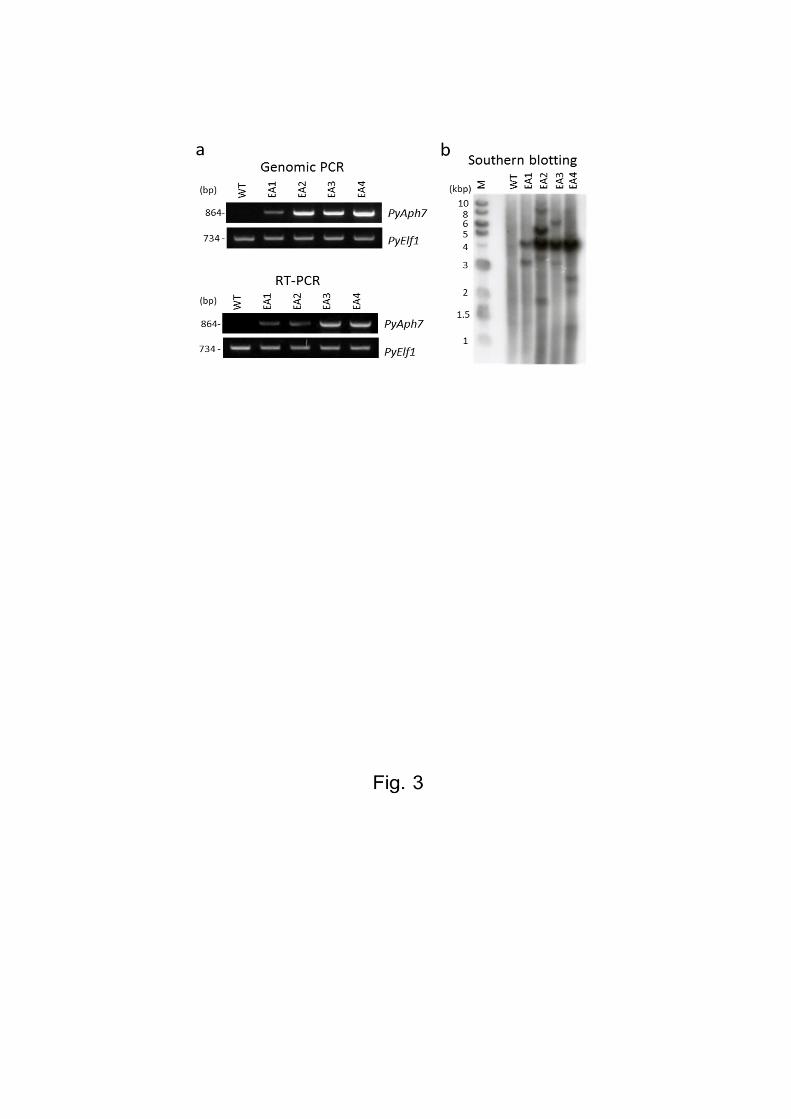
Finally, to verify whether the exogenous PyAph7 gene had been successfully introduced
and expressed in these isolated transformants, genomic PCR and RT-PCR analyses were
performed on four transformant strains (EA1–EA4). Using primers specific for the PyAph7

9
gene sequence, a DNA fragment of the expected size was amplified for all of the examined
strains, whereas this fragment was not observed in a wild-type strain (Fig. 3a). This indicated
that PyAph7 had been successfully introduced and expressed in these transformants. We
further analysed the stable integration of PyAph7 into the genome by Southern blot analysis.
The southern blot analysis revealed that multiple DNA fragments could be detected in all of
the examined transformants, which indicated that PyAph7 had multiplied and randomly
integrated into the genome (Fig. 3b). Strong signals of an approximately 4.2 kbp DNA
fragment that corresponded to the full-length of the pEA7 vector were commonly detected in
all transformed strains. The results intimate the possibility that a part of introduced pEA7
vector might be stably maintained as entire circular plasmids in the transformed cells through
the cell divisions and propagation. Interestingly, there have been several reports on plasmid
DNA isolation from some red macroalgae, including Porphyra tenera, a closely related
species of P. yezoensis (Goff and Coleman 1990, Choi et al. 2000; 2001). Thus, we need to
investigate on mechanisms for maintaining plasmid DNA in P. yezoensis cells.
Taken together, the codon-optimized PyAph7 introduced by particle bombardment was
stably maintained and expressed in P. yezoensis and conferred resistance to hygromycin B.
Our results demonstrate that PyAph7 is useful as an efficient selection marker for stable
nuclear transformation of P. yezoensis. This is the first report of a bacterial antibiotic gene
used as a selection marker for stable transformation in marine macroalgae. Further
development of this stable transformation system in P. yezoensis will overcome some of the
barriers in molecular biological studies of marine red algae.
10
Acknowledgments
We are grateful to Dr. Wolfgang Mages (Lehrstuhl für Genetik, Universität Regensburg,
NWFIII, Germany) for kindly providing the pHyg4 plasmid. This study was supported, in
part, by the Regional Innovation Cluster Program (Global Type) of the Ministry of Education,
Culture, Sports, Science and Technology of Japan to N.S.
11
References
Asamizu E, Nakajima M, Kitade Y, Saga N, Nakamura Y, Tabata S (2003) Comparison of
RNA expression profiles between the two generations of Porphyra yezoensis
(Rhodophyta), based on expressed sequence tag frequency analysis. J. Phycol 39: 923-
30
Berthold P, Schmitt R, Mages W (2002) An engineered Streptomyces hygroscopicus aph 7"
gene mediates dominant resistance against hygromycin B in Chlamydomonas
reinhardtii. Protist 153:401-12
Blouin NA, Brodie JA, Grossman AC, Xu P, Brawley SH (2011) Porphyra: a marine crop
shaped by stress. Trends Plant Sci 16:29-37
Choi HS, Choi KH, Rhew TH (2000) Simple and rapid isolation of plasmids from Porphyra
tenera. Algae 15:133-6
Choi HS, Choi KH, Kim TH, Lee CH, Rhew TH (2001) Characterization of natural plasmid
and construction of putative transformation vector using the plasmid in Korean red alga,
Porphyra tenera. Algae 16: 287-94
Fukuda S, Mikami K, Uji T, Park EJ, Ohba T, Asada K, Kitade Y, Endo H, Kato I, Saga N
(2008) Factors influencing efficiency of transient gene expression in the red
macrophyte Porphyra yezoensis. Plant Sci 174:329-39
Goff L J, Coleman AW (1990) Red algal plasmids. Curr Genet 18:557-65.
Hwang BK, Son SH, Lee JS, Min SR, Ko SM, Liu JR, Choi DS, Jeong WJ (2010) Rapid and
simple method for DNA extraction from plant and algal species suitable for PCR
amplification using a chelating resin Chelex 100. Plant Biotechnol Rep 4:49-52
Hirata R, Takahashi M, Saga N, Mikami K (2011) Transient gene expression system

12
established in Porphyra yezoensis is widely applicable in Bangiophycean algae. Marine
Biotechnol 13:1038-47
Kuwano K, Aruga Y, Saga N (1996) Cryopreservation of clonal gametophytic thalli of
Porphyra (Rhodophyta). Plant Sci 116:117-24
Mikami K, Uji T, Li L, Takahashi M, Yasui H, Saga N (2009) Visualization of
phosphoinositides via the development of the transient expression system of a cyan
fluorescent protein in the red alga Porphyra yezoensis. Marine Biotechnol 11:563-9
Mikami K, Hirata R, Takahashi M, Uji T, Saga N (2011) Transient Transformation of Red
Algal Cells: Breakthrough Toward Genetic Transformation of Marine Crop Porphyra
Species. In María Alvarez (ed) Genetic Transformation. InTech Open Access Publisher
241-58
Nakamura Y, Sasaki N, Kobayashi M, Ojima N, Yasuike M, Shigenobu Y, Satomi M, Fukuma
Y, Shiwaku K, Tsujimoto A, Kobayashi T, Nakayama I, Ito F, Nakajima K, Sano M,
Wada T, Kuhara S, Inouye K, Gojobori T, Ikeo K (2013) The First Symbiont-Free
Genome Sequence of Marine Red Alga, Susabi-nori (Pyropia yezoensis). PLoS One
8(3):e57122
Nikaido I, Asamizu E, Nakajima M, Nakamura Y, Saga N, Tabata S (2000) Generation of
10,154 expressed sequence tags from a leafy gametophyte of a marine red alga,
Porphyra yezoensis. DNA Res 7:223-7
Saga N, Kitade Y (2002) Porphyra: a model plant in marine sciences. Fish Sci
68(suppl):1075-8
Takahashi M, Mikami K, Mizuta H, Saga N (2011) Identification and efficient utilization of
antibiotics for the development of a stable transformation system in Porphyra yezoensis
(Bangiales, Rhodophyta). J Aquac Res Development 2:118
Uji T, Takahashi M, Saga N, Mikami K (2010) Visualization of nuclear localization of

13
transcription factors with cyan and green fluorescent proteins in the red alga Porphyra
yezoensis. Marine Biotechnol 12:150-9
Uji T, Hirata R, Mikami K, Mizuta H, Saga N (2012) Molecular characterization and
expression analysis of sodium pump genes in the marine red alga Porphyra yezoensis.
Mol Biol Rep 39:7973-80
Waaland JR, Stiller JW, Cheney DP (2004) Macroalgal candidates for genomics. J Phycol
40:26-33
Zalacain M, Gonzalez A, Guerrero MC, Mattaliano RJ, Malpartida F, Jimenez A (1986)
Nucleotide sequence of the hygromycin B phosphotransferase gene from Streptomyces
hygroscopicus. Nucleic Acids Res 14:1565-81

14
Figure legends
Fig. 1 Isolation of hygromicin-resistant transformants in P. yezoensis
(a) Schematic diagram of the hygromycin selective vector pEA7. The coding region of
PyAph7 is fused in-frame to 5′ PyElf1 (promoter, 5′-untranslated region of PyElf1 and the
initiation codon). 3′ CrRbcS2 indicates the 3′ untranslated region of the RbcS2 gene from
Chlamydomonas reinhardtii. The position and length of the DNA fragment amplified by
genomic PCR or RT-PCR are indicated. The position of the probe used in Southern blotting is
indicated (probe). (b) Time-line for isolating hygromicin-resistant transformants.(hyg B,
hygromycin B). (c) Macroscopic view of the bottom of the culture flask on which
monospores released from bombarded thalli were attached (arrowheads). Scale bar = 10 mm.
(d) Hygromycin-resistant thalli regenerated from monospores attached to the bottom of a
culture flask (arrowheads). Scale bar = 10 mm. (e) Hygromycin-resistant thalli regenerated
from the vegetative cells of a bombarded thallus (arrowheads). Scale bar = 5 mm.
Fig. 2 Analysis of hygromycin B resistance for wild type and transgenic P. yezoensis
strains Survival rates of wild type and six lines of the hygromycin-resistant strains (EA1–
EA6) when cultured with varying concentrations of hygromycin B (1.0–10.0 mg mL−1). The
survival rate was calculated by counting viable and dead gametophytes during 2 weeks
culture in ESL medium containing hygromycin B. Values are means ± SDs (n = 30).
Fig. 3 PCR and Southern blot analyses of hygromicin-resistant transformants
(a) Expression of the exogenous PyAph7 gene in hygromycin-resistant transformants was
detected by genomic PCR and RT-PCR. PCR was performed using primers specific for the
PyAph7 gene sequence (Fig. 1) and genomic DNA or total RNA from a wild type strain and
15
the transformants EA1–EA4. PyElf1 was used as the internal control gene in P. yezoensis.
Only transformants were expected to yield an 864 bp fragment of PyAph7. (b) Southern blot
analysis of hygromycin-resistant transformants. Genomic DNA from a wild type strain and
the transformants EA1–EA4 were digested with Pst I, separated on agarose gel, transferred to
nylon membrane and hybridized with a labelled probe corresponding to the PyAph7 fragment
(Fig.1). Lane M, molecular weight marker.
Fig. S1 Comparison of the original aph7” and synthetic PyAph7 coding regions
Differences between original aph7”and synthetic PyAph7 are noted below the original
aph7”sequence in red letters. Optimized codons were based on codon usage indicated in:
http://www.kazusa.or.jp/codon/cgibin/showcodon.cgi?species=Porphyra+yezoensis+[gbpln].
The amino acid sequence is shown below the nucleotide sequence by the single letter code.
The nucleotide numbers are indicated on both the left and right hand sides.
Fig. S2 Hygromycin B resistance in wild type and transgenic P. yezoensis strains
Wild type and six hygromycin-resistant strains (EA1–EA6) were cultured in ESL medium
containing hygromycin B (1.0–10.0 mg mL−1) for 2 weeks. Scale bar =50 m.