a salivary gland lymphoma involving contralateral …...region consisted of a portion of parotid...
TRANSCRIPT
Hiroshima Journal of Medical Sciences Vol. 33, No. 2, June, 113 ........ 123, 1984
HIJM 33-14
A Salivary Gland Lymphoma Involving Contralateral Parotid Gland and Submaxillary Gland with a
Review of the Literature*)
Toshio TANAKA1>, Tomoaki SUGIURA2), Shuichi WATANAB£2>, Ryusuke SAIT02>, Yoshio OGURA2>, Kyoichi HAY ASHP>
and Taisuke OHNOSHP>
1) Pathology Section, Central Laboratories, Okayama University Medical School 2) Department of Otorhinolaryngology, Okayama University Medical School 3) Department of Medicine (2nd Clinic), Okayama University Medical School, Shikata,
Okayama 700, Japan
(Received January 7, 1984)
Key words: Malignant lymphoma, Salivary gland
ABSTRACT
A 75-year-old male was pointed out the right submandibular swelling followed by the left pre-auricular swelling, about half-year apart. Parenchyma of the left parotid gland was infiltrated by lymphoid growth, and the right submaxillary gland had a discrete lymphoid growth at a pole of the gland. Inside and outside these lymphoid growths, lymphocytes infiltrated between ducts, acini, adipose tissue, blood vessels and nerves. Patho-immunologically, the case was a follicular lymphoma of medium-sized (or small cleaved) cell type with no cytoplasmic immunoglobulin production, although a B-cell type of the malignant lymphoma was suggested because of a prominent nodular pattern. The patient has been in a complete remission for six months subsequent to chemotherapy. The major literature concerning . malignant lymphoma primarily in parotid or submaxillary gland reported since 1959 was reviewed. The criteria for possible primary malignant lymphoma of the salivary gland, its pathogenesis and its differential diagnosis from other lymphoid and epithelial lesions were discussed.
113
INTRODUCTION
Foote and Frazell7> wrote ''As yet we have not had an unequivocal case of primary malignant lymphoma of a major salivary gland. When such a claim is made, it should be accompanied by proof that the salivary gland was not secondarily involved. This is difficult. There is, of course, no theoretical objection to the existence of a great many mesenchymal tumors of salivary glands, but vast patience is required to accumulate acceptable data. Publication of single case reports of convincing
material of this sort should be encouraged." Although the head and neck is one of the most common place for malignant lymphoma, the salivary gland per se is rarely involved. We report an elderly man, who started the right submandibular swelling followed by the left pre-auricular swelling, about half-year apart; the pathologic findings confirmed their salivary gland origin.
CASE PRESENTATION
This is an elderly man born in December 1906. In April 1981, he was pointed out the
*> EB11P~tt, tirffljz~B. Vl~?l;Na-. *~~g1r, 1J\;t~~~. tt '$-, ::k~~~ : )e;f{P,~O)JfrJ}~:fo .t Lf~1ffJP~~·fft· l-td~tt y :/,.~Ill

114 T. Tanaka et al.
Table 1. Pertinent Laboratory Data on Admission
Blood picture
RBC
Hg
Ht WBC
4. 69x 106/ µl 15.7 g/dl
43.7%
7. Ox 103/ µl
Differentials (%)
Seg 68
Band
Lympho
Mono
Platelet
Serum protein (%)
2
22
8
22. 7x10⁴/ µl
Albumin 59. 1
Globulin
alpha 1
alpha 2 2.9
11.4 beta 10. 2
gamma 16.3
A/G 1.45
right submandibular swelling, followed by the left pre-auricular swelling in September of the same year. He then was referred to the Otolaryngology Department, Okayama University Hospital, in November. On admission, physical examinations including lymphography indicated the clinical stage, IIE. Laboratory data. (Table 1) showed increased lgM (normal ranges, 60-250 mg/di), slightly elevated CH50 (normal ranges, 30. 0-40. 0 U /ml) and positive serology of syphilis; other pertinent data were not contributory. The right submandibular tumor measured approximately 3. 0 x 2. 5 cm and was entirely removed in the mid-November. The left pre-auricular tumor measured approximately 4. 0 x 3. 5 cm and the superficial part of the parotid gland was excised in the mid-December.
Postoperatively, three courses of a combination chemotherapy consisting of cyclophosphamide, adriamycin, vincristine and prednisolone was given and followed by 4, 000 rads of Linac x-rays to the whole neck including Waldeyer's ring. The following three courses of the same combination chemotherapy as above was terminated because of its toxicity. In June 1983, relapse appeared at bilateral cervical, axillary and inguinal regions. He is currently in a complete remission with peroral ~dministration
Immunoglobulin (mg/di)
lg A 349
lgG 1644
lgl\1 355
Peripheral blood (%)
T cell 85
B cell 11
Serology (syphilis)
Ogata
VDRL
(-tt)
( ti-)
TPHA (+)
l\1antaux reaction
CH50
LDH
lOx 10 lOxlO
(±)
46. 3 U/ml
217 IU/l
of etopocide (VP-16-213) 22 >, 150 mg for five consecutive days and fourth-weekly.
PATHOLOGIC FINDINGS
The specimen from the left pre-auricular region consisted of a portion of parotid gland measuring approximately 1. 7 x 1. 7 cm. The specimen from the right submandibular region consisted of submaxillary gland measuring 3. 0 x 2. 0 cm in the largest dimensions, and two lymph nodes which were adjacent to the submaxillary gland and measured 3. 0 x 2. 0 cm and 2. 0 x 1. 0 cm, respectively.
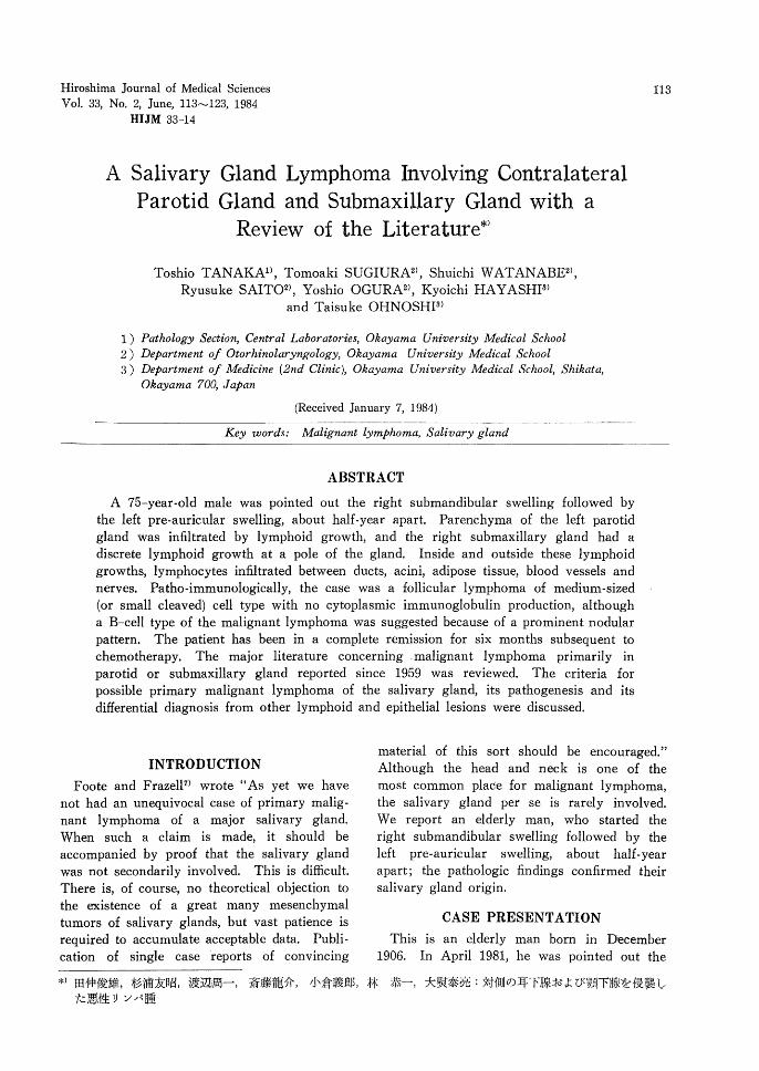
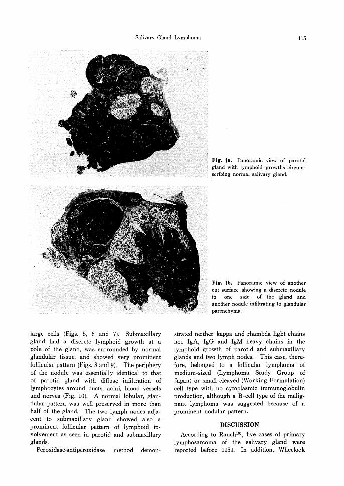
As to parotid gland, nodular lymphoid growth circumscribed normal salivary gland which appeared like a few islands (Fig. la). In a different cut surface, a discrete nodule was embedded in the gland, whereas another nodule invaded to glandular parenchyma (Fig. lb). In the periphery of nodules, lymphocytes infiltrated between ducts, acini and adipose tissue (Fig. 2). There was capsular thickening with perineural growth of lymphocytes (Fig. 3). Inside nodules, glandular architecture was entirely effaced due to intense lymphocyte infiltration with periductal, perivascular and perineural growths (Fig. 4). The tumor cells consisted of mediumsized, cleaved lymphocytes and very occasional
Salivary Gland Lymphoma 115
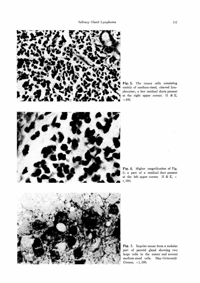
large cells (Figs. 5, 6 and 7). Submaxillary gland had a discrete lymphoid growth at a pole of the gland, was surrounded by normal glandular tissue, and showed very prominent follicular pattern (Figs. 8 and 9). The periphery of the nodule was essentially identical to that of parotid gland with diffuse infiltration of lymphocytes around ducts, acini, blood vessels and nerves (Fig. 10). A normal lobular, glandular pattern was well preserved in more than half of the gland. The two lymph nodes adjacent to submaxillary gland showed also a prominent follicular pattern of lymphoid involvement as seen in parotid and submaxillary glands.
Peroxidase-an ti peroxidase method demon-
Fig. la. Panoramic view of parotid gland with lymphoid growths circum -scribing normal salivary gland.
Fig. lb. Panoramic view of another cut surface showing a discrete nodule in one side of the gland and another nodule infiltrating to glandular parenchyma.
strated neither kappa and rhambda light chains nor IgA, IgG and IgM heavy chains in the lymphoid growth of parotid and submaxillary glands and two lymph nodes. This case, therefore, belonged to a follicular lymphoma of medium-sized (Lymphoma Study Group of Japan) or small cleaved (Working Formulation) cell type with no cytoplasmic immunoglobulin production, although a B-cell type of the malignant lymphoma was suggested because of a prominent nodular pattern.
DISCUSSION
According to Rauch19>, five cases of primary lymphosarcoma of the salivary gland were reported before 1959. In addition, Wheelock

116 T. Tanaka et al.
Fig. 2 The periphery of nodules showing lymphocytes infiltrating to ducts, acini and adipose tissue, H & E, x 40.
Fig. 3. Capsular thickening with perineural growth of lymphocytes. H & E, x40.
Fig. 4. The detail of nodules showing entirely effaced glandular architecture due to intense lymphocyte infiltration. H & E, x40.
Salivary Gland Lymphoma 117
Fig. 5. The tumor cells consisting mainly of medium-sized, cleaved lymphocytes; a few residual ducts present at the right upper corner. H & E, x400.
Fig. 6. Higher magnification of Fig. 5; a part of a residual duct present at the left upper corner. H & E, x 1, 000.
Fig. 7. Imprint smear from a nodular part of parotid gland showing two large cells in the center and several medium-sized cells. May-GrtinwaldGiemsa, x 1, 000,
118 T. Tanaka et al.
Fig. 8. Panoramic view ot submaxillary gland showing a discrete lymphoid growth, surrounded by normal glan · dular tissue, at a pole of the gland.
Fig. 9. The detail of the lymphoid growth showing several, very prominent follicular patterns. Silver, x 40.
Fig. 10. The periphery of the nodule with lymphocytes infiltrating between ducts, blood vessels, nerves (at the left lower corner) and adipose tissue. H & E, x40,

Salivary Gland Lymphoma 119
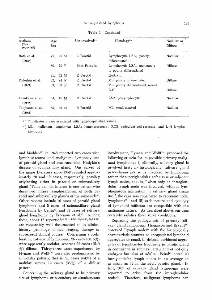
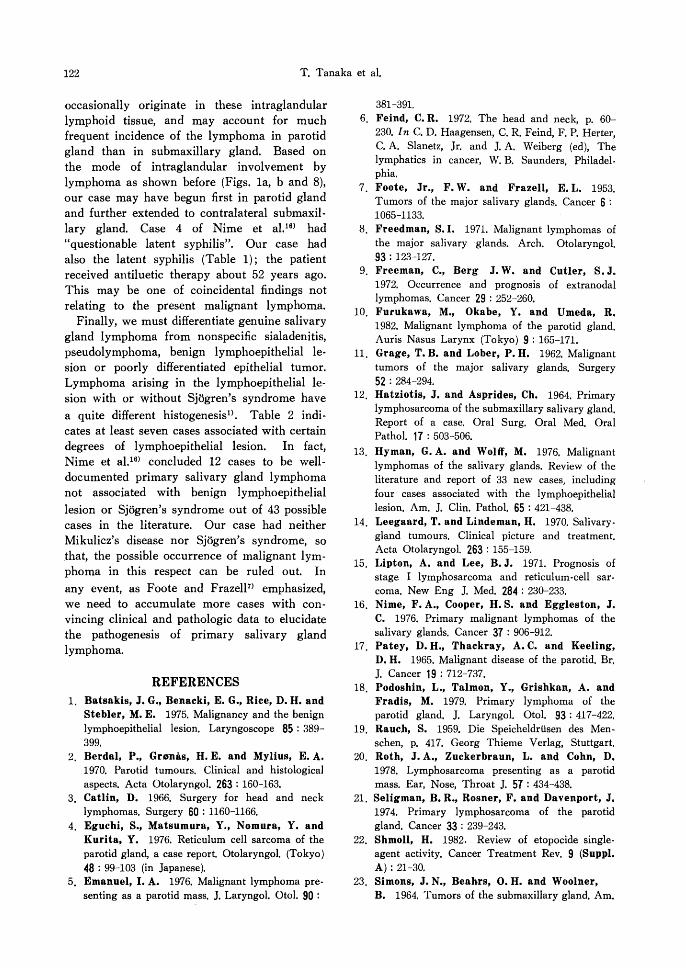
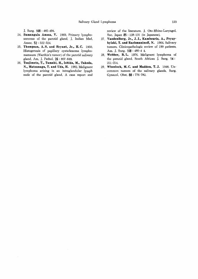
Table 2. Survey of Major Literature Concerning Malignant Lymphoma Primarily in the Parotid or Submaxillary Gland Reported Since 1959
Authors Age Site involveda> Histologyb> Nodular or {Year reported) Sex Diffuse
Grage & Lober ] . 35 M R Parotid Giant follicular lymphoblastoma
(1962) 2. 77 F L Submaxilla Hodgkin
3. 31 M L Parotid LSA
4, 64 M L Parotid Lymphoblastoma, intermediate type
5, 55 F R Parotid Lymphoblastoma, unclassified
Hatziotis &
Asprides 6. 60 M L Submaxilla LSA Diffuse (1964)
Simons et al. 7. Submaxilla LSA
(1964) 8, Submaxilla LSA
9, Sub maxilla LSA
VandenBerg et al. 10, Parotid ML
(1964) 11. Parotid ML
12. Sub maxilla ML
Patey et al. 13. 46 F Parotid* ML
(1965) 14. 66 F Parotid* LSA
15. 63 F Parotid LSA
16, 73 F Parotid RCS
Sumangala Amma
(1969) 17. 55 M R Parotid LSA
Berdal et al. 18. Parotid ML (1970) 19, Parotid ML
20, Parotid ML Leegaard & 21. Parotid RCS
Lindeman 22, Parotid RCS
(1970)
Freedman 23. 54 F R Parotid LSA (1971) 24. 50 M L Parotid LSA
25, 32 F L Parotid Lymphoma Nodular 26. 50M R Parotid Lymphoma Nodular 27. 71 F R Parotid Lymphoma
28. 77 F R Parotid RCS
29. 37 F R Submaxilla LSA
30. 62 F R Parotid &
R Submaxilla LSA Diffuse
31. 43 M R Parotid LSA
32. 77 F R Parotid LSA
Lipton & Lee 33. R Parotid LSA
(1971) 34. R Parotid LSA
35. R Parotid RCS
Seligman et al.
(1974) 36. 20 M R Parotid LSA

120 T. Tanaka et al.
Table 2. Continued
Authors Age Site involved 3) Histologyb) Nodular or
(Year reported) Sex Diffuse
Eguchi et al.
(1976) 37. 65 M R Parotid RCS Diffuse
Emanuel 38. 74 M L Parotid ML, poorly differentiated
(1976) 39. 55 F L Parotid ML, well differentiated Diffuse
40. 75 F L Parotid ML, undifferentiated Diffuse
Hyman & Wolff 41. ;37 M R Parotid Lymphocytic LSA Nodular
(1976) 42. 78 F R Parotid Lymphocytic LSA Nodular
43. 65 F L Parotid Histiocytic LSA Diffuse
44. 38 M Submaxilla * Lymphocytic LSA Nodular
45. 60 F L Parotid Histocytic LSA Nodular
46. 36 F R Parotid Hodgkin, lymphocyte depleted
47. 46 F L Parotid Lymphocytic LSA Nodular
48. 24 F R Parotid Histiocytic LSA Diffuse
49. 56 M L Parotid Histiocytic LSA Diffuse
50. 55 F R Parotid LSA, mixed L-H Nodular
51. 58 F R Parotid Lymphocytic LSA Nodular
52. 21 M R Parotid LSA, mixed L-H Nodular
53. 62 M L Parotid LSA, mixed L-H Diffuse
54. 50 M R Parotid Histiocytic LSA Nodular
55. 73 F L Submaxilla Histiocytic LSA Nodular
56. 66 M L Parotid Lymphocytic LSA Nodular
57. 43 F L Parotid Histiocytic LSA Nodular
58. Parotid LSA, mixed L-H Nodular
59. 60 F Parotid LSA, mixed L-H Nodular
60. 33 F Sub maxilla Histiocytic LSA Diffuse
61. 36 M R Parotid Histiocytic LSA Nodular
62. 82 F L Parotid Histiocytic LSA Nodular
63. Histiocytic LSA Diffuse
64. 68 M Parotid LSA, mixed L-H Nodular
65. 46 F -* Histiocytic LSA Diffuse
66. 53 F L Parotid* Histiocytic LSA Nodular
67. 62 F L Parotid Histiocytic LSA Diffuse
68. 34 M R Parotid Histiocytic LSA Diffuse
69. 62 M R Parotid Lymphocytic LSA Nodular
70. 68 F L Parotid Histiocytic LSA Nodular
71. 57 M R Parotid* Histiocytic LSA Diffuse
72. 57 F R Parotid Histiocytic LSA Diffuse
73. 50 F Parotid Lymphocytic LSA Nodular
Nime et al. 74. 15 F L Parotid Hodgkin, lymphocyte predominant
(1976) 75. 51 M L Parotid Lymphocytic LSA Diffuse
76. 51 F L Parotid* LSA, mixed L-H Diffuse
77. 84 F R Parotid LSA, mixed L-H Diffuse
Webber 78. 49 M R Parotid ML
(1976)
Sail vary: Gland Lymphoma 121'
Table 2. Continued
Authors (Year reported)
Roth et al.
(1978)
Podoshin et al.
(1979)
Furukawa et al.
(1982)
Tsujimoto et al.
(1982)
Age
Sex
79. 29 M
80. 70 F
81. 52 M
82. 74 F 83. 80 F
84. 15 M
85. 35 M
Site involved a>
L Parotid
Bilat Parotids
R Parotid
R Parotid
R Parotid
R Parotid
R Parotid
Histologyb>
Lyrnphocytic LSA, poorly
differentiated
Lymphocytic LSA, moderately
to poorly differentiated
Hodgkin
ML, poorly differentiated
ML, poorly differentiated, mixed
L-H
LSA, prolymphocytic
ML. small cleaved
Nodular or
Diffuse
Nodular
Diffuse
Diffuse
Diffuse
Nodular
Nodular
a ) * indicates a case associated with lymphoepithelial lesions.
b) ML: malignant lymphoma; LSA: lymphosarcoma; RCS: reticulum cell sarcoma; and L-H:lympho
histiocytic
and Madden29l in 1948 reported two cases with lymphosarcoma and malignant lymphocytoma of parotid gland and one case with Hodgkin's disease of submaxillary gland. Our survey of the major literature since 1959 revealed approximately 70 and 10 cases, respectively, possibly originating either in parotid or submaxillary gland (Table 2). Of interest is one patient who developed diffuse lymphosarcoma of both parotid and submaxillary glands of the same side8l. Other reports include 16 cases of parotid gland lymphoma and 5 cases of submaxillary gland lymphoma by Catlin3l, and 69 cases of salivary gland lymphoma by Freeman et al. 9l Among these, about 15 reports4, s, 8, 10-13, 16-18, 20, 21, 24, 26, 28l
are rea~onably well documented as to clinical history, pathology, clinical staging, therapy or subsequent clinical course. Concerning a proliferating .pattern of lymphoma, 26 cases (30. 6%) were apparently nodular, whereas 22 cases (25. 9 %) diffuse. Thirty-three cases experienced by Hyman and Wolff13 i were also predominated by a nodular pattern, that is, 21 cases (64%) of a nodular versus 12 cases (36%) of a diffuse pattern.
Concerning the salivary gland to be primary site of lymphoma or secondary or simultaneous
involvement, Hyman and Wolff13l proposed the following criteria for its possible primary malignant lymphoma: i) clinically, salivary gland is involved first; ii) histologically, salivary gland parenchyma per se is involved by lymphoma rather than periglandular soft tissue or adjacent lymph nodes, that is, "when only an intraglandular lymph node was involved, without lymphomatous infiltration of salivary, . gland tissue itself, the case was considered to .:represent nodal lymphoma"; and iii) architecture and cytology of lymphoid infiltrate are compatible with the malignant nature. As described above, our case certainly sa:fisfies these three conditions.
Regarding the pathogenesis of primary salivary glCJ.nd lymphoma, Thompson and Bryant25l observed "lymph nodes" with the histologically characteristic features or encapsulated lymphoid aggregates or small, ill-defined, periductal aggregates of lymphocytes frequently in parotid gland in contrast to in submaxillary gland of not only embryos but also of adults. Feind6l noted 20 intraglandular lymph nodes in an average to as many as 32 in the adult parotid gland. In fact, 30 % of salivary gland lymphoma were reported to arise from the intraglandular nodes8l. Therefore, malignant lymphoma can
122 T. Tanaka et al.
occasionally originate in these intraglandular lymphoid tissue, and may account for much frequent incidence of the lymphoma in parotid gland than in submaxillary gland. Based on the mode of intraglandular involvement by lymphoma as shown before (Figs. la, b and 8), our case may have begun first in parotid gland and further extended to contralateral submaxillary gland. Case 4 of Nime et al. 16> had "questionable latent syphilis". Our case had also the latent syphilis (Table 1); the patient received antiluetic therapy about 52 years ago. This may be one of coincidental findings not relating to the present malignant lymphoma.
Finally, we must differentiate genuine salivary gland lymphoma from nonspecific sialadenitis, pseudolymphoma, benign lymphoepithelial lesion or poorly differentiated epithelial tumor. Lymphoma arising in the lymphoepithelial lesion with or without Sjogren's syndrome have
a quite different histogenesis0 • Table 2 indicates at least seven cases associated with certain degrees of lymphoepithelial lesion. In fact, Nime et al. 16> concluded 12 cases to be welldocumented primary salivary gland lymphoma not associated with benign lymphoepithelial
lesion or Sjogren's syndrome out of 43 possible cases in the literature. Our case had neither Mikulicz's disease nor Sjogren's syndrome, so that, the possible occurrence of malignant lymphoma in this respect can be ruled out. In any event, as Foote and Frazell7> emphasized, we need to accumulate more cases with convincing clinical and pathologic data to elucidate the pathogenesis of primary salivary gland lymphoma.
REFERENCES 1. Batsakis, J. G., Benacki, E.G., Rice, D. H. and
Stebler, M. E. 1975. Malignancy and the benign lymphoepithelial lesion. Laryngoscope 85 : 389-399.
2. Berdal, P., Gr"nas, H. E. and Mylius, E. A. 1970. Parotid tumours. Clinical and histological aspects. Acta Otolaryngol. 263 : 160-163.
3. Catlin, D. 1966. Surgery for head and neck lymphomas. Surgery 60: 1160-1166.
4. Eguchi, S., Matsumura, Y., Nomura, Y. and Kurita, Y. 1976. Reticulum cell sarcoma of the parotid gland, a case report. Otolaryngol. (Tokyo) 48 : 99-103 (in Japanese).
5, Emanuel, I. A. 1976. Malignant lymphoma presenting as a parotid mass. J. Laryngol. Otol. 90 :
381-391. 6, Feind, C.R. 1972. The head and neck, p. 60-
230. In C. D. Haagensen, C.R. Feind, F. P. Herter, C. A. Slanetz, Jr. and J. A. Weiberg (ed), The lymphatics in cancer, W. B. Saunders, Philadelphia.
7. Foote, Jr., F. W. and Frazell, E. L. 1953. Tumors of the major salivary glands. Cancer 6 : 1065-1133.
8. Freedman, S. I. 1971. Malignant lymphomas of the major salivary glands. Arch. Otolaryngol. 93 : 123-127.
9. Freeman, C., Berg J. W. and Cutler, S. J. 1972. Occurrence and prognosis of extranodal lymphomas. Cancer 29 : 252-260.
10. Furukawa, M., Okabe, Y. and Umeda, R. 1982. Malignant lymphoma of the parotid gland, Auris Nasus Larynx (Tokyo) 9: 165-171.
11. Grage, T. B. and Lober, P.H. 1962. Malignant tumors of the major salivary glands. Surgery 52 : 284-294.
12. Hatziotis, J. and Asprides, Ch. 1964. Primary lymphosarcoma of the submaxillary salivary gland, Report of a case. Oral Surg. Oral Med. Oral Pathol. 17 : 503-506.
13. Hyman, G. A. and Wolff, M. 1976. Malignant lymphomas of the salivary glands. Review of the literature and report of 33 new cases, including four cases associated with the lymphoepithelial lesion. Am. J. Clin. Pathol. 65 : 421-438.
14. Leegaard, T. and Lindeman, H. 1970. Salivarygland tumours. Clinical picture and treatment. Acta Otolaryngol. 263 : 155-159.
15. Lipton, A. and Lee, B. J. 1971. Prognosis of stage I lymphosarcoma and reticulum-cell sarcoma. New Eng J. Med. 284 : 230-233.
16. Nime, F. A., Cooper, H. S. and Eggleston, J. C. 1976. Primary malignant lymphomas of the salivary glands. Cancer 37 : 906-912.
17. Patey, D. H., Thackray, A. C. and Keeling, D. H. 1965. Malignant disease of the parotid. Br. J. Cancer 19 : 712-737.
18. Podoshin, L., Talmon, Y., Grishkan, A. and Fradis, M. 1979. Primary lymphoma of the parotid gland. J. Laryngol. Otol. 93 : 417-422.
19. Rauch, S. 1959. Die Speicheldriisen des Menschen, p, 417. Georg Thieme Verlag, Stuttgart.
20. Roth, J. A., Zuckerbraun, L. and Cohn, D. 1978. Lymphosarcoma presenting as a parotid mass. Ear, Nose, Throat J. 57 : 434-438.
21. Seligman, B. R., Rosner, F. and Davenport, J. 1974. Primary lymphosarcoma of the parotid gland. Cancer 33 : 239-243.
22. Shmoll, H. 1982. Review of etopocide singleagent activity. Cancer Treatment Rev. 9 (Suppl. A): 21-30.
23. Simons, J. N., Beah.rs, 0. H. and Woolner, B. 1964. Tumors of the submaxillary gland. Am.
Salivary Gland Lymphoma 123
J. Surg. 108 : 485-494. 24. Sumangala Amma, V. 1969. Primary lympho
sarcoma of the parotid gland. J. Indian Med. Assoc. 52 : 522-524.
25. Thompson, A. S. and Bryant, Jr., H. C. 1950. Histogenesis of papillary cystadenoma lymphomatosum (Warthin's tumor) of the parotid salivary gland. Am. J. Pathol. 26 : 807-849.
26. Tsujimoto, T., Tamaki, H., Ishida, M., Takeda, N., Matsunaga, T. and Uda, H. 1982. Malignant lymphoma arising in an intraglandular lymph node of the parotid gland. A case report and
review of the literature. J. Oto-Rhino-Laryngol. Soc, Japan 85 : 128-131 (in Japanese).
27. VandenBerg, Jr., J. J., Kambouris, A., Pryzy· bylski, T. and Rachmaninoff, N. 1964. Salivary tumors. Clinicopathologic review of 190 patients. Am. J. Surg. 108 : 480-4 4.
28. Webber, B. L. 1976. Malignant lymphoma of the parotid gland. South African J. Surg. 14 : 211-214.
29. Wheelock, M. C. and Madden, T. J. 1948. Uncommon tumors of the salivary glands. Surg. Gynecol. Obst. 88: 776-782.