รายชื่อคณะอนุกรรมการบริหาร · 3 vol. 25 no.1...
TRANSCRIPT


3Jornal of Thai Association of Radiation Oncologyvol. 25 No.1 January - June 2019
รายชอคณะอนกรรมการบรหารสมาคมรงสรกษาและมะเรงวทยาแหงประเทศไทย
วาระป 2561-2563
คณะอนกรรมการฝายวชาการ
รองศาสตราจารยแพทยหญง จนจรา เพชรสขศร โรงพยาบาลศรราช ประธานฝายวชาการ
รองศาสตราจารยแพทยหญง มณฑนา ธนะไชย โรงพยาบาลรามาธบด ผชวยฝายวชาการ
ผชวยศาสตราจารยแพทยหญง นนทกานต อภวโรดมภ โรงพยาบาลศรราช ผชวยฝายวชาการ
นายแพทย จกรพงษ จกกาบาตร โรงพยาบาลจฬาลงกรณ ผชวยฝายวชาการ
คณะอนกรรมการฝายวจย
ศาสตราจารยแพทยหญง อมใจ ชตาพนารกษ โรงพยาบาลมหาราชนครเชยงใหม ประธานฝายวจย
ผชวยศาสตราจารยแพทยหญง กนยรตน
กตญญ วชรพยาบาล ผชวยฝายวจย
นายแพทย อรรถพล พนจพชรเลศ โรงพยาบาลธรรมศาสตรเฉลมพระเกยรต
ผชวยฝายวจย
คณะอนกรรมการฝายปฏคม
น.อ.แพทยหญงหมอมหลวง อภรด กฤดากร โรงพยาบาลภมพลอดลยเดช ปฏคม
นายแพทย เพชร อลสานนท โรงพยาบาลจฬาลงกรณ ผชวยปฏคม
คณะอนกรรมการฝายวารสาร
รองศาสตราจารยนายแพทย ชวลต เลศบษยานกล โรงพยาบาลจฬาลงกรณ ประธานฝายวารสาร
แพทยหญง พมพขวญ กำาเนดศภผล โรงพยาบาลมหาราชนครเชยงใหม ผชวยฝายวารสาร
ดร. พวงเพญ ตงบญดวงจตร โรงพยาบาลรามาธบด ผชวยฝายวารสาร
ดร. ทวป แสงแหงธรรม โรงพยาบาลจฬาลงกรณ ผชวยฝายวารสาร
คณะอนกรรมการฝายสารสนเทศ
รองศาสตราจารยนายแพทย เตมศกด พงรศม โรงพยาบาลสงขลานครนทร ประธานฝายสารสนเทศ
แพทยหญง กตตวด ศกดศรชย โรงพยาบาลจฬาลงกรณ ผชวยฝายสารสนเทศ

4 มะเรงววฒน วารสารสมาคมรงสรกษาและมะเรงวทยาแหงประเทศไทยปท 25 ฉบบท 1 มกราคม - มถนายน 2562
ศาสตราจารยเกยรตคณนายแพทย ไพรช เทพมงคล
ศาสตราจารยเกยรตคณแพทยหญง พวงทอง ไกรพบลย
ศาสตราจารยเกยรตคณนายแพทย วชาญ หลอวทยา
ศาสตราจารยเกยรตคณแพทยหญง ลกษณา โพชนกล
พลอากาศตรนายแพทย เอกชย วเศษศร
ศาสตราจารยเกยรตคณแพทยหญง วมล สขถมยา
นายแพทย ยงยทธ คงธนารตน
รองศาสตราจารยพลตรแพทยหญง พรศร คดชอบ
รองศาสตราจารยพลตรนายแพทย ประมข พรหมรตนพงศ
ศาสตราจารยนายแพทยพทยภม ภทรนธาพร
ผชวยศาสตราจารยนายแพทย นพดล อศวเมธา
รองศาสตราจารยนายแพทย ประเสรฐ เลศสงวนสนชย
อาจารยอาวโสสมาคมรงสรกษาและมะเรงวทยาแหงประเทศไทย
ศาสตราจารยเกยรตคณแพทยหญง พศมย อรามศร
ศาสตราจารยเกยรตคณแพทยหญง สายสงวน อณหนนทน
นายแพทย สรศกด ภรพฒน
ผชวยศาสตราจารยนายแพทย ภญโญ กำาภ ณ อยธยา
ผชวยศาสตราจารยแพทยหญง สรย ฐตะฐาน
รองศาสตราจารยนายแพทย วสทธ วฒพฤกษ
ผชวยศาสตราจารยแพทยหญง ประภสสร รชตะปต
นายแพทย พศษฐ ศรสข
รองศาสตราจารยนายแพทย จงด สขถมยา
ผชวยศาสตราจารยนายแพทย อนนต โทนสน
รองศาสตราจารยแพทยหญง สพตรา แสงรจ
ผชวยศาสตราจารยนายแพทย โรจนรง สวรรณสทธ
นายแพทย สมชาย วฒนาอาภรณชย
ผชวยศาสตราจารยนายแพทย ประยทธ โรจนพรประดษฐ
นายแพทย ณรงคพล เทยนทอง
แพทยหญง สายพน ตงครชต
นายแพทย ธนเดช สนธเสก
นายแพทย สมคด เพญพธนกล
นายแพทย ศกดพสษฐ นวสร
รายชอทปรกษา
ทปรกษาสมาคมรงสรกษาและมะเรงวทยาแหงประเทศไทย
ป พ.ศ. 2561- 2563

5Jornal of Thai Association of Radiation Oncologyvol. 25 No.1 January - June 2019
มะเรงววฒนวารสารสมาคมรงสรกษาและมะเรงวทยาแหงประเทศไทยฉบบท 25 ปท 1 มกราคม - มถนายน 2562
Content03
04
06
07
10
12
29
43
58
71
86
100
คณะอนกรรมการบรหารสมาคมรงสรกษาและมะเรงวทยาแหงประเทศไทยป2561-2563
ทปรกษาสมาคมรงสรกษาและมะเรงวทยาแหงประเทศไทยป2561-2563
บรรณาธการแถลง
สารสนเทศสำาหรบผเขยน
เนองมาจากปก
Treatment Outcomes of Acute Lymphoblastic Leukemia in both children and adults
using the Thai Pediatric Oncology Group-based protocol at Chiang Mai University hospital
Walaithip Bunyatisai, Bongkot Jia-Mahasap and Imjai Chitapanarux
Application of the RapidPlan knowledge-based treatment planning system for radiation
therapy of prostate cancer patients
Kanokkarn Kuekkong, Siwadol Pleanarom, Pittaya Dankulchai, Puangpen Tangboonduangjit, and
Lalida Tuntipumiamorn
คณสมบตเชงรงสคณตและอตราผานคาแกมมาของอปกรณวดรงสรนอารคเชคสำาหรบการทวนสอบแผนรงสปรบ
ความเขมเชงปรมาตร
Dosimetry Characteristics and Gamma Passing Rate of ArcCHECK for Volumetric Modulated Arc
Therapy Treatment Planning Verification
นนทวฒ สดลอย, เอกสทธ ธราวจตรกล, สมศกด วรรณวไลรตน
FF and FFF patient specific QA using new Varian aS1200 portal imager and compare with
PTW Octavius 1500
Putthipong Chanwichu, Puangpen Tangboonduangjit, Sawwanee Asavaphatiboon,
Nuanpen Damrongkijudom, Porntip Iampongpaiboon, and Chumpot Kakanaporn
Dose Tracking Accuracy of the Dose on the Treatment Day using Cone-beam Computed
Tomography for Radiation Therapy of Prostate Cancer: Pilot Study
Achawee Suwannarat, Tanwiwat Jaikuna, Pittaya Dankulchai, Wiwatchai Sittiwong,
Nuanpen Damrongkijudom, and Lalida Tuntipumiamorn
Implementation of the CatalystTM Optical Surface Imaging System for Left-Sided Breast Cancer
Patients with Deep Inspiration Breath Hold Technique
Sawanya Suwandee, Pitchayut Nakkrasae, Pittaya Dankulchai, Puangpen Tangboonduangjit, and
Lalida Tuntipumiamorn
THASTRO ANNUAL SURVEY 2017-18

6 มะเรงววฒน วารสารสมาคมรงสรกษาและมะเรงวทยาแหงประเทศไทยปท 25 ฉบบท 1 มกราคม - มถนายน 2562
ตงแตตนป พ.ศ.2562 ขาวใหญระดบชาตในหนาหนงสอพมพ
และสอสงคมออนไลนหนไมพนเรองมลพษทางอากาศ และการตรวจวด
PM2.5 ในพนทตางๆ ในประเทศไทย ซงมผสนใจมากกวาขาวการเมอง
เรองการเลอกตงทจะเกดขนในเดอนมนาคมนเสยอก มลพษทางอากาศ
มผลกระทบตอสขภาพของประชาชนโดยเฉพาะอยางยง เดกและผสงวย
ผลกระทบทชดเจนคอตอระบบทางเดนหายใจ สาเหตทเกดมลพษ
ดงกลาวไดแก จากการเผาไหมตางๆเชน เครองยนตสนดาปภายใน
การเผาตางๆ โรงงานอตสาหกรรม โรงไฟฟา ซงในประเทศไทยม
รายงานวา PM2.5 มอตราสวนประมาณ 0.5 เทาของ PM10
นอกจากน PM2.5 ยงมผลตออตราการเสยชวตจากมะเรงปอด
และโรคหลอดเลอดหวใจอกดวย ตวเลขอตราการเสยชวต ซงการคำานวณ
ผลกระทบตออตราการเสยชวตจากโรค ทำาไดโดยการ mapping ปรมาณ
PM2.5 และระยะทาง รวมกบระยะเวลาทไดรบ exposure ซงเปนเรองท
คำานวณไดยากพอสมควร แมในประเทศทพฒนาแลว โดยมรายงานของ
ประเทศไทยวา PM2.5 เปนสาเหตใหเกดการเสยชวตรอยละ 6 ของผเสย
ชวตในแตละป (PLOS ONE 12(12): e0189909)
ตวเลขทเกยวกบมะเรงปอดมรายงานวาคา relative risks
ของการเกดมะเรงปอดอยระหวาง 1.08 (95% confidence interval
(CI): 1.07-1.09) ถง 1.60 (95%CI: 1.09-2.33) ในการเพมขนของ PM2.5
ทกๆ 10 µg/m3 (Environ Res. 2018 Jul;164:585-596.)
สำาหรบการปองกนตนเองนอกจากการใชหนากากอนามย
N95 แลว ยงควรมการเฝาตดตามปรมาณ PM2.5 ทงในททำางานและ
ทบานดวย หากพบวาคา PM2.5 หรอ PM10 ควรพจารณาซอเครอง
กรองอากาศครบ เพอใหพวกเรามสขภาพด จะไดดแลผปวยไดนานๆ ครบ
บรรณาธการ
เจาของสมาคมรงสรกษาและมะเรงวทยาแหงประเทศไทย
วตถประสงค 1. เพอสงเสรมการวจยทางการแพทย โดยเฉพาะอยางยงโรคมะเรง
2. เพอแลกเปลยนขอคดเหนทางวชาการ ดานการแพทย การสาธารณสข
และวทยาศาสตรสขภาพของประเทศไทย โดยเฉพาะดานโรคมะเรง
3. เพอเผยแพรและประชาสมพนธความรทางวชาการแพทย
แลวทยาศาสตรสขภาพ เกยวกบโรคมะเรง
4. เพอเปนวารสารเผยแพรกจกรรมของสมาคม รงสรกษาและมะเรงวทยา
แหงประเทศไทย
กำาหนดออกปละ 2 เลม ในเดอนมนาคมและกนยายน
สำานกงานกองบรรณาธการ
สาขารงสรกษาและมะเรงวทยา ภาควชารงสวทยา
คณะแพทยศาสตร จฬาลงกรณมหาวทยาลย
โทรศพท : 66-2-2564334 โทรสาร : 66-2-2564590
E-mail : [email protected]
Webpage : WWW.THASTRO.ORG
ทปรกษา
พสมย อรามศร สายสงวน อณหนนท
สรศกด ภรพฒน สรย ฐตะฐาน
ประภสสร รชตะปต พศษฐ ศรสข
อนนต โทนสน พวงทอง ไกรพบลย
เอกชย วเศษศร วชาญ หลอวทยา
สพตรา แสงรจ พรศร คดชอบ
ยงยทธ คงธนารตน โรจนรง สวรรณสทธ
วมล สขถมยา
บรรณาธการ รองบรรณาธการ
รศ.นพ.ชวลต เลศบษยานกล รศ.พญ.พมพขวญ กำาเนดศภผล
อ.ดร.พวงเพญ ตงบญดวงจตร
อ.ดร.ทวป แสงแหงธรร
กองบรรณาธการ ประมข พรหมรตนพงศ ประเสรฐ เลศสงวนสนชย
ชนวธน เทศะวบล ชลเกยรต ขอประเสรฐ
เตมศกด พงรศม กาญจนา โชตเลอศกด
อมใจ ชตาพนารกษ นนทน สนทรพงศ
วชรวธ มะลกล ธนตม กวยเจรญพานชก
ปยะ ประทปะเสน ไพรช เทพมงคล
ลกษณา โพชนกล สายพน ตงครชต
นภดล อศวเมธา ศรชย ครสนธ
ศกดพศษฎ นวสร สมใจ แดงประเสรฐ
สมคด เพญพธนกล ธนาทพย ตนตวฒนะ
ฝายศลปกรรม
วรญญา อาศยศาสน
พมพท ปตพานช73 ซอยบางแวก 80 แขวงคลองขวาง เขตภาษเจรญ กรงเทพ 10160
บรรณาธการแถลงวารสารมะเรงววฒน

7Jornal of Thai Association of Radiation Oncologyvol. 25 No.1 January - June 2019
สารสนเทศสำาหรบผเขยน
ขอเชญสงบทความเพอลงวารสารมะเรงววฒนมรายละเอยดดงน ใหผประพนธสงผลงานทจะตพมพมายง [email protected] เทานน
เรองทจะตพมพ 1. บทบรรณาธการ (Editorial) เปนบทแสดงความคดเหนทางวชาการหรอแนวทางการรกษา ศกษา คนควาวจยทาง วชาการทยงใหม 2. นพนธตนฉบบ (Original articles) และรายงานผปวย (Case Report) ซงเปนผลงานการศกษา หรอวจยของผเขยนเอง หรอ รายงานผปวยทนาสนใจ 3. บทฟนฟทางวชาการ (Review articles) เปนการรวบรวมสรปหรอวจารณความกาวหนาทาง วชาการ ในเรองใด เรองหนง 4. ปกณกะ (Miscellary) เปน บทความทวไปเกยวกบการแสดงความคดเหน ซกถามปญหา หรอการรวบรวมบนทก การอภปราย บท คดยอวารสารทนาสนใจ หรอจดหมายถงบรรณาธการ (Letter to editor)
การพจารณาเชงจรยธรรม ผประพนธตองไมเปดเผยชอของผปวยในผลงานทจะตพมพสำาหรบรายงานการศกษาเชงปฏบตการ (experimental report) ทมการใชเนอเยอของมนษย ใหระบวาไดมการปฏบตตามมาตรฐานทางจรยธรรมใน ปจจบน และสำาหรบรายงานการศกษาเชงปฏบตการทมการใชเนอเยอของสตวทดลองใหระบวาได ทำาการศกษาภายใตหลกการทไดรบการอนมตโดยคณะกรรมการผรบผดชอบในเรองการดแลและการปฏบต ตอสตวทดลอง โดยใหระบไวในสวนของวสดและวธการ (materials and methods)
ผลประโยชนทบซอน(ConictsofInterest) ผประพนธตองเปดเผยเปนลายลกษณอกษร (ระบในรายงาน) ถงทกปจจยรวมทงปจจยดานการเงนทอาจมอทธผลตอการศกษา ผล การศกษาหรอขอสรปจากรายงานการศกษาวจย และจำาเปนตองระบหากไดรบ การสนบสนนทางการเงนจากแหลงทนภายนอก เพอให สอดคลองกบคำาประกาศของบรรณาธการ ผรวมประพนธทกทานตองมสวนรวมในผลงาน การศกษาวจย และควรมการระบไวอยางชดเจนในหนงสอปะหนาประกอบการสงเรองทจะตพมพ รวมทง ระบไวในสวนของกตตกรรมประกาศ (acknowledgements) ในตอนทายของ รายงานตนฉบบ
การเตรยมตนฉบบ 1. ตนฉบบสามารถพมพไดทงภาษาไทยและภาษาองกฤษ หากเลอกใชภาษาองกฤษ กองบรรณาธการคาดหวงวาผประพนธจะเตรยม ตนฉบบ โดยใชภาษาองกฤษไดอยางเหมาะสม และหากมความจำาเปนผประพนธควรพจารณาสงผลงานใหแกเจาของภาษาตรวจทาน กอนสง 2. ขอใหผประพนธสงตนฉบบ เปน electronic file ในรปแบบ Microsoft word เทานนในการพมพ ใหใชตวอกษร Angsana New ขนาด 16 พอยต โดยหากเปนนพนธตนฉบบใหเรยงลำาดบเนอหาดงน a. บทนา (Introduction) b. วสดและวธการ (Materials and methods) c. ผลการศกษา (Results) d. บทวจารณ (Discussion) e. ขอสรป (Conclusion) f. กตตกรรมประกาศ (Acknowledgements) g. ตาราง (Tables) เรยงตามลำาดบ h. รปภาพ หรอแผนภม (Figures) 3. การอางองเอกสารในบทความใหใชระบบตวเลขยกระดบอยเหนอขอความทอางองในเรองและเอกสารทอางถงในบทความนน การทำา citation ใหใสในรปแบบ [citation] เปนตวยก เชน มาตรฐานท กำาหนดตามระเบยบปฏบตการควบคมคณภาพสารกมมนตรงส จากองคกรปรมาณเพอสนตระหวางประเทศ ทแนะนา[1,2] วา “กอนใชคา.....
8 มะเรงววฒน วารสารสมาคมรงสรกษาและมะเรงวทยาแหงประเทศไทยปท 25 ฉบบท 1 มกราคม - มถนายน 2562
บทคดยอ(Abstract) บทความประเภทนพนธตนฉบบและรายงานผปวยจะตองมบทคดยอเปนทงภาษาองกฤษและภาษาไทย ไมวาตนฉบบจะเปนภาษาไทยหรอภาษาองกฤษกตาม โดยบทคดยอตองมจำานวนไมเกน 300 คำา ภายใต 5 หวขอดงตอไปน 1. หลกการและเหตผล Backgrounds 2. วตถประสงค objective(s) 3. วสดและวธการ materials and methods 4. ผลการศกษา results 5. ขอสรป conclusion
คำาสำาคญ(Keywords) เปนภาษาองกฤษเทานน สามารถระบคำาสำาคญไดไมเกน 5 คำา โดยใหเรยงตามลำาดบอกษร
คำายอ(Abbreviations) การใชคำายอ ใหเขยนคำาเตมกบคำายอไวในวงเลบเมอมการใช ณ ตำาแหนงแรกสด ทงในตนฉบบตาราง และรปภาพ ยกเวนกรณทใชคำายอสำาหรบมาตราวดทเปนสากล
สญลกษณและหนวยมาตราวด ควรใชสญลกษณและหนวยมาตราวดทเปนสากล โดยอางองตาม The American Medical Association Manual of Style (9th edition) การระบถงยา ใหใชชอสามญ (generic หรอ chemical name) และไมตองใชคำายอ สำาหรบรหสทางยา ควรใช เฉพาะกรณทยงไมม ชอสามญ กรณของสทธบตรหรอลขสทธ (copyright) รวมทงชอทางการคา (trade name) สามารถระบไดโดยใชอกษรพมพใหญในวงเลบ หลงชอสามญ สำาหรบชอและสถานทของบรษทผลตยา รวมทงวสดอปกรณหรอเครองมอทกลาวถงในตนฉบบใหเปนไปตามกฎหมาย ทาง การคา (trademark laws) และควรระบไวในวงเลบ ขอมลเชงปรมาณ (quantitative data) สามารถรายงานในหนวยทเลอกใชแตแรก ดงเชน ขอมลของ นำาหนกตวมวล หรออณหภม
ตาราง ควรพมพตารางในรปแบบของ Excel หรอ Word การเวนระยะในแตละตารางควรใช double-spaced และแยกหนา โดยใหกำาหนดหมายเลขตารางตามลำาดบทกลาวถงในตนฉบบ รวมทงระบชอของแตละตารางสนๆ ซงสามารถเขยนคำาบรรยายกำากบไวในตอนลางของตารางได
รปภาพ ผประพนธตองสงรปภาพโดยแนบไวสวนทายของตนฉบบ ซงผนพนธควรสงเฉพาะภาพขาวดำา เนองจากวารสารตพมพเฉพาะสขาวดำา เทานน หากเปนภาพส ควรแตงรปเปนภาพขาวดำากอน
ภาคผนวก(Appendices) เอกสารหรอขอมลเสรมควรรวบรวมไวในภาคผนวก และจดลำาดบใหอยกอนหนาเอกสารอางอง
กตตกรรมประกาศ(Acknowledgement) การกลาวคำาประกาศเกยรตคณถงบคคล แหลงทน ทนอดหนนวจยและผลประโยชนทบซอน ควรรวบรวมไวในกตตกรรมประกาศ และจดลำาดบใหอยกอนหนาเอกสารอางอง เอกสารอางอง(References) เอกสารอางองตางๆ ใหรวบรวมไวในตอนทายสด โดยใหเรยงตามลำาดบทไดกลาวถงในตนฉบบ ซงสามารถอาศยหลกการเขยนเอกสารอางอง แตละรปแบบไดจากตวอยางดงตอไปน ทานสามารถดาวนโหลด endnote style ไดท https://www.thastro.org/มะเรงววฒน/ Entirebook Sherlock S, Dooley J. Diseases of the liver and biliary system. 9th ed. London: Backwell; 1993.

9Jornal of Thai Association of Radiation Oncologyvol. 25 No.1 January - June 2019
Bookchapter Hewlett EL. Microbial virulence factors. In: Mandell GL, Douglas RG, Benette JE, editors. Principles and practice of infectious diseases 3rd ed. New York: Churchill Livingstone; 1990. p. 2-9.
Journalarticle - Mutirangura A. Quantitative PCR analysis for methylation level of genome: clinical implications in cancer. Asian Biomed. 2007; 1:121-8. - Futrakul N, Butthep P, Patumraj S, Siriviriyakul P, Futrakul P. Microvascular diseases and endothelial dysfunction in chronic kidney diseases: therapeutic implication. Clin Hemorheol Microcirc. 2006; 34:265-71. - Udomsawaengsup S, Pattana-arun J, Tansatit T, Pungpapong SU, Navicharern P, Sirichindakul B, et al. Minimally invasive surgery training in soft cadaver (MIST-SC). J Med Assoc Thai. 2005;88(Suppl 4):S189-94.
Proceedingsarticles Bunnag SC. Microcirculation, endothelial cell injury and pathogenesis of non-insulin dependent diabetes mellitus (NIDDM). Proceedings of the third Asian Congress for Microcirculation; 1997 Oct 2325; Bangkok, Thailland. Bolonga: Monduzzi; 1998. p. 27-32.
Electronicjournalarticles Bos R. Health impact assessment and health promotion. Bull World Health Organ [on line]. 2006 [cited 2007 Feb 12]; 84(11):914-5, Available from: http://www.scielosp.org/pdf/bwho/v84n11/v84n11a19.pdf การเขยนเอกสารอางองในสวนของชอผแตงใหระบไดจำานวนสงสด 6 ชอแรก สวนทเหลอใหใชเปน et al. โดยการใชอกษรยอควรใชตาม Index Medicus ในกรณของ articles in preparation or articles submitted for publication, unpublished observations, personal communications และอนๆ นนไมควรรวมอย ในสวนของเอกสารอางอง แตหากมการอางถงใหระบไวในตนฉบบเลย ดงเชน (P. Futrakul, personal communication) อกทงการไดรบอนญาตเมอมการอางถงขอมลทไมไดรบการตพมพถอเปนความรบผดชอบของผประพนธ
เชคลสตการสงบทความถงบรรณาธการ 1. สงไดท [email protected] เทานน หากสงผานมาจากชองทางอนทานจะไมไดรบการพจารณา 2. ทำาจดหมายถงบรรณาธการ (letter to editor) ระบ a. หวขอเรองทจะขอตพมพความสำาคญของบทความทจะตพมพ b. ขอความแสดงวาบทความดงกลาวไมเคยตพมพมากอน และตองไมอยระหวางการพจารณา เพอลงตพมพในวารสารอน และไมได ลอกเลยนผลงานวชาการของบคคลทสาม c. ขอความแสดง conflict of interest 3. แนบไฟลในรปแบบของ Microsoft word เทานน (Font Angsana New ขนาด 16 พอยต) เรยงตามลำาดบ ดงคำาแนะนำา ดานบน 4. ตรวจสอบลำาดบของ citation และ format ในการอางองทงหมด กรณาตรวจสอบตามขนตอนท 1-4 ใหเรยบรอยแลว แลวจงสงบทความมาทบรรณาธการ หากบรรณาธการเหนวาครบถวนตามขนตอน 1-4 ทานจะไดรบการตอบรบบทความภายใน 14 วน หากไมไดรบ การ ตอบกลบภายใน 14 วน แสดงวาการสงบทความของทานไมสมบรณครบถวนตามเชคลสต บทความจะไมไดรบการพจารณาตพมพในวารสารมะเรงววฒน

10 มะเรงววฒน วารสารสมาคมรงสรกษาและมะเรงวทยาแหงประเทศไทยปท 25 ฉบบท 1 มกราคม - มถนายน 2562
อาจารยสมชาย วฒนอาภรณชยหนวยรงสรกษา ภาควชารงสวทยา คณะแพทยศาสตร มหาวทยาลยสงขลานครนทร
ประวตการศกษาพ.ศ.2531 อนมตบตรสาขารงสวทยาพ.ศ.2529 Certificate of Residency training in Therapeutic Radiology University of illnois Hospital U.S.Aพ.ศ.2515 พ.บ.มหาวทยาลยเชยงใหม ความผกพนกบมหาวทยาลยสงขลานครนทร30 มกราคม 2530 อาจารย(แพทย) ระดบ 6 ภาควชารงสวทยา1 มนาคม 2532 ลาออกจากราชการ1 พฤศจกายน 2532 บรรจกลบเขารบราชการตำาแหนงอาจารย (แพทย)ระดบ 7 จนเกษยณอายราชการป 2550 เกษยณอายราชการ
งานบรหาร - หวหนาหนวยรงสรกษา - อดตหวหนาภาควชารงสวทยา (ธ.ค.2539-ธ.ค.2547) - อดตผชวยคณบดฝายพฒนาบคลากร (พ.ศ. 2535) - อดตรองคณบดฝายพฒนาบคลากร (พ.ศ.2535 – พ.ศ.2539)
งานอนๆ - กรรมการบรหารโรงพยาบาล - กรรมการประจำาคณะแพทยศาสตร - กรรมการพจารณาอาจารยตวอยาง ป พ.ศ.2549 และ 2550 - กรรมการพจารณาโครงการพฒนาหนวยงานของคณะแพทย - ประธานกรรมการเลอกตงองคกรแพทย - ประธานจดทำาเพลงประจำาคณะแพทยศาสตร - ประธานจดการแขงขนกฬาส - ประธานกฬาสฟา ป 2541 และประธานกฬาสมวง ป 2542 - ประธานชมรมเปตอง - กรรมการคดเลอกแพทยดเดนภาคใตป 2547 - กรรมการกำาหนดหลกเกณฑการประเมนผลการปฏบตงานขาราชการและลกจางประจำา เพอการพฒนาของ ม.อ. - กรรมการรณรงคการประหยดวสดทางการแพทยของคณะแพทยศาสตร ป 2542 – 2543
เนองมาจากปก
11Jornal of Thai Association of Radiation Oncologyvol. 25 No.1 January - June 2019
ความรสกในการทำางานในคณะแพทยศาสตร
ผมรสกภาคภมใจทไดทำางานในคณะแพทยศาสตร ม.อ. ซงผบรหารทงในอดตและปจจบนไดใชนโยบายยดถอคณภาพ
และผปวยเปนศนยกลาง โดยดำาเนนตามพระราชปณธานของสมเดจพระราชบดาตลอดชวตการทำางานทน มบางครงทเหลอ
แพทยปฏบตงานเพยงคนเดยวคอ ผม จนกระทงขณะนมอาจารยแพทย 4 ทาน ถงจะเกษยณไป กจะมแพทยทไดไปศกษา
ตอและจะทยอยกลบมาปละคน ซงภายในป 2552 กจะมอาจารยแพทยถง 6 ทาน ทำาใหสามารถรองรบจำานวนผปวยทม
จำานวนเพมขนทกป ปจจบนทางหนวยรงสรกษาบรการผปวยแตละวนประมาณ 150 คน ทำาใหทางหนวยตองเปดบรการ
ฉายรงสทงในและนอกเวลาราชการ ถงแมจะทำางานหนกบคลากรในหนวยทกระดบกทำางานอยางมความสขพอสมควร ทาง
คณะกใหการสนบสนนเปนอยางด กตองยอมรบอยางหนงวาทางหนายเนนหนกไปทางบรการเพราะดานการเรยนการสอน
มคอนขางนอยตามหลกสตรแพทยศาสตรทน แตทางหนวยไดยดมนการปฏบตงานทผปวยเปนศนยกลาง ดวยคณภาพตาม
นโยบายของคณะ ทำาใหผลการประเมนความพงพอใจของผปวยอยในระดบแนวหนาเสมอ
ความประทบใจในการเปนอาจารยแพทยม.อ.
ผมมความรสกวาทางคณะไดใหความกรณาและไววางใจใหผมมโอกาสทำาหนาทดานบรหารมาตลอด ตงแตเปน
หวหนาหนวย หวหนาภาควชา และรองคณบดฝายพฒนาบคลากรในสมยททานอาจารยพนธทพยเปนคณบด สวนงาน
ทประทบใจทสด คอ ไดรบการแตงตงใหเปนประธานจดทำาเพลงประจำาคณะแพทยศาสตรสงขลานครนทร ในวาระ
ครบรอบ 25 ปคณะแพทยศาสตร ซงมเพลงทงหมด 14 เพลง โดยเปดการประกวดใหประชาชนและบคลากรสงเพลง
เขารวมประกวดและดำาเนนการจดทำาจนเปนผลสำาเรจ
ขอคดฝากถงอาจารยแพทยรนนองและนกศกษาแพทย
ผมคงไมมอะไรมากครบ เพราะคณะแพทยทนมนโยบายทดอยแลว เพยงแตขอใหทกคนปฏบตงานโดยยดถอแนว
พระราชปณธานของสมเดจพระราชบดา เปนหลกในการทำางานครบ ซงจะเปนประโยชนเปนบญเปนกศลตอตวผปวย,
ตวเรา ตลอดจนแกคณะแพทย ม.อ. อนเปนทรกของพวกเราทกคน อนสดทายทอยากจะฝาก กคอ ปนเปนปมหามงคล
ทในหลวงพระชนมายครบ 80 พรรษา พวกเราปวงชนชาวไทยควรจะสนองพระราชปณธานของในหลวง เกยวกบเศรษฐกจ
พอเพยง ซงผบรหารประเทศไดนำามาเปนแนวทางการบรหาร ผมกขอฝากบทกลอนทผมแตงขนมาใชในการบรรยาย
เกยวกบเศรษฐกจพอเพยง ใหกบบคลากรในภาควชารงสฯ เมอ26 มกราคม 2550 ในหวขอ
นายแพทยสมชาย วฒนอาภรณชย
“พอเพยงเพอเพยงพอ”
พอ อยพอกนพอสนทรพย
เพยง พอกบยงชพไดไมตองขอ
เพยง ละโลภโกรธหลงคำาเยนยอ
พอ เพยงพอเพอพอเพยงเพยงเพราะพอ

12 มะเรงววฒน วารสารสมาคมรงสรกษาและมะเรงวทยาแหงประเทศไทยปท 25 ฉบบท 1 มกราคม - มถนายน 2562
Treatment Outcomes of Acute Lymphoblastic Leukemia in both children and adults using the Thai Pediatric Oncology
Group-based protocol at Chiang Mai University hospital
Walaithip Bunyatisai1, Bongkot Jia-Mahasap2 , and Imjai Chitapanarux3*
Institutions:
1 Department of Statistics, Faculty of Science, Chiang Mai University, Chiang Mai, Thailand;
2 Division of Hematology and Oncology, Department of Pediatrics, Faculty of Medicine, Chiang Mai
University, Chiang Mai, Thailand;
3 Division of Therapeutic Radiology and Oncology, Department of Radiology, Faculty of Medicine,
Chiang Mai University, Chiang Mai, Thailand;
Corresponding author: Imjai Chitapanarux
Maharaj Nakorn Chiang Mai Hospital, 110 Suthep Road, Su Thep, Chiang Mai 50200, Thailand.
Tel: +66 5393 5450
Fax: +66 5393 6136
Email: [email protected]
Abstract
Backgrounds: Acute lymphoblastic leukemia (ALL) occurs in both children and adults.
It is the most common type of cancer in children and its prognosis is not optimistic
in adults. According to the advent of effective ALL therapy, long-term survival rates
and cured rates could be more than 80%. However, that in Thailand between 1995
and 2009 ranged from 51–59%.
Objective: This study analyses the outcomes of the National Health Security Office
(NHSO) ALL national protocols in Thai children and adults, using data collected from
the Chiang Mai Cancer Registry, Faculty of Medicine, Chiang Mai University.
Materials and methods: Participants were 83 children and 54 adult patients with
ALL between 2005 and 2012. The newly-diagnosed high-risk patients might have
been received prophylactic cranial irradiation (PCI). The demographic, clinical char-
13Jornal of Thai Association of Radiation Oncologyvol. 25 No.1 January - June 2019
acteristics, treatment outcomes, incidence and site of relapse were analyzed using
descriptive statistics. The analyses were separated into two parts between children and
adults ALL patients: estimated disease-free survival (DSF) and the relapse rates using
Kaplan-Meier method and compared the relapse rates in adults using log-rank test.
Results: The five-year overall survival rate for children was 59.04% (45.65% in the
high-risk and 70.27% in the standard-risk groups) and for those who relapsed was
21.69% (26.09% and 12.22%, respectively). The five-year DFS rates were 62.6% and
81.2% in the high-risk and standard-risk groups, respectively. In adults, the five-year
survival rate was 25.93% and it was 46.30% in those who relapsed, and the five-year
DFS rate was 36.2%. The most relapses occurred in central nervous system (CNS).
Conclusion: The standard national protocols for ALL could not improve the outcomes.
The relapse occurrence of ALL was still quite common in both children and adults.
PCI has been shown a slightly better outcome in terms of prevention or delayed
CNS relapse.
Keywords: Acute lymphoblastic leukemia, Chiang Mai Cancer Registry, outcomes,
ThaiPOG,
Introduction
In 2012, the International Agency
for Research on Cancer estimated that
351,965 new leukemia cases were diag-
nosed (age–standardized incidence rate
[ASR] = 4.7) and 265,471 people died
from leukemia (ASR=3.4) globally. While in
Thailand, leukemia is the fifth most com-
mon cancer in males and the eighth most
common in females with 3,744 new cases
(ASR = 5.1) and 3,071 deaths (ASR = 4.0).[1]
Acute lymphoblastic leukemia (ALL)
usually progresses rapidly and is likely fatal
within weeks or a few months if a patient is
not treated.[2–3] The rate of ALL incidence is
approximately 1 to 1.5 per 100,000 persons
and exhibits a bimodal age distribution,
with the first peak in children aged 4 to
5 years (4 to 5 per 100,000) followed by
a second peak at around 50 years of age

14 มะเรงววฒน วารสารสมาคมรงสรกษาและมะเรงวทยาแหงประเทศไทยปท 25 ฉบบท 1 มกราคม - มถนายน 2562
(2 per 100,000). [4–5] ALL is the most
common cancer in children and accounts
fo r a round 80% of ch i ld ren w i th
leukemia.[6–9]
Due to the effectiveness of ALL
therapy, the complete remission (CR),
disease-free survival (DFS), long-term
overall survival (OS), and cured rates have
reached more than 80% in children[6–8,10–12].
However, the prognosis in adults is still
inferior to children,[9, 13] with a long-term
DFS rate of less than 39-40%,[6, 14–15,16] and
a cured rate of only 50%.[1] There are some
differences between children and adults in
the clinical and biological characteristics
which have resulted in differences in the
types of cancer, metastasis, treatment, and
response to treatment. Therefore, analyses
of children (age <15) and adults should be
separated.
Most treatment failures for ALL
remain relapse, which occurs 15–20% of
children,[18] in one-third of adults with a
standard risk of ALL, and two-thirds of
adults with a high risk of ALL.[17] Most
relapses occurred in the bone marrow
(BM), either in an isolated form or
combined with the involvement of another
site, mainly central nervous system (CNS)
or the testes; isolated CNS or testicular
relapse or, much less frequently, relapse
involving other extramedullary sites might
also occur.[10, 17–19] The site of the relapse
and the duration of the first complete
remission have influenced on both DFS
and OS in children.[18]
The treatment of ALL is chemo-
therapeutic agent in combination with
medication to treat complications, includ-
ing blood and blood components, which
can be required for up to three years.[20]
The treatment affects the vital organs of
the body, including the liver, kidney, heart,
brain, etc. Some patients require radio-
therapy to prevent or treat CNS diseases
or improve outcome of children with T-cell
ALL.[21,22] In adult, PCI is performed in highly
aggressive (Philadelphia-chromosome
posit ive) ALL. [23] Now prophylact ic
cranial irradiation (PCI) has been recently
replaced by other treatment strategies
(e.g. intrathecal and systemic chemo-
therapy) because of its association with
long term toxicities in ALL survivors,[22] such
as learning disability,[24] late neurocognitive
sequelae,[24–25] endocrinopathy,[26] and
secondary cancers.[27–28] It also psycholo-
gically, socially, and economically affects
the patients’ family members.
Thailand has different protocols
among the various institutions caring for
leukemia patients[29–30]. To standardize the

15Jornal of Thai Association of Radiation Oncologyvol. 25 No.1 January - June 2019
treatment of leukemia, the Thai Pediatric
Oncology Group (ThaiPOG) proposed a
package of national protocols for child-
hood leukemia treatment in 2006 to the
National Health Security Office (NHSO) of
Thailand.[20] The protocols were sub-classi-
fied according to the subtype of leukemia
and the clinical risk factors that had inter-
national acceptance and reproducibility:
age and initial white blood cell (WBC)
count at diagnosis, and new patients were
treated according to the new protocols
starting in March 2006.[31] Treatment of ALL
in adults has largely been based on the
adaptation of pediatric regimens, but the
success rate has been considerably lower
than in children.[32–33]
Herein, we report the outcomes of
the NHSO ALL national protocols in Thai
children and adults, using data collected
from the Chiang Mai Cancer Registry,
Faculty of Medicine, Chiang Mai University.
Materials and Methods
Patients
We performed a retrospective
analysis of patients who had been diag-
nosed with ALL at Maharaj Nakorn Chiang
Mai Hospital between 1 January 2007
and 31 December 2012. The data were
collected by the Chiang Mai Cancer
Registry, Faculty of Medicine, Chiang Mai
University, which is a population-based
cancer registry that collects the data on
many types of cancer, including ALL.[34]
Treatment
The child ALL patients (0–15 years
of age) were treated by applying the Thai
national protocols. The selection of the
appropriate protocol was carried out
according to stratified risk factors. Thai
national protocol ALL-01-05 (standard-risk
ALL) was used for patients ranging 1–10
years of age with an initial WBC < 50,000/
mm3. Protocol ALL-02-05 (high-risk ALL)
was used for patients > 10 years or <
1 year of age with (a) an initial WBC >
50,000/mm3, (b) CNS or testicular disease
at diagnosis, (c) T-cell ALL, and/or (d) the
specific abnormal chromosome. Protocol
NHL-04-06 was used for patients with
Burkitt-type acute lymphoblastic leukemia
(L3 morphology).[20] For adult ALL, the
patients were also treated by modified
Thai national protocols.
Variables
Demographic and clinical charac-
teristics were extracted from the medical
records, including gender, age at diagnosis,
level of education, occupation, number of
16 มะเรงววฒน วารสารสมาคมรงสรกษาและมะเรงวทยาแหงประเทศไทยปท 25 ฉบบท 1 มกราคม - มถนายน 2562
family members, anemia, weight loss, fever,
fatigue, lymphadenopathy, hepatomegaly,
splenomegaly, bone pain, dyspnea,
immunophenotype, RBC, WBC, blood
platelets, prognosis, and family cancer
history.
Ethical approval and consents of
patients
This study received ethical from
Faculty of Medicine, Chiang Mai University
Ethics Committees.
Statistical analysis
All patients were followed up until
January 2018. The demographic and
clinical characteristics of the patients,
treatment outcome and site of relapse
were analyzed using descriptive statistics.
Death was defined as all-cause mortality;
relapses were defined on the basis of
morphologic evidence of leukemia in the
BM or other sites; OS was defined as the
time from either the date diagnosis or the
started treatment until death or the end
of study; DFS was defined as the time from
diagnosis to the event; and events were
defined either as relapse or death. Differ-
ences in outcome distribution between
the risk groups for the child patients were
tested using the log-rank test. Due to the
difference in the clinical and biological
characteristics of ALL between children
and adults, the analyses in our study were
separated into two parts: the Kaplan-Meier
method was used to estimate DFS and the
relapse rate, and the differences in the
relapse rate in adult patients were tested
using the log-rank test to evaluate the
results of receiving PCI. All statistical
analyses were performed using STATA
version 13.
Results
A total of 155 patients were
enrolled, 18 of whom were subsequently
excluded because of a revised diagnosis
of acute myelogenous leukemia (one
case), relapse (three cases), denial of treat-
ment with chemotherapy and radiation
to the brain (13 cases), and not treated
with chemotherapy (one case). There-
fore, the data on 137 patients with newly
diagnosed ALL were included in the
analyses.
All patients had received chemo-
therapy, and 57 of them were assigned
to receive PCI with the prescribed total
radiation dose of 18–36 Gy. The median
duration of follow-up was 3.41 years
(range: 7 days to 11 years).

17Jornal of Thai Association of Radiation Oncologyvol. 25 No.1 January - June 2019
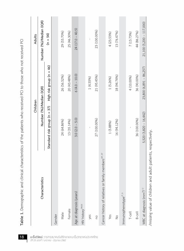
The characteristics of child patients
We enrolled 83 children with ALL,
with boys being more common than girls
(1.5:1). The median age at diagnosis was 4
years. Four (4.82%) were classified as T-cell
lineage immunophenotype, while one
(4.55%) and two (5.56%) were recorded as
having a history of HIV history and a family
history of cancer, respectively. The median
initial WBC was 12,800/mm3 (range:
1,400–515,000/mm3). Characteristics are
summarized in Table 1. Overall, more
than half of the children (n = 46) were
categorized in the “high-risk” group. Among
these, 80.43% (n = 37) were assigned to
receive PCI with a prescribed total radiation
dose of 18–36 Gy.
The results of the treatment of the child
ALL patients
These results are reported in Table
2. The death rates showed no statistically
significant difference between the stan-
dard-risk and high-risk ALL groups (16.22%
vs. 13.04%). The common causes of death
include ALL and uncontrolled brain metas-
tasis (8.70% in the high-risk and 16.22% in
the standard-risk ALL groups). The OS rate
was 85.54% with a median survival time of
69.87 months (86.96% with median 62.18
months and 83.78% with median 84.83
months in the high-risk and standard-risk
ALL groups, respectively, p = 0.004).
The five-year survival rate of the child
patients was 59.04% (45.65% in the high-
risk and 70.27% in the standard-risk ALL
groups, p = 0.023). In the high-risk ALL
group, 62.16% of patients who received
PCI survived with controlled brain metas-
tasis, while those who did not receive PCI
survived with controlled brain metastasis
only 22.22%.
Of the 21.69% child ALL patients
who relapsed, 26.09% were in the high-risk
and 12.22% were in the standard-risk ALL
groups. Relapses of child ALL patients were
mostly isolated in the CNS (21.74% and
8.11%), while other relapses were 4.35%
and 8.11% in the high-risk and standard-risk
groups, respectively. The median survival
time of the relapsed patients was 40.57
months (medians of 38.25 months (n = 12)
and 43.20 months (n = 6) in the high-risk
and standard-risk ALL groups, respectively).
About Thirty-three percent (n = 6) of the
relapsed patients died (three were high-
risk ALL patients). Among the 18 relapsed
patients, 10 had received PCI (all of them
were high-risk ALL patients), and eight
had not (two of them were high-risk ALL
patients). The median duration of relapse
was 37.43 months. The five-year DFS rates
18 มะเรงววฒน วารสารสมาคมรงสรกษาและมะเรงวทยาแหงประเทศไทยปท 25 ฉบบท 1 มกราคม - มถนายน 2562
Tabl
e 1.
Dem
ogra
phic
and
clin
ical c
hara
cter
istics
of t
he p
atie
nts
who
rece
ived
PCI
to th
ose
who
not
rece
ived
PCI
Char
acte
ristic
s
Child
ren
Adul
ts
Num
ber (
%)/
Med
ian
(IQR)
Num
ber (
%)/
Med
ian
(IQR)
(n =
54)
Stan
dard
risk
gro
up (n
= 3
7)Hi
gh ri
sk g
roup
(n =
46)
Gend
er
M
ale
24 (6
4.86
%)
26 (5
6.52
%)
29 (5
3.70
%)
F
emal
e13
(35.
14%
)20
(43.
48%
)25
(46.
30%
)
Age
at d
iagn
osis
(yea
rs)
3.0
(2.0
– 5
.0)
6 (4
.0 –
10.
0)24
(17.
0 –
40.5
)
HIV
hist
ory/3
4,31
y
es-
1 (4
.55%
)-
n
o27
(100
.00%
)21
(95.
45%
)23
(100
.00%
)
Canc
er h
istor
y of
rela
tives
or f
amily
mem
bers
/47,
37
Y
es1
(5.8
8%)
1 (5
.26%
)4
(23.
53%
)
N
o16
(94.
12%
)18
(94.
74%
)13
(76.
47%
)
Imm
unop
heno
type
/7, 3
T
-cel
l-
4 (1
0.00
%)
7 (1
3.73
%)
B
-cel
l36
(100
.00%
)36
(90.
00%
)44
(86.
27%
)
WBC
at d
iagn
osis
(mm
3 )/-,1
5,52
0 (3
,820
– 1
6,44
2)23
,800
(6,8
91 –
86,
257)
21,1
00 (5
,200
– 1
17,0
00)
/miss
ing
valu
e of
chi
ldre
n an
d ad
ult p
atie
nts,
resp
ectiv
ely.

19Jornal of Thai Association of Radiation Oncologyvol. 25 No.1 January - June 2019
were 62.6% and 81.2% in the high-risk
and standard-risk ALL groups, respectively
(Figure 1(a)).
The characteristics of the adult ALL
patients
We enrolled 54 adults with ALL.
The median age at diagnosis was 24 years.
Seven patients (12.89%) were classified as
T-cell lineage immunophenotype and four
patients (7.41%) as having a family history
Table 2. The Treatment Results of the Child and Adult ALL patients
ALL Treatment Results
ChildrenAdults
(n = 54)High Risk
(n = 46)
Standard Risk
(n = 37)
Death 6 (13.04%) 6 (16.22%) 15 (27.78%)
ALL 4 (8.70%) 6 (16.22%) 14 (25.93%)
Complications 1 (2.17%) - -
Other causes 1 (2.17%) - 1 (1.85%)
Relapse 12 (26.09%) 6 (16.22%) 25 (46.30%)
Central nervous system (CNS) 10 (21.74%) 3 (8.11%) 21 (38.89%)
Bone marrow (BM) - 3 (8.11%) 2 (3.70%)
CNS+BM 1 (2.17%) - -
Acute myelogenous leukemia (AML) 1 (2.17%) - -
Acute promyelocytic leukemia (APL) - - 1 (1.85%)
Testes - - 1 (1.85%)
of cancer. The median WBC, RBC, and
platelet count at diagnosis were 21,100/
mm3, 9.2 g/dL and 64,250 /mm3, respec-
tively. Characteristics are summarized in
Table 1. Among these, 37.04% (n = 20)
were assigned to receive PCI with a
prescribed total radiation dose of 18–36
Gy.
The results of the treatment of the
adult ALL patients

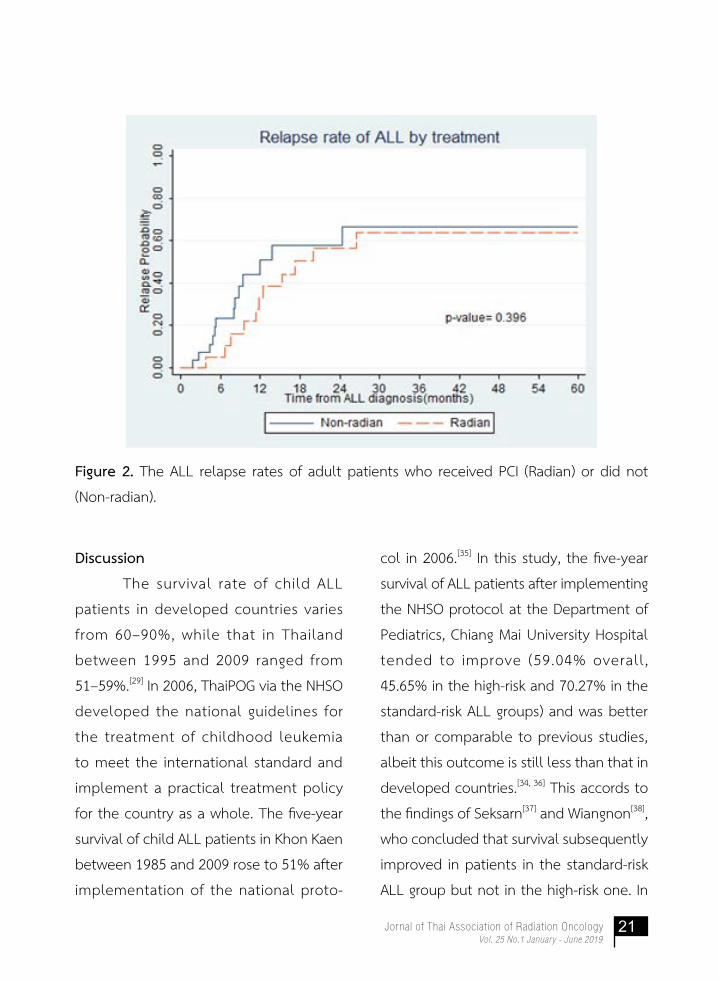
20 มะเรงววฒน วารสารสมาคมรงสรกษาและมะเรงวทยาแหงประเทศไทยปท 25 ฉบบท 1 มกราคม - มถนายน 2562
overall relapse rates of adult patients was
relative high but not related to whether
PCI was administered (no PCI: 66.4% and
with PCI: 63.8%, p = 0.396) (Figure 2).
However, the patients who received PCI
had a slightly better outcome in terms
of prevention or a delay in CNS relapse
(median duration of CNS relapse = 8.23
months in patients with no PCI vs. 11.93
months in patients with PCI) and a longer
survival time (median survival time = 21.75
months in patients with no PCI vs. 48.29
months in patients with PCI). Furthermore,
35.0% of patients who received PCI sur-
vived with controlled brain metastasis
compared to only 20.59% in who did not.
These results are reported in Table
2. The death rate was 27.78%, most of
which were due to ALL and uncontrolled
brain metastasis (25.93%). The overall
and five-year survival rates were 72.22%
and 25.93%, respectively, with a median
survival time of 26.17 months.
Of the 46.30% adult ALL patients
who relapsed, the median duration of
relapse was 9.53 months and the five-year
DFS rate was 36.2% (Figure 1(b)). Relapses
in adult ALL patients were mostly isolated
in the CNS (38.89%), while other relapses
comprised 7.41%. The five-year survival
rate of the relapsed patients was 2.5%.
The median survival time of relapsed
patients was 15.43 months. The five-year
(a) (b)
Figure 1. The ALL Disease free survival rates of (a) child and (b) adult patients who
received PCI or did not.
21Jornal of Thai Association of Radiation Oncologyvol. 25 No.1 January - June 2019
Figure 2. The ALL relapse rates of adult patients who received PCI (Radian) or did not
(Non-radian).
Discussion
The survival rate of child ALL
patients in developed countries varies
from 60–90%, while that in Thailand
between 1995 and 2009 ranged from
51–59%.[29] In 2006, ThaiPOG via the NHSO
developed the national guidelines for
the treatment of childhood leukemia
to meet the international standard and
implement a practical treatment policy
for the country as a whole. The five-year
survival of child ALL patients in Khon Kaen
between 1985 and 2009 rose to 51% after
implementation of the national proto-
col in 2006.[35] In this study, the five-year
survival of ALL patients after implementing
the NHSO protocol at the Department of
Pediatrics, Chiang Mai University Hospital
tended to improve (59.04% overall,
45.65% in the high-risk and 70.27% in the
standard-risk ALL groups) and was better
than or comparable to previous studies,
albeit this outcome is still less than that in
developed countries.[34, 36] This accords to
the findings of Seksarn[37] and Wiangnon[38],
who concluded that survival subsequently
improved in patients in the standard-risk
ALL group but not in the high-risk one. In

22 มะเรงววฒน วารสารสมาคมรงสรกษาและมะเรงวทยาแหงประเทศไทยปท 25 ฉบบท 1 มกราคม - มถนายน 2562
our study, 21.69% of child ALL patients
relapsed, which corresponds to previous
studies,[18–19, 22] thus it is not certain whether
the nationwide implementation of the
standard protocols has been beneficial.[31]
Recurrence occurred in the high-risk group
approximately twice as often as the stan-
dard-risk group, resulted in five-year DFS
rates of 62.6% and 81.2%, respectively,
which is better than the results reported
by Seksarn,[31] in which the event free
surv ival rates f rom 12 inst i tut ions
throughout Thailand were 51.2% and
66.5%, respectively. Finally, 62.16% of
patients who received PCI survived with
controlled metastasis.
For the adult ALL patients, the
five-year survival rate was 25.93%, which
is considered as relatively low compared
to prev ious studies that reported
25–47%.[33, 38–40] Relapse developed in
46.30% of adult ALL patients and the
five-year DFS rates was 36.2%, which is
s imilar to the results in for adults
diagnosed with ALL from 2005 to 2015
at a reference center in Mexico.[33]
In this study, the relapse occurrence
of ALL was still quite common in both
child and adult ALL patients and were high-
er than in previous studies.[11, 17, 19, 41] The
majority of relapse occurrence in this study
was due to CNS relapse (14 (77.78%) out
of 18 relapses in children, and 21 (87.50%)
out of 24 relapses in adults). The relatively
high rate of CNS relapse in our study might
have been related to inadequate system-
ic chemotherapy[42] and most enrolled
patients were high risk of relapse in the
CNS or at other sites.[24, 41–42, 43]
There was no difference in the
frequency or site of relapse between
patients who received PCI and those who
did not. However the trend in the rate of
CNS relapse was not significantly different,
although the patients who received PCI
had a slightly better outcome in terms
of prevention or delayed CNS relapse.
Retrospective data was used in this study
and unfortunately, some useful demo-
graphic variables were not available for our
analysis. Future research with alternative
data sources could address the possible
relationship between other covariates and
relapse occurrence. A study of adolescents
and young adults might be useful since
the ALL occurring in this age group could
be biologically different from that in
younger and older ALL patients, and an
understanding of the biology of ALL in
all of these patient groups could help to
improve outcomes and prognosis. The
limitation of this study are that its based
23Jornal of Thai Association of Radiation Oncologyvol. 25 No.1 January - June 2019
on a retrospective analysis. Therefore, in
the next study should be added toxicities
of pirarubicin in the treatment of childhood
acute lymphoblastic leukemia.
References
1. The International Agency for
Research on Cancer. Population Fact
Sheets: Estimated Incidence, mortality
and prevalence worldwide in 2012
[Online]. 2012 [cited 2018 Mar 30],
Available from: http://globocan.iarc.
fr/Pages/fact_sheets_population.
aspx
2. PDQ Adult Treatment Editorial
Board. Adult Acute Lymphoblastic
Leukemia Treatment (PDQ®): Health
Professional Version [Online]. [cited
2018 March 30], Available from:
https://www.ncbi.nlm.nih.gov/
pubmedhealth/PMH0032602/
3. American Cancer Society. Acute
Lymphocytic Leukemia (ALL) in
Adults [Online]. [cited 2018 March
30]. Available from: https://www.
cancer.org/cancer/acute-lymphocytic-
leukemia.html.
4. Jemal A, Tiwari RC, Murray T,
Ghafoor A, Samuels A, Ward E, et al.
Cancer statistics, 2004. CA Cancer J
Clin. 2004; 54:8–29.
5. Jemal A, Siegel R, Ward E, Murray T,
Xu J, Smigal C, et al. Cancer
statistics, 2006. CA Cancer J Clin.
2006; 56:106–130.
6. Fullmer A, O’Brien S, Kantarjian H,
Jabbour E. Novel Therapies for
Relapsed Acute Lymphoblastic
Leukemia. Curr Hematol Malig Rep.
2009; 4: 148–156.
7. Joyce M, Pollock BH, Devidas M,
Buchanan G, Camitta B. Chemo-
therapy for Initial Induction Failures
in Children Acute Lymphoblastic
Leukemia: a Children’s Oncology
Group Study (POG 8764). J Pediatr
Hematol Oncol. 2013; 35:32–35.
8. Lee JW, Cho B. Prognostic factors
and treatment of children acute
lymphoblastic leukemia. Korean J
Pediatr. 2017; 60:129–137.
9. Terwilliger T, Abdul-Hay M. Acute
lymphoblastic leukemia: a com-
prehensive review and 2017 update.
Blood Cancer J. 2017; 7:e577.
10. Schrappe M, Reiter A, Ludwig WD,
Ha rbot t J , Z immermann M,

24 มะเรงววฒน วารสารสมาคมรงสรกษาและมะเรงวทยาแหงประเทศไทยปท 25 ฉบบท 1 มกราคม - มถนายน 2562
Hiddemann W, et al. Improved
outcome in children acute lympho-
blastic leukemia despite reduced
use of anthracyclines and cranial
radiotherapy: results of trial ALL-BFM
90. German-Austrian-Swiss ALL-BFM
Study Group. Blood. 2000; 95:
3310-3322.
11. Silverman LB, Gelber RD, Dalton VK,
Asselin BL, Barr RD, Clavell LA,
et al. Improved outcome for children
with acute lymphoblastic leukemia:
results of Dana-Farber Consortium
Protocol 91-01. Blood. 2001; 97:
1211-1218.
12. Pui CH, Evans WE. Treatment of
acute lymphoblastic leukemia. N
Engl J Med. 2006; 354:166–178.
13. Stock W, La M, Sanford B, Bloom-
field CD, Vardiman JW, Gaynon P,
et al. What determines the out-
comes for adolescents and young
adults with acute lymphoblastic
leukemia treated on cooperative
group protocols? A comparison of
Children’s Cancer Group and Cancer
and Leukemia Group B studies.
Blood. 2008; 112: 1646–1654.
14. Hoelzer D, Thiel E, Loffler H,
Büchner T, Ganser A, Heil G, et al.
Prognostic factors in a multicenter
study for treatment of acute lymph-
blastic leukemia in adults. Blood.
1988; 71:123-131.
15. Goldstone AH, Richards SM, Lazarus
HM, Tallman MS, Buck G, Fielding AK,
et al. In adults with standard-risk
acute lymphoblastic leukemia, the
greatest benefit is achieved from a
matched sibling allogeneic trans-
plantation in first complete remis-
sion, and an autologous transplan-
tation is less effective than conven-
tional consolidation/maintenance
chemotherapy in all patients: final
results of the International ALL Trial
(MRC UKALL XII/ECOG E2993). Blood
journal. 2008; 111:1827-1833.
16. Pulte D, Redaniel MT, Jansen L,
Brenner H, Jeffreys M. Recent trends
in survival of adult patients with
acute leukemia: overall improve-
ments, but persistent and partly
increasing disparity in survival of
patients from minority groups.
Haematologica. 2013; 98: 222–229.
17. Oriol A, Vives S, Hernández-Rivas
JM, Tormo M, Heras I, Rivas C, et al.
Outcome after relapse of acute
lymphoblastic leukemia in adult
25Jornal of Thai Association of Radiation Oncologyvol. 25 No.1 January - June 2019
patients included in four consecu-
tive risk-adapted trials by the
PETHEMA Study Group. Haemato-
logica. 2010; 95: 589–596.
18. Locatelli F, Schrappe M, Bernardo
ME, Rutella S. How I treat relapsed
children acute lymphoblastic leu-
kemia. Blood journal. 2012; 120:
2807-2816.
19. Cancela CS, Murao M, Viana MB, de
Oliveira BM. Incidence and risk
factors for central nervous system
relapse in children and adolescents
with acute lymphoblastic leukemia.
Rev Bras Hematol Hemoter. 2012;
34: 436–441.
20. Thai Pediatric Oncology Group.
Practical guidelines: Leukemia in
children. Bangkok, National Health
Security Office (NHSO). [Online].
2006 [cited 2018 Mar 30], Available
from: https://www.nhso.go.th/
frontend/page-forhospital_cpg.aspx
21. Conter V, Schrappe M, Aricó M,
Reiter A, Rizzari C, Dördelmann M,
et al. Role of cranial radiotherapy
for children T-cell acute lympho-
blastic leukemia with high WBC
count and good response to pred-
nisone. Associazione Ital iana
Ematologia Oncologia Childrena and
the Berlin-Frankfurt-Münster groups.
J Clin Oncol. 1997;15:2786-91.
22. Hata M, Ogino I, Aida N, Saito K,
Omura M, Kigasawa H, et al .
Prophylactic cranial irradiation
of acute lymphoblastic leukemia in
children: outcomes of late effects
on pituitary function and growth in
long-term survivors. Int J Cancer.
2001; 96 Suppl:117-124.
23. Giordano FA, Welzel G, Abo-Madyan
Y, Wenz F. Potential toxicities of
prophylactic cranial irradiation.
Transl Lung Cancer Res. 2012; 1:
254–262.
24. Meshref MM, Elshazly N, Nasr M,
Abdelhai R. Effect of prophylactic
cranial irradiation dose on CNS
relapse, late cognitive decline and
learning disabilities in children treated
for acute lymphoblastic leukemia.
J Clin Oncol. 2006 24:18_suppl,
9032-9032.
25. Waber DP, Turek J, Catania L,
Stevenson K, Robaey P, Romero I,
et al. Neuropsychological outcomes
from a randomized trial of triple
intrathecal chemotherapy com
pared with 18 Gy cranial radiation as

26 มะเรงววฒน วารสารสมาคมรงสรกษาและมะเรงวทยาแหงประเทศไทยปท 25 ฉบบท 1 มกราคม - มถนายน 2562
CNS treatment in acute lymphoblas-
tic leukemia: findings from Dana-
Farber Cancer Institute ALL Consor-
tium Protocol 95-01. J Clin Oncol.
2007; 25:4914-21.
26. Chemaitilly W, Li Z, Huang S, Ness
KK, Clark KL, Green DM, et al.
Anterior hypopituitarism in adult
survivors of children cancers treated
with cranial radiotherapy: a report
from the St Jude Lifetime Cohort
study. J Clin Oncol. 2015; 33:
492-500.
27. Löning L, Zimmermann M, Reiter A,
Kaatsch P, Henze G, Riehm H,
et al. Secondary neoplasms sub-
sequent to Berlin-Frankfurt-Münster
therapy of acute lymphoblastic
leukemia in children: significantly
lower risk without cranial radio-
therapy. Blood. 2000; 95:2770-5.
28. Pui CH, Cheng C, Leung W, Rai SN,
Rivera GK, Sandlund JT, et al.
Extended follow-up of long-term
survivors of children acute lympho-
blastic leukemia. N Engl J Med. 2003;
349:640-649.
29. Allemani C, Weir HK, Carreira H,
Harewood R, Spika D, Wang XS,
et al. Global surveillance of cancer
survival 1995-2009: analysis of
individual data for 25676887
patients from 279 population-based
registries in 67 countries (CON-
CORD-2). Lancet. 2015;385:977-1010.
30. Hunger SP, Lu X, Devidas M,
Camitta BM, Gaynon PS, Winick NJ,
et al. Improved survival for children
and adolescents with acute
lymphoblastic leukemia between
1990 and 2005: a report from the
children’s oncology group. J Clin
Oncol. 2012; 30: 1663-9.
31. Seksarn P, Wiangnon S , Veerakul G,
Chotsampancharoen T, Kanjana-
pongkul S, Chainansamit S. Outcome
of Children Acute Lymphoblastic
Leukemia Treated Using the Thai
National Protocols. Asian Pac J
Cancer Prev. 2015:16, 4609-4614.
32. Hallböök H, Gustafsson G, Smedmyr
B, Söderhäll S, Heyman M; Swedish
Adult Acute Lymphocytic Leukemia
Group. Treatment outcome in young
adults and children >10 years of age
with acute lymphoblastic leukemia
in Sweden: a comparison between
a pediatric protocol and an adult
protocol. cancer. 2006; 107:1551-61.
27Jornal of Thai Association of Radiation Oncologyvol. 25 No.1 January - June 2019
33. Jaime-Pérez JC, Jiménez-Castillo
RA, Herrera-Garza JL, Gutiérrez-
Aguirre H, Marfil-Rivera LJ, Gómez-
Almaguer D. Survival Rates of
Adults with Acute Lymphoblastic
Leukemia in a Low-Income Popula-
tion: A Decade of Experience at a
Single Institution in Mexico. Clin
Lymphoma Myeloma Leuk. 2017;
17:60-68.
34. Chiang Mai Cancer Registry, Maharaj
Nakorn Chiang Mai Hospital, Faculty
of medicine, Chiang Mai University,
Chiang Mai, Thailand. CANCER
INCIDENCE AND MORTALITY IN
CHIANG MAI 2011. Published by the
Academic Publishing Unit, Faculty of
Medicine, Chiang Mai University,
Chiang Mai, Thailand
35. Wiangnon S , Jets r i suparb A,
Komvilaisak P, Suwanrungruang K.
Childhood cancer incidence and
survival 1985- 2009, Khon Kaen,
Thailand. Asian Pac J Cancer Prev.
2014; 15: 7989-93.
36. Gatta G, Botta L, Rossi S, Aareleid
T, Bielska-Lasota M, Clavel J, et al.
Childhood cancer survival in Europe
1999–2007: results of EUROCARE-
5—a population- based study.
Avaliable from: http://www.euro
care.it/LinkClick.aspx?fileticket=
hcgmq6vAq6Y%3D&tabid=61.
37. Seksarn P. Outcome of childhood
leukemia, ThaiPOG study. Proceed-
ings of the 5th St. Jude-VIVA forum
in pediatric oncology; 2011 March
21; Shangri-La Hotel, Singapore;
2011.
38. Wiangnon S, Veerakul G, Nuchpra-
yoon I, Seksarn P, Hongeng S,
Krutvecho T, et al. Childhood cancer
incidence and survival 2003-5,
Thailand: study from the Thai
Pediatric Oncology Group. Asian Pac
J Cancer Prev. 2011; 12:2215-20.
39. Ghimire KB, Shah BK. Survival in
Adult Acute Lymphoblastic Leuke-
mia Survival by Age, Gender and
Ethnicity. Blood 2013;122:1696.
40. Gökbuget N and Hoelzer D. Treat-
ment of adult acute lymphoblastic
leukemia. Semin Hematol. 2009 ;
46:64-75.
41. Kozlowski P, Åström M, Ahlberg L,
Bernell P, Hulegårdh E, Hägglund H,
et al. High relapse rate of T cell
acute lymphoblastic leukemia in

28 มะเรงววฒน วารสารสมาคมรงสรกษาและมะเรงวทยาแหงประเทศไทยปท 25 ฉบบท 1 มกราคม - มถนายน 2562
adults treated with Hyper-CVAD
chemotherapy in Sweden. Eur J
Haematol. 2014; 92:377-81.
42. Pui CH, Campana D, Pei D. Treat-
ment of Children Acute Lympho-
blastic Leukemia Without Prophy-
lactic Cranial Irradiation. N Engl J
Med. 2009; 360: 2730–2741.
43. Conter V, Schrappe M, Aricó M,
Reiter A, Rizzari C, Dördelmann M,
et al. Role of cranial radiotherapy
for childhood T-cell acute lympho-
blastic leukemia with high WBC
count and good response to pred-
nisone. Associazione Italiana Ema-
tologia Oncologia Pediatrica and
the Berlin-Frankfurt-Münster groups.
J Clin Oncol. 1997;15:2786-91.
29Jornal of Thai Association of Radiation Oncologyvol. 25 No.1 January - June 2019
Application of the RapidPlan knowledge-based treatment planning system for radiation therapy of prostate cancer patients
Kanokkarn Kuekkong1, Siwadol Pleanarom2, Pittaya Dankulchai2, Puangpen Tangboonduangjit1, and Lalida
Tuntipumiamorn2
Institutions: 1Master of Science Program in Medical Physics, Department of Therapeutic and Diagnostic Radiology, Faculty of
Medicine Ramathibodi Hospital, Mahidol University, Bangkok, 10400, Thailand2Division of Radiation Oncology, Department of Radiology, Faculty of Medicine Siriraj Hospital, Mahidol University,
Bangkok, 10700, Thailand
ABSTRACT
Background: RapidPlan (RP) knowledge-based treatment planning was developed
and adopted in volumetric arc modulated radiotherapy (VMAT) planning to
improve plan quality and planning efficiency. RP used plan database to train a
model for predicting organ-at-risk (OAR) dose-volume-histograms (DVHs) of the new
treatment plan.
Objectives: The purpose of this study was to develop and evaluate the performance
of the RP knowledge-based treatment planning to generate VMAT for definitive
radiotherapy of prostate cancer.
Materials and methods: Three RP models based on a number of 20, 40, and 60
previously VMAT plans were trained and validated on 10 new prostate cancer
patients. Dosimetric parameters of the target volume and organs at risks (OARs)
between models and manually optimized method (MO) from experienced planner
were compared. The D2%, D95%, D98%, homogeneity index (HI), and conformation

30 มะเรงววฒน วารสารสมาคมรงสรกษาและมะเรงวทยาแหงประเทศไทยปท 25 ฉบบท 1 มกราคม - มถนายน 2562
number (CN) for planning target volume (PTV), V65Gy, V70Gy, V75Gy for bladder
and V50Gy, V60Gy, V65Gy, V70Gy, V75Gy for rectum were collected and analyzed
(one-way repeated measures ANOVA, p<0.05).
Results: VMAT plans between models and MO showed similar results of D95%,
D98% for PTV but a significant higher of D2%, CN, and HI from RP (105.4%-105.7%
for D2%, 0.06-0.07 for HI, and 0.9 for CN) when compared with MO (104% for D2%,
0.05 for HI, and 0.8 for CN). For bladder and rectum, all dose-volume parameters
of RP were significantly lower than MO (p<0.05), only in RPmodel20 which bladder
V75Gy, was similar to MO. Dosimetric analysis for model training based on a different
number of VMAT plans showed no statistical difference in plan quality.
Conclusion: RP knowledge-based treatment planning in this investigation presented
acceptable VMAT plan quality for definitive radiotherapy prostate in only single op-
timization. Twenty historic plans were found to be an acceptable minimum number
of plans for the model training.
Keyword: Knowledge-based planning, Prostate planning, RapidPlan, VMAT
บทคดยอ
หลกการและเหตผล: RapidPlan(RP)ถกพฒนาและประยกตใชเพอเพมคณภาพและประสทธภาพ
ของแผนการรกษา โดย RP จะใชขอมลแผนการรกษาทถกใชกบผปวยในทางคลนกมาแลว เพอ
สรางเปนแบบจำาลองการรกษาและประเมน DVHs แกอวยวะสำาคญในแผนการรกษาใหม
วตถประสงค: เพอประเมนประสทธภาพของระบบวางแผนการรกษาแบบจดฐานความรดวย
RP สำาหรบการรกษาแบบปรบความเขมหมนรอบตว(VMAT)ในผปวยมะเรงตอมลกหมาก
วสดและวธการ: สรางแบบจำาลอง RP 3 แบบจากแผนการรกษาVMATทถกใชกบผปวยในทาง
คลนกมาแลวจำานวน 20, 40, และ 60 แผนตามลำาดบ จากนนนำาแบบจำาลองดงกลาวมาทดสอบ
กบผปวยรายใหมจำานวน 10 ราย และเปรยบเทยบแผนการรกษาทไดจาก RPและวธดวยมอจากผ
มประสบการณ (MO) ขอมลปรมาณรงสสำาหรบกอนมะเรง (PTV) ทนำามาวเคราะหไดแกคา
D2%, D95%, D98%, conformation number(CN) และhomogeneity index(HI) สวน

31Jornal of Thai Association of Radiation Oncologyvol. 25 No.1 January - June 2019
INTRODUCTION
Intensity-modulated radiation
therapy (IMRT) and volumetric modulated
arc radiotherapy (VMAT) utilized the inverse
planning process that is needed to solve
this complexity through the optimization
process. To get the satisfied results, an
appropriate set of optimization parameters
for organs at risk (OARs) and the target
volume has to be specified by planners
through a repeated trial-and-error process.
Therefore, the planning outcome was
strongly based on the experience and skills
กระเพาะปสสาวะไดแกคา V65Gy, V70Gy, V75Gy และไสตรงท V50Gy, V60Gy, V65Gy, V70Gy,
V75Gy จากนนทำาการวเคราะหทางสถตโดยใช one-way repeated measures ANOVA, p<0.05
ผลการศกษา: แผนการรกษาRPและMO แสดงผลของ D95%, D98% สำาหรบ PTV ทเหมอนกน
แตมคา D2%, CN, และ HI จากแผนRP (105%-106% สำาหรบ D2%, 0.06-0.07 สำาหรบ HI และ
0.9 สำาหรบ CN) ทมากกวาMO (104% สำาหรบ D2%, 0.05 สำาหรบ HI, และ 0.8 สำาหรบ CN)
อยางมนยสำาคญทางสถต สำาหรบปรมาณรงสทกระเพาะปสสาวะและไสตรงไดรบจาก RP มคาตำากวา
MO อยางมนยสำาคญดวยเชนกน ยกเวนปรมาณรงสท V75Gy ของกระเพาะปสสาวะท RP 20 แผน
มผลเหมอนกบการวางแผนแบบ MO ในการเปรยบเทยบแผนการรกษาทสรางจาก RP 3 รปแบบใน
ผปวยรายเดยวกนไมพบความแตกตางทางสถตของปรมาณรงส
ขอสรป: ระบบวางแผนการรกษาแบบจดฐานความรดวย RPสามารถสรางแผนการรกษา VMAT
ในผปวยมะเรงตอมลกหมากไดอยางมคณภาพและแผนการรกษาจำานวนขนตำา 20 แผนเพยงพอ
สำาหรบการสรางแบบจำาลอง RP ไดอยางเหมาะสมและมประสทธภาพ
คำาสำาคญ: ระบบการวางแผนการรกษาแบบจดฐานความร, การวางแผนการรกษามะเรงตอม
ลกหมาก, RapidPlan, การฉายรงสแบบปรบความเขมหมนรอบตว
of the planners[1, 2]. The treatment planning
could take several hours in trial-and-
error optimization to achieve the planning
goals. Currently, various tools were
developed for IMRT and VMAT radiation
therapy treatment planning. Knowledge-
based (KB) approaches were introduced
and adopted in treatment planning to
improve planning consistency in IMRT
and VMAT. KB treatment planning (KBTP)
method was used to predict the
dosimetric features of the new treatment
plan by utilizing a database of prior plans
32 มะเรงววฒน วารสารสมาคมรงสรกษาและมะเรงวทยาแหงประเทศไทยปท 25 ฉบบท 1 มกราคม - มถนายน 2562
determined via the spatial relationship between anatomical, geometric and dosimetric features of targets and OARs [3, 4]. This method was useful to reduce varia- tions in plan quality. Previous studies[5-7]
have demonstrated that KBTP resulted in superior treatment plans in terms of planning time and dose distributions as compared to conventional IMRT and VMAT plans. Furthermore, this approach is able to decrease optimization time. RapidPlan version 13.6 (Varian Medi-cal System, Palo Alto, CA, USA; RP) is a commercially KB planning application software which is an optional applica-tion from the Eclipse treatment planning system (TPS). Therefore, the purpose of this paper was to study and demonstrate the potential of the RP knowledge-based TPS for VMAT plans in prostate cancer in terms of plan quality and efficiency at the Division of Radiation Oncology, Depart-ment of Radiology, Faculty of Medicine, Siriraj Hospital.
MATERIALS AND METHODSPreviously clinical VMAT plans of pros-tate cancer selection Sixty VMAT planning of prostate cancer patients who treated only prostate gland, not involved lymph node during January 2016 to March 2018 were collected and analyzed. All treatment plans were created with two or three arcs, using 10 MV photon beams, and total prescribed dose to PTV was 78 Gy in 39 fractions. Dose-volume constraints for the planning target volume (PTV- prostate gland), the OARs such as bladder, rectum, and the PTV overlap OARs were assigned as the planning goal for the optimization process. All treatment plans were approved by the radiation oncologist according to Quantitative Analyses of Normal Tissue Effects in the Clinic (QUANTEC) guideline[8] as shown in Table 1. Volume data of the PTV and all OARs were also collected for the analysis.
Table 1. Dose-volume constraints assigned for VMAT prostate cancer optimization
Organ Dose constraints
PTV D2%≤ 107%, D95%≥ 95%, and D98%≥ 93%
Bladder V65Gy≤50%, V70Gy≤35%, and V75Gy≤25%
Rectum V50Gy≤ 50%, V60Gy≤35%, V65Gy≤25%, V70Gy≤20%, and V75Gy≤15%

33Jornal of Thai Association of Radiation Oncologyvol. 25 No.1 January - June 2019
Model configuration
RP system consists of two main
components: model configuration and
model validation. To configure a model,
the geometric and dosimetric parameters
of the historic treatment plans are
extracted and trained by using a com-
bination of Principal Component Analysis
and regression tecniques in the RP
algorithm [9]. For model val idation,
estimated dose volume histogram (DVH)
and optimization objectives for the
optimization process of new patients were
generated. In this study, sixty previously
VMAT prostate plans were used to
generate three RP models. Model20 is
the minimum number of 20 random
previous plans that used for building the
model as suggested by the vendor. To
examine whether a number of plans
result in model quality and consistency
or not, Model40 and Model60 were
generated from using 40 and 60 previous
plans for training.
Model Validation
CT dataset of 10 new prostate
cancer patients were used to validate
the model. For each patient, three VMAT
treatment planning were created from a
total of 3 RP models. The VMAT planning
parameters including the field geometry
(2 arcs), 10 MV photon energy, and dose
prescription 78 Gy to PTV were set. In the
optimization process, the RP system was
used to perform an estimation for the
DVHs in any new patient. The workflow of
the DVHs estimation started with a selec-
tion of the RP model. Then, the outlined
structures of new patient auto-matched
to the model structures using a structure
code. The system automatically generated
optimization priorities, setting of the
upper and lower objectives to the PTV,
DVH estimated boundary to the OARs, and
line objectives, which placed along the
inferior DVH estimated boundary[9]. The
examples of the resulting RP predictive
DVH estimated and priorities are shown
in Figure 1. Generating estimated DVHs
and priorities based on patient geometry
of new patient and previous knowledge
contained in a model database and were
used in the optimization process. A single
optimization without any planner inter-
vention was performed to assess the
quality of the models.
Performance of the RP plan compared
to the manually optimized plans
Two expert planners with their
experience in VMAT planning of more

34 มะเรงววฒน วารสารสมาคมรงสรกษาและมะเรงวทยาแหงประเทศไทยปท 25 ฉบบท 1 มกราคม - มถนายน 2562
Figure 1. (a) priorities and objectives (b) the estimated range and line objectives of RP prediction
than 60 prostate cancer plans were
participated in this study. Manually
optimized (MO) VMAT plans were created
by the planners using the same technique
as the RP plans. Dosimetric parameter
results of MO plans from two planners
were comparable. Then, the MO plans
results were averaged and compared
with plans from 3 RP models. Dosimetric
parameters in term of (1) dose to 2%
of the PTV volume (D2%); (2) dose to
95% of the PTV volume (D95%); (3) dose
to 98% of the PTV volume (D98%); (4)
homogeneity index (HI) [10] of the PTV
defined as HI =[D2%-D98]/D50%, the HI
value is 0, representing dose homogeneous
in target ; (5) conformation number (CN)[11]
of the PTV defined as CN = [TVRI/TV] ×
[TVRI/VRI] where TVRI = target volume
covered by the prescription isodose,
TV = target volume, and VRI = volume of
the prescription isodose, the CN value is
1, representing conformity of target; (6)
dose-volume parameters to the bladder
as V65Gy, V70Gy, and V75Gy; and (7)
dose-volume parameters to the rectum as
V50Gy, V60Gy, V65Gy, V70Gy, and V75Gy
were used for the analysis.
The one-way repeated measures
ANOVA (p < 0.05) was used to test the
significance of the plan comparison results.
RESULTS
Collecting the structure’s volume
from the historic plans, the results in
Table 2 shows mean + SD of PTV, OARs,
and OARs overlapping with PTV volumes
for each RP model, compared with 10
new patients. The mean of the PTV
registered in each model and test group

35Jornal of Thai Association of Radiation Oncologyvol. 25 No.1 January - June 2019
Table 2. Data of the structure volumes in each RP model and a test group
Volumes (cm3)
Mean ± S.D.
Model20
Model40
Model60
New Patients
PTV 112.2 ± 33.9 112.6 ± 30.8 116.1 ± 35.1 110.0 ± 35.3
Bladder 264.5 ± 161.6 251.3 ± 135.4 251.1 ± 117.1 266.2 ± 141.8
Rectum 56.6 ± 24.0 57.6 ± 23.9 56.3 ± 21.6 52.6 ± 14.1
Bladder Overlap
PTV
11.4 ± 4.2 14.9 ± 9.0 14.7 ± 7.8 11.31 ± 4.0
Rectum Overlap
PTV
2.8 ± 1.3 3.0 ± 1.5 3.0 ± 1.7 2.7 ± 1.9
were 112.2 ± 33.9 cm3, 112.6 ± 30.8 cm3,
116.1 ± 35.1 cm3 and 110.0 ± 35.3 cm3 for
Model20
, Model40
, Model60
, and test
group respectively. The mean ± SD of the
bladder were 264.5 ± 161.6 cm3, 251.3
± 135.4 cm3, 251.1 ± 117.1 cm3, and
266.2 ± 141.8 cm3 for Model20, Model
40,
Model60, and test group, respectively. In
the rectum volume, the mean ± SD were
56.6 ± 24.0 cm3, 57.6 ± 23.9 cm3, 56.3 ±
21.6 cm3, and 52.6 ± 14.1 cm3 for Model20,
Model40, Model
60, and test group, respec-
tively. It can be seen that the volumes
of the target and organs at risks among
3 models and in a test group were quite
similar.
The isodose distribution for VMAT
planning from both RP & MO optimiza-
tion methods are shown in Figure 2. All
plans were evaluated and passed the
clinical planning goal as prescribed from
QUANTEC guideline. All dosimetric results
were summarized as shown in Table 3.
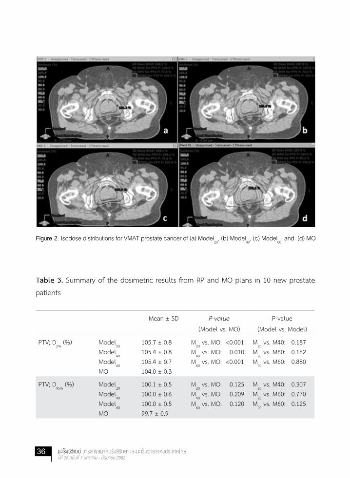
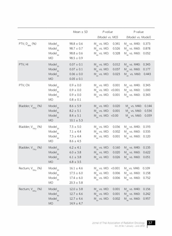
The comparison of RP plans to MO plans,
RP improved D95%
and D98%
of PTV, but
no significant difference was seen. A
significant higher of D2%
, CN, and HI from
all 3 RP models (D2%
: 105.7 %, 105.4 %,
and 105.4%, HI: 0.07, 0.07, and 0.06, CN:
0.9, 0.9, and 0.9 for Model20, Model
40, and
Model60, respectively) was shown when
compared with MO (D2%: 104%, HI: 0.05,
and CN: 0.8), (p<0.05). The higher D2%
and
HI showed more dose variation. However,
the higher CN represented more con-
formity of target. For bladder, almost all
36 มะเรงววฒน วารสารสมาคมรงสรกษาและมะเรงวทยาแหงประเทศไทยปท 25 ฉบบท 1 มกราคม - มถนายน 2562
Figure 2. Isodose distributions for VMAT prostate cancer of (a) Model20
, (b) Model40
, (c) Model60
, and (d) MO
Table 3. Summary of the dosimetric results from RP and MO plans in 10 new prostate
patients
Mean ± SD P-value
(Model vs. MO)
P-value
(Model vs. Model)
PTV; D2%
(%) Model20
Model40
Model60
MO
105.7 ± 0.8105.4 ± 0.8105.4 ± 0.7104.0 ± 0.3
M20 vs. MO: <0.001
M40 vs. MO: 0.010
M60 vs. MO: <0.001
M20 vs. M40: 0.187
M20 vs. M60: 0.162
M40 vs. M60: 0.880
PTV; D95%
(%) Model20
Model40
Model60
MO
100.1 ± 0.5100.0 ± 0.6100.0 ± 0.599.7 ± 0.9
M20 vs. MO: 0.125
M40 vs. MO: 0.209
M60 vs. MO: 0.120
M20 vs. M40: 0.307
M20 vs. M60: 0.770
M40 vs. M60: 0.125
37Jornal of Thai Association of Radiation Oncologyvol. 25 No.1 January - June 2019
Mean ± SD P-value
(Model vs. MO)
P-value
(Model vs. Model)
PTV; D98%
(%) Model20
Model40
Model60
MO
98.8 ± 0.698.7 ± 0.798.8 ± 0.698.5 ± 0.9
M20 vs. MO: 0.341
M40 vs. MO: 0.526
M60 vs. MO: 0.328
M20 vs. M40: 0.373
M20 vs. M60: 0.878
M40 vs. M60: 0.052
PTV; HI Model20
Model40
Model60
MO
0.07 ± 0.10.07 ± 0.10.06 ± 0.00.05 ± 0.1
M20 vs. MO: 0.012
M40 vs. MO: 0.037
M60 vs. MO: 0.023
M20 vs. M40: 0.343
M20 vs. M60: 0.177
M20
vs. M60: 0.443
PTV; CN Model20
Model40
Model60
MO
0.9 ± 0.00.9 ± 0.00.9 ± 0.00.8 ± 0.1
M20 vs. MO: 0.001
M40 vs. MO: <0.001
M60 vs. MO: 0.001
M20 vs. M40: 0.343
M20 vs. M60: 1.000
M40 vs. M60: 0.343
Bladder; V65Gy
(%) Model20
Model40
Model60
MO
8.6 ± 5.98.2 ± 5.18.4 ± 5.110.1 ± 5.3
M20 vs. MO: 0.020
M40 vs. MO: 0.001
M60 vs. MO: <0.00
M20 vs. M40: 0.144
M20 vs. M60: 0.534
M40 vs. M60: 0.059
Bladder; V70Gy
(%) Model20
Model40
Model60
MO
7.5 ± 5.07.1 ± 4.47.3 ± 4.48.6 ± 4.5
M20 vs. MO: 0.036
M40 vs. MO: 0.002
M60 vs. MO: 0.001
M20 vs. M40: 0.193
M20 vs. M60: 0.535
M40 vs. M60: 0.120
Bladder; V75Gy
(%) Model20
Model40
Model60
MO
6.2 ± 4.16.0 ± 3.86.1 ± 3.86.8 ± 3.5
M20 vs. MO: 0.160
M40 vs. MO: 0.020
M60 vs. MO: 0.026
M20 vs. M40: 0.135
M20 vs. M60: 0.622
M40 vs. M60: 0.051
Rectum; V50Gy
(%) Model20
Model40
Model60
MO
16.1 ± 4.617.5 ± 6.017.4 ± 6.020.3 ± 5.8
M20 vs. MO: <0.001
M40 vs. MO: 0.006
M60 vs. MO: 0.006
M20
vs. M40: 0.109M
20 vs. M60: 0.158
M40 vs. M60: 0.752
Rectum; V60Gy
(%) Model20
Model40
Model60
MO
12.0 ± 3.812.7 ± 4.612.7 ± 4.614.9 ± 4.7
M20 vs. MO: 0.001
M40 vs. MO: 0.001
M60 vs. MO: 0.002
M20 vs. M40: 0.156
M20 vs. M60: 0.242
M40 vs. M60: 0.957

38 มะเรงววฒน วารสารสมาคมรงสรกษาและมะเรงวทยาแหงประเทศไทยปท 25 ฉบบท 1 มกราคม - มถนายน 2562
Mean ± SD P-value
(Model vs. MO)
P-value
(Model vs. Model)
Rectum; V65Gy
(%) Model20
Model40
Model60
MO
10.2 ± 3.410.7 ± 4.010.7 ± 4.112.6 ± 4.2
M20 vs. MO: 0.001
M40 vs. MO: 0.001
M60 vs. MO: 0.001
M20 vs. M40: 0.216
M20 vs. M60: 0.305
M40 vs. M60: 0.957
Rectum; V70Gy
(%) Model20
Model40
Model60
MO
8.4 ± 3.18.7 ± 3.58.7 ± 3.610.1 ± 3.7
M20 vs. MO: 0.002
M40 vs. MO: 0.002
M60 vs. MO: 0.003
M20 vs. M40: 0.237
M20 vs. M60: 0.335
M40 vs. M60: 0.826
Rectum; V75Gy
(%) Model20
Model40
Model60
MO
6.4 ± 2.86.5 ± 3.06.6 ± 3.07.1 ± 3.1
M20 vs. MO: 0.030
M40 vs. MO: 0.028
M60 vs. MO: 0.048
M20 vs. M40: 0.478
M20 vs. M60: 0.375
M40 vs. M60: 0.405
aM20 is 20 plans model training, M
40 is 40 plans model training, and M
60 is 60 plans model training
bThe p-value < 0.05 is the statistical significance of this study
dose-volume parameters of RP were
significantly lower than MO (p<0.05), only
RPmodel20, V75Gy of the bladder was
similar to MO (V75Gy: 6.2% and 6.8% for
RPmodel20 and MO). All dose-volume pa-
rameters to the rectum in RP plans were
significantly lower than MO plans (p<0.05).
In addition, PTV parameters in terms of
D2%, D95%, D98%, HI, and CN illustrated
result among the models insignificantly.
For bladder and rectum, all 3 RP models
provided comparable the dose-volume
parameters.
DISCUSSION
The complicated VMAT treatment
planning needed the efficient optimi-
zation process in the inverse planning
system. However, to reach the planning
goals, the optimization was currently
a trial-and-error approach, and quality
of planning was mostly based on the
experience of planners. To reduce the
planner dependent variability in plan
quality, the RP knowledge-based (KB)
solutions for the inverse planning have
been developed. Best practice models
were able to apply for the clinic to increase
planning efficiency. The performance of
RP had been compared with manually
optimized clinical plans for different
treatment sites and techniques. Previous
39Jornal of Thai Association of Radiation Oncologyvol. 25 No.1 January - June 2019
studies[5-7, 12-14] of RP for VMAT optimization
in head and neck, hepatocellular, lung,
rectum, pelvic, and esophagus cancer
were reported and RP optimized plan is
able to improve plan quality and increase
planning efficiency. For prostate cancer,
better plan quality than the original
c l in ica l ly acceptable plans were
presented, from the study of Fogliata
et al.[5], Hussein et al.[14] , and Kubo et al.[15]
In addition, reduction of planning time,
and independently of planner’s skill
when they used RP was also exhibited.
In this study, after the 3 RP models
for VMAT prostate cancer validation, all
plans from 3 models were clearly shown
the acceptable and better plan quality.
For PTV dose coverage, similar results
of D95%
, D98%
, and higher CN, from the
models were exhibited when compared
with MO. However, in MO plans showed
lower D2%
and HI number due to the
better control of a hotspot area in PTV
from the planner. Kubo et al.[15] also
showed that the dose coverage, D2%
, D98%
,
CN, and HI to the PTV was slightly inferior
in KBP plans when compared with the
manually optimized planning. They
suggested to manually adjust in the RP
optimization process for improving PTV
coverage. For OARs, almost all of the
rectum and bladder doses in RP models
showed significantly better results than
MO, except the V75Gy to the bladder in
Model20 that was comparable to the MO
plans. For the head and neck studied from
Tol et al.[16], they reported that the high
dose-volume of OARs might be increased
due to the overlap region between PTV
and OARs. Therefore, Hussein et al.[14] and
Kubo et al.[15] suggested that the planners
should add the upper objective of OARs
in the optimization process to reduce the
high dose of RP plans.
The number of the plan for
model training and the removing of
outliers was also investigated in this study.
Three different RP models, based on 20,
40, and 60 prostate cancer plans, showed
similar results of PTV coverage, blad-
der, and rectum dose. When the organ
volume used in building the model was
analyzed, it can be seen that all 3 models
presented a very similar volume of the
PTV, bladder, and rectum. A study of
Tol et al.[17], also created head and neck
model using 30 and 60 plans and plan
quality obtained from both models were
the same and concluded that 30 plans
were sufficient for building model. In
addit ion, their study showed OAR
outlier did not influence to OAR dose.

40 มะเรงววฒน วารสารสมาคมรงสรกษาและมะเรงวทยาแหงประเทศไทยปท 25 ฉบบท 1 มกราคม - มถนายน 2562
Hussein et al. [14] as well found that
removing statistical outliers from the
training set was insignificant effect to
modal quality. In our study, removing
the outliers from the model was also
tested, and showed the same result of
no effect on our model quality. Besides,
the application of RP for VMAT optimiza-
tion was found to reduce the optimiza-
tion time from 30 min in MO plans to
6-7 min. However, the result of this study
was limited from a number of sample
size in the model validation and further
study with more number of data or
applied in other clinical cases should be
conducted.
CONCLUSION
RP knowledge-based treatment
planning system is able to increase
planning efficiency and plan quality.
Satisfactorily and acceptable prostate
VMAT plans from 3 RP trained models in
this investigation can be obtained in only
single optimization. The 20 historic plans
were also found to be an acceptable
minimum number of plans for the model
training.
ACKNOWLEDGEMENT
We would like to thank you staff at
Division of Radiation Oncology, Department
of Radiology, Faculty of Medicine Siriraj
Hospital for help and support throughout
this research.

41Jornal of Thai Association of Radiation Oncologyvol. 25 No.1 January - June 2019
REFERENCES
1. Khan FM. The Physics of Radiation
Therapy. 4 rded. Philadelphia:
Williams & Wilkins. 2010
2. Nelms BE, Robinson G, Markham
J, Velasco K, Boyd S, Narayan S, et al.
Variation in external beam treat-
ment plan qual ity: An inter-
institutional study of planners and
planning systems. P r a c t R ad i a t
Oncol 2012;2:296-305.
3. Wu B, Ricchetti F, Sanguineti G,
Kazhdan M, Simari P, Chuang M,
et al. Patient geometry-driven
information retrieval for IMRT
treatment plan quality control.
Med Phys 2009;36:5497-505.
4. Sutherland K. A Dosimetrist Per-
spective on the Role of Knowledge
Based Planning [online]. 2014 [cited
2017 Jul 12]. Available from: http://
pubs.medicaldosimetry.org/pub/
ddbcc4e6-f920-82a8-7e3e-e2c37
d5eaaec.
5. Fogliata A, Belosi F, Clivio A,
Navarria P, Nicolini G, Scorsetti M,
et al. On the pre-clinical validation
of a commercial model-based
optimisation engine: Application to
volumetric modulated arc therapy
for patients with lung or prostate
cancer. Radiat Oncol 2014;113:
385-91.
6. Fogliata A, Nicolini G, Clivio A,
Vanett i E, Laksar S, Tozzi A,
et al. A broad scope knowledge
based model for optimization of
VMAT in esophageal cancer: valida-
tion and assessment of plan quality
among different treatment centers.
Radiat Oncol 2015;10:220.
7. Fogliata A, Wang P-M, Belosi F, Clivio
A, Nicolini G, Vanetti E, et al.
Assessment of a model based
optimization engine for volumetric
modulated arc therapy for patients
with advanced hepatocellular
cancer. Radiat Oncol 2014;9:236.
8. Marks LB, Yorke ED, Jackson A, Haken
RT, Constine LS, Eisbrruch A, et al.
Use of normal tissue complication
probability models in the clinic. Int
J Radiat Oncol Biol Phys 2010;76:
10-9.
9. Luca C. RAPIDPLAN: The clinical
value and DVH estimation algorithm
[online]. 2014 [cited 2015 Jul 16].
Available from: Varian Medical
System Source.
42 มะเรงววฒน วารสารสมาคมรงสรกษาและมะเรงวทยาแหงประเทศไทยปท 25 ฉบบท 1 มกราคม - มถนายน 2562
10. Kataria T, Sharma K, Subramani V,
Karrthick KP, Bisht SS. Homo-
geneity Index: An objective tool
for assessment of conformal
radiation treatments. J Med Phys
2012;37:207-13.
11. Riet A, Mak A, Moerland M, Elders
L, van W. A conformation number
to quantify the degree of confor-
mal ity in brachytherapy and
external beam irradiation: Applica-
tion to the prostate. Int J Radiat
Oncol Biol Phys 1997;37:731-6.
12. Fogliata A, Reggiori G, Stravato A,
Lobefalo F, Franzese C, Franceschini
D, et al. RapidPlan head and neck
model: the objectives and possible
cl inical benefit. Radiat Oncol
2017;12:73.
13. Wu H, Jiang F, Yue H, Zhang H, Wang
K, Zhang Y. Applying a RapidPlan
model trained on a technique and
orientation to another: a feasibility
and dosimetric evaluation. Radiat
Oncol 2016;11:108.
14. Hussein M, South CP, Barry MA,
Adams EJ, Jordan TJ, Stewart AJ,
et al. Clinical validation and bench-
mark ing of knowledge-based
IMRT and VMAT treatment planning
in pelvic anatomy. Radiat Oncol
2016;120:473-9.
15. Kubo K, Monzen H, Ishii K, Tamura
M, Kawamorita R, Sumida I, et al.
D o s i m e t r i c c o m p a r i s o n o f
RapidPlan and manually optimized
plans in volumetric modulated arc
therapy for prostate cancer. Phys
Med 2017;1120-1790:30216–8
16. Tol JP, Dahele M, Delaney AR,
Slotman BJ, Verbakel WF. Can
knowledge-based DVH predictions
be used for automated, individua-
lized quality assurance of radio-
therapy treatment plans. Radiat
Oncol 2015;10:234.
17. Tol JP, Delaney AR, Dahele M,
Slotman BJ, Verbakel WFAR. Evalua-
tion of a Knowledge Based Planning
Solution for Head and Neck Cancer.
Int J Radiat Oncol Biol Phys 2015;
91:612-20.
43Jornal of Thai Association of Radiation Oncologyvol. 25 No.1 January - June 2019
คณสมบตเชงรงสคณตและอตราผานคาแกมมาของอปกรณวดรงสรนอารคเชคสำาหรบการทวนสอบแผนรงสปรบความเขมเชงปรมาตร
Dosimetry Characteristics and Gamma Passing Rate of ArcCHECK for Volumetric Modulated Arc Therapy Treatment Planning Verification
นนทวฒ สดลอย1, เอกสทธ ธราวจตรกล2, สมศกด วรรณวไลรตน2
Nuntawuth Sudloy1, Ekkasit Tharavichitkul2, Somsak Wanwilairat2
1หลกสตรฟสกสการแพทย ภาควชารงสวทยา คณะแพทยศาสตร มหาวทยาลยเชยงใหม2หนวยรงสรกษาและมะเรงวทยา ภาควชารงสวทยา คณะแพทยศาสตร มหาวทยาลยเชยงใหม1Medical Physics Program, Department of Radiology, Chiang Mai University2Division of Radiation Oncology, Department of Radiology, Faculty of Medicine Chiang Mai University
ABSTRACT
Background: Volumetric Modulated Arc Therapy (VMAT) is a complicated radiation
technique in both the treatment planning and the machine delivery. A verification
dosimeter with appropriate dosimetry characteristic is essential for treatment
planning verification and quality assurance.
Objectives: To study the dosimetry characteristics of ArcCHECK diode array detector
and evaluate gamma passing rate of Volumetric Modulated Arc Therapy treatment
planning in Nasopharyngeal cancer and Prostate cancer.
Materials and Methods: Dosimetry characteristics of ArcCHECK include short-term
reproducibility, dose linearity, field size dependence, dose rate dependence,
directional dependence and out of field dependence were studied. Volumetric
Modulated Arc Therapy technique using Synergy 6 MV photon beam were planned
for each 10 cases of Nasopharyngeal and Prostate cancer. Then treatment planning
verification were performed using the gamma passing rate of 3%/3mm and the 10%
threshold level.

44 มะเรงววฒน วารสารสมาคมรงสรกษาและมะเรงวทยาแหงประเทศไทยปท 25 ฉบบท 1 มกราคม - มถนายน 2562
Results: The result showed ArcCHECK has consistency response for short-term
reproducibility with 0.05% standard deviation, good linearity response to radiation
dose (R2=1.0) and increase response with increasing field size and dose rate. For
directional dependence study, ArcCHECK was showed good consistency response
with 1% dose difference for gantry angle between -55o to 55o. The scattered
radiation measured out of radiation field has variation by field size and maximum
dose was 6.9% with 15×15 cm2. This scattered dose decrease with distance from
the edge of the radiation field. For Volumetric Modulated Arc Therapy treatment
planning verification, it was found that the average gamma passing rate was
96.74%±2.04% and the action level was 92.74%.
Conclusion: ArcCHECK diode array detector has good dosimetry characteristics and
suitable for Volumetric Modulated Arc Therapy treatment planning verification.
The action level of gamma passing for the department is 92.74%.
Keyword: ArcCHECK, Diode detector, Gamma passing rate, Treatment planning
verification, VMAT
บทคดยอ
หลกการและเหตผล: การฉายรงสปรบความเขมเชงปรมาตรเปนเทคนคทมความซบซอนทงแผน
รงสรกษาและการทำางานของเครองฉายรงส อปกรณวดรงสทมคณสมบตเชงรงสคณตทเหมาะสม
จงจำาเปนและมความสำาคญสำาหรบใชทวนสอบและประกนคณภาพแผนรงสปรบความเขมเชงปรมาตร
วตถประสงค: เพอศกษาคณลกษณะเชงรงสคณตของอปกรณวดรงสรน ArcCHECK และใชหา
คาอตราผานคาแกมมาของแผนรงสปรบความเขมเชงปรมาตรในผปวยโรคมะเรงหลงโพรงจมก
และมะเรงตอมลกหมาก
วสดและวธการ: ศกษาคณลกษณะเชงรงสคณตของอปกรณวดรงสรน ArcCHECK ไดแก การ
ตอบสนองตอการวดปรมาณรงสซำาในระยะเวลาสน การตอบสนองตอขนาดปรมาณรงส การ
ตอบสนองตอขนาดของพนทรงส การตอบสนองตออตราปรมาณรงสทไดรบ การตอบสนองตอ
ทศทางของลำารงส และการตอบสนองตอปรมาณรงสกระเจงนอกพนทรงส จากนนนำาอปกรณวดรงส

45Jornal of Thai Association of Radiation Oncologyvol. 25 No.1 January - June 2019
ArcCHECK ไปใชทวนสอบแผนรงสปรบความเขมเชงปรมาตรและหาคาอตราผานคาแกมมาใน
ผปวยโรคมะเรงหลงโพรงจมกและมะเรงตอมลกหมากโรคละ 10 แผน โดยกำาหนดเกณฑคา
ดชนแกมมา 3%/3mm เกณฑปรมาณรงสขนตำา 10%
ผลการศกษา: อปกรณวดรงส ArcCHECK มการตอบสนองตอการวดรงสซำาในระยะเวลาสนคงท
โดยมสวนเบยงเบนมาตรฐานเทากบ 0.05% มการตอบสนองตอปรมาณรงสเปนแบบเชงเสนทด
(R2=1.0) และมการตอบสนองมากขนตอขนาดพนทรงสและอตราปรมาณรงสทเพมขน การตอบ
สนองตอทศทางของลำารงสพบวามการตอบสนองคงทในชวงมม -55o ถง 55o โดยมความแตกตาง
ของปรมาณรงสไมเกน 1% การตอบสนองตอปรมาณรงสกระเจงนอกพนทรงสพบวา ArcCHECK
วดไดคาปรมาณสงขนเมอพนทรงสมขนาดใหญขน และคาลดลงตามระยะหางจากขอบของพนทรงส
โดยมคามากสดเทากบ 6.9% ทขนาดพนทรงส 15×15 ตร.ซม. สำาหรบการทวนสอบแผนรงสปรบ
ความเขมเชงปรมาตรไดอตราผานคาแกมมาเฉลยเทากบ 96.74%±2.04% และคาระดบเกณฑ
ดำาเนนการเทากบ 92.74%
ขอสรป: อปกรณวดรงสรน ArcCHECK มคณลกษณะเชงรงสคณตทดและมความเหมาะสมใน
การนำามาใชทวนสอบแผนรงสปรบความเขมเชงปรมาตร คาระดบเกณฑดำาเนนการของหนวยงาน
เทากบ 92.74%
คำาสำาคญ: อปกรณวดรงสรน ArcCHECK, หววดรงสไดโอด, อตราผานคาแกมมา, การทวนสอบ
แผนรงสรกษา, รงสปรบความเขมเชงปรมาตร
บทนำา
เทคนครงสปรบความเขมเชงปรมาตร
(Volumetric Modulated Arc Therapy :VMAT)
เปนเทคนคฉายรงสทแขนฉายรงสหมนเปน
แนวโคงรอบตวผปวยพรอมกบมการเปลยนแปลง
รปรางของลำารงสโดยการเคลอนทของซกำาบงรงส
(MLCs) อยางตอเนอง และมการปรบเปลยนอตรา
ปรมาณรงสและความเรวของการเคลอนทของแขน
ฉายรงส เมอเปรยบเทยบกบเทคนคการฉาย
รงสปรบความเขมแบบดงเดม (Conventional
static field IMRT)[1] เทคนครงสปรบความเขม
เชงปรมาตรสามารถใหการกระจายปรมาณรงส
ทมความเขารปและครอบคลมรอยโรคสง สามารถ
หลกเลยงไมใหอวยวะสำาคญขางเคยงไดรบปรมาณ
รงสสง นอกจากนยงใชเวลาในการฉายรงส และ
หนวยนบวดรงส (MU) ทลดลง
เทคนครงสปรบความเขมเชงปรมาตร
เปนเทคนคทมความซบซอนของแผนรงสรกษา

46 มะเรงววฒน วารสารสมาคมรงสรกษาและมะเรงวทยาแหงประเทศไทยปท 25 ฉบบท 1 มกราคม - มถนายน 2562
และการทำางานของเครองฉายรงส ทงซกำาบงรงส
การหมนของแขนหมน และการปรบเปลยนอตรา
ปรมาณรงส ดงนนการทวนสอบปรมาณรงส
และการประกนคณภาพของแผนรงสรกษาจงม
ความสำาคญ[2] วธการทวนสอบแผนรงสรกษาท
เปนทนยมคอการวเคราะหคาแกมมา (Gamma
analysis)[3] อปกรณวดรงสทนำามาใชในการทวน
สอบแผนรงสรกษาทดนอกจากจะมรปรางของ
อปกรณทถกออกแบบมาใหเหมาะสมกบเทคนค
การฉายรงสแลวตองมคณสมบตเชงรงสคณตทด
เชน วดปรมาณรงสซำาไดถกตองแมนยำา มการ
ตอบสนองตอปรมาณรงสแบบเชงเสน ตอบสนอง
ตอขนาดพนทลำารงสและอตราปรมาณรงส และ
ตอบสนองตอปรมาณรงสไดทกทศทาง เปนตน
อปกรณวดรงสรน ArcCHECK มลกษณะ
เปนแฟนทอมรปทรงกระบอกทำาจากวสด PMMA
ภายในมหววดรงสชนดไดโอดจำานวน 1,386 ตว
วางเรยงกนอยในลกษณะเกลยวหมนเปนรปทรง
กระบอก ดวยลกษณะนจงทำาใหอปกรณวดรงสรน
ArcCHECK สามารถวดรงสไดทกทศทางสอดคลอง
กบเทคนครงสปรบความเขมเชงปรมาตร แตกอน
นำาอปกรณวดรงสมาใชในการทวนสอบแผนรงส
รกษา จำาเปนตองมการศกษาคณลกษณะเชงรงส
คณตของอปกรณ เพอใหทราบถงประสทธภาพ
ลกษณะเฉพาะ ขอจำากด และผลของตวแปรตางๆ
ตอปรมาณรงสทวดได
งานวจยนตองการศกษาคณลกษณะเชง
รงสคณตของอปกรณวดรงสรน ArcCHECK และ
ใชทวนสอบแผนรงสปรบความเขมเชงปรมาตร โดย
หาอตราผานคาแกมมา (Gamma passing rate)
ในแผนรงสรกษาผปวยโรคมะเรงหลงโพรงจมก
และมะเรงตอมลกหมาก นำาคาทไดมาใชเปนแนวปฏบตในการประเมนแผนรงสรกษาของหนวยงานตอไป
วสดและวธการทดลอง ในการศกษานใชเครองฉายรงสโฟตอนพลงงาน 6 MV รน Synergy ทมซกำาบงรงส ขนาด 0.5 ซม. และเครองวางแผนรงสรกษา (TPS) รน Monaco Version 5.11.01 (Elekta, Crawley, UK) อปกรณวดรงสรน ArcCHECK โมเดล 1220 และโปรแกรม SNC PatientTM version 6.6.0 (Sun Nuclear, Melbourne, USA) และหววดรงสชนดประจแตกตวรน FC65P (IBA, Schwarzen-bruck, Germany) ขอมลแผนรงสปรบความเขมเชงปรมาตรของผปวยโรคมะเรงหลงโพรงจมกและมะเรงตอมลกหมาก ทไดรบการฉายรงสทหนวยรงสรกษาและมะเรงวทยา คณะแพทยศาสตร มหาวทยาลยเชยงใหม จำานวนโรคละ 10 แผนรงสรกษา รวม 20 แผนรงสรกษา การศกษาไดทำาในหวขอตอไปน
การตอบสนองตอการวดปรมาณรงสซำาในระยะเวลาสน จดตำาแหนงของอปกรณวดรงส Arc-CHECK โดยใหจดกงกลางของอปกรณอยทตำาแหนง Isocenter ของเครองฉายรงส ทระยะ SAD = 100 ซม. (SSD = 86.7 ซม.) ตงหวเครองฉายรงส ท 0o เปดพนทรงส 10×10 ตร.ซม. ฉายรงส 100 MU อานคาปรมาณรงสของไดโอด ทตำาแหนงกงกลางของ ArcCHECK ภายในพนทรงสจำานวน 6 ตว ดงแสดงในภาพท 1 วดซำา 10 ครง โดยแตละครงเวลาหางกน 3 นาท
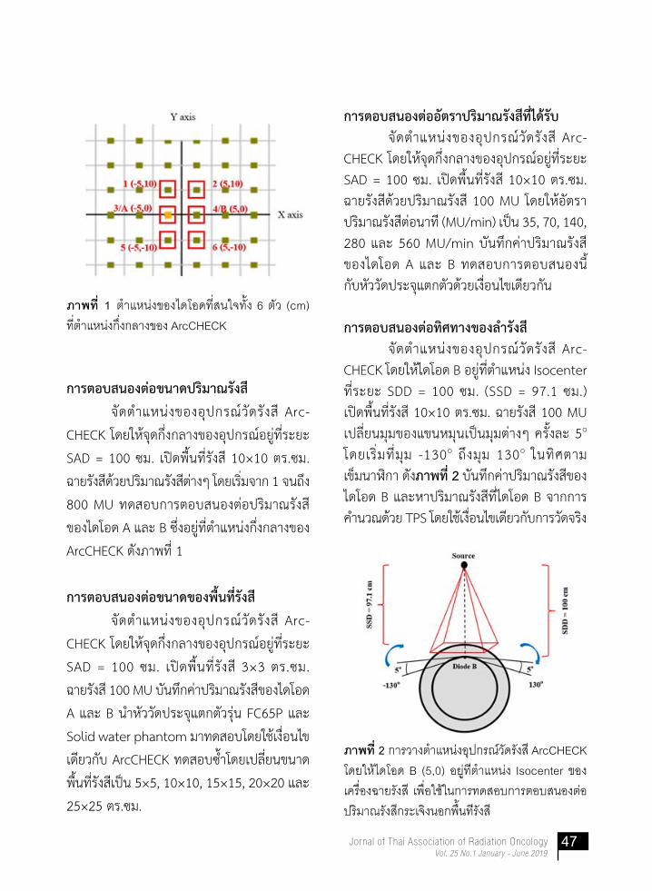
47Jornal of Thai Association of Radiation Oncologyvol. 25 No.1 January - June 2019
ภาพท 1 ตำ�แหนงของไดโอดทสนใจทง 6 ตว (cm)
ทตำ�แหนงกงกล�งของ ArcCHECK
การตอบสนองตอขนาดปรมาณรงส
จดตำาแหนงของอปกรณวดรงส Arc-
CHECK โดยใหจดกงกลางของอปกรณอยทระยะ
SAD = 100 ซม. เปดพนทรงส 10×10 ตร.ซม.
ฉายรงสดวยปรมาณรงสตางๆ โดยเรมจาก 1 จนถง
800 MU ทดสอบการตอบสนองตอปรมาณรงส
ของไดโอด A และ B ซงอยทตำาแหนงกงกลางของ
ArcCHECK ดงภาพท 1
การตอบสนองตอขนาดของพนทรงส
จดตำาแหนงของอปกรณวดรงส Arc-
CHECK โดยใหจดกงกลางของอปกรณอยทระยะ
SAD = 100 ซม. เปดพนทรงส 3×3 ตร.ซม.
ฉายรงส 100 MU บนทกคาปรมาณรงสของไดโอด
A และ B นำาหววดประจแตกตวรน FC65P และ
Solid water phantom มาทดสอบโดยใชเงอนไข
เดยวกบ ArcCHECK ทดสอบซำาโดยเปลยนขนาด
พนทรงสเปน 5×5, 10×10, 15×15, 20×20 และ
25×25 ตร.ซม.
การตอบสนองตออตราปรมาณรงสทไดรบ จดตำาแหนงของอปกรณวดรงส Arc-CHECK โดยใหจดกงกลางของอปกรณอยทระยะ SAD = 100 ซม. เปดพนทรงส 10×10 ตร.ซม. ฉายรงสดวยปรมาณรงส 100 MU โดยใหอตราปรมาณรงสตอนาท (MU/min) เปน 35, 70, 140, 280 และ 560 MU/min บนทกคาปรมาณรงส ของไดโอด A และ B ทดสอบการตอบสนองน กบหววดประจแตกตวดวยเงอนไขเดยวกน
การตอบสนองตอทศทางของลำารงส จดตำาแหนงของอปกรณวดรงส Arc-CHECK โดยใหไดโอด B อยทตำาแหนง Isocenter ทระยะ SDD = 100 ซม. (SSD = 97.1 ซม.) เปดพนทรงส 10×10 ตร.ซม. ฉายรงส 100 MU เปลยนมมของแขนหมนเปนมมตางๆ ครงละ 5o โดยเรมทมม -130o ถงมม 130o ในทศตาม เขมนาฬกา ดงภาพท 2 บนทกคาปรมาณรงสของไดโอด B และหาปรมาณรงสทไดโอด B จากการคำานวณดวย TPS โดยใชเงอนไขเดยวกบการวดจรง
ภาพท 2 ก�รว�งตำ�แหนงอปกรณวดรงส ArcCHECK
โดยใหไดโอด B (5,0) อยทตำ�แหนง Isocenter ของ
เครองฉ�ยรงส เพอใชในก�รทดสอบก�รตอบสนองตอ
ปรม�ณรงสกระเจงนอกพนทรงส

48 มะเรงววฒน วารสารสมาคมรงสรกษาและมะเรงวทยาแหงประเทศไทยปท 25 ฉบบท 1 มกราคม - มถนายน 2562
การตอบสนองตอปรมาณรงสกระเจงนอกพนท
รงส
จดตำาแหนงของอปกรณวดรงส Arc-
CHECK โดยใหจดกงกลางของอปกรณอยทระยะ
SAD = 100 ซม. เปดพนทรงส 3×3 ตร.ซม.
ฉายรงส 100 MU วดปรมาณรงสของไดโอดทอย
หางจากขอบพนทรงสในแนวหว-เทาเปนระยะ
1.66-8.66 ซม. ทดสอบซำาโดยเปลยนขนาดพนท
รงสเปน 5×5, 10×10 และ 15×15 ตร.ซม. และ
วดปรมาณรงสของไดโอดทอยหางจากขอบพนท
รงสเปนระยะ 1.76-7.76, 1.52-5.52 และ
1.28-3.28 ซม. ตามลำาดบ ทดสอบการตอบสนอง
นกบหววดประจแตกตวดวยเงอนไขเดยวกน
การหาคาอตราผานแกมมาของแผนรงสรกษา
เทคนครงสปรบความเขมเชงปรมาตร
การหาคาอตราผานแกมมาของแผนรงส
ปรบความเขมเชงปรมาตร ทไดจากเครองวางแผน
รงสรกษา Monaco ในผปวยโรคมะเรงหลง
โพรงจมกและมะเรงตอมลกหมาก โดยเรมจาก
การนำาแผนรงสปรบความเขมเชงปรมาตรซงได
รบความเหนชอบจากรงสแพทยและฉายรงส
ผปวยครบแลว มาคำานวณปรมาณรงสใหมโดยใช
ภาพรงสตดขวางของอปกรณวดรงส ArcCHECK
สงออกไฟลขอมลแผนรงสรกษา (DICOM RT
Plan) และขอมลปรมาณรงส (DICOM RT Dose)
ไปยงโปรแกรม SNC PatientTM นำาอปกรณ
วดรงส ArcCHECK ไปฉายรงสดวยรงสโฟตอน
พลงงาน 6 MV จากเครองฉายรงส Synergy
ทำาการวเคราะหเพอหาคาอตราผานแกมมาดวย
โปรแกรม SNC PatientTM โดยกำาหนดเกณฑ
คาดชนแกมมา 3%/3mm ตงเกณฑปรมาณรงส
ขนตำา 10% นำาอตราผานคาแกมมาเฉลยและ
คาสวนเบยงเบนมาตรฐานทไดมาคำานวณหาคา
ขดจำากดความเชอมน (Confidence Limit: CL)
และคาเกณฑระดบการดำาเนนการ (Action
Level) เพอใชเปนเกณฑในการกำาหนดอตราผาน
คาแกมมาของการทวนสอบแผนรงสรกษาเทคนค
รงสปรบความเขมเชงปรมาตรของหนวยงาน โดย
คำานวณจากสมการ[4]
Confidence Limit = (100 – mean) + 1.96SD
โดยท mean คออตราผานคาแกมมา
เฉลย และ SD คอคาสวนเบยงเบนมาตรฐานของ
อตราผานคาแกมมา
Action Level = (100 – CL)%
ผลการทดลอง
ผลการตอบสนองตอการวดปรมาณรงสซำาใน
ระยะเวลาสน
การทดสอบการตอบสนองตอการวด
ปรมาณรงสซำาในระยะเวลาสนของไดโอดทง 6 ตว
นำาคาปรมาณรงสเฉลยทวดไดแตละครงมาเทยบ
กบคาปรมาณรงสทวดไดครงแรก พบคา Relative
dose อยระหวาง 99.88% ถง 100.02% และมคา
สวนเบยงเบนมาตรฐานเปน 0.0497 คา Relative
dose ของการวดปรมาณรงสซำาทง 10 ครง แสดง
ในภาพท 3
ผลการตอบสนองตอขนาดปรมาณรงส
การทดสอบการตอบสนองตอขนาด
ปรมาณรงสของไดโอด A และ B โดยใหปรมาณ
รงสตงแต 1 ถง 800 MU นำาขอมลปรมาณรงสท
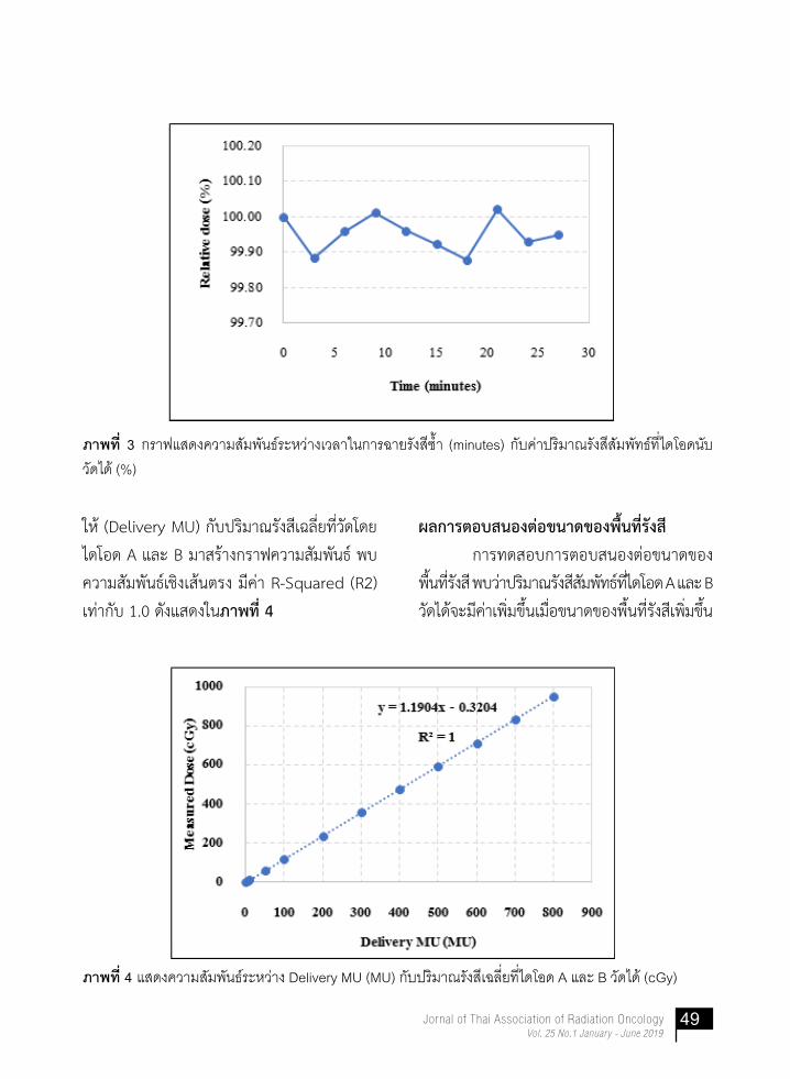
49Jornal of Thai Association of Radiation Oncologyvol. 25 No.1 January - June 2019
ภาพท 3 กร�ฟแสดงคว�มสมพนธระหว�งเวล�ในก�รฉ�ยรงสซำ� (minutes) กบค�ปรม�ณรงสสมพทธทไดโอดนบ
วดได (%)
ภาพท 4 แสดงคว�มสมพนธระหว�ง Delivery MU (MU) กบปรม�ณรงสเฉลยทไดโอด A และ B วดได (cGy)
ให (Delivery MU) กบปรมาณรงสเฉลยทวดโดย
ไดโอด A และ B มาสรางกราฟความสมพนธ พบ
ความสมพนธเชงเสนตรง มคา R-Squared (R2)
เทากบ 1.0 ดงแสดงในภาพท 4
ผลการตอบสนองตอขนาดของพนทรงส
การทดสอบการตอบสนองตอขนาดของ
พนทรงส พบวาปรมาณรงสสมพทธทไดโอด A และ B
วดไดจะมคาเพมขนเมอขนาดของพนทรงสเพมขน
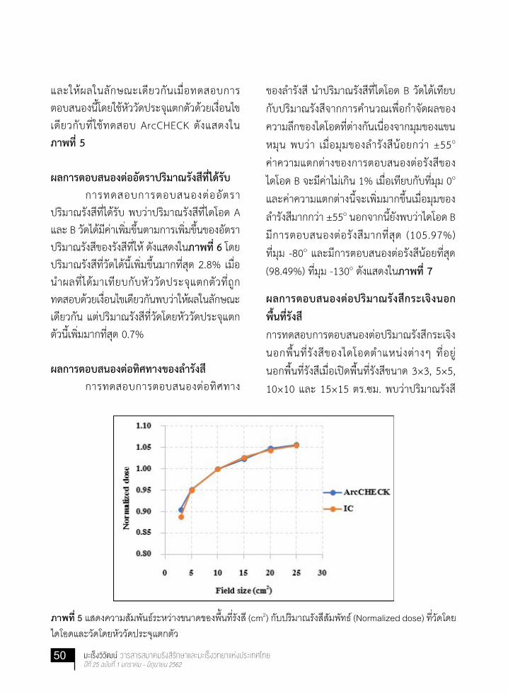
50 มะเรงววฒน วารสารสมาคมรงสรกษาและมะเรงวทยาแหงประเทศไทยปท 25 ฉบบท 1 มกราคม - มถนายน 2562
และใหผลในลกษณะเดยวกนเมอทดสอบการตอบสนองนโดยใชหววดประจแตกตวดวยเงอนไขเดยวกบทใชทดสอบ ArcCHECK ดงแสดงใน ภาพท 5
ผลการตอบสนองตออตราปรมาณรงสทไดรบ การทดสอบการตอบสนองตออตราปรมาณรงสทไดรบ พบวาปรมาณรงสทไดโอด A และ B วดไดมคาเพมขนตามการเพมขนของอตราปรมาณรงสของรงสทให ดงแสดงในภาพท 6 โดยปรมาณรงสทวดไดนเพมขนมากทสด 2.8% เมอ นำาผลทไดมาเทยบกบหววดประจแตกตวทถกทดสอบดวยเงอนไขเดยวกนพบวาใหผลในลกษณะเดยวกน แตปรมาณรงสทวดโดยหววดประจแตกตวนเพมมากทสด 0.7%
ผลการตอบสนองตอทศทางของลำารงส การทดสอบการตอบสนองตอทศทาง
ของลำารงส นำาปรมาณรงสทไดโอด B วดไดเทยบ
กบปรมาณรงสจากการคำานวณเพอกำาจดผลของ
ความลกของไดโอดทตางกนเนองจากมมของแขน
หมน พบวา เมอมมของลำารงสนอยกวา ±55o
คาความแตกตางของการตอบสนองตอรงสของ
ไดโอด B จะมคาไมเกน 1% เมอเทยบกบทมม 0o
และคาความแตกตางนจะเพมมากขนเมอมมของ
ลำารงสมากกวา ±55o นอกจากนยงพบวาไดโอด B
มการตอบสนองตอรงสมากทสด (105.97%)
ทมม -80o และมการตอบสนองตอรงสนอยทสด
(98.49%) ทมม -130o ดงแสดงในภาพท 7
ผลการตอบสนองตอปรมาณรงสกระเจงนอก
พนทรงส
การทดสอบการตอบสนองตอปรมาณรงสกระเจง
นอกพนท รงสของไดโอดตำาแหนงตางๆ ทอย
นอกพนทรงสเมอเปดพนทรงสขนาด 3×3, 5×5,
10×10 และ 15×15 ตร.ซม. พบวาปรมาณรงส
ภาพท 5 แสดงคว�มสมพนธระหว�งขน�ดของพนทรงส (cm2) กบปรม�ณรงสสมพทธ (Normalized dose) ทวดโดย
ไดโอดและวดโดยหววดประจแตกตว
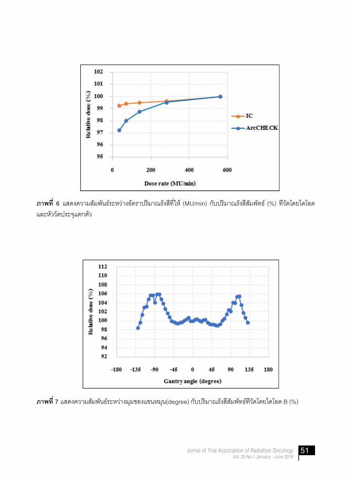
51Jornal of Thai Association of Radiation Oncologyvol. 25 No.1 January - June 2019
ภาพท 6 แสดงคว�มสมพนธระหว�งอตร�ปรม�ณรงสทให (MU/min) กบปรม�ณรงสสมพทธ (%) ทวดโดยไดโอด
และหววดประจแตกตว
ภาพท 7 แสดงคว�มสมพนธระหว�งมมของแขนหมน(degree) กบปรม�ณรงสสมพทธทวดโดยไดโอด B (%)

52 มะเรงววฒน วารสารสมาคมรงสรกษาและมะเรงวทยาแหงประเทศไทยปท 25 ฉบบท 1 มกราคม - มถนายน 2562
วจารณผลการทดลอง
หววดไดโอดของอปกรณวดรงส Arc-
CHECK มการตอบสนองทคงทตอการวดปรมาณ
รงสซำาในระยะเวลาสน มคาสวนเบยงเบนมาตรฐาน
เทากบ 0.05% สอดคลองกบงานวจยของ Li G.
และคณะ[2], Thiyagarajan R. และคณะ[5], สรธญ
และคณะ[6] และเฉลมชาต และคณะ[7] ทไดคา
สวนเบยงเบนมาตรฐานของไดโอดในการตอบ
สนองตอการวดปรมาณรงสซำาเทากบ 0.05%,
0.03%, 0.04% และ 0.02% ตามลำาดบ
หววดไดโอดของอปกรณวดรงส Arc-
CHECK มการตอบสนองตอปรมาณรงสเปน
แบบเชงเสน มคา R2 เทากบ 1.0 และไมพบ
จดอมตวตอปรมาณรงส สอดคลองกบงานวจยของ
Li G. และคณะ[2] ทศกษาเครองฉายรงส Synergy,
Elekta ไดคา R2 มากกวา 0.99 งานวจยของ
นอกพนทรงสจะลดลงเมอระยะหางจากขอบพนท
รงสมากขนและใหผลทเหมอนกนกบหววดประจ
แตกตว นอกจากนยงพบวาปรมาณรงสนอกพนท
รงสมแนวโนมสงขนเมอพนทรงสมขนาดใหญขน
ดงแสดงในภาพท 8
ผลการทวนสอบแผนรงสปรบความเขมเชง
ปรมาตร
จากผลการศกษาพบวา อตราผานคา
แกมมาของแผนรงสปรบความเขมเชงปรมาตร
ทกแผนมคามากกวา 90% โดยมคาตำาทสด
เทากบ 92.3% สงทสดเทากบ 99.6% และมคา
เฉลยเทากบ 96.74% นำาอตราผานคาแกมมา
เฉลยและคาสวนเบยงเบนมาตรฐานมาคำานวณหา
คาขดจำากดความเชอมนไดเทากบ 7.26 และ
คาระดบเกณฑดำาเนนการไดเทากบ 92.74%
ภาพท 8 แสดงคว�มสมพนธระหว�งปรม�ณรงสกระเจงนอกพนทรงส (cGy) กบระยะห�งจ�กขอบของพนทรงสขน�ด
ต�งๆ ของหววดไดโอดและหววดประจแตกตว (cm)

53Jornal of Thai Association of Radiation Oncologyvol. 25 No.1 January - June 2019
Thiyagarajan R. และคณะ [5] ทศกษาเครอง
ฉายรงส Clinac iX, Varian และสรธญ และคณะ[6]
ทศกษาเครองฉายรงส Primus, Siemens รายงาน
วาคา R2 เทากบ 1.0 และงานวจยของเฉลมชาต
และคณะ[7] ทศกษาเครอง Tomotherapy Hi-Art
รายงานคา R2 เทากบ 0.999
การตอบสนองตอขนาดของพนท รงส
พบวาปรมาณรงสทหววดไดโอดวดไดมคาสงขน
เมอพนทรงสมขนาดเพมขนเนองจากผลของรงส
กระเจงทมากขน โดยมความแตกตางกบคาท
วดโดยหววดชนดประจแตกตวอยในชวงไมเกน
2.0% ใกลเคยงกบงานวจยของ Li G. และคณะ[2]
และ Thiyagarajan R. และคณะ [5] ทพบวา
ความแตกตางอยในชวงไมเกน 1.7% และ 1.6%
ตามลำาดบ ทงนความแตกตางของหววดไดโอด
และหววดชนดประจแตกตวสวนหนงมาจาก
รปทรงเลขาคณตของแฟนทอมทตางกนทำาใหเกด
รงสกระเจงทตางกน ความแตกตางของปรมาณ
รงสเมอพนทเปลยนไปสอดคลองกบงานวจยของ
สรธญ และคณะ[6] ทพบวาปรมาณรงสทหววด
ไดโอดวดไดเพมขนไมเกน 7% ในชวงพนทรงส
5×5 ถง 25×25 ตร.ซม. ในขณะทงานวจยน
พบวาเพมขนไมเกน 6%
การตอบสนองตออตราปรมาณรงส
พบวาหววดไดโอดมการตอบสนองมากขนเมอ
อตราปรมาณรงสทใหสงขน โดยการตอบสนอง
เพมขนอยในชวง 2.8% และใหผลในลกษณะ
เดยวกนกบหววดร งสชนดประจแตกตว ท
พบวาการตอบสนองเพมขนไมเกน 0.7% ใกล
เคยงกบงานวจยของ Li G. และคณะ[2] ทรายงาน
วาการตอบสนองของ ArcCHECK เพมขนอยใน
ชวงไมเกน 1.8% เมอฉายรงสดวยอตราปรมาณ
รงสในชวง 29 ถง 473 MU/min ในขณะทหววด
รงสชนดประจแตกตวมการตอบสนองเพมขน
ไมเกน 0.6% และงานวจยของ Thiyagarajan
R. และคณะ[5] ทรายงานวาการตอบสนองของ
หววดไดโอดทอตราปรมาณรงสตำาจะมคานอยกวา
เมอเทยบกบอตราปรมาณรงส 300 MU/min และ
จะใหผลในทางตรงขามเมอใหอตราปรมาณรงส
สงกวา 300 MU/min
การตอบสนองตอทศทางของลำารงส
พบวาหววดไดโอดมการตอบสนองคงทระหวาง
มม -55o ถงมม 55o โดยมคาความแตกตางไมเกน
1% และพบวาการตอบสนองมความแปรปรวน
อยางมากเมอทศทางลำารงสทำามมมากกวา 55o
โดยการตอบสนองมากสดไมเกน 6.0% ในชวงมม
-80o ถง -100o และการตอบสนองนอยสดเปน
-1.51% เมอลำารงสทำามม -130o กบหววดไดโอด
และการตอบสนองมแนวโนมลดลงเมอมมของ
ลำารงสมากกวา ±130o หรอเมอฉายรงสจาก
ดานหลงของหววดไดโอด สอดคลองกบงานวจย
ของ Li G. และคณะ[2] ทศกษาการตอบสนอง
ตอทศทางรงสของ ArcCHECK ในชวง -180o
ถง 180o พบวาหววดไดโอดมการตอบสนองคงท
ในชวง ±60o โดยมความแปรปรวนนอยกวา 1%
และมการตอบสนองมากทสด 9.1% ทมม 105o
และนอยทสด -4.9% ทมม 180o ทงนเปนผล
มาจากการออกแบบหววดไดโอดทไมเปน iso-
tropic (non-isotropic) ของบรเวณปลอดพาหะ
ของไดโอด, การเกด backscatter รวมถงความ
ไมสมำาเสมอของพนททเปนอากาศของไดโอด
แตละตวใน ArcCHECK ทมสวนทำาใหการ

54 มะเรงววฒน วารสารสมาคมรงสรกษาและมะเรงวทยาแหงประเทศไทยปท 25 ฉบบท 1 มกราคม - มถนายน 2562
ตอบสนองของหววดไดโอดเปนแบบ non-iso-
tropic หรอขนกบทศทางของลำารงส[8] แตการ
ตอบสนองตอทศทางลำารงสนมผลนอยมากตอ
การวดปรมาณรงสของหววดไดโอด เนองจากขนาด
ความกวางของพนทรงส (ตามแนวแกน X ของ
ArcCHECK) ทแคบในเทคนครงสปรบความเขม
เชงปรมาตร จงทำาใหอนมาณไดวาพนผวของ
ArcCHECK ยงคงตงฉากกบทศทางรงสอยเสมอ
ทกมมของแขนหมน ซงยนยนไดดวยผลการทดลอง
ของ Li G. ทไดทดสอบพนท รงสรปส เหลยม
ขนาดตางๆ แลวพบวาปรมาณรงสทไดจากการวด
สอดคลองกบการคำานวณเมอพนทรงสมขนาดเลก
โดยมอตราผานคาแกมมาเฉลยเทากบ 94.6% และ
เมอพนทรงสมขนาดใหญกวา 20 ซม. โดยเฉพาะ
เมอใหญกวาอปกรณซงครอบคลมหววดไดโอด
ทกตว พบวาอตราผานคาแกมมามคาลดลง และ
ลดลงถง 64.9% ทขนาดพนทรงส 24 ซม. ทงน
เปนผลจากทศทางของลำารงส และสอดคลองกบ
งานวจยของ Feygelman V. และคณะ[9] ทพบวา
การตอบสนองของหววดไดโอดมความแตกตาง
มากทสดถง 7% เมอเทยบกบการคำานวณทพนท
รงสขนาด 25×25 ตร.ซม. ในขณะทหววดชนด
ประจแตกตวมความแตกตางไมเกน 1.3% และ
ความแตกตางนจะลดลงเมอพนทรงสมขนาด
เลกลง และพบวาความแตกตางนมคาไมเกน 1%
เมอความกวางของพนทรงสเลกกวา 15 ซม.
สำาหรบหววดไดโอดและหววดรงสชนดประจ
แตกตว
การตอบสนองตอปรมาณรงสกระเจงนอก
พนทรงส พบวาปรมาณรงสทหววดไดโอดวดไดม
คาสงขนเมอพนทรงสมขนาดใหญขน ทงนเปนผล
มาจากปรมาณรงสกระเจงทมากขน นอกจากน
ยงพบวาทระยะไกลออกไปจากขอบพนทรงส
ปรมาณรงสทหววดไดโอดวดไดมคาลดลงแบบ
Exponential ดงแสดงในภาพท 8 และเมอเทยบ
กบหววดประจแตกตวพบวามคาความแตกตาง
เฉลยเปน -0.08%, 0.17%, 0.98% และ 1.39%
สำาหรบพนทรงส 3×3, 5×5, 10×10 และ 15×15
ตร.ซม. ตามลำาดบ จากผลการทดสอบแสดงให
เหนวาหววดไดโอดมการตอบสนองทตำาตอปรมาณ
รงสกระเจง สอดคลองกบงานวจยของ Li G. และ
คณะ[2] ทรายงานคาความแตกตางเฉลยระหวาง
หววดไดโอดกบหววดประจแตกตวเทากบ -0.09%,
0.40% และ 0.88% สำาหรบพนทรงส 5×5,
10×10 และ 15×15 ตร.ซม. ตามลำาดบ
เมอนำาอปกรณวดรงส ArcCHECK ท
ผานการประเมนคณสมบตเชงรงสคณตไปทวน
สอบแผนรงสปรบความเขมเชงปรมาตร พบวาท
เกณฑคาดชนแกมมา 3%/3mm ไดอตราผาน
คาแกมมาตำาสดเทากบ 92.3% สงสดเทากบ
99.6% และคาเฉลยเทากบ 96.74%±2.04%
ซง AAPM Task Group 119 แนะนำาใหแผนรงสปรบ
ความเขมควรมเกณฑอตราผานคาแกมมามากกวา
รอยละ 90[4] เมอพจารณาอตราผานคาแกมมาเฉลย
แยกแตละโรคพบวาในโรคมะเรงตอมลกหมาก
[97.5%±1.92%) มอตราผานคาแกมมาเฉลยท
สงกวาในโรคมะเรงหลงโพรงจมก [96.0%±1.93%)
แตไมมความแตกตางอยางมนยสำาคญทางสถต
จากงานวจยของ Aristophanous M. และคณะ[10]
ทศกษาคาพารามเตอรตางๆ ทมผลตอการผาน
เกณฑคาแกมมาท 90% เขาพบวาอตราผานคา
แกมมามความสมพนธกบขนาดความกวางของ

55Jornal of Thai Association of Radiation Oncologyvol. 25 No.1 January - June 2019
maximum Y jaw โดยพบวาขนาดของ Y jaw ทใหญสงผลใหอตราผานคาแกมมามคาลดลง ในแผนรงสรกษาทการทวนสอบไมผานเกณฑคาแกมมา 90% มขนาดความกวางเฉลยของ Y jaw มากกวาแผนรงสรกษาทผานเกณฑคาแกมมา
ตารางท 1 อตราผานคาแกมมาเฉลยและคาสวนเบยงเบนมาตรฐานของงานวจยตางๆ
Researchศกษาใน Average Gamma
Passing Rate (%)SD
เทคนค เครองฉายรงส
Li G. และคณะ (2012) VMAT Synergy, Elekta 95.94 1.54
Jin X. (2014) VMAT Synergy, Elekta 97.11 1.54
Thiyagarajan R. และคณะ (2015) VMAT Clinac iX, Varian 98.53 1.51
สรธญ และคณะ (2017) sIMRT Primus, Siemens 97.40 1.34
เฉลมชาต และคณะ (2017) HT TomoHD, Accuray 98.70 1.76
งานวจยน VMAT Synergy, Elekta 96.74 2.04
90% ถง 2 เทา ในงานวจยนมขนาดความกวาง
เฉลยของ Y jaw ในแผนการทวนสอบโรคมะเรง
ตอมลกหมากและโรคมะเรงหลงโพรงจมกเทากบ
14.9 และ 22.9 ซม. ตามลำาดบ
จากตารางท 1 พจารณาอตราผานคา
แกมมาพบวาในงานวจยนมคาใกลเคยงกบงานวจย
ของ Li G. และคณะ[2] และงานวจยของ Jin X.
และคณะ [11] โดยไมมความแตกตางอยางม
นยสำาคญทางสถต (P-Value > 0.05) เมอ
เปรยบเทยบกบงานวจยของ Thiyagarajan R.
และคณะ [5] ททำาการศกษาในเครองฉายรงส
Clinac iX, Varian เมอเทยบกบงานวจยน
งานวจยของ Thiyagarajan R. มอตราผานคา
แกมมาสงกวา เชนเดยวกบงานวจยของ Utitsarn
K. และคณะ (97.04%)[12] และ Laojunun P.
และคณะ (97.6%)[13] ทศกษาในเครองฉายรงส
รน Clinac iX, Varian แตงานวจยดงกลาวศกษา
ในกลมโรคทแตกตางจากงานวจยน
เมอเปรยบเทยบอตราผานคาแกมมากบ
งานวจยของสรธญ และคณะ[6] และเฉลมชาต และ
คณะ[7] ทเคยศกษาอปกรณวดรงส ArcCHECK น
ในเทคนครงสปรบความเขมแบบแขนหมนหยดนง
และเทคนครงสตดขวางแบบเกลยวหมนตามลำาดบ
พบวาอตราผานคาแกมมาจากงานวจยของสรธญ
มคาสงกวางานวจยน สอดคลองกบงานวจยของ
Li G. ทพบวาอตราผานคาแกมมาของเทคนค
sIMRT มคาสงกวาเทคนค VMAT แตตรงขาม
กบผลงานวจยของ Aristophanous M. ทพบวา

56 มะเรงววฒน วารสารสมาคมรงสรกษาและมะเรงวทยาแหงประเทศไทยปท 25 ฉบบท 1 มกราคม - มถนายน 2562
เทคนค sIMRT มเปอรเซนตของจำานวนแผนทวน
สอบทไมผานเกณฑคาแกมมา 90% มากกวา
เทคนค VMAT ถง 2 เทาอยางมนยสำาคญทาง
สถต เชนเดยวกบผลงานวจยของ P.Laojunun
ทไดอตราผานคาแกมมาสำาหรบเทคนค IMRT
(92.7) ตำากวาในเทคนค VMAT (97.6) อยางม
นยสำาคญทางสถต แตงานวจยของสรธญและงาน
วจยนศกษาในกลมโรคทตางกน สวนงานวจยของ
เฉลมชาตทศกษาในกลมโรคเดยวกบงานวจยน
พบวามอตราผานคาแกมมาทสงกวางานวจยน
อยางมนยสำาคญทางสถต เปนผลมาจากแผนรงส
รกษาเทคนครงสตดขวางแบบเกลยวหมนนนขนาด
ของพนทรงส (Field width) จะถกจำากดใหมขนาด
ไมเกน 5.02 ซม. (2.50 และ 5.02 ซม.) ซงมขนาด
เลกกวาขนาดความกวางเฉลยของพนทรงสของ
งานวจยน เมอนำาอตราผานคาแกมมาของงานวจย
นมาหาคาระดบเกณฑดำาเนนการสำาหรบหนวยงาน
พบวามคาเทากบ 92.74%
สรปผล
การวจยนพบวาอปกรณวดรงสรน Arc-
CHECK มคณลกษณะเชงรงสคณตทด มความ
เหมาะสมตอการนำามาใชทวนสอบแผนรงสปรบ
ความเขมเชงปรมาตร ผลการหาคาอตราผาน
แกมมาของแผนรงสปรบความเขมเชงปรมาตร
ในผปวยโรคมะเรงหลงโพรงจมกและมะเรงตอม
ลกหมาก ไดอตราผานคาแกมมาเฉลยเทากบ
96.74% และคาระดบเกณฑดำาเนนการของ
หนวยงานเทากบ 92.74%
เอกสารอางอง
1. Teoh M, Clark C H, Wood K, Whitaker
S, Nisbet A. Volumetric modulated
arc therapy: a review of current
literature and clinical use in prac-
tice. BJR 2011; 84:967–96.
2. Li G, Zhang Y, Jiang X, Bai S, Peng G,
Wu K, et al. Evaluation of the Arc-
CHECK QA system for IMRT and
VMAT verification. Physica Medica
2013; 29:295-303.
3. Van Dyk J, Barnett R B, Cygler J E,
Shragge P C. Commissioning and
Quality Assurance of Treatment
Planning Computers. International
Int J Radiat Oncol Biol Phys 1993;
26: 261-73.
4. Ezzell G A, Burmeister J W, Dogan N,
LoSasso T J, Mechalakos J G,
Mihailidis D, et al. IMRT commission-
ing: multiple institution planning
and dosimetry comparisons, a
report from AAPM Task Group 119.
Med Phys 2009; 36:5359-72.
5. Thiyagarajan R, Nambiraj A, Sinha S
N, Yadav G, Kumar A, Subramani
V, et al. Analyzing the performance
of ArcCHECK diode array detector

57Jornal of Thai Association of Radiation Oncologyvol. 25 No.1 January - June 2019
for VMAT plan. Rep Pract Oncol
Radiother 2016; 21:50-6.
6. สรธญ ชาญจระเข, สมศกด วรรณวไลรตน,
พชญาภรณ กลนกลน. คณลกษณะเชง
รงสคณตและอตราผานคาแกมมาของหว
วดรงสรนอารคเชคสำาหรบการทวนสอบ
แผนรงสรกษาเทคนครงสปรบความเขม
แบบแขนหมนอยนง. วารสารเทคนคการ
แพทยเชยงใหม 2560;50:300-6.
7. เฉลมชาต บญสง, อมใจ ชตาพนารกษ,
สมศกด วรรณวไลรตน. คณลกษณะเชง
รงสคณตและอตราผานคาแกมมาของหว
วดรงสรนอารคเชคสำาหรบการทวนสอบ
การฉายรงสตดขวางแบบเกลยวหมน.
[วทยานพนธปรญญาวทยาศาสตรมหา
บณฑต]. เชยงใหม: มหาวทยาลยเชยงใหม;
2560.
8. Yan G, Lu B, Kozelka J, Liu C, Li JG.
Cal ibrat ion of a novel four-
dimensional diode array. Med Phys
2010;37:108-15.
9. Feygelman V, Zhang G, Stevens C,
Nelms BE. Evaluation of a new
VMAT QA device, or the “X” and
“O” array geometries. J Appl Clin
Med Phys 2011;12:146-68.
10. Aristophanous M, Suh Y, Chi PC,
Whittlesey LJ, LaNeave S, Martel
MK. Initial clinical experience with
Arc-CHECK for IMRT/VMAT QA. J of
Appl Clin Med Phys 2016;17:20-33.
11. Jin X, Yan H, Han C, Zhou Y, Yi J,
Xie C. Correlation between gamma
index passing rate and clinical
dosimetric difference for pre-treat-
ment 2D and 3D volumetric modu-
lated arc therapy dosimetric verifica-
tion. Br J Radiol 2014;88.
12. Utitsarn K, Suriyapee S, Oonsiri S,
Oonsiri P. Dosimetric verification
using 2D planar diode arrays and
3D cylindrical diode arrays in IMRT
and VMAT. In: Anchali Krisanachinda
editor. 6th Annual Scientific Meeting;
2012 Feb 23-26; Phitsanulok,
Thailand. Phitsanulok: Naresuan
University; 2012:69-71.
13. Laojunun P, Iampongpaiboon P,
Kakanaporn C, Tuntipumiamorn L,
Chansilpa Y. Evaluation of planar
and cylindrical diode arrays for IMRT
and VMAT plan verification. In:
Anchali Krisanachinda editor. 6th
Annual Scientific Meeting; 2012
Feb 23-26; Phitsanulok, Thailand.
Phitsanulok: Naresuan University;
2012:73-76.

58 มะเรงววฒน วารสารสมาคมรงสรกษาและมะเรงวทยาแหงประเทศไทยปท 25 ฉบบท 1 มกราคม - มถนายน 2562
FF and FFF patient specific QA using new Varian aS1200 portal imager and compare with
PTW Octavius 1500
Putthipong Chanwichu1, Puangpen Tangboonduangjit1, Sawwanee Asavaphatiboon1,
Nuanpen Damrongkijudom2, Porntip Iampongpaiboon3, and Chumpot Kakanaporn3
1Master degree of Science Program in Medical Physics, Faculty of Medicine Ramathibodi Hospital,
Mahidol University, Thailand.2Department of Radiological Technology, Faculty of Medical Technology, Mahidol University, Thailand.3Department of Radiology, Faculty of Medicine Siriraj Hospital, Mahidol University, Thailand.
AbstractBackground: An electronic portal imaging device (EPID) is an effective tool for patient specific quality assurance (QA) because of its performance and user-friendly. However, signal saturation can occur when using imager in high dose rate range, especially in flattening filter free (FFF) beams.
Objective: To investigate a new Varian EPID (aS1200) with Portal Dosimetry implementation for patient specific QA in FF and FFF beams with a new gamma index criteria, which proposed by AAPM TG-218, and compared the result with PTW Octavius 1500 with PTW Octavius 4D system.
Materials and methods: Randomly selected thirty patient’s VMAT plans were recalculated into sixty plans by the other beam mode using Eclipse TPS (V.13.6). Therefore, sixty plans were composed of thirty FF plans and thirty FFF plans in vari-ous tumor sites and applied for patient specific QA implementation by aS1200 EPID and PTW Octavius 1500. Gamma index criteria from AAPM TG-119 (global normaliza-tion with 3% DD/3 mm DTA and 10% low dose threshold) and AAPM TG-218 (global normalization with 3% DD/2 mm DTA and 10% low dose threshold) were applied for result interpretation.

59Jornal of Thai Association of Radiation Oncologyvol. 25 No.1 January - June 2019
Results: In FF and FFF plans, the results showed most of the percent gamma
passing rate of aS1200 EPID were slightly lower with higher SD than PTW Octavi-
us 1500, except in stringent criteria for FFF plans. According to the AAPM TG-119,
the mean gamma passing rate of aS1200 EPID and PTW Octavius 1500 was 99.02%
± 1.60% and 99.80% ± 0.52%, respectively. For AAPM TG-218, the results of aS1200
EPID and PTW Octavius 1500 was 98.36% ± 2.50% and 98.66% ± 1.86%, respectively.
Conclusion: The results of the study revealed that both systems showed comparable
results for a new gamma criteria. Both systems can be used as alternative detectors
for patient specific QA.
Keywords: aS1200 EPID, Patient specific QA, PTW Octavius 1500
บทคดยอ
หลกการและเหตผล: ปจจบนอปกรณรบภาพอเลกทรอนกส (EPID) มความสำาคญในการทำา
ประกนคณภาพแผนการรกษาเนองจากใชงานงายและมคณสมบตทเหมาะสม อยางไรกตามการใช
EPID อาจเกดภาวะอมตวไดเมอใชในการวดรงทใหอตราปรมาณรงสตอหนวยเวลาสง โดยเฉพาะ
อยางยงในลำารงสชนดทไมมตวกรอง
วตถประสงค: ศกษาการใชงานอปกรณรบภาพอเลกทรอนกสรนใหม aS1200 ในการใช
ประกนคณภาพแผนการรกษาทงแบบมตวกรองรงสและไมมตวกรองรงส และประเมนผลโดยใช
วธ gamma evaluation ซงถกเสนอโดย AAPM TG-218 โดยเปรยบเทยบผลการทดลองกบหว
วดรงสระนาบแบบแตกประจ PTW Octavius 1500
วสดและวธการ: เลอกแผนการรกษาแบบสมจำานวน 30 แผน และนำามาคำานวณใหมโดยใชรงส
อกรปแบบหนง ดงนนแผนการรกษาทงหมด 60 แผน แบงเปนแผนการรกษาแบบมตวกรองรงส
30 แผน และไมมตวกรองรงส 30 แผน จะถกใชในการประกนคณภาพแผนการรกษาโดยใช aS1200
EPID และ PTW Octavius 1500 และประเมนผลโดยใชเกณฑ gamma evaluation จาก AAPM
TG-119 และ 218

60 มะเรงววฒน วารสารสมาคมรงสรกษาและมะเรงวทยาแหงประเทศไทยปท 25 ฉบบท 1 มกราคม - มถนายน 2562
ผลการศกษา: aS1200 EPID ไดคา γ% เทยบเทากบ PTW Octavius 1500 ทงในแผนการรกษา
ทใชตวกรองรงสและไมใชตวกรองรงส โดยสำาหรบ AAPM TG-119 คาเฉลย γ% ของ aS1200 EPID
และ PTW Octavius 1500 คอ 99.02% ± 1.60% และ 99.80% ± 0.52% ตามลำาดบ และสำาหรบ
AAPM TG-218 คาเฉลย γ% ของ aS1200 EPID และ PTW Octavius 1500 คอ 98.36% ±2.50%
และ 98.66% ± 1.86% ตามลำาดบ
ขอสรป: จากผลการศกษาพบวาอปกรณวดรงสทง 2 ชนดไดผลการศกษาทใกลเคยงกนในเกณฑ
การประเมนผลของ AAPM TG-218 ดงนนอปกรณวดรงสทง 2 ชนด สามารถใชในการประกนคณภาพ
แผนการรกษาได
คำาสำาคญ: การประกนแผนการรกษา, อปกรณรบภาพแนวระนาบแบบแตกประจ รน PTW
Octavius 1500, อปกรณรบภาพอเลกทรอนกสรน aS1200
Introduction
Electronic Portal Imaging Devices
(EPIDs) have been used for patients’
positioning verification. By reason of the
excellent dose linea rity response, large
detection area, high spatial resolution,
digital data acquisition, and convenience
to use of the EPID, so EPIDs are an attrac-
tive tool for patient specific quality
assurance (QA)[1]. Nonetheless, EPIDs have
a limited condition for dose measurement
such as energy dependence[2], ghosting
artifact[3], backscattering from supporting
arm [4] and especially the detector
saturation while using flattening filter free
(FFF) beams[5].
At Siriraj Hospital, we have used
an aS1000 EPID for a patient specific QA
in flattening filter (FF) plans and PTW
Octavius 1500 with PTW Octavius 4D
phantom for FFF plans. At present,
an aS1200 EPID with a new version of
Portal Dosimetry has been developed
for overcoming the pitfalls, which occur
in aS1000 EPID, such as improvement of
spatial resolution, additional backscatter
shielding plate and better charge readout
performance. Therefore, aS1200 EPID is
an attractive tool for a patient specific QA,
especially in FFF beams.
Furthermore, we have used
gamma analysis with global normalization
of 3% DD/3mm criterion which proposed

61Jornal of Thai Association of Radiation Oncologyvol. 25 No.1 January - June 2019
by AAPM TG-119[6] for analyzing the
QA results. Nowadays, AAPM TG-218[7]
suggests a new gamma index criterion
which is global normalization of 3%
DD/2mm DTA with 10% dose threshold
for using in conventional technique QA.
However, this can cause a questionable for
using criteria for gamma analysis.
The goal of present study is to
investigate the feasibility of aS1200 EPID
implementation for a patient specific QA
and also a new gamma criterion proposed
by AAPM TG-218. The results of the test
were compared against PTW Octavius 1500.
Materials and Methods
Patient’s VMAT plan preparation
Randomly selected 30 clinical
patient’s VMAT plans of different sites
were duplicated and recalculated to the
other beam mode. For example, the
original plan using 6 MV FF beams, was
duplicated and recalculated using 6 MV
FFF beams. Therefore, the treatment
plans were composed of 60 plans of 18
brain tumor, 4 head & neck cancer, 4 lung
cancer, 16 GI cancer, 4 GU cancer, and 14
prostate cancer plans. All the treatment
plans were from Division of Radiation
Oncology, Department of Radiology,
Faculty of Medicine Siriraj Hospital.
The VMAT plans (with a total of
165 arcs) were generated with Eclipse
treatment planning system version 13.6
(Varian Medical System, Palo Alto, CA).
The analytical anisotropic algorithm (AAA)
and Acuros XB were used for calculation
of dose according to the plans, which
generated by 6 and 10 MV for flattening
filter (FF) and flattening filter free (FFF)
beams. The maximum dose rate of 6
and 10 MV plans was 600 MU/min. For 6
FFF and 10 FFF, the maximum dose rate
used in the plans were 1400 and 2400 MU/
min, respectively. The grid size was set at
2.5 mm.
Pre-treatment patient specific quality
assurance
A TrueBeam linear accelerator
with Millennium™ 120 multileaf collimator
(MLC) (Varian Medical System, Palo
Alto, CA) was used for performing all
measurements using 6 and 10 MV with
FF and FFF beams. The latest version of
Varian electronic portal imaging device
(aS1200 EPID) with Portal Dosimetry
software version 13.6 (Varian Medical
System, Palo Alto, CA) and PTW Octavius
1500 with 4D phantom (PTW, Freiberg,
Germany) were used in our study.

62 มะเรงววฒน วารสารสมาคมรงสรกษาและมะเรงวทยาแหงประเทศไทยปท 25 ฉบบท 1 มกราคม - มถนายน 2562
The aS1200 EPID, which is mounted
on TrueBeam LINAC, has a maximum
resolution of 1280×1280 over maximum
imager area of 43×43 cm2, so the spatial
pixel resolution of aS1200 EPID is 0.336 mm.
The maximum frame rate is 25 frames
per second, which is about five times
higher than the previous version (aS1000
EPID). An additional absorber plate, which
locates between the detection layers
and supporting arm, is added for blocking
the backscatter from the supporting arm.
The standard calibration and configura-
tion of the imager, described by Van Esch
et al. [8], were performed before the
imager utilization. The EPID was set to
100 cm SDD, as shown in figure 1A. The
Integral image was used for acquiring the
measured portal dose image (PDI). The
measured PDI was sent to Portal Dosimetry
software for the result evaluation.
A PTW Octavius 1500 with PTW
4D phantom, which is used as a routine
detector for the QA of FFF beams at
Siriraj Hospital, were utilized for comparing
the results from aS1200 EPID. A spatial
resolution of the detector has 5 mm
center-to-center distance in each row
and 7.5 mm for diagonal direction. The
maximum detection area of the detector
is 27×27 cm2. PTW Octavius 1500 consists
of 1405 vented ionization chamber and
the area for each chamber is 4.4×4.4 mm
2. Therefore, the volume in each chamber
is 0.058 cm3. In the study, the detector
was placed in the 4D phantom and set
to 100 cm SAD, as shown in figure 1B. A
wireless inclinometer was used for the
gantry rotation tracking, which makes the
detector always located perpendicular
to the gantry. In the study, PTW Octavius
1500 was used as a reference detector due
Figure 1. Machine set up. (A) aS1200 EPID and (B) PTW Octavius 1500 with 4D phantom.

63Jornal of Thai Association of Radiation Oncologyvol. 25 No.1 January - June 2019
to the performance of the detector, as
proposed by Stellijes et al.[9]. Before using
the detector, calibration of the signal for
each energy must be performed.
The VMAT plans were recalculated
for plan verification, which used for
both detectors. The portal dose image
prediction (PDIP) algorithm, which is a dose
calculation algorithm in Portal Dosimetry,
was used for calculation of predicted PDI.
Evaluation of patient’s VMAT plans QA
To evaluate the results, the gamma
evaluation method proposed by Low
et al.[10], was used in the study. The
gamma evaluation method is composed
of dose difference (DD) and distance-to-
agreement (DTA) in a single measurement.
This evaluation method utilizes the
mathematics equation for analyzing the
discrepancy between predicted dose
image in TPS and measured dose image.
Sixty patient’s VMAT plans were
divided into two categories, which are
thirty FF plans (with a total of eighty three
arcs) and thirty FFF (with a total of eighty
two arcs) plans. A gamma index was set
at 3% DD/3mm DTA wi th g lobal
normalization, which has used for a
patient specific QA at Siriraj Hospital, and
3% DD/2 mm with global normalization,
which proposed by AAPM TG-218, with
10% dose threshold for evaluation in every
arc. The percent gamma passing rate (γ)
and mean γ with standard deviation (SD)
were used for evaluation of the QA results.
Furthermore, a traditional gamma index
criteria (3% DD/3mm DTA a global
normalization with 10% dose threshold)
and AAPM TG-218 criteria (3% DD/2mm
DTA a global normalization with 10%
dose threshold) were analyzed for all sixty
patient’s VMAT plans. The plan was
analyzed arc-by-arc based on the
traditional gamma criteria and compared
the results with the new gamma criteria.
Wilcoxon Signed-Rank with α = 0.05 was
used for statistical analysis in this study.
Results
Patient specific QA in flattening filter
and flattening filter free beams
Table 1 shows the percent gamma
passing rate of a pre-treatment patient
specific QA of aS1200 EPID measurements
and compared against PTW Octavius
1500. The verification plans were planned
using FF and FFF beam techniques. The
traditional criteria gamma analysis was
used and compared with the new gamma
analysis criteria. The results showed that
mean γ of aS1200 EPID are likely to the
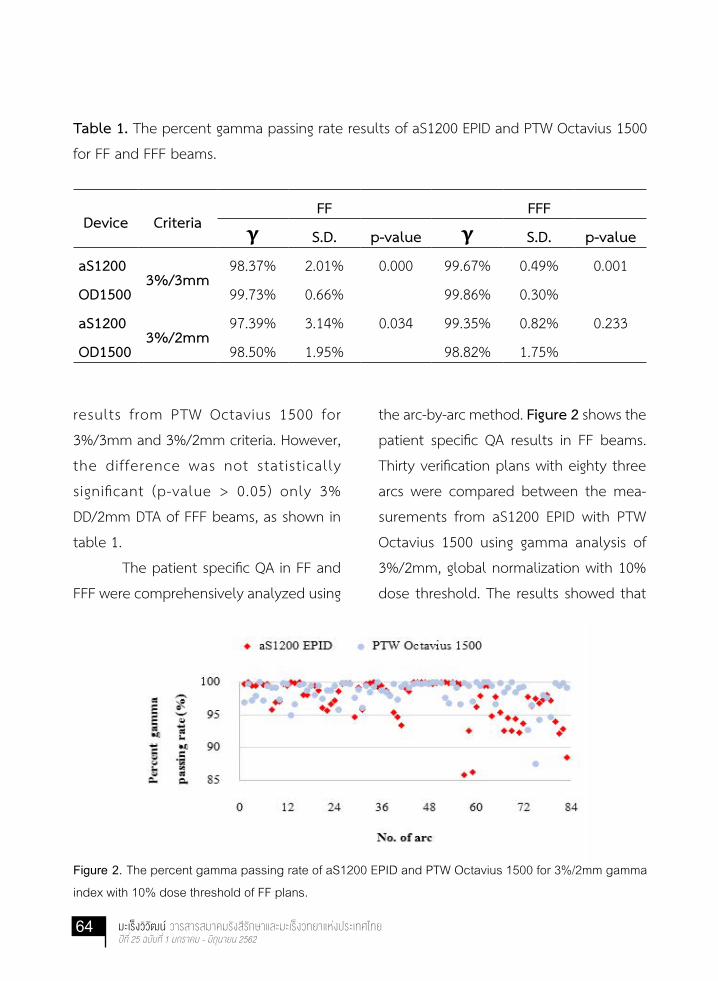
64 มะเรงววฒน วารสารสมาคมรงสรกษาและมะเรงวทยาแหงประเทศไทยปท 25 ฉบบท 1 มกราคม - มถนายน 2562
Table 1. The percent gamma passing rate results of aS1200 EPID and PTW Octavius 1500
for FF and FFF beams.
Device CriteriaFF FFF
γ S.D. p-value γ S.D. p-value
aS12003%/3mm
98.37% 2.01% 0.000 99.67% 0.49% 0.001
OD1500 99.73% 0.66% 99.86% 0.30%
aS12003%/2mm
97.39% 3.14% 0.034 99.35% 0.82% 0.233
OD1500 98.50% 1.95% 98.82% 1.75%
results from PTW Octavius 1500 for
3%/3mm and 3%/2mm criteria. However,
the difference was not statistically
significant (p-value > 0.05) only 3%
DD/2mm DTA of FFF beams, as shown in
table 1.
The patient specific QA in FF and
FFF were comprehensively analyzed using
the arc-by-arc method. Figure 2 shows the
patient specific QA results in FF beams.
Thirty verification plans with eighty three
arcs were compared between the mea-
surements from aS1200 EPID with PTW
Octavius 1500 using gamma analysis of
3%/2mm, global normalization with 10%
dose threshold. The results showed that
Figure 2. The percent gamma passing rate of aS1200 EPID and PTW Octavius 1500 for 3%/2mm gamma
index with 10% dose threshold of FF plans.

65Jornal of Thai Association of Radiation Oncologyvol. 25 No.1 January - June 2019
most of γ of aS1200 EPID measurements
for arc-by-arc were lower with greater SD
value from PTW Octavius 1500 results.
The minimum gamma passing rate of
aS1200 EPID was 85.8% while the results
from PTW Octavius 1500 was 87.5%.
The patient specific QA for FFF
beams technique was also comprehen-
sively evaluated. Thirty verification plans
with eighty two arcs were compared with
the results from the measurements using
PTW Octavius 1500. The new gamma index
criteria (3%/2mm) was used for analyzing
the results. Figure 3 shows the percent
gamma passing rate results from aS1200
EPID and PTW Octavius 1500 measure-
ment. The results showed that the percent
gamma passing rate from aS1200 EPID were
slightly higher than the results from PTW
Octavius 1500 with a lower spread out
data. The minimum gamma passing rate of
aS1200 EPID was 95.5% while the results
from PTW Octavius 1500 was 90%.
A traditional gamma index criteria vs
new gamma index criteria
All sixty patient’s VMAT verifica-
tion plans with a total of 165 arcs were
measured. The mean and SD of the γ were
used for evaluation of the results. The
results of the test are shown in figure 4.
For 3% DD/3mm DTA, the mean
percent gamma passing rate ± SD of aS1200
and PTW Octavius 1500 were 99.02% ±
Figure 3. The percent gamma passing rate of aS1200 EPID and PTW Octavius 1500 for 3%/2mm gamma
index with 10% dose threshold of FFF plans.

66 มะเรงววฒน วารสารสมาคมรงสรกษาและมะเรงวทยาแหงประเทศไทยปท 25 ฉบบท 1 มกราคม - มถนายน 2562
Figure 4. The mean gamma passing rate of aS1200 EPID and PTW Octavius 1500 for a global
normalization with 3%/3mm and 3%/2mm gamma analysis criteria for a 10% dose threshold.
1.60% and 99.80% ± 0.52% (p-value =
0.000), respectively. For 3% DD/2mm DTA,
the mean percent gamma passing rate
± SD of aS1200 and PTW Octavius 1500
were 98.36% ± 2.50% and 98.66% ± 1.86%
(p-value = 0.416), respectively.
Discussion
About the patient specific QA
implementation of aS1200 EPID for FF
and FFF beams, we studied aS1200 EPID
and compared the results with PTW
Octavius 1500. The γ results of the
study showed that most of the γ results
from PTW Octavius 1500 were slightly
higher than aS1200 EPID because PTW
Octavius 1500 used 3D gamma analysis,
while aS1200 EPID used 2D gamma analy-
sis, so the effect from 2D and 3D gamma
analysis have proposed from elsewhere [11].
Another reason which affected
to the γ results were the method to
configure the Portal Dosimetry. In our
configured verification process based on
the standard method, as described by Van
Esch et al.[12], the percent gamma passing
rate results of the AIDA plan which planned
by FF beams (6 and 10 MV) was dropped
and showed red dot in large area, yet
did not present in FFF plans, as shown in
figure 5 (A) and (B). However, this results
occurred in another institute where uti-

67Jornal of Thai Association of Radiation Oncologyvol. 25 No.1 January - June 2019
Figure 5. The gamma evaluation result of AIDA pattern. (A) 10 MV FF beams and (B) 10 MV FFF beams.
lizes Varian EDGE LINAC with aS1200 EPID
(Varian Medical System, Palo Alto, CA) too
(private communication). Therefore, the
mean γ and SD of the FF plans which
measured by aS1200 EPID are worse than
the other detector for both criteria, as
shown in table 1 and figure 2. In the other
hand, the mean γ and SD of aS1200 EPID
for FFF plans which did not have the issue
from the configuration process showed
that the γ and SD from both detectors
were likely to each other and especially
greater results for FFF plans at the new
gamma index criteria, as shown in table
1 and figure 3. This is able to conclude
that aS1200 EPID is able to do a patient
specific QA in FF and FFF beams without any
special condition. The detector resolution
may be dominated factor in stringent
gamma criteria, which increased percent
gamma passing rate of aS1200 EPID, as
shown in table 1.
The effects of the stringent criteria
are shown in figure 4. The results showed
0.66% ± 0.90% decreasing of the gam-
ma passing rate from aS1200 EPID when
using the new gamma criteria, while the
percent gamma passing rate of PTW
Octavius 1500 decreased by 1.14% ±
1.34%, which indicated that PTW Octavius
1500 was more sensitive to the stringent
gamma analysis criteria than aS1200
EPID. These results might be caused by
the spatial resolution for each detector.
Beyer et al.[13] studied aS1200 EPID for
the pre-treatment patient specific QA in
prostate and head & neck plans, which
planned by FF and FFF beam techniques,
comparing with PTW Octavius 1500 by
using gamma analysis with 2%/2mm and

68 มะเรงววฒน วารสารสมาคมรงสรกษาและมะเรงวทยาแหงประเทศไทยปท 25 ฉบบท 1 มกราคม - มถนายน 2562
10% dose threshold criteria. The results
showed that aS1200 EPID had slightly
better gamma passing rate than PTW
Octavius 1500 because aS1200 EPID
has higher spatial resolution than PTW
Octavius 1500, as the same way to our
results. Therefore, the difference between
aS1200 EPID and PTW Octavius 1500 was
not statistically significant (p-value > 0.05)
in 3%/2mm criteria (p-value = 0.416). It is
able to indicate that aS1200 EPID plays a
critical role to do a patient specific QA in
the new gamma index criteria.
Conclusion
The new version of Varian EPID,
aS1200 EPID, is an effective tool for a
patient specific QA due to its performance
such as high spatial resolution, ease of use
and reduction of QA time that is able to
decrease medical physicists’ workload.
As a result from the study, it is able to
conclude that aS1200 EPID is not only
suitable for QA using a new gamma
index tolerance, which was proposed by
AAPM TG-218, but also performs a patient
specific QA for a verification plan of
flattening filter free technique with the
comparable results to PTW Octavius
1500 planar ionization chamber detector.
Therefore, aS1200 EPID is a powerful
detector for a patient specific QA in both
FF and FFF beams, especially in the busy
department of radiation oncology.
Acknowledgments
We wish to thank our colleague
at Division of Radiation Oncology, Depart-
ment of Radiology, Faculty of Medicine
Siriraj Hospital for supporting us about
data in the research. In addition, I wish to
thank Faculty of Graduate Studies, Mahidol
University for supporting in part by the
Thesis Grant.
69Jornal of Thai Association of Radiation Oncologyvol. 25 No.1 January - June 2019
References
1. Stevens S, Dvorak P, Spevacek V,
Pilarova K, Bray-Parry M, Gesner J,
et al. An assessment of a 3D
EPID-based dosimetry system using
conventional two-and three-
dimensional detectors for VMAT.
Phys Med 2018;45:25-34.
2. Greer PB, Vial P, Oliver L, Baldock
C. Experimental investigation of
the response of an amorphous
silicon EPID to intensity modu-
lated radiotherapy beams. Med
Phys 2007;34:4389-98.
3. McDermott L, Nijsten S, Sonke JJ,
Partridge M, Van Herk M, Mijnheer
B. Comparison of ghosting effects
for three commercial a-Si EPIDs.
Med Phys 2006;33:2448-51.
4. Rowshanfarzad P, McCurdy B,
Sabet M, Lee C, O’Connor DJ, Greer
PB. Measurement and modeling
of the effect of support arm
backscatter on dosimetry with a
Varian EPID. Med Phys 2010;37:
2269-78.
5. Nicolini G, Clivio A, Vanetti E, Krauss
H, Fenoglietto P, Cozzi L, et al.
Evaluation of an aSi‐EPID with
flattening filter f ree beams:
Applicability to the GLAaS algorithm
for portal dosimetry and first
experience for pretreatment QA of
RapidArc. Med Phys 2013;40:111719.
6. Ezzell GA, Burmeister JW, Dogan N,
LoSasso TJ , Mechalakos JG,
Mihailidis D, et al. IMRT commis-
sioning: multiple institution planning
and dosimetry comparisons, a
report from AAPM Task Group 119.
Med Phys 2009;36:5359-73.
7. Miften M, Olch A, Mihailidis D, Moran
J, Pawlicki T, Molineu A, et al.
Tolerance limits and methodo-
logies for IMRT measurement-based
verification QA: Recommendations
of AAPM Task Group No. 218. Med
Phys 2018;45:e53-e83.
8. Van Esch A, Depuydt T, Huyskens
DP. The use of an aSi-based EPID
for routine absolute dosimetric
pre-treatment ver ificat ion of
dynamic IMRT fields. Radiother
Oncol 2004;71:223-34.
9. Stelljes T, Harmeyer A, Reuter J,
Looe H, Chofor N, Harder D, et al.
Dosimetric characteristics of the
novel 2D ionization chamber array
OCTAVIUS Detector 1500. Med Phys
2015;42:1528-37.
70 มะเรงววฒน วารสารสมาคมรงสรกษาและมะเรงวทยาแหงประเทศไทยปท 25 ฉบบท 1 มกราคม - มถนายน 2562
10. Low DA, Harms WB, Mutic S, Purdy
JA. A technique for the quantitative
evaluation of dose distributions.
Med Phys 1998;25:656-61.
11. Rajasekaran D, Jeevanandam P,
Sukumar P, Ranganathan A, John
jothi S, Nagarajan V. A study on
correlation between 2D and 3D
gamma evaluation metrics in
patient-specific quality assurance for
VMAT. Med Dosim 2014;39:300-8.
12. Van Esch A, Huyskens DP, Hirschi L, Scheib S, Baltes C. Optimized Varian aSi portal dosimetry: deve- lopment of datasets for collective use. J Appl Clin Med Phys 2013;14: 82-99.13. Beyer G, Gago P, Ruiz G. PD-0380: FFF pre-t reatment QA us ing TrueBeam Portal Dosimetry with DMI panel and comparison with PTW Octavius 1500. Radiother Oncol 2015;115:S182.

71Jornal of Thai Association of Radiation Oncologyvol. 25 No.1 January - June 2019
Dose Tracking Accuracy of the Dose on the Treatment Day using Cone-beam Computed Tomography for Radiation Therapy of
Prostate Cancer: Pilot Study
Achawee Suwannarat1, Tanwiwat Jaikuna2, Pittaya Dankulchai2, Wiwatchai Sittiwong2,
Nuanpen Damrongkijudom3, and Lalida Tuntipumiamorn2
1Master degree of Science Program in Radiological Technology, Faculty of Medical Technology, Mahidol University, Thailand. 2Department of Radiology, Faculty of Medicine Siriraj Hospital, Mahidol University, Thailand.3Department of Radiological Technology, Faculty of Medical Technology, Mahidol University, Thailand.
Abstract
Background: The benefit of cone-beam computed tomography (CBCT) is not
only for pretreatment setup verification, but also their potential in providing the
three-dimensional anatomical information to present the organ deformation and
daily actual dose distribution.
Objective: This study aimed to investigate the dose tracking on the treatment
day for 5 volumetric modulated arc therapy (VMAT) prostate cancer patients using
CBCT-based dose calculation.
Materials and methods: The CBCT HU to electron density calibration curve was
firstly generated. Then, the accuracy of CBCT-based dose calculation (DCBCT
) was
verified by comparing with dose measurements on CIRS pelvic phantom using IC
(0.13cc) and Gafchromic EBT3 film. Velocity 3.2.0 deformable image registration
(DIR) software was applied to map organ contours from planning CT (PCT) to the
fractionated CBCT and modified by the radiation oncologist. Isodose distribution
on CBCT images was calculated and variation of the organ volume as well as the
dosimetric parameters following the QUANTEC-guideline for the planning target
volume (PTV) and organ at risks were collected and analyzed.

72 มะเรงววฒน วารสารสมาคมรงสรกษาและมะเรงวทยาแหงประเทศไทยปท 25 ฉบบท 1 มกราคม - มถนายน 2562
Results: Point dose and dose distribution difference in phantom, between the
measurements and DCBCT
, 2.28% and 87.7% (3%/3mm) gamma passing rate was
presented. In clinical investigation, the volume ratio of CBCT/PCT for PTV, bladder
and rectum were found to be 1.04, 1.11 and 0.91. DCBCT
was found to be deviated
from DPCT
about -8.26% for D95%
of PTV, 1.12% and 9.12% for Dmean
and V70Gy
of
bladder, 6.60%, and 7.10%, for Dmean
and V70Gy
of rectum, respectively.
Conclusion: Dose tracking on treatment day using CBCT-based dose calculation and
DIR to guide organ contouring is found to be feasible and adequate to predict the
actual dose delivery to the VMAT prostate cancer.
Keywords: CBCT, Dose tracking, Prostate cancer, VMAT
บทคดยอ
หลกการและเหตผล: ระบบภาพนำาวถ cone-beam computed tomography (CBCT) ถกนำามา
ใชตรวจสอบความถกตองของตำาแหนงการรกษา และขอมลกายวภาคแบบสามมตทแสดงตำาแหนง
และรปรางทเปลยนไปของอวยวะยงนำามาใชคำานวณหาปรมาณรงสจรงทผปวยไดรบอยางม
ประสทธภาพ
วตถประสงค: ศกษาปรมาณรงสในผปวยมะเรงตอมลกหมาก 5 รายไดรบจากการฉายรงสประจำา
วนดวยเทคนคปรบความเขมเชงปรมาตร (VMAT) ดวยวธคำานวณปรมาณรงสบนภาพ CBCT (DCBCT
)
วสดและวธการ: สรางกราฟความสมพนธระหวางคา HU ของภาพ CBCT กบความหนาแนน
อเลกตรอน ตรวจสอบความถกตองของ DCBCT
โดยเปรยบเทยบกบปรมาณรงสในหนจำาลองเชงกราน
CIRS ดวยหววดรงส IC (0.13cc) และฟลม Gafchromic EBT3 การศกษาในผปวยใชโปรแกรม
Velocity รน 3.2.0 สำาหรบ deformable image registration (DIR) เพอสงผานขอบเขตอวยวะ
ทกำาหนดในภาพวางแผนการรกษา (PCT) ไปยงภาพ CBCT และปรบแกไขโดยรงสแพทย จากนน
คำานวณหา DCBCT
พรอมเกบขอมลปรมาตรอวยวะและพารามเตอรแสดงปรมาณรงสของ PTV และ
อวยวะขางเคยงบนภาพ CBCT

73Jornal of Thai Association of Radiation Oncologyvol. 25 No.1 January - June 2019
ผลการศกษา: การวดปรมาณรงสแบบจดและกระจายตวเทยบกบ DCBCT
ในหนจำาลอง พบความ
แตกตาง 2.28% สำาหรบการวดปรมาณรงสแบบจด และ 87.7% ทผานการวเคราะหดวยแกมมา
3%/3mm การศกษาในผปวยอตราสวนระหวาง CBCT/PCT สำาหรบปรมาตร PTV กระเพาะปสสาวะ
และลำาไสตรง คอ 1.04, 1.11 และ 0.91 ตามลำาดบ ความแตกตางของปรมาณรงสทไดจาก DCBCT
กบ DPCT เทากบ -8.26% สำาหรบ D95%
ใน PTV, 1.12% และ 9.12% สำาหรบ Dmean
และ V70Gy
ใน
กระเพาะปสสาวะ 6.60% และ 7.10 % สำาหรบ Dmean
และ V70Gy
ในลำาไสตรง ตามลำาดบ
ขอสรป: การคำานวณปรมาณรงสลงบนภาพ CBCT และการใชโปรแกรม DIR ในการกำาหนดขอบเขต
อวยวะ สามารถทำาไดและมประสทธภาพเพยงพอสำาหรบใชประเมนปรมาณรงสทผปวยมะเรง
ตอมลกหมากไดรบประจำาวนดวยเทคนค VMAT
Introduction
Currently, radiotherapy prostate
cancer patients are mostly treated with
the intensity modulated radiotherapy
(IMRT), and volumetric modulated arc
therapy (VMAT), from their main dosimetric
advantage to create highly conformal dose
distributions to the planning target volume
(PTV) while minimizing the dose given to
the organs at risk (OARs)[1,2].
To enhance the treatment accuracy,
the three-dimensional anatomical images
obtained from treatment room cone beam
computed tomography (CBCT) for image-
guided radiation therapy (IGRT), has been
introduced to reduce the daily patient
positioning uncertainties-maintaining
interfraction set-up reproducibility[3].
However, for prostate cancer, the problem
of organ motion, changeable volume and
shape of the prostate gland and nearby
organs such as bladder and rectum are
resulted in dosimetric change. Thus, the
accuracy of daily dose to the PTV and
OARs were questioned, and the attempts
to determine the actual dose which the
patient received in each treatment frac-
tion was mentioned and reported[4,5].
The CBCT image-guided system which is
commonly used for patient positioning
verification, also presented their advantage
in providing the three-dimensional anato-
mical information on treatment position.
The Hounsfield unit (HU) on CBCT was

74 มะเรงววฒน วารสารสมาคมรงสรกษาและมะเรงวทยาแหงประเทศไทยปท 25 ฉบบท 1 มกราคม - มถนายน 2562
found to be a meaningful method for
recalculation dose distribution for the
patient receiving radiation therapy on that
treatment day[6]. Organ delineation on
CBCT can also be performed with reduced
time consuming, via the contour deforma-
tion which is one of the applications in
deformable image registration (DIR)
software.
In this study, dose tracking on the
treatment day for prostate cancer patients
using CBCT-based dose calculation was
investigated and reported.
Materials and Methods
VMAT treatment planning technique
and acquisition of CBCT data set
Five prostate cancer patients
treated with VMAT technique with total
prescribed dose 78 Gy in 39 fractions
(2 Gy per fraction) were retrospective-
ly investigated. The planning CT (PCT)
images for all patients were acquired using
Philips Bigbore-16 slice CT simulator
(Philips Healthcare, Cleveland, OH) with
3 mm slice thickness and full bladder
protocol. The VMAT treatment planning
was generated with the Eclipse treat-
ment planning system version 13.6 (Var-
ian Medical Systems, Palo Alto, CA) using
2.5 mm grid size and analytical anisotropic
algorithm. All VMAT treatment plans
were evaluated following the QUANTEC-
guideline[7]. For CBCT data set, the images
were acquired with Truebeam STx acce-
lerator equipped with on-board imag-
ing (OBI) system (Varian Medical System,
Palo Alto, CA). In this study, all patients
underwent the treatment with CBCT
acquisition on the first three days and
then once a week using clinical pelvic
exposure parameters: 125 kV, 1080 mAs and
slice thickness 2.5 mm. For every patient,
10-12 CBCT images were used to recalcu-
late the isodose distribution. The existing
plans were transferred to the CBCT without
any change of MUs. For organ volume (PTV,
bladder volume and rectum volume) on
CBCT, the Velocity AITM version 3.2.0 DIR
(Varian Medical Systems, Palo Alto, CA)
was used to deform the contour of PTV-
prostate bed including the seminal
vesicles, and organ at risks-rectum and
bladder from the PCT image to every
fractionated CBCT image. The deformed
contours were then reviewed and modified
by radiation oncologist.
Accuracy verification of CBCT-based
dose calculation
Using CBCT-based dose calculation
to represent treatment dose, quality of

75Jornal of Thai Association of Radiation Oncologyvol. 25 No.1 January - June 2019
the CBCT image following the AAPM Task
Group 179 guideline[8] was assessed with
the Catphan® 600 phantom (CatPhan®,
Phantom Laboratory, Salem, NY). A
calibration of CBCT HU to electron density
for inhomogeneity correction was also
created using CIRS-062A CBCT electron
density phantom (CIRS Tissue Simulation
and Phantom Technology, Norfolk, VA,
USA)
To verify the accuracy of dose
calculation on CBCT, prostate VMAT treat-
ment plan was created on CIRS pelvic
phantom using PCT-based and CBCT-based
dose calculation (Figure 1) for comparing
with the measurement dose. The point
dose at the treatment isocenter was
measured using a CC13 ionization chamber
(IBA Dosimetry, Schwarzenbruck, Germany)
and a Gafchromic EBT3 film (Ashland
ISP Advanced Materials, NJ, USA) was
placed at 0.3 cm above the isocenter
(Figure 2) to measure dose distribution.
Point dose difference and dose distribu-
tion gamma analysis (10% threshold)
between CBCT-based dose calculation
and measurement as well as the calcula-
tion on CBCT and PCT were determined
and analyzed.
Variation of the organ volumes and do-
simetric parameters evaluation
Dosimetric parameters presented on the
treatment day from a total number of
55 CBCT images from 5 prostate cancer
patients (10-12 images per patient) were
collected. For each patient, dose differ-
ences between the PCT and CBCT were
Figure 1. The isodose distribution of prostate VMAT plan on CIRS pelvic phantom, comparison between
the measurements doses with (a) PCT-based dose calculation and (b) CBCT-dose calculation using the
EclipseTM treatment planning system.
(a) (b)

76 มะเรงววฒน วารสารสมาคมรงสรกษาและมะเรงวทยาแหงประเทศไทยปท 25 ฉบบท 1 มกราคม - มถนายน 2562
Figure 2. The position of (a) ionization chamber and (b) Gafchromic EBT3 film on the CIRS
pelvic phantom for point dose and dose distribution measurement.
(a) (b)
evaluated using dosimetric results from
dose volume histogram (DVH); D2%
, D95%
and D98%
for PTV; D2%
, Dmean
, V40Gy
, V65Gy
and
V70Gy
for both bladder and rectum for the
analysis. Moreover, the volume changes
in PTV, bladder and rectum on each
CBCT image during the treatment course
were also compared to the PCT and the
significance of the comparison results was
evaluated using the independent t-test on
Statistical Package for the Social Sciences
(SPSS) version 18. (IBM SPSS Statistics)
Results
CBCT image quality
From quality assessment, the CBCT
image from TrueBeam STx OBI system
presented the excellent and acceptable
results as described in AAPM Task Group
179. The image uniformity and HU accuracy
was within 1%, 5-line pair/cm, and 0.7 cm
of low contrast resolution were detected
for spatial accuracy at 1% contrast object.
Accuracy of CBCT-based dose calculation
To calculate the daily dose distri-
bution accurately, a calibration of CBCT
HU to electron density for inhomogene-
ity was generated as shown in Figure 3.
Moreover, when the calculated point dose
was compared with the measurements,
percentage of dose difference at the

77Jornal of Thai Association of Radiation Oncologyvol. 25 No.1 January - June 2019
Figure 3. The generated CBCT HU and PCT HU to electron density calibration curve in this investigation.
treatment isocenter, 2.03% and 2.28% were seen for the PCT-based and CBCT-based dose calculation, respectively. The accuracy of dose distribution was evaluated, the percent gamma passing rate of CBCT and PCT were obtained at 87.69 and 91.23, respectively, for 3%/3mm gamma criteria and that both of them increased when the gamma criteria strength was lower as shown in Table 1. For the comparison between the CBCT and PCT isodose distribution, 99.9-100% passing rate in all gamma criteria was exhibited as also presented in Table 1.
Variation of the organ volumes The variation of PTV and OARs
volumes in each patient, found on the
daily CBCT images was shown in Table 2.
For PTV, ratio of the volume averaged
from 55 CBCT of all patients to the PCT
was 1.04, 1.11 for bladder and 0.91 for
rectum. Small variation of volume change
for PTV was observed. This was in contrast
with the bladder, as seen from Figure 4,
CBCT/PCT volume ratio for bladder,
especially in patient no.3 exhibited the
higher variation of data, even the same
full bladder protocol was controlled for
the entire treatment. For rectum, the
volume presented on CBCT were found to
be smaller than PCT, and showed more
consistent from fraction to fraction.

78 มะเรงววฒน วารสารสมาคมรงสรกษาและมะเรงวทยาแหงประเทศไทยปท 25 ฉบบท 1 มกราคม - มถนายน 2562
Table 1. Percentage of gamma passing rate at different gamma criteria for dose
distribution calculated between CBCT (DCBCT
) and PCT (DPCT
) comparison with the
measurement dose.
Gamma criteriaThe percentage of gamma pass rate
DCBCT
vs DMeasurement
DPCT
vs DMeasurement
DCBCT
vs DPCT
3%/3 mm 87.69 91.23 99.9
3%/4 mm 93.63 94.17 100
3%/5 mm 96.22 95.99 100
4%/3 mm 91.09 93.30 100
4%/4 mm 95.59 95.29 100
4%/5 mm 97.60 96.68 100
5%/3 mm 93.82 94.50 100
5%/4 mm 97.20 96.17 100
5%/5 mm 98.81 97.24 100
Table 2. The ratio and percentage difference of average PTV and OARs volume on CBCT/PCT.
Organ volume(cc) PCT CBCT Ratio (CBCT/PCT) %Difference p-value*
PTV 130.62 136.37±58.84 1.04 4.40 0.750
Bladder 227.91 253.31±82.14 1.11 11.14 0.664
Rectum 52.23 47.55±15.87 0.91 -8.96 0.691
p-value <0.05 is significant difference

79Jornal of Thai Association of Radiation Oncologyvol. 25 No.1 January - June 2019
Dosimetric parameters evaluation
The results of percentage differ-
ence of dosimetric parameters between
dose tracking on the treatment day using
CBCT-based dose calculation and PCT in
each patient was shown in Table 3. For
PTV, all patients presented about 1%
higher of D2%
, 15% lower of D95%
and
24% lower of D98%
. For bladder and
rectum, the varied results of both the
lower and higher of the Dmean
,V40Gy
, V65Gy
and V70Gy
among the patients were
presented. However, the average+SD of
the dosimetric parameter change between
(a)
(b) (c)
Figure 4. The volume ratio of CBCT/PCT for (a) PTV, (b) bladder and (c) rectum.
CBCT and PCT as summarized in Table 4,
showed the significant lower of D2%
D95%
D98%
for PTV from DCBCT
, while no significant
of dosimetric results was observed in
bladder and rectum.
Discussion
There are several studies suggested
that the application of CBCT is not only
for patient posit ioning but also for
the actual dose assessment for patient
receiving dose in each treatment fraction.
Yoo et al.[6] and Tuntipumiamorn et al.[9]
demonstrated the potential of using the
80 มะเรงววฒน วารสารสมาคมรงสรกษาและมะเรงวทยาแหงประเทศไทยปท 25 ฉบบท 1 มกราคม - มถนายน 2562
Tabl
e 3.
Dosim
etric
para
met
er
Patie
nt 1
Patie
nt 2
Patie
nt 3
Patie
nt 4
Patie
nt 5
PCT
DCBC
T%
Diff
PCT
DCBC
T%
Diff
PCT
DCBC
T%
Diff
PCT
DCBC
T%
Diff
PCT
DCBC
T%
Diff
PTV
D2%
(Gy)
81.5
482
.57±
0.21
1.27
%81
.63
82.8
8±0.
441.
53%
82.5
082
.76±
0.22
0.31
%80
.89
81.8
5±0.
311.
07%
81.3
482
.58±
0.25
1.51
%
D95%
(Gy)
76.0
064
.65±
6.29
-14.
94%
77.0
067
.90±
1.54
-11.
82%
77.7
8072
.54±
2.36
-6.7
4%78
.81
77.6
8±1.
24-1
.43%
77.6
673
.11±
3.36
-5.8
5%
D98%
(Gy)
74.6
758
.44±
8.24
-23.
07%
75.5
663
.58±
2.48
-15.
86%
77.0
1068
.50±
4.00
-11.
05%
78.6
776
.47±
2.49
-2.8
3%76
.36
65.8
9±6.
41-1
3.73
%
Blad
der
D2%
(Gy)
79.8
880
.38±
0.74
0.63
%80
.02
81.4
9±0.
871.
84%
79.6
380
.34±
1.67
0.90
%78
.14
77.1
7±1.
72-1
.25%
80.2
581
.04±
0.37
0.98
%
Dmea
n (G
y)25
.54
30.3
2±6.
5918
.71%
54.6
857
.29±
6.59
4.78
%44
.42
43.4
8±25
.26
-2.1
2%21
.61
18.8
9±3.
12-1
2.59
%39
.13
37.6
6±5.
95-3
.77%
V40G
y (c
c)93
.85
77.1
1±4.
68-1
7.83
%10
9.27
135.
13±4
1.65
23.6
9%78
.07
105.
98±5
.22
35.7
6%65
.30
54.3
8±11
.79
-16.
72%
65.7
169
.01±
7.10
5.02
%
V65G
y (c
c)62
.13
51.2
2±3.
51-1
7.56
%46
.91
58.1
9±4.
0524
.05%
41.0
058
.65±
4.88
43.0
5%29
.72
24.4
9±9.
43-1
7.58
%35
.07
41.1
7±6.
6017
.40%
V70G
y (c
c)56
.17
46.0
3±3.
36-1
8.05
%36
.58
47.4
6±3.
0029
.75%
27.9
940
.26±
4.77
43.8
5%22
.89
17.5
5±9.
97-2
3.55
%30
.58
37.3
2±6.
4822
.07%
Rect
um
D2%
(Gy)
77.9
078
.31±
0.89
0.52
%78
.06
77.9
7±2.
82-0
.11%
79.0
378
.77±
2.50
-0.3
3%78
.65
79.1
5±0.
980.
64%
78.3
580
.95±
0.83
3.32
%
Dmea
n (G
y)32
.28
35.5
4±2.
5110
.07%
48.6
445
.86±
3.98
-5.7
1%43
.57
42.2
0±4.
40-3
.14%
30.0
032
.63±
3.23
8.75
%32
.77
43.3
5±4.
6832
.27%
V40G
y (c
c)21
.63
17.7
4±1.
36-1
7.98
%26
.38
26.7
6±4.
761.
46%
35.3
837
.76±
17.0
06.
73%
19.9
624
.08±
8.94
20.6
6%14
.53
18.1
2±2.
6624
.73%
V65G
y (c
c)9.
296.
40±1
.02
-31.
16%
6.52
4.92
±2.0
8-2
3.36
%13
.14
12.0
3±4.
40-8
.46%
11.2
313
.59±
7.10
20.9
8%5.
4110
.42±
1.63
92.7
5%
V70G
y (c
c)7.
064.
55±0
.90
-35.
51%
4.72
3.57
±1.7
6-2
4.26
%8.
737.
82±4
.06
-10.
42%
9.34
11.3
5±6.
5821
.63%
3.93
8.92
±1.5
012
7.05
%
Note
: %Di
ff m
ean
the
perc
enta
ge d
iffer
ence
bet
wee
n CB
CT b
ased
-dos
e ca
lcul
atio
n an
d pl
anni
ng d
ose

81Jornal of Thai Association of Radiation Oncologyvol. 25 No.1 January - June 2019
Table 4. Average+SD of dosimetric parameter from 5 VMAT patients between planning
dose (DPCT
) and CBCT-base dose calculation (DCBCT
).
Dosimetric
parameter
DPCT DCBCT %Difference p-value*
PTV
D2%
82.58±0.59 Gy 82.51±0.44 Gy -0.08% 0.023
D95%
77.59±1.01 Gy 71.18±5.03 Gy -8.26% 0.045
D98%
76.45±1.52 Gy 66.37±6.97 Gy -13.19% 0.030
Bladder
D2%
79.58±0.84 Gy 80.08±1.70 Gy 0.63% 0.571
Dmean
37.08±13.60 Gy 37.54±14.36 Gy 1.24% 0.959
V40Gy
82.44±18.99 cc 88.33±32.23 cc 7.14% 0.734
V65Gy
42.97±12.49 cc 46.74±14.31 cc 8.77% 0.668
V70Gy
33.76±13.56 cc 36.84±12.21 cc 9.12% 0.716
Rectum
D2%
78.40±0.45 Gy 79.03±1.16 Gy 0.80% 0.291
Dmean
37.45±8.17 Gy 39.92±5.58 Gy 6.60% 0.593
V40Gy
23.58±7.84 cc 24.89±8.17 cc 5.56% 0.801
V65Gy
9.10±3.23 cc 9.47±3.69 cc 4.07% 0.869
V70Gy
6.76±2.39 cc 7.24±3.19 cc 7.10% 0.792
* p-value <0.05 is significant difference
% Dose difference = (DCBCT
- DPCT
)/ DPCT*100

82 มะเรงววฒน วารสารสมาคมรงสรกษาและมะเรงวทยาแหงประเทศไทยปท 25 ฉบบท 1 มกราคม - มถนายน 2562
CBCT and its accuracy for dose calculation
in clinical used. In this study, our results
also showed the appropriate quality of
CBCT image as suggested by AAPM Task
Group 179 and with acceptable accuracy
level for dose calculation.
There are two main approaches
to calculate dose on the treatment day.
Firstly, treatment dose was obtained by
recalculating dose on CBCT images us-
ing the image registration application
that warping PCT HU into the daily CBCT.
Zhang et al.[10] reported the accuracy
of the dose calculation based on the
deformed HU from PCT to CBCT performed
on static phantom. With 2%, 2 mm criteria,
average the gamma passing rate between
CBCT-based dose and planning dose
were 98.36%. In clinical, the accuracy of
deformed CT HU into CBCT image might
be influenced by patient’s organs move-
ment and resulted in the image registration
accuracy. Another method is using the
generated CBCT HU to electron density
calibration curve for DCBCT
calculation. The
study from Yeng et al.[12] showed that
the uncertainty less than 1% of dose
difference between TPS calculation and
point dose measurement was obtained
when using directly generated CBCT HU
to electron density for dose calculation.
Nevertheless, the accuracy of DCBCT
may
be affected by the scatter dose from high
density, the image artifact and distortion
as well as the limited field size which is
shown to be inferior when compared to
the PCT.
Accuracy of CBCT-based dose
calculation in this study agreed with
the other reports[11-13] that 2-3% of dose
difference between CBCT-based and
PCT-based calculation were presented.
Furthermore, in this study the dose
distribution was further investigated by
comparing with dose measurements from
film. More than 90% (4%/3mm) gamma
passing rate was found which indicated
the accuracy level obtained from dose
calculation on CBCT.
Changes in the location and volume
of the prostate and organ at risks during
the course of radiation therapy result in the
dosimetric consequence which may im-
pact the tumor control and normal tissue
complication. From our results, due to
a small number of data, no statistically
significant difference of volumetric and
dosimetric parameters between the PCT
and CBCT was detected. However, the
tendency of the CBCT dose coverage at
D95%
of PTV about 8% lower than the
prescribed dose and lower near maximum
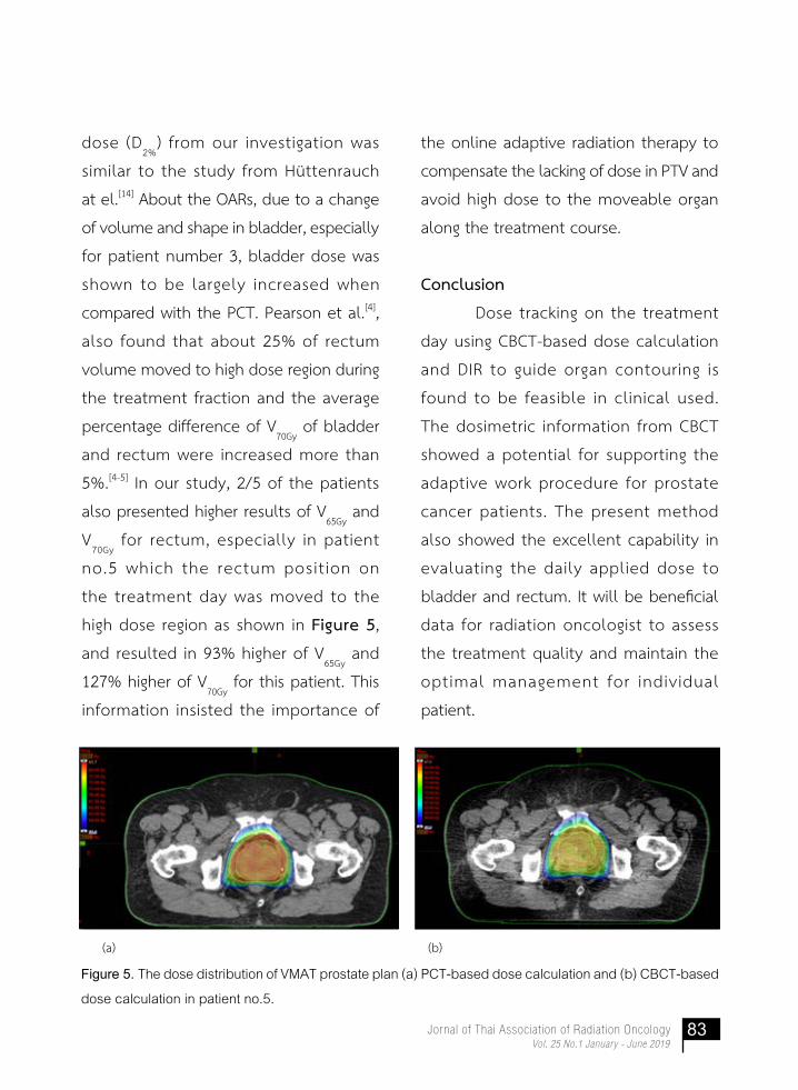
83Jornal of Thai Association of Radiation Oncologyvol. 25 No.1 January - June 2019
dose (D2%
) from our investigation was
similar to the study from Huttenrauch
at el.[14] About the OARs, due to a change
of volume and shape in bladder, especially
for patient number 3, bladder dose was
shown to be largely increased when
compared with the PCT. Pearson et al.[4],
also found that about 25% of rectum
volume moved to high dose region during
the treatment fraction and the average
percentage difference of V70Gy
of bladder
and rectum were increased more than
5%.[4-5] In our study, 2/5 of the patients
also presented higher results of V65Gy
and
V70Gy
for rectum, especially in patient
no.5 which the rectum posit ion on
the treatment day was moved to the
high dose region as shown in Figure 5,
and resulted in 93% higher of V65Gy
and
127% higher of V70Gy
for this patient. This
information insisted the importance of
the online adaptive radiation therapy to
compensate the lacking of dose in PTV and
avoid high dose to the moveable organ
along the treatment course.
Conclusion
Dose tracking on the treatment
day using CBCT-based dose calculation
and DIR to guide organ contouring is
found to be feasible in clinical used.
The dosimetric information from CBCT
showed a potential for supporting the
adaptive work procedure for prostate
cancer patients. The present method
also showed the excellent capability in
evaluating the daily applied dose to
bladder and rectum. It will be beneficial
data for radiation oncologist to assess
the treatment quality and maintain the
optimal management for individual
patient.
Figure 5. The dose distribution of VMAT prostate plan (a) PCT-based dose calculation and (b) CBCT-based
dose calculation in patient no.5.
(a) (b)
84 มะเรงววฒน วารสารสมาคมรงสรกษาและมะเรงวทยาแหงประเทศไทยปท 25 ฉบบท 1 มกราคม - มถนายน 2562
References
1. Bhide SA, Nutting CM. Recent
advances in radiotherapy. BMC
medicine. 2010;8:25.
2. Ahmad SS, Duke S, Jena R, Williams
MV, Burnet NG. Advances in radio-
therapy. BMJ. 2012;345.
3. Sterzing F, Engenhart-Cabillic R,
Flentje M, Debus J. Image-guided
radiotherapy: a new dimension in
radiation oncology. Dtsch Arztebi
Int. 2011;108:274-80.
4. Pearson D, Gill SK, Campbell N,
Reddy K. Dosimetric and volumetric
changes in the rectum and bladder
in patients receiving CBCT-guided
prostate IMRT: analysis based on
daily CBCT dose calculation. J Appl
clin Med Phys. 2016;17:107-17.
5. Orlandini LC, Coppola M, Fulcheri
C, Cernusco L, Wang P, Cionini L.
Dose tracking assessment for
image-guided radiotherapy of the
prostate bed and the impact on
clinical workflow. Radiat Oncol 2017;
12:78.
6. Yoo S, Yin FF. Dosimetric feasibility
of cone-beam CT-based treatment
planning compared to CT-based
treatment planning. International J
of Rad Oncol Biol Phys. 2006;66(5):
1553-61.
7. Emami B. Tolerance of Normal
Tissue to Therapeutic Radiation.
Rep Radiother Oncol. 2013;1:123-7.
8. Bissonnette JP, Balter PA, Dong L,
Langen KM, Lovelock DM, Miften M,
et al. Quality assurance for image--
guided radiation therapy utilizing
CT-based technologies: a report of
the AAPM TG-179. Med Phys. 2012;
39:1946-63.
9. Tuntipumiamorn L, Iampongpaiboon
P, Damrongkijudom N, Krongyuth K,
Polwatsatian V, Jaikreng S. Accuracy
of intensity-modulated radiation
therapy dose calculations: verifi-
cation based on-board conebeam
CT imaging. Asian Biomed. 2011;5:
537-41
10. Zhang GS, Huang SM, Chen C, Xu
SK, Zhang DD, Deng XW. Evaluating
the Therapeutic Dose Distribution
of Intensity-Modulated Radiation
Therapy for Head and Neck with
Cone-Beam Computed Tomography
85Jornal of Thai Association of Radiation Oncologyvol. 25 No.1 January - June 2019
Image: A Methodological Study.
Biomed Res Int. 2014;2014:8.
11. Yang Y, Schreibmann E, Li T, Wang C,
Xing L. Evaluation of on-board kV
cone beam CT (CBCT)-based dose
calculation. Phys Med Biol 2007;
52:685-705.
12. Sriram P, Vivekanandan N, Prabakar
S. A Study on Evaluation of kV-CBCT-
image-based Treatment Planning
using Anthropomorphic Phantom. J
Med Bio Eng. 2010;31:429-35.
13. Rong Y, Smilowitz J, Tewatia D,
Tome WA, Paliwal B. Dose calcula-
tion on kV cone beam CT images:
an investigation of the Hu-density
conversion stability and dose
accuracy using the site-specific
calibration. Med Dosim 2010;35:
195-207.
14. Chen Z, Yang Z, Wang J, Hu W.
Dosimetric impact of different
bladder and rectum filling during
prostate cancer radiotherapy. Radiat
Oncol 2016;11:103.
15. Huttenrauch VVP. Dose calculation
on daily Cone Beam CT data sets for
the investigation of the target
volume coverage and dose to the
organs at risk for prostate cancer
patients [Master thesis]. University
Hospital of Giessen and Marburg.
2013.https://www.thm.de/lse/images
/user/KZink105/Abschlussarbeiten/
Masterarbeit_Petra_Huettenrauch_
2013.pdf.

86 มะเรงววฒน วารสารสมาคมรงสรกษาและมะเรงวทยาแหงประเทศไทยปท 25 ฉบบท 1 มกราคม - มถนายน 2562
Implementation of the CatalystTM Optical Surface Imaging System for Left-Sided Breast Cancer Patients with Deep Inspiration Breath
Hold Technique
Sawanya Suwandee1, Pitchayut Nakkrasae2, Pittaya Dankulchai2, Puangpen Tangboonduangjit1, and
Lalida Tuntipumiamorn2
1Master of Science Program in Medical Physics, Department of Diagnostic and Therapeutic Radiology, Faculty of
Medicine Ramathibodi Hospital, Mahidol University, Bangkok, 10400, Thailand;2Division of Radiation Oncology, Department of Radiology, Faculty of Medicine Siriraj Hospital, Mahidol University,
Bangkok, 10700, Thailand
Abstract
Backgrounds: Optical surface imaging (OSI) system is currently introduced for patient’s
position verification, monitoring during treatment as well as for gating radiotherapy.
The use of this technology is found to be an attractive IGRT for deep inspiration
breathe hold (DIBH) technique in radiotherapy of breast cancer patient.
Objective: The aim of this study is to evaluate the performance and reliability of the
OSI system for image guidance in DIBH for left-sided breast cancer patient.
Materials and methods: A comprehensive acceptance and commissioning was
performed according to the AAPM TG-147 guideline to check communication
system, accuracy and reproducibility of the detection system. For OSI implemen-
tation, the overall process accuracy was examined using the end-to-end test with
thorax phantom. For the patient positioning, the set up deviations from the OSI in
16 left-breast cancer patients were retrospectively compared with the routine CBCT
and kV planar IGRT.
Results: The investigation showed the patient data from OSI can be transferred
correctly to the peripheral equipment. Accuracy verification in the static movement,

87Jornal of Thai Association of Radiation Oncologyvol. 25 No.1 January - June 2019
mean±SD of the deviations in lateral, longitudinal, and vertical were 0.13±0.10 mm,
0.19±0.09 mm, and 0.23±0.17 mm, respectively. For pitch, roll, and rotation, the
deviations were presented to be 0.01±0.07°, 0.07±0.03°, and 0.03±0.05°, respectively.
For the dynamic localization, the mean deviated amplitude was 0.05±0.41mm.
Whole treatment process with OSI showed excellent integration and agreement
within 2 mm. Using OSI for patient positioning, the average deviation in lateral,
longitudinal, and vertical axis were 2.3±1.7 mm, 2.8±3.1 mm, 2.2±1.9 mm when
compared with 72 CBCT images, and were 2.4±2.2 mm, 3.5±2.6 mm, and 2.3±2.2 mm
when compared with 130 kV-image pairs.
Conclusion: The CatalystTM OSI system is found to be an efficient image guidance for
patient setup and accurate for implementing in DIBH technique.
Keywords: deep inspiration breath hold, left-sided breast cancer, optical surface
imaging, patient setup
บทคดยอ
หลกการและเหตผล: ระบบภาพพนผวผปวยทจดเกบดวยเทคโนโลยแสง (Optical Surface
Imaging; OSI) สำาหรบการจดทาผปวย ตรวจจบการเคลอนทระหวางการฉายรงสและใชในการรกษา
แบบ Gating ไดถกนำามาประยกตใชในผปวยมะเรงเตานมทรกษาดวยเทคนค Deep Inspiration
Breathe Hold (DIBH)
วตถประสงค: เพอประเมนประสทธภาพการทำางานของระบบ OSI และตรวจสอบความถกตองใน
การจดทาผปวยมะเรงเตานมดานซายทรกษาดวยเทคนค DIBH
วสดและวธการ: ตรวจสอบการทำางานของ OSI ตามคำาแนะนำาของ AAPM TG-147 โดยประเมน
การเชอมตอของ OSI กบเครองมอและระบบทเกยวของ ศกษาความถกตองและความคงทในระบบ
การตรวจจบพนผว ความถกตองทงระบบของ OSI ในการจดทาผปวยดวยการใชหนจำาลองทรวงอก
ของมนษย ทำาการศกษายอนหลงโดยรวบรวมขอมลความคลาดเคลอนของตำาแหนงการฉายรงส
จากระบบ OSI ในผปวยมะเรงเตานมดานซายจำานวน 16 ราย มาเปรยบเทยบและวเคราะหกบขอมล
ทไดจากภาพ CBCT จำานวน 72 ภาพ และภาพ kilo-voltage Planar Image จำานวน 130 ภาพ

88 มะเรงววฒน วารสารสมาคมรงสรกษาและมะเรงวทยาแหงประเทศไทยปท 25 ฉบบท 1 มกราคม - มถนายน 2562
ผลการศกษา: ระบบ OSI สามารถสงขอมลผปวยไปยงเครองมอและอปกรณททำางานรวมกนได
อยางถกตอง มความถกตองในการตรวจจบวตถทอยนงในตำาแหนงตางๆ โดยมคาเฉลยในแนว
Lateral, Longitudinal และ Vertical เทากบ 0.13±0.10, 0.19±0.09 และ 0.23±0.17มม.
ตามลำาดบ คาเฉลยแนว Pitch, Roll และ Rotation เทากบ 0.01±0.07, 0.07±0.03, และ 0.03±0.05
องศา ตามลำาดบ สวนความถกตองในการตรวจจบการเคลอนไหวแบบตอเนองมคาความแตกตาง
ของ Amplitude เทากบ 0.05±0.41มม. เมอเทยบกบคาทกำาหนดไว ความถกตองทงระบบของ
การจดทาผปวยดวยระบบ OSI มคาความคลาดเคลอนนอยกวา 2 มม.ในทกทศทาง การศกษา
ยอนหลงในผปวยเมอใชระบบ OSI เทยบกบภาพ CBCT พบความแตกตางเฉลยในแนว Lateral,
Longitudinal และ Vertical เทากบ 2.3±1.7, 2.8±3.1 และ 2.2 ± 1.9 มม. ตามลำาดบ และ
เมอเปรยบเทยบกบภาพถาย kV มคาความแตกตางในแนว Lateral, Longitudinal และ Vertical
เทากบ 2.4±2.2, 3.5±2.6 และ 2.3±2.2มม. ตามลำาดบ
ขอสรป: ระบบ Catalyst OSI มประสทธภาพในการประเมนตำาแหนงผปวยกอนการรกษาและม
ความถกตองสำาหรบการนำาไปใชในเทคนค DIBH ในผปวยมะเรงเตานมดานซาย
คำาสำาคญ: การกลนหายใจเขาลก, ผปวยมะเรงเตานมดานซาย, ระบบการถายภาพพนผวดวยแสง,
การจดตำาแหนงผปวย
Introduction
There is the evidence that radia-
tion therapy in breast cancer significantly
reduced the risk of loco-regional recur-
rences after surgery and reduced breast
cancer death rate[1-2]. Unfortunately, for
the left-sided breast cancer patients,
whose their heart and left-anterior
descending arteries are involved in the
radiation treatment fields, the increasing
of card iac mortal i ty and mobi l i ty
associated breast radiotherapy were
presented[3]. To reduce the cardiac dose,
deep inspiration breath-hold (DIBH)
technique by increasing the heart-to-chest
wall distance was proposed. The technique
was designed to have the patient inspiring
to a specified threshold and then holding
their breath at the level during dose
delivery [4-5]. Several breathing control
technologies include the use of active
breathing control (ABC), real-time position
management (RPM) and three-dimensional
optical surface imaging system (OSI) were
proposed for DIBH[6-7]. However, the method
of the OSI has been shown to be attrac-
89Jornal of Thai Association of Radiation Oncologyvol. 25 No.1 January - June 2019
tive for verifying the patient’s pretreat-
ment position, monitoring and for gating
treatment. The system works through a
visible light project on patient’s skin, and
the re-projection was captured by high-
speed camera to generate the real-time
3D surface image. The OSI technique was
found to be a simple, fast, reproduce
and accurate verification for daily patient
setup. Several studies had reported the
application of OSI in many treated sites[8-14].
Moreover, with their real-time imaging,
the feasibility and accuracy of the OSI for
patient positioning and respiratory motion
monitoring in radiotherapy of breast
cancer, was as well presented[15-16].
The CatalystTM OSI system (C-RAD
AB, Uppsala, Sweden) was currently
installed at Division of Radiation Onco-
logy, Department of Radiology, Faculty of
Medicine Siriraj Hospital, and applied for
DIBH technique in left-sided breast cancer
patients. Before starting the clinical activity,
a test to ensure the performance of the
OSI system which consists of the SentinelTM
camera at computed tomography (CT)
simulation room, and three CatalystTM
cameras at treatment room, in terms of
precision and reproducibility of the
measures was conducted according to
recommendation from American Associa-
tion of Physicists in Medicine (AAPM) task
group report number 147[17]. The capability
evaluation of the OSI system in patient
setup compared with the image-guided
radiation therapy (IGRT) method was also
performed and reported.
Materials and Methods
1. Acceptance and Commissioning Test
of the CatalystTM OSI System
To commission the CatalystTM
OSI system, the origin of the SentinelTM
and three CatalystTM cameras were firstly
calibrated to the isocenter of CT simulator
(Brilliant Big Bore, Philips Healthcare,
Andover, MA, USA) and the linac treatment
machine (TrueBeam, Varian, Palo Alto, CA,
USA) using the daily check phantom (C-RAD
AB, Uppsala, Sweden). Dimension of the
localization field of view and, the com-
munication of the OSI with the oncology
information system were then examined
to assess their performance in creating
surface image and transferring of the
patient data. The reproducibility test
were also carried out by the thorax
humanoid phantom was scanned in every
1 minute for 10 times in all translational
and rotational directions, and recorded
the system reading of couch deviations.
For static localization accuracy test, the

90 มะเรงววฒน วารสารสมาคมรงสรกษาและมะเรงวทยาแหงประเทศไทยปท 25 ฉบบท 1 มกราคม - มถนายน 2562
thorax phantom was aligned by the room
laser and displaced at 1, 2, 3, 5 and 10 cm
from the treatment isocenter in all trans-
lational, as well as 1, 2 and 3 degree in all
rotational directions. All measurements
were repeated 3 times in each position,
and the couch deviations were detected
by the c4D Client software (C-RAD AB,
Uppsala, Sweden) and compared with the
experiment results. About the dynamic
accuracy test, the reference surface
image and gating point of the CIRS dynamic
phantom (CIRS, Norfolk, VA, USA) was
created with the SentinelTM system. At
the treatment room, the phantom was
positioned at the treatment isocenter,
and move vertically to the well-defined
amplitude at 5, 10, 15, 20 and 25 mm, by
the CIRS motion control software. The
amplitude data which detected by the
OSI were recorded and compared with
the known setting. Same procedure was
repeated by shifting the phantom at 5
to 25 cm laterally from the treatment
isocenter.
To ensure the overall accuracy
of patient setup with the OSI, the fully
end-to-end test with thorax humanoid
phantom was also undertaken. The
phantom was positioned on the breast
board positioning device (CIVCO support
system, Orange City, Iowa, USA) and aligned
with the CT room laser. The reference
surface image of phantom was captured
by using the SentinelTM system, and then
the phantom was underwent CT scan
with breast exposure protocol with 3mm
slice thickness. The CT dataset of the
phantom was sent to the Eclipse treat-
ment planning system (TPS) version 13.6.23
(Varian, Palo Alto, CA, USA) for contouring
and planning. After that, the reference
surface image and treatment isocenter
from the planning system were sent to
the CatalystTM workstation for setup. At
the treatment room, the phantom was
initially setup relative to treatment isocenter
as shown in Figure 1A. Then, the actual
surface image was created and compared
to the reference surface image to determine
the setup error (Figure 1B). The couch
correction vector from CatalystTM system
was delivered to the linac control system
and using the couch shift to correct the
position. Subsequently, CBCT image was
acquired to verify the phantom position,
and the residual setup error from the
CatalystTM system were recorded.
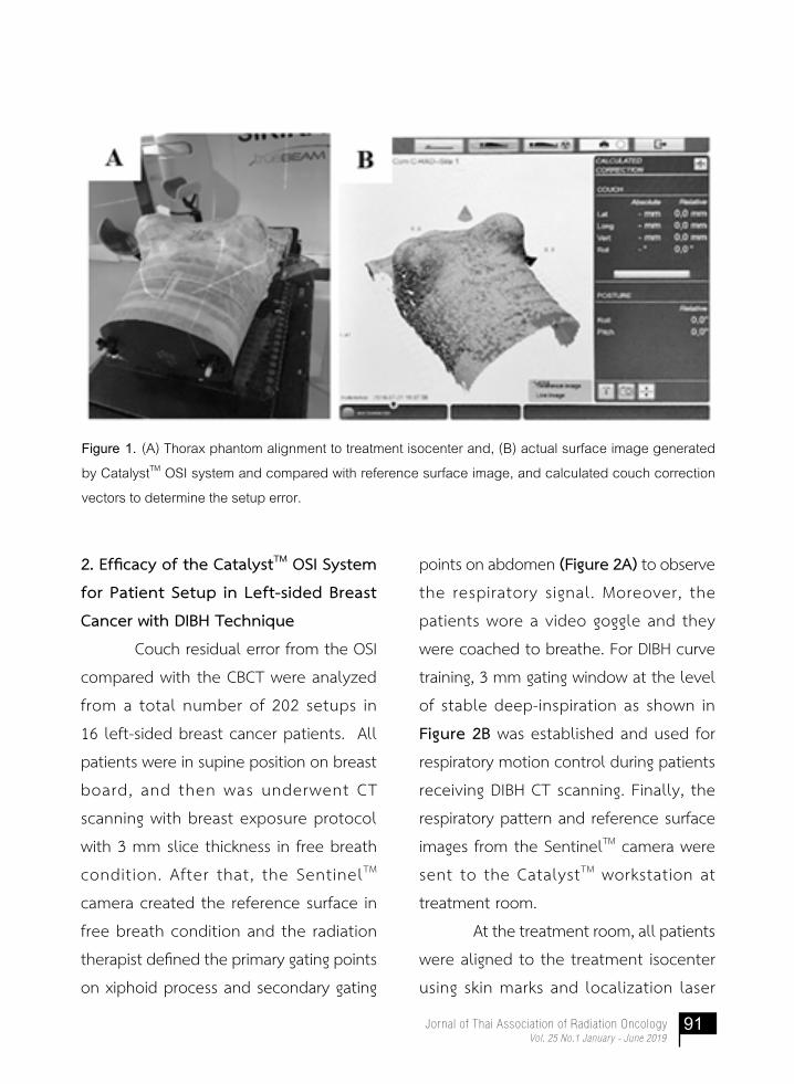
91Jornal of Thai Association of Radiation Oncologyvol. 25 No.1 January - June 2019
Figure 1. (A) Thorax phantom alignment to treatment isocenter and, (B) actual surface image generated
by CatalystTM OSI system and compared with reference surface image, and calculated couch correction
vectors to determine the setup error.
2. Efficacy of the CatalystTM OSI System
for Patient Setup in Left-sided Breast
Cancer with DIBH Technique
Couch residual error from the OSI
compared with the CBCT were analyzed
from a total number of 202 setups in
16 left-sided breast cancer patients. All
patients were in supine position on breast
board, and then was underwent CT
scanning with breast exposure protocol
with 3 mm slice thickness in free breath
condition. After that, the SentinelTM
camera created the reference surface in
free breath condition and the radiation
therapist defined the primary gating points
on xiphoid process and secondary gating
points on abdomen (Figure 2A) to observe
the respiratory signal. Moreover, the
patients wore a video goggle and they
were coached to breathe. For DIBH curve
training, 3 mm gating window at the level
of stable deep-inspiration as shown in
Figure 2B was established and used for
respiratory motion control during patients
receiving DIBH CT scanning. Finally, the
respiratory pattern and reference surface
images from the SentinelTM camera were
sent to the CatalystTM workstation at
treatment room.
At the treatment room, all patients
were aligned to the treatment isocenter
using skin marks and localization laser
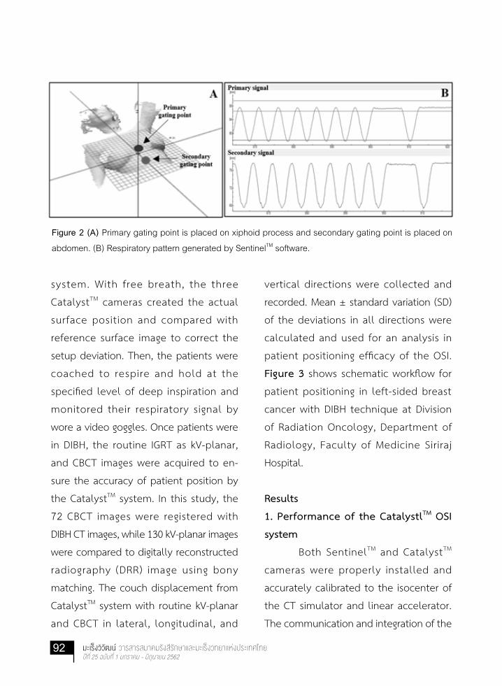
92 มะเรงววฒน วารสารสมาคมรงสรกษาและมะเรงวทยาแหงประเทศไทยปท 25 ฉบบท 1 มกราคม - มถนายน 2562
Figure 2 (A) Primary gating point is placed on xiphoid process and secondary gating point is placed on
abdomen. (B) Respiratory pattern generated by SentinelTM software.
system. With free breath, the three
CatalystTM cameras created the actual
surface position and compared with
reference surface image to correct the
setup deviation. Then, the patients were
coached to respire and hold at the
specified level of deep inspiration and
monitored their respiratory signal by
wore a video goggles. Once patients were
in DIBH, the routine IGRT as kV-planar,
and CBCT images were acquired to en-
sure the accuracy of patient position by
the CatalystTM system. In this study, the
72 CBCT images were registered with
DIBH CT images, while 130 kV-planar images
were compared to digitally reconstructed
radiography (DRR) image using bony
matching. The couch displacement from
CatalystTM system with routine kV-planar
and CBCT in lateral, longitudinal, and
vertical directions were collected and
recorded. Mean ± standard variation (SD)
of the deviations in all directions were
calculated and used for an analysis in
patient positioning efficacy of the OSI.
Figure 3 shows schematic workflow for
patient positioning in left-sided breast
cancer with DIBH technique at Division
of Radiation Oncology, Department of
Radiology, Faculty of Medicine Siriraj
Hospital.
Results
1. Performance of the CatalystlTM OSI
system
Both SentinelTM and CatalystTM
cameras were properly installed and
accurately calibrated to the isocenter of
the CT simulator and linear accelerator.
The communication and integration of the

93Jornal of Thai Association of Radiation Oncologyvol. 25 No.1 January - June 2019
Figure 3 Schematic workflow for patient positioning in left-sided breast cancer with DIBH technique at
Division of Radiation Oncology, Department of Radiology, Faculty of Medicine Siriraj Hospital.
system with all peripheral equipment was
fully functioned. The scan volume was
found to be acceptable for clinical use,
by the dimension in lateral, longitudinal
and vertical axis was 75 × 100 × 55 cm3 for
the SentinelTM, and 70 × 75 × 70 cm3 for
the CatalystTM system. The reproducibility
test also showed the results with mean
variation ± SD in lateral, longitudinal,
and vertical directions were 0.18±0.06
mm, 0.11±0.06 mm, and 0.14±0.05 mm,
respectively. The rotational deviation in
pitch, roll, and rotations were 0.22±0.06°,
0.03±0.05°, and 0.04±0.05°, respectively.

94 มะเรงววฒน วารสารสมาคมรงสรกษาและมะเรงวทยาแหงประเทศไทยปท 25 ฉบบท 1 มกราคม - มถนายน 2562
For static localization accuracy test, the
data showed mean variation ± SD in
lateral, longitudinal, and vertical of
0.13±0.10 mm, 0.19±0.09 mm, and
0.23±0.17 mm, respectively. The rotational
deviation in pitch, roll, and rotation were
presented to be 0.01±0.07°, 0.07±0.03°,
and 0.03±0.05°, respectively. About
dynamic localization accuracy test, the
data presented the mean variation ± SD
was 0.05±0.41 mm.
2. Efficacy of the CatalystTM OSI System
for Patient Setup in Left-sided Breast
Cancer with DIBH Technique
Table 1 and Figure 4 present
the mean ± SD of the couch deviations
associated with 202 individual setups
with the OSI and retrospective compared
with kV and CBCT images. The clinical
study showed good agreement between
Table 1 Mean + SD of the couch displacements compared between OSI&kV-planar and
OSI&CBCT images in 202 treatment setups
Directions Couch Displacements (mm)
kV Images (N=130) CBCT (N=72)
Lateral 2.4±2.2 2.3±1.7
Longitudinal 3.5±2.6 2.8±3.1
Vertical 2.3±2.2 2.2±1.9
the CatalystTM OSI and 130-kV images
within 3.5 mm in all directions, while the
couch displacement less than 3 mm was
demonstrated when compared with
72-CBCT.
Discussion
For modern external beam radio-
therapy, the accuracy and precision of
patient positioning play an important role
in an increase of the tumor control and
reduction of normal tissue complications
probabilities. To maintain the patient’s
setup, the IGRT as a standard practice for
patient’s position verification, had demon-
strated their benefit to improve treatment
quality. Using the daily CBCT in locally
advanced lung cancer, an improvement
of 16% in 2-year locoregional failure-free
survival was reported[18]. For head and neck
cancer, the application of CBCT also help

95Jornal of Thai Association of Radiation Oncologyvol. 25 No.1 January - June 2019
Figure 4 Setup deviations comparison between (A) OSI & kV-planar and (B) OSI&CBCT in 202 treatment
setups
in reducing 2-3 mm of the PTV margin[19].
However, the limitation of IGRT came from
the imaging dose, which can result in the
potential risk of secondary malignancies [20-22].
The OSI system, which provided a
real-time imaging, as well as the positioning
verification, was found to be an alterna-
tive approach to improve patient setup.
The analysis of 224 fractions in patients
with head and neck, thoracic, and pelvic
targets using the OSI after patient position
correction by CBCT, the disagreement
was 0.7±2.8 mm in lateral, -1.3±4.0 mm
in longitudinal, and 1.5±3.6 mm in
vertical direction that agree with the study
of Stieler F et al. who found the largest
shifts in longitudinal and vertical direc-
tions[8]. They concluded a good agree-
ment of the OSI detection with CBCT. For
OSI in breast cancer setup, the study of
Gaisberger C et al.[9] and Ma Z et al.[13]
showed the agreement between the OSI
and standard CBCT within 5 mm in all
translational directions. Moreover, the
total time for setup, image registration
and couch correction for OSI (66.8±17.7
sec) was significantly shorter than CBCT
(308.0±10.3 sec)[13]. The OSI was reported
to increase precision of patient positioning
during breast cancer radiotherapy.
About the application of OSI for
DIBH in left-sided breast cancer radio-
therapy, the capability in providing the
respiratory signal as well as the real-time
feedback information was shown to be
the effective tool to maintain patient’s
chest wall during dose delivery. The study
of Schonecker S et al.[16] showed the mean
heart dose could be decreased by 52%
(p=0.011) in patients treated with DIBH

96 มะเรงววฒน วารสารสมาคมรงสรกษาและมะเรงวทยาแหงประเทศไทยปท 25 ฉบบท 1 มกราคม - มถนายน 2562
and OSI system. In our study, with the ret-
rospectively analysis from 202 setups of 16
left-sided breast cancer patients with the
gating window 3 mm, the residual setup
error from CatalystTM OSI was within 3
mm in all translational directions when
compared with CBCT and kV images. The
uncertainty of arm positions of the patients
using the breast board[23] as well as the
accuracy in image registration, which
strongly depends on skills and experience
of the radiation therapist, are factors for
correction the positioning. Appropriate
camera setting is also another important
parameter, that OSI image quality is found
to be influenced by the surface shape,
color and resolution[8].
To ensure the dosimetric benefit
from DIBH technique, well co-operation
and ability to hold their breath from
the patient are essential to maintain the
patient position during treatment. We
found that the audio-visual coaching came
with system help the patients in controlling
their respiratory pattern to be within the
specified gating window. In our protocol,
the 3 mm gating window was observed
to be effective and practicable in clinical
routine.
Conclusion
The CatalystTM OSI system is found
to be a suitable image-guidance for DIBH
radiotherapy in left-sided breast cancer.
It is an effective tool for daily positioning
which no additional radiation dose, as
well as their real-time feedback, which is
helpful for monitoring the respiratory
signal during gating treatment delivery.
Further study with more sample size is
also suggested to affirm the study results.
Acknowledgement
We would like to express my
special thanks of Division of Radiation
Oncology, Department of Radiology,
Faculty of Medicine Siriraj Hospital,
Mahidol University, Bangkok, Thailand.

97Jornal of Thai Association of Radiation Oncologyvol. 25 No.1 January - June 2019
References
1. Speers C, Pierce LJ. Postoperative
Radiotherapy After Breast-Conserv-
ing Surgery for Early-Stage Breast
Cancer: A Review. JAMA oncology.
2016;2:1075-82.
2. McGale P, Taylor C, Correa C,
Cutter D, Duane F, Ewertz M, et al.
Ef fect of rad iotherapy after
mastectomy and axillary surgery
on 10-year recurrence and 20-year
breast cancer mortality: meta-
analysis of individual patient data
for 8135 women in 22 randomised
trials. Lancet 2014;383:2127-35.
3. Paszat LF, Mackillop WJ, Groome PA,
Schulze K, Holowaty E. Mortality
from myocardial infarction following
postlumpectomy radiotherapy for
breast cancer: a population-based
study in Ontario, Canada. Int j radiat
oncol biol phys 1999;43:755-62.
4. Hayden AJ, Rains M, Tiver K. Deep
inspiration breath hold technique
reduces heart dose from radio-
therapy for left-sided breast cancer.
J Med Imaging Radiat Oncol. 2012;56:
464-72.
5. Nissen HD, Appelt AL. Improved
heart, lung and target dose with
deep inspiration breath hold in a
large clinical series of breast cancer
patients. Radiotherapy and oncology :
Radiother Oncol. 2013;106:28-32.
6. Latty D, Stuart KE, Wang W, Ahern V.
Review of deep inspiration breath--
hold techniques for the treatment
of breast cancer. J Med Radiat Sci.
2015;62:74-81.
7. Bergom C., Currey A., Desai N., Tai A.,
& Strauss J. B. (2018). Deep Inspi-
ration Breath Hold: Techniques and
Advantages for Cardiac Sparing
During Breast Cancer Irradiation.
Front Oncol. 8, 87.
8. Stieler F, Wenz F, Shi M, Lohr F. A
novel surface imaging system for
patient positioning and surveillance
during radiotherapy. A phantom
study and clinical evaluation.
Strahlenther Onkol 2013;189:
938-44.
9. Gaisberger C, Steininger P, Mitter
lechner B, Huber S, Weichenberger
H, Sedlmayer F, et al. Three-dimen-
sional surface scanning for accurate
98 มะเรงววฒน วารสารสมาคมรงสรกษาและมะเรงวทยาแหงประเทศไทยปท 25 ฉบบท 1 มกราคม - มถนายน 2562
patient positioning and monitoring
during breast cancer radiotherapy.
Strahlenther Onkol 2013;189:
887-93.
10. Pallotta S, Vanzi E, Simontacchi
G, Marrazzo L, Ceroti M, Paiar F,
et al. Surface imaging, portal imaging,
and skin marker set-up vs. CBCT for
radiotherapy of the thorax and
pelvis. Strahlenther Onkol 2015;191:
726-33.
11. Walter F, Freislederer P, Belka C,
Heinz C, Söhn M, Roeder F. Evalua-
tion of daily patient positioning
for radiotherapy with a commercial
3D su r f ace - imag ing s y s tem
(Catalyst™). Radiat Oncol 2016;11:
154
12. Stanley DN, McConnell KA, Kirby
N, Gutierrez AN, Papanikolaou N,
Rasmussen K. Comparison of initial
patient setup accuracy between
surface imaging and three point
localization: A retrospective analysis.
J Appl Clin Med Phys. 2017;18:58-61.
13. Ma Z, Zhang W, Su Y, Liu P, Pan Y,
Zhang G, et al. Optical Surface
Management System for Patient
Positioning in Interfractional Breast
Cancer Radiotherapy. Biomed Res
Int. 2018;2018:6415497
14. Hoisak JDP, Pawlicki T. The Role
of Optical Surface Imaging Systems
in Radiation Therapy. Semin radiat
oncol 2018;28:185-93.
15. Hepp R, Ammerpohl M, Morgenstern
C, Nielinger L, Erichsen P, Abdallah A,
et al. Deep inspiration breath-
hold (DIBH) radiotherapy in left-
sided breast cancer. Strahlenther
Onkol 2015;191:710-6.
16. Schonecker S, Walter F, Freislederer
P, Marisch C, Scheithauer H, Harbeck
N, et al. Treatment planning and
evaluation of gated radiotherapy
in left-sided breast cancer patients
using the CatalystTM/SentinelTM
system for deep inspiration breath-
hold (DIBH). Radiat Oncol (London,
England). 2016;11:143.
17. Willoughby T, Lehmann J, Bencomo
JA, Jani SK, Santanam L, Sethi A,
et al. Quality assurance for non-
radiographic radiotherapy localiza-
tion and positioning systems:
Report of Task Group 147. Med. Phys.
2012;39:1728-47.

99Jornal of Thai Association of Radiation Oncologyvol. 25 No.1 January - June 2019
18. Kilburn JM, Soike MH, Lucas JT,
Ayala-Peacock D, Blackstock W, Isom
S, et al. Image guided radiation
therapy may result in improved
local control in locally advanced
lung cancer patients. Pract Radiat
Oncol. 2016;6:e73-e80.
19. Den RB, Doemer A, Kubicek G,
Bednarz G, Galvin JM, Keane WM,
et al. Daily image guidance with
cone-beam computed tomography
for head-and-neck cancer intensity-
modulated radiotherapy: a prospec-
tivestudy. Int J Radiat Oncol Biol
Phys 2010;76:1353-9.
20. Murphy MJ, Balter J, Balter S,
BenComo JA. Jr., Das IJ, Jiang SB,
et al. The management of imaging
dose during image-guided radio-
therapy: report of the AAPM Task
Group 75. Med. Phys. 2007;34:
4041-63.
21. Ding GX, Alaei P, Curran B, Flynn
R, Gossman M, Mackie TR, et al.
Image guidance doses delivered
during radiotherapy: Quantification,
management, and reduction:
Report of the AAPM Therapy Physics
Committee Task Group 180. Med.
Phys. 2018;45:e84-e99.
22. Alaei P, Spezi E. Imaging dose from
cone beam computed tomography
in radiation therapy. Phys Med.
2015;31:647-58.
23. Jassal K, Bisht S, Kataria T, Sachdev
K, Choughle A, et al. (2013) Com-
parison of Geometrical Uncertainties
in Breast Radiation Therapy with
Different Immobilization Methods.
J Nucl Med Radiat Ther 4:140.

100 มะเรงววฒน วารสารสมาคมรงสรกษาและมะเรงวทยาแหงประเทศไทยปท 25 ฉบบท 1 มกราคม - มถนายน 2562
THASTRO ANNUAL SURVEY 2017-2018Services, Manpower and Equipment in Thailand Radiation Oncology Centers
พญ.กตตวด ศกดศรชย โรงพยาบาลจฬาลงกรณ กทม. สกญญา มงไชย, นาตยา ศมศานต
โรงพยาบาลศรราช กทม. สจจา ใจเครง
โรงพยาบาลศรราชปยมหาราชการณย กทม. นพ.ธต สวางศลป, พวงเพญ ตงบญดวงจตร
โรงพยาบาลรามาธบด กทม. นพ.ภรวฒน เมองวงศ
โรงพยาบาลมหาราชนครเชยงใหม เชยงใหม อมพร ฝนเซยน, นพ.เตมศกด พงรศม
โรงพยาบาลสงขลานครนทร สงขลา นพ. คมสนต ธำารงอนนตสกล
โรงพยาบาลศรนครนทร ขอนแกน พญ.อรวรรณ คำาเจรญ, รชดาภรณ ปองชาล
โรงพยาบาลมหาวทยาลยนเรศวร พษณโลก ร.ต.หญง ปยกานต วงศาโรจน
โรงพยาบาลภมพลอดลยเดช พญ.สพชา วงษพล
โรงพยาบาลพระมงกฎเกลา กทม. พญ.คณศา รองศรแยม โรงพยาบาลวชระ กทม. นนทวด เตยวเจรญ โรงพยาบาลราชวถ กทม. พญ.ปรางระว แสงจนทร, ณชาภทร พทธจรรยา
โรงพยาบาลธรรมศาสตรเฉลมพระเกยรต ปทมธาน นพ.พทธ พยพพานนท, ชยสทธ สนทกลาง
โรงพยาบาลมหาราชนครราชสมา นครราชสมา นพ.ชวลต หลกด เกศราภรณ คงสขโข
ธนยภรณ โพธโชต เบญจรตน พรจนทรารกษ โรงพยาบาลพทธชนราช พษณโลก พญ.ฐตารย สวรรณาลย
โรงพยาบาลอตรดตถ อตรดตถ
ไชยา ชางทงใหญ โรงพยาบาลราชบร ราชบร นพ.วรพนฐ พาลสข โรงพยาบาลขอนแกน ขอนแกน พญ.สวนย นรนดรศรผล
โรงพยาบาลรอยเอด รอยเอด พญ.สภาพรรณ วทยาณวฒน
โรงพยาบาลสรนทร สรนทร นพ.เกยรตศกด พรหมเสนสา
โรงพยาบาลสกลนคร สกลนคร นพ.สทธนนท รงจรสศร
โรงพยาบาลสวรรคประชารกษ นครสวรรค พญ.กลฉตร ภโกสย
โรงพยาบาลมหาวชราลงกรณ ปทมธาน ชตกาญจน โหสข โรงพยาบาลจฬาภรณ กทม. ธดา นยมไทย สถาบนมะเรงแหงชาต กทม. พเชษฐ อเบอร โรงพยาบาลมะเรงชลบร ชลบร นพ.ธราธร ตงคะสมต
โรงพยาบาลมะเรงอดรธาน อดรธาน พญ.อจฉรยาพร ทองเถาว
โรงพยาบาลมะเรงอบลราชธาน อบลราชธาน ผกาทพย บณยะตลานนท, พญ.ดรณ สวนพล
โรงพยาบาลมะเรงสราษฎรธาน สราษฎรธาน ณรงค ชมภ โรงพยาบาลมะเรงลำาปาง ลำาปาง นพ.จรศกด สขาบรณ
โรงพยาบาลมะเรงลพบร ลพบร บญรตน วฒประเสรฐพงศ
โรงพยาบาลบำารงราษฎร กทม. เกรยงเดช บญลอลกษณ
โรงพยาบาลวฒโนสถ กทม. กนกรตน ประกอบกจ
สถานพยาบาลศนยมะเรงกรงเทพ
ขาดขอมลของโรงพยาบาลรงสรกษาและเวชศาสตรนวเคลยรพษณโลก

INSTITUTE รวม รพ. จฬาลงกรณ รพ. ศรราช รพ. ศรราชปยมหาฯ รพ. รามาธบด รพ. มหาราช รพ.สงขลานครนทร
กทม. กทม. กทม. กทม. เชยงใหม สงขลา
MANPOWER 1
Number of radiation oncologists (fulltime) 132 12 9 1 10 6 5Number of radiation oncologists (parttime) 42 2 1 6 1 0 1Number of medical physicists (fulltime) 129 12 11 1 14 5 5Number of medical physicists (parttime) 10 0 1 1 0 1 1Number of radiotherapy technicians (fulltime) 295 26 27 6 23 14 14Number of radiotherapy technicians (parttime) 4 0 0 1 0 0 0Number of dosimetrists (fulltime) 0 0 0 0 0 0 0Number of dosimetrists (parttime) 0 0 0 0 0 0 0Number of registered nurses (fulltime) 178 8 19 3 9 9 7Number of practical nurses (fulltime) 58 1 17 4 3 0 2Number of nurses (parttime) 6 0 0 0 0 0 1
EQUIPMENTS 1Low-energy X ray 3 1 0 0 1 0 0Co-60 10 0 0 0 0 0 0Linear accelerator 72 6 3 1 3 3 4Gamma Knife 0 0 0 0 0 0 0Robotic radiosurgery 2 0 0 0 1 0 0Helical radiotherapy (tomotherapy) 3 0 0 0 1 1 0Treatment planning system (system) 70 1 2 1 4 4 3Treatment planning system (workstation) 253 16 25 4 20 10 16Conventional simulator 28 1 1 0 1 1 1CT simulator 33 2 2 1 1 1 1MRI simulator 6 2 1 0 1 0 0Cone beam CT 32 5 3 1 2 1 0Brachytherapy manual after loading unit 1 0 0 0 0 0 0Brachytherapy remote after loading unit LDR 2 0 0 0 0 0 0Brachytherapy remote after loading unit MDR 0 0 0 0 0 0 0Brachytherapy remote after loading unit HDR 30 1 1 1 1 1 1Intravascular brachytherapy system 0 0 0 0 0 0 0Other …….. 6 0 1 0 2 0 0
PATIENTS Number of patients consulted per year 46570 2886 3044 895 2605 2222 3197Number of treated with radiotherapy per year - new cases 37151 2791 1861 317 1663 2093 2903Number of treated with radiotherapy per year - old cases 7082 1018 284 73 242 294
RADIOTHERAPY SERVICES TELETHERAPY 3Leukaemia 107 20 14 0 8 3 14Lymphoma Hodgkin 348 22 7 1 12 96Lymphoma Non-Hodgkin 790 127 43 19 75 81 46Eye 64 2 2 0 13 4 5Breast 9854 951 420 103 480 364 579Lung/thorax tumours 4447 488 164 84 242 163 332Esophagus 1046 115 30 6 37 26 122Cervix 4201 195 127 13 80 244 285Uterus 1011 81 44 0 59 33 79Ovary 134 24 5 1 6 7 10Other gynaecological tumours 426 13 16 6 5 0 19Nasopharynx 1571 175 69 15 59 34 101Head/neck tumours Non-Nasopharynx 5320 429 190 20 156 38 404Brain (primary only) 1258 100 39 19 186 82 99Skin 430 16 17 2 19 22 33Bladder 353 25 17 5 24 25 14Prostate 1274 155 182 36 81 42 35Kidney 145 21 3 8 11 3 11Testis 35 0 0 0 1 0 2Other urological tumours 134 3 4 2 2 0 3Colon and rectum 3695 256 118 28 139 264 188Other digestive tumours (stomach, liver) 1208 159 36 16 19 74 57Bone and soft tissue tumours (primary only) 700 105 34 11 32 31 77Paediatric brain tumours 131 22 7 0 20 3 20Paediatric lymphoma & leukemia 59 6 9 0 3 3 4Paediatric Neuroblastoma 17 0 0 0 2 1 2Paediatric Rhabdomyosarcoma 15 3 1 0 5 1 0Paediatric Wilm's tumour 25 0 3 0 4 0 7Other paediatric tumours 71 10 14 0 8 0 7Benign diseases 495 118 163 0 31 46 12Unknown primary tumours 686 62 73 0 15 2 27Other …….. 1756 106 10 0 71 401 39TOTAL 42090 3809 1,861 395 1905 2093 2633Brain metastasis 2335 NA 204 56 164 84 234Bone metastasis 2938 NA 130 95 241 86 240
BRACHYTHERAPY 4Head/neck 36 0 0 0 0 6 0Eye 12 0 0 0 12 0 0Skin 5 0 1 0 0 0 0Brain 0 0 0 0 0 0 0Lung 2 0 0 0 0 2 0Colorectal 6 0 0 0 2 0 0Other GI (e.g. liquid brachytherapy for liver metastases) 0 0 0 0 0 0 0Breast 43 3 0 0 0 0 0Gynaecological tumours 16562 496 878 30 399 706 1083Prostate 87 0 15 0 0 0 0Intravascular brachytherapy 0 0 0 0 0 0 0Other …….. 12 0 0 0 7 5 0TOTAL 16764 499 894 30 420 719 1083SPECIAL TECHNIQUES 3Intraoperative radiation therapy 64 23 17 0 0 0 0Total body irradiation 19 9 0 0 7 0 0Total lymphoid irradiation 2 2 0 0 0 0 0Stereotactic intracranial irradiation 496 132 59 0 190 26 13Stereotactic extracranial irradiation 214 75 9 0 35 15 5Intensity-modulated radiation therapy (IMRT) 8592 1306 545 194 1156 415 542Image-guided radiation therapy (IGRT) 15221 2285 1,509 385 1466 426 1284
1 Data in June 20182 Data of patients from January-December 20173 number of patients4 number of insertions5 look at definition in DEFINITATION file
C-arm 1
included Hodgkin+ Non Hodgkin
include benign primary brain tumor
1 superficial x-ray1 c-arm fluoroscope
Ru-106 opthalmic Plaques 2

INSTITUTE รพ. ศรนครนทร รพ. ม.นเรศวร รพ. พระมงกฎเกลา รพ.ภมพลอดลยเดช รพ.วชระ รพ.ราชวถ รพ.ธรรมศาสตรฯ
ขอนแกน พษณโลก กทม. กทม. กทม. กทม. ปทมธาน
MANPOWER 1
Number of radiation oncologists (fulltime) 6 2 3 3 4 2 2Number of radiation oncologists (parttime) 0 0 2 1 0 1 2Number of medical physicists (fulltime) 3 2 2 2 3 4 2Number of medical physicists (parttime) 0 0 1 0 1 0 0Number of radiotherapy technicians (fulltime) 11 6 4 3 6 6 4Number of radiotherapy technicians (parttime) 0 0 0 0 0 0 0Number of dosimetrists (fulltime) 0 0 0 0 0 0 0Number of dosimetrists (parttime) 0 0 0 0 0 0 0Number of registered nurses (fulltime) 6 3 1 3 3 3 2Number of practical nurses (fulltime) 5 0 4 0 0 0 2Number of nurses (parttime) 0 0 0 0 3 1 0
EQUIPMENTS 1Low-energy X ray 1 0 0 0 0 0 0Co-60 0 0 1 0 1 0 1Linear accelerator 4 1 2 1 1 1 1Gamma Knife 0 0 0 0 0 0 0Robotic radiosurgery 0 0 0 0 0 0 0Helical radiotherapy (tomotherapy) 0 0 0 0 0 0 0Treatment planning system (system) 3 2 3 1 1 1 2Treatment planning system (workstation) 15 4 4 2 4 3 7Conventional simulator 1 1 1 0 2 1 0CT simulator 1 1 1 1 1 1 1MRI simulator 0 0 0 0 0 0 1Cone beam CT 2 0 1 0 0 1 1Brachytherapy manual after loading unit 0 0 0 0 0 0 0Brachytherapy remote after loading unit LDR 0 0 0 0 0 0 0Brachytherapy remote after loading unit MDR 0 0 0 0 0 0 0Brachytherapy remote after loading unit HDR 1 1 1 1 1 1 1Intravascular brachytherapy system 0 0 0 0 0 0 0Other …….. 0 0 0 0 0 0 0
PATIENTS Number of patients consulted per year 1473 309 861 620 1368 1261 183Number of treated with radiotherapy per year - new cases 1104 270 662 416 1108 960 553Number of treated with radiotherapy per year - old cases 51 39 99 36 146 1347 4
RADIOTHERAPY SERVICES TELETHERAPY 3Leukaemia 2 2 0 1 6 3 6Lymphoma Hodgkin 9 0 11 6 6 16 14Lymphoma Non-Hodgkin 23 7 23 13 12 7 1Eye 2 0 0 0 5 24 1Breast 184 51 169 101 192 114 74Lung/thorax tumours 59 64 109 63 65 40 27Esophagus 12 11 16 23 38 19 23Cervix 94 16 56 37 168 130 54Uterus 29 2 24 9 48 94 1Ovary 5 3 2 0 5 10 0Other gynaecological tumours 3 0 3 1 5 50 11Nasopharynx 127 8 18 12 86 30 10Head/neck tumours Non-Nasopharynx 205 29 85 75 262 154 41Brain (primary only) 81 17 14 6 44 28 31Skin 12 7 8 1 14 12 1Bladder 4 3 8 10 34 12 10Prostate 25 28 58 20 42 43 35Kidney 2 0 2 0 3 0 0Testis 1 0 1 3 3 1 0Other urological tumours 3 3 2 0 3 18 7Colon and rectum 78 27 53 36 61 57 24Other digestive tumours (stomach, liver) 26 10 23 8 57 15 10Bone and soft tissue tumours (primary only) 24 9 46 8 31 23 4Paediatric brain tumours 27 0 4 1 4 0 3Paediatric lymphoma & leukemia 14 0 3 0 4 0 0Paediatric Neuroblastoma 3 0 3 0 4 0 1Paediatric Rhabdomyosarcoma 2 0 0 2 0 0 0Paediatric Wilm's tumour 3 0 2 0 0 0 1Other paediatric tumours 8 0 2 1 2 0 3Benign diseases 14 1 4 2 9 0 7Unknown primary tumours 23 7 6 6 25 12 26Other …….. 0 4 6 7 16 246 0TOTAL 1104 307 761 452 1254 1344 426Brain metastasis 51 20 70 31 128 70 44Bone metastasis 42 38 108 39 288 119 83
BRACHYTHERAPY 4Head/neck 3 0 0 0 0 0 0Eye 0 0 0 0 0 0 0Skin 0 0 0 0 1 0 0Brain 0 0 0 0 0 0 0Lung 0 0 0 0 0 0 0Colorectal 0 0 0 0 0 0 0Other GI (e.g. liquid brachytherapy for liver metastases) 0 0 0 0 0 0 0Breast 0 0 0 0 0 0 0Gynaecological tumours 385 9 196 198 506 582 63Prostate 0 0 6 0 0 0 0Intravascular brachytherapy 0 0 0 0 0 0 0Other …….. 0 0 0 0 0 0 0TOTAL 388 9 202 198 506 582 63SPECIAL TECHNIQUES 3Intraoperative radiation therapy 0 0 0 0 0 0 0Total body irradiation 0 0 0 0 0 0 0Total lymphoid irradiation 0 0 0 0 0 0 0Stereotactic intracranial irradiation 21 0 0 0 0 0 3Stereotactic extracranial irradiation 0 0 0 0 0 0 0Intensity-modulated radiation therapy (IMRT) 246 32 0 124 4 60 312Image-guided radiation therapy (IGRT) 1155 0 1 0 0 163 658
1 Data in June 20182 Data of patients from January-December 20173 number of patients4 number of insertions5 look at definition in DEFINITATION file

INSTITUTE รพ.มหาราช รพ.พทธชนราช รพ.อตรดตถ รพ.ราชบร รพ.สรนทร รพ.สกลนคร รพ.รอยเอด
นครราชสมา พษณโลก อตรดตถ ราชบร สรนทร สกลนคร รอยเอด
MANPOWER 1
Number of radiation oncologists (fulltime) 4 3 1 3 2 1 2Number of radiation oncologists (parttime) 0 0 0 0 0 0 0Number of medical physicists (fulltime) 1 3 0 2 0 3 3Number of medical physicists (parttime) 0 0 0 1 1 0 0Number of radiotherapy technicians (fulltime) 7 6 0 5 4 3 4Number of radiotherapy technicians (parttime) 0 0 0 0 0 0 0Number of dosimetrists (fulltime) 0 0 0 0 0 0 0Number of dosimetrists (parttime) 0 0 0 0 0 0 0Number of registered nurses (fulltime) 3 3 0 4 0 3 3Number of practical nurses (fulltime) 0 0 0 0 4 0 1Number of nurses (parttime) 0 0 0 0 0 0 0
EQUIPMENTS 1Low-energy X ray 0 0 0 0 0 0Co-60 2 1 0 0 0 0Linear accelerator 1 1 1 1 1 1Gamma Knife 0 0 0 0 0 0Robotic radiosurgery 0 0 0 0 0 0Helical radiotherapy (tomotherapy) 0 0 0 0 0 0Treatment planning system (system) 1 2 2 1 1 1Treatment planning system (workstation) 4 2 1 1 2 4Conventional simulator 1 1 1 1 0 0CT simulator 1 1 0 0 1 1MRI simulator 0 0 0 0 0 0Cone beam CT 0 0 0 0 0 1Brachytherapy manual after loading unit 0 0 0 0 0 0Brachytherapy remote after loading unit LDR 0 0 0 0 0 0Brachytherapy remote after loading unit MDR 0 0 0 0 0 0Brachytherapy remote after loading unit HDR 1 1 1 1 0 1Intravascular brachytherapy system 0 0 0 0 0 0Other …….. 0 0 0 0 0 0
PATIENTS 6
Number of patients consulted per year 1992 1923 339 1732 1017 47 315Number of treated with radiotherapy per year - new cases 1764 976 107 867 719 33 99Number of treated with radiotherapy per year - old cases 121 100 0 69 41 0 1
RADIOTHERAPY SERVICES TELETHERAPY 3Leukaemia 3 1 0 4 0 0 0Lymphoma Hodgkin 30 0 8 2 1 0Lymphoma Non-Hodgkin 15 24 3 2 10 0 2Eye 4 0 0 0 1 0 0Breast 272 334 43 233 147 3 8Lung/thorax tumours 96 137 19 27 45 6 2Esophagus 60 0 0 39 35 0 2Cervix 206 163 3 102 66 4 12Uterus 0 15 0 5 10 1 4Ovary 6 1 0 0 2 0 0Other gynaecological tumours 65 48 0 41 2 0 0Nasopharynx 40 30 0 5 35 1 1Head/neck tumours Non-Nasopharynx 299 226 5 104 142 5 6Brain (primary only) 48 47 4 17 10 0 3Skin 1 3 0 1 2 0 1Bladder 18 0 0 7 5 1 0Prostate 1 1 3 3 12 0 2Kidney 35 0 0 0 0 0 1Testis 0 1 0 0 2 0 0Other urological tumours 12 0 1 1 1 2 1Colon and rectum 207 149 10 64 152 5 18Other digestive tumours (stomach, liver) 35 95 10 7 2 3 1Bone and soft tissue tumours (primary only) 21 12 3 1 10 0 2Paediatric brain tumours 1 4 0 0 2 0 0Paediatric lymphoma & leukemia 3 0 0 0 0 0 0Paediatric Neuroblastoma 0 0 0 0 0 0 0Paediatric Rhabdomyosarcoma 0 0 0 0 0 0 0Paediatric Wilm's tumour 0 0 0 1 0 0 0Other paediatric tumours 3 2 0 0 0 0 0Benign diseases 3 0 1 0 2 0 2Unknown primary tumours 13 0 0 1 3 1 0Other …….. 85 334 2 70 0 0 7TOTAL 1579 1627 107 733 700 33 75Brain metastasis 65 NA 15 64 32 2 10Bone metastasis 117 NA 20 76 28 0 14
BRACHYTHERAPY 4Head/neck 0 0 0 0 0 0 0Eye 0 0 0 0 0 0 0Skin 0 0 0 0 0 0 0Brain 0 0 0 0 0 0 0Lung 0 0 0 0 0 0 0Colorectal 0 0 0 0 0 0 0Other GI (e.g. liquid brachytherapy for liver metastases) 0 0 0 0 0 0 0Breast 0 0 0 0 0 0 0Gynaecological tumours 728 506 8 1328 172 0 0Prostate 0 0 0 0 0 0 0Intravascular brachytherapy 0 0 0 0 0 0 0Other …….. 0 0 0 0 0 0 0TOTAL 728 506 8 1328 172 0 0SPECIAL TECHNIQUES 3Intraoperative radiation therapy 0 0 0 0 0 0 0Total body irradiation 0 0 0 0 0 0 0Total lymphoid irradiation 0 0 0 0 0 0 0Stereotactic intracranial irradiation 0 0 0 0 0 0 0Stereotactic extracranial irradiation 0 0 0 0 0 0 0Intensity-modulated radiation therapy (IMRT) 0 43 0 0 0 11 383Image-guided radiation therapy (IGRT) 1134 0 0 0 0 33 0
1 Data in June 20182 Data of patients from January-December 20173 number of patients4 number of insertions5 look at definition in DEFINITATION file6 service start from Dec 5, 2017
All equipments used at Buddhachinaraj Phitsanulok Hospital
Jan-Jun 2017 treated at Ubon Cancer Hospital 42 cases, service at Surin H. started in July 6, 2017.
included Hodgkin+ Non Hodgkin
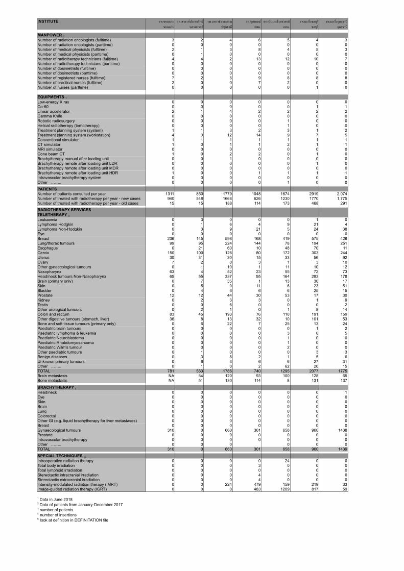
INSTITUTE รพ.ขอนแกน รพ.สวรรคประชารกษ รพ.มหาวชราลงกรณ รพ.จฬาภรณ สถาบนมะเรงแหงชาต รพ.มะเรงชลบร รพ.มะเรงอดรธาน
ขอนแกน นครสวรรค ปทมธาน กทม. กทม. ชลบร อดรธาน
MANPOWER 1
Number of radiation oncologists (fulltime) 3 2 4 6 5 4 3Number of radiation oncologists (parttime) 0 0 0 0 0 0 0Number of medical physicists (fulltime) 2 1 3 8 4 5 3Number of medical physicists (parttime) 0 1 0 0 0 0 0Number of radiotherapy technicians (fulltime) 4 4 2 13 12 10 7Number of radiotherapy technicians (parttime) 0 0 0 0 0 0 0Number of dosimetrists (fulltime) 0 0 0 0 0 0 0Number of dosimetrists (parttime) 0 0 0 0 0 0 0Number of registered nurses (fulltime) 7 2 5 9 8 8 8Number of practical nurses (fulltime) 2 0 0 7 2 0 0Number of nurses (parttime) 0 0 0 0 0 1 0
EQUIPMENTS 1Low-energy X ray 0 0 0 0 0 0 0Co-60 0 0 0 0 0 1 1Linear accelerator 2 1 4 2 2 2 2Gamma Knife 0 0 0 0 0 0 0Robotic radiosurgery 0 0 0 0 1 0 0Helical radiotherapy (tomotherapy) 0 0 0 0 1 0 0Treatment planning system (system) 1 1 3 2 3 1 2Treatment planning system (workstation) 4 3 12 14 9 7 5Conventional simulator 1 1 1 1 1 1 1CT simulator 1 0 1 1 2 1 1MRI simulator 0 0 0 0 0 0 0Cone beam CT 1 0 2 2 0 1 0Brachytherapy manual after loading unit 0 0 1 0 0 0 0Brachytherapy remote after loading unit LDR 0 0 0 0 0 1 0Brachytherapy remote after loading unit MDR 0 0 0 0 0 0 0Brachytherapy remote after loading unit HDR 1 0 0 1 1 1 1Intravascular brachytherapy system 0 0 0 0 0 0 0Other …….. 0 0 0 0 1 0 0PATIENTS Number of patients consulted per year 1311 850 1779 1048 1674 2919 2,074Number of treated with radiotherapy per year - new cases 940 548 1668 626 1230 1770 1,775Number of treated with radiotherapy per year - old cases 15 15 188 114 173 468 291RADIOTHERAPY SERVICES TELETHERAPY 3Leukaemia 0 3 0 0 0 1 0Lymphoma Hodgkin 0 1 6 4 9 21 4Lymphoma Non-Hodgkin 0 3 9 21 5 24 38Eye 0 0 0 0 0 0 0Breast 236 145 598 168 419 575 426Lung/thorax tumours 99 95 224 144 78 194 251Esophagus 0 21 60 10 48 70 11Cervix 150 100 126 80 172 303 244Uterus 30 31 30 15 33 56 92Ovary 7 2 0 3 1 3 10Other gynaecological tumours 0 1 10 1 11 10 12Nasopharynx 63 4 52 23 55 72 73Head/neck tumours Non-Nasopharynx 65 55 337 95 164 283 178Brain (primary only) 0 7 35 1 13 30 17Skin 0 5 0 11 6 23 51Bladder 0 4 6 6 6 25 15Prostate 12 12 44 30 53 17 30Kidney 0 2 3 3 0 1 9Testis 0 0 6 0 0 0 2Other urological tumours 0 2 1 0 1 8 14Colon and rectum 83 45 193 76 110 191 159Other digestive tumours (stomach, liver) 36 8 13 32 10 101 53Bone and soft tissue tumours (primary only) 0 6 22 7 25 13 24Paediatric brain tumours 0 0 0 0 0 1 2Paediatric lymphoma & leukemia 0 0 0 0 3 0 5Paediatric Neuroblastoma 0 0 0 0 1 0 0Paediatric Rhabdomyosarcoma 0 0 0 0 1 0 0Paediatric Wilm's tumour 0 0 0 0 2 0 0Other paediatric tumours 0 1 0 0 0 3 3Benign diseases 0 3 8 2 1 5 6Unknown primary tumours 0 6 3 6 6 27 31Other …….. 0 1 0 2 62 20 15TOTAL 781 563 1786 740 1295 2077 1775Brain metastasis NA 54 120 93 100 128 65Bone metastasis NA 51 130 114 8 131 137
BRACHYTHERAPY 4Head/neck 0 0 0 0 0 0 1Eye 0 0 0 0 0 0 0Skin 0 0 0 0 0 0 0Brain 0 0 0 0 0 0 0Lung 0 0 0 0 0 0 0Colorectal 0 0 0 0 0 0 0Other GI (e.g. liquid brachytherapy for liver metastases) 0 0 0 0 0 0 0Breast 0 0 0 0 0 0 0Gynaecological tumours 310 0 660 301 658 960 1438Prostate 0 0 0 0 0 0 0Intravascular brachytherapy 0 0 0 0 0 0 0Other …….. 0 0 0 0 0 0TOTAL 310 0 660 301 658 960 1439SPECIAL TECHNIQUES 3Intraoperative radiation therapy 0 0 0 0 24 0 0Total body irradiation 0 0 0 3 0 0 0Total lymphoid irradiation 0 0 0 0 0 0 0Stereotactic intracranial irradiation 0 0 0 4 0 0 0Stereotactic extracranial irradiation 0 0 0 4 0 0 0Intensity-modulated radiation therapy (IMRT) 0 0 224 479 159 219 33Image-guided radiation therapy (IGRT) 0 0 0 483 1209 817 59
1 Data in June 20182 Data of patients from January-December 20173 number of patients4 number of insertions5 look at definition in DEFINITATION file

INSTITUTE รพ.มะเรงอบลราชธาน รพ.มะเรงสราษฎรธาน รพ.มะเรงลำปาง รพ.มะเรงลพบร รพ.วฒโนสถ รพ.บำรงราษฎร สพ. มะเรงกรงเทพฯ
อบลราชธาน สราษฎรธาน ลำปาง ลพบร กทม. กทม. กทม.
MANPOWER 1
Number of radiation oncologists (fulltime) 6 2 4 3 4 2 3Number of radiation oncologists (parttime) 0 1 0 3 8 7 6Number of medical physicists (fulltime) 3 2 3 6 6 3 2Number of medical physicists (parttime) 1 0 0 0 0 0 0Number of radiotherapy technicians (fulltime) 10 6 12 14 10 8 4Number of radiotherapy technicians (parttime) 0 0 0 0 0 0 3Number of dosimetrists (fulltime) 0 0 0 0 0 0 0Number of dosimetrists (parttime) 0 0 0 0 0 0 0Number of registered nurses (fulltime) 11 0 10 11 5 2 0Number of practical nurses (fulltime) 0 4 0 0 0 0 0Number of nurses (parttime) 0 0 0 0 0 0 0
EQUIPMENTS 1Low-energy X ray 0 0 0 0 0 0 0Co-60 1 1 0 0 0 0 0Linear accelerator 3 2 3 5 3 2 2Gamma Knife 0 0 0 0 0 0 0Robotic radiosurgery 0 0 0 0 0 0 0Helical radiotherapy (tomotherapy) 0 0 0 0 0 0 0Treatment planning system (system) 2 1 3 2 5 6 2Treatment planning system (workstation) 5 4 5 14 14 9 4Conventional simulator 1 1 1 1 0 1 1CT simulator 1 1 1 1 1 1 1MRI simulator 1 0 0 0 0 0 0Cone beam CT 1 1 2 2 2 0Brachytherapy manual after loading unit 0 0 0 0 0 0 0Brachytherapy remote after loading unit LDR 0 0 0 1 0 0 0Brachytherapy remote after loading unit MDR 0 0 0 0 0 0 0Brachytherapy remote after loading unit HDR 1 1 1 1 1 1 1Intravascular brachytherapy system 0 0 0 0 0 0 0Other …….. 0 0 0 0 0 2 0PATIENTS Number of patients consulted per year 2282 1276 1719 3423 NA NA NANumber of treated with radiotherapy per year - new cases 1895 982 1518 1972 515 455 1766Number of treated with radiotherapy per year - old cases 387 1100 78 68 106 124 281RADIOTHERAPY SERVICES TELETHERAPY 3Leukaemia 3 6 3 1 0 0 3Lymphoma Hodgkin 7 3 4 0 4 44Lymphoma Non-Hodgkin 58 4 36 24 0 11 24Eye 0 0 1 NA 0 0 0Breast 438 289 403 440 160 106 629Lung/thorax tumours 197 147 182 158 104 50 292Esophagus 11 24 20 110 8 8 31Cervix 227 78 136 242 27 20 241Uterus 63 17 38 51 1 0 16Ovary 5 1 3 NA 4 3 5Other gynaecological tumours 21 2 8 6 9 17 30Nasopharynx 97 28 55 83 21 8 81Head/neck tumours Non-Nasopharynx 331 200 109 397 61 32 138Brain (primary only) 84 18 39 42 16 20 61Skin 54 17 16 40 2 4 29Bladder 15 3 8 20 3 5 15Prostate 39 23 11 29 52 46 72Kidney 0 2 3 NA 4 4 14Testis 1 4 0 NA 2 1 4Other urological tumours 18 1 1 11 1 0 8Colon and rectum 189 76 183 192 39 26 199Other digestive tumours (stomach, liver) 109 9 52 42 20 24 36Bone and soft tissue tumours (primary only) 32 17 8 18 6 3 35Paediatric brain tumours 9 0 0 NA 0 1 0Paediatric lymphoma & leukemia 2 0 0 NA 0 0 0Paediatric Neuroblastoma 0 0 0 NA 0 0 0Paediatric Rhabdomyosarcoma 0 0 0 NA 0 0 0Paediatric Wilm's tumour 2 0 0 NA 0 0 0Other paediatric tumours 4 0 0 NA 0 0 0Benign diseases 4 0 0 NA 0 51 0Unknown primary tumours 258 12 0 23 4 2 6Other …….. 4 1 83 43 77 10 34TOTAL 2279 982 1518 1972 621 456 2047Brain metastasis 74 85 57 NA 65 NA 150Bone metastasis 91 83 59 NA 112 NA 258
BRACHYTHERAPY 4Head/neck 26 0 0 0 0 0 0Eye 0 0 0 0 0 0 0Skin 3 0 0 0 0 0 0Brain 0 0 0 0 0 0 0Lung 0 0 0 0 0 0 0Colorectal 0 0 0 0 0 0 4Other GI (e.g. liquid brachytherapy for liver metastases) 0 0 0 0 0 0 0Breast 0 0 0 0 0 20 20Gynaecological tumours 1130 221 561 978 129 89 854Prostate 0 0 0 0 0 66 0Intravascular brachytherapy 0 0 0 0 0 0 0Other …….. 0 0 0 0 0 0 0TOTAL 1159 221 561 978 129 175 878SPECIAL TECHNIQUES 3Intraoperative radiation therapy 0 0 0 0 0 0 0Total body irradiation 0 0 0 0 0 0 0Total lymphoid irradiation 0 0 0 0 0 0 0Stereotactic intracranial irradiation 0 0 5 0 38 5 0Stereotactic extracranial irradiation 0 0 4 0 63 4 0Intensity-modulated radiation therapy (IMRT) 305 193 242 505 356 307 197Image-guided radiation therapy (IGRT) 305 0 16 505 599 532 197
1 Data in June 20182 Data of patients from January-December 20173 number of patients4 number of insertions5 look at definition in DEFINITATION file
outsource
included Hodgkin+ Non Hodgkin
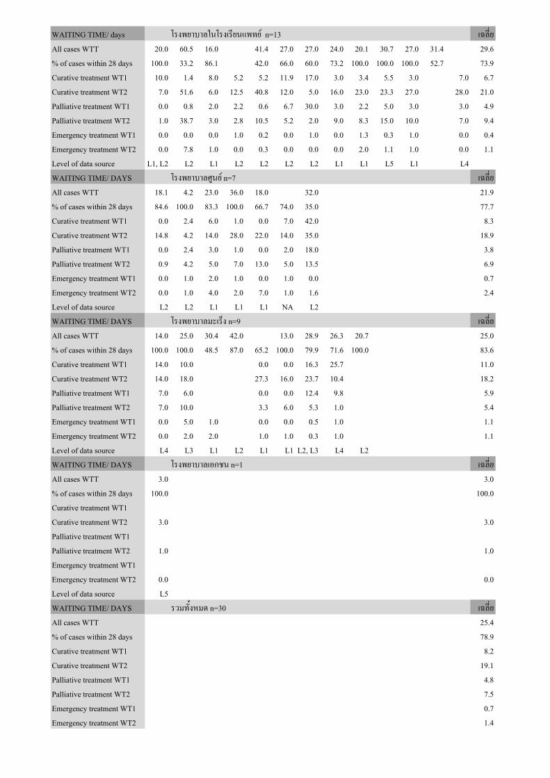
WAITING TIME/ days โรงพยาบาลในโรงเรยนแพทย n=13 เฉลย
All cases WTT 20.0 60.5 16.0 41.4 27.0 27.0 24.0 20.1 30.7 27.0 31.4 29.6
% of cases within 28 days 100.0 33.2 86.1 42.0 66.0 60.0 73.2 100.0 100.0 100.0 52.7 73.9
Curative treatment WT1 10.0 1.4 8.0 5.2 5.2 11.9 17.0 3.0 3.4 5.5 3.0 7.0 6.7
Curative treatment WT2 7.0 51.6 6.0 12.5 40.8 12.0 5.0 16.0 23.0 23.3 27.0 28.0 21.0
Palliative treatment WT1 0.0 0.8 2.0 2.2 0.6 6.7 30.0 3.0 2.2 5.0 3.0 3.0 4.9
Palliative treatment WT2 1.0 38.7 3.0 2.8 10.5 5.2 2.0 9.0 8.3 15.0 10.0 7.0 9.4
Emergency treatment WT1 0.0 0.0 0.0 1.0 0.2 0.0 1.0 0.0 1.3 0.3 1.0 0.0 0.4
Emergency treatment WT2 0.0 7.8 1.0 0.0 0.3 0.0 0.0 0.0 2.0 1.1 1.0 0.0 1.1
Level of data source L1, L2 L2 L1 L2 L2 L2 L2 L1 L1 L5 L1 L4
WAITING TIME/ DAYS โรงพยาบาลศนย n=7 เฉลย
All cases WTT 18.1 4.2 23.0 36.0 18.0 32.0 21.9
% of cases within 28 days 84.6 100.0 83.3 100.0 66.7 74.0 35.0 77.7
Curative treatment WT1 0.0 2.4 6.0 1.0 0.0 7.0 42.0 8.3
Curative treatment WT2 14.8 4.2 14.0 28.0 22.0 14.0 35.0 18.9
Palliative treatment WT1 0.0 2.4 3.0 1.0 0.0 2.0 18.0 3.8
Palliative treatment WT2 0.9 4.2 5.0 7.0 13.0 5.0 13.5 6.9
Emergency treatment WT1 0.0 1.0 2.0 1.0 0.0 1.0 0.0 0.7
Emergency treatment WT2 0.0 1.0 4.0 2.0 7.0 1.0 1.6 2.4
Level of data source L2 L2 L1 L1 L1 NA L2
WAITING TIME/ DAYS โรงพยาบาลมะเรง n=9 เฉลย
All cases WTT 14.0 25.0 30.4 42.0 13.0 28.9 26.3 20.7 25.0
% of cases within 28 days 100.0 100.0 48.5 87.0 65.2 100.0 79.9 71.6 100.0 83.6
Curative treatment WT1 14.0 10.0 0.0 0.0 16.3 25.7 11.0
Curative treatment WT2 14.0 18.0 27.3 16.0 23.7 10.4 18.2
Palliative treatment WT1 7.0 6.0 0.0 0.0 12.4 9.8 5.9
Palliative treatment WT2 7.0 10.0 3.3 6.0 5.3 1.0 5.4
Emergency treatment WT1 0.0 5.0 1.0 0.0 0.0 0.5 1.0 1.1
Emergency treatment WT2 0.0 2.0 2.0 1.0 1.0 0.3 1.0 1.1
Level of data source L4 L3 L1 L2 L1 L1 L2, L3 L4 L2
WAITING TIME/ DAYS โรงพยาบาลเอกชน n=1 เฉลย
All cases WTT 3.0 3.0
% of cases within 28 days 100.0 100.0
Curative treatment WT1
Curative treatment WT2 3.0 3.0
Palliative treatment WT1
Palliative treatment WT2 1.0 1.0
Emergency treatment WT1
Emergency treatment WT2 0.0 0.0
Level of data source L5
WAITING TIME/ DAYS รวมทงหมด n=30 เฉลย
All cases WTT 25.4
% of cases within 28 days 78.9
Curative treatment WT1 8.2
Curative treatment WT2 19.1
Palliative treatment WT1 4.8
Palliative treatment WT2 7.5
Emergency treatment WT1 0.7
Emergency treatment WT2 1.4

107Jornal of Thai Association of Radiation Oncologyvol. 25 No.1 January - June 2019
ขอตกลงความหมายของขอมลสถตบคลากรและเครองมอสมาคมรงสรกษาและมะเรงวทยาแหงประเทศไทย
MANPOWER จำ�นวนบคล�กร ร�ยง�นขอมล ณ วนท 30 มถน�ยน พ.ศ. 2561
แยกจำ�นวน full time และ part time
Number of radiation oncologists แพทยรงสรกษา หรอรงสวทยาทวไปทปฏบตงานดานรงสรกษาในสถาบน
ไมนบแพทยสาขาอน เชน แพทยรงสวนจฉยทปฏบตงานเฉพาะงานรงสรกษา
ไมนบแพทยประจำาบาน แพทยใชทน แพทย fellowship
Number of medical physicists นกฟสกสการแพทยทกสาขาวชา ทปฏบตงานในสถาบน
ไมนบนกศกษาปรญญาโท นกศกษาฝกงาน
Number of radiotherapy technologists นกรงสเทคนคหลกสตร 2-4 ป ทปฏบตงานในสถาบน ไมนบนกศกษาฝกงาน
Number of dosimetrists นกคำานวณปรมาณรงสทปฏบตงานในสถาบน หากปฏบตงานเปนนกรงสเทคนคดวย
ใหนบอยในนกรงสเทคนคเทานน ไมนบนกศกษาฝกงาน
Number of registered nurses พยาบาลวชาชพ registered nurse ทปฏบตงานเฉพาะในแผนกผปวยนอกของหนวยฯ
ไมนบพยาบาลหอผปวยใน ทงหอรวมและหอผปวยแยกรงสรกษา
Number of practical nurses ผชวยพยาบาล practical nurse ทปฏบตงานเฉพาะในแผนกผปวยนอกของหนวยฯ
ไมนบพยาบาลหอผปวยใน ทงหอรวมและหอผปวยแยกรงสรกษา
ไมนบพนกงานผชวยเหลอผป. หรอ nursing aids
EQUIPMENTS จำ�นวนเครองมอ ร�ยง�นขอมล ณ วนท 30 มถน�ยน พ.ศ. 2561 เฉพ�ะเครองทยงใชง�นไดเท�นน
Low-energy X ray kilovoltage, orthovoltage radiation machine รวมทงเครอง deep และ
superficial x-ray ไมรวมเครอง C-arm machine
Cobalt-60 เครองโคบอลต-60
Linear accelerator linear accelerator ทกพลงงาน
Gamma Knife
Robotic radiosurgery
Helical radiotherapy (tomotherapy)
Treatment planning system (system) แสดงจำานวนของระบบปฏบตการ เชน Plato, Pinnacle, Eclipse, Theraplan
Treatment planning system (workstation) แสดงจำานวน monitor workstation ทมใหแพทย
นกฟสกสสามารถใชงานระบบไดพรอมกน นบทงเครองทใช planning หรอ
contouring
Conventional simulator
CT simulator
MRI simulator
Cone beam CT เครองฉายรงสทสามารถทำา Cone beam CT ได นบซำากบเครองฉายได
Brachytherapy manual after loading unit
Brachytherapy remote after loading unit LDR < 2 Gy/hour
Brachytherapy remote after loading unit MDR 2-12 Gy/hour
Brachytherapy remote after loading unit HDR >12 Gy/hour
Intravascular brachytherapy system

108 มะเรงววฒน วารสารสมาคมรงสรกษาและมะเรงวทยาแหงประเทศไทยปท 25 ฉบบท 1 มกราคม - มถนายน 2562
Others เครองมออนๆ ทไมมในรายการขางบน
PATIENTS จำ�นวนผปวย ร�ยง�นขอมล วนท 1 มกร�คม- 31 ธนว�คม พ.ศ. 2560
Number of patients consulted per year จำานวนผป.ทรบปรกษาจากแผนกอนของโรงพยาบาล รวมถงผป. ทปฏเสธการรกษา
หรอสงตอมาจากสถาบนอนเพอตดตามผลการรกษาเทานน
Number of treated with radiotherapy per year - new
cases
จำานวนผป.ใหมทมารบการรกษาทง teletherapy หรอ brachytherapy
ในปนนๆ รวมถงผป.เกาของสถาบนอนทมาเรมการรกษาใหม
Number of treated with radiotherapy per year - old
cases
จำานวนผป.เกาของสถาบนตนเองทกลบมารบการรกษาทง teletherapy หรอ
brachytherapy ใหม ในปนนๆ ผป.เกา นบตงแตการรกษาตามแผนการรกษาเดมครบ
หากเรมรกษาในตำาแหนงอนในวนถดไป กนบเปนผป.เกาดวย
และนบเปน 1 คน ถงแมจะมารกษาใหมหลายครงในปนน
RADIOTHERAPY SERVICES
จำ�นวนก�รใหบรก�ร
ร�ยง�นขอมล วนท 1 มกร�คม- 31 ธนว�คม พ.ศ. 2560
TELETHERAPY รายงานขอมล วนท 1 มกราคม- 31 ธนวาคม พ.ศ. 2560
นบทงผปวยใหมและผปวยเกาทกลบมารบการรกษาใหมในปนนๆ
เปนจำ�นวนคน ไมใชจำ�นวนครง
Tumours by primary sites ผป. metastases ใหใสตาม primary site
หากไมทราบ ใหใสในชอง unknown primary tumours
Brain metastasis
Bone metastasis
ผป. metastases นบซำากบ Tumour by primary sites ได
ผป.ทมทง brain met และ multiple bone met นบ 1 คน ทงชอง brain และ bone
BRACHYTHERAPY รายงานขอมล วนท 1 มกราคม- 31 ธนวาคม พ.ศ. 2560
นบทงผปวยใหมและผปวยเกาทกลบมารบการรกษาใหมในปนนๆ
เปนจำ�นวนครง ไมใชจำ�นวนคน
SPECIAL TECHNIQUES รายงานขอมล วนท 1 มกราคม- 31 ธนวาคม พ.ศ. 2560
นบทงผปวยใหมและผปวยเกาทกลบมารบการรกษาใหมในปนนๆ
เปนจำ�นวนคน ไมใชจำ�นวนครง
Intraoperative radiation therapy
Total body irradiation
Total lymphoid irradiation
Stereotactic irradiation นบเฉพาะการฉายรงสทใช stereotactic protocol ทง intracranial และ body หรอ
ใชเครอง radiosurgery
- Intracranial
- Extracranial
IMRT เชน VMAT, Rapid Arc, TrueBeam
IGRT ผป. ทม onboard image check ใน treatment room กอนเรมฉายรงส
เพอให target จรงตรงกบท plan ไว โดยใช KV imaging หรอ MV imaging กได เทคนค
อาจใชเปน 2D (planar image, EPID) หรอ 3D (conebeam CT) กได การ correction
อาจเปน on-line (auto or semi-auto) หรอ off-line กได และมการตรวจสอบอยาง
สมำาเสมอตลอดการรกษานบผป. ซำากบชอง stereotactic หรอ IMRT ได

109Jornal of Thai Association of Radiation Oncologyvol. 25 No.1 January - June 2019
WTT:
WAITING TIME ระยะเวลารอการบรการรงสรกษา
โดยไมตอง exclude
ผป.ทระยะเวลารอเกดจากสาเหตอน (elective delay)
ออกไป
- รายงานเปนคาเฉลย หรอมธยฐาน จำานวนวน
- รายงานเปนสดสวนรอยละของผป.ท WTT
อยในเกณฑมาตรฐานทสมาคมกำาหนด คอ 28 วน
- หมายถง ระยะเวลาทเกดจากกระบวนการทางรงสรกษาทงหมด ไดแก การรอพบแพทย
ขนตอนทางรงสรกษา เชน simulation, treatment planning และการรอคว
inclusion criteria
- ผปวยใหมและผปวยเกาทแพทยสาขาอนสงปรกษาใหมทกราย ทไดรบการรกษา
ทางรงสทง complete และ incomplete treatment ในชวงเวลาทเกบรวบรวมขอมล
โดยไมตอง exclude
ผป.ทระยะเวลารอเกดจากสาเหตอน (elective delay) ออกไป
WTT = T2-T0
T0 : วนทผปวยตดตอ สงปรกษาโดยแพทยสาขาอน
หรอถกสงตวมาถงหนวยรงสรกษาครงแรก
T2 : วนเรมรงสรกษาครงแรก (D1 or first fraction)
WTO = WT1+WT2:
WAITING TIME ระยะเวลารอการบรการรงสรกษา
โดยไมรวม
* ผปวย elective delay(1) ซงหมายถง
ผปวยทระยะเวลารอเกดจากสาเหตอน เชน
เปนความตองการของผปวยเองทยงไมพรอม
สภาพรางกายผปวย ไมพรอม รอผลชนเนอ
รอหลงการผาตด รอการใหเคมบำาบดพรอมกน
* ผปวยทในทสดไมไดรบการรกษาทางรงสเลย
* ผปวยเกาทแพทยรงสรกษาตดตามผลการรกษา
และพจารณาใหรกษาใหมเอง
- รายงานเปนคาเฉลย หรอมธยฐาน จำานวนวน ของ
WT1 และ WT2 แบงกลมเปน curative, palliative และ
emergency treatment
สถตทสถาบนตางๆ ควรมเพอเปนตวชวดสำาหรบการ
พฒนา
- รายงานเปนสดสวนรอยละของผป.ท WTO
อยในเกณฑมาตรฐานตาม JCCO 1993 (2)
Curative treatment 28 วน
Palliative treatment 14 วน
Emergency treatment 2 วน
WT1 : ระยะเวลารอ กอนแพทยตดสนใจรกษา = T1-T0
WT2 : ระยะเวลารอ หลงแพทยตดสนใจรกษา = T2-T1
WTO : ระยะเวลารอ การบรการรงสรกษา = T2-T0
T0 : วนทผปวยตดตอ สงปรกษาโดยแพทยสาขาอน
หรอถกสงตวมาถงหนวยรงสรกษาครงแรก
T1 : วนทแพทยตดสนใจใหรกษาผปวย อาจไมใชวนแรกทพบแพทยกได
T2 : วนเรมรงสรกษาครงแรก (D1 or first fraction)
ตวอยางท 1 : อายรแพทยสงปรกษารงสรกษา ผปวย cord compression
ฉกเฉน วนท 27 มย. 2557 (T0) แพทยรงสรกษาพบผปวย (T1)
และเรมฉายรงสได (T2) ในวนเดยวกน
WT1=0, WT2=0, WTO = 0
ตวอยางท 2 : แพทยโรงพยาบาลศนย refer ผปวยมา ใบ refer
ลงวนท 27 มย. 2557 แตผปวยเดนทางมาถงวนท 9 กค. (T0)
พบแพทยรงสรกษาครงแรก 20 กค. แตสงตรวจเพมเตมเพอ staging
ไดผลครบและตดสนใจใหฉายรงสเมอวนท 30 กค. (T1)
ทำา CT simulation และ treatment planning
และเรมฉายรงสครงแรกในวนท 14 สค. 2557 (T2)
WT1=21, WT2=15, WTO = 36
Level of data source ระดบทมาของขอมล
ใหเลอกตอบวา ขอมลทรายงานครงน ไดมาจากอะไร
L1 : สถตจากการเกบขอมลผปวยทกราย ตอเนองจนถงปจจบน
L2 : สถตจากการสมเกบขอมลผปวยบางราย เชน 1 เดอน ในปจจบน
L3 : สถตจากการเกบขอมลผปวยทกรายในอดต เชน สถตเกาในป 2559
L4 : สถตจากการสมเกบขอมลผปวยบางราย เชน 1 เดอน ในอดต
L5 : คาโดยประมาณจากการคาดการณ ไมไดเกบขอมลสถต
1. Drinkwater KJ, Williams MV. Re-audit of Radiotherapy Waiting Times in the United Kingdom 2007.
2.. The Royal College of Physicians, The Royal College of Radiologisrts. Reducing Delays in Cancer Treatment: Some Targets. 1993.