acta odontologica...
TRANSCRIPT

ACTA ODONTOLOGICALATINOAMERICANAVol. 31 Nº 1 2018
ISSN 1852-4834 on line versionversión electrónica
AOL12018:32011 18/07/2018 17:30 Página 1

AOL12018:32011 18/07/2018 17:30 Página 1
Scientific EditorsEditores CientíficosMaría E. ItoizRicardo Macchi(Universidad de Buenos Aires, Argentina)
Associate EditorsEditores AsociadosAngela M. Ubios(Universidad de Buenos Aires, Argentina)Amanda E. Schwint(Comisión Nacional de Energía Atómica, Argentina)
Assistant EditorsEditores AsistentesPatricia MandalunisSandra J. Renou(Universidad de Buenos Aires, Argentina)
Technical and Scientific AdvisorsAsesores TécnicoCientíficosLilian Jara TracchiaLuciana M. SánchezTammy SteimetzDelia Takara(Universidad de Buenos Aires, Argentina)
Editorial BoardMesa EditorialEnri S. Borda (Universidad de Buenos Aires, Argentina)
Noemí E. Bordoni (Universidad de Buenos Aires, Argentina)
Fermín A. Carranza (University of California, Los Angeles, USA)
José Carlos Elgoyhen (Universidad del Salvador, Argentina)
Andrea Kaplan (Universidad de Buenos Aires, Argentina)
Andrés J.P. KleinSzanto (Fox Chase Cancer Center, Philadelphia, USA)
Susana Piovano (Universidad de Buenos Aires, Argentina)
Guillermo Raiden (Universidad Nacional de Tucumán, Argentina)
Sigmar de Mello Rode (Universidade Estadual Paulista,Brazil)
Hugo Romanelli (Universidad Maimónides, Argentina)
Cassiano K. Rösing (Federal University of Rio Grande do Sul, Brazil)
PublisherProducción Gráfica y PublicitariaImageGraf / email: [email protected]
Acta Odontológica Latinoamericana is the officialpublication of the Argentine Division of the InternationalAssociation for Dental Research.
Revista de edición argentina inscripta en el RegistroNacional de la Propiedad Intelectual bajo el N° 284335.Todos los derechos reservados.Copyright by:ACTA ODONTOLOGICA LATINOAMERICANAwww.actaodontologicalat.com
ACTA ODONTOLÓGICA LATINOAMERICANAAn International Journal of Applied and Basic Dental Research
POLÍTICA EDITORIAL
El objetivo de Acta OdontológicaLatinoamericana (AOL) es ofrecer a lacomunidad científica un medio adecuadopara la difusión internacional de los trabajos de investigación, realizados preferentemente en Latinoamérica, dentro delcampo odontológico y áreas estrechamente relacionadas. Publicará trabajos originales de investigación básica, clínica yepidemiológica, tanto del campo biológico como del área de materiales dentales ytécnicas especiales. La publicación de trabajos clínicos será considerada siempreque tengan contenido original y no seanmeras presentaciones de casos o series. Enprincipio, no se aceptarán trabajos de revisión bibliográfica, si bien los editorespodrán solicitar revisiones de temas departicular interés. Las ComunicacionesBreves, dentro del área de interés de AOL,serán consideradas para su publicación.Solamente se aceptarán trabajos no publicados anteriormente, los cuales no podránser luego publicados en otro medio sinexpreso consen timiento de los editores.
Dos revisores, seleccionados por lamesa editorial dentro de especialistas encada tema, harán el estudio crítico de losmanuscritos presentados, a fin de lograr elmejor nivel posible del contenido científico de la revista.
Para facilitar la difusión internacional,se publicarán los trabajos escritos eninglés, con un resumen en castellano o portugués. La revista publicará, dentro de laslimitaciones presupuestarias, toda información considerada de interés que se lehaga llegar relativa a actividades conexasa la investigación odontológica del árealatinoamericana.
EDITORIAL POLICY
Although Acta Odontológica Lati noamericana (AOL) will accept originalpapers from around the world, the principal aim of this journal is to be an instrumentof communication for and among LatinAmerican investigators in the field of dental research and closely related areas.
AOL will be devoted to original articlesdealing with basic, clinic and epidemiological research in biological areas or thoseconnected with dental materials and/orspecial techniques.
Clinical papers will be published aslong as their content is original and notrestricted to the presentation of singlecases or series.
Bibliographic reviews on subjects ofspecial interest will only be published byspecial request of the journal.
Short communications which fall within the scope of the journal may also besubmitted. Submission of a paper to thejournal will be taken to imply that it presents original unpublished work, not underconsideration for publication elsewhere.
By submitting a manuscript the authorsagree that the copyright for their article istransferred to the publisher if and whenthe article is accepted for publication. Toachieve the highest possible standard inscientific content, all articles will be refereed by two specialists appointed by theEditorial Board. To favour internationaldiffusion of the journal, articles will bepublished in English with an abstract inSpanish or Portuguese.
The journal will publish, within budgetlimitations, any data of interest in fieldsconnected with basic or clinical odontological research in the Latin America area.
Este número se terminó de editar el mes de Junio de 2018
Vol. 31 Nº 1 / 2018 ISSN 1852-4834 Acta Odontol. Latinoam. 2018
AOL12018:32011 18/07/2018 17:30 Página 1

CONTENTS / ÍNDICE
Association between history of orthodontic treatment and sociodemographic factors in adolescentsAssociação entre histórico de tratamento ortodôntico e fatores sociodemográficos em adolescentesGustavo H.S. Merlo, Carla C. Piardi, Ezequiel Gabrielli, Francisco Wilker M.G. Muniz, Cassiano K. Rösing, Paulo Roberto G. Colussi ................................................................................................................ 3
Cytotoxicity assessment of 1% peracetic acid, 2.5% sodium hypochlorite and 17% EDTA on FG11 and FG15 human fibroblastsAvaliação da citotoxicidade de ácido peracético a 1%, hipoclorito de sódio a 2,5% e EDTA a 17% em fibroblastos humanos FG11 e FG15Pedro A. Teixeira, Marcelo S. Coelho, Augusto S. Kato, Carlos E. Fontana, Carlos E.S. Bueno, Daniel G. PedroRocha .......................................................................................................................................... 11
Comparison of instruments used to select and classify patients with temporomandibular disorderAvaliação de instrumentos utilizados para selecionar pacientes com disfunção temporomandibularGabriel P Pastore, Douglas R Goulart, Patrícia R Pastore, Alexandre J Prati, Márcio de Moraes ................................................................................................................................................................................ 16
Parental perceptions of impact of oral disorders on Colombian preschoolers’ oral healthrelated quality of lifePercepción de padres del impacto de desórdenes orales de preescolares Colombianos sobre calidad de vida relacionada con la salud oralShyrley Díaz, María Mondol, Angélica Peñate, Guillermina Puerta, Marcelo Boneckër, Saul Martins Paiva, Jenny Abanto .................................................................................................................................... 23
The effect of gingival aging in diabetic and nondiabetic status. An experimental studyEl efecto del envejecimiento gingival en el estado diabetico y no diabetico. Estudio experimentalOrlando L. Catanzaro, Ana Andornino, Nicole Brasquet, Irene Di Martino, Alejandra Arganiaraz ............................................................................................................................................................................ 32
Oral healthrelated quality of life in Colombian children with MolarIncisor HypomineralizationCalidad de vida relacionada con la salud oral en niños Colombianos con Hipomineralización Inciso MolarLina M.Velandia, Laura V. Álvarez, Lofthy P. Mejía, Martha J. Rodríguez.................................................................................................................................................................................................................. 38
Factors associated to apical overfilling after a thermoplastic obturation technique – Calamus®
or Guttacore®: a randomized clinical experimentFactores asociados con la presencia de sobreobturación apical posterior a una técnica de obturación termoplástica Calamus® o Guttacore®: experimento clínico aleatorizadoJavier L. NinoBarrera, Luis F. GamboaMartinez, Horacio LasernaZuluaga, Jessy Unapanta, Daniela HernándezMejia, Carolina Olaya, Diana AlzateMendoza ........................................................................................................................................................................................................................................................................ 45
Inflamatory response in pregnant women with high risk of preterm delivery and its relationship with periodontal disease. A pilot studyRespuesta inflamatoria en pacientes embarazadas con alto riesgo de parto pretérmino y su relación con la enfermedad periodontal. Estudio pilotoFrancina EscobarArregoces, Catalina LatorreUriza, Juliana VelosaPorras, Nelly RoaMolina, Alvaro J Ruiz, Jaime Silva, Estefania Arias, Juliana Echeverri ............................................................................................................................................................................................................................................................ 53
Risk factors for early childhood caries experience expressed by ICDAS criteria in Anapoima, Colombia. A crosssectional studyFactores de riesgo de experiencia de caries temprana expresada con criterios ICDAS en niños de Anapoima, Colombia. Estudio de corte transversalStefania Martignon, Margarita UsugaVacca, Fabián Cortés, Andrea Cortes, Luis Fernando Gamboa, Sofía JacomeLievano, Jaime Alberto RuizCarrizosa, María Clara GonzálezCarrera, Luis Fernando RestrepoPerez, Nicolás Ramos ........................................................................................................................................................ 58
Comparison of canal transportation and centering ability of Xp Shaper, WaveOne and Oneshape: A cone beam computed tomography study of curved root canalsComparación del transporte apical y la capacidad de conservación de la anatomía del conducto de XpShaper, WaveOne y Oneshape: Estudio de conductos curvos por tomografía computarizada de haz cónicoReham Hassan, Nehal Roshdy, Noha Issa .................................................................................................................................................................................................................................................................... 67
Acta Odontol. Latinoam. 2018 ISSN 1852-4834 Vol. 31 Nº 1/ 2018
ACTA ODONTOLÓGICA LATINOAMERICANAAn International Journal of Applied and Basic Dental Research
Contact us Contactos: Cátedra de Anatomía Patológica, Facultad de Odontología, Universidad de Buenos AiresM.T. de Alvear 2142 (1122) Buenos Aires, Argentina Fax: (5411) 4 508[email protected] http://www.actaodontologicalat.com/contacto.html
ACTA ODONTOLÓGICA LATINOAMERICANA
A partir del Volumen 27 (2014) AOL se edita en formato digital con el Sistema de Gestión de Revistas Electrónicas (Open Journal System, OJS). La revista es de accesoabierto (Open Access). Esta nueva modalidad no implica un aumento en los costos de publicación para los autores.
Comité Editorial
ACTA ODONTOLÓGICA LATINOAMERICANA
From volume 27 (2014) AOL is published in digital format with the Open Journal System (OJS). The journal is Open Access. This new modality does not implyan increase in the publication fees.
Editorial Board
AOL12018:32011 18/07/2018 17:30 Página 2

Associação entre histórico de tratamento ortodôntico e fatores sociodemográficos em adolescentes
Association between history of orthodontic treatment and sociodemographic factors in adolescents
Gustavo H.S. Merlo1, Carla C. Piardi2, Ezequiel Gabrielli1, Francisco Wilker M.G. Muniz2, Cassiano K. Rösing2, Paulo Roberto G. Colussi1
1 Universidade de Passo Fundo, Faculdade de Odontologia, Passo Fundo, Rio Grande do Sul, Brasil.
2 Universidade Federal do Rio Grande do Sul, Faculdade de Odontologia, Porto Alegre, Rio Grande do Sul, Brasil.
Vol. 31 Nº 1 / 2018 / 3-10 ISSN 1852-4834 Acta Odontol. Latinoam. 2018
3
ABSTRACTThe aim of this study was to assess history of orthodontictreatment and its determinants in adolescents. This was a crosssectional study conducted in the city of Passo Fundo, Brazil,on a representative sample of adolescents aged 15 to 19 years, regularly enrolled in 20 high schools. A structuredquestionnaire was applied to assess demographic, behavioraland health variables. The association between history oforthodontic treatment and the dependent variables wasanalyzed by the chisquare test or Fisher’s exact test.Additionally, multivariate regression with robust variance wasperformed. A total 736 students were examined andinterviewed, of whom 57.6% had undergone orthodontictreatment, while 42.4% had not. In the multivariable analysis,
the following variables were significantly associated withhistory of orthodontic treatment: female (PR= 1.26; 95% CI:1.11 – 1.43), white (PR= 1.32; 95% CI: 1.11 – 1.56), motherswith higher level of education (PR=1.49; 95% CI: 1.28 – 1.74),tooth loss (PR=1.21; 95% CI: 1.06 – 1.39), and concern aboutoral health (PR=0.69; 95% CI: 0.61 – 0.78). Attending aprivate school was not significantly associated with history oforthodontic treatment (p>0.05). This study found a high ratefor history of orthodontic treatment among adolescents,associated with gender, ethnicity, adolescent’s mother withhigher education, and tooth loss. Concern about oral healthwas a protective factor for orthodontic treatment.
Key words: Adolescence; Esthetics, dental; Orthodontics.
RESUMOEsse estudo objetivou verificar o histórico de tratamentoortodôntico e seus fatores associados. Esse estudo transversal foiconduzido na cidade de Passo Fundo, Brasil, com uma amostrarepresentativa dos adolescentes regularmente matricu lados noensino médio, com idades entre 15 e 19 anos, de 20 escolas. Umquestionário estrutura foi aplicado para acessar variáveisdemográficas, comportamentais e de saúde. As associações entrehistórico de tratamento ortodôntico e as variáveis independentesforam analisadas pelos testes de quiquadrado ou exato deFisher. Além disso, regressão multivariada com variância robustafoi realizada. 736 estudantes foram examinados e entrevistados.Tratamento ortodôntico foi reali zado por 57,6% dosadolescentes, enquanto que 42,4% dos participantes não omencionaram. Na análise multivariada, as seguintes variáveis
estiveram significativamente associadas com histórico detratamento ortodôntico: sexo feminino (PR= 1,26; 95%CI: 1,11– 1,43), branco (PR= 1,32; 95%CI: 1,11 – 1,56), mães com altonível educacional (PR=1,49; 95%CI: 1,28 – 1,74), perdadentária (PR=1,21; 95%CI: 1,06 – 1,39) e preocupação com asaúde bucal (PR=0,69; 95%CI: 0,61 – 0,78). Ir a uma escolaprivada não esteve significativamente associado com históricode tratamento ortodôntico (p>0,05). Esse estudo demonstrou quealtos níveis de histórico de tratamento ortodôntico sãoencontrados em adolescentes e isso está associado com sexo,etnia, alta escolaridade da mãe do adolescente e perda dentária.Preocupação com a saúde bucal mostrouse como um fatorprotetor para o tratamento ortodôntico.
Autores: AdolescênciaEstética, Dentária Ortodontia.
INTRODUCTIONMalocclusion is the third largest oral health problemin terms of public health1, and may influenceappearance, with impact on selfesteem and qualityof life2,3. Epidemiological studies on occlusionpatterns in a given population are important becausethey reveal the prevalence of malocclusions and
their impairment severity, indicating the normativeneed for orthodontic treatment4. Such data are usedfor planning treatment priorities in public health,based on the principle of equity in oral healthcare5.The Index of Orthodontic Treatment Need (IOTN)developed by Brook and Shaw6, is one of theinstruments that assess treatment need. It classifies
AOL12018:32011 18/07/2018 17:30 Página 3

the need for orthodontic treatment considering theimportance and severity of occlusal aspects for health and dental function7. According to thelatest national survey in Brazil8, prevalence ofmalocclusions among 15 to 19yearolds was35.6%, of which 20.3% were classified as definitemalocclusion, 6.2% as severe malocclusion, and9.1% as very severe malocclusion. Followingthese criteria based on the DAI, 64.4% ofBrazilian adolescents would have little or notreatment need, 20.3% would have electivetreatment need, 6.2% would have highly desirabletreatment need, and 9.1% would have mandatorytreatment need9. It is estimated that about onethird of the population presents an obvious needfor orthodontic treatment10.The national survey in Brazil did not reportexposure to orthodontic treatment, either current orin the past. Generally, the country’s public systemdoes not provide such treatment, and these patientsusually seek private clinicians. Moreover, theirtreatment needs are evaluated on an individualbasis.Prevalence of malocclusion varies widely dependingon the method of evaluation. A study in India foundthat the prevalence of children with malocclusionwho do not require immediate orthodontic treatmentwas 63% higher than those who do not needtreatment11. Another study, conducted at theUniversity of Leuven in Belgium, evaluated theorthodontic treatment needs of 11 to 16yearolds12, finding orthodontic treatment need for theesthetic component in 38.3% of the subjects. Whenthe dental component was evaluated separately, thetreatment need increased to 80.3%. However, few studies have addressed the historyof orthodontic treatment, either completed or inprogress. There seems to be a difference betweenthe prevalence of malocclusion and the normativeneed for treatment. The aim of this study wastherefore to assess history of orthodontic treatmentamong high school adolescent students frompublic and private schools in a city in southernBrazil. The null hypothesis is that there is nostatistically significant difference in the historyof orthodontic treatment in adolescents regardinggender, ethnicity, mother’s level of education,type of school, selfreported health problems,tooth loss, and concern about oral health ingeneral.
MATERIAL AND METHODSStudy design and locationThis crosssectional study examined 15 to 19yearolds enrolled in public and private highschools of the city of Passo Fundo, which islocated in the north of the state of Rio Grande do Sul, Brazil, 300 km from the state capital, Porto Alegre. The population is about 190,000,according to the Brazilian Institute of Geographyand Statistics13. More than 95% of the populationlives in the urban area, with a poverty incidenceof 27.91% and a Gini Index of 0.41. In 2012, thecity recorded 7,558 students enrolled in regularhigh school in 23 schools, comprising 16 publicschools and 7 private schools, all in the urban area.Of this total, 6,256 (82.78%) students attendedpublic schools, and 1,302 (17.22%) attendedprivate schools14.
Ethical considerationsThe Institutional Review Board of the Universityof Passo Fundo approved the present studyfollowing the authorization from the 7th RegionalOffice of Education to carry out the study atpublic schools, and after formal approval by theprincipals of the private schools. All selectedstudents provided an Informed Consent Formsigned by their parents or legal guardians, andeveryone present on the day of the survey wasexamined.
SampleThe study coordinator visited all 23 high schoolsand invited them to participate. The sampleconsisted of thirty percent of the students fromeach accepting school, who were randomlychosen by draw from the lists of all students aged15 to 19 years, regardless of their school schedule.Randomization was performed in two blocks,according to the distribution of male and femalestudents.The research team visited all classrooms thatincluded selected students to present the aims of thestudy. After the explanation, the selected studentsreceived the Informed Consent Form to be signedby their parents or guardians. In case of absence, asecond contact was made. The study included 736students, who are representative of the adolescentsregularly enrolled in the Passo Fundo high schoolsystem.
4 Gustavo H.S. Merlo, et al.
Acta Odontol. Latinoam. 2018 ISSN 1852-4834 Vol. 31 Nº 1 / 2018 / 3-10
AOL12018:32011 18/07/2018 17:30 Página 4

Clinical examination and questionnairecompletion A structured questionnaire, including demographicdata, socioeconomic condition, general health behavior, health record, and oral health selfperception,was applied with the use of a set of questions fromthe PCAToolSB Brazil adult version, validated in Brazil15. Aspects regarding oral esthetics and concern about oral health were obtained through the application of a questionnaire validated forBrazilians16. The questionnaire explores the perception of the appearance of teeth and oral healthconcerns, which consider the last two months or theadolescent’s current selfperception. We used fourquestions of the abovementioned questionnaire toassess the adolescents’ concern regarding oral health,the appearance of their teeth, and tooth alignmentand color16.All participants were asked the following question:“Have you ever been under any type of orthodontictreatment?” The answer to this question was used to classify the sample into “no orthodontictreatment”, “history of orthodontic treatment”, and“current orthodontic treatment”. The research teamasked the adolescents all the questions andcompleted the questionnaire.Interviews followed by clinical examinations wereconducted at the schools from April to July 2012.Clinical examinations were performed with the helpof a wooden spatula, to verify ongoing orthodontictreatment and count all teeth except third molars. The examinations and interviews were conducted byteams of five dental students who had been trainedand validated by the study coordinator to ensurestandard procedures. The training consisted oftheoretical classes including a review of the literatureon the subject, as well as reading and receiving anexplanation of each question from the questionnaire.In order to assess reproducibility, after one week, thestudy coordinator reexamined and reinterviewedten percent of the participating students randomlychosen by draw. The agreement rate between testsfor tooth count was 98%. Teeth that could somehowbe restored were counted, and teeth or roots indicatedfor extraction were not counted.
Statistical analysisHistory of orthodontic treatment was considered themain outcome of this study. Adolescents underorthodontic treatment at the time of the examination
and those who reported previous treatment werecategorized as having a history of orthodontictreatment. All the independent variables analyzedare shown in Figure 1. The independent variables were categorized asfollows. Ethnicity was classified as white or nonwhite, with nonwhite including subjects whoreferred to themselves as being black, yellow,brown or indigenous. Socioeconomic condition wasassessed by information on income and education.Mother’s level of education was classified into threegroups: complete or incomplete higher education,complete or incomplete high school, and up toelementary school. Type of school (public orprivate) was used as an income proxy, as studentsfrom public schools were considered to come fromlower income families.General health problem was dichotomized as eitheryes or no. Tooth loss was dichotomized as “yes”with at least one tooth loss, and as “no” for thosewho had 28 teeth. Concern about oral health wasdichotomized as “yes” for those who wereconcerned about the health of their teeth, and “no”for those who were not concerned. Dental estheticswas assessed through three questions: whether thesubjects were bothered by the appearance of theirteeth, and whether they were concerned about toothalignment and tooth color. These answers weredichotomized as yes or no.Associations between the dependent variable andindependent variables were assessed by either the chisquare test or Fisher’s exact test. Uni and multivariate analyses were performed usingPoisson regression to assess the associationbetween dependent and independent variables. The
Orthodontic treatment in adolescents 5
Vol. 31 Nº 1 / 2018 / 3-10 ISSN 1852-4834 Acta Odontol. Latinoam. 2018
Fig. 1: Explanatory variables for history of orthodontic treatment.
AOL12018:32011 18/07/2018 17:30 Página 5

covariates were chosen based on either associationin univariate analysis (p<.25) or from conceptualbasis. The significance level applied was 5%. Dataanalysis was performed using the statistical packageSPSS 18 (SPSS Inc., Chicago, United States).
RESULTSOf the 23 schools invited, 20 accepted to take partin the study, including all 16 public and 4 privateschools. Of the 6,122 students eligible for the studyat the 20 schools selected, 1,836 students were
chosen by draw and invited to participate. Sevenhundred and thirtysix adolescents (736) acceptedthe invitation, all of whom were interviewed andreceived oral examination, generating a 40.08% rateof participation. The main reasons for exclusion areshown in Figure 2, and all adolescent were includedin the final statistical analyses. Of these, 323(43.9%) were males, and 413 (56.1%) werefemales. Of the total 736 participants, 620 (84.2%)were from public schools and 116 (15.8%) fromprivate schools. These rates were similar to overall
6 Gustavo H.S. Merlo, et al.
Acta Odontol. Latinoam. 2018 ISSN 1852-4834 Vol. 31 Nº 1 / 2018 / 3-10
Fig. 2:Flowchart of the participants through the study.
AOL12018:32011 18/07/2018 17:30 Página 6

city rates. History of orthodontic treatment was57.6%. Of the total study population, 32.7% wereunder orthodontic treatment, 24.9% had completedorthodontic treatment, and only 42.4% had noexperience of orthodontic treatment.Sociodemographic aspects were associated withhistory of orthodontic treatment. Subjects who werefemale, white, enrolled at private schools, and withmothers with higher level of education wereassociated with higher history of orthodontictreatment (Table 1). Aspects related to oral healthpresented inverse association. Adolescents whoreported no concern about oral health had 70.02%rate of history of orthodontic treatment, while46.53% were concerned about oral health(p=0.0001). Tooth loss was not associated withhistory of orthodontic treatment in this analysis(p=0.13). Aspects related to appearance and
esthetics were also associated with history oforthodontic treatment. Those who reported concernabout appearance had 62.00% rate of history oftreatment, compared to 51.46% for those notconcerned about appearance (p=0.003). Concernsabout alignment and color of the teeth wereinversely and significantly associated with historyof orthodontic treatment (p=0.0001 for bothassociations).Table 2 shows the univariate analysis of this study.Female gender, white ethnicity, mother’s level ofeducation, attending to private schools, concernabout oral health, appearance of the teeth botheringthe adolescent, and concern about teeth alignmentand color were significantly associated with historyof orthodontic treatmentAll those variables and tooth loss were included inthe multivariable analyses. Only female gender
Orthodontic treatment in adolescents 7
Vol. 31 Nº 1 / 2018 / 3-10 ISSN 1852-4834 Acta Odontol. Latinoam. 2018
Table 1: Frequency distribution of exposures regarding history of orthodontic treatment among adolescents aged 15 to 19 years, Passo Fundo, Brazil.
History of orthodontic treatment?
Yes No P-value*
n (%) n (%)
Age 15 123 (52.11%) 113 (47.89%) 0.0716 161 (64.14%) 90 (35.86%)17 99 (56.57%) 76 (43.43%)18 34 (58.62%) 24 (41.38%)19 7 (43.75%) 9 (56.25%)
Gender Male 164 (50.78%) 159 (49.22%) 0.0001Female 260 (62.95%) 153 (37.05%)
Ethnicity/skin colour White 330 (64.57%) 181 (35.43%) 0.0001Non-white 94 (41.78%) 131 (58.22%)
Mother's level of education Complete or incomplete higher education 123 (75.00%) 41 (25.00%) 0.0001Complete or incomplete high school 162 (61.13%) 103 (38.87%)Up to elementary school 139 (45.27%) 168 (54.73%)
Type of school Public 333 (53.70%) 287 (46.30%) 0.0001Private 91 (78.44%) 25 (21.56%)
General health problem Yes 53 (58.24%) 38 (41.76%) 0.50No 366 (57.63%) 269 (42.37%)
Tooth loss Yes 96 (61.94%) 59 (38.06%) 0.13No 328 (56.45%) 253 (43.55%)
Concern about oral health Yes 181 (46.53%) 208 (53.47%) 0.0001No 243 (70.02%) 104 (29.98%)
Bothered by appearance Yes 266 (62.00%) 163 (38.00%) 0.003No 158 (51.46%) 149 (48.54%)
Concern about tooth alignment Yes 156 (48.29%) 167 (51.71%) 0.0001No 268 (64.90%) 145 (35.10%)
Concern about tooth color Yes 211 (51.84%) 196 (48.16%) 0.0001No 213 (64.74%) 116 (35.26%)
*Chi-square test or Fisher's exact test
AOL12018:32011 18/07/2018 17:30 Página 7

(PR=1.26; 95% CI: 1.11 – 1.43), white ethnicity(PR=1.32; 95% CI: 1.11 – 1.56), higher mother’slevel of education (PR=1.49; 95% CI: 1.08 – 1.46),tooth loss (PR=1.21; 95% CI: 1.06 – 1.39) weresignificantly associated with history of orthodontictreatment in this analysis (Table 3). On the other hand,concern with oral health presented as a protectivefactor to history of orthodontic treatment (PR=0.69;95% CI: 0.61 – 0.78).
DISCUSSIONThe aim of this study was to assess the history oforthodontic treatment among adolescents and toestablish factors associated with such history,including sociodemographic factors, oral andgeneral health factors, and oral esthetic factors.The results showed that 57.6% of adolescents hadat some time undergone orthodontic treatment,including both those who had already received
treatment and those currently under treatment.These results show a high orthodontic treatmentrate among Brazilian adolescents compared tostudies that assess the true need for treatment.According to the latest national survey9, the needfor treatment obtained through the DAI was about35%, of which 15% represented highly desirableand mandatory treatment. It is important tohighlight that the DAI presents many disadvan tages, such as its subjective nature, high variabilityin its classification, and the lack of assessment of all occlusal traits17. The higher history oforthodontic treatment in this sample is a matter ofconcern and should be contrasted with the true needof treatment. The results of the present study show thatadolescents who claimed to be white were morelikely to receive this type of treatment. Whiteadolescents in Brazil have a higher level of
8 Gustavo H.S. Merlo, et al.
Acta Odontol. Latinoam. 2018 ISSN 1852-4834 Vol. 31 Nº 1 / 2018 / 3-10
Table 2: Univariate analysis model associating history of orthodontic treatment among adolescents 15 to 19 years old, Passo Fundo, 2012.
Prevalence Ratio (95%CI) P-value
Gender Female 1.24 (1.09 – 1.41) 0.001
Age 1.01 (0.953 – 1.08) 0.683
Ethnicity White 1.55 (1.31 – 1.83) <0.001
Mother’s level of education Complete or incomplete higher education 1.66 (1.42 – 1.93) <0.001Complete or incomplete high school 1.35 (1.16 – 1.58) <0.001
Type of school Private 1.46 (1.30 – 1.65) <0.001
General health problems Yes 1.01 (0.84 – 1.22) 0.913
Tooth loss Yes 1.10 (0.95 – 1.27) 0.203
Concern about oral health Yes 0.66 (0.59 – 0.75) <0.001
Bothered by appearance Yes 1.21 (1.06 – 1.37) 0.005
Concern about tooth alignment Yes 0.74 (0.65 – 0.85) <0.001
Concern about tooth color Yes 0.80 (0.71 – 0.91) <0.001
Table 3: Multivariate analysis model associating history of orthodontic treatment among adolescents 15 to 19 years old, Passo Fundo, 2012.
Prevalence Ratio (95%CI) P-value
Gender Female 1.26 (1.11 – 1.43) <0.001
Ethnicity White 1.32 (1.11 – 1.56) 0.001
Mother’s level of education Complete or incomplete higher education 1.49 (1.28 – 1.74) <0.001Complete or incomplete high school 1.26 (1.08 – 1.46) 0.003
Tooth loss Yes 1.21 (1.06 – 1.39) 0.006
Concern about oral health Yes 0.69 (0.61 – 0.78) <0.001
AOL12018:32011 18/07/2018 17:30 Página 8

education and income than adolescents who claimto be nonwhite 13. Thus, they have more access todental care, more knowledge, and more attituderegarding oral hygiene care and esthetic needs.Female adolescents were also associated withgreater orthodontic experience, which may beexplained by the fact that they seek more dentalservices, and are more concerned with oral healthand aspects related to esthetics18.Adolescents with mothers with higher level ofeducation were associated with higher orthodontictreatment experience. The association betweenmother’s level of education and better oral healthconditions of their children has been previouslydemonstrated in the literature19. This may include ahigher concern from mothers about matters relatedto their children’s appearance. The multivariable analysis showed that tooth loss was significantly associated with history of orthodontic treatment, with tooth loss being 21% higher in adolescents who had undergoneorthodontic treatment than in those who had not.Another study showed that 23.5% of tooth loss inchildren and adolescents was due to orthodonticreasons20. Although extractions may be neededduring orthodontic treatment to gain space easily21,it should be considered that they may not providesignificant esthetic improvement.Adolescents who reported concern about their oralhealth presented 31% less history of orthodontictreatment. The literature shows that the primarymotivation for orthodontic treatment is improvingdental appearance, and improvement in oral functionis not necessarily involved in this process22.However, esthetic factors were not significant in the multivariate analysis. Furthermore, it may behypothesized that adolescents who have alreadyundergone orthodontic treatment and probably haveregular access to oral health services could respondthat they are not concerned about their generalhealth. In contrast, adolescents who do not haveaccess to these services may for that very reasonexpress concern.The literature reports that adults who express concernabout their oral health tend to have more symptomaticvisits to the dentist23. It may be speculated that the situation is similar in adolescents. Moreover,adolescents from private schools also presented morehistory of orthodontic treatment only in the univariateanalysis. Another study found that the type of school
is associated with oral health differences amongadolescents24. Not only do these differences concernproper oral health behavior, but also show thatstudents from private schools have greater access tosophisticated and expensive treatments.Among adolescents who reported concern aboutappearance, 62% had orthodontic treatment history,while among those who were not concerned aboutappearance, only 51.46% had orthodontic treatmenthistory (p=0.003). However, in the multivariableanalysis, none of the factors related to oral estheticsdiffered significantly regarding history of orthodontictreatment. Another study showed that dissatisfactionwith appearance is directly related to the desire for treatment, leading people to seek orthodontictreatment25. It should be noted that overtreatment isundesirable not only because of the expense involved,but also due to its potential association with adverseeffects. Moreover, awareness needs to be raisedregarding the fact that orthodontic therapy is notindicated strictly for esthetic problems.Sample size was not calculated in this study.Analytical epidemiological studies need a minimumamount of 4050 individuals for each variable tested.We estimated that inviting 30% of the adolescents atthe schools would suffice for the different analysesto be performed. A census would not be possible,and other studies in this field have used smallersample size than this study26,27. The literature alsoreports studies with similar sample sizes28,29.Among strengths of this study, we can highlight thenumber of participants, the reliability and use ofvalidated methods, and the use of a random samplefrom all schools, achieving similar proportions tothe actual distribution of enrolled students. Themain limitation was the response rate, which wasprobably limited by the change of school and theneed to provide informed consent from parents. Studies providing associations are important inorder to frame questions for further research. Theevidence found in the current study contributes tounderstanding which factors predict whether or notadolescents have experience of orthodontic therapy.The findings of this study showed a high rate ofhistory of orthodontic treatment among Brazilianadolescents. It was associated with sociodemo graphicfactors, tooth loss and absence of concern about oralhealth. The history of orthodontic treatment in thissample does not match the data for malocclusion inBrazil, so overtreatment may be suspected.
Orthodontic treatment in adolescents 9
Vol. 31 Nº 1 / 2018 / 3-10 ISSN 1852-4834 Acta Odontol. Latinoam. 2018
AOL12018:32011 18/07/2018 17:30 Página 9

ACKNOWLEDGEMENTSThis study was selffunded and all the authors report no conflict of interest.
CORRESPONDENCEDr. Cassiano K. RösingRamiro Barcelos, 2492. 90035003Porto Alegre, RS, Brazil [email protected]
10 Gustavo H.S. Merlo, et al.
Acta Odontol. Latinoam. 2018 ISSN 1852-4834 Vol. 31 Nº 1 / 2018 / 3-10
REFERENCES1. Singh VP, Sharma A. Epidemiology of Malocclusion and
Assessment of Orthodontic Treatment Need for NepaleseChildren. Int Sch Res Notices 2014; 2014:768357.
2. Choi SH, Kim JS, Cha JY, Hwang CJ. Effect ofmalocclusion severity on oral healthrelated quality of lifeand food intake ability in a Korean population. Am J OrthodDentofacial Orthop 2016; 149:384390.
3. Masood Y, Masood M, Zainul NN, Araby NB, Hussain SF,Newton T. Impact of malocclusion on oral health relatedquality of life in young people. Health Qual Life Outcomes2013; 11:25.
4. Peres KG, Traebert ES, Marcenes W. Differences betweennormative criteria and selfperception in the assessment ofmalocclusion. Rev Saude Publica 2002; 36:230236.
5. BorzabadiFarahani A. A review of the oral healthrelatedevidence that supports the orthodontic treatment needindices. Prog Orthod 2012; 13:314325.
6. Brook PH, Shaw WC. The development of an index oforthodontic treatment priority. Eur J Orthod 1989; 11:309320.
7. Cons NC, Jenny J, Kohout FJ. DAI: The Dental Aesthetic Index.Iowa City: College of Dentistry University of Iowa, 1986.
8. Brasil. Ministério da Saúde. Projeto SB Brasil 2010.Pesquisa Nacional de Saúde Bucal. Resultados Principais.In: Brasília, Brasil, 2012.
9. Garbin A, Perin P, Garbin C, Lolli L. Prevalência deoclusopatias e comparação entre a Classificação de Angle eo Índice de Estética Dentária em escolares do interiordo estado de São PauloBrasil. Dental Press Journal ofOrthodontics 2010; 4:94102.
10. BorzabadiFarahani A. An insight into four orthodontictreatment need indices. Prog Orthod 2011; 12:132142.
11. Prabhakar RR, Saravanan R, Karthikeyan MK, VishnuchandranC, Sudeepthi. Prevalence of malocclusion and need forearly orthodontic treatment in children. J Clin Diagn Res2014; 8:ZC6061.
12. Ghijselings I, Brosens V, Willems G, Fieuws S, ClijmansM, Lemiere J. Normative and selfperceived orthodontictreatment need in 11 to 16yearold children. Eur J Orthod2014; 36:179185.
13. IBGE. Instituto Brasileiro de Geografia e Estatística.Ministério do Planejamento, Orçamento e Gestão. CensoDemográfico 2010: Características da população e domicílios– resultados gerais. Rio de Janeiro, Brasil, 2011.
14. Passo Fundo. 7ª Coordenadoria Regional de Educação –Passo Fundo – Setor de Estatística PF, RS: Secretaria daEducação, Passo Fundo, Brasil, 2011.
15. Fontanive VT. Adaptação do Instrument Primary CareAssessment Tool – Brasil versão usuários dirigido à saúdebucal. [Master degree dissertation]. Porto Alegre: Facul dade de Medicina, Universidade Federal do Rio Grande doSul; 2011.
16. Furtado GE, Sousa ML, Barbosa TS, Wada RS, MartínezMier EA, Almeida ME. Perceptions of dental fluorosis andevaluation of agreement between parents and children:validation of a questionnaire. Cad Saude Publica 2012;28:14931505.
17. BorzabadiFarahani A. A review of the evidence supportingthe aesthetic orthodontic treatment need indices. ProgOrthod 2012; 13:304313.
18. Barbato PR, Peres MA. Tooth loss and associated factorsin adolescents: a Brazilian populationbased oral healthsurvey. Rev Saude Publica 2009; 43:1325.
19. Perera I, Ekanayake L. Social gradient in dental cariesamong adolescents in Sri Lanka. Caries Res 2008; 42:105111.
20. Olatosi OO, Sote EO. Causes and pattern of tooth loss inchildren and adolescents in a Nigerian tertiary hospital. NigQ J Hosp Med 2012; 22:258262.
21. Rinchuse DJ, Busch LS, DiBagno D, Cozzani M. Extractiontreatment, part 1: the extraction vs. nonextraction debate. JClin Orthod 2014; 48:753760.
22. Tuominen ML, Tuominen RJ. Factors associated withsubjective need for orthodontic treatment among Finnishuniversity applicants. Acta Odontol Scand 1994; 52:106110.
23. Azodo CC, Onyeagba MI, Odai CD. Does concern abouthalitosis influence individual’s oral hygiene practices?Niger Med J 2011; 52:254259.
24. Campus G, Cagetti MG, Senna A, Spano G, Benedicenti S,Sacco G. Differences in oral health among Italian adolescentsrelated to the type of secondary school attended. OralHealth Prev Dent 2009; 7:323330.
25. Hamamci N, Başaran G, Uysal E. Dental Aesthetic Indexscores and perception of personal dental appearance amongTurkish university students. Eur J Orthod 2009; 31:168173.
26. AlJobair AM, Baidas LF, AlHamid AA, AlQahtani SG,AlNajjar AT, AlKawari HM. Orthodontic treatment needamong young Saudis attending public versus private dentalpractices in Riyadh. Clin Cosmet Investig Dent. 2016;8:121129.
27. Pithon MM, Nascimento CC, Barbosa GC, Coqueiro RaS.Do dental esthetics have any influence on finding a job?Am J Orthod Dentofacial Orthop. 2014; 146:423429.
28. Feldens CA, Dos Santos Dullius AI, Kramer PF, Scapini A,Busato AL, VargasFerreira F. Impact of malocclusion anddentofacial anomalies on the prevalence and severity ofdental caries among adolescents. Angle Orthod. 2015;85:10271034.
29. Tumurkhuu T, Fujiwara T, Komazaki Y, Kawaguchi Y, et al.Association between maternal education and malocclusionin Mongolian adolescents: a crosssectional study. BMJOpen. 2016; 6:e012283.
AOL12018:32011 18/07/2018 17:30 Página 10
RESUMENO objetivo do presente estudo foi o de avaliar os efeitoscitotóxicos de hipoclorito de sódio (NaOCl) a 2,5%, ácidoetilenodiaminotetracético (EDTA) a 17% e ácido peracético(PAA) a 1% em fibroblastos humanos. As linhagens celularesFG11 e FG15 foram colonizadas em 24well cell plates paraavaliação da proliferação celularr e em 96well cell plates parao ensaio de MTT; O médio modificado Dulbecco’s Eagle’s(DMEM) foi usado como controle. As soluções experimentaisforam usadas com diluições de 0,01%, 0.05%, e 0,1% e avaliadascom 1, 2 e 4 horas de intervalo. Os dados foram submetidos àanálise estatística pelo twoway ANOVA, seguido do teste deBonferroni com nível de significância de p <0.05. A avaliação da
proliferação celular neste estudo mostrou o NaOCL a 2,5% comocitotóxico nas 3 diluições e 3 intervalos de tempo, o EDTA a 17%nas diluições de 0,05% e 0,1% nos intervalos de 2 e 4 horas, e oPAA a 1% em todas as diluições no intervalo de 4 horas. O testede viabilidade cellular (MTT) mostrou o NaOCl a 2,5% citotóxicoa 0,05% e 0,1% em todos os intervalos, o EDTA a 17% citotóxiconas diluições de 0,1% nos intervalos de 2 e 4 horas e o PAA a 1%citotóxico na diluição de 0,1% nos intervalos de 2 e 4 horas.Como conclusão o PAA a 1% mostrouse menos citotóxico que oNaOCl a 2,5% e o EDTA a 17%.
Palavras Chave: EDTA, ácido peracético, hipoclorito de sódio,toxicidade.
INTRODUCTIONRemoval of bacteria is the key to successfullytreating necrotic teeth. Root canal instrumentationalone cannot render a root canal completelydisinfected, but needs to be complemented by theuse of irrigants to achieve disinfection and preventor heal apical periodontitis1.
The ideal irrigant should meet the followingrequirements: effectiveness against a broad spectrumof bacteria, ability to dissolve vital and necrotic pulptissue and remove the smear layer, and biologicalcompatibility with periodontal tissues. Cytotoxicityis important because during irrigation procedures asolution may extrude and contact periodontal
ABSTRACTThe aim of this study was to evaluate the cytotoxic effects of2.5% sodium hypochlorite (NaOCl), 17% ethylenediaminetetraacetic acid (EDTA), and 1% peracetic acid (PAA) onhuman fibroblasts. FG11 and FG15 cell lines were cultured in24well cell culture plates for cell proliferation assessment and96well cell culture plates for the methylthiazolyldiphenyltetrazolium bromide (MTT) assay; Dulbecco’s modified Eagle’smedium (DMEM) was used as control data. The experimentalsolutions were used at 0.01%, 0.05%, and 0.1% dilutions andassessed at 1, 2, and 4hour intervals. Data were subjected tostatistical analysis by twoway analysis of variance (ANOVA),followed by the Bonferroni test at a significance level of p
<0.05. The assessment of cell proliferation in this study showedcytotoxicity to the fibroblasts with 2.5% NaOCl for all threedilutions at all time intervals, 17% EDTA for the 0.05% and0.1% dilutions at the 2 and 4hour intervals, and 1% PAA forall three dilutions at the 4hour interval. The cell viability assay(MTT assay) for fibroblasts showed 2.5% NaOCl to becytotoxic at the 0.05% and 0.1% dilutions at all time intervals,17% EDTA to be cytotoxic at the 0.1% dilution at the 2 and 4hour intervals, and 1% PAA to be cytotoxic at the 0.1% dilutionat the 2 and 4hour intervals. In conclusion, 1% PAA was lesscytotoxic than 2.5% NaOCl and 17% EDTA.
Key words: EDTA, peracetic acid, sodium hypochlorite, toxicity.
11
Vol. 31 Nº 1 / 2018 / 11-15 ISSN 1852-4834 Acta Odontol. Latinoam. 2018
Cytotoxicity assessment of 1% peracetic acid, 2.5% sodium hypochlorite and 17% EDTA on FG11 and FG15 human fibroblasts
Pedro A. Teixeira¹, Marcelo S. Coelho¹, Augusto S. Kato¹, Carlos E. Fontana ², Carlos E.S. Bueno¹, Daniel G. Pedro-Rocha¹
¹ Universidade São Leopoldo Mandic, Faculdade de Odontologia, Departamento de Endodontia, Campinas, Sao Paulo, Brazil.
² Pontifícia Universidade Católica de Campinas, Faculdade de Odontologia, Departamento de Endodontia, Campinas, Sao Paulo, Brazil
Avaliação da citotoxicidade de ácido peracético a 1%, hipoclorito de sódio a 2,5% e EDTA a 17% em fibroblastos humanos FG11 e FG15
AOL12018:32011 18/07/2018 17:30 Página 11

tissues2,3. For many years, sodium hypochlorite(NaOCl) has been the most commonly used irrigantsolution. Despite its antibacterial and tissuedissolution characteristics, NaOCl is not able toremove the smear layer, so chelant solutions havebeen used for this purpose. Ethylenediaminetetraacetic acid (EDTA) and Citric Acid (CA) havebeen recommended for smear layer removal4, 5.Several solutions that are effective against bacteriawhile removing the smear layer have recently been pro posed. Chlorhexidine (CHX), tetraacetylethylenediaminewith sodium perborate (TAE+P), maleic acid (MA),and iodine potassium Iodide (IPI) have beensuggested as alternative irrigant solutions, althoughnone of them possess all of the desired properties tobe an ideal single irrigant solution1,6. CHX has beenwidely recommended; however, it cannot dissolvepulp tissue, and recent studies have shown it to becytotoxic7. In addition, several new endodonticirrigants have failed to significantly remove or killbiofilm6. Citric acid has been used for root canalirrigation due to its ability to remove the smear layerand effectiveness against several bacterial species8
10. However, it is not an efficient antimicrobialsolution, requiring more contact time to kill bacteriain vitro; therefore, its use as a single irrigant shouldbe evaluated carefully11.Peracetic acid (PAA) has been used to disinfectmedical devices12 and has low levels of cytotoxicity13.Furthermore, 4% PAA has demonstrated efficacy inthe reduction of live bacteria in biofilm6, and 2% PAAhas been shown to be effective against Enterococcusfaecalis14. A low concentration of PAA has beenshown to be useful in dissolving the smear layer15,disinfecting guttapercha cones16, removing calciumhydroxide from the apical third17, and providingantimicrobial activity against Enterococcus faecalisbiofilm18. Additionally, recent studies have shownthat 2.25% PAA increases dislodgment resistance ofBiodentine filling material19 and not does not affectthe pushout strength of MTA Fillapex20, Some studies have shown the appropriate propertiesof a low concentration of PAA. In fact, a previousstudy of our research group demonstrated theefficacy of 1% PAA against Entroccocus faecalisbiofilm; however, little is known about thecytotoxicity of 1% PAA on human fibroblasts. Theaim of this study is therefore to assess and comparethe cytotoxicity of 1% PAA, 2.5% NaOCl and 17%EDTA on human fibroblasts.
MATERIALS AND METHODSFG11 and FG15 fibroblast cells (110/mm2) weremaintained in Dulbecco’s modified Eagle’s medium(DMEM) (Invitrogen Srl, Milan, Italy) with 10% fetal bovine serum (Hyclone Laboratories Inc.,Logan, UT) and 1% penicillinstreptomycin solution(Invitrogen). The cells were kept in a humidifiedatmosphere containing 5% CO2 at 37°C until theyreached confluence. Then they were enzymaticallyremoved from the wells, and 10 uL of trypan blue(SigmaALdrich, St. Louis, MO) was added to 10 uLof the cells; 1 uL of this solution was counted bymicroscope in a Neubauer chamber. Cell prolife ration, with the experimental solutions at 0.01%,0.05%, and 0.1% dilutions, was evaluated by thetrypan blue exclusion method in a hemocytometer(Neubauer Improved Brightline, HBG, WesternGermany) at 1, 2, and 4hour intervals. Eachdilution was assessed in sextuplicate per test.Cytotoxicity was examined for the 0.01%, 0.05%,and 0.1% dilutions for each experimental solutionafter the 1, 2, and 4hour intervals using the methylthiazolyldiphenyltetrazolium bromide(MTT) assay (Sigma Chemical Company, St Louis,MO). For the cytotoxicity assay, 110 cells/mm2 in96well plates were cultured with the experimentalsolutions after 1, 2, and 4 hours. Then the culturedcells were added to 10 uL of the MTT assay (5mg/mL) diluted in 90 uL of DMEM for 3 hours at37°C. After this period, 100 uL of dimethylsulfoxide (DMSO) was added; and after 15 minutes,the reading was performed. Cellular viability wasdetermined using a microplate spectrophotometerreader (Epoch; BioTek Instruments Inc., Winooski,VT) at 590 nm, and the percentage of cell viabilityat each concentration was compared to the control.All the experimental procedures were conducted insterile conditions under a laminar flow hood(Nuaire, Fernbrook Lane, Plymouth, MN).Data were evaluated by twoway analysis ofvariance (ANOVA) followed by the Bonferroni testusing Prism 5 software (GraphPad Software, SanDiego, CA) at a significance level of p < 0.05.
RESULTSThe trypan blue and MTT assays showed that theuntreated cells remained vital at all time intervals.The trypan blue assay showed values for cellproliferation of the control group of (0.53 ± 0.03 x10⁴) at 1 hour, (0.60 ± 0.05 x 10⁴) at 2 hours, and
12 Pedro A Teixeira, et al.
Acta Odontol. Latinoam. 2018 ISSN 1852-4834 Vol. 31 Nº 1 / 2018 / 11-15
AOL12018:32011 18/07/2018 17:30 Página 12

(1.29 ± 0.16 x 10⁴) at 4 hours (Fig. 1). The MTTassay showed absorbance values for the controlgroup of (0.34 ± 0.01) at the 1hour interval, (0.34± 0.02) at the 2hour interval, and (0.34 ± 0.04) atthe 4hour interval (Table 1).Cell proliferation in the 2.5% NaOCl group wasdifferent from the control group with the 0.01%dilution at the 1hour interval (0.30 ± 0.07 x 10⁴),the 2hour interval (0.22 ± 0.05 x 10⁴), and 4hourinterval (0.92± 0.06 x 10⁴); the values for the 0.05%and 0.1% dilutions were the same (0 x 104) at alltime intervals and differed significantly from thecontrol group (p <0 .05). The MTT assay showed
differences in cell viability for the 0.05% and 0.1%dilutions at all time intervals; no difference wasfound for the 0.01% dilution (p< 0.05).Cell proliferation in the 17% EDTA group wasdifferent from the control group with the 0.05%dilution at the 2hour interval (0.40 ± 0.06 x 10⁴) andthe 4hour interval (0.73 ± 0.06 x 10⁴) and with the0.1% dilution at the 2hour interval (0.37 ± 0.04 x10⁴) and the 4hour interval (0.22 ± 0.11 x 10⁴). Nodifference was found for the 0.01% dilution at anytime interval. The MTT assay showed differences incell viability for the 0.1% dilution at the 2hourinterval (0.24 ± 0.03) and the 4hour interval (0.21 ±
Cytotoxicity of 1% Peracetic Acid 13
Vol. 31 Nº 1 / 2018 / 11-15 ISSN 1852-4834 Acta Odontol. Latinoam. 2018
Table 1: Cell Viability for the Experimental Solutions at Different Time Intervals and Dilutions According to Optical Absorbance (590 nm).
Interval Dilution Control 2.5% NaOCL 17% EDTA 1% PAA
1-hour 0.01% 0.34 ± 0.03 0.40 ± 0.09 0.33 ± 0.03 0.33 ± 0.03
0.05% 0.20 ± 0.02* 0.31 ± 0.03 0.35 ± 0.03
0.1% 0.10 ± 0.03* 0.35 ± 0.06 0.32 ± 0.02
2-hour 0.01% 0.34 ± 0.02 0.32 ± 0.04 0.34 ± 0.02 0.34 ± 0.04
0.05% 0.09 ± 0.004* 0.35 ± 0.07 0.35 ± 0.03
0.1% 0.08 ± 0.003* 0.24 ± 0.03* 0.25 ± 0.03*
4-hour 0.01% 0.34 ± 0.04 0.30 ± 0.06 0.43 ± 0.06 0.38 ± 0.08
0.05% 0.07 ± 0.004* 0.40 ± 0.09 0.34 ± 0.03
0.1% 0.08 ± 0.01* 0.21 ± 0.05* 0.13 ± 0.007*
*Values statistically different from the control group at p < 0.05.
Fig. 1: Number of cells (x 104) for each experimental solution at different time intervals and dilutions. *Values statisticallydifferent from control group at p < 0.05.
AOL12018:32011 18/07/2018 17:30 Página 13
0.05). No difference was found for the 0.01% and0.5% dilutions at any time interval (p <0 .05).Cell proliferation in the 1% PAA group at the 4hourinterval was different from the control group with the0.01% (0.66 ± 0.11 x 10⁴), 0.05% (0.58 ± 0.25 x 10⁴),and 0.1% (0.14 ± 0.12 x 10⁴) dilutions. No differencewas found at the 1hour and 2hour intervals (p< 0.05).The MTT assay showed differences from the controlgroup at the 2hour (0.25 ± 0.03) and 4hour intervals(0.13 ± 0.007) for the 0.1% dilution (p < 0.05).
DISCUSSIONThe aim of this study was to assess the cytotoxicityof different irrigant solutions used in endodontics.The trypan blue assay stains dead cells and is largelyapplied as a confirmatory test for measuring changesin viable cell number caused by a drug. The MTTassay measures the mitochondrial function and iscommonly used to detect the loss of cell viability21.It is important to emphasize that the dilutions usedin the present study (0.01%, 0.05%, and 0.1%) arebased on the fact that in vitro, these cells do notpresent other shields such as phagocytic cells,lymphatic system or blood to decrease the cytotoxiceffect of the experimental solutions20. Different cellshave been used to evaluate cytotoxicity12,21; ourstudy used FG11 and FG15 human periodontalfibroblasts for better simulation of cells that mightbe clinically affected by irrigant solutions.The results of the present study confirm previousfindings that NaOCl is cytotoxic to fibroblastscells7. NaOCl was able to maintain cell viabilityonly with the 0.01% dilution. These findings are inaccordance with Simbula et al.23, who have shownthat higher concentrations are related to lowerpercentages of cell viability. It has also beensuggested that low concentrations of NaOCl
possess the antibacterial characteristics required tobe an appropriate solution24. The present study is inagreement with a previous study that showed 17%EDTA to be less cytotoxic than 2.5% NaOCl7.PAA has been shown to be efficient against Ebolavirus in culture plates and in dried blood25; andin its different dilutions has been suggested for use as a disinfectant solution for dental devices due to its rapid action against all microorganismsand absence of harmful products12. A previous study recommended PAA as a single irrigationsolution in the treatment of teeth presenting necrotic pulp tissue26. Another study showed that0.3% PAA was not cytotoxic when polyvinyl chloride (PVC), polyurethane, silicone tubes andpolytetrafluoroethylene (PTFE) tubes were soakedin the solution and immersed in a culture ofHenrietta Lacks cells (HeLa)13. To the best of ourknowledge, no study has yet evaluated the cytotoxiceffects of 1% PAA against human periodontalfibroblasts. Our results showed that 1% PAApresented lower cytotoxicity than 2.5% NaOCl and17% EDTA. While our results suggest that 1% PAAappears to be an appropriate solution, another recentstudy showed 1% PAA to be more cytotoxic than2.5% NaOCL, in disagreement with our findings27.PAA has been used in the past as a single irrigantsolution for root canal therapy. Recently, PAA hasattracted the attention of the endodontic community.Further clinical studies using current instrumentation,irrigation and filling protocols are necessary toevaluate whether 1% PAA can be used as a singleirrigant solution. The present study evaluated onlyshortterm cytotoxicity, but longterm evaluations arealso recommended.Within the limitations of this study, 1% PAA is lesscytotoxic than 2.5% NaOCl and 17% EDTA.
14 Pedro A Teixeira, et al.
Acta Odontol. Latinoam. 2018 ISSN 1852-4834 Vol. 31 Nº 1 / 2018 / 11-15
ACKNOWLEDGMENTSThe authors deny any conflict of interest related to this study.
CORRESPONDENCEDr. Marcelo Santos CoelhoRua Emilio Ribas 776 sala 13 Campinas, SP, BrazilCEP 13025[email protected]
REFERENCES1. Zehnder M. Root canal irrigants. J Endod 2006;32:389398.2. Hulsmann M, Hahn W. Complications during root canal
irrigation—literature review and case reports. Int Endod J2000; 33:186193.
3. Kleier DJ, Averbach RE, Mehdipour O. The sodium hypochloriteaccident: experience of diplomates of the American Board ofEndodontics. J Endod 2008; 34:13461350.
4. Torabinejad M, Handysides R, Khademi AA, Bakland LK.Clinical implications of the smear layer in endodontics: areview. Oral Surg Oral Med Oral Pathol Oral Radiol Endod2002; 94:658666.
5. Lottanti S, Gautschi H, Sener B, Zehnder M. Effects ofethylenediaminetetraacetic, etidronic and peracetic acidirrigation on human root dentine and the smear layer. IntEndod J 2009; 42:335343.
AOL12018:32011 18/07/2018 17:30 Página 14

6. OrdinolaZapata R, Bramante CM, Garcia RB, de AndradeFB, Bernardineli N, de Moraes IG, Duarte MA. Theantimicrobial effect of new and conventional endodonticirrigants on intraorally infected dentin. Acta OdontolScand 2013; 71:424431.
7. Vouzara T, Koulaouzidou E, Ziouti F, Economides N.Combined and independent cytotoxicity of sodiumhypochlorite, ethylenediaminetetraacetic acid andchlorhexidine. Int Endod J, 2016; 49:764773.
8. Turk T, Kaval ME, Sen BH. Evaluation of the smear layerremoval and erosive capacity of EDTA, boric acid, citricacid and desy clean solutions: an in vitro study. BMC OralHealth 2015;15:104. doi: 10.1186/s129030150090y.
9. Sceiza MF, Daniel RL, Santos EM, Jaeger MM. Cytotoxiceffects of 10% citric acid and EDTAT used as root canalirrigants: an in vitro analysis. J Endod 2001; 27:741743.
10. Yamaguchi M, Yoshida K, Suzuki R, Nakamura H. Root canalirrigation with citric acid solution. J Endod 1996; 22:2729.
11. GuerreiroTanomaru JM, Morgental RD, Flumignan DL,Gasparini F, Oliveira JE, TanomaruFilho M. Evaluation ofpH, available chlorine content, and antibacterial activity of endodontic irrigants and their combinations againstEnterococcus faecalis. Oral Surg Oral Med Oral Pathol OralRadiol Endod 2011; 112:132135.
12. Rutala WA, Weber DJ and the Healthcare Infection ControlPractices Advisory Committee (HICPAC). Guideline forDisinfection and Sterilization in Healthcare Facilities, 2008.
13. Ryu M, Matsumura R, Quan G, Furuta T. Comparison of thecytotoxicity of highlevel disinfectants by the MTT assayand direct contact assay. Biocontrol Sci 2013; 18:221225.
14. AriasMoliz MT, OrdinolaZapata R, Baca P, RuizLinaresM, et al. Antimicrobial activity of Chlorhexidine, Peraceticacid and Sodium hypochlorite/etidronate irrigant solutionsagainst Enterococcus faecalis biofilms. Int Endod J 2015;48:11881193.
15. DeDeus G, Souza EM, Marins JR, Reis C, Paciornik S,Zehnder M. Smear layer dissolution by peracetic acid oflow concentration. Int Endod J 2011; 44:485490.
16. Subha N, Prabhakar V, Koshy M, Abinaya K, Prabu M,Thangavelu L. Efficacy of peracetic acid in rapiddisinfection of Resilon and guttapercha cones comparedwith sodium hypochlorite, chlorhexidine, and povidoneiodine. J Endod 2013; 39:12611264.
17. Sağsen B, Ustün Y, Aslan T, Canakçi BC.The effect ofperacetic acid on removing calcium hydroxide from theroot canals. J Endod 2012; 38:11971201.
18. Cord CB, Velasco RV, Ribeiro Melo Lima LF, Rocha DG,da Silveira Bueno CE, Pinheiro SL Effective analysis ofthe use of peracetic acid after instrumentation of root canalscontaminated with Enterococcus faecalis. J Endod 2014;40:11451148.
21. Sumantran VN. Cellular chemosensitivity assays: anoverview. Methods Mol Biol 2011; 731:219236.
22. Wall GL, Dowson J, Shipman C Jr. Antibacterial efficacyand cytotoxicity of three endodontic drugs. Oral Surg OralMed Oral Pathol 1972; 33:230241.
23. Simbula G, Dettori C, Camboni T, Cotti E. Comparison oftetraacetylethylendiamine + sodium perborate and sodiumhypochlorite cytotoxicity on L929 fibroblasts. J Endod2010; 36:15161520.
24. Siqueira JF Jr, Rocas IN, Favieri A, Lima KC.Chemomechanical reduction of the bacterial population inthe root canal after instrumentation and irrigation with 1%,2.5%, and 5.25% sodium hypochlorite. J Endod 2000;26:331334.
25. Smither SJ, Eastaugh L, Filone CM, Freeburger D et al.TwoCenter Evaluation of Disinfectant Efficacy againstEbola Virus in Clinical and Laboratory Matrices. EmergInfect Dis 2018; 24:135139
26. Kuhlfluck I, Klammt J. Suitability of peracetic acid for rootcanal disinfection. Stomatol DDR 1980;30:558563.
27. Viola KS, Rodrigues EM, TanomaruFilho M, Carlos IZ,Ramos SG, GuerreiroTanomaru JM, Faria G. Cytotoxicityof peracetic acid: evaluation ofeffects on metabolism,structure and cell death. Int Endod J. 2017, doi: 10.1111/iej.12750.
Cytotoxicity of 1% Peracetic Acid 15
Vol. 31 Nº 1 / 2018 / 11-15 ISSN 1852-4834 Acta Odontol. Latinoam. 2018
AOL12018:32011 18/07/2018 17:30 Página 15

16
Acta Odontol. Latinoam. 2018 ISSN 1852-4834 Vol. 31 Nº 1 / 2018 / 16-22
RESUMOO objetivo deste estudo foi identificar a relação entre osinstrumentos utilizados para selecionar e diagnosticar ospacientes com disfunção temporomandibular (DTM). Foirealizado um estudo retrospectivo utilizando prontuáriosodontológicos de pacientes atendidos devido a dor e disfunçãona articulação temporomandibular, que haviam procurado ainstituição para uma avaliação inicial entre janeiro e dezembrode 2015. Foram coletados dados da história médica e do exame físico, particularmente aqueles que se concentraram nodiagnós tico de DTM. Os seguintes instrumentos foramutilizados para avaliar a gravidade dos sinais e sintomas da DTM: o índice anamnésico de Fonseca (FAI); O índiceHelkimo (HI); o questionário da Associação Americana de DorOrofacial (AAOPQ) eo Questionário de Sintomas e HábitosOrais (JSOHQ). Foram incluídos trinta e oito prontuários depacientes, com prevalência de mulheres (84,6%) e idade médiade 37,42 ± 14,32 anos. Os pacientes que foram classificados
com DTM severa pela FAI apresentaram maior número derespostas positivas no AAOPQ (6,25 ± 1,42; ANOVA F =15,82), com diferença estatisticamente significativa emcomparação com pacientes com DTM leve (3,0 ± 1,22; p <0,01). Foi encontrada uma correlação positiva (r = 0,78; p<0,01) entre o número de respostas positivas no AAOPQ e asoma dos escores no JSOHQ. Os pacientes que foramclassificados com DTM severa na FAI exibiram pontuaçõesmais altas no JSOHO (18,58 ± 4,96 / ANOVA F = 14,43), comdiferença estatisticamente significativa quando comparados apacientes com DTM média (12,08 ± 5,64; p <0,01) e leve (7,46± 4,89; p <0,01). Na amostra estudada, houve congruênciaentre os instrumentos utilizados para diferenciar os pacientescom DTM grave e leve. A seleção de instrumentos deve serracional, a fim de melhorar a qualidade dos resultados.
Palavras chave: disfunção temporomandibular, sinais esintomas, inquéritos e questionários, dor facial.
INTRODUCTIONTemporomandibular disorders (TMDs) is a collectiveterm that defines a subgroup of painful orofacialdisorders involving pain in the temporoman dibular
joint (TMJ), fatigue of the craniofacial and cervicalmuscles and limited mandibular movements.1
Musclerelated conditions account for the largestsubgroup.2
ABSTRACTThe aim of the present study was to identify the relationshipamong instruments used to screen and diagnose temporoman dibular disorders (TMD). A retrospective study was conductedusing medical records of patients with temporomandibulardisorder who had visited the institution for initial assessmentbetween January and December 2015. Medical history andphysical examination data were collected, particularly thosefocusing on the diagnosis of TMD and TMJ (temporomandibularjoint) function. The following instruments were used to assess theseverity of the TMD signs and symptoms: the Fonseca Anamnesticindex (FAI), the Helkimo index (HI), the American Association ofOrofacial Pain Questionnaire (AAOPQ) and the Jaw Symptom& Oral Habit Questionnaire (JSOHQ). Thirtyeight patientrecords were included, with prevalence of women (84.6%) andmean age 37.42 ± 14.32 years. The patients who were classifiedas having severe TMD by the FAI exhibited more positive
responses on the AAOPQ (6.25 ±1.42; oneway ANOVAF=15.82), with a statistically significant difference whencompared to patients with mild TMD (3.0 ±1.22; p<0.01). Apositive correlation (r=0.78; p<0.01) was found between thenumber of positive responses on the AAOPQ and the sum of theJSOHQ scores. Patients who were classified with severe TMD onthe FAI exhibited higher scores on the JSOHO (18.58 ±4.96/ oneway ANOVA F=14.43), with a statistically significant differencewhen compared to patients with moderate (12.08 ±5.64; p<0.01)and mild TMD (7.46 ±4.89; p<0.01). Conclusion: In the studysample, there was consistency among the instruments used todifferentiate patients with severe and mild TMD. The selection ofinstruments should be rational, in order to improve the quality ofthe results.
Key words: Temporomandibular joint disorders, signs andsymptoms, surveys and questionnaires, facial pain.
Comparison of instruments used to select and classify patients with temporomandibular disorder
Gabriel P Pastore1,2, Douglas R Goulart3,4, Patrícia R Pastore1,2, Alexandre J Prati1, Márcio de Moraes3
1 Universidade Paulista (UNIP), Departmento de Cirurgia Bucomaxilofacial, São Paulo, Brasil.2 Hospital SírioLibanês, Instituto de Ensino e Pesquisa - IEP, Brasil,3 Universidade Estadual de Campinas (UNICAMP), Faculdade de Odontologia de Piracicaba, Departamento de Diagnóstico Oral, Divisão de Cirurgia Bucomaxilofacial, Brasil.
4 Centro Universitário Euro-Americano (UNIEURO), Departamento de Odontologia, Brasília, Brasil.
Avaliação de instrumentos utilizados para selecionar pacientes com disfunção temporomandibular
AOL12018:32011 18/07/2018 17:30 Página 16

A number of assessment tools have been proposedfor use in clinical practice and research on individu als with TMD, including the American Academy ofOrofacial Pain questionnaire (AAOPQ), theHelkimo Index (HI), the Fonseca anamnestic index(FAI), and the Research Diagnostic Criteria forTemporomandibular Disorders (RDC/TMD), whichcan be used with clinical assessments, radiography,Magnetic Resonance Imaging, Computed Tomographyand electromyography.3,4
An effective scale must identify patients correctly anddiscriminate normal subjects. Helkimo constructedan index by adding up the presence of symptoms andassigning a degree of severity when a certain levelwas exceeded. This index seems to provide asatisfactory indication of the severity of TMD.Helkimo also introduced a fixed set of symptoms,with welldefined assignments in the segments of theindex and computation of the indexclass, there byenabling the comparison of results. 5
The severity of TMD is often analyzed. The Fonsecaanamnestic index (FAI) has been widely employedfor such purpose in clinical and epidemiologicalstudies.1,3,4,6 However, Chaves et al.4 suggested thatthe FAI has not yet been completely validated anddoes not providea diagnostic classification of TMD.The data obtained using the FAI are thereforerestricted to the classification of the severity of TMDsigns and symptoms. A number of authors have used two or moreinstruments to determine the level of agreementbetween them and with clinical findings.7,8,4 It isessential to select a reliable instrument to assessTMD. Only scales that provide reliable reflectionsof the underlying problems can be used todifferentiate between healthy and clinically affectedindividuals.5 The aim of the present study was toassess the epidemiological profile of TMD patientstreated at the dental clinic of Paulista University(Brazil). In addition, this study sought to identifythe relationship among instruments used to screenand diagnose temporomandibular disorders.
Materials and Methods A retrospective study was conducted using medicalrecords of patients with TMD who had visited theinstitutionforan initial assessment between Januaryand December 2015. It included male and femalepatients aged 18 to 60 years who exhibited at leastmild TMD according to the FAI. The following
exclusion criteria were applied: missing teethwithout proper rehabilitation; deep bite; crossbite;use of misfit prostheses (partial or total dentures);history of trauma to the face or TMJ; systemicdiseases (arthritis, arthrosis or dystonia).The medical history and physical examination data,particularly those related to the diagnosis of TMDand TMJ function, were collected, including mouthopening (interincisor distance) and pain duringmuscle palpation (recorded on a scale of 0 to 10).The following instruments were used to assess theseverity of the TMD signs and symptoms: FonsecaAnamnestic index (FAI), Helkimo index (HI),American Association of Orofacial Pain Question naire (AAOPQ), and Jaw Symptom & Oral HabitQuestionnaire (JSOHQ).The FAI was used to assess the severity of TMDbased on signs and symptoms. It consists of tenitems with three response options: yes (10 points),sometimes (5 points) and no (0 points). The scoreis determined by adding the scores of all items andprovides the following classifications: absence ofTMD signs and symptoms (015 points); mild TMD(2045 points); moderate TMD (5065 points) andsevere TMD (70100 points).3
Concerning the Helkimo index,9 the present studyused the clinical dysfunction index, which involvesa functional assessment of the masticatory system.According to the presence and intensity of thesymptom, a score of 0, 1 or 5 points was assignedto each patient. The following symptoms wereanalyzed: 1 Range of mandibular motion; 2 TMJfunctional impairment; 3 Muscle pain duringpalpation; 4 TMJ pain during palpation; 5 pain during mandibular movement. The sum of the scores was used to classify the subjects asfollows: 0 points clinically free from symptoms;14 points– mild dysfunction symptoms; 59 points –moderate dysfunction symptoms; 1025 points –severe dysfunction symptoms. The AAOP Questionnaire contains 10 selfexplanatoryquestions (“yes” and “no” answers) on the mostfrequent signs and symptoms of oro facial pain andTMD. The Helkimo patienthistory index (modifiedby Fonseca) contains 10 selfexplanatory questions(“yes” and “no” answers) based on different symptomsof masticatory dysfunction.7
The Jaw Symptom & Oral Habit Questionnaire(JSOHQ) contains 13 questions, eight of which arerelated to jaw pain and five related to jaw function.
Comparison of instruments used in TMD 17
Vol. 31 Nº 1 / 2018 / 16-22 ISSN 1852-4834 Acta Odontol. Latinoam. 2018
AOL12018:32011 18/07/2018 17:30 Página 17

There are five possible answers to each question,ranging from no sign or symptom to extreme signsor symptoms. For analysis, the answers wereconverted into an ordinal ranking system (0 to 4). All instruments were completed by dentistryundergraduate students and checked and correctedby a single researcher (PRP). The data were analyzedusing descriptive and correlational statistics andSPSS v. 18.0 for Windows (SPSS Inc, Chicago, IL).The results were considered statistically significantfor p<0.05. The present study was approved by theResearch Ethics Committee of the Faculty ofDentistry of the UNIP.
RESULTS In the present study, the records of 57 patients whohad received care for the first time during the studyperiod were gathered. Of these, 38 fulfilled theinclusion criteria. There was prevalence of women(84.6%), white skin (76.9%), mean age 37.42 ±14.32 years and mean body mass index 23.94 ± 3.98kg/cm2. Most patients reported some form ofsystemic disease (60.5%), with 18.4% mentioningdepression. Ten women reported using oralcontraceptives. Five main categories of pain wereidentified: facial pain (31.6%); difficulty whilechewing (28.9%); headache (10.5%); bruxism andtooth clenching (7.9%) and clicking noises in theTMJ (5.3%). Twentytwo of the patient recordsmentioned difficulties while chewing and 21
patients reported parafunction. The maximummouth opening values ranged from 31 to 60 mm(mean of 42.15 ± 9.34 mm).Concerning the Helkimo index or clinical cranioman dibular dysfunction, the most common form ofdisorder was severe (18 patients), distributed amongthe indices 3, 4 and 5 (n=11/5/2), followed by mild(11 patients) and moderate (9 patients). Concerningthe FAI, there was a balanced distribution among thepatients, who were classified as follows: mild TMD(n=14); severe TMD (n=13) and moderate TMD(n=11). In the AAOPQ, there was a greater number ofpositive responses for question 7, referring to thepresence of headaches, toothaches and neck pain(n=29), and question 5, referring to the presence ofstiffness and fatigue in the jaw (n=26). The resultsrelated to the frequency of positive responses areshown in Fig. 1. The mean score on the Mandibular Symptoms andOral Habits Questionnaire was 12.34 ± 6.65. Higherscores were obtained for the questions related todifficulty while opening the mouth, discomfortwhile chewing and joint pain and noises. Fig. 2shows the sum of the scores for each question. Pain during muscle palpation was reported in 16 ofthe records. A greater intensity of pain in thepalpated muscles was noted during the intraoralexamination. During muscle palpation, the patientswere asked to report zero for pain absence and ten
18 Gabriel P. Pastore, et al.
Acta Odontol. Latinoam. 2018 ISSN 1852-4834 Vol. 31 Nº 1 / 2018 / 16-22
Fig. 1: Frequency of affirmativeresponseson the American Academy of Orofacial Pain questionnaire.
AOL12018:32011 18/07/2018 17:30 Página 18

for worst pain experienced. The patients weredivided according to average (above and belowfive). Table 1 shows the results. Table 2 shows thepain results for TMJ palpation.
Higher scores were recorded on the MandibularSymptoms and Oral Habits Questionnaire for patientswith severe TMD, according to the Helkimo index(oneway ANOVA, 15.94±5.05, F=7.05; p<0.01),when compared with those with mild TMD (7.10±4.65; p=0.002). This difference was not found forpatients with moderate TMD, when compared withthose with severe TMD (12.27 ±7.87; p=0.258) orthose with mild TMD (p=0.127). Table 3 shows thecorrelation between the Helkimo index and the FAI. A positive correlation (r=0.78; p<0.01) was found between the number of positive responses
Comparison of instruments used in TMD 19
Vol. 31 Nº 1 / 2018 / 16-22 ISSN 1852-4834 Acta Odontol. Latinoam. 2018
Fig. 2: Frequency of the scores for each question of the Mandibular Symptoms and Oral Habits Questionnaire.
Table 1: Patient response in relation to pain during muscle palpation.
Muscle/ Side Right (n) Left (n)
Score Above 5 Below 5 Above 5 Below 5
Anterior temporal 05 11 06 10
Medial temporal 02 14 05 11
Posterior temporal 02 14 05 11
Masseter 08 08 09 07
Sternocleidomastoid 06 10 05 11
Digastric 02 14 04 12
Platysma 04 12 04 12
Temporal (Intraoral) 06 10 09 07
Medial pterygoid 05 11 08 08
Lateral medial pterygoid 06 10 08 08
Table 2: Patient response in terms of pain during palpation of the TMJ.
Right (n) Left (n)
Score Above 5 Below 5 Above 5 Below 5
Lateral pole 05 11 03 13
Posterior pole 04 12 07 09
Table 3: Distribution and classification of patients according to the Helkimo Index (The Clinicaldysfunction component) and the FonsecaAnamnestic Index.
Fonseca Anamnestic Index (n)
Mild Moderate SevereTMD TMD TMD
Helkimo Score 20-40 45-65 70-100 TotalIndex (n)
Mild 1-4 7 3 1 11
Moderate 5-9 4 3 2 9
Severe 10-13 2 3 6 11
15-17 1 2 2 5
20-25 0 0 2 2
Total 14 11 13 38
n – number of patients
AOL12018:32011 18/07/2018 17:30 Página 19

on the AAOPQ and the sum of the scores on the Mandibular Symptoms and Oral Habitsquestionnaire. Patients who were classified withsevere TMD according to the FAI exhibited higherscores on the Mandibular Symptoms and OralHabits questionnaire (18.58 ±4.96/ one wayANOVA F=14.43), with a statistically significantdifference when compared to patients withmoderate TMD (12.08 ±5.64; p<0.01) and mildTMD (7.46 ±4.89; p<0.01). Patients who were classified with severe TMD bythe FAI exhibited more positive responses on theAAOPQ (6.25 ±1.42; one way ANOVA F=15.82),with a statistically significant difference whencompared to patients with mild TMD (3.0 ±1.22;p<0.01). No significant difference was foundbetween patients with severe and moderate TMD(5.69 ±1.93; p=0.648).
DISCUSSIONIn general, all the indices used sought to assess thefrequency and severity of the symptoms associatedwith TMD.4,7 Patients with TMD may suffer frommyalgia and joint disorders, which contribute to thediversity of the signs and symptoms reported. It wasnoted that the indices exhibited statisticallysignificant differences when mild and severedisorders were compared using the Helkimo Indexand the JSOHQ and when using the FAI and theAAOPQ, with no significant difference found forindividuals classified with moderate TMD. Thismay be due to the fact that the moderate form of thedisorder does not differ greatly from the otherstages in terms of the frequency of symptoms. Helkimo was a pioneer in developing indices tomeasure TMD severity. In an epidemiological study,Helkimo developed an index that was further dividedinto anamnesis, clinical and occlusal dysfunction.The index sought to identify the prevalence andseverity of TMD in the general population.5,9,10
However, the relationship between the anamnesis,occlusal and dysfunction components of the Helkimoindex was not clear.10 Thus, in the present study, onlythe dysfunction index was used, similarly to aprevious study.11 The Fonseca Anamnestic Question naire is a modified version of the Helkimoanamnestic index and is one of the few instrumentsavailable in Portuguese that assesses the severity ofTMD symptoms12 Despite the similarities in theresults for TMD severity calculated by the FAI and
the HI, they were not identical. These indices exhibitcertain similarities among the symptoms studied,such as pain upon opening the mouth; pain in theTMJ and joint noises. Nevertheless, the HI is anobjective clinical assessment, whereas the FAI is aquestionnaire in which the patient indicates thepresence or absence of the symptom studied. Inaddition, none of the indices provide a completeassessment and consequently, flaws are to beexpected. A positive correlation was found between positiveresponses on the AAOPQ and the sum of the scoreson the JSOHQ. This can be explained by the factthat the questions deal with equivalent subjects,which contributed to the similarity of the results.The equivalent JSOHQ and AAOPQ questions are(question/question): 1/1 – pain or difficulty openingthe mouth; 2/3 – pain during mandibular function/while chewing; 9/4 – joint noises; 1112/5 – lockingof the jaws; 5/6 – pain in the TMJ.Manfredi et al. 8 assessed the sensitivity andspecificity of the questionnaire used for screeningorofacial pain and TMD, as recommended by theAmerican Academy of Orofacial Pain. A correlationwas found between positive responses and the clinicalfindings of the specific anamnesis for TMD.Questions 3 and 5 deal with the characteristic painsof TMD such as difficulty and/or pain when chewingor talking, as well as a feeling of tiredness in the jaws.These are the most significant in the questionnaire,due to the link with occlusal conditions and thepresence of habits such as grinding or clenching teeth.The authors noted that the questionnaire is sensitiveand correlated with extracapsular pathologies ormyogenic disorders in which the main complaint isdiffuse facial pain. FrancoMicheloni et al. 13 showedthat questions 8 and 10 of the AAOPQ demonstratedlow and nonsignificant interitem correlations withthe clinical findings, corroborating their lowcontribution to the questionnaire. More than twopositive answers for the eight item questionnairecould be used as a threshold for the detection of TMD.Campos et al.6 conducted a study on 700 women toassess the validity and reliability of the FAI. Theyidentified that questions 4, 8 and 10 hindered theinternal consistency of the instrument. When these questions were excluded, the FAI exhibitedsatisfactory internal consistency. The FAI exhibiteda high degree of diagnostic accuracy and can beused to identify myogenous TMD in women.3
20 Gabriel P. Pastore, et al.
Acta Odontol. Latinoam. 2018 ISSN 1852-4834 Vol. 31 Nº 1 / 2018 / 16-22
AOL12018:32011 18/07/2018 17:30 Página 20

Chaves et al.4 suggested that the FAI has not yet beencompletely validated and does not offer a diagnosticclassification of TMD. Thus, data obtained usingthis index are restricted to the classification of theseverity of the signs and symptoms of TMD. In the present study, the classification of severity(according to the FAI) enabled us to establish astatistically significant difference in relation to theJSOHQ scores for mild, moderate and severe stagesof the disorder. This was possible due to the similarityof the questions concerning the presence of signs andsymptoms, such as (FAI question/ JSOHQ question):1/1 pain upon opening the mouth; 2/2 pain whilechewing/moving the mandible; 3/3 muscle fatigue;6/5 pain in the TMJ region; and 7/9 – joint noises. The AAOPQ and the FAI seem to be ideal tools forinitial patient screening because they are quick toapply and costeffective, and are thus alsoappropriate for large epidemiological studies. Theseverity of TMD in the FAI and the number ofpositive responses in the AAOPQ can helpclinicians decide whether a more comprehensiveassessment is required to obtain a definitive TMDdiagnosis.13 The FAI and the AAOPQ are somewhatsimilar in terms of the signs and symptoms assessed(FAI question/AAOPQ question): 1/1: difficulty inopening the mouth; 2/3 – difficulty in moving themandible; 3/5 – fatigue in the jaws; 4/7 headaches;6/6 – pain in the TMJ region (preauricular); 9/9 –abnormal occlusion/bite.Several studies have sought to analyze the relevanceof certain questions within the instruments used in thepresent study.6,8 Several authors have proposed theremoval of questions that do not seem to contributeto the diagnosis or classify the severity of the disorder.In fact, none of these instruments are flawless.However, considering that these indices include verysimilar questions and provide consistent results, is itnecessary to use all of them? Which instrumentshould be selected for diagnosis and which should beused for classification? How should the results of acertain treatment protocol be monitored?
There is consensus in the literature concerningdiagnostic instrument: the RDC/TMD has beenaccepted as a universal diagnostic instrument forTMD. It was proposed in 1992 by Dworkin andLeresche14 and has been accepted and used inseveral clinical and epidemiological studies.15
Moreover, it is continuously being improved.16
Concerning classification, the present studyconsidered two instruments (Helkimo and FAI), ofwhich the latter seemed to be more adequate sinceit reducesthe number of categories and has beenused recently in the literature. The authors of thepresent study monitored the results. No diagnostic or assessment instrument should beused in place of a physical examination. Unfortu nately, clinical data were not found for all of thepatients, which prevented comparisons with theindices. The present study has the limitations thatare inherent to retrospective studies: limited samplesize; it did not use RDC/DTM in the diagnosis; and the inclusion of patients. Further studies could rationalize the selection of the assessmentinstrument in accordance with the objective, eitherto identify whether or not patients have the disorderor to classify, diagnose or monitor/compare theresults of different treatment protocols. This studyshould be viewed as a preliminary study seeking tohighlight an issue of paramount importance: how toselect instruments correctly when assessing TMD. In the study sample, there was consistency amongthe instruments used to distinguish between patientswith severe TMD symptoms and patients with mildTMD symptoms, since the same topics (signs andsymptoms) are covered by most of the instruments.The use of two or three of these instruments doesnot guarantee a more accurate diagnosis becausemost of them were created as a selection tool or toclassify the severity of disease. Further studiescould associate instruments for diagnosis such asRDC/TMD with instruments to classify the severityof disease such as Fonseca Anamnestic index andHelkimo index.
Comparison of instruments used in TMD 21
Vol. 31 Nº 1 / 2018 / 16-22 ISSN 1852-4834 Acta Odontol. Latinoam. 2018
CORRESPONDENCEProf. Douglas Rangel GoulartCentro Universitário EuroAmericano Departamento de Odontologia St de Clubes Sul Trecho 1Brasília, Distrito Federal – BrasilCEP 70200[email protected]
AOL12018:32011 18/07/2018 17:30 Página 21

REFERENCES 1. Habib SR, Al Rifaiy MQ, Aean KH, Alsaif A, Alshalan A,
Altokais Y. and severity of temporomandibular disorders amonguniversity students in Riyadh. Saudi Dent J 2015; 27: 125130.
2. Reiter S, Goldsmith C, EmodiPerlman a., FriedmanRubinP, Winocur E. Masticatory muscle disorders diagnosticcriteria: The American Academy of Orofacial Pain versusthe research diagnostic criteria/temporomandibular disorders(RDC/TMD). J Oral Rehabil 2012; 39: 941947.
3. Berni KC dos S, Dibaifilho AV, RodriguesBigaton D.Accuracy of the Fonseca anamnestic index in the identificationof myogenous temporomandibular disorder in femalecommunity cases. J. Bodyw Mov Ther 2015;19: 404409.
4. Gomes CA, DibaiFilho AV, Silva JR, Oliveira PM, Politti F,BiasottoGonzalez DA. Correlation between severity oftemporomandibular disorder and mandibular range ofmotion. J Bodyw Mov Ther 2014;18: 306310.
5. van der Weele LT, Dibbets JM. Helkimo’s index: a scale orjust a set of symptoms? J Oral Rehabil 1987; 14: 229237.
6. Campos JA, Carrascosa AC, Bonafé FS, Maroco J. Severityof temporomandibular disorders in women: validity andreliability of the Fonseca Anamnestic Index. Braz Oral Res2014; 28: 1621.
7. de Santis TO, Motta LJ, BiasottoGonzalez DA, MesquitaFerrari RA, Fernandes KPS, de Godoy CH, Alfaya TA,Bussadori SK. Accuracy study of the main screening toolsfor temporomandibular disorder in children and adolescents.J Bodyw Mov Ther 2014; 18: 8791.
8. Manfredi APS, da Silva AA, Vendite LL.Avaliação da sensibi lidade do questionário de triagem para dor orofacial e desordenstemporomandibulares recomendado pela Academia Americanade Dor Orofacial. Rev Bras Otorrinolaringol 2001; 67:763768.
9. Helkimo M. Studies on function and dysfunction of themasticatory system. II. Index for anamnestic and clinical
dysfunction and occlusal state. Sven Tandlak Tidskr 1974;67: 101121.
10. da Cunha SC, Nogueira R V, Duarte AP, Vasconcelos BC,Almeida Rde A. Analysis of helkimo and craniomandibularindexes for temporomandibular disorder diagnosis onrheumatoid arthritis patients. Braz J Otorhinolaryngol2007; 73: 1926.
11. Perillo L, Femminella B, Farronato D, Baccetti T, Contardo L,Perinetti G. Do malocclusion and Helkimo Index ≥5 correlatewith body posture? J Oral Rehabil 2011; 38: 242252.
12. Minghelli B, Cardoso I, Porfírio M, Gonçalves R,Cascalheiro S, Barreto V, Soeiro A, Almeida L. Prevalenceof temporomandibular disorder in children and adolescentsfrom public schools in southern portugal. N Am J Med Sci2014; 6: 126132.
13. FrancoMicheloni AL, Fernandes G, Gonçalves DA,Camparis CM. Temporomandibular disorders amongBrazilian adolescents: reliability and validity of a screeningquestionnaire. J Appl Oral Sci 2014; 22: 314322.
14. Dworkin S, LeResche L: Research diagnostic criteria fortemporomandibular disorders: review, criteria, examinationsand specifications, critique. J Craniomandib Disord 1992;6: 301355.
15. Manfredini D, GuardaNardini L, Winocur E, Piccotti F,Ahlberg J, Lobbezoo F. Research diagnostic criteria fortemporomandibular disorders: a systematic review of axis Iepidemiologic findings. Oral Surg Oral Med Oral PatholOral Radiol Endod 2011; 112: 453462.
16. Michelotti A, Alstergren P, Goulet JP, Lobbezoo F, OhrbachR, Peck C, Schiffman E, List T. Next steps in developmentof the diagnostic criteria for temporomandibular disorders(DC/TMD): Recommendations from the InternationalRDC/TMD Consortium Network workshop. J Oral Rehabil2016; 43: 453467.
22 Gabriel P. Pastore, et al.
Acta Odontol. Latinoam. 2018 ISSN 1852-4834 Vol. 31 Nº 1 / 2018 / 16-22
AOL12018:32011 18/07/2018 17:30 Página 22

RESUMENNo existen estudios que evalúen el impacto de la caries dental(CD), el trauma dentoalveolar (TDA) y las maloclusionesdentales (MD) sobre la calidad de vida relacionada con lasalud bucal (CVRSB) en niños preescolares en muestras depoblaciones de países hispanohablantes. El propósito de esteestudio fue evaluar el impacto de CD, TDA y MD sobre laCVRSB en niños colombianos en edad preescolar a través deun estudio transversal. Las evaluaciones clínicas se realizaronen colegios privados y públicos de Cartagena, Colombia, enuna muestra de 643 niños en edad de 15 años y sus padresquienes respondieron la versión colombiana de la Escala EarlyChildhood Oral Health Impact Scale (CECOHIS) y uncuestionario socioeconómico. Tres examinadores calibradosrealizaron la evaluación clínica de la severidad de CD acordecon el Indice ceod para dentición decidua, TDA y MD. Laregresión de Poisson asoció las condiciones clínicas y
socioeconómicas al puntaje total del CECOHIS y susdominios. En general, el 48,2% de los padres reportaronimpactos orales de los niños (puntuación CECOHIS total ≥1).La media (DE) del CECOHIS fué de 2,20 (0,15). El modelomultivariado ajustado mostró que los niños de familias nonucleares (RR = 1,51; p = 0,003), que tienen baja y altaseveridad de CD (RR = 1,51, p = 0,003; RR = 1,53, p = 0,009)y TDA(RR = 1,56, p = 0,003) tuvieron mayor probabilidad deexperimentar un impacto negativo en las puntuaciones totalesde CECOHIS.La CD y la TDA tienen un impacto negativo sobre la CVRSBen niños preescolares colombianos. Los niños de familias nonucleares tienen peor CVRSB a esta edad, independientementede la presencia de las condiciones orales.
Palabras clave: Caries dental, traumatismos de los dientes,calidad de vida, maloclusión, preescolar.
ABSTRACTThere is no study assessing the impact of dental caries (DC),traumatic dental injuries (TDI) and dental malocclusions(DM) on the oral healthrelated quality of life (OHRQoL) ofpreschool children from Spanishspeaking countries inpopulationbased samples. The purpose of this study was toassess the impact of DC, TDI and DM, on Colombianpreschool children’s OHRQoL through a crosssectional study.The clinical setting included private and public preschools inCartagena,Colombia. The sample included 643 preschoolchildren aged 15 years and their parents, who answered theColombian version of the Early Childhood Oral Health ImpactScale (CECOHIS) and socioeconomic questionnaire. Threecalibrated examiners performed clinical assessment ofseverity of DC according to decayed, missing and filledprimary teeth index, TDI and DM. Poisson regression
associated clinical and socio economic conditions to theoutcome. Overall, 48.2% of parents reported children’s oralimpacts (total CECOHIS score ≥1). The mean (standarddeviation) CECOHIS scores were 2.20 (0.15). The multivariateadjusted model showed that children from nonnuclearfamilies (RR=1.51; p=0.003),with low and high DC severity(RR=1.51, p=0.003; RR=1.53, p=0.009) and TDI (RR=1.56,p=0.003), were more likely to experience negative impact ontotal CECOHIS scores.DC and TDI have negative impact on Colombian preschoolchildren’s OHRQoL. Children from nonnuclear families haveworse OHRQoL at this age, independently of the presence oforal conditions.
Key words: Dental caries; tooth injuries; quality of life;malocclusion; child, preschool.
23
Vol. 31 Nº 1 / 2018 / 23-31 ISSN 1852-4834 Acta Odontol. Latinoam. 2018
Parental perceptions of impact of oral disorders on Colombian preschoolers’ oral health-related quality of life
Shyrley Díaz1, María Mondol1, Angélica Peñate1, Guillermina Puerta1, Marcelo Boneckër2, Saul Martins Paiva3, Jenny Abanto2,4
1 Universidad de Cartagena, Facultad de Odontología, Departamento de Odontología Preventiva y Social, Cartagena, Colombia
2 Universidade de São Paulo, Faculdade de Odontologia, Departamento de Odontopediatria, Sao Paulo, Brasil
3 Universidade Federal de Minas Gerais, Faculdade de Odontologia, Departamento de Odontopediatria, Minas Gerais, Brasil
4 Universidade de Sao Paulo, Faculdade de Saúde Pública, Departamento de Epidemiologia, Sao Paulo , Brasil.
Percepción de padres del impacto de desórdenes orales de preescolares Colombianos sobre calidad de vida relacionada con la salud oral
AOL12018:32011 18/07/2018 17:30 Página 23

INTRODUCTIONOral healthrelated quality of life (OHRQoL) isimportant for measuring the impact of oral healthdisparities and access to care in different populations1.Children are the main priority of Public HealthDentistry, so urgent efforts should be made to applythe OHRQoL concept to children around the world,considering that their OHRQoL can be influenced bysocial, cultural and economic factors2.In Colombia, the latest national epidemiologicalsurvey for Oral Health indicated that the prevalenceof dental caries (DC), traumatic dental injuries(TDI) and dental malocclusions (DM) in childrenaged 1 to 5 years is 38.3%, 34.5% and 15.8%,respectively3. At present, there is concrete evidenceof the negative impact of DC on preschoolchildren’s OHRQoL48, while the evidence for TDIand DM at this age remains controversial914.However, as far as we know, all the studies availablein the literature are on nonHispanic children.The ECOHIS is the most frequently used instrument in the literature to assess OHRQoL inpreschool children. Recently, the Latin AmericanSpanish version of the ECOHIS has been crossculturally adapted and validated to evaluate parents’perceptions of their preschool children’s OHRQoL15. Nevertheless, to the best of our knowledge,it has not been tested among preschoolers in Spanishspeaking countries to assess the impact oforal diseases and disorders on preschool children’sOHRQoL. The first use of the Latin American Spanish version of the ECOHIS would enableOHRQoL comparisons with other cultural and ethnic groups for which the ECOHIS has beenadapted and validated, as well as being an outcomemeasure for evaluating service initiatives and oralhealth promotion programs in different countries. The aim of this study was to evaluate the impact ofDC, TDI and DM on the OHRQoL of Colombianpreschool children and their parents in a populationbased sample.
MATERIALS AND METHODSThis study was approved by the Research EthicsCommittee of the Public Health School of theUniversity of Cartagena in compliance with ColombianNational Health Council Resolution 8430/1993, andabided by the Helsinki Convention. Parents signedinformed consent forms allowing their children toparticipate in the study.
SamplingA preschoolbased crosssectional study wasperformed in 2015 on children aged 1 to 5 yearsenrolled at private and public preschools inCartagena,Colombia. The city of Cartagena has anestimated population of 1,001,755 inhabitants,including 116,293 preschool children.16
A 2stage random sampling method was adopted toselect the sample. The first stage units were all thepublic and private preschools in the city. A total ofsix schools, three public and three private, wererandomly selected with a chance proportional to thenumber of enrolled children and considering theirstrategic distribution among the city districts. Sincethe preschools were of different sizes, an equalprobability selection method (probability proportionalto size) was used to ensure that all children wouldhave the same probability of being selected17. Thesecond stage units were the 1to 5yearold childrenenrolled in each selected preschool. Sample size was calculated based on a 5% marginof error, 95% confidence interval and 1.4 correctionfactor for design effect. As there was no data on theprevalence of oral impacts on the OHRQoL ofColombian preschool children, a prevalence of 50%was used to obtain the largest possible sample andincrease the power of the study. The minimumsample was determined to be 538 under fiveyearold preschool children, to which 20% was added tocompensate for possible dropouts, totaling 641children. Children of both sexes and with parents/caregivers who were fluent in Spanish language andwho agreed to participate in the study wereincluded. Children undergoing dental treatment inthe past 3 months or with systemic and/or neurologicaldiseases were excluded.
Data collectionOne of the parents, preferably the one who spentmost time with the child, was invited to answer twostructured questionnaires in facetoface interviewsat schools: one on socioeconomic conditions andanother on the child’s OHRQoL. Interviews wereconducted by two dental assistants who wereblinded to the children’s oral examinations. Theywere trained in the reading and intonation of eachquestion and answer options to the OHRQoLinstrument, and in the use of modalizers.Socioeconomic conditions such as parental age,number of siblings, and family income were collected
24 Shyrley Díaz, et al.
Acta Odontol. Latinoam. 2018 ISSN 1852-4834 Vol. 31 Nº 1 / 2018 / 23-31
AOL12018:32011 18/07/2018 17:30 Página 24

as discrete quantitative variables, whereas parentallevel of education and family structure were collectedas ordinal and nominal qualitative variables, respecti vely. All these variables were then categorized forstatistical analyses as follows: child’s age [12 yearsold and 35 years old]; child’s sex [female, male]; typeof school [public, private]; age of parents [≤ 44 yearsold,> 44 years old]; number of siblings [≤ 2 children,>2 children]; parental education [< 10 years, ≥ 10years]; family income [as measured in terms of the Colombia minimum wageCMW, a standard for this type of assessment, which corresponds toapproximately US$ 255.4 per month and was dividedinto ≤ 01 CMW, ≥ 02 CMW]; household crowding[≤ 02 members per room, >02 members per room]and family structure [Nuclear family, Nonnuclearfamily (living with single parents or others)].
OHRQoL instrumentThe Latin American Spanish version of the EarlyChildhood Oral Health Impact Scale (ECOHIS)assesses preschool children’s OHRQoL accordingto parents’ proxyreports15. It contains 13 itemscorresponding to four domains for items includedin the child impact section: symptoms 01 item;function – 04 items; psychological – 02 items; selfimage ∕ social interaction – 02 items; and twodomains for the family impact section: parentdistress – 02 items and family function – 02 items.The responses to each item are coded as follows: 0= never; 1 = almost never; 2 = sometimes; 3 = often;4 = very often; 5 = don’t know. Total scores arecalculated as the sum of the response codes. Theanswers “do not know” are counted, but excludedfrom the total ECOHIS score for each patient. Ahigh score means a high degree of negative oralimpacts on the child’s OHRQoL.In this study, the Latin American Spanish versionof the ECOHIS15 was adapted to the Colombiancontext. The Latin American Spanish version of the ECOHIS was pilottested on a conveniencesample of 30 parents of children aged 15 years.These parents did not take part in the final sample.Parents suggested the substitution of some wordsor expressions by synonyms to facilitate decomprehension of the questionnaire; modificationswere made according the parents’ comments. Onlyone expression related to question 7, “has your childbeen annoyed or bad tempered?” was adapted to“has your child expressed frustration of been sad?”.
A Revision Panel consisting of three postgraduateprofessors in Pediatric Dentistry and Family HealthCare, all fluent in Spanish, who knew the objectivesof the study and had experience in OHRQoLstudies, reviewed the results and in consensusdeveloped the Colombian version of the ECOHIS(CECOHIS), which was the instrument used in thisstudy.
Children’s oral examinationFour previously calibrated dental examiners independently carried out the children’s oral examination.The examiners were all graduate dentists with experience in previous epidemiological surveys. Allexaminers underwent two 6h sessions of trainingexercises with pictures of clinical cases for the studyclinical conditions. The calibration process was established with an interval of 1 week between oralclinical sessions to obtain intra and interexaminerreliability Kappa values using all examiners’ assessments of 20 children who received dental treatmentat Dental School of Cartagena University. These children did not form part of the study sample. Intraandinterexaminer Kappa values were calculated for allclinical conditions.DC was assessed according to the World HealthOrganization criteria17 and calculated in terms ofdecayed, indicated for extraction owing to cariesand filled primary teeth (dmft). The dmft wascategorized according to the severity of DC, basedon the previously proposed scores4,18: dmft 0 =caries free; dmft 1–5 = low severity; and dmft> 6 =high severity. The children were then classified bydental caries experience according to the Knutsonindex19.TDI was performed using the criteria proposed byAndreasen et al.20, which is based on the systemadopted by the WHO. It includes injuries to harddental tissues and pulp; injuries to hard dentaltissues, pulp and alveolar process; and injuries toperiodontal tissues. The TDI data were thenanalyzed according to the presence of at least onekind of trauma or the absence (tooth present andsound) of TDI in the upper and lower arches.DM were diagnosed according to published clinicalcriteria:anterior open bite – lack of a vertical overlapof the incisors in the occlusal position4,11,21,22;anterior cross bite – when the lower incisors wereobserved in front of the upper ones4,23,24 posteriorcross bite – when the upper primary molars
Quality of life and oral disorders in preschoolers 25
Vol. 31 Nº 1 / 2018 / 23-31 ISSN 1852-4834 Acta Odontol. Latinoam. 2018
AOL12018:32011 18/07/2018 17:30 Página 25

occluded in lingual relationship to the lowerprimary molars in centric occlusion4,10,21,23, andincreased overjet – horizontal distance between theincisal edges of upper and lower central incisors >3 mm4,10,24. The presence of at least one of thesemalocclusions classified the child as havingmalocclusions.
Data analysisData were analyzed using STATA 9.0 (Stata Corp.College Station. TX. USA). Descriptive analysesassessed measures of central tendency (mean,standard deviation and observed range) of the totaland individual domains of the CECOHIS scores.Poisson regression with robust variance wasperformed to associate domains and total scores ofthe CECOHIS to oral clinical conditions (DC, TDIand DM) and socioeconomic conditions. UnivariatePoisson regression analysis was performed to selectvariables with a pvalue ≤0.20 to enter the model.Then the explanatory variables selected were testedin the multivariate adjusted model and retained onlywhen p ≤ 0.05. In these analyses, the outcome wasemployed as a count outcome, and rate ratios (RR)and 95% confidence intervals (95% CI) werecalculated.
RESULTSInternal consistency of the CECOHIS wasanalyzed using Cronbach’s alpha coefficient, andwas 0.87 for total CECOHIS scores in the pilot testphase and 0.90 for total CECOHIS scores in thefinal sample size of the study, showing the stabilityof the instrument.As a result of the calibration process, interexaminer reliability values for Cohen’s Kappaagreement were 0.86 for DC, 0.90 for TDI and 0.91for DM. Intraexaminer agreement kappa valueswere 0.87, 0.86 and 0.90 for DC, TDI and DM,respectively.Altogether, 664 parents were invited to participatein the study, of whom 9 were excluded because theydid not meet the study criteria. Of the 655 eligibleparticipants, 643 provided signed parental informedconsent to participate in the study (positive responserate = 98.2%).Table 1 shows the socioeconomic and clinicalconditions of the sample. It was observed that 197children had dental caries experience (30.6%),whereas TDI and DM were present in 14% and23.5% of the sample, respectively. All questionnaireswere fully completed without omitting any answers.Most surveys were answered by children’s mothers(84.1%).Overall, 48.2% of parents reported children’s oralimpacts (total CECOHIS scores ≥ 1). The highestimpact scores reported were 25 on the child impact section and 16 on the family impact section.
26 Shyrley Díaz, et al.
Acta Odontol. Latinoam. 2018 ISSN 1852-4834 Vol. 31 Nº 1 / 2018 / 23-31
Table 1: Sociodemographic features of the sample (n = 643).
Variables n (%)
Child’s ageInfant (1 – 2 years old) 157 (24.2)Preschool (3 – 5 years old) 486 (75.6)
Child’s sexFemale 310 (48.2)Male 333 (51.8)
Type of SchoolPublic 326 (50.7)Private 317 (49.3)
Age of parents≤ 44 years old 632 (98.3)> 44 years old 11 (1.7)
Number of siblings≤ 2 children 509 (79.3)> 2 children 134 (20.8)
Parental education level< 10 years 121 (18.8)≥ 10 years 522 (81.2)
Mother’s education level< 10 years 93 (14.5)≥ 10 years 550 (85.5)
Family income≤ 01 CMW 277 (43.1)≥ 02 CMW 366 (56.9)
Household crowding≤ 2 members per room 476 (74.0)> 2 members per room 167 (26.0)
Family structureNuclear family 476 (64.8)Non-Nuclear family 167 (26.0)
Dental caries experienceAbsence (dmft = 0) 446 (69.4)Presence (dmft ≥1) 197 (30.6)
Dental caries severityCaries free: dmft = 0 446(69.4)Low severity: dmft 1-5 160(24.9)High severity; dmf<6 37(5.7)
Traumatic dental injuriesAbsence 573 (86.0)Presence 90 (14.0)
Dental malocclusionAbsence 492 (76.5) Presence 151 (23.5)
AOL12018:32011 18/07/2018 17:30 Página 26

Table 2 shows the mean, standard deviation, andthe range observed for the total CECOHIS scoresand individual domains. The most strongly affecteddomains were the function domain and the parentdistress domain in the child section and familysection, respectively.Table 3 shows the univariate unadjusted analysis ofsocioeconomic and clinical variables associatedwith total and individual domains of the CECOHIS.There was significant association between someindependent variables, total scores, and individualdomains (P < 0.05). In general, total CECOHISscores was associated with child’s age, type ofschool, mother’s education, family structure, DCexperience and its severity, and TDI.The multivariate adjusted model (Table 4) showedthat children from nonnuclear families, those withlow and high DC severity and with presence of TDIwere more likely to experience a negative impacton total CECOHIS scores (RR = 1.51, p = 0.003;RR = 1.53, p = 0.009; RR = 3.38, p =<0.0001;RR = 1.56; p = 0.003, respectively). Children fromnonnuclear families, with low and high DCseverity and TDI showed a negative impact on theoral symptoms domain (RR = 1.40, p = 0.034;RR = 1.46, p = 0.04; RR = 3.26, p = <0.0001 andRR = 1.60, p = 0.012, respectively). For thefunctional limitations domain, 3 and 5yearoldchildren, with mother’s education ≥ 10 years, fromnonnuclear families and those with high DCseverity and TDI showed a negative impact(RR = 1.92, p = 0.002; RR = 1.72, p = 0.002;RR = 1.38, p = 0.03; RR = 1.69, p = 0.001;RR = 2.89, p< 0.0001; RR = 1.95, p <0.0001,respectively). Children from nonnuclear families,with low and high DC severity were associated withnegative impact on the psychological domain(RR = 1.76, p = 0.036; RR = 1.48, p = 0.021; RR = 3.22,p = 0.003, respectively). The negative impact on theselfimage social interaction domain was associatedwith children from nonnuclear families (RR = 2.54,p = 0.01), low and high DC severity (p<0.05). Thelow and high DC severity, and presence of TDIshowed negative impact on the parent distressdomain (RR = 1.75, p = 0.002; RR = 2.85,p<0.0001; RR = 1.57, p = 0.017, respectively). Forthe family function domain, 3 and 5yearoldchildren and with DC experience showed negativeimpact on the family function domain (RR = 2.53,p = 0.038;RR = 2.09, p = 0.009, respectively).
DISCUSSIONThis is the first study to asses parents’ perceptionsof the impact of DC, TDI and DM on the OHRQoLof Colombian preschool children using the CECOHIS in a populationbased sample of 1to 5yearold children. The CECOHIS showed semanticequivalence and excellent internal consistency forthis study. Our adjusted results show that in general, low andhigh DC severities were associated with negativeimpact on total CECOHIS scores and most of theCECOHIS domains, while DC prevalence wasonly associated with the family function domain.This may be explained by the similarity of the twovariables, which confound the associations in theanalysis. Moreover, DC severity appears to be amore sensitive measure because the effect of thedisease on children’s OHRQoL can be observed indetail. Our results are also in accordance withprevious studies assessing the impact of DC onpreschool children’s OHRQoL using other ECOHISversions4,5,79,25. However, although there is con clu sive evidence of the impact of DC on preschoolchildren’s OHRQoL, never before had this associationbeen confirmed in Spanishspeaking preschoolers.This suggests that oral health perceptions con cerning DC are not influenced by ethnic andcultural issues.TDI showed negative impact on preschoolchildren’s OHRQoL, but only on the oral symptomsdomain, functional limitations domain, parentdistress domain and total CECOHIS scores. Moststudies in the literature1113 found no associationbetween the presence of TDI and preschool children’sOHRQoL in total ECOHIS scores. Conversely, and
Quality of life and oral disorders in preschoolers 27
Vol. 31 Nº 1 / 2018 / 23-31 ISSN 1852-4834 Acta Odontol. Latinoam. 2018
Table 2: Mean,standard deviation, possible range and range observed overall and for each C-ECOHIS domain score (n = 643).
C-ECOHIS (variances) Means(SD) Range Observed
Oral symptoms (0-4) 0.37 (0.03) 0-4
Functional limitations (0-16) 0.65 (0.05) 0-9
Psychological (0-8) 0.21 (0.02) 0-7
Self-image and social 0.12 (0.2) 0-8interaction(0-8)
Parent distress (0-8) 0.63 (0.05) 0-8
Family function(0-8) 0.19 (0.02) 0-8
Total Score 2.2(0.15) 0-33
AOL12018:32011 18/07/2018 17:30 Página 27

in line with our results, only one study9 found anegative impact of the presence of TDI on totalscores. Moreover, few studies12,13 have assessed the impact of the presence of TDI on the ECOHISdomains using regression analysis, but no associa tion was found. Notwithstanding, one of these studiesalso used a classification for severity of TDI13,showing that complicated TDI has negative impacton the oral symptoms and selfimage/social
interaction domains. Our results corroborate thefindings for the symptoms domain, however, we alsofound associations for other domains of the CECOHIS. Discrepancies between our results andprevious studies can be explained by the use ofdifferent clinical criteria and analysis for TDI.Nevertheless, although some types of TDI can be expected to have negative effects on symptomsand to cause functional limitations and parental
28 Shyrley Díaz, et al.
Acta Odontol. Latinoam. 2018 ISSN 1852-4834 Vol. 31 Nº 1 / 2018 / 23-31
Table 3: Univariate analysis of sociodemographic variables and clinical conditions associated with total ECOHIS score and total scores for domains.
Child age
Infant (1 - 2 years old)
Preschool (3 - 5
years old)
Type of school
Private
Public
Number of siblings≤ 2 children
> 2 children
Father’s education
< 10 years
≥ 10 years
Mother’s education
< 10 years
≥ 10 years
Family income
≤ One CSW
≥ Two CSW
Household crowding
≤ 2 members
> 2 members
Family structure
Nuclear family
Non-nuclear family
Dental caries
experience
Absence
Presence
Dental caries
severity
Caries free: dmft 0
Low severity: dmft 1-5
High severity; dmf<6
Traumatic dental
injuries
Absence
Presence
Dental malocclusion
Absence
Presence
Oral Symptoms
Domain
RR (95% IC) P
1.29 (0.84–1.97) 0.235
1.33 (0.97–1.83) 0.071
1.29 (0.88–1.88) 0.178
0.99 (0.64 – 1.53) 0.984
1.21 (0.77 – 1.90) 0.387
0.96 (0.69 – 1.32) 0.804
1.32 (0.93–1.87) 0.119
1.43 (1.04–1.97) 0.027
1.81 (1.32–2.50) <0.0001
1.52 (1.05-2.18) 0.024
3.28 (2.13-5.06) <0.001
1.53 (1.03– 2.28) 0.035
0.92 (0.65– 1.31) 0.656
Functional Limitations
Domain
RR (95% IC) P
1.07 (1.19–2.93) 0.006
1.58 (1.15- 2.18) 0.005
1.39 (0.96–2.00) 0.074
1.43 (0.99–2.07) 0.055
1.86 (1.29-2.69) 0.001
1.24 (0.90-1.70) 0.183
1.56 (1.11-2.18) 0.009
1.78 (1.30-2.44) <0.001
1.62 (1.16-2.26) 0.004
1.19 (0.81-1.73) 0.356
3.67 (2.35-5.71) <0.001
1.67 (1.12-2.49) 0.012
1.08 (0.74-1.56) 0.672
Psychological
Domain
RR (95% IC) P
0.96 (0.53-1.74) 0.917
2.05 (1.22-3.44) 0.006
1.62 (0.88-2.98) 0.012
1.27 (0.63-2.58) 0.495
2.12(1.10-4.10) 0.025
1.43 (0.85-2.42) 0.170
1.84 (1.07-3.17) 0.027
1.84 (1.09-3.09) 0.021
1.94 (1.14-3.30) 0.014
1.57 (0.86-2.87) 0.137
3.33 (1.50-7.39) 0.003
0.90 (0.39-2.09) 0.818
0.96 (0.56-1.66) 0.899
Self-Image And Social
Interaction Domain
RR (95% IC) P
1.77 (0.67-4.69) 0.247
1.94 (0.90-4.20) 0.091
1.13 (0.51-2.53) 0.749*
1.48 (0.69-3.18) 0.307
1.90 (0.86-4.17) 0.108
0.96 (0.46-2.00) 0.932
1.50 (0.70-3.24) 0.292
2.79 (1.36-5.74) 0.005
3.80 (1.82-7.95) <0.001
2.78 (1.30-5.94) 0.008
7.23 (2.37-22.0) <0.001
2.11 (0.79-5.64) 0.133
0.59 (0.26-1.31) 0.198
Parent Distress
Domain
RR (95% IC) P
1.11 (0.74-1.65) 0.597
0.93 (0.67-1.28) 0.661
1.02 (0.70-1.49) 0.893
1.04 (0.70–1.56) 0.816
1.01 (0.63–1.62) 0.945
0.84 (0.61-1.16) 0.286
1.18 (0.83–1.67) 0.342
1.26 (0.90–1.75) 0.169
1.94 (1.40–2.68) <0.001
1.76 (1.23-2.53) 0.002
2.80 (1.76-4.45) <0.001
1.56 (1.06–2.30) 0.024
0.21 (0.86-1.71) 0.269
Family Function
Domain
RR (95% IC) P
3.12 (1.26-7.70) 0.013
1.42 (0.78-2.55) 0.242
0.74 (0.33-1.63) 0.464
1.56 (0.79-3.04) 0.193
1.89 (0.94-3.76) 0.070
1.09 (0.61-1.93) 0.755
0.87 (0.44-1.70) 0.691
1.34 (0.76-2.38) 0.308
2.69 (1.53-4.72) 0.001
2.41 (1.28-4.52) 0.006
3.21 (1.42-7.25) 0.005
1.00 (0.50-1.98) 0.987
1.59 (0.84-3.00) 0.152
Total Ecohis
Score
RR (95% IC) P
1.42 (1.00-2.04) 0.049
1.34 (1.025.77) 0.032
1.20 (0.85-1.70) 0.279
1.23 (0.86-1.76) 0.237
1.51 (1.03-2.22) 0.033
1.05 (0.79-1.38) 0.715
1.35 (0.99-1.84) 0.051
1.56 (1.18-2.06) 0.002
1.03 (1.00-1.06) 0.011
1.61 (1.17-2.21) 0.003
3.42 (2.28-5.14) <0.001
1.49 (1.05-2.10) 0.022
1.09 (0.81-1.45) 0.555
AOL12018:32011 18/07/2018 17:30 Página 28

distress, our study did not assess whether this wastrue. Overall, the association between TDI andpreschoolers’ OHRQoL is still controversial. Thenegative impact of TDI on preschoolers seems to bemore frequently reported in Spanishspeakingpreschoolers, since presence of TDI affected not onlytotal CECOHIS scores, but also most of its domains.DM did not show a negative impact on theOHRQoL of preschool children and their parents.Our results are in line with a recent systematicreview14 that did not find any type of associationbetween malocclusions and impact on preschoolchildren’s OHRQoL according to the ECOHIS. Atthis age, children probably do not perceive thisimpact, which is often related to nonnutritivesucking habits. In addition, parents may attributegreater importance to oral symptoms than to anyocclusal and aesthetic changes caused by the badposition of primary teeth13 which will be replacedby permanent teeth in the future. Thus, the effect ofmalocclusions on OHRQoL is modified by the age
of the children and their cultural environment14 .Ourstudy indicates that perceptions concerning theimpact of DM on preschool children’s OHRQoL issimilar around the world when assessed by proxyreports. However, as this is the first study using theCECOHIS on Spanishspeaking preschoolers,further studies in other Spanish populations wouldbe required to confirm our results.A systematic review2 that assessed the influence ofparental socioeconomic status and home environmenton children’s OHRQoL suggested that children fromfamilies with high income, parental education andfamily economy had better OHRQoL. In addition,family structure, among other factors, was a signifi cant predictor of children’s OHRQoL2. In accordancewith the systematic review, we have also foundsignificant associations between CECOHIS scores,its domains and some socio economic and homeenvironment conditions such as family structure,child’s age and mother’s education. Our study foundthat the variable most strongly associated with worse
Quality of life and oral disorders in preschoolers 29
Vol. 31 Nº 1 / 2018 / 23-31 ISSN 1852-4834 Acta Odontol. Latinoam. 2018
Table 4: Multivariate analysis of sociodemographic variables and clinical conditions associated with total scores for domains and ECOHIS.
Child age
Infant (1 - 2 years old)
Preschool (3 - 5
years old)
Mother’s education
< 10 years
≥ 10 years
Household crowding
≤ 2 members
> 2 members
Family structure
Nuclear family
Non-nuclear family
Dental caries
experience
Absence
Presence
Dental caries
severity
Caries free: dmft 0
Low severity: dmft 1-5
High severity; dmf<6
Traumatic dental
injuries
Absence
Presence
†Variables not associated with the respective domains in the final multivariate model after the adjustment.
Oral Symptoms
Domain
RR (95% IC) P
†
†
†
1.40 (1.02-1.91) 0.034
†
1.46(1.01-2.10) 0.040
3.26(2.16-4.92) <0.0001
1.60 (1.10-2.31) 0.012
Functional Limitations
Domain
RR (95% IC) P
1.92(1.26-2.93) 0.002
1.72 (1.21-2.44) 0.002
1.38(1.03-1.86) 0.03
1.69 (1.25-2.29) 0.001
†
0.94(0.66-1.34) 0.75
2.89 (1.93-4.34) <0.0001
1.95 (1.40-2.71) <0.0001
Psychological
Domain
RR (95% IC) P
†
†
†
1.76 (1.03-3.01) 0.036
†
1.48(0.79-2.76) 0.21
3.22(1.47-7.02) 0.003
†
Self-Image And Social
Interaction Domain
RR (95% IC) P
†
†
†
2.54 (1.23-5.26) 0.001
†
2.52 (1.15-5.51) 0.021
6.84 (2.36-19.81) <0.0001
†
Parent Distress
Domain
RR (95% IC) P
†
†
†
†
†
1.75 (1.23-2.51) 0.002
2.85 (1.80-4.51) <0.0001
1.57 (1.08-2.30) 0.017
Family Function
Domain
RR (95% IC) P
2.53 (1.05-6.17) 0.038
†
†
†
2.09(1.20-3.6) 0.009
1.09(0.62-1.91) 0.74
1.34(0.54-3.34) 0.52
†
Total Ecohis
Score
RR (95% IC) P
†
†
†
1.51(1.15-1.99) 0.003
†
1.53(1.11-2.13) 0.009
3.38(2.35-4.86) <0.0001
1.56(1.16-2.11) 0.003
AOL12018:32011 18/07/2018 17:30 Página 29

preschool children’s OHRQoL was family structure.Children from nonnuclear families were more likelyto experience negative impact not only on total CECOHIS scores, but also on all the domains relatedto the child impact section. In this regard, living in anonnuclear family predisposes preschool children tohigher prevalence of DC26, reflecting a situation ofunstable economic circumstances and less ability toaccess health services and preventive tools, whichcould lead to higher oral impacts. Thus, mothers andchildren in nonnuclear families require substantiallymore attention and support than those from nuclearfamilies to eliminate their disadvantage in developingoral diseases.26
This study is the first to test the CECOHIS inSpanishspeaking preschoolers and indicates theneed for public policies providing comprehensive
oral care to children at this age and redirecting effortsfor prevention for DC and TDI in order to improveOHRQoL. In addition, the first use of the CECOHIShas important implications for research and practice.In this regard, the first use of the CECOHIS in aSpanishspeaking country enables comparisons withother cultural and ethnic groups around the world, aswell as providing support for public oral healthprograms and dental care services for this age group.
CONCLUSIONSDental caries and traumatic dental injuries havenegative impact on Colombian preschool children’sOHRQoL according to proxy reports, whereasmalocclusions do not. In general, children from nonnuclear families have worse OHRQoL at this age,independently of the presence of oral conditions.
30 Shyrley Díaz, et al.
Acta Odontol. Latinoam. 2018 ISSN 1852-4834 Vol. 31 Nº 1 / 2018 / 23-31
ACKNOWLEDGMENTSWe thank Beverly Hills School and John F. Kennedy School inCartagena, Colombia, for allowing us to conduct this study,and the children’s parents for cooperating with the necessaryinformation. We also thank David Madrid for helping us toevaluate the children clinically.
CORRESPONDENCEDra. Shyrley Díaz Cárdenas, Facultad de Odontología, Universidad de CartagenaDepartamento de Odontología Preventiva y Social, Campus de la Salud, Zaragocilla, Cartagena, [email protected]
REFERENCES1. Sischo L, Broder HL. Oral healthrelated quality of life:
what. why. how. and future implications. J Dent Res 2011;90:126470.
2. Kumar S, Kroon J, Lalloo R. A systematic review of the impactof parental socioeconomic status and home environmentcharacteristics on children’s oral health related quality oflife. Health Qual Life Outcomes 2014; 12: 41.
3. ENSAV IV. Ministerio de salud y protección social. IVEstudio Nacional de Salud Bucal. 2014.URL: https://www.minsalud.gov.co/ sites/rid/Lists/BibliotecaDigital/RIDE/VS/PP/ENSABIVSituacionBucalActual.pdf
4. Abanto J, Carvalho TS, Mendes FM, Wanderley MT, et al.Impact of oral diseases and disorders on oral healthrelatedquality of life of preschool children. Community Dent OralEpidemiol 2011; 39:105114.
5. Wong HM, McGrath CP, King NM, Lo EC. Oral healthrelated quality of life in Hong Kong preschool children.Caries Res 2011; 45:370376.
6. Gradella CM, Bernabé E, Bönecker M, Oliveira LB. Cariesprevalence and severity. and quality of life in Brazilian 2 to4yearold children. Community Dent Oral Epidemiol 2011;39:498504.
7. Li MY, Zhi QH, ZhouY, Qiu RM, et al. Impact of earlychildhood caries on oral healthrelated quality of life ofpreschool children. Eur J Paediatr Dent 2015; 16: 6572.
8. CorrêaFaria P, PaixãoGonçalves S, Paiva SM, MartinsJúnior PA, et al. Dental caries. but not malocclusion or
developmental defects. negatively impacts preschoolers’quality of life. Int J Paediatr Dent 2016; 26:211219.
9. Kramer PF, Feldens CA, Ferreira SH, Bervian J, et al.Exploring the impact of oral diseases and disorders onquality of life of preschool children. Community Dent OralEpidemiol 2013; 41:327335.
10. Aldrigui JM, Abanto J, Carvalho TS, Mendes FM, et al.Impact of traumatic dental injuries and malocclusions onquality of life of young children. Health Qual Life Outcomes2011; 9:78. doi: 10.1186/14777525978.
11. Viegas CM, Scarpelli AC, Carvalho AC, Ferreira F de M,et al. Impact of traumatic dental injury on quality of lifeamong Brazilian preschool children and their families.Pediatr Dent 2012; 34:300306.
12. Goettems ML, Ardenghi TM, Romano AR, Demarco FF, etal. Influence of maternal dental anxiety on oral healthrelated quality of life of preschool children. Qual Life Res2011; 20:951959.
13. Abanto J, Tello G, Bonini GC, Oliveira LB, et al. Impact oftraumatic dental injuries and malocclusions on quality oflife of preschool children: a populationbased study. Int JPaediatr Dent 2015; 25:1828.
14. Kragt L, Dhamo B, Wolvius EB, Ongkosuwito EM. Theimpact of malocclusions on oral healthrelated quality oflife in children a systematic review and metaanalysis. ClinOral Investig 2016; 20:18811894.
15. Bordoni N, Ciaravino O, Zambrano O, Villena R, BeltranAguilar E, Squassi A. Early Childhood Oral Health Impact
AOL12018:32011 18/07/2018 17:30 Página 30

Scale (ECOHIS). Translation and validation in Spanishlanguage. Acta Odontol Latinoam. 2012; 25:270278.
16. Vélez Trujillo D SC. Plan Sectorial de Educación Ahora SiCartagena 20132015. Educación. La gran Estrategia parala inclusion social. Cartagena; 2014. URL:http://servicios.cartagena.gov.co/PlanDesarrollo2013/Documentos/PLANDEDESARROLLOAHORASiFinal.pdf
17. WHO. Oral health surveys: basics methods. 4th edn.Geneva: Word Health Organization; 1997.URL: http://apps.who.int/iris/ bitstream/handle/10665/41905/9241544937.pdf?sequence=1&isAllowed=y
18. Hallet KB, O’rourke PK. Pattern and severity of earlychildhood caries. Community Dent Oral Epidemiol 2006;34:2535.
19. Knutson JW. An index of the prevalence of dentalcaries in school children. Public Health Rep 1944; 59:253263.
20. Andreasen JO, Andreasen FM. Textbook and color atlas oftraumatic injuries to the teeth. Copenhagen: Munksgaard.Wiley Online Library; 1993; 216256.
21. Carvalho AC, Paiva SM, Scarpelli AC, Viegas CM, et al.Prevalence of malocclusion in primary dentition in apopulationbased sample of Brazilian preschool children.Eur J Paediatr Dent 2011; 12:107111.
22. de Vasconcelos Cunha Bonini GA, Marcenes W, Oliveira LB,Sheiham A, et al. Trends in the prevalence of traumatic dentalinjuries in Brazilian preschool children. Dent Traumatol 2009;25:594598.
23. Foster TD, Hamilton MC. Occlusion in the primary dentition.Study of children at 2 and onehalf to 3 years of age. Br DentJ 1969; 126:7679.
24. Robson F, RamosJorge ML, Bendo CB, Vale MP, et al.Prevalence and determining factors of traumatic injuries toprimary teeth in preschool children. Dent Traumatol 2009;25:118122.
25. Naidu R, Nunn J, DonnellySwift E. Oral healthrelatedquality of life and early childhood caries among preschoolchildren in Trinidad. BMC Oral Health 2016; 16:128.
26. Plutzer K. Keirse MJ. Incidence and prevention of earlychildhood caries in one and twoparent families. ChildCare Health Dev 2011; 37:510.
Quality of life and oral disorders in preschoolers 31
Vol. 31 Nº 1 / 2018 / 23-31 ISSN 1852-4834 Acta Odontol. Latinoam. 2018
AOL12018:32011 18/07/2018 17:30 Página 31
32
Acta Odontol. Latinoam. 2018 ISSN 1852-4834 Vol. 31 Nº 1 / 2018 / 32-37
RESUMENSe han observados importantes cambios gingivoperiodontalesen función de la edad tanto en ratas no diabéticas como en ratas diabéticas. Ratas machos Wistar de 200220 g de peso corporal fueron separadas en dos grupos: 1) Nodiabéticas(ND) ; 2) Diabéticas (D), por haber recibido unadosis intraperitoneal (ip) de estreptozotocina (STZ) (50 mg/kg). Ambos grupos de ratas (ND y D) fueron sacrificados alas 4, 8, 12, 17 y 25 semanas de edad después del tratamientocon solución salina o con STZ. En ratas ND las los valores deglucemia fueron de 5 a 6 mmol/L, en tanto que en las D lasglucemias se observaron progresivamente aumentadas entrelas 4 y las 25 semanas con valores entre 18.3±2.1 a 39.3±2.7mmol/L. El estrés oxidativo mostró diferencias significativasentre las encías de animales ND respecto a los D; ND:peroxidacion lipidica: Malondihaldeido (MDA): 8.52±1.2 a15.5±2(nmol/mgP);superoxido dismutasa (SOD):37.1±4.2 a21.2±1.3 (U/100mgP); D : MDA 13.1±1.6 a 22.9±2.7 (nmol/L);
Superoxidodismutasa :SOD 17.7±0.8 a 9.±0.2 (U/100mgP). Lapermeabilidad vascular(PV) y el edema(E) gingival mostraroncambios significativos entre las 4 y las 25 semanas de edadentre los animales ND respecto a los D : ND : PV : 10±0.2 a16.1±1.3 (EB ug/g t seco); E :0.9±0.1 a 4.1±1.3 ml; D: PV:12±1.2 a 24.4±1.6 (EB ug/g t seco); E 2.2_/ 0.2 a 8.4± 1.3ml. El envejecimiento produjo cambios progresivos naturalesen el estrés oxidativo, PV y E gingival. En tanto que en el estadodiabético los cambios del estrés oxidativo, PV y E gingival seobservan temprano y fueron progresivamente más significa tivos comparados con los ND. De acuerdo a estos resultadoslas modificaciones gingivales no diabéticas se desarrollannaturalmente en función de la edad, en cambio en la senectudasociada con enfermedad diabética la hiperglucemia aumentaprogresiva y tempranamente.
Palabras clave: Diabetes mellitus, gingiva, periodontits, estrésoxidativo, permeabilidad capilar, edema.
ABSTRACTMajor gingivalperiodontal changes according to age have beenobserved in both diabetic and nondiabetic rats. Male Wistar ratsweighing 200220 g were divided into two groups: 1) Nondiabetic (ND) and 2) Diabetic (D) by receiving an intraperitoneal(ip) dose of streptozotocin (STZ) (50 mg /kg). Animals from bothgroups (ND and D) were euthanized at 4, 8, 12, 17 y 25 weeksafter treatment with saline solution or STZ. Glycemia values inND rats were 5 to 6 mmol/L, while in D, glycemia increasedprogressively between weeks 4 and 25, with values ranging from18.3±2.1 to 39.3±2.7 mmol/L. Oxidative stress differedsignificantly in gums of ND and D rats. ND: lipid peroxidation:Malondialdehyde (MDA): 8.52±1.2 to 15.5±2(nmol/mgP);superoxide dismutase (SOD): 37.1±4.2 to 21.2±1.3 (U/100mgP);D: MDA 13.1±1.6 to 22.9±2.7 (nmol/L); superoxide dismutase
(SOD): 17.7±0.8 to 9.±0.2 (U/100mgP). Vascular permeability(VP) and gingival edema (E) showed significant changes betweenND and D rats from 4 to 25 weeks. ND: PV: 10±0.2 to 16.1±1.3(EB ug/g dry t); E: 0.9±0.1 to 4.1±1.3 ml; D: PV: 12±1.2 to24.4±1.6 (EB ug/g dry t); E: 2.2±0.2 to 8.4±1.3 ml. Agingproduced progressive natural changes in oxidative stress, VP andgingival E. In diabetic animals, changes in oxidative stress, VPand gingival E were observed early and were progressively moresignificant than for ND. According to these results, nondiabeticgingival modifications develop naturally with age, while in agingassociated to diabetic disease, hyperglycemia increasesprogressively and early.
Key words: Diabetes Mellitus, gingiva, periodontitis, oxidativestress, capillary permeability, edema.
The effect of gingival aging in diabetic and non-diabetic status. An experimental study
Orlando L. Catanzaro1,2, Ana Andornino2, Nicole Brasquet1, Irene Di Martino2, Alejandra Arganiaraz1,2
1 Universidad Del Salvador, Facultad de Ciencias Médicas, Escuela de Odontologia, Cátedra de Fisiología General y Neurofisiología. Buenos Aires, Argentina.
2 Universidad Argentina John F. Kennedy, Laboratorio de Diabetes Experimental.
El efecto del envejecimiento gingival en el estado diabetico y no diabetico. Estudio experimental
INTRODUCTIONAccording to Van der Velden, the tissues surroundingthe periodontium undergo major changes withaging1. Aging affects all tissues in the body,producing major anatomical and structural changes,
and is associated to a decline in the control of tissuehomeostasis with progressive deterioration and lossof ability to repair2,3. Diabetes mellitus (DM) is a wellknown metabolic disorder due to whichchronic inflammatory reactions affect and/or
AOL12018:32011 18/07/2018 17:30 Página 32

modulate the structures and function of differenttissues in patients4,5. DM occurs in various forms;however, all forms of DM are characterized byhyperglycemia and abnormal glucose metabolism,resulting in a deficit in insulin production or insulinaction. When the prevalence of diabetic disease is associated to aging, there is an increase insusceptibility to a number of autoimmune andinfectious diseases. Diabetes has been suspected ofcontributing to the deterioration of oral andsystemic health because there is higher prevalenceof infections in diabetics than in nondiabetics.Moreover, DM and periodontal disease are commonchronic diseases especially in the elderly and are related to each other. Old age alone is notconsidered to be a risk factor for the developmentof gingivalperiodontal pathology6,7, but alterationsin the gingival tissue may be caused by nutritionalfactors, metabolic factors and/or systemic diseaseswhich determine gingivalperiodontal alterations8,9.When the local oral system is modified, there maybe gingivalperiodontal inflammatory alterationsaccording to age, while in the associated diabeticcondition, old age fosters early production ofinflammatory cytokines as an effect of hypergly cemia. Considering old age and diabetic status, ourhypothesis is that individuals with gingivalperio dontal disease may be at high risk of developingother systemic inflammatory diseases beyondgingivalperiodontal disease.
MATERIALS AND METHODSMale Wistar rats weighing 200220 g were obtainedfrom the Animal Facility at Instituto Malbran (BuenosAires, Argentina). Animals were placed in boxes withcontrolled room temperature (23±2 º C) and a 12hourlight cycle. They were fed ad libitum on standardpellets and water. All experiments were conducted inagreement with the Ethics Committee of UniversidadArgentina John F Kennedy (Code 621).
Treatment of animalsThe animals were divided into 2 groups: 1) nondiabetic (ND) and 2) diabetic (D) by streptozotocin(STZ) (50 mg/kg). Five control animals and 6diabetic animals were used for each experimentaltime. They were euthanized at 4, 8, 12, 17 and 25weeks from the beginning of the experiment.Diabetic status was confirmed as from 76 hoursafter STZ injection and throughout the experimental
period by means of an automatic glycemia analyzer(Contour TS, Bayer Diagnostic, BA, Argentina).Gingival tissue was obtained at the level of lowerincisors and molar zone, to be used in biochemicaldeterminations, vascular permeability and edema.
Experimental procedureControl and diabetic rats were anesthetized ip withurethane (200mg/kg). Extracted tissue was placedin 50 mM trisHCl buffer (Ph 7) at 4 ºC, dried onfilter paper and homogenized in 10 mM cold TrisHCl buffer pH 7.2. Samples were centrifuged at10.000 rpm for 30 minutes. The supernatant wasused for biochemical determinations.
Biochemical determinationsProtein concentration was determined using Bradford’smethod10. Lipid peroxidase (MDA) concentrationswere determined following Okhawa et al 11. Lipidperoxidase levels were measured by thiobarbituricacid (TBA) reaction. Tissue supernatant (50 ul) was added to tubes containing 2 ul butylatedhydroxytoluene (BHT) in methanol. Then 50 ul 1M phosphoric acid were added and finally 100 ul0.8% TBA aqueous solution. The mixture wasincubated for 60 min at 60 ºC. Absorbance wasmeasured at 532 nm. TBA levels were expressed innmol/mg P. The following reagents were used todetermine superoxide dismutase (SOD): 0.3mMxanthine oxidase, 0.6 mM diethylenetriaminepenta acetic acid (DETAPAC), 150 uM nitrobluetetrazolium (NBT), 400 nM sodium carbonate and1g/L BSA. The principle of the method is based onthe inhibition of NBT reduced by the superoxideradicals produced by the xanthine oxidase/xanthinesystem. For the assay, 10 ul supernatant were added to 200 ul NBT diluted in 1.9 ml 50mM tris_HCl, pH 8.0, 0.1 mM DETAPAC and 0.1 mMhypoxanthine. Finally, 20 ul xanthine oxidase wereadded in 20 sec. It was incubated at 25 ºC for 20min. At the end of the reaction, 0.8 mM cupricchloride was added and it was read at 560 nm. SODactivity was expressed in U/100mg12.
Vascular permeability and tissue edema Similar groups of control rats (n=5) and diabeticrats (n=6) were prepared with the same timesequence to measure vascular permeability anddetermine gingival edema, following Simard et al13.Animals in both groups, not anesthetized, were
Gingival aging and diabetes 33
Vol. 31 Nº 1 / 2018 / 32-37 ISSN 1852-4834 Acta Odontol. Latinoam. 2018
AOL12018:32011 18/07/2018 17:30 Página 33

injected with Evans Blue (EB) (20 mg/kg) throughthe tail vein. After 20 min the animals wereeuthanized, the thoracic cavity opened and 15 mlsaline solution –heparin perfused through the rightventricle. Extracted tissues were divided into twoportions and weighed. The first portion wasdesiccated at 60 ºC for 24 hs, while the secondportion was immersed in 2 ml formamide for 24 hat 24 ºC to extract the dye (EB). Dye concentrationwas measured at 620 nm against an EB standard.The second portion was used to determine tissueedema, calculated by the difference between wetweight and dry weight, which reflects the amount ofwater (g) retained and calculated per ml (1g=1ml).Statistical analysisStatistical data were analyzed as means ± standard errorof the mean (SEM). Analysis of variance (ANOVA)was accompanied by the StudentNewmanKeuls testfor multiple comparisons, which was used to evaluateat a significance of p<0.05.
RESULTSBlood glucose level was found to increaseprogressively in diabetic animals compared to nondiabetic animals from week 4 to week 25 (Fig. 1).At 4 weeks of diabetes, the increase was greaterthan 300%, while at 25 weeks it was 6.5 timeshigher than in controls (p<0.05). MDA concen trations in gingival tissue at 4 and 25 weeks werealso significantly higher in D than in ND, withvalues of 53% at weeks 4 and 25 of diabetes(p<0.01) (Fig. 2). On the other hand, superoxidedismutase (SOD) was significantly lower indiabetic rats than in controls at week 4 (53.1 %)and week 25 (57.6 %) (p<0.05 and 0.01) (Fig. 3).STZ induces diabetes associated to markedalterations of vascular permeability over 4 to 25weeks. Fig. 4 shows that permeability to EB boundto protein increased by over 100% in diabetic ratscompared to nondiabetic controls (p<0.05).Gingival edema also showed alterations at weeks 4and 25. Fig. 5 shows the progressive increase ingingival edema in diabetic rats to over 100%compared to nondiabetic rats.
DISCUSSIONAging is a continuous, complex process thatgradually affects various body tissues. Aging isassociated to a progressive decline in the ability tomaintain tissue homeostasis and alterations in thecomposition of the extracellular matrix14,15. Agingalso causes abnormal functioning as well asqualitative and quantitative modifications andstructural changes in morphology16. Old ageproduces aging changes with atrophies andreduction of the junction between epithelium and
34 Orlando L. Catanzaro, et al.
Acta Odontol. Latinoam. 2018 ISSN 1852-4834 Vol. 31 Nº 1 / 2018 / 32-37
Fig. 1: Changes in blood glucose levels (mmol/L) in nondiabeticrats (White bars) and diabetic rats (Black bars) from 4 to 25weeks. Data represent mean ±SEM of 56 rats. (Black bars vs.white bars) p<0.05.
Fig. 3: Antioxidative enzyme (SOD) activity in gingival tissuein nondiabetic rats (white bars) and diabetic rats (black bars)from 4 to 25 weeks. Data represent mean ± SEM of 56 rats(Black bars vs. white bars) p<0.05.
Fig. 2: Agerelated changes in gingival lipid peroxidation(MDA) in nondiabetic rats (white bars) and diabetic rats(black bars) from 4 to 25 weeks. Data represent mean ± SEMof 56 rats. (Black bars vs. white bars) p<0.05.
AOL12018:32011 18/07/2018 17:30 Página 34

connective tissue. It has been established thatmicrovascular changes in gums and alveolarmucosa are similar to those in other organs andtissues affected by DM17. The association of ageproduces differential expression of oxidative stresson gingival tissues in nondiabetic animalscompared to diabetic animals of the same age.Gingival tissue deteriorates under persistentoxidative stress, inducing an inflammatory reactiondue to the presence of microflora in the oralcavity18. The loss of homeostatic balance betweenproteolytic enzymes and their inhibitors andreactive oxygen species (ROS) and the reduction ofthe antioxidant defense system which protects and repairs living tissues, cells and molecularcomponents, are the main factors in tissue damage.In the aging process, gingival antioxidant defensesdecline late, but when aging is associated todiabetes, the two factors contribute to earlyamplification of the response on tissue damage.Glucose intolerance is a major risk factor forendothelial inflammation in diabetic subjects,compromising microvasculature and alteringwound healing19. Natural aging in ND rat modelsshows that old age produces alterations in lipidperoxidation (MDA) as one of the main freeradicals. It is interesting to note that our resultsshowed that SOD, as an antioxidant defense system,declines significantly in ND rats as from week 12,regardless of their glycemia status. Vascularpermeability and edema showed a significant lateincrease in ND rats. Together, these resultsdocument the fact that phenomena related to agingproduce modifications in the oxidative stress of thegingival microvascular system. Other evidence in
animal models confirms that high levels of lipidperoxidation and oxidative DNA reduce thesynthesis of collagen in the gums of diabeticrats20,21. Previous studies have shown antioxidantreduction in diabetic condition22 and reduction of antioxidant defenses23. Age associated todifferential expression of inflammation in NDgingival tissue may specifically reflect the influenceof age status by changes dependant on global age.Diabetic hyperglycemia measured over longtermdiabetes is associated to damage and complicationsin several organs24. Moreover, early hyperglycemia(4 weeks) induces early oxidative stress in gumsand increases vascular permeability and edema. Theregulation of vascular permeability in normalconditions or pathophysiological conditions suchas diabetes is related to the release of certain agents(NO, eicosanoids, bradykinin, free radicals or ROS)which affect microvascular homeostasis. There isan important relationship between levels ofhyperglycemia and progression of gingivitis indiabetic patients25. Natural aging reduces defensecapacity and develops inflammatory problems.Diabetes and aging produce immunosenescence,and chronic periodontal disease exhibits a twowayrelationship centered by a local increase and asystemic inflammatory response with severity andrapid progression of the gingivalperiodontal tissue,attributed to the possible bacterial increase and host contribution24. Hyperglycemia induces a preinflammatory state in microvasculature andincreases vascular permeability and edema. Tissueand/or organ homeostasis in health is maintainedby the continuous process of microcirculation,defined as the exchange between blood and tissue
Gingival aging and diabetes 35
Vol. 31 Nº 1 / 2018 / 32-37 ISSN 1852-4834 Acta Odontol. Latinoam. 2018
Fig. 5: Edema induced in gingival tissue of nondiabetic rats(white bars) and diabetic rats (black bars) from 4 to 25 weeks.Data represent mean ± SEM of 56 rats in each group (blackbars vs. white bars) p<0.05.
Fig. 4: Changes in vascular permeability (EB) in gingivaltissue in nondiabetic rats (white bars) and diabetic rats (blackbars) from 4 to 25 weeks. Data represent mean ± SEM in eachgroup( 56 rats). Black bars vs. white bars) p<0.05.
AOL12018:32011 18/07/2018 17:30 Página 35
fluids. This process includes the fluid and its spatialdistribution, capillary pressure and permeability of the wall and the potential exchange area. Ifoxidative stress is altered in diabetic condition, oneof the earliest signs of inflammation with increasein fluid filtration from capillaries to tissue, microfil tration and capillary vasodilation increase26. Theseassessments are consistent with hyperfiltrationproducing alterations in the microcirculation ofgingival tissue in diabetic patients27. Hyper glycemia causes intravascular accumulation ofpolyols, leading to an increase in osmolarity with intramural water retention28. Moreover, thepotential role of an immune receptor or molecularsignal in the gingival tissue during the inflamma tory process is also suggested as important in aging
and progression of diabetic disease. The increase in the expression of C5a and TREM in old age contributes to elevated gingivalperiodontalinflammation because these receptors participateactively in the amplification of the host inflamma tory response29,30.To conclude, this study showed that with age, theactivity of free radicals, vascular permeability andedema increase more significantly and earlier indiabetics than in nondiabetics. Aging and diabeteshave greater impact on the gingival tissue ofdiabetics than nondiabetics. In both cases, oxida tive stress is present, but long periods of diabeticdisease produce early modifications and seriousdamage, with declining capacity to maintain tissuehomeostasis.
36 Orlando L. Catanzaro, et al.
Acta Odontol. Latinoam. 2018 ISSN 1852-4834 Vol. 31 Nº 1 / 2018 / 32-37
CORRESPONDENCEDr. Orlando Luis Catanzaro Rivadavia 5126 P183 CP1424 Buenos Aires, Argentina [email protected]
REFERENCES1. Van de Velden U. Effect of age on the periodontium. J Clin
Periodontol 1984; 11:281294.2. Van der Velden U. The onset age of periodontal destruction.
J Clin Periodontol 1991; 18:380383.3. Tsalikis L. The effect of age on the gingival crevicular fluid
composition during experimental gingivitis. A pilot study.Open Dent J 2010; 4:1326
4. Genco RJ, Bornakke W. Risk factors for periodontaldisease. Periodontology 2000 2013; 62:5994.
5. Brownlee M, Cerami A. The biochemistry of the compli cationsof diabetes mellitus. Annu Rev Biochem 1981; 50: 385432.
6. Locker D, Slade GD, Murray H. Epidemiology of periodontal disease among older adults: a review. Periodontol 20001998;4:1633.
7. De Angelis D, Gaudio D, Guercini N, Cipriani F, Gibelli D,Caputi S, Cattaneo C. Age estimation from canine volumes.Radiol Med 2015; 120:731736.
8. Osterberg T, Ohman A, Hayden G, Svanborg A. The conditionof the oral mucosa at age 70: a population study. Gerontology1985; 4:7175.
9. Hill MW. Influence of age on the morphology and transittime of murine stratified squamous epithelia. Arch OralBiol 1988; 33:221229.
10. Bradford MM. A rapid and sensitive method for the quantitationof micrograms quantities of protein utilizing the principle ofprotein dye binding. Anal Biochem 1976; 72:248254.
11. Ohkawa H, Ohishi N, Yagi K. Assay for lipid peroxidase inanimal tissues by thiobarbituric acid reaction. Anal Biochem1979; 95:351358.
12. Sun Y, Oberley LW, Li Y. A simple method for clinicalassay of superoxide dismutase. Clin Chem 1988; 34:497500.
13. Simard B, Bichoy G, Sirois P. Inhibitory effect of a novelbradykinin B1 receptor antagonist ,R954, on enhancedvascular permeability in type 1 diabetic mice. Can J PhysiolPharmacol 2002; 80:12031207.
14. Newman MG, Takei HH, Klokkevold PR, Carranza FA.(eds) Carranza’s clinical Periodontology 10a edition.Elsevier Saunders 2006, 5354.
15. Bartold PM, Boyd PR, Page RC. Proteoglycans synthesizedby gingival fibroblasts derived fromhuman donors ofdifferent ages. J Cell Physiol 1986; 126: 3746.
16. HolmPedersen P, Loe H. Textbook of geriatric den tistry, 2a edition, Munksgaard, Copehagen 1996, 263301.
17. Severson JA, Moffet BC, Kokich V, Selipsky H. A histologicalstudy of age changes in the adult human periodontal joint(ligament). J Periodontol 1978;49:189200.
18. D’Aiuto F, Niblai L, Parkar M, Patel K, Suvan J, Donos N.Oxidative stress, systemic inflammation, and severePeriodontitis. J Dent Res 2010; 89:12411246.
19. Berezin AE, Kremzer AA. Relationship between circulationendothelial progenitor cells and resistance in nondiabeticpatients with ischemic chronic heart failure. DiabetesMetab Syndr 2014;8: 138144.
20. Catanzaro OL, Dziubecki D, Hanisch I, Diaz N, MartinezCeron C, F et al. Las complicaciones de la Diabetes: Laenfermedad Periodontal, Rev Soc Argent Diabetes. 2005;39:20102015.
AOL12018:32011 18/07/2018 17:30 Página 36

21. Joaquin AM, Gollapudi S. Functional decline in aging anddisease: a role for apoptosis. J Am Geriatr Soc 2001; 49:12341240.
22. Chapple IL, Matthews JB. The role of reactive oxygen andantioxidant species in periodontal tissue destruction.Periodontol 2000; 43:160232.
23. Konopka T, Krol K, Kopec W, Gerberg H. Total antioxidantstatus and 8 –hydroxy2deoxyguanosine levels in gingivaland peripheral blood of periodontitis patients. Arch ImmunolTher Exp (Warsz). 2007;55:417422. URL: https://doi.org/10.1007/s0000500700471
24. Taylor GW, Borgnakke WS. Periodontal disease: Associationwith diabetes, glycemic control and complications. Oral Dis2008;14: 191203.
25. Ongradi J, Kovesdi V. Factors that may impact on immuno se nescence: an appraisal. Immun Ageing 2010; 7:7. doi:10.1186/1742493377.
26. Del Fabro M, Francetti L, Bulfamante G, Cribiu M,Miserocchi G, Weisten RL. Fluid dynamics of gingivaltissues in transition from physiological conditions ininflammation. J Periodontol 2001; 72:6573.
27. Chakir M, Plante GE. Endothelial dysfunction in diabetesmellitus. Prostaglandins Leukot Essent Fatty Acids 1996;54: 4551.
28. Lalla E, Lamster IB, Drury S, Fu C, Schmidt AM.Hyperglycemia, glycoxidation and receptor for advancedglycation endproducts: potential mechanisms underlyingdiabetic complications, including diabetesassociatedperiodontitis. Periodontol 2000. 2000; 23: 5062.
29. KlesneyTait J, Turnbull JR, Colonna M. The TREMreceptor family and signal integration. Nat Inmmunol 2006;7: 12661273.
30. Guo RF, Ward PA. Role of C5a in inflammatory response.Ann Rev Immunol 2005; 23:821852.
Gingival aging and diabetes 37
Vol. 31 Nº 1 / 2018 / 32-37 ISSN 1852-4834 Acta Odontol. Latinoam. 2018
AOL12018:32011 18/07/2018 17:30 Página 37

38
Acta Odontol. Latinoam. 2018 ISSN 1852-4834 Vol. 31 Nº 1 / 2018 / 38-44
ResumenEl objetivo de este trabajo fue evaluar el impacto de laHipomineralización Inciso Molar sobre la Calidad de VidaRelacionada con la Salud Oral (CVRSO) en escolares vinculadoscon una institución educativa pública de Bucaramanga,Colombia. Ochenta y ocho menores de 7 a 10 años hicieron partedel estudio, la mitad presentaba HIM; a todos se les aplicó laversión traducida y adaptada del Child Perceptions Questionnaire(CPQ 810) mediante entrevista. La variable de salida fue laCVRSO y las variables explicatorias, la presencia y severidad deHIM, el género, la edad, el estrato socioeconómico y la seguridadsocial. Se calcularon frecuencias y proporciones para lasvariables cualitativas, y medidas de tendencia central, dispersióny posición para las cuantitativas. Se utilizaron las pruebas Chi2, Test Exacto de Fisher, U. de Mann Whitney y KruskalWallissegún fuera apropiado. Un valor de p<0,05 fue considerado
estadísticamente significativo. Los padres o cuidadores de losmenores participantes firmaron un consentimiento informado ylos niños y niñas, un asentimiento. Se encontró una diferenciaestadísticamente significativa en los grupos de edad (p<0,001),el estrato socioeconómico (p=0,015), y la seguridad social(p=0,045) según la presencia de HIM. Así mismo, se obtuvo unadiferencia estadísticamente significativa en cada uno de los cuatrodominios del cuestionario y en el puntaje global del CPQ 810(p<0,0001) de acuerdo con la presencia de HIM. Según laspercepciones de los participantes al responder al CPQ 810, sepodría sugerir la presencia de la Hipomineralización Inciso Molarinfluye de forma negativa sobre la Calidad de Vida Relacionadacon la Salud Oral en los niños participantes.
Palabras clave: Calidad de vida, Hipoplasia del esmaltedental, Salud bucal, Desmineralización dental.
INTRODUCTIONQuality of Life (QoL) is defined as the perception ofwellbeing and subjective, personal manifestation offeeling well within the cultural and social context in
which one lives. According to the World HealthOrganization (WHO) it is influenced in a complexway by physical health, psychological status, socialrelationships and relationship with essential elements
ABSTRACTThe aim of this study was to assess the impact of MolarIncisorHypomineralization (MIH) on Oral HealthRelated Quality ofLife (OHRQoL) in schoolchildren from a public educationalinstitution in Bucaramanga, Colombia. Eightyeight 7 to 10yearolds took part in the study; of whom half had MIH. The translated and adapted version of the Child PerceptionsQuestionnaire (CPQ 810) was applied by means of an inter view. The dependent variable was OHRQoL and explanatoryvariables were presence and severity of MIH, sex, age,socioeconomic status and social security. Frequencies andproportions were calculated for qualitative variables, andmeasures of central tendency, dispersion and position werecalculated for quantitative variables. Chisquare, Fisher’sExact Test, MannWhitney U Test and KruskalWallis tests
were used, as appropriate. A pvalue <0.05 was consideredstatistically significant. Parents or caregivers of participatingchildren signed informed consent, and children signed anassent. A statistically significant difference was found for agegroups (p<0.001), socioeconomic status (p=0.015) and socialsecurity (p=0.045) according to the presence of MIH. Likewise,statistically significant differences were found for each of thefour domains of the questionnaire and for the overall CPQ 810 score (p<0.0001) according to the presence of MIH. Thepresence of the MolarIncisor Hypomineralization may havenegative impact on the Oral HealthRelated Quality of Life ofthe participating children.
Key words: Quality of Life, Dental enamel hypoplasia, Oralhealth, Tooth demineralization.
Oral health-related quality of life in Colombian children with Molar-Incisor Hypomineralization
Lina M.Velandia1, Laura V. Álvarez1, Lofthy P. Mejía1, Martha J. Rodríguez2
1 Universidad Santo Tomás, Facultad de Odontología, Área de Odontopediatría, Colombia.
2 Universidad Santo Tomás, Facultad de Odontología, Área de Investigación, Colombia.
Calidad de vida relacionada con la salud oral en niños Colombianos con Hipomineralización Inciso- Molar
AOL12018:32011 18/07/2018 17:30 Página 38

in the environment, among others1. Oral health also affects QoL, with major impact on physical,psychological and social aspects. This is especiallytrue for children, who are undergoing physical,mental and social growth, which is why some papersclaim that oral diseases can have negative impact onchildren’s QoL, in contrast to children who do nothave any oral pathology2,3.Developmental Defects of Enamel (DDE) havebeen reported to impact QoL because they affectboth aesthetics and function. These effects includeMolarIncisor Hypomineralization (MIH), definedas a hypomineralized lesion of the enamel as a resultof different causes, mainly affecting permanent firstmolars and frequently associated to similar lesionson upper and/or lower permanent incisors, whichcauses deterioration and destruction of affectedteeth because the enamel is fragile, and dependingon the severity, may cause teeth to be lost4,5. MIHon permanent incisors compromises aesthetics andMIH on first molars alters the eruption guide forother molars, and hence, occlusion4,6.Masticatory function is also altered, since depend ing on how severely the enamel is affected and theforces applied during mastication, dental wear and fractures may cause dentin to be exposed, with subsequent tooth sensitivity7. This leads to the child brushing their teeth less and thereby having inappro priate hygiene, leading to greatersusceptibility to carious lesions and increasingdeterioration of affected teeth8,9. MIH also creates adental clinical problem because it is difficult toeliminate dental sensitivity, and it causes marginaldegradation of restorations due to lack of adequate
adhesion between tooth structure and restorativematerial4,10.In 2003, Weerheijm et al. proposed the criteria usedby the European Academy of Paediatric Dentistry(EAPD), identifying lesions according to: presenceor absence of demarcated opacity, posteruptivebreakdown, atypical restoration, premature extractionof first molars due to MIH, failure of eruption of amolar or incisor11. In 2006, MathuMujuy andWright12 classified MIH as mild, moderate or severe.Many other classifications have been developedconsidering the severity, size, depth and extent ofhypomineralization13.This lack of uniformity fordiagnosing the lesion has meant that the results ofstudies are not consistent and not comparableepidemiologically6. Considering this situation,reports on prevalence of MIH differ widely amongpopulations. Table 1 shows the values reported insome studies7,1422.As mentioned above, MIH affects Oral HealthRelated Quality of Life (OHRQoL). DantasNeta etal. (2016) evaluated perception of OHRQoL in 594schoolchildren and their parents by applying theChild Perceptions Questionnaire (CPQ 1114) andthe ParentalCaregiver Perceptions Questionnaire(PCPQ). They found that there was a negativeimpact in the domains of “oral symptoms” [RR 1.30CI 95% 1.06 – 1.60] and “functional limitation”[RR 1.42 CI 95% 1.08 – 1.86] in schoolchildrenwith severe MIH compared to those without MIH23. Arrow applied the Parental PerceptionsQuestionnaire (PPQ) to parents of 522 children andfound no association between OHRQoL andDevelopmental Defects of Enamel (DDE) in first
Oral health-related quality of life 39
Vol. 31 Nº 1 / 2018 / 38-44 ISSN 1852-4834 Acta Odontol. Latinoam. 2018
Table 1: Evaluation criteria and prevalence of MIH.
Author, year Country, city Sample Criterion Prevalence
Murrieta-Pruneda et al., 201614 Mexico, Mexico City 435 EAPD 13.9%
Escobar et al., 201515 Colombia, Medellin 1075 EAPD 11.2%
Oyedele et al., 201516 Nigeria, Ile Ife 469 EAPD 17.7%
Ng et al., 201517 Singapore 1083 EAPD 12.5%
De Lima et al., 201518 Brazil, Teresina 594 EAPD 18.4%
Bhaskar & Hedge, 201419 India, Udaipur 1173 EAPD 9.5%
Garcia-Margarit et al., 201320 Spain, Valencia 840 EAPD 21.8%
Biondi et al., 201221 Argentina, Buenos Aires 1098 DDE Index 15.8%
Da Costa-Silva et al, 20107 Brazil, Botelhos 918 EAPD 19.8%
Calderara et al., 200522 Italy, Lissone 227 Weerheijm et al., 2001 13.7%
AOL12018:32011 18/07/2018 17:30 Página 39
permanent molars24. In this regard, it is importantto mention that Arrow did not discriminate thesedefects in MIH and that it is not a good idea to useparents’ answers as a proxy because their view maybe based on external factors unrelated to what thechild feels25. Since few studies have been published on theinfluence of MolarIncisor Hypomineralization onOral HealthRelated Quality of Life in children, theaim of this study was to evaluate this relationshipby applying the Colombian version of the ChildPerceptions Questionnaire (CPQ 810) to childrenattending a public school in the city of Bucaramanga(Colombia).
MATERIALS AND METHODSAn analytical observational crosssectional studywas performed, with nonprobability sampling of88 7 to 10 yearold schoolchildren from a publiceducational institution in the city of Bucaramanga.Bucaramanga is the capital of the department ofSantander, located in northeast Colombia, andconsidered in the JulySeptember 2016 quarter to be the city with lowest unemployment in thecountry26.The sample was calculated using the OpenEpisoftware version 3.1 with 97% confidence and 5%type I error based on a population of 928 students,and expected prevalence of 5.4% according to astudy performed in the city of Medellín (Colombia)27,28. Participants were selected by conveniencesampling to ensure equitable, proportional repre sentation, with half the sample with MIH and theother half without MIH. Schoolchildren in this agerange (7 to 10 years) were included because theirpermanent first molars and incisors have erupted.Children with systemic compromise, physical ormental disability, severe malocclusions, presenceof fixed orthodontic appliances, teeth with cavities,fillings in first molars and incisors, and teeth withenamel developmental defects other than MIH(enamel fluorosis, amelogenesis imperfect) wereexcluded.Output variable was Oral HealthRelated Qualityof Life evaluated by the CPQ 810. Explanatoryvariables were presence and severity of MIH, sex,socioeconomic level (tool to classify housingaccording to the National Statistics Administra tive Department in Colombia) and type of socialsecurity.
Clinical examinationDental clinical examination was performed at theschool nurse’s office by an examiner previouslycalibrated by an expert (Cohen’s Kappa coefficient= 0.68). Children brushed their teeth, after whichpresence/absence (yes/no) of MIH and its severitywere evaluated following the criteria of MathuMuju and Wright12. MIH was considered presentwhen at least one affected molar was foundaccording to the guidelines proposed by the EAPD11.It is important to note that dental hypersensitivitywas not investigated. Inspection was performedusing mouth mirror, gauze for drying, tonguedepressor and very good lighting. Children withoutMIH were selected as controls.
Evaluation of Oral HealthRelatedQuality of LifeThe version of the Child Perceptions Questionnaire(CPQ 810) created by Jokovic et al.29,30andtranslated and adapted to Colombian Spanish wasused. It consists of 25 questions divided into fourdomains: “oral symptoms” (five items), “functionallimitation” (five items), “emotional wellbeing”(five items) and “social wellbeing” (10 items).Answer options are arranged on a Likert scale withfive categories: 0 = never, 1 = once or twice, 2 =sometimes, 3 = often, and 4 = nearly every day. TheCPQ 810 was applied in an interview. The closerthe score was to zero, the better oral healthrelatedquality of life was considered to exist.In addition, sociodemographic information wascollected by means of a questionnaire sent tochildren’s parents or caregivers.
Statistical analysisThe information collected was entered in duplicateto an Excel database to be validated subsequentlyin EpiData 3.1. The fully refined database wasexported to the Stata IC 12.0 statistical package3133.Univariate analysis was used to calculate centraltendency values and dispersion for quantitativevariables. Frequency tables were made for catego rical variables. Bivariate analysis was used toanalyze presence of MIH with relation to sex, age,socioeconomic status and social security by meansof Chisquare or Fisher’s exact test. The distributionof each domain in the questionnaire was reviewedand the mean score for each domain and totalquestionnaire score were calculated to be associated
40 Lina M.Velandia, et al.
Acta Odontol. Latinoam. 2018 ISSN 1852-4834 Vol. 31 Nº 1 / 2018 / 38-44
AOL12018:32011 18/07/2018 17:30 Página 40

with presence of MIH and sex using MannWhitney’s test. The KruskalWallis test was used toestablish association between questionnaire domainsand MIH degree of severity. A value of p<0.05 wasconsidered statistically significant.
Ethical considerationsThis study was classified as “research withminimum risk” according to Resolution 8430 ofOctober 1993 which establishes the scientific,technical and administrative standards for healthresearch in Colombia34. In addition, it was approvedby the Research Ethics Committee of UniversidadSanto Tomás. Authorization was requested from theschool, and participants’ parents signed an informedconsent after receiving an explanation of the aimand procedure of the study. Children were asked forassent to participate. The principles of autonomy,beneficence, justice and nonmaleficence wereobserved.
RESULTSHalf of the 88 children in the sample had MIH.Fortyseven (47; 53.4%) were female. Average agewas 8.6 ± 1.2 years [CI 95% 8.4 – 8.9]. Average agewas 8.8 ± 1.2 years for males and 8.5 ± 1.1 years forfemales; with no statistically significant differencefor age according to sex (p=0.3167). Table 2 showsthe demographics of the study population according
to presence of MIH. A statistically significantdifference (p<0.001) was found according to agegroup, with a higher proportion of 7 to 8yearoldshaving MIH.Average CPQ 810 score for participants with MIHwas17.4 ± 14.1 [CI 95% 13.121.7] (Median =12.5),ranging from 2 to 57. Average overall scorefor the questionnaire in children without MIH was4.3 ± 4.1 [CI 95% 3.1 – 5.6] (Median = 4.0), rangingfrom 0 to 22. There was a statistically significantdifference between groups (p<0.0001). Table 3 shows Median (Me) and Interquartile Range(IQR) for the scores in each dimension and for theoverall questionnaire according to presence ofMIH. Median score was 2.0 or higher for alldomains when children had MIH. Median score for the full questionnaire accordingto presence of MIH and sex was higher in females,regardless of presence of MIH. However, thedifference was not statistically significant (Fig. 1).For MIH severity, 24 (54.6%) of the children hadmoderate severity (demarcated opacities on theocclusal/incisal third without breakdown, posteruptive loss of enamel or carious lesions limited toone or two zones, without participation of cusps).There were 16 (33.4%) cases of isolated opacitieswithout loss of dentin in these areas (mild). Therewas no statistically significant difference in CPQ 810 scores according to severity (p=0.4420) (Table 4).
Oral health-related quality of life 41
Vol. 31 Nº 1 / 2018 / 38-44 ISSN 1852-4834 Acta Odontol. Latinoam. 2018
Table 2: Sample demographics according to presence of MIH (n=88).
Variables With MIH n (%) Without MIH n (%) P
Sex 0.831a
Female 23 (52.3) 24 (54.5)Male 21 (47.7) 20 (45.5)
Age <0.001b
7 14 (31.8) 6 (13.6)8 14 (31.8) 5 (11.4)9 3 (6.8) 18 (40.9)10 13 (29.6) 15 (34.1)
Socioeconomic status 0.015b
1 4 (9.1) 13 (29.6)2 28 (63.6) 15 (34.1)3 11 (25.0) 13 (29.6)4 1 (2.3) 3 (6.8)
Social security 0.045b
Subsidiated 31 (70.5) 21 (47.7)Contributive 10 (22.7) 13 (29.6)Prepaid medical care 3 (6.8) 10 (22.7)
a: Chi-square test. b: Fisher’s exact testStatistical significance p<0.05
AOL12018:32011 18/07/2018 17:30 Página 41

DISCUSSIONPresence of MolarIncisor Hypomineralizationaffected Oral HealthRelated Quality of Life in thechildren who participated in the study. This is inagreement with Oyedele et al.8 who report thatchildren with MIH presented a series of associatedentities such as dental caries, dentin hypersensitivityand aesthetic compromise, which have a negativeinfluence on OHRQoL.DantasNeta et al. report that severe MIH was foundto have a negative impact on OHRQoL when theChild Perceptions Questionnaire (CPQ 1114) wasapplied to a population of 594 11 to 14yearoldschoolchildren; with Risk Ratio (RR) 1.30 [CI 95%1.06 – 1.60] in the domain “oral symptoms” and RR
42 Lina M.Velandia, et al.
Acta Odontol. Latinoam. 2018 ISSN 1852-4834 Vol. 31 Nº 1 / 2018 / 38-44
Table 2: Sample demographics according to presence of MIH (n=88).
Variables With MIH n (%) Without MIH n (%) P
Sex 0.831a
Female 23 (52.3) 24 (54.5)Male 21 (47.7) 20 (45.5)
Age <0.001b
7 14 (31.8) 6 (13.6)8 14 (31.8) 5 (11.4)9 3 (6.8) 18 (40.9)10 13 (29.6) 15 (34.1)
Socioeconomic status 0.015b
1 4 (9.1) 13 (29.6)2 28 (63.6) 15 (34.1)3 11 (25.0) 13 (29.6)4 1 (2.3) 3 (6.8)
Social security 0.045b
Subsidiated 31 (70.5) 21 (47.7)Contributive 10 (22.7) 13 (29.6)Prepaid medical care 3 (6.8) 10 (22.7)
a: Chi-square test. b: Fisher’s exact testStatistical significance p<0.05
Table 3: Median and interquartile range for scores in each domain and full questionnaire according topresence of MIH.
DOMAIN (items) With MIH Me (IQR) Without MIH Me (IQR) P
Oral symptoms (5) 5.0 (3.0) 1.0 (1.0) <0.0001
Functional limitation (5) 2.0 (4.5) 0.0 (1.0) 0.0003
Emotional wellbeing (5) 4.0 (6.5) 0.0 (1.0) <0.0001
Social wellbeing (10) 2.0 (5.5) 0.0 (2.0) 0.0005
Total CPQ (25) 12.5 (17) 4.0 (3.5) <0.0001
Me: Median. IQR: Interquartile range. Wilcoxon Rank-sum (Mann-Whitney) Test.
Fig. 1: Mean and interquartile range for CPQ 810 scoresaccording to presence of MIH and sex.
AOL12018:32011 18/07/2018 17:30 Página 42

1.42 [CI 95% 1.08 – 1.86] in the domain “functionallimitation”23. It is important to note that participantage was higher in that study than in ours,considering that it has been suggested that untreatedMIH worsens with age due to plaque accumulation,hypersensitivity, enamel breakdown and dentalcaries4. VargasFerreira and Ardenghi35 also foundassociation between the dimension “functionallimitation” in CPQ 1114 and enamel defects. Incontrast, Arrow36 applied the CPQ 1114 to childrenwith enamel defects in first molars and found noeffect on OHRQoL, although there was associationwith presence of dental caries. We found that MIHaffected females more than males, considering thatthe CPQ 810 score were higher for females. Thishas also been reported by other authors23,37. Girlsare considered to be more concerned with theirpersonal appearance and selfperception36.Socioeconomic status and type of social securityrevealed a statistically significant difference betweenparticipants with and without MIH. A higher propor tion of children with MIH had low socioeconomicstatus and used the social security system subsidizedby the Colombian State. DantasNeta et al23 relatedlow socioeconomic status with children’s difficulty toaccess to oral hygiene products and information, aswell as timely dental care, with a negative impact onOral HealthRelated Quality of Life. These variablesare therefore considered to be confounding becausethey are directly related to the OHRQoL, as reported.As mentioned above, one of the difficulties incomparing results is the variation in methods used
to identify hypomineralization, as it is included inthe Defects in Development of Enamel (DDE)classification3840. In addition, there is influence ofage difference between populations evaluated, theways in which clinical examination is performed,and recording methods6. It should be noted that theEuropean Academy of Paediatric Dentistry suggeststaking into account its recommendations to determinepresence of MIH11. In participants with MIH, severity did not differstatistically between groups with relation to the fourdomains of the questionnaire and overall score,possibly because very few participants (less than10%) presented degree of severity 3. Nevertheless,the median score for the whole questionnaire forthis group was slightly lower than the score for thegroup with severity 2 (Me=14.0 vs. Me=15.5). This study has some limitations. Participants wereselected for convenience, so the results found cannotbe generalized. In addition, the population study wasfrom a public educational institution that did notinclude all socioeconomic levels, and participationof children with dental caries lesions was restricted.Among the strengths of the study, it is one of the fewstudies evaluating OHRQoL in children with MIH.Moreover, the clinical examination was performedcarefully by a calibrated examiner, and children’sage was appropriate for evaluation of MIH6.According to the results, it may be concluded thatpresence of MIH in 7 to 10yearolds has negativeimpact on all dimensions of OHRQoL as reflectedby the CPQ 810.
Oral health-related quality of life 43
Vol. 31 Nº 1 / 2018 / 38-44 ISSN 1852-4834 Acta Odontol. Latinoam. 2018
ACKNOWLEDGMENTSWe thank the children who took part, their parents and/or caregivers and the school that allowed this study to be conducted atits facilities.
CORRESPONDENCEMg. Martha Juliana RodriguezUniversidad Santo Tomás, Facultad de Odontología, 3°p Ed SantanderKm. 6 vía a Piedecuesta (Campus Floridablanca), Floridablanca, Santander, [email protected]
REFERENCES1. Fayers PM, Machin D. Quality of Life: The assessment,
analysis and interpretation of patienreported outcomes.Great Britain Second ed.: Wiley, 2007.
2. Castro F, Raggio D, Imparato J, Piovesan C, et al. Impactodos problemas bucais emqualidade de vida em preesco lares.Pesq Bras Odontoped Clin Integr, João Pessoa 2013; 13:361369.
3. Barbosa TS, Gavião MB. Oral healthrelated quality of lifein children: part II. Effects of clinical oral health status. Asystematic review. Int J Dent Hyg 2008; 6:100107.
4. William V, Messer LB, Burrow MF. Molar incisorhypomine ra li zation: review and recommendations forclinical management. Pediatr Dent 2006;28:224232.
5. Silva MJ, Scurrah KJ, Craig JM, Manton DJ, et al. Etiologyof molar incisor hypomineralization A systematic review.Community Dent Oral Epidemiol 2016; 44:342353.
6. Jalevik B. Prevalence and diagnosis of MolarIncisorHypomineralisation (MIH): a systematic review. Eur ArchPaediatr Dent 2010; 11:5964.
7. da CostaSilva CM, Jeremias F, de Souza JF, Cordeiro RdeC,et al. Molar incisor hypomineralization: prevalence, severity
AOL12018:32011 18/07/2018 17:30 Página 43

and clinical consequences in Brazilian children. Int JPaediatr Dent 2010; 20:426434.
8. Oyedele TA, Folayan MO, AdekoyaSofowora CA,Oziegbe EO. Comorbidities associated with molarincisorhypomi ne ra lisation in 8 to 16 year old pupils in IleIfe,Nigeria. BMC Oral Health 2015; 15:37.
9. Americano GC, Jacobsen PE, Soviero VM, Haubek D. Asystematic review on the association between molar incisorhypomineralization and dental caries. Int J Paediatr Dent2017; 27:1121.
10. Elhennawy K, Schwendicke F. Managing molarincisorhypomi neralization: A systematic review. J Dent 2016;55:1624.
11. Weerheijm KL, Duggal M, Mejàre I, Papagiannoulis L, etal. Judgement criteria for molar incisor hypomineralisation(MIH) in epidemiologic studies: a summary of theEuropean meeting on MIH held in Athens, 2003. Eur JPaediatr Dent 2003; 4:110113.
12. MathuMuju K, Wright JT. Diagnosis and treatment ofmolar incisor hypomineralization. Compend Contin EducDent 2006; 27:604610.
13. Allam E, Ghoneima A, Kula K. Definition and scoringsystem of molar incisor hypomineralization: a review. DentOral Craniofac Res 2017; 3:19.
14. MurrietaPruneda JF, TorresVargas J, SánchezMeza JDC.Frecuencia y severidad de hipomineralizacion incisivomolar (HIM) en un grupo de ninos mexicanos, 2014. RevNac Odontol 2016; 12:714.
15. Escobar A, Mejia J, Castaño J, González S, et al. Preva lencia yseveridad de la hipomineralización molarincisivo (HMI) enpacientes escolarizados de la ciudad de Medellin. RepositorioDigital Universidad CES. 2015. URL: http://bdigital.ces.edu.co:8080/repositorio/bitstream/10946/4106/1/Prevalencia_Severidad_Hipomineralizacion.pdf
16. Oyedele TA, Folayan MO, AdekoyaSofowora CA, OziegbeEO, et al. Prevalence, pattern and severity of molar incisorhypomineralisation in 8 to 10yearold school children inIleIfe, Nigeria. Eur Arch Paediatr Dent 2015; 16:277282.URL: http://bdigital.ces.edu.co:8080/repositorio/bitstream/10946/3556/1/Prevalencia_Hipomineralizacion.pdf
17. Ng JJ, Eu OC, Nair R, Hong CH. Prevalence of molarincisor hypomineralization (MIH) in Singaporean children.Int J Paediatr Dent. 2015; 25:738.
18. Lima MdeD, Andrade MJ, DantasNeta NB, Andrade NS, etal. Epidemiologic study of Molarincisor Hypomineralizationin schoolchildren in Northeastern Brazil. Pediatr Dent 2015;37:513519.
19. Bhaskar SA, Hedge S. Molarincisor hypomineralization:prevalence, severity and clinical characteristics in 8 to 13yearold children of Udaipur, India. J Indian Soc PedodPrev Dent 2014; 32:322329.
20. GarciaMargarit M, CatalaPizarro M, MontielCompanyJM, AlmerichSilla JM. Epidemiologic study of molarincisor hypomineralization in 8yearold Spanish children.Int J Paediatr Dent 2014; 24:1422.
21. Biondi AM, Cortese SG, Martinez K, Ortolani AM, et al.Prevalence of molar incisor hypomineralization in thecity of Buenos Aires. Acta Odontol Latinoam 2011;24:8185.
22. Calderara PC, Gerthoux PM, Mocarelli P, Lukinmaa PL, etal. The prevalence of Molar Incisor Hypomineralization
(MIH) in a group of Italian School children. Eur J PediatrDent 2005; 6:7983.
23. DantasNeta NB, Moura LF, Cruz PF, Moura MS, et al. Impactof molarincisor hypomineralization on oral healthrelatedquality of life in schoolchildren. Braz Oral Res 2016; 30:e117.
24. Arrow P. Child oral healthrelated quality of life (COHQoL),enamel defects of the first permanent molars and cariesexperience among children in Western Australia. CommunityDent Health 2013; 30:183188.
25. Eiser C, Morse R. A review of measures of quality of life forchildren with chronic illness. Arch Dis Child 2001; 84:205211.
26. Bucaramanga: ciudad con menor desempleo en ColombiaRevista Semana; 2016. URL:https://www.semana.com/economia/articulo/bucaramangaciudadconmenordesempleoencolombia/ 504308
27. Dean A, Sullivan K, Soe M. OpenEpi: Open Source Epide miologic Statistics for Public Health Version 3.01; URL:http://www.openepi.com/Menu/OE_Menu.htm
28. Escobar A, Mejia J, Villegas M, Portacio K. Prevalencia dela hipomineralización en pacientes escolarizados de la ciudadde Medellín. Repositorio Digital Universidad CES. 2015.URL:http://bdigital.ces.edu.co:8080/repositorio/bitstream/10946/3556/1/Prevalencia_Hipomineralizacion.pdf
29. Jokovic A, Locker D, Tompson B, Guyatt G. Questionnairefor measuring oral healthrelated quality of life in eight totenyearold children. Pediatr Dent 2004; 26:512518.
30. Téllez M, Martignon S, Lara J, Zuluaga J, et al. Correlaciónde un instrumento de Calidad de Vida relacionado conSalud Oral entre niños de 8 a 10 años y sus acudientes enBogotá. CES Odont 2010; 23:9 15.
31. Microsoft Excel Corporation. Version 2016;URL https://products.office.com/en/excel
32. Christiansen TB, Lauritsen JM. EpiData ComprehensiveData Management and Basic Statistical Analysis System.Version 3.1; 2008, URL: http://www.epidata.dk/download.php
33. StataCorp 2011. Stata Statistical Software United States;2011.1 CDROM: color, 4 3/4 in.
34. República de Colombia.Ministerio de Salud.Resolución N°8430, 1993 URL: https://www.minsalud.gov.co/sites/rid/Lists/BibliotecaDigital/RIDE/DE/DIJ/RESOLUCION8430DE1993.PDF.
35. VargasFerreira F, Ardenghi TM. Developmental enameldefects and their impact on child oral healthrelated qualityof life. Braz Oral Res 2011;25:531537.
36. Arrow P. Dental enamel defects, caries experience and oralhealthrelated quality of life: a cohort study. Aust Dent J2017;62:165172.
37. Piovesan C, Antunes JL, Guedes RS, Ardenghi TM. Impact ofsocioeconomic and clinical factors on child oral healthrelatedquality of life (COHRQoL). Qual Life Res 2010; 19:13591366.
38. Clarkson J, O’Mullane D. A modified DDE Index for usein epidemiological studies of enamel defects. J Dent Res1989; 68:445450.
39. Naranjo MC. Terminología, clasificación y medición de losdefectos en el desarrollo del esmalte. Revisión de la literatura.Univ Odontol 2013; 32:3344.
40. Balmer R, Toumba J, Godson J, Duggal M. The prevalence ofmolar incisor hypomineralisation in Northern England and itsrelationship to socioeconomic status and water fluoridation.Int J Paediatr Dent 2012; 22:250257.
44 Lina M.Velandia, et al.
Acta Odontol. Latinoam. 2018 ISSN 1852-4834 Vol. 31 Nº 1 / 2018 / 38-44
AOL12018:32011 18/07/2018 17:30 Página 44

RESUMENLos objetivos de esta investigación fueron: Analizar si factores
como la edad, el tipo de diente y el género están asociados con laextrusión de material obturador radiográficamente visible comodesenlace imprevisto en dientes sometidos a tratamientoendodóntico con una técnica de obturación termoplástica(Calamus® o GuttaCore®) y determinar si la presencia de estasobreobturación está asociada con dolor posoperatorio. Seseleccionaron 120 dientes con diagnósticos que implicaban pulpavital y con indicación de tratamiento de endodoncia. Lapreparación biomecánica se llevó a cabo con el sistemaProtaperNext® con lima apical principal X2 o X3. Los dientes sedividieron aleatoriamente de la siguiente forma (n=60): El grupo1 se obturó con el sistema Guttacore® y el grupo 2 se obturó conuna técnica de obturación de cono único y el sistema deobturación Calamus®. Se observó quelas técnicas de obturacióntermoplástica generaron sobreobturación en 53.33% del total(64 dientes), 56.66% se presentaron en el grupo de obturación
con Guttacore® y 50% en el grupo de la técnica con el sistemaCalamus®. Se encontró que los dientes anteriores presentaronasociación con la presencia de sobreobturación (p= 0.024)(OR=4.35). De los 120 dientes tratados, 10 (8.33 %) presentarondolor posoperatorio y sobreobturación radiográficamentevisible. La asociación entre la presencia de extrusión de materialde obturación y dolor levemoderado fue estadísticamentesignificativa con un valor p= 0.002.Como conclusión las técnicas de obturación termoplástica enendodoncia Guttacore® y el sistema Calamus® están asociadassignificativamente con la probabilidad de extrusión de materialobturador y dicha sobreobturación está asociada con dolorposoperatorio, los dientes anteriores presentan 4 veces másriesgo de sobreobturación con las técnicas de obturaciónutilizadas.
Palabras clave: Gutapercha; Dolor; Materiales para obturacióndel conducto radicular.
ABSTRACTThe aims of this study were to analyze whether age, tooth typeand sex are related to radiographically visible extrusion offilling material as an unintended outcome in teeth undergoingendodontic treatment with a thermoplastic obturation technique(Calamus® or GuttaCore®) and to determine whether thepresence of such overfilling is associated to postoperative pain.We selected 120 teeth with diagnoses involving vital pulp andindication for endodontic treatment. Biomechanical preparationwas performed using the Protaper Next® system with X2 or X3master apical file. Teeth were randomly divided into 2 groups(n=60). Teeth in Group 1 were filled with the Guttacore® systemand teeth in Group 2 were filled with single cone technique and Calamus® obturation system. Thermoplastic obturationtechniques were found to cause overfilling in 53.33% of the total
cases (64 teeth) (56.66% in Guttacore® group and 50% in theCalamus® group). Anterior teeth were found to be associated topresence of overfilling (p= 0.024) (OR = 4.35). Of the 120 teethtreated, 10 (8.33%) presented postoperative pain andradiographically visible overfilling. The association betweenpresence of extruded filling material and mild/moderate painwas statistically significant with p = 0.002. To conclude, endodontic thermoplastic obturation withGuttacore® and Calamus® systems are significantly associatedwith the probability of filling material extrusion, andoverfilling is associated to postoperative pain. Anterior teethare 4 times more likely to be overfilled with the obturationtechniques tested.
Key words: Guttapercha; Pain; Root Canal Filling Materials.
45
Vol. 31 Nº 1 / 2018 / 45-52 ISSN 1852-4834 Acta Odontol. Latinoam. 2018
Factors associated to apical overfilling after a thermoplastic obturation technique – Calamus® or Guttacore®: a randomized clinical experiment
Javier L. Nino-Barrera1,2, Luis F. Gamboa-Martinez1, Horacio Laserna-Zuluaga1, Jessy Unapanta1, Daniela Hernández-Mejia1, Carolina Olaya1, Diana Alzate-Mendoza1
1 Universidad el Bosque, Facultad de Odontología, Posgrado de Endodoncia Bogotá, Colombia.
2 Universidad Nacional de Colombia, Facultad de Odontología, Bogotá, Colombia.
Factores asociados con la presencia de sobreobturación apical posterior a una técnica de obturación termoplástica Calamus® o Guttacore®: experimento clínico aleatorizado
AOL12018:32011 18/07/2018 17:30 Página 45

INTRODUCTIONThe aim of root canal obturation is to preventpathogenic microorganisms from invading andrecolonizing the root canal by after its preparation andbiomechanical cleaning by sealing it hermetically,both apically and laterally, with biocompatiblematerials1, 2. Threedimensional obturation techniqueshelped by sealers are used to reduce the presence ofvoids in the obturation and achieve appropriate fit andadherence to root canal walls 1, 35.The concept of 3D root canal filling with warmguttapercha condensation technique was introducedby Schilder in 19676 and has developed over timewith the advent of devices facilitating it1, 3, 4. Thesenew techniques enable root canals to be filled withthermoplasticized guttapercha, which favors 3Dfilling7.Among recently developed techniques for dispensingthermoplasticized guttapercha inside the root canalwith the aim of facilitating and optimizing verticalcondensation are Calamus® (Dentsply®, Maillefer®,Switzerland), which works as an integrated systemwith “Downpack” for the apical third creating anapical plug and “Backfill” for the middle andcoronal thirds, providing 3D filling throughout the root canal5, 8, 9, and theGuttacore® system,(Dentsply® Tulsa Dental Specialties, Tulsa, OK,USA), which uses a thermally stable crosslinkedguttapercha core which enables the material topreserve its properties during and after theprocedure, thereby achieving 3D obturation1012.Overfilling has been defined as obturation whichreaches the radiographic apex and extrudes withinthe periapical tissue13 and has been classified as aprocedural accident14.Extrusion of filling material has also been called“apical puff”, and is radiographically visible as asmall quantity of sealer protruding through theapical foramen, sometimes with remains of guttapercha15, 16.With regard to the influence of factors such as toothtype or age on presence of apical overfilling,AlRahabi reports that upper molars are the teethmost likely to have accidental apical overfilling,showing that the type of tooth does influencewhether or not there is apical extrusion of fillingmaterial. However that study considered lateralcondensation techniques, not modern thermoplas ticized guttapercha techniques17. With regard toage, a higher number of endodontic failures has
been reported in patients aged 41 to 50 years,although the cause of endodontic failure has notbeen specified as overfilling18.Tennert et al. report presence of apical extrusion offilling material in 80% with Thermafil® and 42% withwarm vertical compaction technique4. However, todate there is no study in the literature evaluatingpresence of overfilling in vivo with the technologicaldevelopment of the techniques used by Tennert et al.4,including thermoplasticized guttapercha obturationsystems such as Calamus® and Guttacore®.Prognosis for endodontic treatments with overfillingof material (sealer and/or guttapercha) has been asubject of controversy. Ricucci et al. report that thetype of extruded sealer does not affect treatmentprognosis and that overfilling has better prognosis ifthe tooth does not have preoperative apical lesion19.Sadaf et al. report low association betweenoverfilling and postoperative pain (3.3%) andbetween pain and sealer extrusion (26.7%)20. Swartz et al.2 report 63.41% success rate in overfilledteeth, which is significantly lower than the 89.77%observed for teeth with adequate obturation length.Marquis et al.21 report 86% success rate for caseswithout extruded sealer and 73% success rate withextruded sealer. In a metaanalysis to determineoptimal obturation length, Schaeffer et al. report thatteeth without overfilling have significantly betterprognosis than overfilled teeth22. Similarly, the extrusion of obturation material hasbeen reported as a factor that delays bone repair inteeth with periapical pathology23. It has beenreported that apical extrusion of sealer can damagethe nerve structures in the periapical tissue, alteringthe sensitivity of the nerves involved2427.The Thermafil® system, which is the predecessor ofGuttacore®, has been reported to have higher riskof extrusion of filling material compared to the coldlateral condensation technique, and this extrusionis associated with the occurrence of postoperativepain 24 to 48 hours after the procedure28. This mightbe explained by apical overfilling stimulating andactivating sensory neurons, which are related to themanifestation of acute postoperative pain in thetreated tooth.According to the above, postoperative pain may beassociated to overfilling in root canal treatment;however, there is no report on how these symptomsare related to extrusion of filling material as a resultof thermoplastic techniques such as Calamus® and
46 Javier I. Nino-Barrera, et al.
Acta Odontol. Latinoam. 2018 ISSN 1852-4834 Vol. 31 Nº 1 / 2018 / 45-52
AOL12018:32011 18/07/2018 17:30 Página 46

Guttacore®. Neither are there any reports on theassociation between overfilling with Calamus® andGuttacore® techniques and factors such as age, toothtype or sex.The aims of this study were thus to analyze whetherage, tooth type and sex are associated withradiographically visible extrusion of filling materialas an unintended outcome in teeth subjected toendodontic treatment with thermoplastic obturation(Calamus® or Guttacore®) and to determine whetherthe presence of overfilling is associated to postope rative pain.
MATERIALS AND METHODSThis was a randomized clinical experiment. It wasapproved by the Research and Ethics Committee ofUniversidad el Bosque (Bogotá, Colombia), duringan Institutional Ethics Committee session onDecember 4, 2014.The study considered patients who visited the postgraduate endodontics clinic at Universidad ElBosque (Bogotá Colombia) with indication for rootcanal treatment between March 1, 2015 and January30, 2016. (Fig.1)Patients were informed and invited to participate in the research project. They participated aftercompleting the regulatory clinical history used atUniversidad El Bosque (Bogota, Colombia) andcompleting the informed consent for the clinical study.Sample size was calculated probabilistically usingthe asymptotic normal method withtype I error 0.05 and type II error0.20. Sample size was determined as60 teeth per group, including a 20%dropout rate. The study analyzed 120 teeth inpatients aged 18 to 60 years of bothsexes who had been referred to thepostgraduate endodontics clinic atUniversidad El Bosque (Bogota,Colombia).Inclusion criteria were teeth with vitalpulp with indication for endodontictreatment, with formed apex, and forwhich during biomechanical prepara tion, maximum master apical filediameter was X2 or X3 (ProtaperNext®).Exclusion criteria were teeth diagnosedwith apical periodontitis, teeth with
open apex or which during biomechanical prepara tion presented a master apical file larger than X2 orX3 (Protaper Next®).Biomechanical preparation protocol for teeth usingthe Protaper Next® system included the followingsteps: local anesthetic block with 2% lidocaine and1:80,000 epinephrine; opening and shaping theaccess cavity with highspeed handpiece, rounddiamond bur and endoZ bur; locating root canalentrance with endodontic probe. The operating fieldwas completely isolated with rubber sheet. Rootcanal apical patency was verified with a size 10 file,working length was determined with a Root ZX II®
apex locator (J. Morita®) and confirmed with aconductometry radiograph. Biomechanical preparation was performed with theProtaper Next® system, and for the selected teeth itwas confirmed that they presented master apical fileX2 (025.06) or X3 (030.07) by continuing withpecking motion until working length was reached,irrigating constantly with 5.25% sodium hypochlorite. Lastly, the final irrigation protocol was performed,which consisted of drying the canal with sterile paperpoints, irrigating with 17% EDTA for one minute,drying the canal again with sterile paper points andirrigating with 5.25% sodium hypochlorite, anddrying the canal once again with sterile paper points.The manufacturer’s instructions were followed forroot canal obturation: working length was confirmed,the canal was coated with 1:1 Top Seal® cement29
Overfilling and associated factors 47
Vol. 31 Nº 1 / 2018 / 45-52 ISSN 1852-4834 Acta Odontol. Latinoam. 2018
Fig. 1: Distribution and randomization of the groups.
AOL12018:32011 18/07/2018 17:30 Página 47

with a Protaper Next® system (Denstply®) X2 or X3paper point to working length30.Group 1 was filled with the Guttacore® system(Dentsply®) and Group 2 was filled with a hybridobturation technique using Protaper Next® single cone(Dentsply®) and obturation technique with Calamus®
thermoplasticized guttapercha (Dentsply®), followingthe manufacturer’s instructions. Lastly, a finalradiograph was taken. In order to standardize the digital periapical radio graphs (initial, conductometry, conometry and finalradiograph) a mold was made with heavy siliconeon which to position the sensor and hold it in fixedposition, allowing reproducibility in the angle ofthe radiographs. Presence of overfilling or extrusion of filling mate rial was evaluated in the final radiograph by anendodontics specialist (JN) with 16 years’ expe rience, who was blinded to the filling procedureperformed on each patient. All patients had a followup visit 24 hours after theendodontic procedure, in order to establish whetherthere was postoperative pain. If there was, it wasmanaged with occlusal adjustment and analgesic31.Intensity and presence of pain were measured on avisual analog scale (VAS) with values ranging from1 mm to 170 mm, classified as follows: 0 mm to 54mm mild pain, 54 mm to 114 mm moderate painand 114 mm to 170 mm severe pain32.In order to ensure that the process would beunpredictable, subjects were assigned randomly tothe study groups by means of numbers generatedusing the STATA® software RALLOC commandand sealed in opaque, sequentially numberedenvelopes. Following diagnosis and signature ofinformed consent, envelopes were opened and the
procedure performed at the postgraduate Endo dontics Clinic at Universidad del Bosque (Bogotá,Colombia) by postgraduate residents under teachersupervision. Under no circumstances was patientassignment to one of the experimental groupsknown prior to endodontic treatment. The endo dontist who read and evaluated the radiographs wasblinded.
Statistical analysisPearson’s chisquare test was used for differencesbetween proportions. Subsequently, bivariate testswere performed to determine relationships betweendependent variables (presence of overfilling) andindependent variables (age, sex, tooth type andfilling system). The quantitativequalitative natureof the study variables was taken into account.Multivariate analysis was adjusted according to sociodemographic variables, confounding variables andpossible interaction, in order to model the effect ofdifferent risk factors on presence of overfilling anddetermine whether potential differences betweenexperimental groups are maintained after controllingthe various factors associated to the presence of thestudy event. As the outcomes were dichotomic(presence or absence of overfilling or extrusion offilling material), a logistic regression analysis wasperformed.A pvalue < 0.05 was determined in all cases. Allstatistical analyses were performed on STATA©.The information obtained was entered directly intoan EXCEL© electronic database. Names were keptseparate from the rest of the information, which wasstored under unique identifying numbers. STATA©
files were created to analyze all the information inthe registers and individual results.
48 Javier I. Nino-Barrera, et al.
Acta Odontol. Latinoam. 2018 ISSN 1852-4834 Vol. 31 Nº 1 / 2018 / 45-52
Table 1: Baseline characteristics of patients and teeth in the study. There is no significant difference in distribution of patients and teeth between groups.
Calamus® Guttacore® Total P value =(n=60) (n=60) (n=120)
Age (years) 51.43±11.72 50.7±10.27 51.06±10.98 0.258
Sex 0.073Female 38 (63.33%) 48 (80%) 86 (71.67)Male 22 (36.66%) 12 (20%) 34 (28.33)
Tooth type 0.086Anterior 18 (30%) 8 (13.33%) 26 (21.67%)Premolars 17 (28.33%) 21 (35%) 38 (31.67%)Molars 25 (41.67%) 31 (51.67%) 56 (46.67%)
AOL12018:32011 18/07/2018 17:30 Página 48
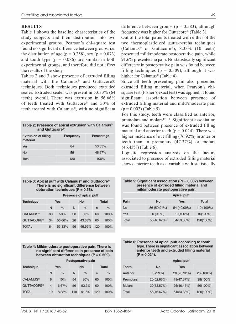
RESULTSTable 1 shows the baseline characteristics of thestudy subjects and their distribution into twoexperimental groups. Pearson’s chisquare testfound no significant difference between groups, i.e.the distribution of age (p = 0.258), sex (p = 0.073)and tooth type (p = 0.086) are similar in bothexperimental groups, and therefore did not affectthe results of the study. Tables 2 and 3 show presence of extruded fillingmaterial with the Calamus® and Guttacore®techniques. Both techniques produced extrudedsealer. Extruded sealer was present in 53.33% (64teeth) overall. There was extrusion in 56.66% of teeth treated with Guttacore® and 50% of teeth treated with Calamus®, with no significant
difference between groups (p = 0.583), althoughfrequency was higher for Guttacore® (Table 3).Out of the total patients treated with either of thetwo thermoplasticized guttapercha techniques(Calamus® or Guttacore®), 8.33% (10 teeth)presented mild/moderate postoperative pain, while91.6% presented no pain. No statistically significantdifference in postoperative pain was found betweenfilling techniques (p = 0.509), although it washigher for Calamus® (Table 4).Since all teeth presenting pain also presentedextruded filling material, when Pearson’s chisquare test (Fisher’s exact test) was applied, it foundsignificant association between presence ofextruded filling material and mild/moderate pain(p = 0.002) (Table 5). For this study, teeth were classified as anterior,premolars and molars17, 33. Significant associationwas found between presence of extruded fillingmaterial and anterior teeth (p = 0.024). There washigher incidence of overfilling (76.92%) in anteriorteeth than in premolars (47.37%) or molars(46.43%) (Table 6). Logistic regression analysis on the factorsassociated to presence of extruded filling materialshows anterior teeth as a variable with statistically
Overfilling and associated factors 49
Vol. 31 Nº 1 / 2018 / 45-52 ISSN 1852-4834 Acta Odontol. Latinoam. 2018
Table 2: Presence of apical extrusion with Calamus®
and Guttacore®.
Extrusion of filling Frequency Percentagematerial
Yes 64 53.33%
No 56 46.67%
Total 120 100%
Table 3: Apical puff with Calamus® and Guttacore®. There is no significant difference betweenobturation techniques (P = 0.58).
Presence of apical puff
Technique Yes No Total
N % N % n %
CALAMUS® 30 50% 30 50% 60 100%
GUTTACORE® 34 56.66% 26 43.33% 60 100%
TOTAL 64 53.33% 56 46.66% 120 100%
Table 4: Mild/moderate postoperative pain. There is no significant difference in presence of painbetween obturation techniques (P = 0.509).
Postoperative pain
Technique Yes No Total
N % N % n %
CALAMUS® 6 10% 54 90% 60 100%
GUTTACORE® 4 6.67% 56 93.3% 60 100%
TOTAL 10 8.33% 110 91.6% 120 100%
Table 5: Significant association (Pr = 0.002) between presence of extruded filling material andmild/moderate postoperative pain.
Apical puff
Pain No Yes Total
No 56 (50.91%) 54 (49.09%) 110 (100%)
Yes 0 (0.0%) 10(100%) 10(100%)
Total 56(46.67%) 64(53.33%) 120(100%)
Table 6: Presence of apical puff according to tooth type. There is significant association betweenanterior teeth and extruded filling material (P = 0.024).
Apical puff
Tooth No Yes
Anterior 6 (23%) 20 (76.92%) 26 (100%)
Premolars 20(52.63%) 18(47.37%) 38(100%)
Molars 30(53.57%) 26(46.43%) 56(100%)
Total 56(46.67%) 64(53.33%) 120(100%)
AOL12018:32011 18/07/2018 17:30 Página 49

significant association in the raw model (OR 3.70;P = 0.01). In the model adjusted with the variablessex, age and filling system, the same factor (anteriorteeth) was the only one with statistically significantassociation (p = 0.01) with OR 4.35, which meansthat the risk of extruded filling material from a rootcanal in an anterior tooth is 4 times higher than in apremolar or molar.
DISCUSSIONThis randomized clinical experiment was conductedto evaluate what factors could influence the pre sence of accidental apical overfilling or extrusionof sealer. Following randomization, a statisticalverification confirmed that factors were distributedbetween groups in similar proportions, enablingdismissal of the possibility of results being affectedby any imbalance. This is important to note becauseapical foramen diameter varies according to age34
and differs between anterior and posterior teeth35,so confirmation of similar distribution betweengroups provides reliability to the results. Overall, with the Calamus® and Guttacore® obtu ration techniques used in this study there wasextrusion of filling material in 53.33%. This valuediffers from Tennert et al.4, who report 80%extrusion of material with Thermafil® and 42% with a vertical condensation filling technique4. Inaddition, the present study found no significantdifference in extrusion of filling material betweenthe two techniques. The difference between Tennertet al. and the current study may be due to studymethodological design. Tennert et al. conducted aretrospective study with little control over inclusioncriteria, whereas the current study was prospective,with randomized distribution into groups, andcontrolled both the procedures performed and theinclusion criteria. De Chevigny et al.36 found 53% extrusion of fillingmaterial in a sample grouping the Toronto studyphases 1 to 4, which is in agreement with the53.33% reported in the current study. However, DeChevigny et al. 36 used lateral condensation andvertical condensation filling techniques, so clearly,filling material extrusion or overfilling may occurregardless of the filling technique and without thepossibility of being predicted with total certainty14,although the outcome is generally functional19, 21, 36,so that it may be considered an accident with goodprognosis.
Said good prognosis may be owed to the periapicaltissue’s tolerance to the cytotoxicity of the fillingmaterial (guttapercha and sealer)37, 38, although iffilling material extrudes and affects neighboringanatomical structures, it may lead to consequenceswhich could cause patient discomfort16, 2427, 39. Here,it is important to stress that it is not possible to predicthow much filling material (sealer or guttapercha)will extrude or how far it will reach14, 39, in additionto which extruded material can only be detectedradiographically after the obturation has beencompleted. It is therefore important to endeavor tokeep the obturation material within the root canal,limiting extrusion to what it really is: an accidentwith good prognosis, not an aim of the obturation. Wong et al.40 conducted a metaanalysis observingoverfilling and postoperative pain in teeth with rootcanals filled with Thermafil® system, findingpostoperative pain in 24%, higher than the 8.33%found in the current study. Wong et al.40 claim thatpostoperative pain was due to the extrusion offilling material40. This explanation may also applyin the current study, which found that all teethpresenting postoperative pain also had extrudedfilling material. AlRahabi17 report average overfilling of 44.15% inmolars (62.5% in upper molars and 25.8% in lowermolars), similar to the overall 46.43% found formolars in the current study. However, for anteriorteeth, AlRahabi, report 16.25% (20% for upperincisors and 12.5% for lower incisors), which ismuch lower than the 76.92% found in the currentstudy. Differences in treatment protocols mayexplain this inconsistency between results. Thesedifferences include that the current study determinedworking length with an apex locator (Root ZX II® JMorita®.) and confirmed it radiographically, whileAlRahabi17 only took a conductometry radiograph,and that in AlRahabi the root canal treatments wereperformed by undergraduate fourth or fifth yearstudents, while in the current study they wereperformed by postgraduate students of endodontics. The statistically significant association found betweenextrusion of filling material and anterior teeth may beexplained by two reasons: (a) as has been reportedpreviously, it is easier to fill anterior than posteriorteeth,14, 17 and (b) the difference in the diameter of theapical foramen between anterior and posterior teeth35. Statistically significant association between extru sion of filling material and anterior teeth has not
50 Javier I. Nino-Barrera, et al.
Acta Odontol. Latinoam. 2018 ISSN 1852-4834 Vol. 31 Nº 1 / 2018 / 45-52
AOL12018:32011 18/07/2018 17:30 Página 50

been reported previously in the literature, soanterior teeth should be taken into account as a riskfactor for extruded filling material. Special careshould thus be taken to maintain a stable apical limitduring biomechanical preparation of anterior teethto prevent extrusion of filling material.Within the limitations of this study, it may beconcluded that the endodontic thermoplastic filling
techniques with Guttacore® and the Calamus®
system are significantly associated to the probabilityof filling material extrusion. In this regard, there isno difference between techniques and overfilling is associated with mild/moderate postoperativepain. Anterior teeth are 4 times more likely to beoverfilled than premolars and molars with thesefilling techniques.
Overfilling and associated factors 51
Vol. 31 Nº 1 / 2018 / 45-52 ISSN 1852-4834 Acta Odontol. Latinoam. 2018
ACKNOWLEDGMENTSWe thank Universidad El Bosque and its directorship of postgraduate degrees for making this study possible by means offinancial support received through the Sixth Internal Call forresearch and technological innovation funding projects.
CORRESPONDENCEDr. Javier NiñoBarreraCalle 135 # 58B 21 Interior 2 Apartamento 302Bogotá, [email protected]
REFERENCES 1. Tomson RM, Polycarpou N, Tomson PL. Contemporary
obturation of the root canal system. Br Dent J 2014;216:315322.
2. Swartz DB, Skidmore AE, Griffin JA, Jr. Twenty years ofendodontic success and failure. J Endod 1983;9:198202.
3. Farias AB, Pereira KF, Beraldo DZ, Yoshinari FM, ArashiroFN, Zafalon EJ. Efficacy of three thermoplastic obturationtechniques in filling ovalshaped root canals. Acta OdontolLatinoam 2016;29:7681.
4. Tennert C, Jungback IL, Wrbas KT. Comparison betweentwo thermoplastic root canal obturation techniquesregarding extrusion of root canal filling—a retrospective invivo study. Clin Oral Investig 2013;17:449454.
5. Gupta R, Dhingra A, Panwar NR. Comparative Evaluationof Three Different Obturating Techniques Lateral Compaction,Thermafil and Calamus for Filling Area and Voids UsingCone Beam Computed Tomography: An Invitro study. JClin Diagn Res 2015;9:ZC1517.
6. Schilder H. Filling root canals in three dimensions. DentClin North Am 1967:723744.
7. Brothman P. A comparative study of the vertical and thelateral condensation of guttapercha. J Endod 1981;7:2730.
8. Ruddle CJ. Filling root canal systems: the Calamus 3Dobturation technique. Dent Today 2010;29:76, 7881.
9. Jindal D, Sharma M, Raisingani D, Swarnkar A, Pant M,Mathur R. Volumetric analysis of root filling with coldlateral compaction, Obtura II, Thermafil, and Calamususing spiral computerized tomography: An In vitro Study.Indian J Dent Res 2017;28:175180.
10. Zogheib C, Hanna M, Pasqualini D, Naaman A. Quantitativevolumetric analysis of crosslinked guttapercha obturators.Ann Stomatol (Roma) 2016;7:4651.
11. Schafer E, Schrenker C, Zupanc J, Burklein S. Percentageof Guttapercha Filled Areas in Canals Obturated withCrosslinked Guttapercha Corecarrier Systems, SingleCone and Lateral Compaction Technique. J Endod 2016;42:294298.
12. MarquesFerreira M, Abrantes M, Ferreira HD, CarameloF, Botelho MF, Carrilho EV. Sealing efficacy of system B
versus Thermafil and Guttacore obturation techniquesevidenced by scintigraphic analysis. J Clin Exp Dent 2017;9:e56e60.
13. Kandemir Demirci G, Caliskan MK. A ProspectiveRandomized Comparative Study of Cold Lateral Conden sation Versus Core/Guttapercha in Teeth with PeriapicalLesions. J Endod 2016;42:206210.
14. Mozayeni MA, Asnaashari M, Modaresi SJ. Clinical andRadiographic Evaluation of Procedural Accidents and Errorsduring Root Canal Therapy. Iran Endod J 2006;1:97100.
15. Koch K, Brave D. A new endodontic obturation technique.Dent Today 2006;25:102, 104107.
16. Morse DR. Infectionrelated mental and inferior alveolarnerve paresthesia: literature review and presentation of twocases. J Endod 1997;23:457460.
17. AlRahabi MK. Evaluation of complications of root canaltreatment performed by undergraduate dental students.Libyan J Med 2017;12:1345582.
18. Iqbal A. The Factors Responsible for Endodontic TreatmentFailure in the Permanent Dentitions of the PatientsReported to the College of Dentistry, the University ofAljouf, Kingdom of Saudi Arabia. J Clin Diagn Res 2016;10:ZC146148.
19. Ricucci D, Rocas IN, Alves FR, Loghin S, Siqueira JF, Jr.Apically Extruded Sealers: Fate and Influence onTreatment Outcome. J Endod 2016;42:243249.
20. Sadaf D, Ahmad MZ. Factors associated with postoperativepain in endodontic therapy. Int J Biomed Sci 2014;10:243247.
21. Marquis VL, Dao T, Farzaneh M, Abitbol S, Friedman S.Treatment outcome in endodontics: the Toronto Study.Phase III: initial treatment. J Endod 2006;32:299306.
22. Schaeffer MA, White RR, Walton RE. Determining theoptimal obturation length: a metaanalysis of literature. JEndod 2005;31:271274.
23. Sarin A, Gupta P, Sachdeva J, Gupta A, Sachdeva S, NagpalR. Effect of Different Obturation Techniques on the Prognosisof Endodontic Therapy: A Retrospective ComparativeAnalysis. J Contemp Dent Pract 2016;17:582586.
24. Rosen E, Goldberger T, Taschieri S, Del Fabbro M, CorbellaS, Tsesis I. The Prognosis of Altered Sensation after
AOL12018:32011 18/07/2018 17:30 Página 51

Extrusion of Root Canal Filling Materials: A SystematicReview of the Literature. J Endod 2016;42:873879.
25. Nitzan DW, Stabholz A, Azaz B. Concepts of accidentaloverfilling and overinstrumentation in the mandibular canalduring root canal treatment. J Endod 1983;9:8185.
26. Blanas N, Kienle F, Sandor GK. Injury to the inferioralveolar nerve due to thermoplastic gutta percha. J OralMaxillofac Surg 2002;60:574576.
27. Blanas N, Kienle F, Sandor GK. Inferior alveolar nerveinjury caused by thermoplastic guttapercha overextension.J Can Dent Assoc 2004;70:384387.
28. AlonsoEzpeleta LO, GascoGarcia C, CastellanosCosanoL, MartinGonzalez J, LopezFrias FJ, SeguraEgea JJ.Postope rative pain after onevisit rootcanal treatment onteeth with vital pulps: comparison of three differentobturation techniques. Med Oral Patol Oral Cir Bucal 2012;17:e721727.
29. Top Seal Instructions for use Maillefer Instruments SA CH1338 Ballaigues Switzerland [Internet]2014.URL: http://www.dentsplymaillefer.com/wpcontent/uploads/2015/Dentsply/Obturation/Ciments/TOPSEAL/produit49_EN.pdf.
30. DENTSPLY Tulsa Dental Specialties DI, Inc.DentsplySirona, Inc. GuttaCore® Crosslinked GuttaPercha CoreObturators Directions for Use. StepbyStep Instructions, 7Drying the Canal and Applying Sealer. 608 Rolling HillsDr. Johnson City, TN 37604: 2017. p. pag 7.URL: www.dentsplysirona.com;
31. Aminoshariae A, Kulild JC, Donaldson M, Hersh EV. Evidencebased recommendations for analgesic efficacy to treat pain ofendodontic origin: A systematic review of randomizedcontrolled trials. J Am Dent Assoc 2016;147:826839.
32. Mehrvarzfar P, Esnashari E, Salmanzadeh R, Fazlyab M,Fazlyab M. Effect of Dexamethasone IntraligamentaryInjection on PostEndodontic Pain in Patients withSymptomatic Irreversible Pulpitis: A RandomizedControlled Clinical Trial. Iran Endod J 2016;11:261266.
33. Hale R, Gatti R, Glickman GN, Opperman LA. Comparativeanalysis of carrierbased obturation and lateral compaction:a retrospective clinical outcomes study. Int J Dent 2012;2012:954675.
34. Fang Y, Wang X, Zhu J, Su C, Yang Y, Meng L. Influenceof Apical Diameter on the Outcome of RegenerativeEndodontic Treatment in Teeth with Pulp Necrosis: AReview. J Endod 2018;44:414431.
35. Vertucci FJ. Root canal morphology and its relationship toendodontic procedures. Endod Topics 2005;10:329.
36. de Chevigny C, Dao TT, Basrani BR, Marquis V, FarzanehM, Abitbol S, Friedman S. Treatment outcome in endo dontics: the Toronto study—phase 4: initial treatment. JEndod 2008;34:258263.
37. Scotti R, Tiozzo R, Parisi C, Croce MA, Baldissara P.Biocompatibility of various root canal filling materials exvivo. Int Endod J 2008;41:651657.
38. Tavares T, Soares IJ, Silveira NL. Reaction of rat subcu taneous tissue to implants of guttapercha for endodonticuse. Endod Dent Traumatol 1994;10:174178.
39. Yamaguchi K, Matsunaga T, Hayashi Y. Gross extrusion ofendodontic obturation materials into the maxillary sinus: acase report. Oral Surg Oral Med Oral Pathol Oral RadiolEndod 2007;104:131134.
40. Wong AW, Zhang S, Li SK, Zhang C, Chu CH. Clinicalstudies on corecarrier obturation: a systematic review andmetaanalysis. BMC Oral Health 2017;17:167.
52 Javier I. Nino-Barrera, et al.
Acta Odontol. Latinoam. 2018 ISSN 1852-4834 Vol. 31 Nº 1 / 2018 / 45-52
AOL12018:32011 18/07/2018 17:30 Página 52

RESUMEN La enfermedad periodontal y su repuesta inflamatoria ha sidorelacionada con desenlaces adversos del embarazo como elparto pretérmino, preeclampsia y bajo peso al nacer. Lapresente investigación analizó la respuesta inflamatoriasistémica en pacientes embarazadas con alto riesgo de partopretérmino y su relación con la enfermedad periodontal. Serealizó un estudio piloto de casos y controles, en el cual se contócon 23 pacientes que presentaban riesgo de parto pretérminocomo casos y 23 pacientes sin riesgo de parto pretérmino comocontroles. Fueron excluidas las pacientes que hubieran recibidotratamiento periodontal, antibióticos o antimicrobianos en losúltimos tres meses, que tuvieran infecciones, o enfermedades debase como diabetes o hipercolesterolemia. A todas las pacientesse les hicieron valoración periodontal, exámenes de laboratorio
(cuadro hemático, perfil lipídico, glucemia basal) y cuanti ficación de citocinas (IL2, IL4, IL6, IL10, TNF α e INFγ).En las pacientes con periodontitis crónica se encontraronniveles más elevados en las citocinas proinflamatorias (IL2,IL4, IL6, IL10, TNF α e INFγ) en comparación con laspacientes con gingivitis o sanas periodontales. Estas citocinasse encontraron más elevadas en las pacientes con alto riesgode parto pretérmino, en especial la IL2, IL10 y TNF α. Laspacientes con alto riesgo de parto pretérmino presentaronmayor severidad de la enfermedad periodontal y adicional mente niveles aumentados de los marcadores pro inflamatoriosIL2, IL4, IL6, IL10, TNF α e INFγ.
Palabras clave: periodontitis crónica, embarazo, nacimientoprematuro, parto prematuro, citocinas.
ABSTRACTPeriodontal disease and its inflammatory response have beenrelated to adverse outcomes in pregnancy such as preterm birth,preeclampsia and low birth weight. This study analyzed systemicinflammatory response in patients with high risk of pretermdelivery and its relationship to periodontal disease. A pilot studywas conducted for a case and control study, on 23 patients at riskof preterm delivery and 23 patients without risk of pretermdelivery as controls. Exclusion criteria were patients who hadreceived periodontal treatment, antibiotic or antimicrobial agentswithin the past three months, or with infections or baselinediseases such as diabetes or hypercholesterolemia. All patientsunderwent periodontal assessment, laboratory tests (complete
blood count, lipid profile, baseline glycemia) and quantificationof cytokines (IL2, IL4, IL6, IL10, TNFα and INFγ). Higherlevels of proinflammatory cytokines (IL2, IL4, IL6, IL10,TNFα and INFγ) were found in patients with chronicperiodontitis than in patients with gingivitis or periodontal health.These cytokines, in particular IL2, IL10 and TNF α, werehigher in patients at high risk of preterm delivery. Patients withhigh risk of preterm delivery had higher severity of periodontaldisease as well as higher levels of the proinflammatory markersIL2, IL4, IL6, IL10, TNF α and INFγ.
Key words: Chronic periodontitis, Pregnancy, Preterm birth,Preterm labor, cytokines.
53
Vol. 31 Nº 1 / 2018 / 53-57 ISSN 1852-4834 Acta Odontol. Latinoam. 2018
Inflamatory response in pregnant women with high risk of preterm delivery and its relationship with periodontal disease. A pilot study
Francina Escobar-Arregoces1, Catalina Latorre-Uriza1, Juliana Velosa-Porras1, Nelly Roa-Molina1, Alvaro J Ruiz2, Jaime Silva3, Estefania Arias4, Juliana Echeverri4
1 Pontificia Universidad Javeriana. Facultad de Odontología. Centro de Investigaciones Odontológicas, CIO. Bogotá D.C. Colombia.
2 Pontificia Universidad Javeriana. Facultad de Medicina. Departamento de Epidemiologia Clínica. Bogotá D.C. Colombia.
3 Hospital San Ignacio. Departamento de Ginecología y Obstetricia, Bogotá D.C. Colombia.
4 Practica Privada en Periodoncia. Colombia.
Respuesta inflamatoria en pacientes embarazadas con alto riesgo de parto pretérmino y su relación con la enfermedad periodontal. Estudio piloto
AOL12018:32011 18/07/2018 17:30 Página 53

INTRODUCTIONPeriodontal disease is a chronic infectious diseasearound the teeth which triggers a local and systemicinflammatory response1. The bacterial challengegenerates an inflammatory infiltrate of polymorpho nuclear neutrophils, monocytes, Blymphocytesand Tlymphocytes with release of cytokines suchas IL1, IL6 IL8, IL10 and PGE2, among others,which upon entering the bloodstream can reach theuterus and pass through the fetal placental barrier 2 4. Normal labor is controlled by inflammatory signaling.As the pregnancy progresses, PGE2, TNFα and IL1β levels increase to critical levels, which induce therupture of the amniotic sac, followed by the beginningof uterine contractions and delivery 5,6 This immuneprocess, which is selfregulated up to the ninth month, may be modified by external stimuli such asperiodontal infection, which by triggering a systemicinflammatory response during pregnancy, couldinduce labor earlier than necessary. Preterm birth isdefined as birth at fewer than 37 weeks gestational age7. Global incidence of preterm birth is about 9.6%,which is equivalent to 12.9 million preterm babies 79.It is a frequent problem which even affects highincome countries and has a strong impact on maternaland child morbidity and mortality, affecting bothfamilies and healthcare systems10,11.The aim of this study was to analyze the systemicinflammatory response in pregnant patients withhigh risk of preterm delivery and its relationshipwith periodontal disease.
MATERIALS AND METHODS A pilot test was conducted for a case and control study.The sample consisted of 46 patients – 23 at risk ofpreterm delivery and 23 without that risk as controls.Risk of preterm delivery was diagnosed following theClinical Practice of the Obstetrics and GynecologyService at Hospital San Ignacio12, which definepreterm delivery as follows: 1. Risk of preterm delivery: patients with uterineactivity without cervical changes. 2. Preterm labor,initial phase: patients with regular uterine activityand cervical changes less than 4 cm y 3. Pretermlabor, advanced phase: patients with uterine activitywith cervical changes greater than 4 cm.The patients who took part in the study as caseswere hospitalized and at risk of phase 1 and 2preterm labor. The patients in the control groupwere pregnant women who attended their normal
prenatal checkup as outpatients at the same hospitaland Javesalud. Exclusion criteria for both groupswere having received periodontal treatment,antibiotics or antimicrobial agents within the pastthree months, or having active infections or baselinediseases such as diabetes or hypercholesterolemia. According to Colombia’s Ministry of Health13 and theCIOMS14, this study was classified as investi gationwith minimum risk and was approved by the researchethics committees of the School of Medicine (FMCIE841215) and the School of Dentistry (CIEFOUJ OD0131) of Universidad Javeriana. Before enrolling inthe study, each patient signed an informed consent.Blood samples were taken to determine full bloodcount, glycemia, total cholesterol, triglycerides,cHDL, cLDL and quantification of the inflammatory markers IL2, IL4, IL6, IL10, TNFα andINFγ using the BD Cytometric Bead Array HumanTh1/Th2 Cytokine Kit II. The Beckton DickinsonFACSCanto II flow cytometer acquired 50,000events and the data were analyzed using FCAPArray software. Concentrations were expressed inpg/ml. Periodontal disease was diagnosed based onthe Armitage classification, 1999.15 (Fig. 1).To analyze the information, a description was made of the demographics, results of periodontalevaluation and systemic inflammatory markersusing means, medians, ranges, standard deviationsand 95% confidence intervals. For the bivariateanalysis, the Chisquare test was used to establishassociations and the ANOVA test to establishdifferences between the averages for the variablesof interest. In addition, the KolmogorovSmirnov testwas performed to establish whether the inflammatorymarkers had normal behavior. Variables in whichbehavior was not normal were compared using theUMann Whitney test. A difference of p < 0.05(twotailed) was considered significant.
RESULTSGeneral characteristicsFortysix patients were analyzed. Average age was25.91 ± 6.4 years and average gestational age was29.46 ± 4.3 weeks. For periodontal status, 50.0%had chronic periodontitis, 45.7% had gingivitisassociated to biofilm and 4.3% periodontal health.Upon describing according to groups, average agefor both groups was 26 years, and gestational agewas 31 weeks for the group with high risk and 28weeks for the group without risk (p = 0.466).
54 Francina Escobar-Arregoces, et al.
Acta Odontol. Latinoam. 2018 ISSN 1852-4834 Vol. 31 Nº 1 / 2018 / 53-57
AOL12018:32011 18/07/2018 17:30 Página 54

Oral clinical characteristicsNo statistically significant differencewas found for number of teeth orO’Leary’s plaque index. The group withhigh risk of preterm delivery included alarger number of patients diagnosed withchronic periodontitis than the groupwithout risk (Table 1).
Cytokine quantificationPatients in the group with high risk ofpreterm delivery had higher levels ofcytokines than patients without risk,with significant statistical differencesfor TNFα, IL10 and IL2 (Table 2). Forrelationship between cytokines and periodontal diagnoses, patients with chronicperiodontitis tended to have higher levels of proinflammatory cytokinesthan patients with gingivitis or periodontal health. Although the trendwas towards higher values of cytokinesin patients with periodontitis, it shouldbe noted that there was no significantstatistical difference in cytokine concen
tration according to diagnosis. Since the variablesdid not have normal behavior, a Kruskall Wallis testwas used, which showed no significant differencein distribution between groups (Table 3).
DISCUSSION Current evidence suggests that preterm deliveryoriginates mainly from ascending infections from thevagina or uterine cervix, or bloodborne propagation
Periodontal disease and pregnancy 55
Vol. 31 Nº 1 / 2018 / 53-57 ISSN 1852-4834 Acta Odontol. Latinoam. 2018
Fig. 1: Flow diagram showing patient enrollment and testing. The flowdiagram shows patients enrolled in the study, with the subgroups formed forCases (pregnant women with risk of preterm delivery) and controls(pregnant women without risk of preterm delivery). All patients signedinformed consent, underwent periodontal clinical assessment and had ablood sample taken for full blood count, glycemia, total cholesterol, HDL,LDL and cytokine quantification.
Table 1: Clinical characteristics of pregnant women with high risk and without risk of pretermdelivery.
Clinical characteristics High risk Without risk
Number of teeth 28.0 27.9
Plaque index % 61.9 45.7
Patients with chronic periodontitis 14 9
Patients with gingivitis 9 12
Healthy patients 0 2
Table 2: Cytokine quantification in pregnant women with high risk and without risk of pretermdelivery.
Cytokines High risk Without risk P value
IFN-γ 21.74 pg/ml 0.10 pg/ml 0.241
TNF-α 36.63 pg/ml 0.02 pg/ml 0.010
IL-10 21.30 pg/ml 0.34 pg/ml 0.012
IL-6 17.20 pg/ml 2.06 pg/ml 0.429
IL-4 53.79 pg/ml 0.11 pg/ml 0.355
IL-2 20.93 pg/ml 0.19 pg/ml 0.013
Statistically significant p-value < 0.05.
Table 3: Relationship between cytokines and periodontal diagnosis.
Cytokine Periodontal diagnosis
Healthy Gingivitis Chronic P-valueperiodontitis
IFN-γ 0.00 pg/mL 0.05 pg/mL 21.79 pg/mL 0.147
TNF-α 0.00 pg/mL 0.06 pg/mL 36.59 pg/mL 0.459
IL-10 0.00 pg/mL 0.40 pg/mL 21.27 pg/mL 0.053
IL-6 1.11 pg/mL 1.62 pg/mL 17.68 pg/mL 0.155
IL-4 0.00 pg/mL 0.16 pg/mL 53.76 pg/mL 0.699
IL-2 0.00 pg/mL 0.38 pg/mL 20.78 pg/mL 0.213
Statistically significant p-value < 0.05. Kruskal-Wallis test was used,enabling three variables to be compared at the same time.
AOL12018:32011 18/07/2018 17:30 Página 55

from nongenital sources. Maternal periodontitis is anongenital source of microorganisms which areknown to routinely enter the bloodstream and to havepotential to influence the health of the fetoplacentalunit7. This study evaluated systemic inflammatory responsein pregnant patients with and without risk ofpreterm delivery and its relationship with periodon tal disease. The results showed that patients withhigh risk of preterm delivery had higher levels of cytokines IL2, IL4, IL6, IL10, TNFα andINFγ. These findings are in agreement with Ingliset al. 16, Gursoy et al. 17, von Minckwitz et al. 18 andTurhan et al. 19, which associate increased systemicinflammatory marker levels with adverse events inpregnancy. Inflammatory cytokines play a key role in themechanisms that trigger labor. This study foundincreased quantities of IL6 and IL10 in patientswith risk of preterm delivery, in agreement withArntzen et al. 20, Romero et al. 21 and Wommack et al. 22. The current study also found that the groupwith risk of preterm delivery included a largernumber of patients diagnosed with chronic perio don titis than the group without risk. This is inagreement with Offenbacher et al. 23, who report forthe first time a relationship between maternalperiodontal disease and preterm birth. Upon analyzing presence of cytokines with relationto periodontal status, it was found that the group ofpatients with periodontitis tended to have higherlevels of cytokines IL2, IL4, IL6, IL10, TNFαand INFγ; in agreement with Kelso24 and Zadeh etal.25, who relates periodontal infection to a systemicinflammatory response, reporting an increase in IL5, IL6, IL4, IL 10 IL13 IL3 and TNFα inpatients with periodontitis. Similarly, Perunovik et al. 26 found that women with preterm delivery hadsignificantly more periodontitis and higher levelsof IL6 and PGE2 in crevicular fluid than the termdelivery group.
Studies in recent years have focused on the effectof periodontal treatment on the reduction of proinflammatory markers and its relationship withpreterm delivery. Offenbacher et al.27 report thatperiodontal treatment showed a reduction inpreterm delivery rate by 3.8 times, and a decreasein IL1β and IL6. PenovaVeselinovic,28 found thatin the group with periodontal treatment there was asignificant reduction in levels of IL1β, IL10, IL12p70 and IL6 compared to controls. Da Silva et al.29 report in a metaanalysis that nonsurgicalperiodontal therapy during pregnancy reduced thelevel of inflammatory biomarkers in gingivalcrevicular fluid and serum, without influence on thelevel of inflammatory biomarkers in umbilical cordblood and without reducing the occurrence ofadverse gestational outcomes.However, in contrast to the above, studies such as Michalowicz et al. 30, Fiorini T et al.31 and Pirie M et al.32 report that nonsurgical periodontaltreatment in pregnant women did not reducesystemic inflamma tion markers.Considering that preterm delivery is a public healthissue, preventive measures should be taken, includingpromoting periodontal health in pregnant women.Unfortunately, as reported by Duque et al.33 in 2011,physicians’ knowledge and attitudes regardingperiodontal disease and general health status are stilldeficient. Greater efforts are therefore needed to raiseawareness among the different groups of scientists andspecialists regarding the importance of maintaininggood periodontal health during the gestation period,given the biological plausibility for etiology of pretermdelivery shown by periodontal disease. Within the limitations of the current study, it maybe concluded that patients with high risk of pretermdelivery presented greater severity of periodontaldisease as well as increased levels of the proinflammatory markers IL2, IL4, IL6, IL10,TNF α and INFγ, with statistically significantdifferences for IL2, IL10 and TNFα.
56 Francina Escobar-Arregoces, et al.
Acta Odontol. Latinoam. 2018 ISSN 1852-4834 Vol. 31 Nº 1 / 2018 / 53-57
ACKNOWLEDGMENTSThe authors thank Javesalud for help with obtaining the sample, the clinical laboratory Hospital Universitario San Ignacio,Flow Cytometry department, and Beckton Dickinson for adviceand technical support. This study was financed by the Office ofthe ViceRector of Research at Pontificia Universidad Javeriana, Bogotá, Colombia.
CORRESPONDENCE Dr. Francina Maria Escobar ArregocesCarrera 7 # 4062 Universidad Javeriana, Facultad de Odontología Bogotá, Colombia [email protected]
AOL12018:32011 18/07/2018 17:30 Página 56

REFERENCES1. Contreras A, Herrera JA, Soto JE, Arce RM, et al.
Periodontitis is associated with preeclampsia in pregnantwomen. J Periodontol. 2006; 77:182188.
2. Van Dyke TE, Van Winkelhoff AJ. Infection and inflammatorymechanisms. J Clin Periodontol. 2013; 40:S17.
3. Page RC, Offenbacher S, Schroeder HE, Seymour GJ, et al.Advances in the pathogenesis of periodontitis: summary ofdevelopments, clinical implications and future directions.Periodontol 2000. 1997; 14:216248.
4. Ardila CM, Lafaurie GI. Asociación entre porphyromonagingivalis y proteína C reactiva en enfermedades sistémicasinflamatorias. Av Periodon Implantol 2010; 22:4553.
5. Armitage GC. Bidirectional relationship between pregnancyand periodontal disease. Periodontol 2000. 2013; 61:160176.
6. Madianos PN, Bobetsis YA, Offenbacher S. Adversepregnancy outcomes (APOs) and periodontal disease:pathogenic mechanisms. J Clin Periodontol 2013; 40:S170180 doi:10.1111/jcpe.12082
7. Sanz M, Kornman K. Periodontitis and adverse pregnancyoutcomes: consensus report of the Joint EFP/AAP Workshopon Periodontitis and Systemic Diseases. J Clin Periodontol2014; 40:S164169. doi:10.1111/jcpe.1208
8. Saini N., Walia M. Relationship between periodontaldiseases and preterm birth: Recent epidemiological andbiological data. Int J Appl Basic Med Res 2015; .5, 2. doi:10.4103/2229516x.149217
9. Villanueva L, Contreras AK, Pichardo M, Rosales J. Perfilepidemiológico del parto prematuro. Artículo de revisión.Ginecol Obstet Mex 2008; 76:542548.
10. Ide M, Papapanou PN. Epidemiology of association betweenmaternal periodontal disease and adverse pregnancy outcomes systematic review. J Clin Periodontol 2013; 40:181194.
11. Castaldi JL, Bertin MS, Giménez F, Roberto L. Enfermedadperiodontal: ¿es factor de riesgo para parto pretérmino, bajopeso al nacer o preeclampsia? Rev Panam Salud Pública.2006; 19:253258.
12. Guía Institucional Hospital Universitario San Ignacio.Intranet Almera. Guía Práctica Clínica Parto Pretérmino.2014.
13. Normas científicas, técnicas y administrativas para la investi gación en salud, Resolución 8430 de 1993, República deColombia. Ministerio de Salud, (1993).URL: www.dib.unal.edu.co/ promocion/etica_res_8430_1993.pd
14. Consejo de las Organizaciones Internacionales de CienciasMédicas (CIOMS). Guías Éticas Internacionales para laInvestigación Biomédica que involucra Seres Humanos.[Programa Regional de Bioética OPS/OMS]. 282010.URL: www.paho.org/Spanish/BIO/CIOMS
15. Armitage G. Diagnóstico y clasificación de las enferme dades periodontales. Periodontol 2000. 2005; 9:921.
16. Inglis SR. Biochemical markers predictive of pretermdelivery. Infect Dis Obstet Gynecol. 1997; 5:158164.
17. Gürsoy M, Könönen E, Gürsoy UK, Tervahartiala T, et al .Periodontal Status and Neutrophilic Enzyme Levels inGingival Crevicular Fluid During Pregnancy and Postpartum.J Periodontol 2010; 18:17901796.
18. von Minckwitz G, Grischke EM, Schwab S, Hettinger S, etal. Predictive value of serum interleukin6 and 8 levels in
preterm labor or rupture of the membranes. Acta ObstetGynecolo Scand. 2000; 79:667672.
19. Turhan NO, Karabulut A, Adam B. Maternal serum interleukin 6 levels in preterm labor: prediction of admissiontodelivery interval. J Perinat Med. 2000; 28:133139.
20. Arntzen KJ, Kjøllesdal AM, Halgunset J, Vatten L, et al.TNF, IL1, IL6, IL8 and soluble TNF receptors in relationto chorioamnionitis and premature labor. J Perinat Med.1998; 26:1726.
21. Romero R, Avila C, Santhanam U, Sehgal P. Amniotic fluidInterleukin 6 in preterm labor. Association with infection. JClin Invest. 1990; 85:13921400.
22. Wommack JC, Ruiz RJ, Marti CN, Stowe RP et al.Interleukin10 predicts preterm birth in acculturatedhispanics. Biol Res Nurs. 2013; 15:7885.
23. Offenbacher S, Jared HL, O’Reilly PG, Wells SR, et al.Potential pathogenic mechanisms of periodontitis associatedpregnancy complications. Ann Periodontol. 1998; 3:233250.
24. Kelso A. Th1 and Th2 subsets: paradigms lost? ImmunolToday. 1995; 16:374379.
25. Zadeh HH, Nichols FC, Miyasaki KT. The role of the cellmediated immune response to Actinobacillusactinomycetemcomitans and Porphyromonas gingivalis inperiodontitis. Periodontol 2000. 1999; 20:239288.
26. Perunovic ND, Rakic MM Nikolic LI, Jankovic SM, et al.The Association Between Periodontal Inflammation andLabor Triggers (Elevated Cytokine Levels) in PretermBirth: A CrossSectional Study. J Periodontol 2016; Mar;87:248256.
27. Offenbacher S, Lin D, Strauss R, McKaig R, et al. Effectsof periodontal therapy during pregnancy on periodontalstatus, biologic parameters, and pregnancy outcomes: apilot study. J Periodontolol 2006; 77:20112024.
28. PenovaVeselinovic B, Keelan JA, Wang CA, NewnhamJP, et al. Changes in inflammatory mediators in gingivalcrevicular fluid following periodontal disease treatment inpregnancy: Relationship to adverse pregnancy outcome. JReprod Immunol 2015; 112:110.
29. Da Silva HEC, Stefani CM, de Santos Melo N, de Almeidade Lima A, et al. Effect of intrapregnancy nonsurgicalperiodontal therapy on inflammatory biomarkers andadverse pregnancy outcomes: a systematic review withmetaanalysis. Syst Rev. 2017 Oct 10;6:197. doi: 10.1186/s1364301705873.
30. Michalowicz BS, Novak MJ, Hodges JS, DiAngelis A, etal. Serum inflammatory mediators in pregnancy: changesafter periodontal treatment and association with pregnancyoutcomes. J Periodontol 2009; 80:17311741.
31. Fiorini T, Susin C, da Rocha JM, Weidlich P, et al. Effect ofnonsurgical periodontal therapy on serum and gingivalcrevicular fluid cytokine levels during pregnancy andpostpartum. J Periodontal Res 2013; 48 :126133.
32. Pirie M, Linden G, Irwin C. Intrapregnancy nonsurgicalperiodontal treatment and pregnancy outcome: a randomizedcontrolled trial. J Periodontol 2013; 84:13911400.
33. Duque A, Tirado M, Arbeláez C, Garcia, S Conocimientos yactitudes sobre periodontitis como factor de riesgo de algunasenfermedades y condiciones médicas en médicos. RevColomb Investig en Odontol 2011; 12:262272.URL:https://www.rcio. org/index.php/rcio/article/viewFile/9/90
Periodontal disease and pregnancy 57
Vol. 31 Nº 1 / 2018 / 53-57 ISSN 1852-4834 Acta Odontol. Latinoam. 2018
AOL12018:32011 18/07/2018 17:30 Página 57

58
Acta Odontol. Latinoam. 2018 ISSN 1852-4834 Vol. 31 Nº 1 / 2018 / 58-66
RESUMENEl objetivo de este estudio fue evaluar en la primera infancia la asociación entre el riesgo de caries, el estado nutricional, el acceso ala consulta odontológica y los factores sociocomporta mentales ydos variables de desenlace de experiencia de caries usando el Sistema epidemiológico Internacional de Detección y Valoración deCaries (ICDASepi), que incluye lesiones de caries en el esmalte: 1La presencia de experiencia de caries ICDASepi (ceodICDASepi≥ 1) y, 2 Tener una experiencia de caries ICDASepi (ceodICDASepi) mayor que los datos nacionales correspondientes parala misma edad.La muestra fue de 316 niños del municipio deAnapoima, Colombia, de 8 a 71 meses de edad. Las valoracionesincluyeron: prevalencia y promedio de experiencia de caries usando el sistema ICDASepi sin secado de las superficies de los dientescon aire comprimido (dmfICDASepi); riesgo de caries y estadonutricional. Los cuidadores respondieron un cuestionario de onceítems que valoraba en relación con salud oral, determinantessociales, prácticas y calidad de vida y, el acceso de los niños a laconsulta odontológica. Los datos fueron analizados con la prueba
de suma de rangos de Wilcoxon, la prueba de χ2, la prueba exactade Fisher y, modelos de regresión logística multivariada tiposbivariantelineal y no condicionada. La prevalencia de experienciade caries (ceodICDASepi) fue de 65.18% y el promedio de dientescon ceodICDASepi de 3.5 ± 4.13. El estado nutricional por fuerade rangos de normalidad, el bajo nivel educativo de los cuidadoresy la edad se asociaron significativamente con ceoICDASepi ≥ 1. Seencontró asociación estadísticamente significativa entre tener unceodICDASepi mayor que el promedio nacional y, calidad de vidarelacionada con salud oral, barreras de acceso para perder y asistir a consulta odontológica, tratamiento operatorio o urgencia comomotivo de consulta odontológica, alto riesgo de caries y edad. Lasasociaciones estadísticamente significativas encontradas en esteestudio entre la experiencia de caries de infancia temprana y demásvariables representan inequidades en salud oral en la primera infancia en Anapoima, Colombia.
Palabras clave: Caries dental, Atención odontológica, Estadonutricional, Accesibilidad a los servicios de salud, Calidad de vida.
ABSTRACTThe aim of this study was to assess whether caries risk,nutritional status, access to dental care and sociobehavioralfactors are associated to two caries experience outcomevariables using the Epidemiologic International CariesDetection and Assessment System (ICDASepi), which includesinitial enamel caries lesions: 1 The presence of ICDASepicaries experience (dmfICDASepi ≥ 1), and 2 Having anICDASepi caries experience (dmftICDASepi) higher thannational figures for the same age. The sample included 316eight to 71monthold children from the municipality ofAnapoima, Colombia. Assessments included: prevalence andmean of caries experience using the ICDASepi system withoutcompressed airdrying of teeth surfaces (dmfICDASepi), cariesrisk and nutritional status. Caregivers completed an elevenitem questionnaire assessing oral healthrelated socialdeterminants, practices and quality of life (OHRQoL), and
children’s access to dental care. Data were analysed using theWilcoxonranksum test, the test, the Fisherexact test, andbivariatelinear and nonconditioned logisticmultivariateregression models. Prevalence and mean number of teeth withdmftICDASepi were 65.2% and 3.5±4.13, respectively.Nutritional status outside the normal status, lower educationallevel of caregivers and age were significantly associated withdmfICDASepi≥ 1. OHRQoL, access barriers to miss and toattend dental care, operativetreatment or emergency being thereason to attend dental care, high caries risk, and age weresignificantly associated with a higherthannational dmftICDASepi. The significant associations found between earlychildhood caries experience and other variables represent oralhealth inequalities in early childhood in Anapoima, Colombia.
Key words: Dental caries; dental care; nutritional status;health services accessibility; quality of life.
Risk factors for early childhood caries experience expressed by ICDAS criteria in Anapoima, Colombia. A cross-sectional study
Stefania Martignon1,2, Margarita Usuga-Vacca1, Fabián Cortés3, Andrea Cortes1, Luis Fernando Gamboa1, Sofía Jacome-Lievano1, Jaime Alberto Ruiz-Carrizosa1, María Clara González-Carrera4, Luis Fernando Restrepo-Perez4, Nicolás Ramos5
1 Universidad El Bosque, Vicerrectoría de Investigaciones, Unidad de Investigación en Caries, Bogotá, Colombia.
2 King’s College London Dental Institute, Dental Innovation and Translation Centre, London, United Kingdom.
3 Universidad El Bosque, Vicerrectoría de Investigaciones, Bogotá, Colombia. 4 Universidad El Bosque, Facultad de Odontología, Bogotá, Colombia. 5 Universidad El Bosque Programa de Pediatría, Bogotá, Colombia.
Factores de riesgo de experiencia de caries temprana expresada con criterios ICDAS en niños de Anapoima, Colombia. Estudio de corte transversal
AOL12018:32011 18/07/2018 17:30 Página 58

INTRODUCTIONOver half the children in Latin America are affectedby the most severe form of caries(cavitation/dentinal caries lesions)1. Studies haveshown significant association between caries inchildren and caries experience, oral healthrelatedquality of life (OHQoL), socioeconomic determinants,access to dental care, oral health care practices, dietand nutritional status26. In Colombia, the recent National Oral Health Study(NOHS) reported conventional caries experience(with cavitation/dentinal caries lesions) (WHO basicreporting codes) as well as ICDAS (InternationalCaries Detection and Assessment System) cariesexperience with the Epidemiologic modification (nocompressed air available) (ICDASepi). In additionto the conventional caries experience, it reportedinitial caries lesions on enamel with no cavitation(ICDAS Initial: codes 12)7 (Table 1). Prevalence ofconventional caries experience (dmft) in 5yearoldchildren was 62% with mean dmft of 2.8 (dt: 1.9),increasing to 89% and 6.8, respectively, when initialcaries lesions were included (dmftICDASepi)8. Thesame study reported inequalities and barriers toaccess to healthcare. With regard to the nutritional status of Colombianchildren, national data show that young children (up to the age of 6 years) present2.5% acutemalnutrition and 12% chronic malnutrition, withrates higher than 20% for subgroups in ruralareas9.In its effort to achieve better general and oral health care, for more than three decades the ColombianGeneral Social Healthcare Security System (abbreviatedSGSSS in Spanish) has developed better general/oralhealthcare legislation and programs: Statutory Law1751 of 201510, which guarantees the fundamentalright to health; Law 1438 of 201111, which guaranteeshealthcare; Law 100 of 199312, which extendscoverage to the entire population; the positioning oforal health as a third priority in public healthcare13;the 2010 Primary Healthcare14, and reinforcement ofhealthcare through the programmes “De 0 a Siempre”(From 0 to Always) and “Soy generación sonriente”(“I belong to a smiling generation”) implemented bythe Ministry of Health and Social Protection15. A fouruniversity team recently launched theColombian Chapter of the Alliance for a CavityFreeFuture (2011)16, which is a worldwide initiativeseeking cariesfree cohorts. Thus, El BosqueUniversity agreed with AnapoimaCundinamarca’s
municipal government and the community to jointlyconstruct an earlychildhood oral/general healthmodel, which is part of an ongoing implementationstudy. Taking into account the importance of better generaland local understanding of the associations betweenrisk factors and caries experience, the aim of thisstudy was to assess, in young children, how cariesrisk, nutritional status, access to dental care andsociobehavioral factors are associated to twooutcome variables: (1) ICDASepi caries experience ≥1, and (2) ICDASepi caries experience higher thannational figures for the same age.
MATERIALS AND METHODSThis was acrosssectional study conducted onchildren 8 to 71 months old from Anapoima,Cundinamarca, Colombia, with IRB (UEB2011149), following the STROBE Guidelines. AnapoimaMunicipality is located in Cundinamarca State inthe western Andes range, approximately 87 Kmsouthwest of Bogotá (Colombia’s capital city), at 650950 masl and temperatures 2228 ºC.Population is 13,659 (urban area 5,709)17. Samplesize was based on a related prospective study whichreported that there were 1,200 children under 6 years of age in Anapoima (typeI error: 0.05probability, 0% expected population proportion, 5%populationproportion distance), resulting in asample size of 272 children. A 20% oversamplewas added to prevent for dropouts in the futureprogram, resulting in a total sample of 326 children.The municipal government runs a program toimprove nutrition, through which once a month,families with children are invited to receive food supplements. Participation rate is higher than 80%. In connection with this program, radioannouncements were broadcast, inviting childrento participate in an oral and nutritional examination. The children were examined at the healthcare centrein order of attendance until the sample size wascompleted. Informed consent was obtained fromtheir caregivers. Subjects were thus selected from a random sample representative of the studypopulation.Exclusion criteria were: children whose parents didnot agree to participate, children whose behaviourprevented examination, and children whose teethhad not yet erupted. All participants received an oralhygiene toolkit andreferral to a dentist if needed.
Risk factors for caries experience in Colombia 59
Vol. 31 Nº 1 / 2018 / 58-66 ISSN 1852-4834 Acta Odontol. Latinoam. 2018
AOL12018:32011 18/07/2018 17:30 Página 59

Data collectionAssessments were conducted from November toDecember 2012. Clinical assessments includedcaries, caries risk and nutritional status. Fivedentists trained and calibrated in the ICDAS caries assessment system (inter/intraexaminerreproducibility Kappa values ≥ 0.7) assessed carieswith the ICDASepi system under epidemiologicalfield conditions, at the Anapoima health centre,after assisted toothbrushing, with portable units,headlamp, mirror, drying cotton (no compressed airavailable) and WHO ballended probes. Filled (f)and missing (m) surfaces were also included in theexamination. The caries component (“d”) for theconventional caries experience was the same as theone used in the 2015 NOHS8, namely Moderate and Extensive (dentinal) lesions (Conventionalcaries experience: dmfs/t).The ICDASepicariesexperience also included Initial epicaries lesions(dmfs/tICDASepi)7 (Table 1).Individual caries risk was assessed with theCariogram® software18,setting country risk as high,group risk as standard, and assessing eight of tenfactors (excluding salivary buffer capacity andmicrobiological tests) clinically and by interviewingcaregivers, and using national data for meannumber of teeth with caries experience for eachchild’s age8. Each individual’s caries risk wasclassified as low (very low – low), intermediate, orhigh (highvery high). Nutritional status was assessed at the same session,in line with WHO guidelines as adopted forColombia19.Height and weight were measured by 4trained physicians with achild meter and electronic
table scale in children ≤ 2 years of age and with aheight rod and upright scale in children > 2 years ofage. Nutritional status was calculated by means ofthe height to weight indicator in children under 5years old and by means of the body mass index(BMI) age indicatorin 5yearolds. Children’snutritional status was classified as undernutrition,underweight, normal, overweight or obese.
Access to oral healthcare and sociobehavioural aspectsAn elevenquestion survey, modified from nationallyvalidated tools, was applied in the same session toparents/caregivers with regardto the children’s oralhealthrelated access to dental care (3 questions);social determinants (2 questions)8; practices (3questions)20, andquality of life (3 questions)21.
Statistical analysisFor the general description of the variables, centraltendency measures (means) and dispersion(standard deviation) were used in quantitativevariables, with previous confirmation of normalitydistribution by means of the Shapiro–Wilk test. Incase of nonnormal distribution, variables weredescribed using medians and interquartile ranks.For the comparison between subjects with andwithout ICDASepi caries experience, as well as thegroup of individuals with a higherthannational vs. the lower/equal mean for ICDASepi cariesexperience, the Ztest was used for meandifferences in continuous variables with normaldistribution or nonparametric statistics (Wilcoxonrank sum test). The Ztest for difference in
60 Stefania Martignon, et al.
Acta Odontol. Latinoam. 2018 ISSN 1852-4834 Vol. 31 Nº 1 / 2018 / 58-66
Table 1: Clinical ICDASepi Caries Categories, and ICDASepi and Conventional Caries Experience.
ICDASepi Caries merged categories Correspondent Caries Experience
Score
s
Initial-epi
Moderate
Extensive
Sound(ICDAS 0)
ICDASepi 1/2
ICDAS 3,4
ICDAS 5,6
Definition
No evidence of visible caries
Initial/distinct visual change in enamel
observed with no availableair-drying
Localised enamel breakdown or dentine-
underlying shadow
A distinct cavity in opaque or discoloured enamel
with visible dentine
ICDASepi
No caries experience:dmf-ICDASepi= 0
dmf-ICDASepi ≥ 1
Conventional
No caries experience:dmf = 0
dmf ≥ 1
AOL12018:32011 18/07/2018 17:30 Página 60

proportions was used for dichotomous qualitativevariables, and for multiple qualitative variableswhen the expected values in each cell were ≥5,x2;otherwise, the Fisher exact test was used. The association between sociobehavioural aspects(access to oral healthcare, social determinants,practices, and quality of life), and the studyoutcomes (caries experience and higherthannationalmean caries experience) was initiallyesti mated in a bivariate way using OR (odds ratio),followed by a x2 test. In order to adjust such associationaccording to the presence of confounding andinteraction variables, adjusted ORs were obtainedby means of a nonconditioned logistic regressionmultivariate model. For the selection of theregression variables, the stepwise technique wasused, with an entry probability of 0.1 and an exitprobability of 0.25, from a complete model with thevariables described in the literature as effectmodifiers, as those statistically different betweengroups and the possible interactions that couldoccur. The crude and adjusted odds ratio was estimatedusing the logistic regression model (1) and (2),respectively:
where p(y|x) and p(y|x1 , x2 ,…,xn) are the probabilityof presenting the outcome given the explanatoryvariables x or x1 , x2 ,…, xn for crude and adjustedmodels, respectively, and the OR are eβ1 for bothmodels.The reliability of each of these models wasevaluated using the Deviance and the HosmerLemeshov goodness to fit test.In order to correct the type I error by multiplecomparisons test, a corrected type I error α wascalculated using the Bonferroni correction, whereα = ακ , being α = 0.05 the type I error rate and thenumber of null hypotheses; all statistical testswhose were p ≤ α considered to be statisticallysignificant.The rest of the statistical tests were consideredsignificant at a pvalue<0.05. The statisticalanalysis was conducted using STATA software(Version 12 SE; Stata Corporation, College Station,Texas).
RESULTSIn total, 316 eight to 71monthold children wereincluded in the assessment of both study outcomes:presence of dmfsICDASepi ≥ 1, and dmftICDASepi mean higher than the national mean forsame age group. More than half were girls (56.01%)and of urban origin (55.38%). Caries risk in mostchildren were classified as high (61.07%), followedby low (26.88%) and intermediate (12.02%). Overone third of children were classified outside anormal nutritional status (34.17%), with 24.05%classified as presenting overweight or obesity.The prevalence of Conventional caries experience(dmf) was of 37.41%, increasing to 65.18% with the dmfICDASepi. Statistically significantdifferences were found between children with andwithout any level of caries experience (enamel ordentinal) and the variables of age and nutritionalstatus (Table 2).The mean Conventional caries experience was1.4±2.5 (dmft) and 2.5±5.3 (dmfs; ds: 2.0±4.5),increasing to 3.5±4.13 (dmftICDASepi) and 7.5±10.21 (dmfsICDASepi; dsICDASepi: 4.5±6.6).For the outcome higherthannational ICDASepicariesexperience (dmftICDASepi) for same age, Table 2shows that statistically significant differences werefound between children with anequal/lower andhigherthannational mean of dmftICDASepi andthe variables origin, caries risk and age. Table 3 shows the analysis of factors associated topresence/absence of ICDASepi caries experience,both with the crude model and with the modeladjusted by age, caries risk, and nutritional status.A statistically significant association was foundonly with the adjusted model regarding access to oral healthcare for the parents’ maximumeducational level, with children with presence ofcaries experience reporting 1.9 times more parents’maximum educational level as primary school vs.secondary school (p=0.043).Table 2 shows the analysis of factors associated to having a higherthannational mean dmftICDASepi, both with the crude model and with themodel adjusted by patients’ OHRQoL, origin, cariesrisk, and age. In relation to OHRQoL, statisticallysignificant associations were found for all three riskfactors; only with the crude model for the childhaving presented tooth pain (sometimes/frequentlyduring the last three months vs. never) during thelast three months (OR: 2.89; p=0.001); with boththe crude (OR: 24.5; p=0.003) and the adjusted
Risk factors for caries experience in Colombia 61
Vol. 31 Nº 1 / 2018 / 58-66 ISSN 1852-4834 Acta Odontol. Latinoam. 2018
AOL12018:32011 18/07/2018 17:30 Página 61

(OR: 12.02; p=0.022) models regarding the childhaving stopped activities (sometimes/frequently vs.never) during the last 3 months due to tooth pain;and with both the crude (OR: 4.8; p=0.002) and theadjusted (OR: 3.85; p=0.013) models regarding thefamily members having had their sleep interrupted(sometimes/frequently vs. never) during the last 3months due to child’s tooth pain. With the adjustedmodel, the access to oral healthcare variableregarding access barriers for taking the child to dental care showed statistically significantassociation with children with a higherthannational dmftICDASepi(OR: 2.98; p=0.043). Withregard to children not receiving dental care due toaccess barriers, strong statistically significantassociations were found with both the crude (OR:6.55; p<0.001) and the adjusted model(OR: 4.49;p=0.011). A statistically significant association wasfound only with the crude model regarding oralhealth practices for operative treatment (OR: 6.8;p<0.01) and emergency (OR: 4.19;p<0.01) as thereason for child’s last appointment vs. checkup orprevention.
DISCUSSIONThis study explored whether early childhood cariesis associated to caries risk, nutritional status, access to care and sociobehavioural factors inyoung Colombian children in the municipality ofAnapoima, Cundinamarca. Two caries experienceoutcome variables were established for exploringthe associations: one within a more general scenario,where children were split into with/ without cariesexperience, and the other within a more nationalscenario intended to gain further insight into thesituation of more vulnerable children and theirsurrounding factors22, where children were split intowith/withouthigherthannational mean dmftICDASepifor same age group. This study foundsignificant associations between early childhoodICDASepi caries experience and nutritional status,OHRQoL, dental care access barriers, caregivers’educational level, caries risk, and age.The ICDAS caries system was used in this study, asit is compatible with the WHO conventional cariesassessment and at the same time broadens theinformation about disease in a population, enabling
62 Stefania Martignon, et al.
Acta Odontol. Latinoam. 2018 ISSN 1852-4834 Vol. 31 Nº 1 / 2018 / 58-66
Table 2: Sociodemographic characteristics, caries risk and nutritional status of subjects according to the presence of dmft-ICDASepi ≥ 1 and of a higher-than-national mean of teeth with dmft-ICDASepi.
Variable Presence of dmft- p value Higher-than-national p valueICDASepi ≥ 1 mean of dmft-ICDASepi
Sex (n - %)GirlsBoys
Age (months)MedianIQR
Origin (n - %)UrbanRural
Caries risk (n-%)LowModerateHigh
Nutritional status (n-%)ObeseOverweightNormalUnderweightUndernutrition
Abbreviations: dmft-ICDASepi, ICDASepi caries experience including initial caries lesions; IQR, interquartile range.*Differences calculated by means of x2 test.† Differences calculated by means of Wilcoxon sum of ranks tests.‡ Differences calculated by means of Fisher exact test.
Yes (n=206)
120-58.2586-41.75
5040-60
107-51.9499-48.06
47-22.8224-11.65
135-65.53
8-3.8851-24.76
129-62.6213-6.315-2.43
No (n=110)
57-51.8253-48.18
29.517-44
68-61.8242-38.18
38-34.5514-12.7358-52.73
2-1.8214-12.7379-71.8213-11.822-1.82
0.286*
<0.001†
0.092*
0.059*
0.040‡
Yes (n=85)
48-56.4737-43.53
4938-61
36-42.3549-57.65
6-7.066-7.06
73-85.88
3-3.5322-25.8854-63.534-4.712-2.35
No (n=231)
129-55.84102-44.16
4328-55
139-60.1792-39.83
79-34.2032-13.85
120-51.95
7-3.0343-18.61
154-66.6722-9.525-2.16
0.921*
0.0011†
0.005*
<0.001*
0.453‡
AOL12018:32011 18/07/2018 17:30 Página 62

Risk factors for caries experience in Colombia 63
Vol. 31 Nº 1 / 2018 / 58-66 ISSN 1852-4834 Acta Odontol. Latinoam. 2018
Tab
le 3
: Fac
tors
ass
oci
ated
wit
h t
he
pre
sen
ce o
f IC
DA
Sep
i car
ies
exp
erie
nce
(d
mft
-IC
DA
Sep
i ≥1)
.
Varia
ble
Pres
ence
of d
mft-
ICD
ASe
pi ≥
1C
rude
mod
elA
djus
ted
mod
el*
Yes
(n=2
06)
No
(n=1
10)
OR
95%
CI
p v
alue
OR
95%
CI
p v
alue
n%
n%
Acc
ess
to d
enta
l car
e
Pare
nts’
max
imum
edu
catio
nal l
evel
†≥
high
sch
ool
130
64.0
478
71.5
61.
0R
efer
ence
1.0
Ref
eren
ceP
rimar
y sc
hool
6733
.00
2926
.61
1.38
0.82
- 2.
320.
211
1.9
1.01
- 3.5
50.
0043
#N
one
62.
962
1.83
1.8
0.35
- 9.
130.
478
1.25
0.2
- 7.5
70.
802
Acc
ess
barr
iers
to ta
ke th
e ch
ild to
den
tal c
are‡
No
14
089
.74
5289
.66
1.0
Ref
eren
ce1.
0R
efer
ence
Yes
1610
.26
610
.34
0.99
0.36
- 2.
660.
985
0.70
0.22
- 2.
250.
555
Chi
ld m
issi
ng d
enta
l car
e du
e to
acc
ess
barr
iers
§N
o 13
087
.84
5494
.74
1.0
Ref
eren
ce1.
0R
efer
ence
Yes
1812
.16
35.
262.
490.
70 -
8.81
0.15
61.
120.
26 -
4.70
0.87
1
Soc
ial d
eter
min
ants
Mon
thly
inco
me
rang
e (n
-%) ∏
> 1
min
imum
wag
e11
658
.00
6459
.81
1.0
Ref
eren
ce1.
0R
efer
ence
≤ 1
min
imum
wag
e84
42.0
043
40.1
91.
070.
66 -
1.73
0.75
91.
340.
76 -
2.37
0.29
8
Soc
ial s
ecur
ity h
ealth
sch
eme
(n-%
) ¶C
ontri
butin
g / p
rivat
e 79
39.5
038
35.1
91.
0R
efer
ence
1.0
Ref
eren
ceB
enef
icia
ry12
060
.00
7064
.81
0.81
0.5
- 1.
320.
407
1.21
0.68
- 2.
160.
898
Non
e1
0.50
00
****
****
****
Pra
ctic
es
Rea
son
for c
hild
’s la
st v
isit
to th
e de
ntis
t ††
Che
ck-u
p or
pre
vent
ion
119
78.8
149
94.2
31.
0R
efer
ence
1.0
Ref
eren
ceO
pera
tive
treat
men
t20
13.2
50
0**
****
****
**E
mer
genc
y12
7.95
35.
771.
410.
38 -
5.20
0.60
60.
459
0.10
- 2.
040.
307
Who
bru
shes
chi
ld’s
teet
h? ‡
‡Pa
rent
s or
com
bine
d
178
86.8
397
92.3
81.
0R
efer
ence
1.0
Ref
eren
ceC
hild
by
him
/her
self
2713
.17
87.
621.
80.
80 -
4.2
0.14
90.
540.
20 -
1.45
0.22
6
How
ofte
n ar
e ch
ild’s
teet
h br
ushe
d at
nig
ht?
§§A
lway
s
5627
.86
3029
.13
1.0
Ref
eren
ce1.
0R
efer
ence
Alm
ost a
lway
s99
49.2
538
36.8
91.
390.
78 -
2.49
0.26
01.
230.
63 -
2.39
0.54
3N
ever
4622
.89
3533
.98
0.70
0.37
- 1.
310.
271
0.66
0.31
- 1.
400.
283
Ora
l-hea
lth re
late
d Q
ualit
y of
life
¶¶
Dur
ing
the
last
3 m
onth
s, in
rela
tion
with
toot
h pa
in, h
ow o
ften
Has
the
child
had
pre
sent
ed it
?N
ever
166
81.3
798
89.0
91.
0R
efer
ence
1.0
Ref
eren
ceS
omet
imes
-freq
uent
ly38
18.6
312
10.9
11.
860.
93 -
3.7
40.
078
1.01
0.43
- 2.
350.
971
Has
the
child
sto
pped
his
act
iviti
es?
Nev
er19
696
.08
109
99.0
91.
0R
efer
ence
1.0
Ref
eren
ceS
omet
imes
-freq
uent
ly8
3.92
10.
914.
440.
54 -3
6.04
0.16
21.
970.
19 -1
9.61
0.56
1
Hav
e fa
mily
mem
bers
had
thei
r sle
ep in
terr
upte
d?N
ever
193
94.6
110
393
.64
1.0
Ref
eren
ce1.
0R
efer
ence
Som
etim
es-fr
eque
ntly
115.
397
6.36
0.83
0.31
- 2
.22
0.72
40.
580.
17 -
1.95
0.38
7
*Adj
uste
d by
sub
ject
’s a
ge, c
arie
s ris
k an
d nu
tritio
nal s
tatu
s. †
Var
iabl
e av
aila
ble
for 2
03 s
ubje
cts
with
and
109
with
out d
mft-
ICD
AS
epi ≥
1. ‡
Var
iabl
e av
aila
ble
for 1
56 s
ubje
cts
with
and
58
with
out d
mft-
ICD
AS
epi ≥
1. §
Var
iabl
e av
aila
ble
for 1
48 s
ubje
cts
with
and
57
with
out d
mft-
ICD
AS
epi ≥
1. ∏
Varia
ble
avai
labl
e fo
r 200
sub
ject
s w
ith a
nd 1
07 w
ithou
t dm
ft-IC
DA
Sep
i ≥1.
¶ V
aria
ble
avai
labl
e fo
r 200
sub
ject
s w
ith a
nd 1
08 w
ithou
t dm
ft-IC
DA
Sep
i ≥1.
**I
t was
not
pos
sibl
e to
calc
ulat
e O
R fo
r thi
s ca
tego
ry g
iven
its
num
ber o
f sub
ject
s. †
† Va
riabl
e av
aila
ble
for 1
51 s
ubje
cts
with
and
52
with
out d
mft-
ICD
AS
epi ≥
1. ‡
‡ Va
riabl
e av
aila
ble
for 2
05 s
ubje
cts
with
and
105
with
out d
mft-
ICD
AS
epi ≥
1. §
§ Va
riabl
e av
aila
ble
for 2
01 s
ubje
cts
with
and
103
with
out d
mft-
ICD
AS
epi ≥
1. ¶
¶ Va
riabl
es a
vaila
ble
for 2
04 s
ubje
cts
with
and
110
with
out d
mft-
ICD
AS
epi ≥
1. #
Sig
nific
ant a
t α´=
0.05 4
≈
0.01
25.
AOL12018:32011 18/07/2018 17:30 Página 63
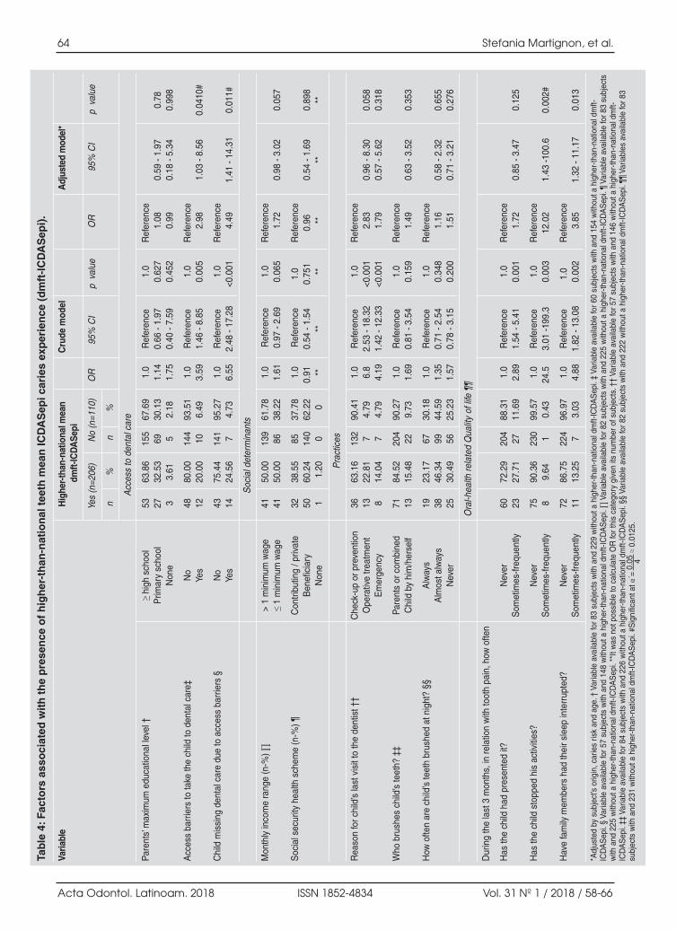
64 Stefania Martignon, et al.
Acta Odontol. Latinoam. 2018 ISSN 1852-4834 Vol. 31 Nº 1 / 2018 / 58-66
Tab
le 4
: Fac
tors
ass
oci
ated
wit
h t
he
pre
sen
ce o
f h
igh
er-t
han
-nat
ion
al t
eeth
mea
n IC
DA
Sep
i car
ies
exp
erie
nce
(d
mft
-IC
DA
Sep
i).
Varia
ble
Hig
her-
than
-nat
iona
l mea
nC
rude
mod
elA
djus
ted
mod
el*
dmft-
ICD
ASe
pi
Yes
(n=2
06)
No
(n=1
10)
OR
95%
CI
p v
alue
OR
95%
CI
p v
alue
n%
n%
Acc
ess
to d
enta
l car
e
Pare
nts’
max
imum
edu
catio
nal l
evel
†≥
high
sch
ool
5363
.86
155
67.6
91.
0R
efer
ence
1.0
Ref
eren
ceP
rimar
y sc
hool
2732
.53
6930
.13
1.14
0.66
- 1.
970.
627
1.08
0.59
- 1.
970.
78N
one
33.
615
2.18
1.75
0.40
- 7.
590.
452
0.99
0.
18 -
5.34
0.99
8
Acc
ess
barr
iers
to ta
ke th
e ch
ild to
den
tal c
are‡
No
48
80.0
014
493
.51
1.0
Ref
eren
ce1.
0R
efer
ence
Yes
1220
.00
106.
493.
591.
46 -
8.85
0.00
52.
981.
03 -
8.56
0.04
10#
Chi
ld m
issi
ng d
enta
l car
e du
e to
acc
ess
barr
iers
§N
o 43
75.4
414
195
.27
1.0
Ref
eren
ce1.
0R
efer
ence
Yes
1424
.56
74.
736.
552.
48 -
17.2
8<0
.001
4.49
1.41
- 14
.31
0.01
1#
Soc
ial d
eter
min
ants
Mon
thly
inco
me
rang
e (n
-%) ∏
> 1
min
imum
wag
e41
50.0
013
961
.78
1.0
Ref
eren
ce1.
0R
efer
ence
≤ 1
min
imum
wag
e41
50.0
086
38.2
21.
610.
97 -
2.69
0.06
51.
720.
98 -
3.02
0.05
7
Soc
ial s
ecur
ity h
ealth
sch
eme
(n-%
) ¶C
ontri
butin
g / p
rivat
e 32
38.5
585
37.7
81.
0R
efer
ence
1.0
Ref
eren
ceB
enef
icia
ry50
60.2
414
062
.22
0.91
0.54
- 1.
540.
751
0.96
0.54
- 1.
690.
898
Non
e1
1.20
00
**
****
****
**
Pra
ctic
es
Rea
son
for c
hild
’s la
st v
isit
to th
e de
ntis
t ††
Che
ck-u
p or
pre
vent
ion
3663
.16
132
90.4
11.
0R
efer
ence
1.0
Ref
eren
ceO
pera
tive
treat
men
t13
22.8
17
4.79
6.8
2.53
- 18
.32
<0.0
012.
830.
96 -
8.30
0.05
8E
mer
genc
y8
14.0
47
4.79
4.19
1.42
- 12
.33
<0.0
011.
790.
57 -
5.62
0.31
8
Who
bru
shes
chi
ld’s
teet
h? ‡
‡Pa
rent
s or
com
bine
d
7184
.52
204
90.2
71.
0R
efer
ence
1.0
Ref
eren
ceC
hild
by
him
/her
self
1315
.48
229.
731.
690.
81 -
3.54
0.15
91.
49
0.63
- 3.
520.
353
How
ofte
n ar
e ch
ild’s
teet
h br
ushe
d at
nig
ht?
§§A
lway
s
1923
.17
6730
.18
1.0
Ref
eren
ce1.
0R
efer
ence
Alm
ost a
lway
s38
46.3
499
44.5
91.
350.
71 -
2.54
0.34
81.
160.
58 -
2.32
0.65
5N
ever
2530
.49
5625
.23
1.57
0.78
- 3.
150.
200
1.51
0.71
- 3.
210.
276
Ora
l-hea
lth re
late
d Q
ualit
y of
life
¶¶
Dur
ing
the
last
3 m
onth
s, in
rela
tion
with
toot
h pa
in, h
ow o
ften
Has
the
child
had
pre
sent
ed it
?N
ever
6072
.29
204
88.3
11.
0R
efer
ence
1.0
Ref
eren
ceS
omet
imes
-freq
uent
ly23
27.7
127
11.6
92.
891.
54 -
5.41
0.00
11.
720.
85 -
3.47
0.12
5
Has
the
child
sto
pped
his
act
iviti
es?
Nev
er75
90.3
623
099
.57
1.0
Ref
eren
ce1.
0R
efer
ence
Som
etim
es-fr
eque
ntly
89.
641
0.43
24.5
3.01
-199
.30.
003
12.0
21.
43 -1
00.6
0.00
2#
Hav
e fa
mily
mem
bers
had
thei
r sle
ep in
terr
upte
d?N
ever
7286
.75
224
96.9
71.
0R
efer
ence
1.0
Ref
eren
ceS
omet
imes
-freq
uent
ly11
13.2
57
3.03
4.88
1.82
- 13
.08
0.00
23.
851.
32 -
11.1
70.
013
*Adj
uste
d by
sub
ject
’s o
rigin
, car
ies
risk
and
age.
† V
aria
ble
avai
labl
e fo
r 83
subj
ects
with
and
229
with
out a
hig
her-
than
-nat
iona
l dm
ft-IC
DA
Sep
i. ‡
Varia
ble
avai
labl
e fo
r 60
subj
ects
with
and
154
with
out a
hig
her-
than
-nat
iona
l dm
ft-IC
DA
Sep
i. §
Varia
ble
avai
labl
e fo
r 57
subj
ects
with
and
148
with
out a
hig
her-
than
-nat
iona
l dm
ft-IC
DA
Sep
i. ∏
Varia
ble
avai
labl
e fo
r 82
subj
ects
with
and
225
with
out a
hig
her-
than
-nat
iona
l dm
ft-IC
DA
Sep
i. ¶
Varia
ble
avai
labl
e fo
r 83
subj
ects
with
and
225
with
out a
hig
her-
than
-nat
iona
l dm
ft-IC
DA
Sep
i. **
It w
as n
ot p
ossi
ble
to c
alcu
late
OR
for t
his
cate
gory
giv
en it
s nu
mbe
r of s
ubje
cts.
††
Varia
ble
avai
labl
e fo
r 57
subj
ects
with
and
146
with
out a
hig
her-
than
-nat
iona
l dm
ft-IC
DA
Sep
i. ‡‡
Var
iabl
e av
aila
ble
for 8
4 su
bjec
ts w
ith a
nd 2
26 w
ithou
t a h
ighe
r-th
an-n
atio
nal d
mft-
ICD
AS
epi.
§§ V
aria
ble
avai
labl
e fo
r 82
subj
ects
with
and
222
with
out a
hig
her-
than
-nat
iona
l dm
ft-IC
DA
Sep
i. ¶¶
Var
iabl
es a
vaila
ble
for 8
3su
bjec
ts w
ith a
nd 2
31 w
ithou
t a h
ighe
r-th
an-n
atio
nal d
mft-
ICD
AS
epi.
#Sig
nific
ant a
t α´=
0.0
5 4 ≈
0.01
25.
AOL12018:32011 18/07/2018 17:30 Página 64

better understanding of related risk factors andsupporting public health measures7,2224.Colombia,among other countries, used the ICDAS cariessystem in its recent National Oral Health Study8.This supports the use of the ICDAS caries systemin the current study to gain further insight intopossible associations of risk factors with caries,including early manifestations of caries, i.e., noncavitated enamel caries lesions (Initial).In this population, the prevalence of conventionalcaries experience was higher than 50% (4yr. olds:55%; 5yr. olds: 53%) but lower than the nationalvalues reported in the recent NOHS for 5year olds(62%)8, and values reported recently in Bogotá in4 (59%) and 6yr. olds (66%)23.When initial carieslesions were included, the prevalence of ICDASepi caries experience in the total population wasslightly lower (65.18%) than equivalent modifiedICDAS caries experience for the country (NOHS)for same ages (67.05%)8.Even though a convenience sample was selected inthis study, the high participation rate in the localgovernmental nutrition programme and call toparticipate in a free oral and nutritional healthexamination provided subjects from both affluentand less affluent areas. It is however possible, thatchildren from less needy families were less likelyto participate in the programme. The significant association between ICDASepi cariesexperience ≥ 1 and malnutrition is in agreement withGaur and Nayak5. Overweight and obesity were morefrequent in children with ICDASepi caries experience(29%) than were lowweight risk and malnutrition(9%), representing a public health alert. Recentevidence shows association between high freesugardaily intake and caries25.Aspects of oral healthrelated quality of life werestrongly associated to both outcomes. Sheiham3
also found an association between caries anddecreased quality of life in preschool children. Thelack of association in this study between the twooutcome variables and social determinants mightbe due to the facts that the study population issmaller and more homogeneous than in othernational municipalities and that formal employmentis higher, possibly with higher income levels. Socialdeterminants have been shown to be important in theNOHS8, and are part of the dental caries ‘Global oralhealth inequalities’ task group agenda worldwide26.The significant association between caries experienceand access to dental care as well as between caries
experience and rural/urban origin (borderline)might reflect social inequalities, in agreement withthe NOHS8. Insurance limitations and a shortage of dentists have been related to low income26. The latter might explain the local situation andreinforces the significant association found with thecrude model between higherthannational mean ofICDASepi experience and visiting the dentist foroperative treatment/emergency.The significant associations found between bothoutcome variables and age, while logical with regardto natural caries history, also refer to a relevant ageincrease in caries experience in this study. This mayreflect problems with dental care at early ages andmay be partially explained by Colombia’s 2000legislation, which established that a child’s firstdental visit should take place at 2 years of age,including only operative treatment of cavitated carieslesions. Since 2011, it has included biannual fluoridevarnish applications starting at the age of one year,among other oral health promotion and preventionactivities8.This aspect is also supported in this studyby the association between a higherthannationalmean number of teeth with ICDASepi caries expe rience and caries risk. It should however be notedthat the Cariogram includes caries experience as oneof its factors, with an implicit association effect.When analysing other relevant risk factors in thispopulation using the Cariogram, most children pre sented high and frequent daily intake of carbohydrates(66.5%), which is common in Colombian diet, andmore than half reported not using fluoride toothpaste(56%) even though there is strong evidence thatfluoride toothpaste prevents and controls caries asfrom the eruption of the first teeth27.This study and the NOHS8show that action is neededto improve oral health in infants and young children.In Anapoima, these actions, among others framed inthe current paradigm of caries, are already beingconducted with an emphasis on early child hood,together with government, health, education, familiesand community bodies, within the Colombian Chapterof the Alliance for a Cavity Free Future16. This study found significant associations between earlychildhood caries experience and nutritional status,OHRQoL, dental care access barriers, caregivers’educational level, caries risk, and age, both in terms ofany ICDASepi caries experience and of having ahigherthannational mean figures of ICDASepi cariesexperience, representing oral health inequalities inearly childhood in Anapoima, Colombia.
Risk factors for caries experience in Colombia 65
Vol. 31 Nº 1 / 2018 / 58-66 ISSN 1852-4834 Acta Odontol. Latinoam. 2018
AOL12018:32011 18/07/2018 17:30 Página 65

ACKNOWLEDGEMENTSThe authors would like to thank the partial funding of the study bythe Colombian Chapter of the Alliance for a Cavity Free Future –Colgate Palmolive; the municipality of Anapoima – Cundinamarca;María Beatriz Ferro from Colgate Palmolive; UNICA, the dentistsand Paediatrics Specialization residents from Universidad ElBosque. The authors declare that they have no conflict of interest.
CORRESPONDENCEDra.Stefania Martignon UNICA Unidad de Investigación en Caries, Vicerrectoría deInvestigaciones, Universidad El BosqueAv. Cra 9 No. 131 A – 02 Edificio de Laboratorios, 2o Piso Bogotá, 110121 Colombia [email protected]
66 Stefania Martignon, et al.
Acta Odontol. Latinoam. 2018 ISSN 1852-4834 Vol. 31 Nº 1 / 2018 / 58-66
REFERENCES1. Gimenez T, Bispo BA, Souza DP, ViganóME, Wanderley
MT, Mendes FM, Bönecker M, Braga MM. Does the Declinein Caries Prevalence of Latin American and CaribbeanChildren Continue in the New Century? Evidence fromSystematic Review with MetaAnalysis. PLoS One 2016;11:e0164903. doi: 10.1371/journal.pone.0164903
2. Fontana M, Santiago E, Eckert GJ, FerreiraZandona AG.Risk Factors of Caries Progression in a Hispanic Schoolaged Population. J Dent Res 2011;90:11891196.
3. Sheiham A. Dental caries affects body weight, growth andquality of life in Preschool children. Br Dent J 2006; 201:625626.
4. Albino J, Tiwari T. Preventing Childhood Caries: A Reviewof Recent Behavioral Research. J Dent Res 2016; 95:3542.
5. Gaur S, Nayak R. Underweight in low socioeconomicstatus preschool children with severe early childhoodcaries. J Indian Soc Pedod Prev Dent 2011; 29:305309.
6. Gao XL, Hsu CY, Xu Y, Hwarng HB, Loh T, Koh D.Building caries risk assessment models for children. J DentRes 2010; 89:637643.
7. ICDAS, International Caries Detection and AssessmentSystem URL:www.icdas.org
8. Ministerio de Salud y Protección Social República deColombia.URL: https://www.minsalud.gov.co/sites/rid/Lists/BibliotecaDigital/RIDE/VS/PP/ENSABIVSituacionBucalActual.pdf
9. Neufeld L, Rubio M, Pinzón L, and Tolentino L. Nutriciónen Colombia: estrategia de país 20112014. Santiago deChile: Banco Interamericano de Desarrollo. 2010. URL:http://www.piaschile.cl/wpcontent/uploads/2015/04/NutricionenColombiaEstrategiadepa%C3%ADs201120141.pdf
10. Ley Estatutaria 1751/2015 de 16 de Febrero. Bogotá.URL:https://www.minsalud.gov.co/Normatividad_Nuevo/Ley%201751%20de%202015.pdf
11. Ley 1438/2011. Bogotá, Congreso de Colombia.URL:http://www.alcaldiabogota.gov.co/sisjur/normas/Norma1.jsp?i=41355
12. Ley 100/1993. Bogotá, Congreso de Colombia.URL:http://www.alcaldiabogota.gov.co/sisjur/normas/Norma1.jsp?i=41355
13. Ministerio de Protección social, Bogotá. Decreto 3039/ 2007. URL: http://www.ins.gov.co/normatividad/Normatividad/DECRETO%203039_2007%20Plan%20Nacional%20SP%2020072010.pdf
14. Estrategia de Atención Integral a la Primera Infancia.Repú blica de Colombia: Gobierno de Colombia. URL:http://www.deceroasiempre.gov.co/QuienesSomos/Documents/Fundamientospoliticostecnicosgestiondeceroasiempre.pdf
15. Lineamientos Estrategia incremental de cuidado bucal yprotección específica en salud bucal, para primera infancia,
infancia y adolescencia “Soy generación sonriente”.República de Colombia: Ministerio de Salud y Protecciónsocial.URL: http://www.deceroasiempre.gov.co/QuienesSomos/Documents/Fundamientospoliticostecnicosgestiondeceroasiempre.pdf
16. The Alliance for a CavityFree Future. Boston: URL:www.allianceforacavityfreefuture.org
17. Anapoima. Sitio oficial de Anapoima en Cundinamarca,Colombia. URL: https://archive.is/20121127162132/h t t p : / / w w w. a n a i m a c u n d i n a m a r c a . g o v . c o /nuestraalcaldia.shtml?apc=a1I1—&m=q
18. Bratthall D, HanselPetersson G, Stjernsward J. CariogramInternet Version 2.01. Malmö, Sweden. URL: https://www.mah.se/fakulteterochomraden/Odontologiskafakulteten/Avdelningochkansli/Cariologi/Cariogram/
19. WHO, World Health Organization]. Geneva. URL:http://www.who.int/childgrowth/standards/Technicalreport.pdf
20. Martignon S, BautistaMendoza G, GonzálezCarrera M,LafaurieVillamil G, Morales V, Santamaría R. Instrumentsfor evaluating oral health knowledge, attitudes and practicefor parents /caregivers of small children. Rev Salud Pública(Bogotá) 2008;10:308314.
21. Tellez M, Martignon S, Lara JS, Zuluaga J, Barrero L,Builes L, Cordoba D, Gomez J. Correlación de un instru mento de calidad de vida relacionado con salud oral entreniños de 8 a 10 años y sus acudientes en Bogotá. CESOdontol. 2010; 23:915. http://revistas.ces.edu.co/index.php/odontologia/article/view/662
22. Pitts NB, Ekstrand KR. International Caries Detection andAssessment System (ICDAS) and its International CariesClassification and Management System (ICCMS) methodsfor staging of the caries process and enabling dentists tomanage caries. Community Dent Oral Epidemiol 2013;41:e4152. doi: 10.1111/cdoe.12025
23. Pitts NB, Zero DT, Marsh PD, Ekstrand K, Weintraub JA,RamosGomez F, Tagami J, et al. Dental caries. Nat RevDis Primers 2017;3:17030. doi: 10.1038/nrdp.2017.30
24. Cortes A, Ekstrand KR, Gamboa LF, González L,Martignon S. Caries status in young Colombian childrenexpressed by the ICCMS™ visual/radiographic combinedcaries staging system. Acta Odontol Scand 2017; 75:1220.
25. Moynihan PJ, and Kelly SA. Effect on caries of restrictingsugars intake: systematic review to inform WHO Guidelines.J Dent Res 2014; 93:818.
26. Pitts N, Amaechi B, Niederman R, Acevedo AM, ViannaR, Ganss C, Ismail A et al.. Global oral health inequalities:dental caries task group—research agenda. Adv Dent Res2011; 23:211220.
27. Marinho VC, Worthington HV, Walsh T, Clarkson JE..Fluoride varnishes for preventing dental caries in childrenand adolescents. Cochrane Database Syst Rev. 2013;7:CD002279. doi: 10.1002/14651858.CD002279.pub2
AOL12018:32011 18/07/2018 17:30 Página 66

RESUMENEl objetivo del presente trabajo fue investigar la capacidad detallado apical de XP Shaper y compararla con dos sistemasde NiTi, rotatorio y reciprocante, mediante tomografíascomputarizadas de haz cónico. Se analizaron los canalesmesio vestibulares de cuarenta y cinco primeros molaresinferiores. Los dientes fueron divididos en tres gruposexperimentales (n=15): WaveOne, OneShape and XP shaper.Se obtuvieron imágenes pre y post instrumentación a 3mm,5mm y 7 mm del ápice utilizando tomografías computadas dehaz cónico para determinar la presencia de transporte apicaly la capacidad de conservación de la anatomía original del
conducto. Se utilizó el test deKruskalWallis para compararlos tres sistemas de intrumentación y el test de Friedman paracomparar las mediciones en los tres niveles de raíz. XP Shapermostró la menor cantidad de transporte apical estadística mentesignificativa mientras que WaveOne y OneShape mostraron elmayor transporte apical sin diferencia estadística mentesignificativa entre los dos grupos. XP shaper permitióconservar la anatomía del canal original mejor que WaveOney OneShape.
Palabras clave: Conducto radicular; Tomografía computadade haz cónico.
INTRODUCTION The ultimate goal of root canal treatment is to removeinfected pulpal remnants, eliminate microorga nisms and adequately shape the root canal system1.Optimum biomechanical preparation can beachieved through uniform enlargement of the rootcanal system in all dimensions to permit thoroughdisinfection while preserving the original curvaturewithout inducing iatrogenic errors2.However, endodontic preparation in narrow andcurved root canals has always been a challenge, due
to the tendency of the prepared canal to deviatefrom its natural axis3. Innovations and techniquesare continuously being developed with the aim ofreducing the difficulties encountered during endo dontic therapy. Nickeltitanium instruments providesatisfactory treatment of curved canals in shortertimes through their enhanced properties of shapememory, super elasticity and cutting efficiency4.Various singlefile systems with different metallurgyand designs have been promoted to prepare the rootcanals with one instrument using either continuous
ABSTRACTThe aim of the present study was to investigate the shapingabilities of XP Shaper and compare it with other single file rotaryNiTi systems utilizing full rotation and reciprocation motion, bycone beam computed tomography. Mesiobuccal canals of fortyfive mandibular first molars, were allocated into three equalgroups (n=15) according to the rotary system applied; WaveOne,OneShape and XP shaper. Preand postinstrumentation imageswere obtained at 3mm, 5mm and 7mm from the apex using conebeam computed tomography and assessed to determine canaltransportation and centering ability.
Data were analyzed using KruskalWallis test to compare the threesystems and Friedman’s test to compare the root levels. Resultsshowed that WaveOne and OneShape rotary systems producedgreatest mean transportation with no statistically significantdifference between them, while XP Shaper produced the loweststatistically significant mean transportation. Canal centeringability differed significantly among the three systems used. It wasconcluded that XP shaper preserved the original canal anatomybetter than WaveOne and OneShape rotary systems.
Key words: Root canal, Cone beam computed tomography.
67
Vol. 31 Nº 1 / 2018 / 67-74 ISSN 1852-4834 Acta Odontol. Latinoam. 2018
Comparison of canal transportation and centering ability of Xp Shaper, WaveOne and Oneshape: A cone beam computed tomography study of curved root canals
Reham Hassan1, Nehal Roshdy2, Noha Issa3
1 Minia University, Faculty of Dentistry, Department of Endodontics, Egypt.2 Cairo University, Faculty of Dentistry, Department of Endodontics, Egypt.3 Cairo University, Faculty of Dentistry, Department of Oral and Maxillofacial Radiology, Egypt.
Comparación del transporte apical y la capacidad de conservación de la anatomía del conducto de XpShaper, WaveOne y Oneshape: Estudio de conductos curvos por tomografía computarizada de haz cónico
AOL12018:32011 18/07/2018 17:30 Página 67
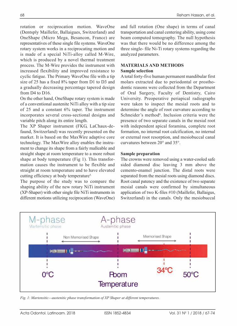
rotation or reciprocation motion. WaveOne(Dentsply Maillefer, Ballaigues, Switzerland) andOneShape (Micro Mega, Besancon, France) arerepresentatives of these single file systems. WaveOnerotary system works in a reciprocating motion andis made of a special NiTialloy called MWire,which is produced by a novel thermal treatmentprocess. The MWire provides the instrument withincreased flexibility and improved resistance tocyclic fatigue. The Primary WaveOne file with a tipsize of 25 has a fixed 8% taper from D1 to D3 anda gradually decreasing percentage tapered designfrom D4 to D16. On the other hand, OneShape rotary system is madeof a conventional austenite NiTi alloy with a tip sizeof 25 and a constant 6% taper. The instrumentincorporates several crosssectional designs andvariable pitch along its entire length. The XP Shaper instrument (FKG, LaChauxdefaund, Switzerland) was recently presented on themarket. It is based on the MaxWire adaptive coretechnology. The MaxWire alloy enables the instru ment to change its shape from a fairly malleable andstraight shape at room temperature to a more robustshape at body temperature (Fig 1). This transfor mation causes the instrument to be flexible andstraight at room temperature and to have elevatedcutting efficiency at body temperature5.
The purpose of the study was to compare theshaping ability of the new rotary NiTi instrument(XPShaper) with other single file NiTi instruments indifferent motions utilizing reciprocation (WaveOne)
and full rotation (One shape) in terms of canaltransportation and canal centering ability, using conebeam computed tomography. The null hypothesiswas that there would be no difference among thethree single file NiTi rotary systems regarding theanalyzed parameters.
MATERIALS AND METHODSSample selectionA total fortyfive human permanent mandibular firstmolars extracted due to periodontal or prostho dontic reasons were collected from the Departmentof Oral Surgery, Faculty of Dentistry, CairoUniversity. Preoperative periapical radiographswere taken to inspect the mesial roots and todetermine the angle of root curvature according toSchneider’s method6. Inclusion criteria were thepresence of two separate canals in the mesial rootwith independent apical foramina, complete rootformation, no internal root calcification, no internalor external root resorption, and mesiobuccal canalcurvatures between 20° and 35°.
Sample preparationThe crowns were removed using a watercooled safesided diamond disc leaving 3 mm above thecementoenamel junction. The distal roots wereseparated from the mesial roots using diamond discs.Root canal patency and the existence of two separatemesial canals were confirmed by simultaneousapplication of two Kfiles #10 (Maillefer, Ballaigus,Switzerland) in the canals. Only the mesiobuccal
68 Reham Hassan, et al.
Acta Odontol. Latinoam. 2018 ISSN 1852-4834 Vol. 31 Nº 1 / 2018 / 67-74
Fig. 1: Martensitic—austenitic phase transformation of XP Shaper at different temperatures.
AOL12018:32011 18/07/2018 17:30 Página 68

canals were used in our study. The working length ofeach canal was determined by subtracting 1 mm fromthe apical foramen.Before scanning, the roots were fixed by mountingthem vertically halfway in transparent autopolymerizing acrylic resin (Acrostone, Dental &Medical Supplies, Cairo, Egypt) mixed accordingto the manufacturer’s instructions in a silicon mold(10 cm x 10 cm). The root apices were sealed withwax (Wilson, Sao Paulo, Brazil) to preserve theapical foramen from resin penetration. To ensurestandardization of the specimens during tomo graphic scanning, each root was placed in the unsetacrylic resin such that its long axis was parallel tothe long axis of the mold. In addition, an amalgamfilling was inserted into the resin at the buccodistalline angle of the roots, to enable the orientation ofthe canal during scanning.
Preinstrumentation scanningAll roots were scanned using cone beam computedtomography (CBCT) (Scanora 3D, Soredex, Palodexgroup, Finland) at 85 kVp and 15 mA to detect canalshape before instrumentation. For each specimen,three tomograms were chosen according to thedistance from the root apex, as follows: 3 mm fromthe root apex (Representing the apical third), 5 mmfrom the root apex (Representing the middle third) and7 mm from the root apex (Representing the cervicalthird). All scans were assessed using a Softwareprogram (OnDemand 3D, Cybermed, South Korea).
Root Canal PreparationA glide path was created using #15 Kfile (Maillefer,Ballaigus, Switzerland). Then samples were randomlydivided into 3 equal groups (n = 15 canals per group)as follows:
• Group I: The WaveOne group, where roots weremechanically prepared using Primary WaveOnefile (size 25) operated by Xsmart plus endodonticmotor (Dentsply, Tulsa Dental, Tulsa, OK) usingthe reciprocation preset mode.
• Group II: The OneShape group, where roots weremechanically prepared using OneShape file (size25, taper 0.06) operated in continuous rotationmotion using an electric motor with a rotationalspeed set at 350 rpm and a 5Ncm torque in acrowndown technique.
Both instruments were used with a slow inandoutpecking motion with an amplitude of about 3 mm.After three pecks, the flutes of the instruments werecleaned and reinserted, and the process wasrepeated until full working length was reached.
• Group III: The XPShaper group, where rootswere shaped using the XPshaper file with theelectric motor set at 900 rpm and 1Ncm torque.The file was inserted into the canal using longgentle strokes (57 mm); 5 strokes were applieduntil working length was reached.
Xsmart plus endodontic motor (Dentsply, TulsaDental, Tulsa, OK) was employed for root canalpreparation of all samples, following the manu facturer’s instructions for each instrument. Eachinstrument was used to prepare only four canals.Freshly prepared 2.6% sodium hypochloritesolution (Clorox, Cairo, Egypt) was used as anirrigant during the instrumentation procedure,placed with 30gauge needle tips (NaviTip,Ultradent, South Jordan, UT, USA) as deeply aspossible into the canal without binding. Apicalpatency was retained using a #10 Kfile. Once therotary instrument reached the working length androtated freely, it was removed. Then 10 ml ofdistilled water were used as a final flush.
Postinstrumentation scanningThe root canals were scanned after mechanical preparation using CBCT, similarly to the preinstru mentation scanning protocol. Pre and postinstru men tation scans were superimposed using theaforementioned software program to evaluate the degree of transportation as well as the centeringability of the tested instruments. The shortest distance from the periphery of the root (mesial anddistal) to the edge of the canal was measured usingthe length measuring tool on the reconstructedcrosssectional scans. The degree of canal transportation was calculatedaccording to the formula provided by Gambill et al.7. Canal transportation (CT) = (M1 M2) (D1 D2)where M1: refers to the shortest distance from themesial edge of the root to the mesial edge of the uninstrumented canal. M2: refers to the shortest distance from the mesialedge of the root to the mesial edge of the instrumentedcanal.
Shaping ability of XP shaper using CBCT 69
Vol. 31 Nº 1 / 2018 / 67-74 ISSN 1852-4834 Acta Odontol. Latinoam. 2018
AOL12018:32011 18/07/2018 17:30 Página 69

D1: refers to the shortest distance from the distal edgeof the root to the distal edge of the uninstrumentedcanal. D2: refers to the shortest distance from the distaledge of the root to the distal edge of the instrumentedcanal (Fig. 2).Regarding the transportation direction, CT equal to 0(zero) denoted lack of transportation, a negativevalue denoted transportation towards the distaldirection, and a positive value denoted transportationtowards the mesial direction. Centering ability ratio was calculated using thesame values obtained during the measurement oftransportation according to the following equation:
Centralization ability ratio = (M1 M2)/ (D1 D2)or (D1D2) /(M1M2)
The formula was selected in such a manner that thelowest of the results acquired through the differenceshould be the numerator. A result equal to 1.0signified perfect centralization. When the value wascloser to zero, it denoted that the instrument hadlower capacity to maintain itself in the central axisof the canal.
Assessment of root canal preparationRoot canals were prepared by one operator, whilecanal curvatures prior to and after instrumentationwere assessed by a second examiner who was blindto all the experimental groups.
Statistical analysisData were presented as mean and standard devia tion (SD) values. KruskalWallis test was used tocompare the three systems. Friedman’s test wasused to compare the different root levels. Dunn’stest was used for pairwise comparisons. Fisher’sExact test was used to compare frequency data ofthe three systems. The significance level was set atP ≤ 0.05.
RESULTS Canal TransportationAt 3 and 5 mm from the apex, WaveOne andOneShape had highest mean transportation, with nostatistically significant difference between them,while the XP Shaper had significantly lower meantransportation (Table 1). At 7 mm from the apex, all three groups differedsignificantly. WaveOne had the highest meantransportation (0.22 ± 0.09), followed by OneShape(0.14 ± 0.11), and finally XP Shaper with the lowestvalue (0.08 ± 0.06).With regard to the root canal levels; our resultsshowed that preparing the canal with WaveOneinstrument created a statistically significant diffe rence between different levels (pvalue = 0.035).The highest distal transportation was found at 3 mmfrom the apex, while the highest mesial transpor tation was observed at the 7 mm level. However,OneShape and XP Shaper instruments showed nostatistically significant difference among thedifferent root levels (pvalue = 0.061 and 0.175respectively) (Table 2).
Centering AbilityThe maintenance of canal curvature was better withOne Shape (0.54 ± 0.11) and XP Shaper (0.41± 0.15)than with WaveOne, which had the statisticallysignificant lowest mean centering ratio (0.31 ± 0.12)(Table 3).Regarding different root canal levels, at the level of3 mm from the apex, One Shape had the highestmean statistically significant centering ratio (0.51±0.32), while there was no statistically significantdifference between WaveOne and XP Shaper instru ments. At the 5 mm level there was no statisticallysignificant difference among the three systems. At 7 mm level, OneShape had the statisticallysignificantly highest mean centering ratio, followedby XP Shaper, and lastly, WaveOne.
70 Reham Hassan, et al.
Acta Odontol. Latinoam. 2018 ISSN 1852-4834 Vol. 31 Nº 1 / 2018 / 67-74
Fig. 2: (A) Preinstrumentation and (B) postinstrumentationCBCT images with markings showing points of measurementsused for determining canal transportation and centeringratio.
AOL12018:32011 18/07/2018 17:30 Página 70

DISCUSSIONEver since Schilder supported the concept ofpreparing the root canal in a funnel shape, whilepreserving its original curve8; ideal cleaning andshaping of the root canal systems has remained avery challenging procedure. The American Association of Endodontics definedtransportation as “removal of the canal wallstructure on the outside of the curve in the apical
half of the canal due to the files’ tendency to restoretheir original shape during canal preparation”9. Theinappropriate pattern of dentin removal adverselyaffects the treatment prognosis, as it causes highrisk of straightening the original canal curvature,and increases the rate of debris extrusion andsubsequent postoperative discomfort10.
In the present study, mesiobuccal canals of extractedfirst mandibular molars were chosen to provide
Shaping ability of XP shaper using CBCT 71
Vol. 31 Nº 1 / 2018 / 67-74 ISSN 1852-4834 Acta Odontol. Latinoam. 2018
Table 1: Mean, standard deviation (SD) and results of Kruskal-Wallis test for comparison between canal transportation values (mm) after using the three systems.
Root level WaveOne One Shape XP Shaper P-value
Mean SD Mean SD Mean SD
3 mm 0.14 A 0.10 0.12 A 0.16 0.07 B 0.06 0.023*
5 mm 0.19 A 0.13 0.21 A 0.15 0.05 B 0.04 0.001*
7 mm 0.22 A 0.09 0.14 B 0.11 0.08 C 0.06 0.001*
Total 0.18 A 0.07 0.16 A 0.11 0.07 B 0.04 <0.001*
*: Significant at P ≤ 0.05. Different superscripts in the same row denote statistically significant differences.
Table 2: Frequencies (n), percentages (%) and results of Friedman’s test for comparison between direction of transportation among different root levels.
System Direction 3 mm 5 mm 7 mm P-value
n % n % n %
WaveOne Distal 10 66.7 6 40 2 13.3 0.035*Mesial 5 33.3 7 46.7 13 86.7
No transportation 0 0 2 13.3 0 0
One Shape Distal 13 86.7 9 60 7 46.7 0.061Mesial 2 13.3 6 40 8 53.3
No transportation 0 0 0 0 0 0
XP Shaper Distal 8 53.3 11 73.3 6 40 0.175Mesial 5 33.3 4 26.7 9 60
No transportation 2 13.3 0 0 0 0
*: Significant at P ≤ 0.05
Table 3: Mean, standard deviation (SD) and results of Kruskal-Wallis test for comparison between centering ratio after using the three systems.
Root level WaveOne One Shape X Shaper P-value
Mean SD Mean SD Mean SD
3 mm 0.29 B 0.19 0.51 A 0.32 0.24 B 0.34 0.016*
5 mm 0.40 0.29 0.46 0.22 0.49 0.26 0.571
7 mm 0.23 C 0.16 0.64 A 0.07 0.51 B 0.30 <0.001*
Total 0.31C 0.12 0.54 A 0.11 0.41 B 0.15 <0.001*
*: Significant at P ≤ 0.05. Different superscripts in the same row denote statistically significant differences.
AOL12018:32011 18/07/2018 17:30 Página 71

conditions similar to the clinical situation and to allowrealistic evaluation of the instruments’ performance11.CBCT was used because it allows detailed threedimensional (3D) observation of the root canalanatomy with highresolution images, fasteracquisition and reconstruction scheme. CBCT is aneffective tool for measuring dentin thickness, apicaltransportation and canal centering 12,13.Two parameters were selected to assess the shapingability of the instruments tested in this study: (1)Apical transportation, which can endanger efficientroot canal sealing, thereby reducing the treatmentoutcomes, and (2) maintenance of canal centering,which is basic in preparing curved canals11
.
Results showed no statistically significant differencebetween WaveOne and OneShape rotary systems atthe levels of 3 and 5 mm from the apex, where bothinstruments produced high mean transportationcompared to the XP Shaper instrument. This couldbe credited to the tip diameter corresponding to a size 25 for both WaveOne and the primaryOneShape instruments, comparable to size 17 whichXP Shaper initially starts with. At the level of 7 mmfrom the apex; WaveOne produced the higheststatistically significantly mean transportation, followedby OneShape, while the XP Shaper produced thelowest mean transportation. There is an inverse relationship between instrumenttapering and canal transportation14. The primaryWaveOne instrument has an 8% taper over the first3 millimeters. This is greater than the other tworotary instruments, which decrease 4.3% and 5.5%respectively. The OneShape instrument has aconstant 6% taper along its entire length and the XPshaper possesses an initial 1% taper along its wholelength, which expands to a final 4% taper5,15
.
For overall canal transportation, this study showedthat the XP Shaper produced the lowest statisticallysignificantly mean transportation, while theWaveOne and OneShape instruments producedhighest mean transportation, with no statisticallysignificant difference between them. The outstanding results of the XP Shaper can beattributed to its Adaptive Core technology, thanks towhich it can expand while preserving the originalcanal anatomy and curvature5. The XP Shaper isbelieved to apply minimal stresses on the dentin walls,and can thus adapt easily to the canal irregularities16
.
Although the results of our investigation cannot becompared directly with those of Azim et al.5 because
of the different systems and methodology applied,their results were consistent with ours. They reportedthat XP Shaper was superior to Vortex Blue in termsof shaping ability, where the file created nonuniform preparation adapting to the complex canalanatomy.On the other hand, the attitude of the OneShapeinstrument in the canal could be explained by itsasymmetrical cutting edges. When combined withcontinuous rotation at a relatively high speed (350 rpm),this design feature causes the instrument to progressinto the curved canals, creating some stress that mightresult in the observed apical transportation 15.
Similar results were drawn in by Agarwal et al.17 andAlrahabi and Alkady18, who found no statisticallysignificant difference between WaveOne andOneShape instruments regarding canal transpor tation. Likewise, You et al. 19 and Capar et al. 20
reported similar transportation results for recipro cation motion and conventional continuous rotationtechnique. However, there have been contradictoryresults with Saber et al.15 who report that the use ofOneShape file caused a significantly greater apicaltransportation than WaveOne file. Still other studiesreport that WaveOne system preserved the originalcanal curvature better than the OneShape systemdid21,22.Our results confirm the increasing tendency ofcanal transportation as the diameter of the filesincreases10. OneShape and XP Shaper instrumentsshowed no statistically significant difference amongthe different root levels. On the other hand,WaveOne instrument showed more distal canaltransportation at the level of 3 mm from the apexand higher tendency toward mesial transportationat the 7 mm level. It is therefore suggested thatinstruments with taper greater than 0.06 should notbe used for apical enlargement of curved canals.Sinai reported that aggressive instrumentation in the cervical third of the root canal may lead tostrip perforations and subsequent inflammatorycomplications23. Less transportation towards thisarea can be considered a favorable feature for theWave One and the XP Shaper instruments. Agarwalet al.17 showed that at 3 mm above the apex,ProTaper and WaveOne groups showed transpor tation towards the lateral side of the canal curvature,while the OneShape group remained centered,which agrees with the results of the present study.This result differs from previous studies that report
72 Reham Hassan, et al.
Acta Odontol. Latinoam. 2018 ISSN 1852-4834 Vol. 31 Nº 1 / 2018 / 67-74
AOL12018:32011 18/07/2018 17:30 Página 72

that the apical segment usually has more canaltransportation toward the outside of the curve24.It should be noted that from a clinical standpoint,Wu et al.25 reported that apical transportation of more than 0.3 mm can negatively affect thesealability of the root canal filling material. In thepresent study none of the tested rotary systemscaused more than 0.2 mm apical transportation. Our results demonstrated that at the level of 3 mmfrom the apex; OneShape instrument showed thehighest mean statistically significant centering ratioand there was no statistically significant differencebetween WaveOne and XP Shaper instruments. Atthe 5 mm level; there was no statistically significantdifference among the three systems. At the 7 mmlevel; OneShape showed the statistically significanthighest mean centering ratio followed by XPShaper, and lastly by WaveOne instrument. Thesefindings proved that instruments with constant taperin the apical section had good centering abilitycompared to instruments with progressive tapersalong the cutting surface 26.
With regard to total centering ratio; OneShapeshowed the statistically significant highest meancentering ratio followed by XP Shaper followed byWaveOne. The superiority of OneShape instrumentcan be credited to its design, which progressively
changes from variable 3cutting edges at the tip to Sshaped 2 cutting edges near the shaft27. Thesnakelike motion helps preserve the original canalanatomy due to the offset rotation center, causingthe file to engage and disengage along the canalwall, thus reducing the stresses between the file andthe canal wall18.WaveOne instrument showed low centering ability,as it is a relatively large rigid single file with moretaper that moves apically until it reaches theworking length, creating a piston effect28. The findings of this research are consistent withprevious results reported by different authors, such asSaleh et al.29 who showed that canals prepared withthe F360 and OneShape systems were better centeredthan those prepared with Reciproc and WaveOnesystems, and Agarwal et al.17 who showed that aOneShape group had less transportation and remainedmore centered than a WaveOne group, although the differences were not statistically significant.However, they are contradicted by Dhingra et al.22
and Tambe et al.,21 who showed the superiority of WaveOne system over OnesShape in terms ofcentering ability.It can be concluded that overall, XP Shaper pro duced less canal transportation than WaveOne andOneShape instruments.
Shaping ability of XP shaper using CBCT 73
Vol. 31 Nº 1 / 2018 / 67-74 ISSN 1852-4834 Acta Odontol. Latinoam. 2018
CORRESPONDENCEDr. Hassan RehamDepartment of Endodontics, Faculty of Dentistry, Minia University,Misr Aswan Agricultural Road. Minia. [email protected]
REFERENCES1. Haapasalo M, Endal U, Zandi H, Coil JM. Eradication of
endodontic infection by instrumentation and irrigationsolutions. Endod Topics 2005; 10:77102.URL: https://onlinelibrary.wiley.com/doi/abs/10.1111/j.16011546.2005.00135.x
2. GonzalezRodrıguez MP, FerrerLuque CM. A comparisonof ProFile, Hero 642, and K3 instrumentation systems inteeth using digital imaging analysis. Oral Surg Oral MedOral Pathol Oral Radiol Endod 2004; 97:112115.
3. Fornari VJ, SilvaSousa YT, Vanni JR. Histological evalua tion of the effectiveness of increased apical enlargement forcleaning the apical third of curved canals. Int Endod J 2010;43:988994.
4. da Frota MF, Filho IB, Berbert FL, Sponchiado EC Jr,Marques AA, Garcia Lda F.Cleaning capacity promoted bymotordriven or manual instrumentation using ProTaper
Universal system: histological analysis. J Conserv Dent2013; 16:7982.
5. Azim AA, Piasecki L, da Silva Neto UX, Cruz ATG, AzimKA. XP Shaper, A Novel Adaptive Core Rotary Instrument:Micro–computed Tomographic Analysis of Its ShapingAbilities. J Endod 2017; 43:15321538.
6. Schneider SW. A comparison of canal preparations instraight and curved root canals. Oral Surg Oral Med OralPathol. 1971; 32:271275.
7. Gambill JM, Alder M, del Rio CE. Comparison of nickeltitanium and stainlesssteel handfile instrumentation usingcomputed tomography. J Endod. 1996;22:369375.
8. Schilder H. Cleaning and shaping the root canal. Dent ClinNorth Am. 1974; 18:269296.
9. Glossary of Endodontic Terms. Chicago: AAE; 9th Ed. 2012.URL:http://www.nxtbook.com/nxtbooks/aae/endodonticglossary2016/index.php#/1
AOL12018:32011 18/07/2018 17:30 Página 73

10. López FU, Fachin EV, Camargo Fontanella VR, BarlettaFB, Só MV, Grecca FS. Apical transportation: A compa rative evaluation of three root canal instrumentationtechniques with three different apical diameters. J Endod2008; 34:15451548.
11. Hülsmann M, Peters O, Dummer PMH. Mechanical prepa ration of root canals. Shaping goals, techniques and means.Endod Topics 2005; 10:3076. (). URL:https://onlinelibrary.wiley.com/doi/abs/10.1111/j.16011546.2005.00152.x
12. Elayouti A, Dima E, Judenhofer MS, Löst C, Pichler BJ.Increased Apical Enlargement Contributes to Excessive DentinRemoval in Curved Root Canals: A Stepwise MicrocomputedTomography Study. J Endod 2011; 37: 15801584.
13. Cotton TP, Geisler TM, Holden DT, Schwartz SA,Schindler WG. Endodontic applications of conebeamvolumetric tomography. J Endod 2007; 33:11211132.
14. Pique F, Ganahl D, Peters OA. Effects of root canal prepa ration on apical geometry assessed by microcomputedtomography. J Endod 2009; 35:10561059.
15. Saber SEDM, Nagy MM, Schafer E. Comparative evalua tion of the shaping ability of WaveOne, Reciproc andOneShape singlefile systems in severely curved root canalsof extracted teeth. Int Endod J 2015; 48: 109114.
16. FKG XP Endo Shaper. URL: http://www.fkg.ch/sites/default/files/201607_fkg_xps_brochure_en_web.pdf.Accessed October 25, 2016
17. Agarwal RS, Agarwal J, Jain P, Chandra A. Comparativeanalysis of canal centering ability of different single filesystems using cone beam computed tomography An invitro study. J Clin Diagn Res 2015;9: ZC06ZC10.
18. Mothanna Alrahabi, Ayman Alkady. Comparison of rootcanal apical transportation associated with Wave One,ProTaper Next, TF, and OneShape nickeltitanium instru ments in curved canals of extracted teeth: A radiographicevaluation. Saudi J Dent Res 2017; 8:14. URL:https://www.sciencedirect.com/science/article/pii/S2352003517300011
19. You SY, Kim HC, Bae KS, Baek SH, Kum KY, Lee W.Shaping ability of reciprocating motion in curved root
canals: A comparative study with microcomputed tomo graphy. J Endod 2011; 37:12961300.
20. Capar ID, Ertas H, Ok E, Arslan H, Ertas ET. Comparativestudy of different novel nickeltitanium rotary systems forroot canal preparation in severely curved root canals. JEndod 2014, 40:852856.
21. Tambe VH, Nagmode PS, Abraham S, Patait M, Lahoti PV,Jaju N. Comparison of canal transportation and centeringability of rotary ProTaper, OneShape system and Wave Onesystem using cone beam computed tomography: an in vitrostudy. J Conserv Dent 2014; 17:561565.
22. Dhingra A, Kochar R, Banerjee S, Srivastava P. Compa rative evaluation of the canal curvature modifications afterinstrumentation with OneShape rotary and Wave Onereciprocating files. J Conserv Dent 2014; 17:138141.
23. Sinai IH. Endodontic perforations: their prognosis andtreatment. J Am Dent Assoc 1977; 95:9095.
24. Silva e Souza PA, das Dores RS, Tartari T, Pinheiro TP, TujiFM, Silva e Souza MH Jr. Effects of sodium hypochloriteassociated with EDTA and etidronate on apical roottransportation. Int Endod J 2014; 47:2025.
25. Wu MK, R’oris A, Barkis D, Wesselink PR. Prevalence andextent of long oval canals in the apical third. Oral Surg OralMed Oral Pathol Oral Radiol Endod 2000 ;89:739743.
26. Yang GB, Zhou XD, Zheng YL, Zhang H, Shu Y, Wu HK.Shaping ability of progressive versus constant taperinstruments in curved root canals of extracted teeth. IntEndod J 2007; 40:707714.
27. Bürklein S, Benten S, Schäfer E. Shaping ability ofdifferent singlefile systems in severely curved root canalsof extracted teeth. Int Endod J 2013; 46:590597.
28. Gambarini G, Testarelli L, De Luca M, Milana V, PlotinoG, Grande NM, et al. The influence of three differentinstrumentation techniques on the incidence of postope rative pain after endodontic treatment. Ann Stomatol(Roma) 2013; 4:152155.
29. Saleh AM, Vakili Gilani P, Tavanafar S, Schäfer E. ShapingAbility of 4 Different Singlefile Systems in Simulated Sshaped Canals. J Endod 2015;4: 548552.
74 Reham Hassan, et al.
Acta Odontol. Latinoam. 2018 ISSN 1852-4834 Vol. 31 Nº 1 / 2018 / 67-74
AOL12018:32011 18/07/2018 17:30 Página 74

AOL12018:32011 18/07/2018 17:30 Página 75

AOL12018:32011 18/07/2018 17:30 Página 1

AOL12018:32011 18/07/2018 17:30 Página 2

AOL12018:32011 18/07/2018 17:31 Página 3

AOL12018:32011 18/07/2018 17:31 Página 4