ahugeplexiformneurofibromaina20monthsoldchild:...
TRANSCRIPT

A Huge Plexiform Neurofibroma in a 20 Months Old Child:Case Report
20 Aylık Çocukta Dev Pleksiform Nörofibrom:Olgu Sunumu
*Murat Sertan ŞAHİN, MD, *Ömer Afşın ÖZMEN, MD, *Fikret KASAPOĞLU, MD,**Özlem SARAYDAROĞLU, MD, *Uygar Levent DEMİR, MD, *Hakan COŞKUN, MD
* Uludağ University Medical Faculty, Department of Otorhinolaryngology Head and Neck Surgery,** Uludağ University Medical Faculty, Department of Pathology, Bursa
ABSTRACT
Plexiform neurofibroma is a rare, benign, peripheric nerve tumor. In the literature, there has been very few reports of it in the childhood period, especiallywith huge size which is not resectable totally. We herein presented a 20-months old male patient with plexiform neurofibroma. The tumor overfilled in everypossible space in the head and neck region and it was not available for total resection. Therefore, an initial conservative-supportive treatment plan was con-sidered. The aim of the present case report was to discuss the management of a massive pediatric head and neck tumor.
KeywordsPlexiform neurofibroma; pediatric; head and neck tumor;
treatment; tracheotomy
ÖZET
Pleksiform nörofibrom nadir görülen, iyi huylu, periferik sinir tümörüdür. Literatürde, çocukluk çağında, özellikle de total olarak rezeke edilemeyecek öl-çüde dev boyutlarda olan çok az olguya rastlanmaktadır. Bu olgu sunumunda pleksiform nörofibromlu 20 aylık bir erkek hasta sunulmuştur. Tümör baş veboyun bölgesinin bütün olası boşluklarını doldurmaktaydı ve cerrahi olarak total rezeksiyona izin vermeyecek büyüklükteydi. Bu nedenle, ilk aşamada kon-servatif ve destekleyici bir tedavi planı düşünüldü. Bu olgu sunumunun amacı dev boyutlu bir pediatrik baş boyun tümörüne yaklaşımın tartışılmasıdır.
Anahtar SözcüklerPleksiform nörofibrom; pediatrik; baş boyun tümörleri;
tedavi; trakeotomi
This report was presented as an electronic poster in 32th Turkish Otorhinolaryngology Congress.
Çalıșmanın Dergiye Ulaștığı Tarih: 05.05.2011 Çalıșmanın Basıma Kabul Edildiği Tarih: 21.11.2011
≈≈Correspondence
Murat Sertan ŞAHİN, MDUludağ University Medical Faculty,
Department of Otorhinolaryngology, Bursa, TurkeyE-mail:[email protected]
KBB ve BBC Dergisi 20 (2):94-7, 2012

Turkiye Klinikleri J Int Med Sci 2008, 4 95
95A Huge Plexiform Neurofibroma in a 20 Months Old Child: Case Report 95
INTRODUCTION
lexiform neurofibroma is a benign, periphericnerve tumor which originates from Schwann cells.Mostly, it is found as a component of Neurofibro-
matosis Type 1 (NF-1). It does not metastasize but localrecurrence is very often, Male/Female ratio is 3:2 and itis most commonly found in head and neck region (%14-37).1 Malignant transformation is rare (%10-15) andmostly seen in NF-1 patients who have big and deeplyseated tumors.1 The main treatment is surgery. In litera-ture, reports of plexiform neurofibromas are rare and ma-jority of the reported cases are young adults. We hereinpresented a very young child who had a huge-sized plex-iform neurofibroma that was not resectable totally.
CASE REPORT
20-months old male patient referred to our clinicwith dyspnea, snoring and a diffuse mass occupyingboth sides of the neck which enlarged over the previous2 months. He was born after 8 months of gestation byCaeserian section due to intrauterine exitus of his twin.When he was 3 months old, a biopsy was taken fromthe mass under his tongue and it was reported as nervesheath mixoma. It regressed spontaneously without ther-apy but when he was 6 months old his family noticedsmall masses in his neck which grew progressively. Inthe physical examination, there was a diffuse, solid masswhich overfilled all neck zones bilaterally (Figure 1).The rest of the otolaryngologic and systemic examina-tion was normal. We did not detect any cranial nerveparalysis or neurological deficits. Any intraabdominal
lymph nodes or hepato-splenomegaly were ruled out byabdominal ultrasonography. Tracheotomy was per-formed in order to secure the airway and relieve dysp-nea. Another biopsy was taken transcervically whichwas reported as plexiform neurofibroma (Figure 2a, 2b).Cervical MRI revealed a solid mass without definiteborders bilaterally at neck region which overfilled theparotis, submandibuler and sublingual spaces, reaching
Figure 1. Anterior view of the patient after tracheotomy.
Figure 2. a) Plexiform neurofibroma with tortuous enlargement of the nerves (HE x200), b) Positive S-100 staining of the neural sheath (S-100 x 200).
a b
96 KBB ve BBC Dergisi 20 (2):94-7, 201296
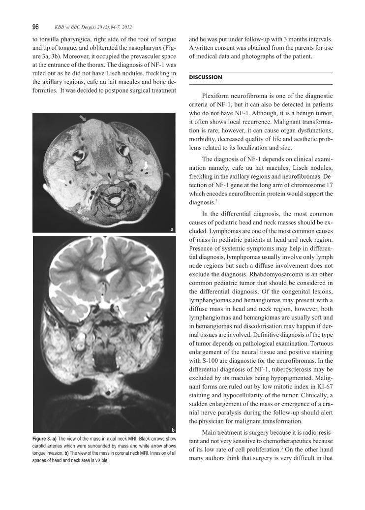
to tonsilla pharyngica, right side of the root of tongueand tip of tongue, and obliterated the nasopharynx (Fig-ure 3a, 3b). Moreover, it occupied the prevasculer spaceat the entrance of the thorax. The diagnosis of NF-1 wasruled out as he did not have Lisch nodules, freckling inthe axillary regions, cafe au lait macules and bone de-formities. It was decided to postpone surgical treatment
and he was put under follow-up with 3 months intervals.A written consent was obtained from the parents for useof medical data and photographs of the patient.
DISCUSSION
Plexiform neurofibroma is one of the diagnosticcriteria of NF-1, but it can also be detected in patientswho do not have NF-1. Although, it is a benign tumor,it often shows local recurrence. Malignant transforma-tion is rare, however, it can cause organ dysfunctions,morbidity, decreased quality of life and aesthetic prob-lems related to its localization and size.
The diagnosis of NF-1 depends on clinical exami-nation namely, cafe au lait macules, Lisch nodules,freckling in the axillary regions and neurofibromas. De-tection of NF-1 gene at the long arm of chromosome 17which encodes neurofibromin protein would support thediagnosis.2
In the differential diagnosis, the most commoncauses of pediatric head and neck masses should be ex-cluded. Lymphomas are one of the most common causesof mass in pediatric patients at head and neck region.Presence of systemic symptoms may help in differen-tial diagnosis, lymphpomas usually involve only lymphnode regions but such a diffuse involvement does notexclude the diagnosis. Rhabdomyosarcoma is an othercommon pediatric tumor that should be considered inthe differential diagnosis. Of the congenital lesions,lymphangiomas and hemangiomas may present with adiffuse mass in head and neck region, however, bothlymphangiomas and hemangiomas are usually soft andin hemangiomas red discolorisation may happen if der-mal tissues are involved. Definitive diagnosis of the typeof tumor depends on pathological examination. Tortuousenlargement of the neural tissue and positive stainingwith S-100 are diagnostic for the neurofibromas. In thedifferential diagnosis of NF-1, tuberosclerosis may beexcluded by its macules being hypopigmented. Malig-nant forms are ruled out by low mitotic index in KI-67staining and hypocellularity of the tumor. Clinically, asudden enlargement of the mass or emergence of a cra-nial nerve paralysis during the follow-up should alertthe physician for malignant transformation.
Main treatment is surgery because it is radio-resis-tant and not very sensitive to chemotherapeutics becauseof its low rate of cell proliferation.3 On the other handmany authors think that surgery is very difficult in that
Figure 3. a) The view of the mass in axial neck MRI. Black arrows showcarotid arteries which were surrounded by mass and white arrow showstongue invasion, b) The view of the mass in coronal neck MRI. Invasion of allspaces of head and neck area is visible.
a
b

Turkiye Klinikleri J Int Med Sci 2008, 4 97
97A Huge Plexiform Neurofibroma in a 20 Months Old Child: Case Report 97
kind of tumors and it often recurs and grows faster thanbefore, following surgery.4,5 In the previous studies itwas reported that recurrence was less than 40% aftersubtotal resection, whereas, it was less than 20% aftertotal resection. Recurrence rate is especially muchhigher in patients under 10 years old.3 Therefore, manysurgeons are against surgery especially in children, iftumor is localized at head and neck region.6
The present case was unique for having such a bigtumor in such a young age. We decided not to operate onthis patient at the moment, because total excision wasnot possible and partial resection had a high risk formore aggressive course. Therefore, we offered only sup-portive treatment including tracheotomy until he grewolder, and drawbacks related to young age ceased. Bythen, even a palliative surgery might offer success.
1. Barnes L. Tumors of the nervous system. In: Barnes L, eds.Surgical Pathology of Head and Neck. 2nd ed. New York-Basel: Marcel Dekker; 2001. p.800-3.
2. Rosai J. Soft tissues. In: Rosai J, eds. Rosai and Ackerman’sSurgical Pathology. 9th ed. Edinburgh - New York: Mosby;2004. p.2266-9.
3. Wise JB, Creyer JE, Belasco JB, Jacobs I, Elden L. Manage-ment of head and neck plexiform neurofibromas in pediatricpatients with neurofibromatisis Type 1. Arch OtolaryngolHead Neck Surg 2005;131(8):712-8.
4. Needle MN, Cnaan A, Dattilo J, Chatten J, Phillips PC, ShochatS, et al. Prognostic signs in the surgical management of plexi-form neurofibroma:The Children’s Hospital of Philadelphia Ex-perience, 1974-1994. J Pediatr 1997;131(5):678-82.
5. Serletis D, Parkin, P, Bouffet E, Shroff M, Drake JM, Rutka JT.Massive plexiform neurofibromas in childhood: natural historyand management issues. J Neurosurg 2007;106(5 Suppl):363-7.
6. Ransom ER, Yoon C, Manolidis S. Single stage near total re-section of massive pediatric head and neck plexiform neurofi-bromas. Int J Pediatr Otorhinolarygol 2006;70(6):1055-61.
REFERENCES