anesthesia osce
DESCRIPTION
Preoperativeassessment• A)Definitionofpreoperativeperiod.• B)Indicationsofpreoperativeevaluation.• C)Evaluationofapatientinthepre-‐operativeperiod:I.Historytaking.II.Examination.III.ASAClassification.IV.Investigations.V.Consent.VI.Premedication.VII.PreoperativestarvationTRANSCRIPT
Anesthesia OSCE
045 Anesthesia team
Page 2
Anesthesia OSCE
Index Topic Page Preoperative assessment
• A) Definition of preoperative period. • B) Indications of preoperative evaluation. • C) Evaluation of a patient in the pre-‐operative
period: I. History taking. II. Examination. III. ASA Classification. IV. Investigations. V. Consent. VI. Premedication. VII. Preoperative starvation.
3 3 3 3 4 6 6 6 6 6
Airway management • A) Basic airway anatomy • B) Methods of supporting the airway:
I. Mouth-‐to-‐mask ventilation with supplemental oxygen. II. Bag mask ventilation. III. Laryngeal Mask Airway (LMA). IV. Endotracheal Tube (ETT). V. Fiber-‐optic laryngoscope. VI. Instruments that ease the process of intubation.
• C) Rapid sequence induction • D) Difficult Airway management • E) Surgical Invasive airway access
7 8 9 10 12 14 15 16 17 18
Regional Anesthesia • A) Definition of regional anesthesia. • B) Indications of regional anesthesia. • C) Contraindications of regional anesthesia. • D) Complications of regional anesthesia. • E) Types of regional anesthesia.
I. Epidural anesthesia. II. Spinal anesthesia. III. Combined spinal and epidural anesthesia.
19 19 19 19 20 21 22
Intravenous Access • A) Central line: • B) Peripheral veins:
23 25
Intravenous Fluids • A) Factors must be taken into account • B) Crystalloids. • C) Colloids. • D) Fluid replacement. • E) Blood loss regimens. • F) Blood transfusion complications.
27 27 27 27 29 29
Page 3
Anesthesia OSCE
Preoperative Care
A) Definition of preoperative period: It is the time from the decision to have surgery until admitted into the OR theatre.
B) Indications of preoperative evaluation:
1. Assess the anesthetic risks in relation to the proposed surgery. 2. To decide the anesthetic technique (general, regional, or a combination). 3. To plan the postoperative care including any analgesic regimens.
C) Evaluation of a patient in the pre-‐operative period:
I. History taking:
1. Introduction: -‐ Introduce yourself to the patient giving your name and status as a student. Ask for permission to take a history and perform a physical examination.
2. Personal history: Ask for the patient’s name, age, occupation, nationality, and marital status.
3. Present illness: Establish the principal symptom or symptoms that caused the patient to seek medical attention, when it first appeared and how it has changed over time.
4. Past medical: Ask about the patient’s previous medical problems including cardiac (IHD, HTN, HF, AF), respiratory (asthma, COPD, TB), neurological (stroke, TIA, epilepsy), gastroenterological (liver disease, jaundice) and haematological (sickle cell, thalassemia) problems. Also ask the patient if they are pregnant if relevant.
5. Past surgical: Ask about any previous operations and post-‐op. complications. Enquire about previous types of anaesthesia received (local, general) and enquire about any anesthetic complications (malignant hyperthermia).
6. Medications: Ask about any prescribed medications the patient is taking including insulin or hypoglycemics, anticoagulants (warfarin, aspirin), β-‐blockers, steroids, ACE inhibitors, diuretics and inhalers. Enquire about any over-‐the-‐counter medication, contraception (COCP) and HRT.
7. Allergies: Enquire about allergies to antibiotics, plasters, latex, eggs and antiseptic solutions.
8. Family history: Check for family history of any illnesses including myotonic dystrophy, malignant hyperpyrexia, porphyria, cholinesterase disorders and sickle cell disease. Enquire about any other anaesthetic complications and allergic reactions in the family.
9. Dental: Ask about any history of dental problems, false teeth, caps, bridges and dentures.
10. Social: Elicit the patient’s alcohol history noting the number of units consumed in a week. Determine if the patient is a smoker and how many cigarettes he smokes per day.
Page 4
Anesthesia OSCE
1. General examination: -‐ BMI: Measure the patient’s height and weight and calculate his body mass index. Ideal BMI is between 18.5 and 24.9. -‐ Document the patient’s blood pressure, oxygen saturation on air, pulse, respiratory rate and temperature. -‐ Perform a brief chest, abdomen, cardiovascular and neurological examination.
2. Airway: (LEMON) • Look: -‐ Ask the patient to flex and extend his neck and to open and close his mouth looking for short immobile neck. Some patients cannot be placed in the “sniffing position” secondary to neck trauma, cervical collar, musculoskeletal disorders like kyphosis and rheumatoid arthritis. -‐ A neck circumference of greater than 45cm in an obese patient with a BMI of greater than 40kg/m^2 is likely to be a difficult intubation. -‐ Women with large pendulous breasts add a degree of difficulty to an intubation because the provider may not be able to position the blade handle appropriately toward the chest -‐ Inspect the mouth and see if there are any obvious abnormalities, buckteeth, high arch palate, receding mandible (may be hidden by a beard), Inability to sublux the jaw (forward protrusion of the lower incisors beyond the upper incisors).
• Evaluate: a. Thyromental distance: -‐ It is the distance from the thyroid cartilage to the mental prominence when the neck is extended fully. -‐ If the distance is more than7cm (around 3 fingerbreadths), problems
Traumatic: Infection Inflammatory Neoplastic Endocrine: Pregnancy • Fractures Of Mandible And Cervical Spine
• Epiglottitis • Dental Or Facial Abscess
• Ankylosing Spondylitis
• Rheumatoid Arthritis
• Tongue • Neck • Mouth
• Thyroid Enlargement • Acromegaly • Obesity
Important Symptoms that you should ask in the history:
• Upper airway obstruction may be found in patients with stridor, dysphagia and hoarseness.
• Snoring may also indicate partial upper airway obstruction.
II. Examination: Initially examine the patient generally then move to airway examination.
I.Complete review of the systems
Page 5
Anesthesia OSCE
should not occur with intubation. -‐ A distance of less than 6 cm suggests laryngoscopy will be impossible and for distances of 6–6.5 cm, laryngoscopy is considered difficult, but possible.
b. Sternomandibular distance: -‐ This test is claimed to predict up to 90% of difficult intubations. -‐ The distance from the upper border of the manubrium sterni to the tip of the chin, with the mouth closed and the head fully extended, is measured. -‐ A distance of less than 12.5 cm indicates a difficult intubation.
c. Alantooceptal joint: -‐ Presence of a gap between the Occiput and C1 is essential. -‐ It should be (15-‐20 degrees).
d. C-‐Spine: -‐ Flexion and extension of the head and neck must be more than 90 degree.
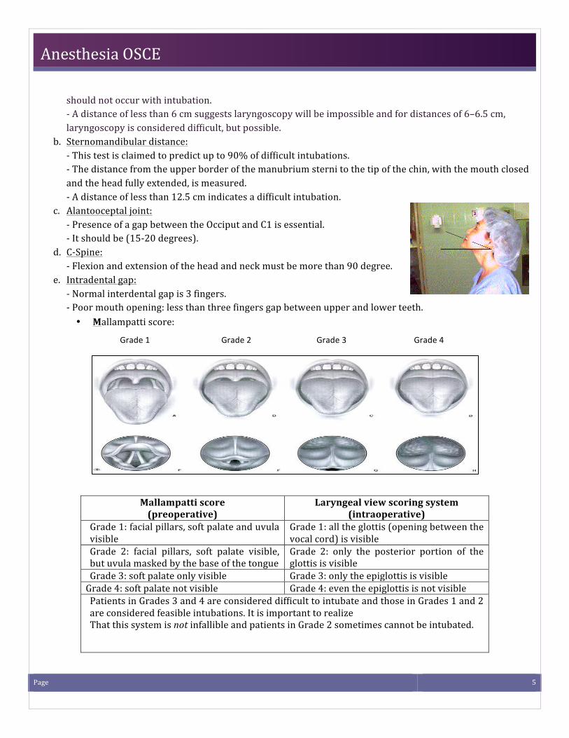
e. Intradental gap: -‐ Normal interdental gap is 3 fingers. -‐ Poor mouth opening: less than three fingers gap between upper and lower teeth. • Mallampatti score:
Mallampatti score (preoperative)
Laryngeal view scoring system (intraoperative)
Grade 1: facial pillars, soft palate and uvula visible
Grade 1: all the glottis (opening between the vocal cord) is visible
Grade 2: facial pillars, soft palate visible, but uvula masked by the base of the tongue
Grade 2: only the posterior portion of the glottis is visible
Grade 3: soft palate only visible Grade 3: only the epiglottis is visible Grade 4: soft palate not visible Grade 4: even the epiglottis is not visible Patients in Grades 3 and 4 are considered difficult to intubate and those in Grades 1 and 2 are considered feasible intubations. It is important to realize That this system is not infallible and patients in Grade 2 sometimes cannot be intubated.
Grade 1 Grade 2 Grade 3 Grade 4
Page 6
Anesthesia OSCE
• Obstruction: Airway edema, tracheal mass, mediastinal mass. • Neck mobility.
III. ASA classification • ASA 1: Healthy patient without organic biochemical or psychiatric disease. • ASA 2: A Patient with mild systemic disease. No significant impact on daily activity. Unlikely impact on
anesthesia and surgery. • ASA 3: Significant or severe systemic disease that limits normal activity. Significant impact on daily activity.
Likely impact on anesthesia and surgery. • ASA 4: Severe disease that is a constant threat to life or requires intensive therapy. Serious limitation of
daily activity. • ASA 5:Moribund patient who is equally likely to die in the next 24 hours with or without surgery. • ASA 6: Brain-‐dead organ donor. • “E” – added to the classifications indicates emergency surgery.
IV. Investigations: State that you would order investigations if clinically appropriate.
• Blood: Hemoglobin concentration. Coagulation profile. Screening for sickle cell disease. Urea. Creatinine. Electrolytes. Glucose.
• Chest X-‐ ray. Respiratory (asthma, COPD) or cardiac disease (heart failure), malignancy, thoracic surgery, respiratory symptoms (cough, SOB, sputum), previous TB.
• ECG. Hypertension, heart disease, arrhythmia, >50 years old, DM.
V. Consent: It should be a written one and it explains the anesthetic options for a given surgical procedure. VI. Premedication: If your patient need any premeditations like anxiolytics mention them. VII. Preoperative starvation:
• From Solid Food = 6-‐8 hours. • From Clear Fluid= 2 hours. • From Breast Milk for Neonates = 4 hours. • From Formula Milk for Neonates = 6 hours.
Page 7
Anesthesia OSCE
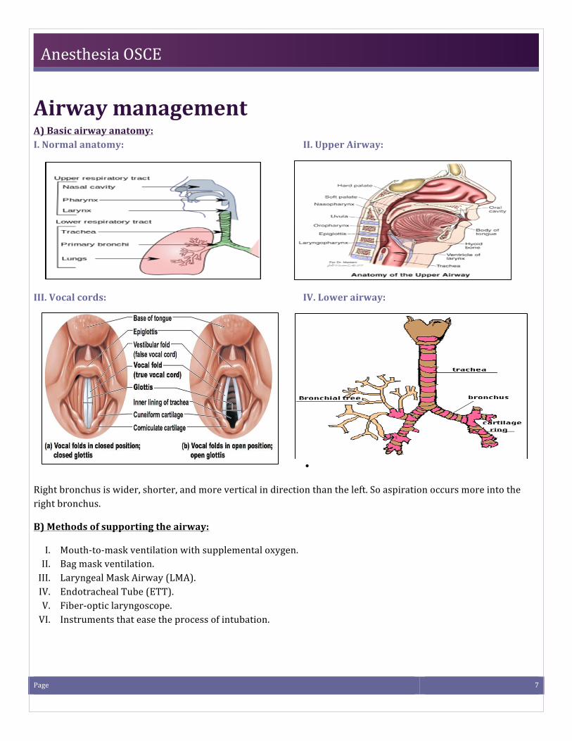
Airway management A) Basic airway anatomy: I. Normal anatomy: II. Upper Airway:
III. Vocal cords: IV. Lower airway:
•
Right bronchus is wider, shorter, and more vertical in direction than the left. So aspiration occurs more into the right bronchus.
B) Methods of supporting the airway:
I. Mouth-‐to-‐mask ventilation with supplemental oxygen. II. Bag mask ventilation. III. Laryngeal Mask Airway (LMA). IV. Endotracheal Tube (ETT). V. Fiber-‐optic laryngoscope. VI. Instruments that ease the process of intubation.
Page 8
Anesthesia OSCE
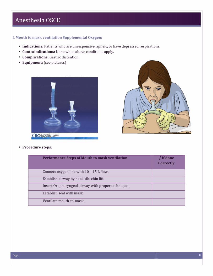
I. Mouth to mask ventilation Supplemental Oxygen:
• Indications: Patients who are unresponsive, apneic, or have depressed respirations. • Contraindications: None when above conditions apply. • Complications: Gastric distention. • Equipment: (see pictures)
• Procedure steps:
Performance Steps of Mouth to mask ventilation √ if done Correctly
Connect oxygen line with 10 – 15 L flow.
Establish airway by head-‐tilt, chin lift.
Insert Oropharyngeal airway with proper technique.
Establish seal with mask.
Ventilate mouth-‐to-‐mask.
Page 9
Anesthesia OSCE
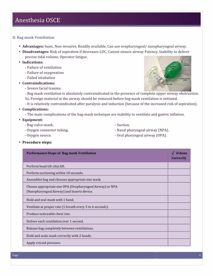
II. Bag mask Ventilation
• Advantages: basic, Non-‐invasive, Readily available, Can use oropharyngeal/ nasopharyngeal airway. • Disadvantages: Risk of aspiration if decreases LOC, Cannot ensure airway Patency, Inability to deliver
precise tidal volume, Operator fatigue. • Indications:
-‐ Failure of ventilation -‐ Failure of oxygenation -‐ Failed intubation
• Contraindications: -‐ Severe facial trauma. -‐ Bag mask ventilation is absolutely contraindicated in the presence of complete upper airway obstruction. So, Foreign material in the airway should be removed before bag mask ventilation is initiated. -‐ It is relatively contraindicated after paralysis and induction (because of the increased risk of aspiration).
• Complications: -‐ The main complications of the bag-‐mask technique are inability to ventilate and gastric inflation.
• Equipment: -‐ Bag-‐valve-‐mask. -‐ Oxygen connector tubing. -‐ Oxygen source.
-‐ Suction. -‐ Nasal pharyngeal airway (NPA). -‐ Oral pharyngeal airway (OPA).
• Procedure steps:
Performance Steps of Bag mask Ventilation √ if done Correctly
Perform head tilt-‐chin lift.
Perform suctioning within 10 seconds.
Assembles bag and chooses appropriate size mask.
Choose appropriate size OPA (Oropharyngeal Airway) or NPA (Nasopharyngeal Airway) and Inserts device.
Hold and seal mask with 1 hand.
Ventilate at proper rate (1 breath every 5 to 6 seconds).
Produce noticeable chest rise.
Deliver each ventilation over 1 second.
Release bag completely between ventilations.
Hold and seals mask correctly with 2 hands.
Apply cricoid pressure.
Page 10
Anesthesia OSCE
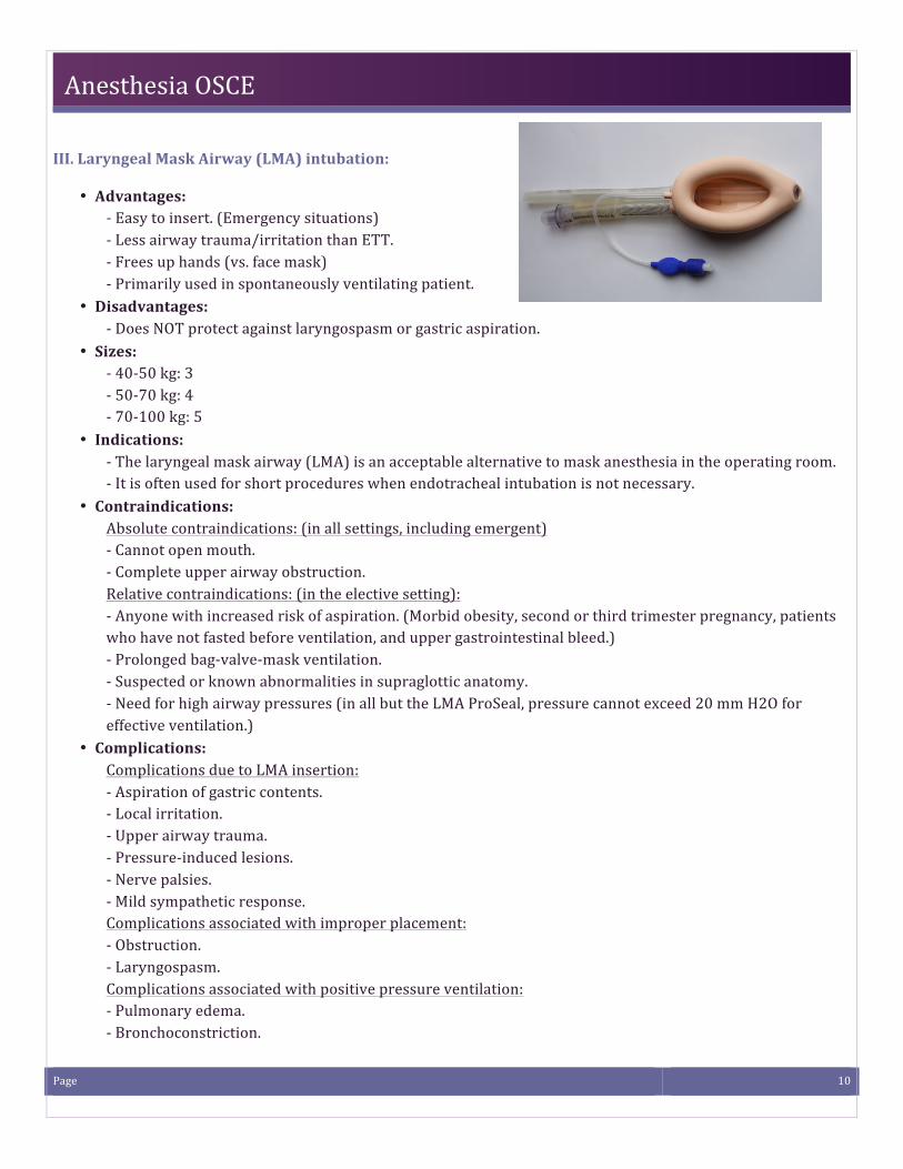
III. Laryngeal Mask Airway (LMA) intubation:
• Advantages: -‐ Easy to insert. (Emergency situations) -‐ Less airway trauma/irritation than ETT. -‐ Frees up hands (vs. face mask) -‐ Primarily used in spontaneously ventilating patient.
• Disadvantages: -‐ Does NOT protect against laryngospasm or gastric aspiration.
• Sizes: -‐ 40-‐50 kg: 3 -‐ 50-‐70 kg: 4 -‐ 70-‐100 kg: 5
• Indications: -‐ The laryngeal mask airway (LMA) is an acceptable alternative to mask anesthesia in the operating room. -‐ It is often used for short procedures when endotracheal intubation is not necessary.
• Contraindications: Absolute contraindications: (in all settings, including emergent) -‐ Cannot open mouth. -‐ Complete upper airway obstruction. Relative contraindications: (in the elective setting): -‐ Anyone with increased risk of aspiration. (Morbid obesity, second or third trimester pregnancy, patients who have not fasted before ventilation, and upper gastrointestinal bleed.) -‐ Prolonged bag-‐valve-‐mask ventilation. -‐ Suspected or known abnormalities in supraglottic anatomy. -‐ Need for high airway pressures (in all but the LMA ProSeal, pressure cannot exceed 20 mm H2O for effective ventilation.)
• Complications: Complications due to LMA insertion: -‐ Aspiration of gastric contents. -‐ Local irritation. -‐ Upper airway trauma. -‐ Pressure-‐induced lesions. -‐ Nerve palsies. -‐ Mild sympathetic response. Complications associated with improper placement: -‐ Obstruction. -‐ Laryngospasm. Complications associated with positive pressure ventilation: -‐ Pulmonary edema. -‐ Bronchoconstriction.
Page 11
Anesthesia OSCE
• Performance Steps Of Laryngeal Mask Airway:
Performance Steps Of Laryngeal Mask Airway: √ if done correctly
Prepare and assemble all necessary equipment.
Choose appropriate size LMA.
Test integrity of cuff by inflating it.
Deflate cuff on a flat surface and lubricate LMA on posterior surface only for use.
Open the mouth using the “crossed fingers” technique or by performing a tongue-‐Jaw lift; do not hyperextend neck.
Clear the airway if needed.
Insert tube into mouth and place it so that the curvature is the same as that of the Pharynx, directing it posteriorly until resistance is felt.
Inflate the cuff with the appropriate amount of air corresponding to the size of the tube , remove syringe.
Insert bite block.
Produce noticeable chest rise; auscultate breath sounds.
Confirm correct positioning of LMA by colorimetric ETCO" capnograph.
Secure LMA in place.
Perform correct ventilation rate for respiratory arrest (1 breath every 5 to 6 seconds).
Perform correct ventilation rate for cardiac arrest (1 breath every 6 to 8 seconds).
Deliver each ventilation over 1 second.
Demonstrate complete release of bag between ventilation.
Page 12
Anesthesia OSCE
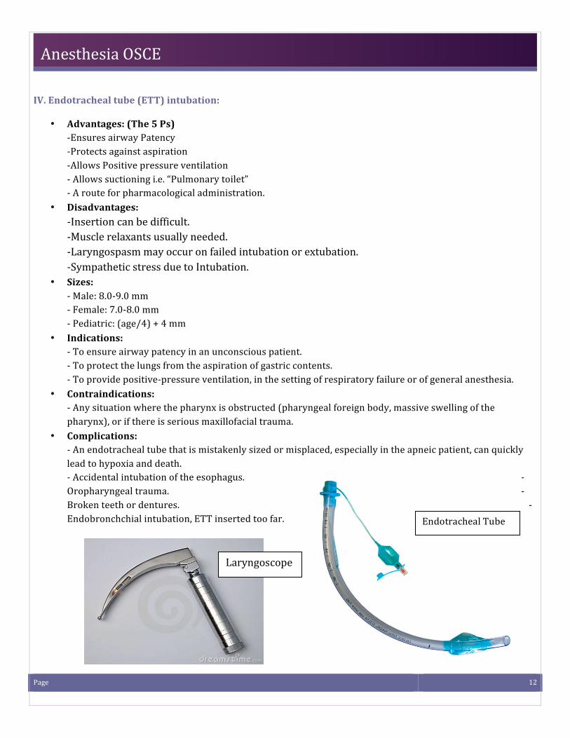
IV. Endotracheal tube (ETT) intubation:
• Advantages: (The 5 Ps) -‐Ensures airway Patency -‐Protects against aspiration -‐Allows Positive pressure ventilation -‐ Allows suctioning i.e. “Pulmonary toilet” -‐ A route for pharmacological administration.
• Disadvantages: -‐Insertion can be difficult. -‐Muscle relaxants usually needed. -‐Laryngospasm may occur on failed intubation or extubation. -‐Sympathetic stress due to Intubation.
• Sizes: -‐ Male: 8.0-‐9.0 mm -‐ Female: 7.0-‐8.0 mm -‐ Pediatric: (age/4) + 4 mm
• Indications: -‐ To ensure airway patency in an unconscious patient. -‐ To protect the lungs from the aspiration of gastric contents. -‐ To provide positive-‐pressure ventilation, in the setting of respiratory failure or of general anesthesia.
• Contraindications: -‐ Any situation where the pharynx is obstructed (pharyngeal foreign body, massive swelling of the pharynx), or if there is serious maxillofacial trauma.
• Complications: -‐ An endotracheal tube that is mistakenly sized or misplaced, especially in the apneic patient, can quickly lead to hypoxia and death. -‐ Accidental intubation of the esophagus. -‐ Oropharyngeal trauma. -‐ Broken teeth or dentures. -‐ Endobronchchial intubation, ETT inserted too far.
Endotracheal Tube
Laryngoscope Laryngoscope
Page 13
Anesthesia OSCE
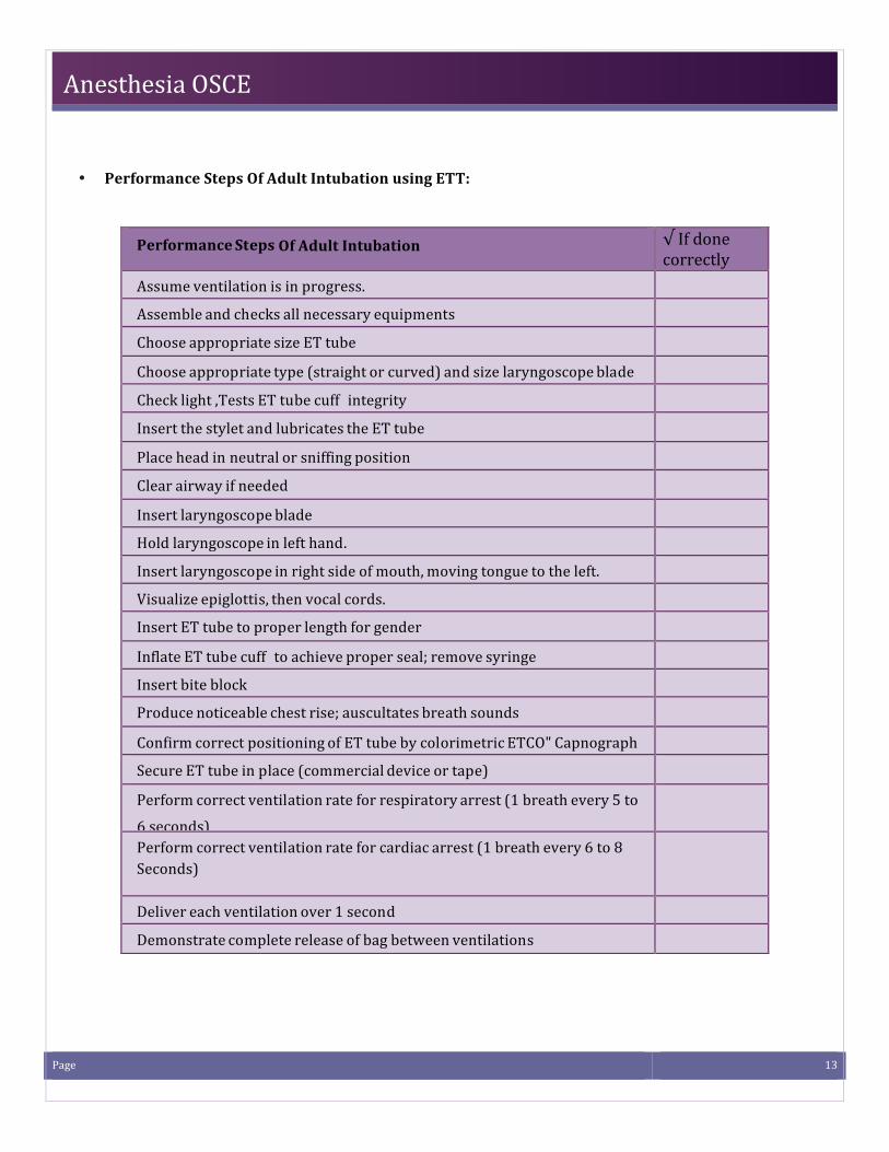
• Performance Steps Of Adult Intubation using ETT:
Performance Steps Of Adult Intubation √ If done correctly Assume ventilation is in progress.
Assemble and checks all necessary equipments
Choose appropriate size ET tube
Choose appropriate type (straight or curved) and size laryngoscope blade
Check light ,Tests ET tube cuff integrity
Insert the stylet and lubricates the ET tube
Place head in neutral or sniffing position
Clear airway if needed
Insert laryngoscope blade
Hold laryngoscope in left hand.
Insert laryngoscope in right side of mouth, moving tongue to the left.
Visualize epiglottis, then vocal cords.
Insert ET tube to proper length for gender
Inflate ET tube cuff to achieve proper seal; remove syringe
Insert bite block
Produce noticeable chest rise; auscultates breath sounds
Confirm correct positioning of ET tube by colorimetric ETCO" Capnograph
Secure ET tube in place (commercial device or tape)
Perform correct ventilation rate for respiratory arrest (1 breath every 5 to 6 seconds)
Perform correct ventilation rate for cardiac arrest (1 breath every 6 to 8 Seconds)
Deliver each ventilation over 1 second
Demonstrate complete release of bag between ventilations
Page 14
Anesthesia OSCE
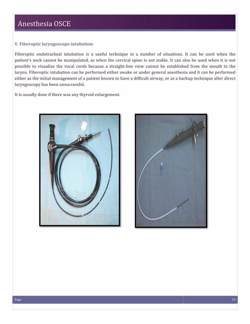
V. Fiberoptic laryngoscope intubation:
Fiberoptic endotracheal intubation is a useful technique in a number of situations. It can be used when the patient's neck cannot be manipulated, as when the cervical spine is not stable. It can also be used when it is not possible to visualize the vocal cords because a straight-‐line view cannot be established from the mouth to the larynx. Fiberoptic intubation can be performed either awake or under general anesthesia and it can be performed either as the initial management of a patient known to have a difficult airway, or as a backup technique after direct laryngoscopy has been unsuccessful.
It is usually done if there was any thyroid enlargement.
Page 15
Anesthesia OSCE
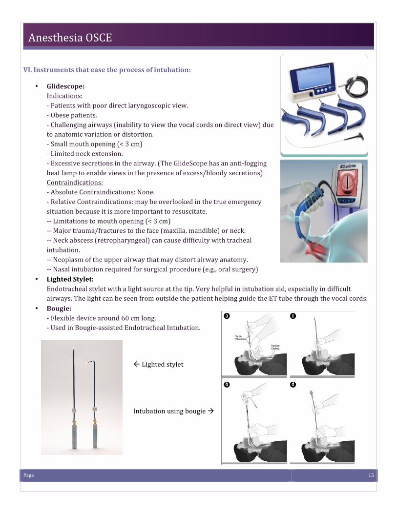
VI. Instruments that ease the process of intubation:
• Glidescope: Indications: -‐ Patients with poor direct laryngoscopic view. -‐ Obese patients. -‐ Challenging airways (inability to view the vocal cords on direct view) due to anatomic variation or distortion. -‐ Small mouth opening (< 3 cm) -‐ Limited neck extension. -‐ Excessive secretions in the airway. (The GlideScope has an anti-‐fogging heat lamp to enable views in the presence of excess/bloody secretions) Contraindications: -‐ Absolute Contraindications: None. -‐ Relative Contraindications: may be overlooked in the true emergency situation because it is more important to resuscitate. -‐-‐ Limitations to mouth opening (< 3 cm) -‐-‐ Major trauma/fractures to the face (maxilla, mandible) or neck. -‐-‐ Neck abscess (retropharyngeal) can cause difficulty with tracheal intubation. -‐-‐ Neoplasm of the upper airway that may distort airway anatomy. -‐-‐ Nasal intubation required for surgical procedure (e.g., oral surgery)
• Lighted Stylet: Endotracheal stylet with a light source at the tip. Very helpful in intubation aid, especially in difficult airways. The light can be seen from outside the patient helping guide the ET tube through the vocal cords.
• Bougie: -‐ Flexible device around 60 cm long. -‐ Used in Bougie-‐assisted Endotracheal Intubation.
ß Lighted stylet
Intubation using bougie à
Page 16
Anesthesia OSCE
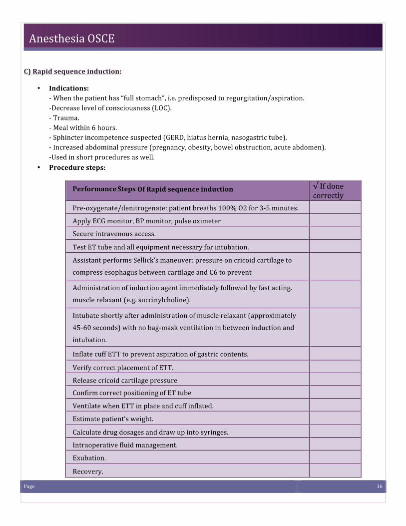
C) Rapid sequence induction:
• Indications: -‐ When the patient has “full stomach”, i.e. predisposed to regurgitation/aspiration. -‐Decrease level of consciousness (LOC). -‐ Trauma. -‐ Meal within 6 hours. -‐ Sphincter incompetence suspected (GERD, hiatus hernia, nasogastric tube). -‐ Increased abdominal pressure (pregnancy, obesity, bowel obstruction, acute abdomen). -‐Used in short procedures as well.
• Procedure steps:
Performance Steps Of Rapid sequence induction √ If done
correctly Pre-‐oxygenate/denitrogenate: patient breaths 100% O2 for 3-‐5 minutes.
Apply ECG monitor, BP monitor, pulse oximeter
Secure intravenous access.
Test ET tube and all equipment necessary for intubation.
Assistant performs Sellick’s maneuver: pressure on cricoid cartilage to
compress esophagus between cartilage and C6 to prevent
reflux/aspiration.
Administration of induction agent immediately followed by fast acting.
muscle relaxant (e.g. succinylcholine).
Intubate shortly after administration of muscle relaxant (approximately
45-‐60 seconds) with no bag-‐mask ventilation in between induction and
intubation.
Inflate cuff ETT to prevent aspiration of gastric contents.
Verify correct placement of ETT.
Release cricoid cartilage pressure
Confirm correct positioning of ET tube
Ventilate when ETT in place and cuff inflated.
Estimate patient’s weight.
Calculate drug dosages and draw up into syringes.
Intraoperative fluid management.
Exubation.
Recovery.
Page 17
Anesthesia OSCE
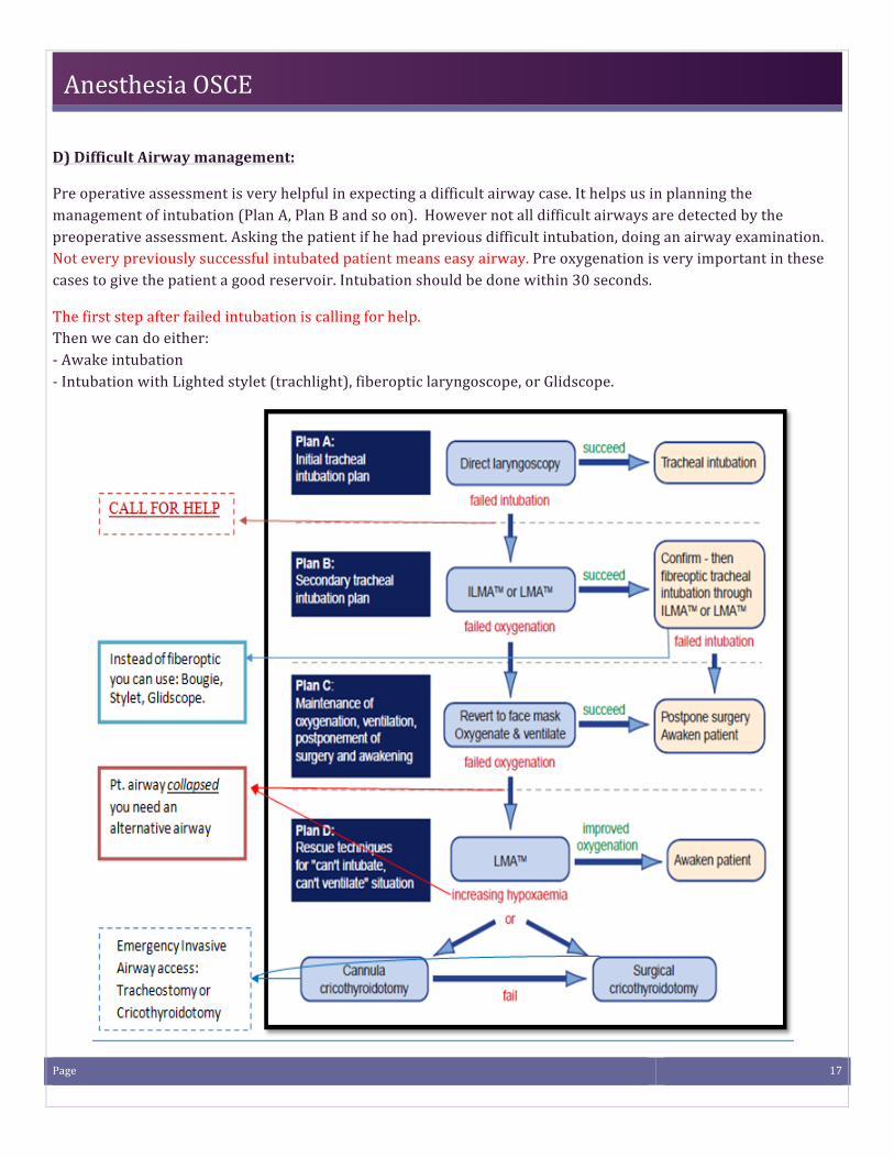
D) Difficult Airway management:
Pre operative assessment is very helpful in expecting a difficult airway case. It helps us in planning the management of intubation (Plan A, Plan B and so on). However not all difficult airways are detected by the preoperative assessment. Asking the patient if he had previous difficult intubation, doing an airway examination. Not every previously successful intubated patient means easy airway. Pre oxygenation is very important in these cases to give the patient a good reservoir. Intubation should be done within 30 seconds.
The first step after failed intubation is calling for help. Then we can do either: -‐ Awake intubation -‐ Intubation with Lighted stylet (trachlight), fiberoptic laryngoscope, or Glidscope.
Page 18
Anesthesia OSCE
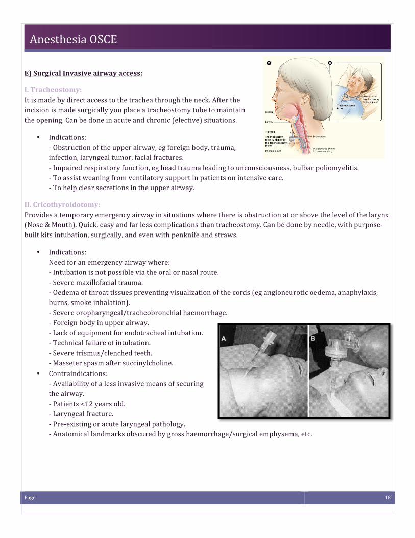
E) Surgical Invasive airway access:
I. Tracheostomy: It is made by direct access to the trachea through the neck. After the incision is made surgically you place a tracheostomy tube to maintain the opening. Can be done in acute and chronic (elective) situations.
• Indications: -‐ Obstruction of the upper airway, eg foreign body, trauma, infection, laryngeal tumor, facial fractures. -‐ Impaired respiratory function, eg head trauma leading to unconsciousness, bulbar poliomyelitis. -‐ To assist weaning from ventilatory support in patients on intensive care. -‐ To help clear secretions in the upper airway.
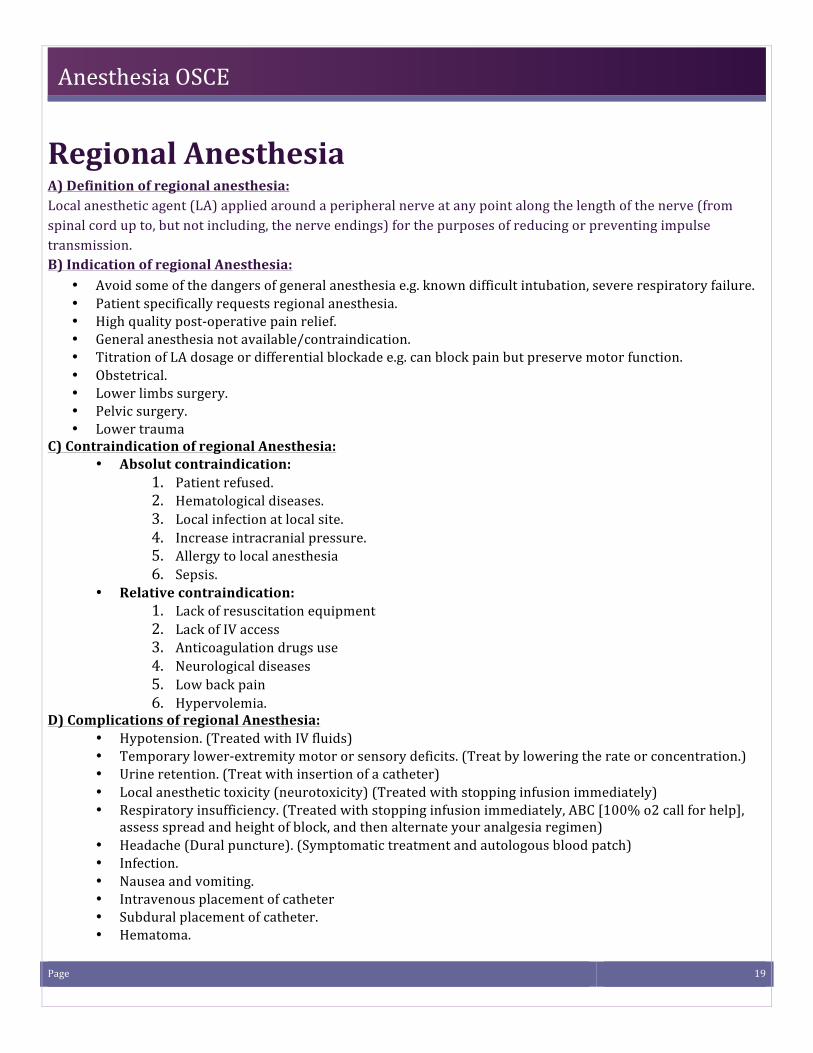
II. Cricothyroidotomy: Provides a temporary emergency airway in situations where there is obstruction at or above the level of the larynx (Nose & Mouth). Quick, easy and far less complications than tracheostomy. Can be done by needle, with purpose-‐built kits intubation, surgically, and even with penknife and straws.
• Indications: Need for an emergency airway where: -‐ Intubation is not possible via the oral or nasal route. -‐ Severe maxillofacial trauma. -‐ Oedema of throat tissues preventing visualization of the cords (eg angioneurotic oedema, anaphylaxis, burns, smoke inhalation). -‐ Severe oropharyngeal/tracheobronchial haemorrhage. -‐ Foreign body in upper airway. -‐ Lack of equipment for endotracheal intubation. -‐ Technical failure of intubation. -‐ Severe trismus/clenched teeth. -‐ Masseter spasm after succinylcholine.
• Contraindications: -‐ Availability of a less invasive means of securing the airway. -‐ Patients <12 years old. -‐ Laryngeal fracture. -‐ Pre-‐existing or acute laryngeal pathology. -‐ Anatomical landmarks obscured by gross haemorrhage/surgical emphysema, etc.
Page 19
Anesthesia OSCE
Regional Anesthesia A) Definition of regional anesthesia: Local anesthetic agent (LA) applied around a peripheral nerve at any point along the length of the nerve (from spinal cord up to, but not including, the nerve endings) for the purposes of reducing or preventing impulse transmission. B) Indication of regional Anesthesia:
• Avoid some of the dangers of general anesthesia e.g. known difficult intubation, severe respiratory failure. • Patient specifically requests regional anesthesia. • High quality post-‐operative pain relief. • General anesthesia not available/contraindication. • Titration of LA dosage or differential blockade e.g. can block pain but preserve motor function. • Obstetrical. • Lower limbs surgery. • Pelvic surgery. • Lower trauma
C) Contraindication of regional Anesthesia: • Absolut contraindication:
1. Patient refused. 2. Hematological diseases. 3. Local infection at local site. 4. Increase intracranial pressure. 5. Allergy to local anesthesia 6. Sepsis.
• Relative contraindication: 1. Lack of resuscitation equipment 2. Lack of IV access 3. Anticoagulation drugs use 4. Neurological diseases 5. Low back pain 6. Hypervolemia.
D) Complications of regional Anesthesia: • Hypotension. (Treated with IV fluids) • Temporary lower-‐extremity motor or sensory deficits. (Treat by lowering the rate or concentration.) • Urine retention. (Treat with insertion of a catheter) • Local anesthetic toxicity (neurotoxicity) (Treated with stopping infusion immediately) • Respiratory insufficiency. (Treated with stopping infusion immediately, ABC [100% o2 call for help],
assess spread and height of block, and then alternate your analgesia regimen) • Headache (Dural puncture). (Symptomatic treatment and autologous blood patch) • Infection. • Nausea and vomiting. • Intravenous placement of catheter • Subdural placement of catheter. • Hematoma.
Page 20
Anesthesia OSCE
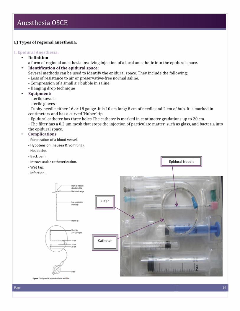
E) Types of regional anesthesia: I. Epidural Anesthesia:
• Definition a form of regional anesthesia involving injection of a local anesthetic into the epidural space.
• Identification of the epidural space: Several methods can be used to identify the epidural space. They include the following: -‐ Loss of resistance to air or preservative-‐free normal saline. -‐ Compression of a small air bubble in saline -‐ Hanging drop technique
• Equipment: -‐ sterile towels -‐ sterile gloves -‐ Tuohy needle either 16 or 18 gauge .It is 10 cm long: 8 cm of needle and 2 cm of hub. It is marked in centimeters and has a curved ‘Huber’ tip. -‐ Epidural catheter has three holes The catheter is marked in centimeter gradations up to 20 cm. -‐ The filter has a 0.2 μm mesh that stops the injection of particulate matter, such as glass, and bacteria into the epidural space.
• Complications -‐ Penetration of a blood vessel. -‐ Hypotension (nausea & vomiting). -‐ Headache. -‐ Back pain. -‐ Intravascular catheterization. -‐ Wet tap. -‐ Infection.
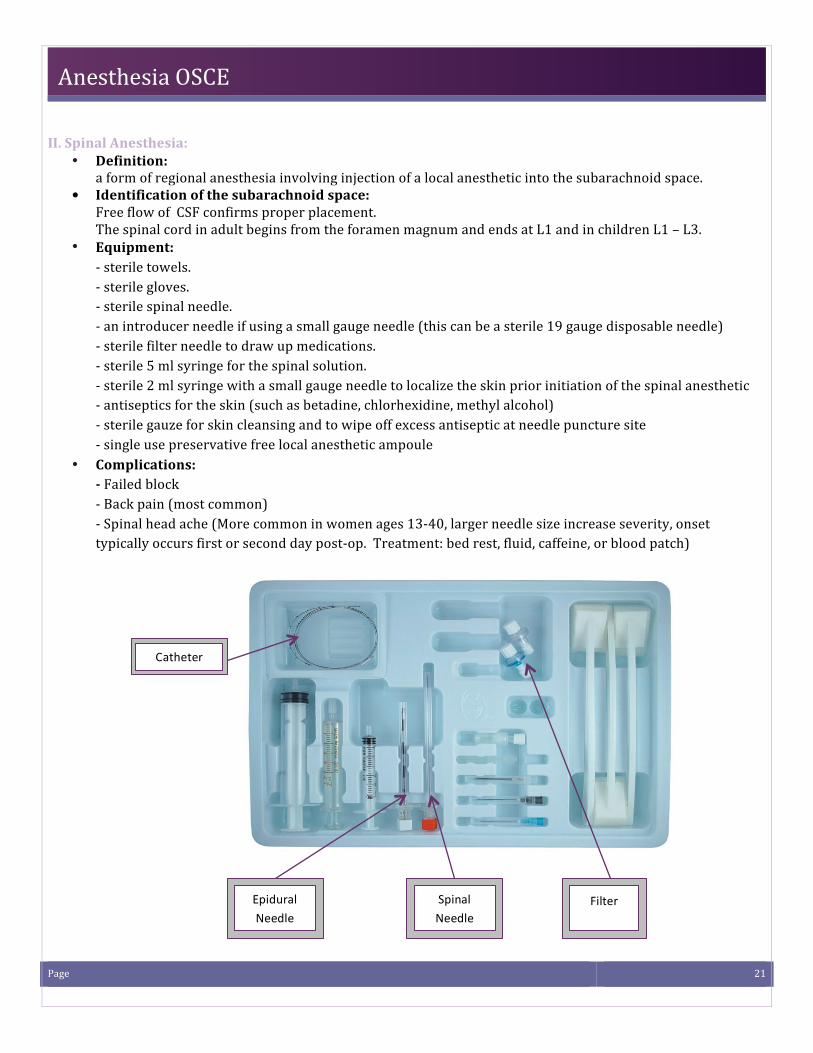
Filter
Catheter
Epidural Needle
Page 21
Anesthesia OSCE
II. Spinal Anesthesia: • Definition:
a form of regional anesthesia involving injection of a local anesthetic into the subarachnoid space. ! Identification of the subarachnoid space:
Free flow of CSF confirms proper placement. The spinal cord in adult begins from the foramen magnum and ends at L1 and in children L1 – L3.
• Equipment: -‐ sterile towels. -‐ sterile gloves. -‐ sterile spinal needle. -‐ an introducer needle if using a small gauge needle (this can be a sterile 19 gauge disposable needle) -‐ sterile filter needle to draw up medications. -‐ sterile 5 ml syringe for the spinal solution. -‐ sterile 2 ml syringe with a small gauge needle to localize the skin prior initiation of the spinal anesthetic -‐ antiseptics for the skin (such as betadine, chlorhexidine, methyl alcohol) -‐ sterile gauze for skin cleansing and to wipe off excess antiseptic at needle puncture site -‐ single use preservative free local anesthetic ampoule
• Complications: -‐ Failed block -‐ Back pain (most common) -‐ Spinal head ache (More common in women ages 13-‐40, larger needle size increase severity, onset typically occurs first or second day post-‐op. Treatment: bed rest, fluid, caffeine, or blood patch)
Epidural Needle
Spinal Needle
Filter
Catheter
Page 22
Anesthesia OSCE
• Procedure steps:
Performance Steps of spinal anesthesia √ if done
correctly
Taking Consent from the patient
Assessment (indications and contraindications)
Insert iv fluids
Mask, cap, gown and gloves
Prepare the back with antiseptic
Place a sterile Drape Over The Area
Identify the anatomical landmarks
Inject local anaesthetic into the skin and deeper tissue
Insert the large introducer needle into the selected spinal interspace
Direct the spinal needle through the introducer and into the Subarachnoid space
Free flow of CSF confirms proper placement
Aspirate for CSF if clear inject the proper anaesthetic
Remove the needle, introducer and drape sheet
Have the patient lie down
III. Combined spinal and epidural anesthesia:
• Definition: Is a regional anesthetic technique, which combines the benefits of both spinal anesthesia and epidural anesthesia and analgesia. The spinal component gives a rapid onset of a predictable block. The indwelling epidural catheter gives the ability to provide long lasting analgesia and to titrate the dose given to the desired effect.
• Indications: -‐ Caesarean sections. -‐ Labor pains. -‐ Post operative pain.
Page 23
Anesthesia OSCE
Intravenous access A) Central line: I. Indications:
• Monitor CVP • Administration of fluids to treat hypovolemia and shock. • Infusion of caustic drugs. • Total parenteral nutrition • Aspiration of air emboli • Insertion of transcutaneous pacing leads • Venous access in cases of poor peripheral veins.
II. Contraindications: • Relative contraindicated in patients who are receiving anticoagulant. • Ipsilateral caroted endartrectomy • Injury, or infection at the site of insertion.
III. Complications: • Local complications associated with femoral vein:
-‐ Thrombosis or phlebitis may extend to deep to iliac veins or vena cava. -‐ Arterial cannulation – loss of limb. -‐ Hematoma
• Local complications associated with subclavian and internal jugular: -‐ Hematoma may compromise airway -‐ Damage to adjacent artery, nerve, or lymphatic duct -‐ Perforation of endotracheal cuff
• Systemic complications: -‐ Pneumothorax. (Need follow-‐up chest X-‐ray) -‐ Hemothorax. -‐ Air embolism. -‐ Infiltration into mediastinum or pleural space. -‐ Arrhythmia from catheter tip. -‐ Infection.
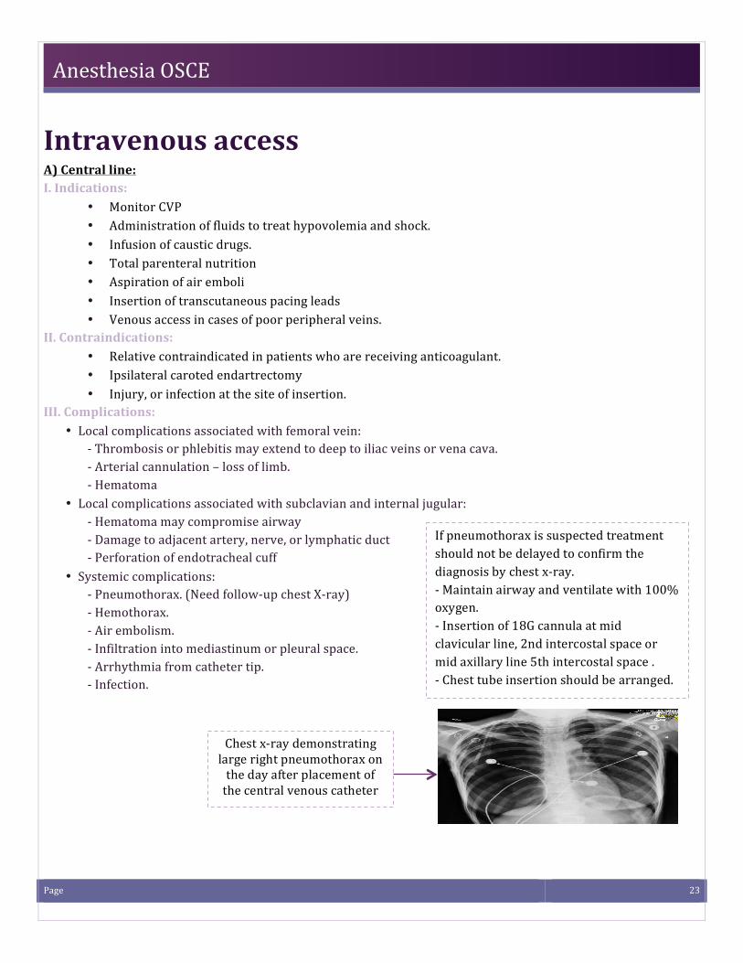
If pneumothorax is suspected treatment should not be delayed to confirm the diagnosis by chest x-‐ray. -‐ Maintain airway and ventilate with 100% oxygen. -‐ Insertion of 18G cannula at mid clavicular line, 2nd intercostal space or mid axillary line 5th intercostal space . -‐ Chest tube insertion should be arranged.
Chest x-‐ray demonstrating large right pneumothorax on the day after placement of the central venous catheter
Page 24
Anesthesia OSCE
IV. Procedure steps:
V. Useful links:
http://www.youtube.com/watch?v=YCOHP5-‐86K0
http://www.youtube.com/watch?v=4-‐uyTUzPSJ8
Performance steps of central line insertion: Done correctly
Introduce yourself Greet the patient Explain procedure Assemble equipments Wash your hands and wear gloves Patient in supine, at least 150 head down position, head turned away
Clean skin, use lidocaine if patient awake. Introduce needle attached to syringe in the center of triangle formed by two lower heads of sternomastoid muscle and clavicle
Direct needle caudally, parallel to sagittal plane, at 300 posterior angle
If vein not entered, withdraw needle and redirect it 5 to 10 degrees laterally
Advance needle while withdrawing plunger of syringe When blood appears and vein entered, remove syringe and insert catheter to predetermined depth.
Remove needle and connect catheter to IV tubing Cover puncture site, and affix catheter in place Documents procedure Ask the patient about any concerns Thank the patient
Page 25
Anesthesia OSCE
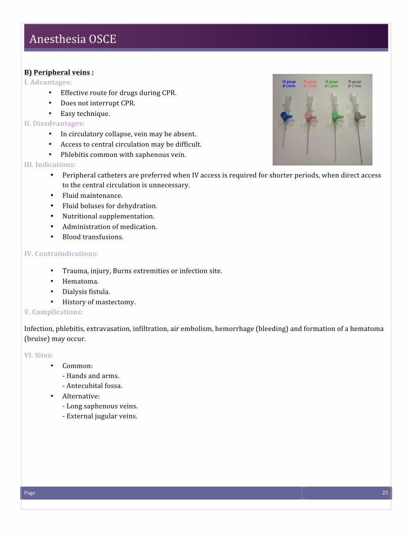
B) Peripheral veins : I. Advantages:
• Effective route for drugs during CPR. • Does not interrupt CPR. • Easy technique.
II. Disadvantages: • In circulatory collapse, vein may be absent. • Access to central circulation may be difficult. • Phlebitis common with saphenous vein.
III. Indications: • Peripheral catheters are preferred when IV access is required for shorter periods, when direct access
to the central circulation is unnecessary. • Fluid maintenance. • Fluid boluses for dehydration. • Nutritional supplementation. • Administration of medication. • Blood transfusions.
IV. Contraindications:
• Trauma, injury, Burns extremities or infection site. • Hematoma. • Dialysis fistula. • History of mastectomy.
V. Complications:
Infection, phlebitis, extravasation, infiltration, air embolism, hemorrhage (bleeding) and formation of a hematoma (bruise) may occur.
VI. Sites: • Common:
-‐ Hands and arms. -‐ Antecubital fossa.
• Alternative: -‐ Long saphenous veins. -‐ External jugular veins.
Page 26
Anesthesia OSCE
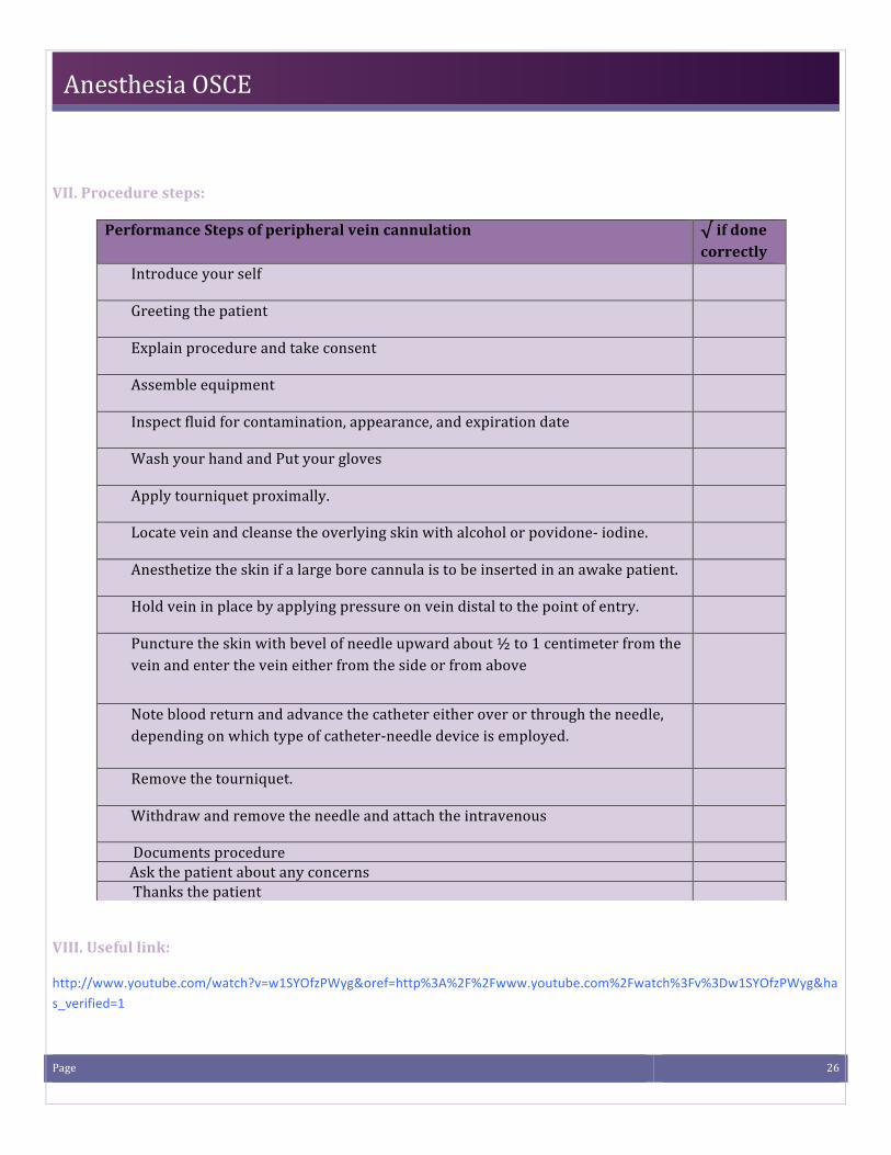
VII. Procedure steps:
VIII. Useful link:
http://www.youtube.com/watch?v=w1SYOfzPWyg&oref=http%3A%2F%2Fwww.youtube.com%2Fwatch%3Fv%3Dw1SYOfzPWyg&has_verified=1
Performance Steps of peripheral vein cannulation √ if done correctly
Introduce your self
Greeting the patient
Explain procedure and take consent
Assemble equipment
Inspect fluid for contamination, appearance, and expiration date
Wash your hand and Put your gloves
Apply tourniquet proximally.
Locate vein and cleanse the overlying skin with alcohol or povidone-‐ iodine.
Anesthetize the skin if a large bore cannula is to be inserted in an awake patient.
Hold vein in place by applying pressure on vein distal to the point of entry.
Puncture the skin with bevel of needle upward about ½ to 1 centimeter from the vein and enter the vein either from the side or from above
Note blood return and advance the catheter either over or through the needle, depending on which type of catheter-‐needle device is employed.
Remove the tourniquet.
Withdraw and remove the needle and attach the intravenous
Documents procedure Ask the patient about any concerns Thanks the patient
Page 27
Anesthesia OSCE
Intravenous fluids A) Factors must be taken into account:
1. Maintenance fluid requirements. 2. NPO and other deficits: NG suction, bowel prep. 3. Third space losses. 4. Replacement of blood loss. 5. Special additional losses: diarrhea.
B) Crystalloid:
• Isotonic: electrolyte composition and osmolality similar to plasma: normal saline, Ringer`s Lactate • Hypotonic: D5W • Hypertonic (Fluids containing sodium concentrations greater than normal saline. Disadvantages:
Hypernatremia, Hyperchloremia) ♦ normal saline:
composition: Na, Cl. Disadvantages: Hyper-‐chloremic acidosis
♦ Lactated Ringer's: MOST Physiological fluid Composition: Na, Cl, K, Ca, lactate. Disadvantages: Not to be used as diluent for blood (Ca citrate) and its low osmolarity can lead to high ICP.
C) Colloids:
• Solutions stay in the space into which they are infused. • Examples: hetastarch (Hespan), albumin, dextran.
D) Fluid replacement:
I. Maintenance fluid requirements:
• It is to maintain the insensible losses such as evaporation of water from respiratory tract, sweat, feces, and urinary excretion. Occurs continually.
• Adults: approximately 1.5 ml/kg/hr “4-‐2-‐1 Rule” -‐ 4 ml/kg/hr for the first 10 kg of body weight. -‐ 2 ml/kg/hr for the second 10 kg body weight. -‐ 1 ml/kg/hr subsequent kg body weight. -‐ Extra fluid for fever, tracheotomy, endued surfaces.
Page 28
Anesthesia OSCE
II. NPO and other deficits:
• NPO deficit = number of hours NPO x maintenance fluid requirement. • Bowel prep may result in up to 1 L fluid loss. • Measurable fluid losses, e.g. NG suctioning, vomiting, ostomy output, biliary fistula and tube.
III. Third space losses
It is the isotonic transfer of ECF from functional body fluid compartments to non-‐functional compartments. Replacing Third Space Losses:
• Superficial surgical trauma: 1-‐2 ml/kg/hr • Minimal Surgical Trauma: 3-‐4 ml/kg/hr
o head and neck, hernia, knee surgery. • Moderate Surgical Trauma: 5-‐6 ml/kg/hr
o hysterectomy, chest surgery. • Severe surgical trauma: 8-‐10 ml/kg/hr (or more)
o AAA repair, nephrectomy.
IV Blood Loss:
• Replace 3 cc of crystalloid solution per cc of blood loss (crystalloid solutions leave the intravascular space). • When using blood products or colloids replace blood loss volume per volume.
V. Other additional losses
Ongoing fluid losses from other sites:
• Gastric drainage. • Ostomy output. • Diarrhea.
Replace volume per volume with crystalloid solutions:
Page 29
Anesthesia OSCE
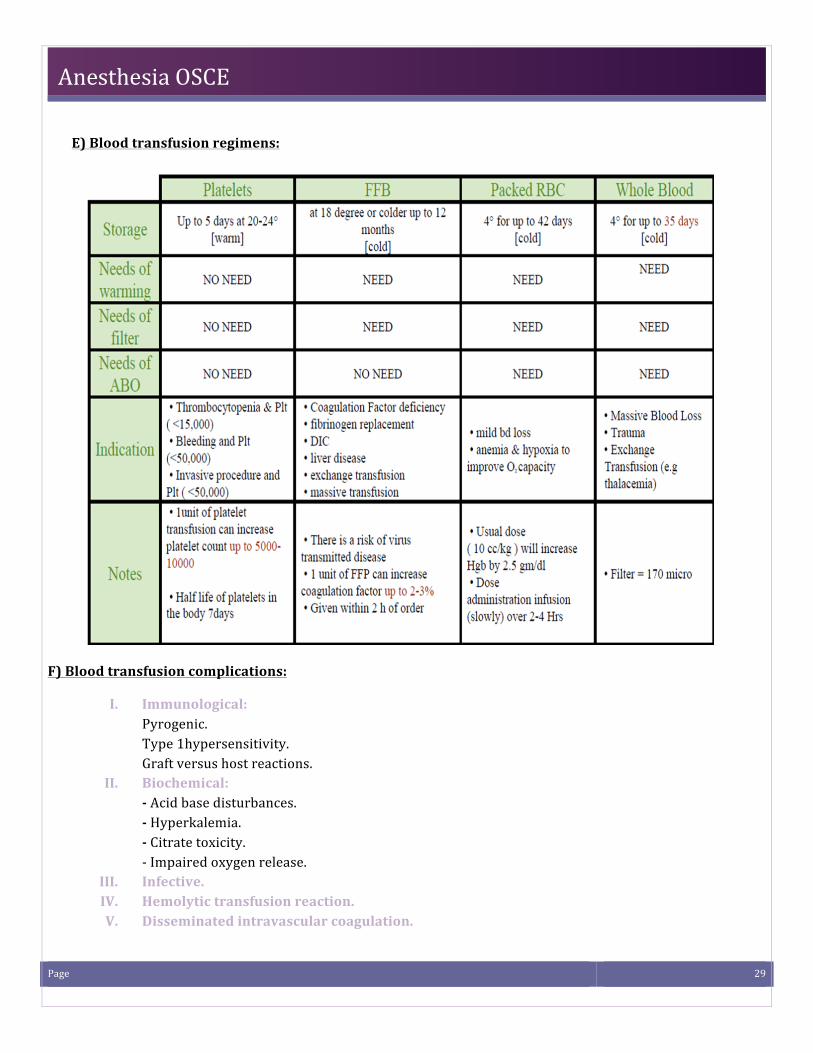
E) Blood transfusion regimens:
F) Blood transfusion complications:
I. Immunological: Pyrogenic. Type 1hypersensitivity. Graft versus host reactions.
II. Biochemical: -‐ Acid base disturbances. -‐ Hyperkalemia. -‐ Citrate toxicity. -‐ Impaired oxygen release.
III. Infective. IV. Hemolytic transfusion reaction. V. Disseminated intravascular coagulation.