articles the genome of the social amoeba dictyostelium
TRANSCRIPT

The genome of the social amoebaDictyostelium discoideumL. Eichinger1*, J. A. Pachebat1,2*, G. Glockner3*, M.-A. Rajandream4*, R. Sucgang5*, M. Berriman4, J. Song5, R. Olsen9, K. Szafranski3, Q. Xu6,7,B. Tunggal1, S. Kummerfeld2, M. Madera2, B. A. Konfortov2, F. Rivero1, A. T. Bankier2, R. Lehmann3, N. Hamlin4, R. Davies4, P. Gaudet10,P. Fey10, K. Pilcher10, G. Chen5, D. Saunders4, E. Sodergren6,8, P. Davis4, A. Kerhornou4, X. Nie5, N. Hall4†, C. Anjard9, L. Hemphill5, N. Bason4,P. Farbrother1, B. Desany5, E. Just10, T. Morio11, R. Rost12, C. Churcher4, J. Cooper4, S. Haydock13, N. van Driessche6, A. Cronin4, I. Goodhead4,D. Muzny8, T. Mourier4, A. Pain4, M. Lu5, D. Harper4, R. Lindsay5, H. Hauser4, K. James4, M. Quiles8, M.Madan Babu2, T. Saito14, C. Buchrieser15,A. Wardroper2,16, M. Felder3, M. Thangavelu17, D. Johnson4, A. Knights4, H. Loulseged8, K. Mungall4, K. Oliver4, C. Price4, M. A. Quail4,H. Urushihara11, J. Hernandez8, E. Rabbinowitsch4, D. Steffen8, M. Sanders4, J. Ma5, Y. Kohara18, S. Sharp4, M. Simmonds4, S. Spiegler4,A. Tivey4, S. Sugano19, B. White4, D. Walker4, J. Woodward4, T. Winckler20, Y. Tanaka11, G. Shaulsky6,7, M. Schleicher12, G. Weinstock6,8,A. Rosenthal3, E. C. Cox21, R. L. Chisholm10, R. Gibbs6,8, W. F. Loomis9, M. Platzer3, R. R. Kay2, J. Williams22, P. H. Dear2, A. A. Noegel1,B. Barrell4 & A. Kuspa5,6
1Center for Biochemistry and Center for Molecular Medicine Cologne, University of Cologne, Joseph-Stelzmann-Str. 52, 50931 Cologne, Germany2Laboratory of Molecular Biology, MRC Centre, Cambridge CB2 2QH, UK3Genome Analysis, Institute for Molecular Biotechnology, Beutenbergstr. 11, D-07745 Jena, Germany4The Wellcome Trust Sanger Institute, Wellcome Trust Genome Campus, Hinxton, Cambridgeshire CB10 1SA, UK5Verna andMarrs McLean Department of Biochemistry andMolecular Biology, 6Department of Molecular and Human Genetics, 7Graduate Program in Structural andComputational Biology and Molecular Biophysics, and 8Human Genome Sequencing Center, Baylor College of Medicine, Houston, Texas 77030, USA9Section of Cell and Developmental Biology, Division of Biology, University of California, San Diego, La Jolla, California 92093, USA10dictyBase, Center for Genetic Medicine, Northwestern University, 303 E Chicago Ave, Chicago, Illinois 60611, USA11Graduate School of Life and Environmental Sciences, University of Tsukuba, Tsukuba, Ibaraki 305-8572, Japan12Adolf-Butenandt-Institute/Cell Biology, Ludwig-Maximilians-University, 80336 Munich, Germany13Biochemistry Department, University of Cambridge, Cambridge CB2 1QW, UK14Division of Biological Sciences, Graduate School of Science, Hokkaido University, Sapporo 060-0810, Japan15Unite de Genomique des Microorganismes Pathogenes, Institut Pasteur, 28 rue du Dr Roux, 75724 Paris Cedex 15, France16Department of Biology, University of York, York YO10 5YW, UK17MRC Cancer Cell Unit, Hutchison/MRC Research Centre, Hills Road, Cambridge CB2 2XZ, UK18Centre for Genetic Resource Information, National Institute of Genetics, Mishima, Shizuoka 411-8540, Japan19Department of Medical Genome Sciences, Graduate School of Frontier Sciences, The University of Tokyo, Minato, Tokyo 108-8639, Japan20Institut fur Pharmazeutische Biologie, Universitat Frankfurt (Biozentrum), Frankfurt am Main 60439, Germany21Department of Molecular Biology, Princeton University, Princeton, New Jersey 08544-1003, USA22School of Life Sciences, University of Dundee, Dow Street, Dundee DD1 5EH, UK
* These authors contributed equally to this work
† Present address: The Institute for Genomic Research, 9712 Medical Center Drive, Rockville, Maryland 20850, USA
...........................................................................................................................................................................................................................
The social amoebae are exceptional in their ability to alternate between unicellular and multicellular forms. Here we describe thegenome of the best-studied member of this group, Dictyostelium discoideum. The gene-dense chromosomes of this organismencode approximately 12,500 predicted proteins, a high proportion of which have long, repetitive amino acid tracts. There are manygenes for polyketide synthases and ABC transporters, suggesting an extensive secondary metabolism for producing and exportingsmall molecules. The genome is rich in complex repeats, one class of which is clustered and may serve as centromeres. Partialcopies of the extrachromosomal ribosomal DNA (rDNA) element are found at the ends of each chromosome, suggesting a noveltelomere structure and the use of a common mechanism to maintain both the rDNA and chromosomal termini. A proteome-basedphylogeny shows that the amoebozoa diverged from the animal–fungal lineage after the plant–animal split, but Dictyosteliumseems to have retained more of the diversity of the ancestral genome than have plants, animals or fungi.
The amoebozoa are a richly diverse group of organisms whosegenomes remain largely unexplored. The soil-dwelling socialamoeba Dictyostelium discoideum has been actively studied for thepast 50 years and has contributed greatly to our understanding ofcellular motility, signalling and interaction1. For example, studies inDictyostelium provided the first descriptions of a eukaryotic cellchemoattractant and a cell–cell adhesion protein2,3.
Dictyostelium amoebae inhabit forest soil and consume bacteriaand yeast, which they track by chemotaxis. Starvation, however,prompts the solitary cells to aggregate and develop as a truemulticellular organism, producing a fruiting body comprised of acellular, cellulosic stalk supporting a bolus of spores. Thus, Dictyo-stelium has evolved mechanisms that direct the differentiation of ahomogeneous population of cells into distinct cell types, regulate
the proportions between tissues and orchestrate the construction ofan effective structure for the dispersal of spores4. Many of the genesnecessary for these processes in Dictyostelium were also inherited byMetazoa and fashioned through evolution for use within manydifferent modes of development.
The amoebozoa are also noteworthy as representing one of theearliest branches from the last common ancestor of all eukaryotes.Each of the surviving branches of the crown group of eukaryotesprovides an example of the ways in which the ancestral genome hasbeen sculpted and adapted by lineage-specific gene duplication,divergence and deletion. Comparison between representatives ofthese branches promises to shed light not only on the nature andcontent of the ancestral eukaryotic genome, but on the diversity ofways in which its components have been adapted to meet the needs
articles
NATURE | VOL 435 | 5 MAY 2005 | www.nature.com/nature 43© 2005 Nature Publishing Group

of complex organisms. The genome of Dictyostelium, as the firstfree-living protozoan to be fully sequenced, should be particularlyinformative for these analyses.
Mapping, sequencing and assemblyAn international initiative to sequence the genome of Dictyosteliumdiscoideum AX4 (refs 5, 6) was launched in 1998. The high repeatcontent and (AþT)-richness of the genome (the latter renderinglarge-insert bacterial clones unstable) posed severe challenges forsequencing and assembly. The response to these challenges was touse a whole-chromosome shotgun (WCS) strategy, partially purify-ing each chromosome electrophoretically and treating it as aseparate project. This approach was supported by novel statisticaltools to recover chromosome specificity from the impure WCSlibraries, and by highly detailed HAPPY maps that provided aframework for sequence assembly. These approaches have enabledthe completion of this difficult genome to a high standard, and arelikely to be valuable in tackling the many other genomes that presentchallenges of composition and complexity.
Genome mapping
To support sequence assembly, we made high-resolution maps ofthe chromosomes using HAPPY mapping7–9, which relies onanalysing the sequence content of single DNA molecules preparedby limiting dilution. A total of 3,902 markers selected mostly fromthe emerging shotgun data were mapped, and maps of all sixchromosomes were assembled (see Methods and Table 1; see alsoSupplementary Fig. 1 and Supplementary Table 1).
Genome sequencing and assembly
Two strategies were used to recover chromosome-specific data fromimpure WCS libraries (see Methods). The first (for chromosomes 1,2 and 3) used enrichment of the respective libraries as the mainstatistical indicator of the chromosomal assignment of contigs, andHAPPY maps were used to guide assembly. The second strategy (forchromosomes 4, 5 and most of 6) used mapping data to assignsequences to chromosomes initially, with detailed HAPPY mapsbeing used to validate final assemblies. A 1,508-kilobase (kb)portion of chromosome 6 was sequenced as a pilot project using acombination of approaches (see Methods).
Repetitive tracts complicated assembly. For chromosomes 1, 2and 3, inspection of polymorphisms, combined with HAPPY maps,allowed unambiguous assembly in many cases. For chromosomes 4,
5 and 6, low-coverage sequencing of AX4-derived yeast artificialchromosomes (YACs) alleviated the problems by providing a localdata set within which the troublesome repeat element was present asa single copy. Nevertheless, some repeat tracts proved intractableand remain as gaps. Thirty-four unlinked (floating) contigs of.1 kb, totalling 225,339 base pairs (bp), remain unpositioned inthe genome, but can be provisionally assigned to specific chromo-somes based on their content of reads from the WCS libraries. Mostor all of these floating contigs are bounded by repetitive regions. Thechromosome 2 sequence in the current assembly supersedes thatpreviously published9, having benefited from further HAPPYmapping and manual sequence finishing.
The six chromosomal assemblies span 33,817 kb (Table 1),including ,156 kb in the form of clone-, sequence- and repeatgaps. Assuming that most of the floating contigs lie beyond thetermini of the assemblies, the total genome size is estimated at34,042,810 bp. In estimating the completeness of the sequence, wenote that of 967 well-characterized D. discoideum genes, 957 (99%)were found initially in the assemblies. Of the remaining ten, seven(cupE, trxA, trxB, trxC, staA, staB and cinB) have close matches,suggesting that their GenBank entries may contain errors orrepresent alternative alleles. Only three (fcpA, wasA and roco5)had no matches in the initial assemblies, although the first two ofthese were recovered by searches of unincorporated sequencefollowed by local reassembly. Of 133,168 ‘qualified’ D. discoideumAX4 expressed sequence tags (ESTs of .200 bp and .20% GþC,and not matching mitochondrial sequence; ref. 10 andH. Urushihara et al., unpublished data), 128,207 (96.3%) arefound in the assemblies (the higher proportion of missing sequencesamong the ESTs probably reflects the higher error rate inherent inEST data).
We conclude that the current assembly represents ..95% of thechromosomal sequence (less than 1% of which is in floating contigs)and $99% of genes, with most of the missing sequence comprisingcomplex or simple repeats. The most stringent test of the medium-to long-range accuracy of the assembly comes from comparisonwith the HAPPY maps. This is particularly true for chromosomes 4,5 and 6, where HAPPY markers were used to nucleate contigs butnot to guide their assembly or ordering, specifically to allow such acomparison to be made without circularity of argument. As can beseen, good agreement between map and sequence confirms theaccuracy of the assembly (Fig. 1).
Table 1 Sequence assembly details
Chromosome
1 2 3 4 5 6 AllFeature...................................................................................................................................................................................................................................................................................................................................................................
Chromosomal assembliesAssembly span (bp)* 4,919,822 8,467,571 6,358,352 5,430,575 5,062,323 3,578,828 33,817,471Assembly sequence (bp)† 4,911,622 8,437,971 6,334,852 5,397,875 5,032,273 3,547,128 33,661,721Total contigs 11 40 32 65 107 44 309Mean contig size (bp) 446,511 210,949 197,964 83,044 47,031 80,617 108,938Number of sequence gaps 4 12 10 34 81 14 155Number of repeat gaps 8 29 23 9 4 11 84Number of clone gaps 0 0 0 22 22 20 64Total estimated gap size (bp)‡ 8,200 29,600 23,500 32,700 30,050 31,700 155,750Number of HAPPY markers (mean spacing in kb) 749 (6.6) 615 (12.5)§ 684 (9.3) 628 (8.6) 628 (8.1) 598 (6.0) 3,902 (8.7)
Floating contigskNumber of floating contigs 0 22 3 9{ 0 34Total size of floating contigs (bp) 0 171,670 16,360 37,309 0 225,339
Combined (assemblies plus floating contigs)Total sequence (bp) 4,911,622 8,609,641 6,351,212 5,416,529{ 5,050,928{ 3,547,128 33,887,060Mean coverage (fold) 9.1 6.5 6.7 9.6 9.9 10.3 8.3
...................................................................................................................................................................................................................................................................................................................................................................
*Total end-to-end length of the chromosomal assembly, including any gaps.†Sequenced bases covered by chromosomal assembly, not counting gaps.‡Sequence, repeat and clone gaps are taken to have average sizes of 50 bp, 1,000 bp and 1,000 bp, respectively.§Does not include the second copy of the 755-kb inverted duplication.k Includes only those contigs that can be assigned to specific chromosomes.{Floating contigs from chromosomes 4 and 5 cannot be distinguished. In calculating total chromosomal sequence, we assume that half of these floating contigs are from each of chromosomes 4 and 5.
articles
NATURE | VOL 435 | 5 MAY 2005 | www.nature.com/nature44© 2005 Nature Publishing Group

Sequence characteristics of the genomeThe genome is (AþT)-rich (77.57%) and has a broadly uniformcomposition, apart from the more (GþC)-rich repeat-denseregions (Fig. 2). On a finer scale, nucleotide composition tracksthe distribution of exons (see below). Among dinucleotides, CpG isunder-represented, not just in absolute terms but also relative to itsisomer GpC (the former occurring only 62% as often as the latter).This bias normally reflects cytosine methylation at CpG sequences,promoting their mutation to TpG (which is over-representedrelative to GpT by 38%). Hence, these observations suggest thatcytosine methylation may occur in Dictyostelium, contrary to earlierfindings11.
Simple sequence repeats are abundant and unusual
Simple sequence repeats (SSRs) are more abundant in Dictyosteliumthan in any other genome sequenced so far, comprising .11% ofbases (Supplementary Fig. 2). In non-coding sequence, tracts ofdinucleotides or longer motifs occur every 392 bp on average andcomprise 6.4% of the bases. There is a bias towards repeat units of3–6 bases, whereas dinucleotide tracts predominate in most othergenomes. Homopolymer tracts are also abundant, comprising afurther 16% of non-coding sequence. The base composition ofnon-coding SSRs and homopolymer tracts (99.2% AþT content) iseven more biased than that of the surrounding sequence, suggestingthat either selection or the mechanism of repeat expansion favours(AþT)-rich repeats.
Notably, SSRs are also abundant in protein-coding sequence,occurring on average every 724 bp within exons. We consider thesecoding SSRs in further detail below, in the context of proteins.
Transposable elements are clustered
The genome is rich in transposable elements9,12. Completion of thesequence confirms the earlier observation that transposableelements of the same type are clustered, suggesting their preferentialinsertion within similar resident elements. However, none of theelements appears to use a specific sequence as a target for insertion:they insert at random within other elements of the same type.Non-long terminal repeat (LTR) retrotransposons are known toinsert next to transfer RNA genes; we find many such instances
(Fig. 2), but again no specific sequences were identified as insertiontargets.
tRNAs are numerous and paired by specificity
The sequenced genome encodes 390 tRNAs, a number at the upperend of the eukaryotic spectrum (for example, Plasmodiumfalciparum ¼ 43, Drosophila melanogaster ¼ 284, Homo sapiens¼496). Allowing for the normal wobble rules in codon–anticodonpairing13,14, every sense codon can be decoded, apart from the rarealanine codon GCG; we infer that the missing tRNA(s) lie in one ormore gaps in the sequence. We also find a possible selenocysteinetRNA in the genome, as well as corresponding selenocysteineinsertion targets in two predicted proteins (see SupplementaryFig. 3).Dictyostelium, in common only with Acanthamoeba castellanii15,
has been shown to lack certain apparently essential tRNAs in itsmitochondrial genome16. It therefore seems likely that at least somechromosomally encoded tRNAs (those for valine, threonine,asparagine and glycine, as well as one arginine and two serinetRNAs) are imported into mitochondria.
Although the gross distribution of tRNAs is uniform, organiza-tion of tRNAs on a finer scale is striking: about 20% occur as pairs ortriplets with identical anticodons (and usually 100% sequenceidentity), separated by ,20 kb and often by ,5 kb (Fig. 2). Thereare 41 such groups in the genome; a random distribution wouldproduce few, if any. This pattern is unique among sequencedgenomes, and suggests a wave of recent duplications. However,tRNA pairs are found in tandem, converging and diverging orien-tations with comparable frequencies, suggesting no straightforwardduplication mechanism; nor is there usually duplication of exten-sive flanking sequences. Whether the preference of TRE elements forinserting adjacent to tRNAs is related to the large number andunusual distribution of tRNAs is unclear.
A chromosomal master copy of the extrachromosomal rDNA element
In Dictyostelium, ribosomal RNA genes lie on an 88-kb palindromicextrachromosomal element17, present at ,100 copies per nucleus(Fig. 2). Evidence also exists of chromosomal copies: at least thecentral 3.2 kb of the element is located17 on chromosome 4, whereas
Figure 1 Chromosomal assemblies compared against HAPPY map data. The locations of
markers as found in the sequence (y axis) are plotted against their location in HAPPY maps
(x axis) for chromosomes 1–6. Markers mapped to one chromosome but found in the
assembled sequence of another are indicated by diamonds on the x axis. The dashed box
indicates a large inverted duplication on chromosome 2: markers in this region are shown
at one of their two possible map locations but are found at two points in the sequence.
articles
NATURE | VOL 435 | 5 MAY 2005 | www.nature.com/nature 45© 2005 Nature Publishing Group
chromosome 2 carries both a partial rDNA sequence and a 5S rRNApseudogene9,18.
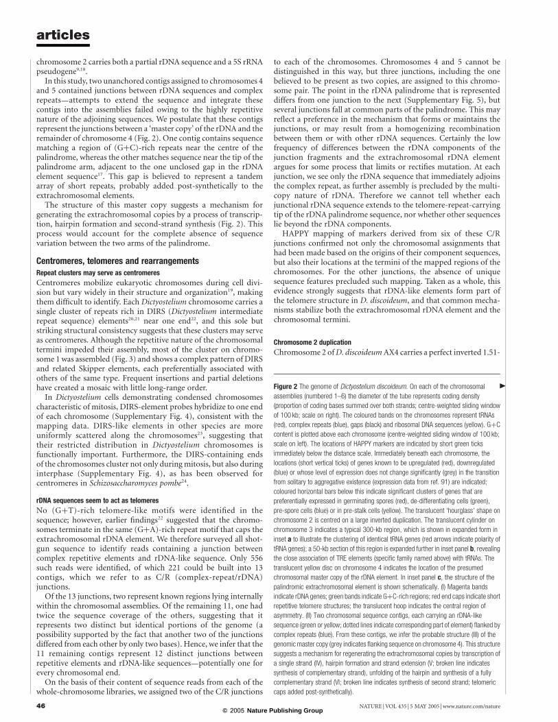
In this study, two unanchored contigs assigned to chromosomes 4and 5 contained junctions between rDNA sequences and complexrepeats—attempts to extend the sequence and integrate thesecontigs into the assemblies failed owing to the highly repetitivenature of the adjoining sequences. We postulate that these contigsrepresent the junctions between a ‘master copy’ of the rDNA and theremainder of chromosome 4 (Fig. 2). One contig contains sequencematching a region of (GþC)-rich repeats near the centre of thepalindrome, whereas the other matches sequence near the tip of thepalindrome arm, adjacent to the one unclosed gap in the rDNAelement sequence17. This gap is believed to represent a tandemarray of short repeats, probably added post-synthetically to theextrachromosomal elements.
The structure of this master copy suggests a mechanism forgenerating the extrachromosomal copies by a process of transcrip-tion, hairpin formation and second-strand synthesis (Fig. 2). Thisprocess would account for the complete absence of sequencevariation between the two arms of the palindrome.
Centromeres, telomeres and rearrangementsRepeat clusters may serve as centromeres
Centromeres mobilize eukaryotic chromosomes during cell divi-sion but vary widely in their structure and organization19, makingthem difficult to identify. Each Dictyostelium chromosome carries asingle cluster of repeats rich in DIRS (Dictyostelium intermediaterepeat sequence) elements20,21 near one end22, and this sole butstriking structural consistency suggests that these clusters may serveas centromeres. Although the repetitive nature of the chromosomaltermini impeded their assembly, most of the cluster on chromo-some 1 was assembled (Fig. 3) and shows a complex pattern of DIRSand related Skipper elements, each preferentially associated withothers of the same type. Frequent insertions and partial deletionshave created a mosaic with little long-range order.
In Dictyostelium cells demonstrating condensed chromosomescharacteristic of mitosis, DIRS-element probes hybridize to one endof each chromosome (Supplementary Fig. 4), consistent with themapping data. DIRS-like elements in other species are moreuniformly scattered along the chromosomes23, suggesting thattheir restricted distribution in Dictyostelium chromosomes isfunctionally important. Furthermore, the DIRS-containing endsof the chromosomes cluster not only during mitosis, but also duringinterphase (Supplementary Fig. 4), as has been observed forcentromeres in Schizosaccharomyces pombe24.
rDNA sequences seem to act as telomeres
No (GþT)-rich telomere-like motifs were identified in thesequence; however, earlier findings22 suggested that the chromo-somes terminate in the same (GþA)-rich repeat motif that caps theextrachromosomal rDNA element. We therefore surveyed all shot-gun sequence to identify reads containing a junction betweencomplex repetitive elements and rDNA-like sequence. Only 556such reads were identified, of which 221 could be built into 13contigs, which we refer to as C/R (complex-repeat/rDNA)junctions.
Of the 13 junctions, two represent known regions lying internallywithin the chromosomal assemblies. Of the remaining 11, one hadtwice the sequence coverage of the others, suggesting that itrepresents two distinct but identical portions of the genome (apossibility supported by the fact that another two of the junctionsdiffered from each other by only two bases). Hence, we infer that the11 remaining contigs represent 12 distinct junctions betweenrepetitive elements and rDNA-like sequences—potentially one forevery chromosomal end.
On the basis of their content of sequence reads from each of thewhole-chromosome libraries, we assigned two of the C/R junctions
to each of the chromosomes. Chromosomes 4 and 5 cannot bedistinguished in this way, but three junctions, including the onebelieved to be present as two copies, are assigned to this chromo-some pair. The point in the rDNA palindrome that is representeddiffers from one junction to the next (Supplementary Fig. 5), butseveral junctions fall at common parts of the palindrome. This mayreflect a preference in the mechanism that forms or maintains thejunctions, or may result from a homogenizing recombinationbetween them or with other rDNA sequences. Certainly the lowfrequency of differences between the rDNA components of thejunction fragments and the extrachromosomal rDNA elementargues for some process that limits or rectifies mutation. At eachjunction, we see only the rDNA sequence that immediately adjoinsthe complex repeat, as further assembly is precluded by the multi-copy nature of rDNA. Therefore we cannot tell whether eachjunctional rDNA sequence extends to the telomere-repeat-carryingtip of the rDNA palindrome sequence, nor whether other sequenceslie beyond the rDNA components.
HAPPY mapping of markers derived from six of these C/Rjunctions confirmed not only the chromosomal assignments thathad been made based on the origins of their component sequences,but also their locations at the termini of the mapped regions of thechromosomes. For the other junctions, the absence of uniquesequence features precluded such mapping. Taken as a whole, thisevidence strongly suggests that rDNA-like elements form part ofthe telomere structure in D. discoideum, and that common mecha-nisms stabilize both the extrachromosomal rDNA element and thechromosomal termini.
Chromosome 2 duplication
Chromosome 2 ofD. discoideumAX4 carries a perfect inverted 1.51-
Figure 2 The genome of Dictyostelium discoideum. On each of the chromosomal
assemblies (numbered 1–6) the diameter of the tube represents coding density
(proportion of coding bases summed over both strands; centre-weighted sliding window
of 100 kb; scale on right). The coloured bands on the chromosomes represent tRNAs
(red), complex repeats (blue), gaps (black) and ribosomal DNA sequences (yellow). GþC
content is plotted above each chromosome (centre-weighted sliding window of 100 kb;
scale on left). The locations of HAPPY markers are indicated by short green ticks
immediately below the distance scale. Immediately beneath each chromosome, the
locations (short vertical ticks) of genes known to be upregulated (red), downregulated
(blue) or whose level of expression does not change significantly (grey) in the transition
from solitary to aggregative existence (expression data from ref. 91) are indicated;
coloured horizontal bars below this indicate significant clusters of genes that are
preferentially expressed in germinating spores (red), de-differentiating cells (green),
pre-spore cells (blue) or in pre-stalk cells (yellow). The translucent ‘hourglass’ shape on
chromosome 2 is centred on a large inverted duplication. The translucent cylinder on
chromosome 3 indicates a typical 300-kb region, which is shown in expanded form in
inset a to illustrate the clustering of identical tRNA genes (red arrows indicate polarity of
tRNA genes); a 50-kb section of this region is expanded further in inset panel b, revealing
the close association of TRE elements (specific family named above) with tRNAs. The
translucent yellow disc on chromosome 4 indicates the location of the presumed
chromosomal master copy of the rDNA element. In inset panel c, the structure of the
palindromic extrachromosomal element is shown schematically. (I) Magenta bands
indicate rDNA genes; green bands indicate GþC-rich regions; red end caps indicate short
repetitive telomere structures; the translucent hoop indicates the central region of
asymmetry. (II) Two chromosomal sequence contigs, each carrying an rDNA-like
sequence (green or yellow; dotted lines indicate corresponding part of element) flanked by
complex repeats (blue). From these contigs, we infer the probable structure (III) of the
genomic master copy (grey indicates flanking sequence on chromosome 4). This structure
suggests a mechanism for regenerating the extrachromosomal copies by transcription of
a single strand (IV), hairpin formation and strand extension (V; broken line indicates
synthesis of complementary strand), unfolding of the hairpin and synthesis of a fully
complementary strand (VI; broken line indicates synthesis of second strand; telomeric
caps added post-synthetically).
Q
articles
NATURE | VOL 435 | 5 MAY 2005 | www.nature.com/nature46© 2005 Nature Publishing Group

megabase (Mb) duplication (Fig. 2; see also refs 9, 25). Thisduplication, containing 608 genes, is known25 to be absent fromthe wild-type isolate NC4 and from one of its direct descendents(AX2), but present in another (AX3); AX4 in turn is derived fromAX3. The sequences adjoining the right-hand end of the dupli-cation—a partial copy of a DIRS element (and a partial DDT-Aelement) and a region identical to part of the rDNA palindrome,both at about 3.74 Mb (Fig. 2)—have been implicated in centro-meric and telomeric functions, respectively, elsewhere in thegenome.
We propose that this duplication arose from a ‘breakage-fusion-bridge’ cycle as first described in maize26 and since observed in manygenomes. The nearby DIRS and rDNA components, in this view,represent abortive attempts to stabilize the halves of the brokenchromosome by establishing new telomeres and centromeres,followed by re-fusion of the pieces to create a restored and enlargedchromosome (Supplementary Fig. 6).
Chromosome 2 (the largest of the chromosomes, even discount-ing the duplication in AX4) may be prone to breakage: in theBonner isolate of NC4, maintained in vegetative growth for 50 years,chromosome 2 is represented by two smaller fragments27. Com-parison with more recent data22 indicates that the break point inNC4-Bonner lies in the same region as the duplication in AX4,suggesting that NC4-Bonner underwent the early stages of thisprocess, but that the chromosome fragments were stabilized andmaintained after the initial breakage. Preliminary results (data notshown) from HAPPY mapping also suggest that although wild-typeisolates V12M2 and NC4 both lack the duplication seen in AX4,NC4 may carry a duplication of ,300 kb near the opposite end ofchromosome 2.
Content and organization of the proteomePrediction of protein-coding genes (see Methods) was performedon the complete set of chromosomes and floating contigs (Table 2).In assessing the completeness and accuracy of the predictions, wefind that of the 957 well-characterized D. discoideum genes that arepresent in the current sequence, 823 (86%) are predicted astranscripts with structures matching the experimentally determinedones. For a further 123 (13%), the predicted transcript differs fromthe experimentally determined one, about one-half of these differ-ing only in their 5
0boundary; the remaining 11 (1%), although
present in the sequence, were not predicted as transcripts. Similarly,of the 128,207 qualified ESTs present in the current sequence,127,097 (99.1%) fall within predicted transcripts. Combining ourestimate of sequence coverage (above) with these estimates of thesuccess of gene prediction, we infer that approximately 98% of allD. discoideum genes are present in the predicted set.
The level of overprediction, conversely, is harder to estimate:prediction was performed generously to ensure that most true geneswere represented. Of the 13,541 predicted proteins, 47.5%are represented by qualified ESTs, reflecting the inevitable bias inEST sampling. Among the shortest predicted proteins, fewer arerepresented by ESTs (for example, 21% of those of ,60 aminoacids); this is at least partly due to a higher level of overprediction.On the basis of the simplifying assumption that 50% of all genescoding for proteins of ,100 amino acids are mis-predictions, weestimate the true number of genes at roughly 12,500. This number iscloser to that seen in multicellular organisms rather than in mostunicellular eukaryotes (Table 2). The same relative complexity isseen in the total number of amino acids encoded by the respectivegenomes; this measure of complexity is less affected by the inclusion
Figure 3 DIRS repeat region of chromosome 1. Complete complex repeat units are
represented by coloured triangles whose size corresponds to the sequence length of the
repeat unit (see key at top of figure). The bottom-left and top-right corners of each triangle
represent 50
and 30
ends of the repeat, respectively. The arrangement of complete and
partial repeat units within the first 187 kb of D. discoideum chromosome 1 is shown
(bottom) by corresponding portions of the triangles; the orientation of the triangles
indicates the direction in which each repeat unit lies. The vertical scale (sizes of repeat
units) is the same as the horizontal scale (chromosomal distances).
Table 2 Comparison between the predicted protein-coding gene set of D. discoideum and those of other organisms
Feature D. discoideum P. falciparum S. cerevisiae A. thaliana D. melanogaster C. elegans Human...................................................................................................................................................................................................................................................................................................................................................................
Genome size (Mb) 34 23 13 125 180 103 2,851Number of genes 12,500* 5,268 5,538 25,498 13,676 19,893 22,287Gene spacing (kb per gene) 2.5 4.3 2.2 4.9 13.2 5.0 127.9Mean gene length (bp) 1,756 2,534 1,428 2,036 1,997 2,991 27,000Mean coding size (amino acids) 518 761 475 437 538 435 509Genes with introns (%) 69 54 5 79 38 5 85Mean intron size (bp) 146 179 ND 170 ND 270 3,365Mean no. of introns (in spliced genes) 1.9 2.6 1.0 5.4 4.0 5.0 8.1Total amino acids encoded (thousands) 7,021 4,009 2,471 11,143 7,358 9,038 11,333Codon A þ T bias† 86 83 62 57 50 64 41Mean A þ T percentage (exons) 73 76 72 72 45 58 55Mean A þ T percentage (introns) 88 87 51 55 38 71 62Mean A þ T percentage (intergenic) 85 86 51 56 38 72 62...................................................................................................................................................................................................................................................................................................................................................................
ND, not determined.*See text. The estimated number of true transcripts for D. discoideum is given here for comparability with other species; however, the total predicted gene number of 13,541 is used in calculating thefigures below.†Percentage of all codons used which have A/T at their third base.
articles
NATURE | VOL 435 | 5 MAY 2005 | www.nature.com/nature 47© 2005 Nature Publishing Group
of shorter (and hence more dubious) gene predictions. Introns inDictyostelium are few and short, and intergenic regions are small,producing a compact genome of which 62% encodes protein.
Genes are distributed approximately uniformly across thegenome (Fig. 2). Although we do not see widespread clustering ofgenes with coordinated expression patterns (see Methods), we dofind statistically significant (P , 0.01) clusters of genes expressedpredominantly at some developmental stages or in specific cell types(Fig. 2).
(A1T)-richness influences protein composition and codon usage
Codon usage in Dictyostelium favours codons of the form NNT orNNA over their NNG or NNC synonyms, the bias being even greaterthan for the (AþT)-rich Plasmodium genome. Comparison of tRNAand codon frequencies (Supplementary Table 2) reveals a similarpicture to that in human28 and other eukaryotes, suggesting that thesame use is made of ‘wobble’ and of base modifications (forexample, of adenine to inosine in some tRNAs) to expand theeffective repertoire of tRNAs.
As in Plasmodium29, the extreme (AþT)-richness is reflected notjust in the choice of synonymous codons, but also in the amino acidcomposition of the proteins. Amino acids encoded solely by codonsof the form WWN (where W indicates A or T and N indicates anybase; these are Asn, Lys, Ile, Tyr and Phe) are much commoner inDictyostelium proteins than in human ones; the reverse is true forthose encoded solely by SSN codons (where S indicates C or G; theseare Pro, Arg, Ala and Gly).
Geometry reflects phylogeny—duplications in the genome
The predicted gene set of Dictyostelium is rich in relatively recentlyduplicated genes. Of the 13,498 predicted proteins analysed, 3,663fall into 889 families clustered by BLASTP similarities of e , 10240.Most (538) families contain only two members, but 351 familiescontain between three and 81 proteins (Supplementary Table 3).Hence, 2,774 (20%) of all predicted proteins have arisen byrelatively recent duplication, potentially accounting for much ofDictyostelium’s excess gene number compared with typical uni-cellular eukaryotes.
We tried to infer the mechanisms by which such duplications
arise and propagate in the genome. Where members of a family areclustered on one chromosome, the physical distance between familymembers often (23 out of 86 families examined) correlates stronglywith their evolutionary divergence (see Methods). Where a family issplit between different chromosomes, members on the samechromosome are often (23 out of 50 families examined) morerelated to each other than to members on different chromosomes;the reverse is never observed.
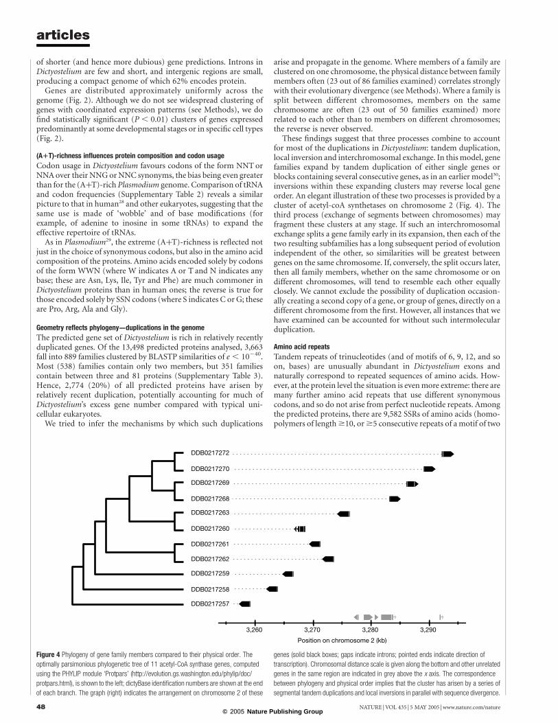
These findings suggest that three processes combine to accountfor most of the duplications in Dictyostelium: tandem duplication,local inversion and interchromosomal exchange. In this model, genefamilies expand by tandem duplication of either single genes orblocks containing several consecutive genes, as in an earlier model30;inversions within these expanding clusters may reverse local geneorder. An elegant illustration of these two processes is provided by acluster of acetyl-coA synthetases on chromosome 2 (Fig. 4). Thethird process (exchange of segments between chromosomes) mayfragment these clusters at any stage. If such an interchromosomalexchange splits a gene family early in its expansion, then each of thetwo resulting subfamilies has a long subsequent period of evolutionindependent of the other, so similarities will be greatest betweengenes on the same chromosome. If, conversely, the split occurs later,then all family members, whether on the same chromosome or ondifferent chromosomes, will tend to resemble each other equallyclosely. We cannot exclude the possibility of duplication occasion-ally creating a second copy of a gene, or group of genes, directly on adifferent chromosome from the first. However, all instances that wehave examined can be accounted for without such intermolecularduplication.
Amino acid repeats
Tandem repeats of trinucleotides (and of motifs of 6, 9, 12, and soon, bases) are unusually abundant in Dictyostelium exons andnaturally correspond to repeated sequences of amino acids. How-ever, at the protein level the situation is even more extreme: there aremany further amino acid repeats that use different synonymouscodons, and so do not arise from perfect nucleotide repeats. Amongthe predicted proteins, there are 9,582 SSRs of amino acids (homo-polymers of length $10, or $5 consecutive repeats of a motif of two
Figure 4 Phylogeny of gene family members compared to their physical order. The
optimally parsimonious phylogenetic tree of 11 acetyl-CoA synthase genes, computed
using the PHYLIP module ‘Protpars’ (http://evolution.gs.washington.edu/phylip/doc/
protpars.html), is shown to the left; dictyBase identification numbers are shown at the end
of each branch. The graph (right) indicates the arrangement on chromosome 2 of these
genes (solid black boxes; gaps indicate introns; pointed ends indicate direction of
transcription). Chromosomal distance scale is given along the bottom and other unrelated
genes in the same region are indicated in grey above the x axis. The correspondence
between phylogeny and physical order implies that the cluster has arisen by a series of
segmental tandem duplications and local inversions in parallel with sequence divergence.
articles
NATURE | VOL 435 | 5 MAY 2005 | www.nature.com/nature48© 2005 Nature Publishing Group

or more amino acids). Of these, the most striking are polyaspar-agine and polyglutamine tracts of $20 residues, present in 2,091 ofthe predicted proteins. Also abundant are low-complexity regionssuch as QLQLQQQQQQQLQLQQ: there are 2,379 tracts of $15residues composed of only two different amino acids. In total,repeats or simple-sequence tracts of amino acids (even by theseconservative definitions) occur in 34% of predicted proteins andencode 3.3% of all amino acids.
It seems likely that these repeats have arisen through nucleotideexpansion, but have been selected at the protein level. Evidence forselection at the protein level is that any given trinucleotide repeatoccurs predominantly in only one of the three reading frames. Forexample, the repeat …ACAACAACAACA… is usually translated aspolyglutamine ([CAA]n) rather than polythreonine ([ACA]n) orpolyasparagine ([AAC]n). Further evidence comes from the manytrinucleotide repeats that have apparently mutated to produce onlysynonymous codons (for example, …GATGACGATGATGAC…,translated as polyaspartate). Moreover, the distribution of repeatsand simple-sequence tracts is nonrandom: most proteins eitherhave no such features (66% of proteins) or have two or more (18%of proteins), suggesting that they are tolerated only in certain typesof protein. The polyasparagine- and polyglutamine-containingproteins appear to be over-represented in protein kinases, lipidkinases, transcription factors, RNA helicases and messenger RNAbinding proteins such as spliceosome components (SupplementaryFig. 9). Protein kinases and transcription factors are also over-represented in the polyasparagine- and polyglutamine-containingproteins of Saccharomyces cerevisiae, so it is possible thatthese homopolymers serve some functional role in these proteinclasses. A more detailed analysis of amino acid homopolymers isgiven in Supplementary Tables 4–6 and Supplementary Figs 7–10.
Phylogeny, evolution and comparative proteomics
The organisms that diverged from the last common ancestor of alleukaryotes followed different evolutionary paths, but all retainedthe basic properties of eukaryotic cells. Their genomes have beensculpted by chromosomal deletions and duplications that led tolineage-specific gene family expansions, reductions and losses, as
well as genes with new functions31,32. Our analysis of Dictyostelium’sproteome shows that similar mechanisms have shaped its genome,augmented by horizontal gene transfer from bacterial species.
Phylogeny of eukaryotes based on complete proteomes
Using morphological criteria, early workers were unsure whether toclassify Dictyostelids as fungi or protozoa33. Molecular methodsindicated that they were amoebozoa and also suggested thatDictyostelium diverged from the line leading to animals atabout the same time as plants34,35. A study of more than 100proteins suggested that Dictyostelium diverged after the plant–animal split, but before the divergence of the fungi36. The recentfinding of a gene fusion encoding three pyrimidine biosyntheticenzymes, shared only byDictyostelium, fungi and Metazoa, indicatesthat the amoebozoa are a true sister group of the fungi andMetazoa37.
To examine the phylogeny ofDictyostelium on a genomic scale, weapplied an improved method for predicting orthologous proteinclusters to complete eukaryotic proteomes38 (for details, see Sup-plementary Information). The data were used to construct aphylogenetic tree that confirms the divergence of Dictyosteliumalong the branch leading to the Metazoa soon after the plant–animal split (Fig. 5). Despite the earlier divergence of Dictyostelium,many of its proteins are more similar to human orthologues thanare those of S. cerevisiae, probably due to higher rates of evolution-ary change along the fungal lineage. Whether the greater similaritybetween amoebozoa and Metazoa proteins translates into a gener-ally higher degree of functional conservation between themcompared to the fungi remains to be seen.
Proteins shared by Dictyostelium and major organism groups
To examine shared functions, we identified eukaryote-specificSuperfamily and Pfam protein domains, and sorted them accordingto their presence or absence within 12 completely sequencedgenomes to arrive at their distribution among the major organismalgroups (see Supplementary Tables 7–10 and Supplementary Fig. 11).Plants, Metazoa, fungi and Dictyostelium all share 32% of theeukaryotic Pfam domains (Fig. 6). The protein domains present
Figure 5 Proteome-based eukaryotic phylogeny. The phylogenetic tree was
reconstructed from a database of 5,279 orthologous protein clusters drawn from the
proteomes of the 17 eukaryotes shown, and was rooted on 159 protein clusters that had
representatives from six archaebacterial proteomes. Tree construction, the database of
protein clusters and a model of protein divergence used for maximum likelihood
estimation are described in Supplementary Information. The relative lengths of the
branches are given as Darwins (where 1 Darwin ¼ 1/2,000 of the divergence between
S. cerevisiae and humans). Species that are not specified are Plasmodium falciparum
(malaria parasite), Chlamydomonas reinhardtii (green alga), Oryza sativa (rice), Zea mays
(maize), Takifugu rubripes (fish) and Anopheles gambiae (mosquito).
articles
NATURE | VOL 435 | 5 MAY 2005 | www.nature.com/nature 49© 2005 Nature Publishing Group
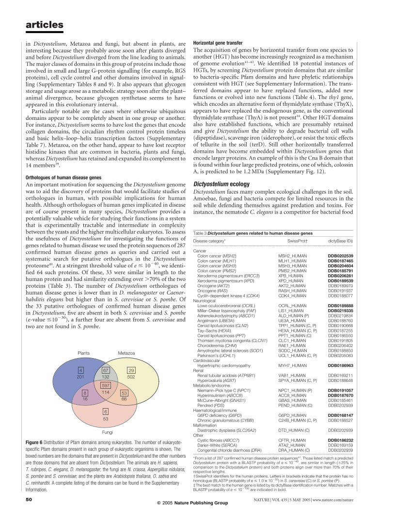
in Dictyostelium, Metazoa and fungi, but absent in plants, areinteresting because they probably arose soon after plants divergedand before Dictyostelium diverged from the line leading to animals.The major classes of domains in this group of proteins include thoseinvolved in small and large G-protein signalling (for example, RGSproteins), cell cycle control and other domains involved in signal-ling (Supplementary Tables 8 and 9). It also appears that glycogenstorage and usage arose as a metabolic strategy soon after the plant–animal divergence, because glycogen synthetase seems to haveappeared in this evolutionary interval.
Particularly notable are the cases where otherwise ubiquitousdomains appear to be completely absent in one group or another.For instance, Dictyostelium seems to have lost the genes that encodecollagen domains, the circadian rhythm control protein timelessand basic helix–loop–helix transcription factors (SupplementaryTable 7). Metazoa, on the other hand, appear to have lost receptorhistidine kinases that are common in bacteria, plants and fungi,whereasDictyostelium has retained and expanded its complement to14 members39.
Orthologues of human disease genes
An important motivation for sequencing the Dictyostelium genomewas to aid the discovery of proteins that would facilitate studies oforthologues in human, with possible implications for humanhealth. Although orthologues of human genes implicated in diseaseare of course present in many species, Dictyostelium provides apotentially valuable vehicle for studying their functions in a systemthat is experimentally tractable and intermediate in complexitybetween the yeasts and the higher multicellular eukaryotes. To assessthe usefulness of Dictyostelium for investigating the functions ofgenes related to human disease we used the protein sequences of 287confirmed human disease genes as queries and carried out asystematic search for putative orthologues in the Dictyosteliumproteome40. At a stringent threshold value of e # 10240, we identi-fied 64 such proteins. Of these, 33 were similar in length to thehuman protein and had similarity extending over .70% of the twoproteins (Table 3). The number of Dictyostelium orthologues ofhuman disease genes is lower than in D. melanogaster or Caenor-habditis elegans but higher than in S. cerevisiae or S. pombe. Ofthe 33 putative orthologues of confirmed human disease genesin Dictyostelium, five are absent in both S. cerevisiae and S. pombe(e-value #10230), a further four are absent from S. cerevisiae andtwo are not found in S. pombe.
Horizontal gene transfer
The acquisition of genes by horizontal transfer from one species toanother (HGT) has become increasingly recognized as a mechanismof genome evolution41–43. We identified 18 potential instances ofHGTs, by screening Dictyostelium protein domains that are similarto bacteria-specific Pfam domains and have phyletic relationshipsconsistent with HGT (see Supplementary Information). The trans-ferred domains appear to have replaced functions, added newfunctions or evolved into new functions (Table 4). The thy1 gene,which encodes an alternative form of thymidylate synthase (ThyX),appears to have replaced the endogenous gene, as the conventionalthymidylate synthase (ThyA) is not present44. Other HGT domainsalso have established functions, which are presumably retainedand give Dictyostelium the ability to degrade bacterial cell walls(dipeptidase), scavenge iron (siderophore), or resist the toxic effectsof tellurite in the soil (terD). Still other horizontally transferreddomains have become embedded within Dictyostelium genes thatencode larger proteins. An example of this is the Cna B domain thatis found within four large predicted proteins, one of which, colossinA, is predicted to be 1.2 MDa (Supplementary Fig. 12).
Dictyostelium ecologyDictyostelium faces many complex ecological challenges in the soil.Amoebae, fungi and bacteria compete for limited resources in thesoil while defending themselves against predation and toxins. Forinstance, the nematode C. elegans is a competitor for bacterial food
Table 3 Dictyostelium genes related to human disease genes
Disease category* SwissProt† dictyBase ID‡.............................................................................................................................................................................
CancerColon cancer (MSH2) MSH2_HUMAN DDB0202539Colon cancer (MLH1) MLH1_HUMAN DDB0187465Colon cancer (MSH3) MSH3_HUMAN DDB0204604Colon cancer (PMS2) PMS2_HUMAN DDB0185791Xeroderma pigmentosum (ERCC3) XPB_HUMAN DDB0206281Xeroderma pigmentosum (XPD) XPD_HUMAN DDB0189539Oncogene (AKT2) AKT2_HUMAN DDB0189970Oncogene (RAS) RASH_HUMAN DDB0191937Cyclin-dependent kinase 4 (CDK4) CDK4_HUMAN DDB0188077
NeurologicalLowe oculocerebrorenal (OCRL) OCRL_HUMAN DDB0189888Miller–Dieker lissencephaly (PAF) LIS1_HUMAN DDB0219335Adrenoleukodystrophy (ABCD1) ALD_HUMAN (P) DDB0219834Angelmann (UBE3A) UE3A_HUMAN DDB0188760Ceroid lipofuscinosis (CLN2) TPP1_HUMAN (C, P) DDB0190668Tay–Sachs (HEXA) HEXA_HUMAN (C, P) DDB0187255Ceroid lipofuscinosis (PPT) PPT1_HUMAN (C) DDB0186550Thomsen myotonia congenita (CLCN1) CLC1_HUMAN DDB0191805Choroideremia (CHM) RAE1_HUMAN DDB0206402Amyotrophic lateral sclerosis (SOD1) SODC_HUMAN DDB0188850Parkinson’s (UCHL1) UCL1_HUMAN (C, P) DDB0205083
CardiovascularHypertrophic cardiomyopathy MYH7_HUMAN DDB0186963
RenalRenal tubular acidosis (ATP6B1) VAB1_HUMAN DDB0169211Hyperoxaluria (AGXT) SPYA_HUMAN (C, P) DDB0188646
Metabolic/endocrineNiemann–Pick type C (NPC1) NPC1_HUMAN (P) DDB0191057Hyperinsulinism (ABCC8) ACC8_HUMAN DDB0187670McCune–Albright (GNAS1) GBAS_HUMAN DDB0185461Pendred (PDS) PEND_HUMAN (C) DDB0202939
Haematological/immuneG6PD deficiency (G6PD) G6PD_HUMAN DDB0168147Chronic granulomatous (CYBB) C24B_HUMAN (C, P) DDB0188527
MalformationDiastrophic dysplasia (SLC26A2) DTD_HUMAN (C) DDB0202939
OtherCystic fibrosis (ABCC7) CFTR_HUMAN DDB0186232Darier–White (SERCA) ATA2_HUMAN DDB0169159Congenital chloride diarrhoea (DRA) DRA_HUMAN (C) DDB0202939
.............................................................................................................................................................................
*From a list of 287 confirmed human disease protein sequences40. Those listed match a predictedDictyostelium protein with a BLASTP probability of e # 10240, are similar in length (^25% incomparison to the Dictyostelium protein) and both proteins align over more than 70% of theirrespective lengths.†SwissProt identifiers for the human proteins. Letters in brackets indicate that the protein has nohomologue (BLASTP probability of e # 1.0 £ 10230) in S. cerevisiae (C) or S. pombe (P).‡The best match to the human gene is listed by its dictyBase identification number. Matches with aBLASTP probability of e # 102100 are indicated in bold.
Figure 6 Distribution of Pfam domains among eukaryotes. The number of eukaryote-
specific Pfam domains present in each group of eukaryotic organisms is shown. The
boxed numbers are the domains that are present in Dictyostelium and the other numbers
are those domains that are absent from Dictyostelium. The animals are H. sapiens,
T. rubripes, C. elegans, D. melanogaster; the fungi are N. crassa, Aspergillus nidulans,
S. pombe and S. cerevisiae; and the plants are Arabidopsis thaliana, O. sativa and
C. reinhardtii. A complete listing of the domains can be found in the Supplementary
Information.
articles
NATURE | VOL 435 | 5 MAY 2005 | www.nature.com/nature50© 2005 Nature Publishing Group
and a predator of Dictyostelium amoebae, but also a potentialdispersal agent for Dictyostelium spores45. Dictyostelium hasexpanded its repertoire of several protein classes that are probablycrucial for such interspecies interactions and for survival andmotility in this complex ecosystem.
Polyketide synthases
A small number of natural products have already been identifiedfrom Dictyostelium, but the gene content suggests that it is a prolificproducer of such molecules. Some of them may act as signals duringdevelopment, such as the dichlorohexanophenone DIF-1, butothers are likely to mediate currently unknown ecological inter-actions46. Many antibiotics and secondary metabolites destined forexport are produced by polyketide synthases, modular proteins ofaround 3,000 amino acids47. We identified 43 putative polyketidesynthases in Dictyostelium (see Supplementary Information). Bycontrast, S. cerevisiae completely lacks polyketide synthases andNeurospora crassa has only seven. Furthermore, two of the Dictyo-stelium proteins have an additional chalcone synthase domain,representing a type of polyketide synthase most typical of higherplants and found to be exclusively shared by Dictyostelium, fungiand plants. In addition to polyketide synthases, the predictedproteome has chlorinating and dechlorinating enzymes as wellas O-methyl transferases, which could increase the diversity ofnatural products made. Thus, Dictyostelium appears to have alarge secondary metabolism, which warrants further investigation.
ABC transporters
ATP-binding cassette (ABC) transporters are prevalent in theproteomes of soil microorganisms and are thought to provideresistance to xenobiotics through their ability to translocatesmall-molecule substrates across membranes against a substantialconcentration gradient48–51. There are 66 ABC transporters encodedby the genome, which can be classified according to the subfamiliesdefined in humans (ABCA, ABCB, ABCC, ABCD, ABCE, ABCF andABCG) based on domain arrangement and signature sequences52.At least 20 of them are expressed during growth and are probablyinvolved in detoxification and the export of endogenous secondarymetabolites.
Cellulose degradation
Many of the predicted cellulose-degrading enzymes in the proteome
(see Supplementary Information) that have secretion signals areexpressed in growing cells that do not produce cellulose53. Theproteome also contains one xylanase enzyme that can degrade thexylan polymers that are often found associated with the cellulose ofhigher plants. Perhaps Dictyostelium uses these enzymes to degradeplant tissue into particles that are then taken up by cells. Theseenzymes may also aid in the breakdown of cellulose-containingmicroorganisms upon which Dictyostelium feeds. Alternatively,these enzymes may promote the growth of bacteria that can serveas food, because Dictyostelium’s habitat also contains cellulose-degrading bacteria.
Specializations for cell motility
During both growth and development, Dictyostelium amoebaedisplay motility that is characteristic of human leukocytes54. As aconsequence, studies of Dictyostelium have contributed significantlyto cytoskeleton research55. Dictyostelium’s survival depends on anability to efficiently sense, track and consume soil bacteria usingsophisticated systems for chemotaxis and phagocytosis. Its multi-cellular development depends on chemotactic aggregation of indi-vidual amoebae and the coordinated movement of thousands ofcells during fruiting body morphogenesis. The proteome reveals anastonishing assortment of proteins that are used for robust,dynamic control of the cytoskeleton during these processes. Assuggested by functional parallels to human cells, these proteins aremost similar to metazoan proteins in their variety and domainarrangements (Fig. 7; see also Supplementary Table 11). Surpris-ingly, although the actin cytoskeleton has been studied for over25 years, 71 putative actin-binding proteins apparently escapedclassical methods of discovery. For example, actobindins had notbeen previously recognized in Dictyostelium. Curiously, the actindepolymerization factor (ADF) and calponin homology (CH)domain proteins appear to have diversified by domain shuffling,a substantial fraction having domain combinations unique toDictyostelium (Supplementary Table 12 and SupplementaryFig. 13). In addition to 30 actin genes, there are also orthologuesof all actin-related protein (ARP) classes present in mammals, aswell as three founding members of a new class (SupplementaryFig. 14).
Cytoskeletal remodelling during chemotaxis and phagocytosis isregulated by a considerable number of upstream signalling com-ponents. Of the 18 Rho family GTPases in Dictyostelium, some are
Table 4 Candidate horizontal gene transfers from bacteria
Function* Pfam† Number of proteins‡ DictyBase ID§ Length (aa)k Region matched{ e-value#...................................................................................................................................................................................................................................................................................................................................................................
Aromatic amino acid lyase Beta_elim_lyase 2 DDB0204031 170 4–170 3.2 £ 10265
Biotin metabolism BioY 1 DDB0184375 338 145–299 5.8 £ 10220
Unknown Cna_B 4 DDB0184530 11,103 Multiple†† 1.1 £ 10210
Peroxidase Dyp_peroxidase 1 DDB0168077 306 3–303 1.4 £ 10282
Insecticide Endotoxin_N 2 DDB0188332 628 38–210 1.2 £ 10232
Isopentenyl transferase IPT 1 DDB0169077 283 1–63 5.1 £ 10212
Siderophore IucA_IucC 2 DDB0219918 739 183–350 2.3 £ 10218
Osmoregulation OsmC 2 DDB0190102 156 16–156 9.8 £ 10222
Dipeptidase/b-lactamase Peptidase M15 1 DDB0205124 897 68–406; 711–879 3.4 £ 10216
Dipeptidase/b-lactamase Peptidase S13 1 DDB0168572 522 337–495 4.2 £ 10225
Polyphosphate synthesis PP_kinase 1 DDB0192001 1,053 372–1045 1.6 £ 102234
Tellurite resistance TerD 2 DDB0169240 287 152–279 2.1 £ 10267
Thymidylate synthesis Thy1 1 DDB0214905 303 38–254 9.9 £ 102117
Unknown DUF84 1 DDB0203145 179 5–175 1.6 £ 10220
Unknown DUF885 2 DDB0205394 689 318–685 1.5 £ 102124
(Prespore protein 3B) DUF1121 3 DDB0169184 226 1–226 8.7 £ 102134
Unknown DUF1289 1 DDB0204782 88 29–85 3.3 £ 10215
Unknown DUF1294 1 DDB0186703 155 2–73 8.9 £ 10218
...................................................................................................................................................................................................................................................................................................................................................................
*Confirmed or proposed function of the prokaryotic orthologue is given. For domains without function information, information on any Dictyostelium protein in the set is given in parentheses.†The Pfam domain designation (http://www.sanger.ac.uk/Software/Pfam/).‡The number of gene models in which the domain appears. Bold numbers indicate gene sets where there are pairs of genes that map within 10 kb of each other.§The gene identification number for the example given in the rest of the table (release v2.0 at http://www.dictybase.org/).kNumber of amino acid (aa) residues in the predicted Dictyostelium protein containing the domain.{The region of the Dictyostelium protein that matched the prokaryotic domain. The amino acid sequence identity between this region and the most highly related prokaryotic protein was 21–52%.#The e-value for the domain against the Pfam model library used to identify it (see Supplementary Information).††The protein colossin A consists of an array of 91 partial Cna_B domains within 18 larger repeats, and the e-value corresponds to one domain.
articles
NATURE | VOL 435 | 5 MAY 2005 | www.nature.com/nature 51© 2005 Nature Publishing Group

clear Rac orthologues and one belongs to the RhoBTB subfamily56.However, the Cdc42 and Rho subfamilies characteristic of Metazoaand fungi are absent, as are the Rho subfamily effector proteins. Theactivities of these GTPases are regulated by two members of theRhoGDI family, by components of ELMO1–DOCK180 complexesand by a large number of proteins carrying RhoGEF and RhoGAPdomains (.40 of each), most of which show domain compositionsnot found in other organisms. Remarkably,Dictyostelium appears tobe the only lower eukaryote that possesses class I phosphatidyl-inositol-3-OH kinases, which are at the crossroad of several criticalsignalling pathways (for details of the regulators and their effectors,see Supplementary Table 13)57. The diverse array of these regulatorsand the discovery of many additional actin-binding proteins suggestthat there are many aspects of cytoskeletal regulation that have yet tobe explored.
Multicellularity and developmentThe evolution of multicellularity was arguably as significant as theorigin of the eukaryotic cell in enabling the diversification of life.The common unicellular ancestor of the crown group of organismsmust have possessed the basic machinery to regulate nutrientuptake, metabolism, cellular defence and reproduction, and it islikely that these mechanisms were adapted to integrate the functionsof cells in multicellular organisms. Dictyostelium achieved multi-cellularity through a different evolutionary route compared withplants and animals, yet the ancestors of these respective groupsprobably started with the same endowment of genes and faced thesame problem of achieving cell specialization and tissueorganization.
When starved, Dictyostelium develops as a true multicellularorganism, organizing distinct tissues within a motile slug andproducing a fruiting body comprised of a cellular, cellulosic stalksupporting a bolus of spores4. Thus, Dictyostelium has evolveddifferentiated cell types and the ability to regulate their proportionsand morphogenesis. A broad survey of proteins required for multi-cellular development shows that Dictyostelium has retained celladhesion and signalling modules normally associated exclusivelywith animals, whereas the structural elements of the fruiting bodyand terminally differentiated cells clearly derive from the control ofcellulose deposition and metabolism now associated with plants.The Dictyostelium genome offers a first glimpse of how multi-cellularity evolved in the amoebozoan lineage. In the followingsections, we consider some of the systems that are particularlyrelevant to cellular differentiation and integration in a multicellularorganism.
Signal transduction through G-protein-coupled receptors
The needs of multicellular development add greatly to those ofchemotaxis in demanding dynamically controlled and highly selec-tive signalling systems. G-protein-coupled cell surface receptors(GPCRs) form the basis of such systems in many species, allowingthe detection of a variety of environmental and intra-organismalsignals such as light, Ca2þ, odorants, nucleotides and peptides. Theyare subdivided into six families, which, despite their conservedsecondary domain structure, do not share significant sequencesimilarity58. Until recently, in Dictyostelium only the seven CAR/CRL (cAMP receptor/ cAMP receptor-like) family GPCRs had beenexamined in detail59,60. Surprisingly, a detailed search uncovered 48additional putative GPCRs of which 43 can be grouped into thesecretin (family 2), metabotropic glutamate/GABAB (family 3) andthe frizzled/smoothened (family 5) families of receptors (Fig. 8; seealso Supplementary Information). The presence of family 2, 3 and 5receptors in Dictyostelium was surprising because they had beenthought to be specific to animals. Their occurrence in Dictyosteliumsuggests that they arose before the divergence of the animals andfungi and were later lost in fungi, and that the radiation of GPCRspre-dates the divergence of the animals and fungi. The secretin
Figure 7 Microfilament system proteins. Proteins with probable interactions with the actin
cytoskeleton are tabulated by their documented or predicted functions. Coloured boxes
indicate the presence of a protein related to the Dictyostelium (D) protein in Metazoa (M),
fungi (F) or plants (P). Dictyostelium-specific proteins have no recognizable relatives or
differ from relatives due to extensions or unusual domain compositions. For details see
Supplementary Information. Actin-binding modules: ACT, actin fold; ADF, actin
depolymerization factor/cofilin-like domain; CAP, capping protein fold; CH, calponin
homology domain; EVH, Ena/VASP homology domain 2; FH2, formin homology 2 domain;
GEL, gelsolin repeat domain; KELCH, Kelch repeat domain; MYO, myosin motor domain;
PRO, profilin fold; TAL, the I/LWEQ actin-binding domain of talin and related proteins; TRE,
trefoil domain; VHP, villin head piece; WH2, Wiskott Aldrich syndrome homology region 2.
articles
NATURE | VOL 435 | 5 MAY 2005 | www.nature.com/nature52© 2005 Nature Publishing Group
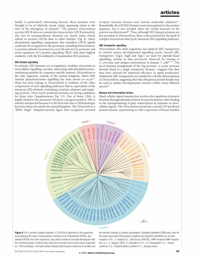
family is particularly interesting because these proteins werethought to be of relatively recent origin, appearing closer to thetime of the divergence of animals61. The putative Dictyosteliumsecretin GPCR does not contain the characteristic GPCR proteolyticsite, but its transmembrane domains are clearly more closelyrelated to secretin GPCRs than to other families (Fig. 8). Manydownstream signalling components that transduce GPCR signalscould also be recognized in the proteome, including heterotrimericG-protein subunits (fourteen Ga, two Gb and one Gg proteins) andseven regulators of G-protein signalling (RGS) that share highestsimilarity with the R4 subfamily of mammalian RGS proteins.
SH2 domain signalling
In animals, SH2 domains act as regulatory modules of proteins inintracellular signalling cascades, interacting with phosphotyrosine-containing peptides in a sequence-specific manner. Dictyostelium isthe only organism, outside of the animal kingdom, where SH2domain phosphotyrosine signalling has been shown to occur62.What has been lacking in Dictyostelium is evidence of the othercomponents of such signalling pathways; that is, equivalents of themetazoan SH2-domain-containing receptors, adaptors and target-ing proteins. Three newly predicted proteins are strong candidatesfor these roles (Supplementary Fig. 15). One of them, CblA, ishighly related to the metazoan Cbl proto-oncogene product. This isentirely unexpected because it is the first time that a Cbl homologuehas been observed outside the animal kingdom. The Cbl protein is a‘RING finger’ ubiquitin-protein ligase that recognizes activated
receptor tyrosine kinases and various molecular adaptors63.Remarkably, the Cbl SH2 domain went unrecognized in the proteinsequence, but it was revealed when the crystal structure of theprotein was determined64. Thus, although SH2 domain proteins areless prevalent in Dictyostelium, there is the potential for the kind ofcomplex interactions that typify metazoan SH2 signalling pathways.
ABC transporter signalling
Dictyostelium, like other organisms, has adapted ABC transportersto control various developmental signalling events. Several ABCtransporters (TagA, TagB and TagC) are used for peptide-basedsignalling, similar to that previously observed for mating inS. cerevisiae and antigen presentation in human T cells65–67. Thenovel domain arrangement of the Tag proteins—a serine proteasedomain fused to a single transporter domain—suggests that theyhave been selected for improved efficiency in signal production.Additional ABC transporters are needed for cell fate determinationin Dictyostelium, suggesting that this ubiquitous protein family maybe used in similar developmental contexts within many differentspecies68.
Kinases and transcription factors
Much cellular signal transduction involves the regulation of proteinfunction through phosphorylation by protein kinases, often leadingto the reprogramming of gene transcription in response to extra-cellular signals. The Dictyostelium proteome contains 295 predictedprotein kinases, representing as wide a spectrum of kinase families
Figure 8 The G-protein-coupled receptors. A CLUSTALX alignment of the sequences
encompassing the seven transmembrane domains of all Dictyostelium GPCRs, and
selected GPCRs from other organisms, was used to create an unrooted dendrogram with
the TreeView program. A black circle marks the innermost node of each branch supported
by .60% bootstraps. The hash symbol indicates that this gene model has to be split, and
the asterisk indicates a putative pseudogene. DictyBase identifiers (DDB) were used for
the newly discovered Dictyostelium receptors and SwissProt identifiers for all other
receptors. A.th., A. thaliana; B.t., Bos taurus; CAR/CRL, cAMP receptor/cAMP receptor-
like; C.e., C. elegans; DICDI, D. discoideum; D.m., D. melanogaster; G.c., Geodia
cydonium; P.p., Polysphondylium pallidum; X.l., Xenopus laevis.
articles
NATURE | VOL 435 | 5 MAY 2005 | www.nature.com/nature 53© 2005 Nature Publishing Group
as that observed in Metazoa (Supplementary Tables 14–16 andSupplementary Fig. 16). Given the presence of SH2-domain-basedsignalling it was surprising that no receptor tyrosine kinases couldbe recognized in the genome. However, Dictyostelium has a numberof other receptor kinases, such as the histidine kinases and a groupof eight novel putative receptor serine/threonine kinases, whichare involved in nutrient and starvation sensing69. Most of theubiquitous families of transcription factors are represented inDictyostelium, with the notable exception of the otherwise ubiqui-tous basic helix–loop–helix proteins (Supplementary Table 17and Supplementary Fig. 17). Compared with other eukaryotes,Dictyostelium appears to have fewer transcription factors relativeto the total number of genes, suggesting that many transcriptionfactors have yet to be defined, or that the activities of a smallerrepertoire of factors are combined and controlled to achieve com-plex regulation (Supplementary Table 18 and SupplementaryFig. 18).
Cell adhesion
Throughout Dictyostelium development, cells must modulate theiradhesiveness to the substrate, to the extracellular matrix and toother cells in order to create tissues and carry out morphogenesis. Toaccomplish this, Dictyostelium uses a surprising number of com-ponents that have been normally only associated with animals. Forexample, disintegrin proteins regulate cell adhesiveness and differ-entiation in a number of Metazoa, and at least one Dictyosteliumdisintegrin, AmpA, is needed throughout development for cell fatespecification70. We also identified distant relatives of vinculin anda-catenin—normally associated with adherens junctions—whichsupport the idea that the epithelium-like sheet of cells thatsurrounds the stalk tube contains such junctions71. Consistentwith this, the Dictyostelium genome encodes numerous proteinspreviously described as components of adherens junctions inMetazoa, such as b-catenin (Aardvark), a-actinin, formins, VASPand myosin VII.
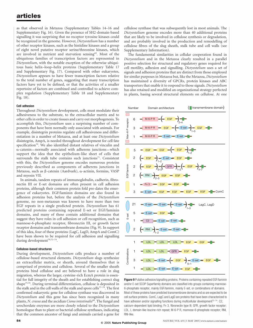
In animals, tandem repeats of immunoglobulin, cadherin, fibro-nectin III or E-set domains are often present in cell adhesionproteins, although their common protein fold pre-dates the emer-gence of eukaryotes. EGF/laminin domains are also found inadhesion proteins but, before the analysis of the Dictyosteliumgenome, no non-metazoan was known to have more than twoEGF repeats in a single predicted protein. Dictyostelium has 61predicted proteins containing repeated E-set or EGF/laminindomains, and many of these contain additional domains thatsuggest they have roles in cell adhesion or cell recognition, such asmannose-6-phosphate receptor, fibronectin III, or growth factorreceptor domains and transmembrane domains (Fig. 9). In supportof this idea, four of these proteins (LagC, LagD, AmpA and ComC)have been shown to be required for cell adhesion and signallingduring development70,72–74.
Cellulose-based structures
During development, Dictyostelium cells produce a number ofcellulose-based structural elements. Dictyostelium slugs synthesizean extracellular matrix, or sheath, around themselves that iscomprised of proteins and cellulose. Several of the smaller sheathproteins bind cellulose and are believed to have a role in slugmigration, whereas the larger, cysteine-rich EcmA protein is essen-tial for full integrity of the sheath and for establishing correct slugshape75,76. During terminal differentiation, cellulose is deposited inthe stalk and in the cell walls of the stalk and spore cells77–79. The firstconfirmed eukaryotic gene for cellulose synthase was discovered inDictyostelium and this gene has since been recognized in manyplants,N. crassa and the ascidian Ciona intestinalis80. The fungal andurochordate enzymes are more closely related to the Dictyosteliumhomologue than to plant or bacterial cellulose synthases, indicatingthat the common ancestor of fungi and animals carried a gene for
cellulose synthase that was subsequently lost in most animals. TheDictyostelium genome encodes more than 40 additional proteinsthat are likely to be involved in cellulose synthesis or degradation,and are probably involved in the production and remodelling ofcellulose fibres of the slug sheath, stalk tube and cell walls (seeSupplementary Information).
The fundamental similarities in cellular cooperation found inDictyostelium and in the Metazoa clearly resulted in a parallelpositive selection for structural and regulatory genes required forcell motility, adhesion and signalling. Dictyostelium uses a set ofsignals and adhesion proteins that are distinct from those employedfor similar purposes in Metazoa but, like the Metazoa, Dictyosteliumhas maintained a diversity of GPCRs, protein kinases and ABCtransporters that enable it to respond to those signals. Dictyosteliumhas also retained and modified an organizational strategy perfectedin plants, basing several structural elements on cellulose. At one
Figure 9 Putative adhesion/signalling proteins. Proteins containing repeated EGF/laminin
and/or E-set SCOP Superfamily domains are classified into groups containing mannose-
6-phosphate receptor, mainly EGF/laminin, mainly E-set, or combinations of domains.
Most of these proteins have predicted transmembrane domains and so are expected to be
cell surface proteins. ComC, LagC and LagD are proteins that have been characterized to
have adhesion and/or signalling functions during multicellular development72–74. C2,
calcium-dependent lipid binding; Fn 3, fibronectin type III; GFR, growth factor receptor;
LDL, L domain-like leucine-rich repeat; M-6-P R, mannose-6-phosphate receptor; RNI,
RNI-like.
articles
NATURE | VOL 435 | 5 MAY 2005 | www.nature.com/nature54© 2005 Nature Publishing Group

level Dictyostelium has achieved multicellularity by using strategiesthat are similar to plants and Metazoa, but the differences betweenthem suggest convergent evolution, rather than lineal descent froman ancestor with overt or latent multicellular capacities.
ConclusionThe complete protein repertoire of Dictyostelium provides a newperspective for studying its cellular and developmental biology. At asystems level, Dictyostelium provides a level of complexity that isgreater than the yeasts, but much simpler than plants or animals.Thus, high-resolution molecular analyses in this system may revealcontrol networks that are difficult to study in more complexsystems, and may presage regulatory strategies used by higherorganisms81–83. At a practical level, the comparative genomics ofDictyostelium and related pathogens, such as Entamoeba histolytica,should aid in the functional definition of amoebozoa-specific genesthat may open new avenues of research aimed at controllingamoebic diseases. Dictyostelium’s adeptness at hunting bacteriaalso renders it susceptible to infections by intracellular bacterialpathogens84,85. Dictyostelium and human macrophages display fun-damental similarities in their cell biology, which has spurred the useof Dictyostelium as a model host for bacterial pathogenesis. It is alsoan attractive model in which to study other disease processes: for anumber of human disease-related proteins, it provides a test-bed forstudying their functions in a model organism that has greatersimilarity to higher eukaryotes than do the yeasts, yet shares thelatter’s experimental tractability.
The high frequency of repeated amino acid tracts in Dictyosteliumproteins has long been known anecdotally, but we can now surveytheir precise nature and number, and find them to be moreabundant than in any other sequenced genome. Many humandiseases result from the expansion of triplet nucleotide repeats,some of which encode polyglutamine tracts that cause cell degenera-tion86,87. Learning how Dictyostelium cells tolerate so many proteinswith amino acid homopolymers will, we hope, help to elucidate theroles of these motifs in protein function and dysfunction.
Comparative genomic studies in eukaryotes are providing theraw material for global examinations of the evolution of cellularregulation and developmental mechanisms88. Many genes have beenlost in one species but retained in others, such that each new genomesequence adds to our understanding of the genetic complement of theeukaryotic progenitor. Thus, our understanding of eukaryotes willcontinue to be refined as more genome sequences become availablefrom representatives of large groups of organisms whose genomesremain largely unexplored, such as the amoebozoa. The surprisingmolecular diversity of the Dictyostelium proteome, which includesprotein assemblages usually associated with fungi, plants or ani-mals, suggests that their last common ancestor had a greaternumber of genes than had been previously appreciated. A
MethodsDetails on the availability of reagents can be found in the Supplementary Information. Allanalyses described here were performed on Version 2.0 of the genome sequence. Updatesto the sequence and annotation are available at http://www.dictybase.org and http://www.genedb.org/genedb/dicty/index.jsp. Further details of analyses not explicitlydescribed below can be found in the Supplementary Information.
HAPPY mappingA short-range (,100-kb), high-resolution (^8.54-kb) mapping panel was prepared asdescribed9. Briefly, 96 aliquots each containing ^0.52 haploid genome equivalents ofsheared AX4 genomic DNA were pre-amplified by PEP (primer extension pre-amplification89). A total of 4,913 STS markers (Supplementary Table 1) were typed by two-phase hemi-nested polymerase chain reaction (PCR; multiplexed for up to 1,200 markersin the first phase) on aliquots of the diluted PEP products. Maps were assembled fromgood-quality data essentially as described previously8. A second, longer-range (^150 kb)mapping panel was used to confirm some linkages on chromosomes 2 and 5. HAPPY mapanalysis and PCR primer design for HAPPY mapping was performed using variouscustom programs (P.H.D. and A.T.B., unpublished).
Chromosome purificationGenomic DNA from D. discoideum strain AX4 was prepared and separated by pulsed fieldgel electrophoresis essentially as described27,9, except that gels were run in stacked pairs;one member of each pair was stained with ethidium bromide, and bands excised from itsunstained counterpart by alignment.
WCS and YAC subclone librariesFor WCS libraries, gel slices (above) were disrupted by several passages through a 30-gaugesyringe needle, digested with b-agarase (NEB) and phenol-extracted. DNA wasconcentrated by ethanol precipitation, sonicated, end-blunted using mung beannuclease and size-fractionated on 0.8% low-melting-point agarose gels. Fractions of 1.4–2 kb and 2–4 kb were excised, DNA extracted as before and ligated into the SmaI site ofpUC18 or pUC19. Clone propagation and template preparation followed standardprotocols.
For YAC subclone libraries, AX4-derived YACs were identified (and their position andintegrity confirmed) by screening the set described by ref. 22 using markers from theHAPPY map. Subclones were prepared from PFG-purified YACs essentially as for the WCSlibraries; contaminating yeast-derived sequences were filtered out in silico.
Sequencing and assemblyDetails of the sequencing and assembly methods can be found in SupplementaryInformation. Generally, mapped sequence features were used to nucleate sequence contigsassembled from the WCS data, and extended using read-pair information and iterativesearches for overlapping sequences, followed by directed gap closure using a range ofapproaches.
Fluorescent in situ hybridizationIn situ hybridization was performed as in ref. 17.
Gene prediction and identification of sequence featuresFull details are provided in the Supplementary Information. Briefly, automated geneprediction was performed using a combination of programs that had been trained onwell-characterized D. discoideum genes, and the results integrated with reference toD. discoideum complementary DNA sequences and homology to genes in other species.Other features in the predicted proteins, and other sequence features, were identified usinga variety of software packages.
Analysis of functional gene clusteringMicroarray targets (refs 53, 90, 91; and N. Van Driessche and G. Shaulsky, unpublisheddata) and gene models were mapped onto the genome sequence using BLAST92 and themodified LIS algorithm93. To look for clustering of genes with correlated temporalexpression profiles, pairwise correlation coefficients were calculated for genes with knownexpression profiles on each chromosome91. Blocks of $6 consecutive genes were sought,for which either (1) all pairwise correlation coefficients were positive and $70% were.0.2 (genes with similar developmental trajectories) or (2) each gene had a partner withan absolute correlation coefficient value of .0.6 (tightly co-regulated genes); nostatistically significant clusters met these criteria.
To look for clustering of genes associated with specific developmental stages94,95 or celltypes90,96, the genome was scanned with various sized windows97 for regions withsignificant (P , 0.01) over-representation of genes in any one of these groups.
Analysis of duplicated genesPredicted protein sequences were clustered using TribeMCL98, using a BLASTPexpectation of ,10240 as a cutoff. A x2 test invalidated the hypothesis that members of afamily are randomly distributed in the genome. Within each family, protein divergences(similarity distances computed using the ‘Protdist’ module of PHYLIP; http://evolution.genetics.washington.edu/phylip.html) and physical intergenic distancesbetween all pairs of family members were tabulated, and the correlation coefficientbetween the former and latter values was calculated. Analysis was performed on the 86gene families (representing 155 gene pairs) with at least 10 intrachromosomal distancepairings to provide robust statistical confidence.
Other sequence analyses and graphical representationOther sequence analyses (nucleotide and dinucleotide composition; identification ofsimple-sequence repeats in nucleotide and protein sequence; coding density computation;tRNA cluster identification) were performed using a range of custom software (P.H.D. andA.T.B., unpublished). Graphical representation of chromosomes in Fig. 2 was doneprimarily using Cinema4D-8.5 (Maxon Computer GmbH) after pre-processing usingcustom software (P.H.D.).
Received 16 September 2004; accepted 17 February 2005; doi:10.1038/nature03481.
1. Kessin, R. H. Dictyostelium—Evolution, Cell Biology, and the Development of Multicellularity, xiv, 294
(Cambridge Univ. Press, Cambridge, 2001).
2. Konijn, T. M. et al. The acrasin activity of adenosine-3 0 ,5 0 -cyclic phosphate. Proc. Natl Acad. Sci. USA
58, 1152–1154 (1967).
3. Muller, K. & Gerisch, G. A specific glycoprotein as the target site of adhesion blocking Fab in
aggregating Dictyostelium cells. Nature 274, 445–449 (1978).
4. Raper, K. B. Pseudoplasmodium formation and organization in Dictyostelium discoideum. J. Elisha
Mitchell Sci. Soc. 56, 241–282 (1940).
articles
NATURE | VOL 435 | 5 MAY 2005 | www.nature.com/nature 55© 2005 Nature Publishing Group

5. Raper, K. B. Dictyostelium discoideum, a new species of slime mold from decaying forest leaves. J. Agr.
Res. 50, 135–147 (1935).
6. Knecht, D. A., Cohen, S. M., Loomis, W. F. & Lodish, H. F. Developmental regulation of Dictyostelium
discoideum actin gene fusions carried on low-copy and high-copy transformation vectors. Mol. Cell.
Biol. 6, 3973–3983 (1986).
7. Dear, P. H. & Cook, P. R. HAPPY mapping—linkage mapping using a physical analog of meiosis.
Nucleic Acids Res. 21, 13–20 (1993).
8. Konfortov, B. A., Cohen, H. M., Bankier, A. T. & Dear, P. H. A high-resolution HAPPY map of
Dictyostelium discoideum chromosome 6. Genome Res. 10, 1737–1742 (2000).
9. Glockner, G. et al. Sequence and analysis of chromosome 2 of Dictyostelium discoideum. Nature 418,
79–85 (2002).
10. Urushihara, H. et al. Analyses of cDNAs from growth and slug stages of Dictyostelium discoideum.
Nucleic Acids Res. 32, 1647–1653 (2004).
11. Smith, S. S. & Ratner, D. I. Lack of 5-methylcytosine in Dictyostelium discoideum DNA. Biochem. J.
277, 273–275 (1991).
12. Glockner, G. et al. The complex repeats of Dictyostelium discoideum. Genome Res. 11, 585–594 (2001).
13. Crick, F. H. Codon-anticodon pairing: the wobble hypothesis. J. Mol. Biol. 19, 548–555 (1966).
14. Soll, D. & RajBhandary, U. (ed.) tRNA: Structure, Biosynthesis and Function (ASM, Washington DC,
1995).
15. Burger, G., Plante, I., Lonergan, K. M. & Gray, M. W. The mitochondrial DNA of the amoeboid
protozoon, Acanthamoeba castellanii: complete sequence, gene content and genome organization.
J. Mol. Biol. 3, 522–537 (1995).
16. Ogawa, S. et al. The mitochondrial DNA of Dictyostelium discoideum: complete sequence, gene
content and genome organization. Mol. Gen. Genet. 263, 514–519 (2000).
17. Sucgang, R. et al. Sequence and structure of the extrachromosomal palindrome encoding the
ribosomal RNA genes in Dictyostelium. Nucleic Acids Res. 31, 2361–2368 (2003).
18. Szafranski, K., Dingermann, T., Glockner, G. & Winckler, T. Template jumping by a LINE reverse
transcriptase has created a SINE-like 5S rRNA retropseudogene in Dictyostelium. Mol. Genet. Genom.
271, 98–102 (2004).
19. Eichler, E. E. & Sankoff, D. Structural dynamics of eukaryotic chromosome evolution. Science 301,
793–797 (2003).
20. Cappello, J., Cohen, S. M. & Lodish, H. F. Dictyostelium transposable element DIRS-1 preferentially
inserts into DIRS-1 sequences. Mol. Cell. Biol. 4, 2207–2213 (1984).
21. Cappello, J., Handelsman, K. & Lodish, H. F. Sequence of Dictyostelium DIRS-1: an apparent
retrotransposon with inverted terminal repeats and an internal circle junction sequence. Cell 43,
105–115 (1985).
22. Loomis, W. F., Welker, D., Hughes, J., Maghakian, D. & Kuspa, A. Integrated maps of the
chromosomes in Dictyostelium discoideum. Genetics 141, 147–157 (1995).
23. Goodwin, T. J. & Poulter, R. T. Multiple LTR-retrotransposon families in the asexual yeast Candida
albicans. Genome Res. 10, 174–191 (2000).
24. Appelgren, H., Kniola, B. & Ekwall, K. Distinct centromere domain structures with separate functions
demonstrated in live fission yeast cells. J. Cell Sci. 116, 4035–4042 (2003).
25. Kuspa, A., Maghakian, D., Bergesch, P. & Loomis, W. F. Physical mapping of genes to specific
chromosomes in Dictyostelium discoideum. Genomics 13, 49–61 (1992).
26. McClintock, B. The production of homozygous deficient tissues with mutant characteristics by means
of the aberrant mitotic behaviour of ring-shaped chromosomes. Genetics 23, 315–376 (1938).
27. Cox, E. C., Vocke, C. D., Walter, S., Gregg, K. Y. & Bain, E. S. Electrophoretic karyotype for
Dictyostelium discoideum. Proc. Natl Acad. Sci. USA 87, 8247–8251 (1990).
28. International Human Genome Sequencing Consortium. Initial sequencing and analysis of the human
genome. Nature 409, 860–941 (2001).
29. Gardner, M. J. et al. Genome sequence of the human malaria parasite Plasmodium falciparum. Nature
419, 498–511 (2002).
30. Trusov, Y. A. & Dear, P. H. A molecular clock based on the expansion of gene families. Nucleic Acids
Res. 24, 995–999 (1996).
31. Kellis, M., Birren, B. W. & Lander, E. S. Proof and evolutionary analysis of ancient genome duplication
in the yeast Saccharomyces cerevisiae. Nature 428, 617–624 (2004).
32. Dujon, B. et al. Genome evolution in yeasts. Nature 430, 35–44 (2004).
33. Raper, K. B. The Dictyostelids (Princeton Univ. Press, Princeton, New Jersey, 1984).
34. Loomis, W. F. & Smith, D. W. Consensus phylogeny of Dictyostelium. Experientia 51, 1110–1115
(1995).
35. Baldauf, S. L. & Doolittle, W. F. Origin and evolution of the slime molds (Mycetozoa). Proc. Natl Acad.
Sci. USA 94, 12007–12012 (1997).
36. Bapteste, E. et al. The analysis of 100 genes supports the grouping of three highly divergent amoebae:
Dictyostelium, Entamoeba, and Mastigamoeba. Proc. Natl Acad. Sci. USA 99, 1414–1419 (2002).
37. Nara, T., Hshimoto, T. & Aoki, T. Evolutionary implications of the mosaic pyrimidine-biosynthetic
pathway in eukaryotes. Gene 257, 209–222 (2000).
38. Olsen, R. & Loomis, W. F. A model of orthologous protein sequence divergence. J. Mol. Evol. (in the
press).
39. Thomason, P. & Kay, R. Eukaryotic signal transduction via histidine-aspartate phosphorelay. J. Cell
Sci. 113, 3141–3150 (2000).
40. Fortini, M. E., Skupski, M. P., Boguski, M. S. & Hariharan, I. K. A survey of human disease gene
counterparts in the Drosophila genome. J. Cell Biol. 150, F23–F30 (2000).
41. Jain, R., Rivera, M. C., Moore, J. E. & Lake, J. A. Horizontal gene transfer in microbial genome
evolution. Theor. Popul. Biol. 61, 489–495 (2002).
42. Richards, T. A., Hirt, R. P., Williams, B. A. & Embley, T. M. Horizontal gene transfer and the evolution
of parasitic protozoa. Protist 154, 17–32 (2003).
43. Iyer, L. M., Aravind, L., Coon, S. L., Klein, D. C. & Koonin, E. V. Evolution of cell-cell signaling in
animals: did late horizontal gene transfer from bacteria have a role? Trends Genet. 20, 292–299 (2004).
44. Myllykallio, H. et al. An alternative flavin-dependent mechanism for thymidylate synthesis. Science
297, 105–107 (2002).
45. Kessin, R. H., Gundersen, G. G., Zaydfudim, V., Grimson, M. & Blanton, R. L. How cellular slime
molds evade nematodes. Proc. Natl Acad. Sci. USA 93, 4857–4861 (1996).
46. Morris, H. R., Taylor, G. W., Masento, M. S., Jermyn, K. A. & Kay, R. R. Chemical structure of the
morphogen differentiation inducing factor from Dictyostelium discoideum. Nature 328, 811–814
(1987).
47. Cane, D. E., Walsh, C. T. & Khosla, C. Harnessing the biosynthetic code: combinations, permutations,
and mutations. Science 282, 63–68 (1998).
48. Holland, I. B. & Blight, M. A. ABC-ATPases, adaptable energy generators fuelling transmembrane
movement of a variety of molecules in organisms from bacteria to humans. J. Mol. Biol. 293, 381–399
(1999).
49. Andrade, A. C., Van Nistelrooy, J. G., Peery, R. B., Skatrud, P. L. & De Waard, M. A. The role of ABC
transporters from Aspergillus nidulans in protection against cytotoxic agents and in antibiotic
production. Mol. Gen. Genet. 263, 966–977 (2000).
50. Mendez, C. & Salas, J. A. The role of ABC transporters in antibiotic-producing organisms: drug
secretion and resistance mechanisms. Res. Microbiol. 152, 341–350 (2001).
51. Schoonbeek, H. J., Raaijmakers, J. M. & De Waard, M. A. Fungal ABC transporters and microbial
interactions in natural environments. Mol. Plant Microbe Interact. 15, 1165–1172 (2002).
52. Anjard, C. & Loomis, W. F. Evolutionary analyses of ABC transporters of Dictyostelium discoideum.
Eukaryot. Cell 1, 643–652 (2002).
53. Iranfar, N., Fuller, D. & Loomis, W. F. Genome-wide expression analyses of gene regulation during
early development of Dictyostelium discoideum. Eukaryot. Cell 2, 664–670 (2003).
54. Devreotes, P. N. & Zigmond, S. H. Chemotaxis in eukaryotic cells: A focus on leukocytes and
Dictyostelium. Annu. Rev. Cell Biol. 4, 649–686 (1988).
55. Noegel, A. A. & Schleicher, M. The actin cytoskeleton of Dictyostelium: a story told by mutants. J. Cell
Sci. 113, 759–766 (2000).
56. Rivero, F. & Somesh, B. P. Signal transduction pathways regulated by Rho GTPases in Dictyostelium.
J. Muscle Res. Cell Motil. 23, 737–749 (2002).
57. Merlot, S. & Firtel, R. A. Leading the way: directional sensing through phosphatidylinositol 3-kinase
and other signalling pathways. J. Cell Sci. 116, 3471–3478 (2003).
58. Bockaert, J. & Pin, J. P. Molecular tinkering of G protein-coupled receptors: an evolutionary success.
EMBO J. 18, 1723–1729 (1999).
59. Ginsburg, G. T. et al. The regulation of Dictyostelium development by transmembrane signalling.
J. Eukaryot. Microbiol. 42, 200–205 (1995).
60. Raisley, B., Zhang, M., Hereld, D. & Hadwiger, J. A. A cAMP receptor-like G protein-coupled receptor
with roles in growth regulation and development. Dev. Biol. 265, 433–445 (2004).
61. King, N., Hittinger, C. T. & Carroll, S. B. Evolution of key cell signaling and adhesion protein families
predates animal origins. Science 301, 361–363 (2003).
62. Kawata, T. et al. SH2 signaling in a lower eukaryote: A STAT protein that regulates stalk cell
differentiation in Dictyostelium. Cell 89, 909–916 (1997).
63. Thien, C. B. & Langdon, W. Y. Cbl: many adaptations to regulate protein tyrosine kinases. Nature Rev.
Mol. Cell Biol. 2, 294–307 (2001).
64. Meng, W. et al. Structure of the amino-terminal domain of Cbl complexed to its binding site on
ZAP-70 kinase. Nature 398, 84–90 (1999).
65. Shaulsky, G., Kuspa, A. & Loomis, W. F. A multidrug resistance transporter serine protease gene is
required for prestalk specialization in Dictyostelium. Genes Dev. 9, 1111–1122 (1995).
66. Anjard, C., Zeng, C., Loomis, W. F. & Nellen, W. Signal transduction pathways leading to spore
differentiation in Dictyostelium discoideum. Dev. Biol. 193, 146–155 (1998).
67. Good, J. R. et al. TagA, a putative serine protease/ABC transporter of Dictyostelium that is required for
cell fate determination at the onset of development. Development 130, 2953–2965 (2003).
68. Good, J. R. & Kuspa, A. Evidence that a cell-type-specific efflux pump regulates cell differentiation in
Dictyostelium. Dev. Biol. 220, 53–61 (2000).
69. Chibalina, M. V., Anjard, C. & Insall, R. H. Gdt2 regulates the transition of Dictyostelium cells from
growth to differentiation. BMC Dev. Biol. 4, 8 (2004).
70. Blumberg, D. D., Ho, H. N., Petty, C. L., Varney, T. R. & Gandham, S. AmpA, a modular protein
containing disintegrin and ornatin domains, has multiple effects on cell adhesion and cell fate
specification. J. Muscle Res. Cell Motil. 23, 817–828 (2002).
71. Grimson, M. J. et al. Adherens junctions and b-catenin-mediated cell signalling in a non-metazoan
organism. Nature 408, 727–731 (2000).
72. Dynes, J. L. et al. LagC is required for cell-cell interactions that are essential for cell-type differentiation
in Dictyostelium. Genes Dev. 8, 948–958 (1994).
73. Wang, J. et al. The membrane glycoprotein gp150 is encoded by the lagC gene and mediates cell-cell
adhesion by heterophilic binding during Dictyostelium development. Dev. Biol. 227, 734–745 (2000).
74. Kibler, K., Svetz, J., Nguyen, T. L., Shaw, C. & Shaulsky, G. A cell-adhesion pathway regulates
intercellular communication during Dictyostelium development. Dev. Biol. 264, 506–521 (2003).
75. Morrison, A. et al. Disruption of the gene encoding the EcmA, extracellular matrix protein of
Dictyostelium alters slug morphology. Dev. Biol. 163, 457–466 (1994).
76. Wang, Y. Z., Slade, M. B., Gooley, A. A., Atwell, B. J. & Williams, K. L. Cellulose-binding modules from
extracellular matrix proteins of Dictyostelium discoideum stalk and sheath. Eur. J. Biochem. 268,
4334–4345 (2001).
77. Freeze, H. & Loomis, W. F. Chemical analysis of stalk components of Dictyostelium discoideum.
Biochim. Biophys. Acta 539, 529–537 (1978).
78. Zhang, P., McGlynn, A., Loomis, W. F., Blanton, R. L. & West, C. M. Spore coat formation and timely
sporulation depend on cellulose in Dictyostelium. Differentiation 67, 72–79 (2001).
79. West, C. M., Zhang, P., McGlynn, A. C. & Kaplan, L. Outside-in signaling of cellulose synthesis by a
spore coat protein in Dictyostelium. Eukaryot. Cell 1, 281–292 (2002).
80. Blanton, R. L., Fuller, D., Iranfar, N., Grimson, M. J. & Loomis, W. F. The cellulose synthase gene of
Dictyostelium. Proc. Natl Acad. Sci. USA 97, 2391–2396 (2000).
81. Thomason, P. A. et al. An intersection of the cAMP/PKA and two-component signal transduction
systems in Dictyostelium. EMBO J. 17, 2838–2845 (1998).
82. Maeda, M. et al. Periodic signaling controlled by an oscillatory circuit that includes protein kinases
ERK2 and PKA. Science 304, 875–878 (2004).
83. Soler-Lopez, M. et al. Structure of an activated Dictyostelium STAT in its DNA-unbound form. Mol.
Cell 13, 791–804 (2004).
84. Solomon, J. M., Rupper, A., Cardelli, J. A. & Isberg, R. R. Intracellular growth of Legionella
pneumophila in Dictyostelium discoideum, a system for genetic analysis of host-pathogen interactions.
Infect. Immun. 68, 2939–2947 (2000).
85. Skriwan, C. et al. Various bacterial pathogens and symbionts infect the amoeba Dictyostelium
discoideum. Int. J. Med. Microbiol. 291, 615–624 (2002).
86. Zoghbi, H. Y. & Orr, H. T. Glutamine repeats and neurodegeneration. Annu. Rev. Neurosci. 23,
217–247 (2000).
articles
NATURE | VOL 435 | 5 MAY 2005 | www.nature.com/nature56© 2005 Nature Publishing Group

87. Brown, L. Y. & Brown, S. A. Alanine tracts: the expanding story of human illness and trinucleotide
repeats. Trends Genet. 20, 51–58 (2004).
88. Rubin, G. M. et al. Comparative genomics of the eukaryotes. Science 287, 2204–2215 (2000).
89. Zhang, L. et al. Whole genome amplification from a single cell: Implications for genetic analysis. Proc.
Natl Acad. Sci. USA 89, 5847–5851 (1992).
90. Iranfar, N. et al. Expression patterns of cell-type-specific genes in Dictyostelium. Mol. Biol. Cell 12,
2590–2600 (2001).
91. Van Driessche, N. et al. A transcriptional profile of multicellular development in Dictyostelium
discoideum. Development 129, 1543–1552 (2002).
92. Altschul, S. F., Gish, W., Miller, W., Myers, E. W. & Lipman, D. J. Basic local alignment search tool.
J. Mol. Biol. 215, 403–410 (1990).
93. Zhang, H. Alignment of BLAST high-scoring segment pairs based on the longest increasing
subsequence algorithm. Bioinformatics 19, 1391–1396 (2003).
94. Katoh, M. et al. An orderly retreat: dedifferentiation is a regulated process. Proc. Natl Acad. Sci. USA
101, 7005–7010 (2004).
95. Xu, Q. et al. Transcriptional transitions during Dictyostelium spore germination. Eukaryot. Cell 3,
1101–1110 (2004).
96. Maeda, M. et al. Changing patterns of gene expression in Dictyostelium prestalk cell subtypes
recognized by in situ hybridization with genes from microarray analyses. Eukaryot. Cell 2, 627–637
(2003).
97. Cohen, B. A., Mitra, R. D., Hughes, J. D. & Church, G. M. A computational analysis of whole-genome
expression data reveals chromosomal domains of gene expression. Nature Genet. 26, 183–186 (2000).
98. Enright, A. J., Van Dongen, S. & Ouzounis, C. A. An efficient algorithm for large-scale detection of
protein families. Nucleic Acids Res. 30, 1575–1584 (2002).
Supplementary Information accompanies the paper on www.nature.com/nature.
Acknowledgements Sequencing and analysis of chromosomes 1, 2 and 3 were supported by
grants from the DFG and by Koln Fortune, and that of chromosomes 4, 5 and 6 in the USA by
grants from NICHD/NIH. Work in the UK/European Union (EU) was supported by a
programme grant from the MRC to J.W., R.R.K., B.B. and P.H.D., and by the EU. Analyses at
dictyBase were supported by a grant from the NIGMS/NIH to R.L.C. The Dictyostelium cDNA
project was supported by Research for the Future of JSPS and by Grants-in-Aid for Scientific
Research on Priority Areas of MEXT of Japan. The German team wishes to thank S. Forste,
N. Zeisse, S. Rothe, S. Landmann, R. Schultz, C. Neuhoff and R. Muller for technical assistance.
The US team thanks S. Kaminsky, S. Klein and T. Hewitt for their scientific foresight in the early
stages of this project and for their support of Dictyostelium as a model system, and H. Hosak,
O. Delgado, L. Lewis, K. Hamilton, J. Hume, C. Kovar Smith, D. Neal, P. Havlak, K. J. Durbin and
P. Burch of the HGSC at Baylor College of Medicine. R.S. thanks L. Cortez, E. Joyner and B. Hill
for their assistance during the course of the project. C.B. thanks M. Veron and P. Glaser for their
support and discussions. P.H.D. thanks H. O’Hare for early involvement in the project, A. Ivens
for discussions, and the MRC Centre Visual Aids department, Cambridge, for advice on graphics.
We also thank S. Bowman and D. Lawson for their contribution to the EUDICT region in the
initial stages of the project, and D. Martin at the Wellcome Trust Biocentre, University of Dundee,
for running the GOtcha search of our gene models. The Japanese cDNA project thanks
N. Ogasawara and I. Takeuchi for comments and encouragement, and others who participated in
earlier stages of the project.
Author contributions M. Platzer, R. R. Kay, J. Williams, P. H. Dear, A. A. Noegel, B. Barrell and
A. Kuspa are co-senior authors.
Competing interests statement The authors declare that they have no competing financial
interests.
Correspondence and requests for materials should be addressed to P.H.D.
([email protected]). Sequence data for the genome were deposited in the GenBank
nucleotide database under the project accession number AAFI00000000. The six chromosomal
assemblies have accession numbers CM000150–CM000155, and their component sequence
contigs have accession numbers AAFI01000001–AAFI01000336.
articles
NATURE | VOL 435 | 5 MAY 2005 | www.nature.com/nature 57© 2005 Nature Publishing Group