assessment of an anterior mandibular protruding …pm3.ro/pdf/64/12 - radescu 140-145.pdf ·...
TRANSCRIPT

140
Palestrica of the third millennium – Civilization and SportVol. 17, no. 2, April-June 2016, 140–145
Assessment of an anterior mandibular protruding dental device in treating obstructive sleep apnea Evaluarea unui aparat dentar de avansare anterioară mandibulară în tratarea apneei în somn de tip obstructiv
Ovidiu Dănuţ Rădescu 1, Silviu Albu 2, Mihaela Băciuţ 3, Simion Bran 3, Andreea Codruţa Coman 4*, Doina Adina Todea 4 1 Department of Orthodontics and Dentofacial Orthopedics, “Iuliu Haţieganu” University of Medicine and Pharmacy, Cluj-Napoca, Romania2 Department of Cervicofacial Surgery and Oto-Rhino-Laryngology, “Iuliu Haţieganu” University of Medicine and Pharmacy, Cluj-Napoca, Romania3 Department of Maxillofacial Surgery and Implantology, “Iuliu Haţieganu” University of Medicine and Pharmacy, Cluj-Napoca, Romania4 Department of Pneumology, “Iuliu Haţieganu” University of Medicine and Pharmacy, Cluj-Napoca, Romania
AbstractBackground. Upper airway narrowing is implicated in the development of obstructive sleep apnea syndrome (OSAS).
The facial appearance is influenced by breathing and multiple craniofacial factors. The use of a mandibular protruding device (MPD) during sleep is one method of establishing a wider air space in the pharynx and improving breathing during sleep.
Aims. The aim of this study was to manage the changes in the physical condition of a young male patient suffering from mild obstructive sleep apnea, after maxillary expansion and mandibular protrusion device treatment.
Methods. The patient presented to the dental office for complex oral rehabilitation. After clinical exams, a comprehensive analysis of dental casts, cephalometric and anteroposterior radiographs, the orthodontist indicated the opinion of a sleep medi-cine specialist. A cardiorespiratory polygraphy analysis was performed pre- and post-mandibular protrusion device treatment.
Results. Transverse skeletal measurements were not significantly affected. The measurement for maxillary lateral inclina-tion was significant because of the active transverse force applied once a week through an expansion screw. During the treat-ment time of 13 months of wearing the mandibular protruding device, AHI decreased significantly from 4.6 to 1.6 events per hour of sleep.
Conclusions. The findings of this case indicate that anterior mandibular protruding dental appliances improve nocturnal breathing in adolescents, their physical and school performance. Functional oral appliances have a direct effect on tongue pos-ture during sleep and help to stabilize the mandible in a more forward position. We cannot induce bone growth as the literature confirms, but a myofunctional appliance opens the pharyngeal airway space, posturing the mandible forwards and improving the respiratory parameters. The success of our orthodontic appliance in improving nocturnal breathing, school performance and the exercise capacity in OSAS patients has been attributed to enlarging the airway, by forward positioning of the mandible and reduced collapsibility of the pharyngeal structures.
Key words: obstructive sleep apnea, cardiorespiratory polygraphy, mandibular advancement, cephalometric measure-ments, dental appliance, exercise
RezumatPremize. Îngustarea căilor aeriene superioare este implicată în dezvoltarea sindromului de apnee obstructivă în somn
(SAOS). Aspectul facial este influenţat de respirație și de multipli factori craniofaciali. Folosirea unui dispozitiv de avansare mandibulară (MAD) în timpul somnului este o metodă de lărgire a spațiului aerian la nivelul faringelui şi de îmbunătățire a respirației în timpul somnului.
Obiective. Scopul acestui studiu a fost acela de a evidenția schimbările în condiţia fizică pentru un tânăr pacient de sex masculin, care suferă de apnee obstructivă în somn forma ușoară, după tratamentul de dilatare a maxilarului și avansare a mandibulei.
Metode. Pacientul s-a prezentat la cabinetul stomatologic pentru reabilitare orală complexă, iar după examene clinice, o analiză cuprinzătoare a modelelor dentare, radiografiilor cefalometrice și antero- posterioare, medicul ortodont a solicitat opinia unui specialist în medicina somnului, astfel efectuându-se o analiză cardio-respiratorie poligrafică înainte și după tratamentul
Copyright © 2010 by “Iuliu Haţieganu” University of Medicine and Pharmacy Publishing
Received: 2016, February 20; Accepted for publication: 2016, March 10; Address for correspondence: Department of Pneumology, “Iuliu Haţieganu” University of Medicine and Pharmacy, Cluj-Napoca,
Romania, Bogdan Petriceicu Haşdeu Str. No. 6, PC 400371, Cluj-Napoca, RomaniaE-mail: [email protected] Corresponding author: Andreea Codruţa Coman [email protected]
141
Assessment of an anterior mandibular protruding dental device in treating obstructive sleep apnea
IntroductionUpper airway narrowing is implicated in the
development of obstructive sleep apnea syndrome (OSAS) (Guilleminault & Chan, 2005). The importance of obstructive sleep apnea during growth is increasingly recognized and much attention has been paid to the influence of maxillofacial form on respiratory function during growth (Iwasaki et al., 2011; Iwasaki et al., 2013; Iwasaki et al., 2014; Warren, 1991). Other authors believe that facial appearance is influenced by breathing and multiple craniofacial factors (Linder-Aronson, 1970; Solow & Kreiborg, 1977), such as retrognathism of the maxilla and mandible, a narrow high-arched palate, increased lower facial height, elongated soft palate, macroglossia, temporomandibular joint abnormalities, decreased posterior airway space and inferiorly positioned hyoid bone (Backer, 2010; Lowe et al., 1986; Reily et al., 1983; Tangugsorn et al., 1995a; Tangugsorn et al., 1995b). Clinical consequences include excessive daytime sleepiness related to sleep disruption, daytime fatigue, behavioral and cognitive impairment or poor school performance (Bradley, 2009; Chan, 2008).
Pharyngeal airway obstruction is expected to improve with forward jaw movement by surgical maxillomandibular advancement or the use of mandibular advancement oral appliances. The use of a mandibular protruding device (MPD) during sleep is a method to establish a wider air space in the pharynx (Gale et al., 2000; Liu et al., 2000) and improve breathing during sleep. The MPD is a non-invasive method frequently used to treat obstructive sleep apnea syndrome (OSAS) or disturbing snoring (Fransson et al., 2001; Wilhelmsson et al., 1999).
The aim of this study was to manage the changes in physical fitness for a young male patient suffering from mild obstructive sleep apnea, after maxillary expansion and mandibular protrusion device treatment.
HypothesisDoes a mandibular protrusion dental device have
a major effect on upper airway structures, improving respiratory function and increasing quality of life in growing children?
Material and methods This case study was approved by the Ethics Committee
of the “Iuliu Hațieganu” University of Medicine and
Pharmacy Cluj-Napoca, and the subject’s written informed consent was obtained from the legal parent before enrollment in the study.
Research protocol a) Period and place of the researchOn 19.12.2013, the patient presented to the dental
office for complex oral rehabilitation, which was carried out until 12.03.2015.
b) SubjectWe analyzed the case of a 10-year-old male with
transverse dentoalveolar deficiency, maxillary protrusion, a large overjet, lip incompetence, an Angle class I skeletal pattern, a mouth breather with long face syndrome, who had been transferred from a school office, because of symptomatic upper airway obstruction, with request for an orthodontic opinion (Fig. 1 a-f).
His sport teacher noticed he had difficulties in performing physical effort, in class he was not paying attention and he could not concentrate on school lessons.
A standard cephalometric radiograph was obtained for the subject, with the teeth in maximum intercuspation position and the Frankfort horizontal plane parallel to the floor, at the end of the treatment phase. The mandibular protruding dental device was produced at the Orthodontics and Dentofacial Orthopedics Clinic of the “Iuliu Haţieganu” University of Medicine and Pharmacy in Cluj-Napoca, Romania.
The oral device used was a custom-made, two-piece mandibular advancement appliance, and bite opening was adapted for the patient according to a wax constructed occlusion; in the sagittal plane, functional protraction of the mandible was in an edge-to-edge incisal position and in the vertical plane, a height of 5-6 mm was reached.
The appliance was fabricated in the orthodontic research laboratory from heat cured diacrylic resin, with an anterior bow for the correction of the frontal teeth, two expansion screws for transversal movement and four Adams retention clasps for maximum anchorage. The patient wore the functional oral appliance for 14 months, at the end of which cardiorespiratory polygraphy tests were repeated.
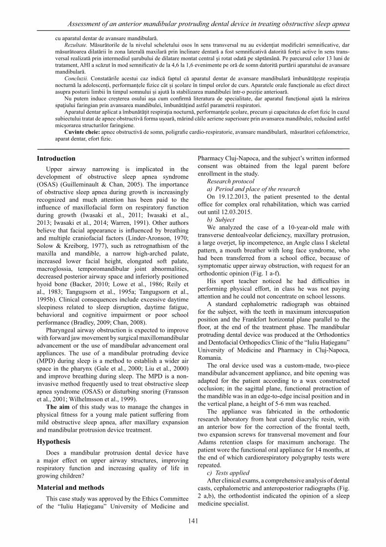
c) Tests appliedAfter clinical exams, a comprehensive analysis of dental
casts, cephalometric and anteroposterior radiographs (Fig. 2 a,b), the orthodontist indicated the opinion of a sleep medicine specialist.
cu aparatul dentar de avansare mandibulară.Rezultate. Măsurătorile de la nivelul scheletului osos în sens transversal nu au evidenţiat modificări semnificative, dar
măsurătoarea dilatării în zona laterală maxilară prin înclinare dentară a fost semnificativă datorită forței active în sens trans-versal realizată prin intermediul șurubului de dilatare montat central și rotat odată pe săptămână. Pe parcursul celor 13 luni de tratament, AHI a scăzut în mod semnificativ de la 4,6 la 1,6 evenimente pe oră de somn datorită purtării aparatului de avansare mandibulară.
Concluzii. Constatările acestui caz indică faptul că aparatul dentar de avansare mandibulară îmbunătățește respirația nocturnă la adolescenți, performanțele fizice cât și școlare în timpul orelor de curs. Aparatele orale funcționale au efect direct asupra posturii limbii în timpul somnului și ajută la stabilizarea mandibulei într-o poziție anterioară.
Nu putem induce creşterea osului aşa cum confirmă literatura de specialitate, dar aparatul funcţional ajută la mărirea spaţiului faringian prin avansarea mandibulei, îmbunătăţind astfel parametrii respiratori.
Aparatul dentar aplicat a îmbunătăţit respiraţia nocturnă, performanţele şcolare, precum şi capacitatea de efort fizic în cazul subiectului tratat de apnee obstructivă forma ușoară, mărind căile aeriene superioare prin avansarea mandibulei, reducând astfel micșorarea structurilor faringiene.
Cuvinte cheie: apnee obstructivă de somn, poligrafie cardio-respiratorie, avansare mandibulară, măsurători cefalometrice, aparat dentar, efort fizic.

142
Ovidiu Dănuţ Rădescu et al.
a bFig. 2 (a, b) – Pre-treatment anteroposterior and lateral cephalometric head film provides information for diagnostic records and planning of orthodontic treatment.
In the Sleep Laboratory of the “Iuliu Hatieganu” University of Medicine and Pharmacy Cluj-Napoca, the standard protocol for clinical assessment was performed and subsequently, the 9-year-old boy underwent overnight cardiorespiratory polygraphy tests. A first time was registered as T1 – before and a second time T2 – after mandibular protrusion device treatment.
The Stardust Sleep Recorder manufactured by Philips/Respironis (Andover, United States) was the diagnostic sleep device which included recording of nasal flow, thoracoabdominal movements, pulse oximetry and snoring. The entire record was manually scored for cardiorespiratory events. Total sleep time, the number and duration of complete upper airway obstructions, partial upper airway obstructions, and central and mixed apneas were scored. The apnea-hypopnea index (AHI) was
calculated as the number of apneas and hypopneas per hour of sleep. According to the AHI, the severity of sleep apnea is as follows: no OSAS (AHI<5 events/hour), mild OSAS (5≤AHI<15 events/hour), moderate OSAS (15≤AHI<30 events/hour), severe OSAS (AHI≥30 events/hour).
The cephalometric landmarks and analysis depend on the methods of Tweed, Steiner and Munster. All cephalometric landmarks were located and digitized by the same observer (DOR). The digital cephalometric imaging system was PaX-Reve3D from Vatech 3D Global, ranked No. 5 (Russelsheim, Germany).
ResultsSkeletal relationsTransverse skeletal measurements were not significantly
affected.Our device moved the mandible forward and had a
restraining effect on the maxilla, as seen in Table I. The mandibular position in relation to the skull base, the SNB angle, was reduced on average by 1.35° and the SNA angle decreased by -1.55°, moving the maxilla back.
Dental relationsInclination of the lower incisors according to the
reference line ML mandibular plane angle increased from 86.56° initially (ILi/ML) to 88°. The inclination of the upper incisors according to the reference line NL (ILs/NL) was affected by 3.55°, from 22.89° to 19.34°.
Only the measurement of maxillary lateral inclination was significant because of the active transverse force applied through the expansion screw once a week (Fig. 3 a-f).
a
b c d
e
f gFig. 1 (a-f) – Pre-treatment facial and intraoral photographs.

143
Assessment of an anterior mandibular protruding dental device in treating obstructive sleep apnea
a bFig. 4 (a, b) – Post-treatment anteroposterior and lateral cephalometric head film provides information about treatment outcome and maxillofacial alteration.
Table IThe results of the patient’s cardiorespiratory polygraphy variables at the initial time (T1) and the final time (T2).
B.O. Pre-treatment (T1)
Post-treatment (T2)
Age (years) 10 11Weight (kg) 33 34Height (m²) 1.47 1.48Body mass index (kg/ m²) 15.3 15.5Total sleep time (min) 546 546Total number of complete airway obstruction events 37 13Apnea/hypopnea index (events/hour) 4.6 1.6Mean oxygen saturation (%) 97 97Minimum oxygen saturation (%) 85 86
Respiratory parametersDuring the treatment period of 13 months of wearing the
mandibular protruding device, AHI decreased significantly
from 4.6 to 1.6 events per hour of sleep. The initial pre-treatment rate of apneas-hypopneas was 37 events/hour of sleep and decreased to 13 events during the 546 minutes of sleep time when the appliance was worn. An improvement in the mean and minimum oxygen saturation after mandibular protruding device treatment T2 was observed compared with T1 (Table I).
DiscussionSome studies reported that the decrease of nasal
resistance values after expansion resulted in a more nasal respiratory pattern, reducing mouth breathing (Gray, 1987; White & Cole, 1989).
Obstructive apnea is the cessation of airflow in the presence of breathing effort. Central apnea represents the cessation of both airflow and breathing effort. Mixed apnea is defined as no respiratory effort for at least 10 seconds, followed by at least three unsuccessful attempts to inspire before breaking the obstruction (Bjork & Skieller, 1972).
In this clinical case, the patient changed his respiratory pattern due to wearing the protruding device, but the measurement did not verify whether the increase of the nasal cavity and active growth of the maxillary structures (Bjork & Skieller, 1972) and growth of the nose (Scott, 1953) had some influence on the width of the nasomaxillary region.
Another study “The effect of rapid maxillary expansion on nasal airway resistance” reported that most patients found that their nasal breathing was improved after expansion, and those who perceived no change were
a
b c d
e
f gFig. 3 (a-f) – Post-treatment facial and intraoral photographs.

144
Ovidiu Dănuţ Rădescu et al.
generally patients whose (nasal airway resistance) NAR was initially nearer to normal, and the change was small (Timms, 1986).
Taking into consideration the results of this study, it is suggested that in the evaluation of the relationship between transverse skeletal and dental effects after expansion, correction of lateral axial inclination was evidenced, without a significant change in skeletal parameters, as seen in Table II.
Table II Cephalometric and posteroanterior radiographic variables
before and after functional treatment indicate positive changes in maxillofacial structures.
Results Unit Minimum Maximum Before AfterFMA ° 16.00 35.00 16.06 20.75IMPA ° 84.00 92.00 119.22 107.97SNA ° 80.00 84.00 83.13 81.58SNB ° 78.00 82.00 78.35 79.70ANB ° 1.00 5.00 7.64 5.87SN-OcP ° 14.00 14.00 14.72 14.41SN-GoGn ° 30.00 30.00 24.94 24.40Max1-NA ° 22.00 22.00 25.41 18.55Mand1-NB ° 25.00 25.00 42.19 32.57Wits mm 0.00 4.00 6.61 4.88ZR - ZL mm 116 +/- 3 112 113JR – JL mm 62 +/- 3 61 62NR – NL mm 27 +/- 3 22 23R1UpMb/6A-L1UpMb/A6 mm 50 +/- 3 44 49R1LoM6/6B-L1LoM6/B6 mm 48 +/- 3 44 46
Go R/L mm 76 +/- 3 69 70
Cephalometric analysisThe measured variables defined below are represented
graphically in Table II.1. FMA: When FMA<25, it indicates a horizontal
growth pattern. When FMA>25, it indicates a vertical growth pattern.
2. Dental: Incisor – IMPA (incisor mandibular plane angle) • It indicates that the upright position of the mandibular incisor is normal • Balance and harmony of the lower facial profile • Mean: 87 degrees profile
3. Skeletal: SNA: The angle formed by the lines connecting the sella, nasion, and A point
4. Skeletal: SNB: The angle formed by the lines connecting the sella, nasion, and B point
5. Skeletal: The ANB angle indicates the magnitude of the discrepancy between the maxilla and the mandible. ANB is affected by the following factors other than anteroposterior discrepancy of jaws.
6. Skeletal: occlusal plane angle (SN - occlusal plane). The mean reading for normal occlusion is 14°.
7. Skeletal: SN-GOGN - mandibular plane angle (30). The anterior angle formed by the intersection of SN and GoGn is measured.
8. Max-NA - angle between the upper incisors to line NA
9. Mand-NB - angle between the lower incisors to line NB
10. The Wits appraisal is a measure of the extent to which the maxilla and the mandible are related to each other in the anterior-posterior (sagittal) plane.
11. ZR - ZL Zygomatic point - the most lateral aspect of the right and left zygomatic arch
12. NR – NL Nasal cavity - the most lateral aspect on the curvature of the nasal cavity
13. JR – JL Jugal point - deepest point on the curve of the molar process of the maxilla
14. R1UpMb/6A – right maxillary first molar – midpoint of the buccal surface of the maxillary first molar
15. L1UpMb/A6 – left maxillary first molar – midpoint of the buccal surface of the maxillary first molar
16. R1LoM6/6B - right mandibular first molar – midpoint of the buccal surface of the mandibular first molar
17. L1LoM6/B6 - left mandibular first molar – midpoint of the buccal surface of the mandibular first molar
18. Go R/L - Gonion – midpoint on curvature at angle of mandible, right and left
Krogman (1979) has mentioned that growth in width of both jaws, including the width of the dental arches, tends to be completed before the adolescent growth spurt and is minimally affected by adolescent growth changes.
This finding is according to the main mechanism of action with a MAS - the protrusion of the mandible and associated soft tissues (Fig. 4) improves the caliber of the upper airway (Chan et al., 2010) (Table I). It appears that the occlusal changes are not only predominantly dental as many authors have suggested. Our results align with those of Marklund (2006), who reported on the progression of occlusal changes in a subset of 51 patients treated for at least 5 years with a mono-block style of MAS. Marklund found overall that dental side effects increased with treatment time as well as more frequent use of the device; he also stated that overjet decreased continuously, but mandibular posturing and maxillary setback were also seen (Table II).
Schooling problems have been repeatedly reported in case series of children with OSAS, and in fact may underlie more extensive behavioral disturbances such as restlessness, aggressive behavior, excessive daytime sleepiness (EDS) and poor test performance. After improving the sleep quality for our subject, his school and physical performance increased, similarly to the results of the studies of some researchers (Ali & Stradling, 1996; Kheirandish-Gozal et al., 2010; Owens et al., 1998; Urschitz et al., 2004).
The body mass index predicts OSAS in older children and youth, especially in those who are overweight or obese (Daar et al., 2016), but our subject had a normal BMI and no significant weight changes were found.
Moreover, exercise could be helpful in improving numerous sequelae of OSA and vigorous physical activity was shown to be associated with a decrease in the OSA prevalence rate and subjective well-being (Ueno et al., 2009).
ConclusionsThe findings of this case indicate that an anterior
mandibular protrusion dental appliance improves nocturnal breathing and school performance in adolescents.
1. Improvement of sleep apnea may be attributed to the effect of the appliance on the oropharyngeal structures.
2. Oral appliances seem to work by enlarging upper airway patency at multiple levels and by improving muscle airway tone, and thus decreasing upper airway

145
Assessment of an anterior mandibular protruding dental device in treating obstructive sleep apnea
collapsibility. 3. We cannot induce bone growth, as confirmed by
the literature, but the myofunctional appliance opens the airway, posturing the mandible forward.
4. Functional oral appliances have a direct effect on tongue posture during sleep and help to stabilize the mandible in a more forward position.
5. Our mandibular advancement dental device changed the lower jaw position, improving respiratory functions, quality of sleep and exercise capacity in patients with mild to moderate OSAS.
Conflicts of interestsThere are no conflicts of interests.
ReferencesAli NJ, Pitson D, Stradling JR. Sleep disordered breathing: effects
of adenotonsillectomy on behaviour and psychological functioning. Eur J Pediatr, 1996;155(1):56-62.
Backer WD. Obstructive sleep apnea hypopnea syndrome. In Palange P, Simonds A. ERS Handbook respiratory medicine. First edition, 2010, 403-404.
Björk A, Skieller V. Facial development and tooth eruption. An implant study at the age of puberty. Am J Orthod. 1972; 62(4):339-383.
Bradley TD, Floras JS. Obstructive sleep apnoea and its cardiovascular consequences. Lancet. 2009;373(9657):82-93. doi: 10.1016/S0140-6736(08)61622-0.
Chan AS, Sutherland K, Schwab RJ, Zeng B, Petocz P, Lee RW, Darendeliler MA, Cistulli PA. The effect of mandibular advancement on upper airway structure in obstructive sleep apnoea. Thorax. 2010;65(8):726-732. doi: 10.1136/thx.2009.131094.
Chan AS, Lee RW, Cistulli PA. Non-positive airway pressure modalities: mandibular advancement devices/positional therapy. Proc Am Thorac Soc. 2008;5(2):179-184. doi: 10.1513/pats.200707-104MG.
Daar GKS, Gencer ZK, Ede H, Aydin R, Saydam L. The relation between childhood obesity and adenotonsillar hypertrophy. Eur Arch Oto-Rhino-Laryngology, 2016;273(2):505-509.
Fransson AM, Isacsson G, Leissner LC, Näsman AB, Alton MK. Treatment of snoring and obstructive sleep apnea with a mandibular protruding device: an open-label study. Sleep Breath. 2001;5(1):23-33.
Gale DJ, Sawyer RH Woodcock A, Stone P, Thompson R, O’Brien K. Do oral appliances enlarge the airway in patients with obstructive sleep apnoea? A prospective computerized tomographic study. Eur J Orthod. 2000 Apr;22(2):159-168.
Gray LP. Rapid maxillary expansion and impaired nasal respiration. Ear, Nose & Throat J, 1987;66(6):248-251.
Guilleminault C, Lee JH, Chan A. Pediatric obstructive sleep apnea syndrome. Arch Pediatric Adolesc Med, 2005;159(8):775-785.
Iwasaki T, Saitoh I, Takemoto Y, Inada E, Kanomi R, Hayasaki H et al. Evaluation of upper airway obstructionin Class II children with fluid-mechanical simulation. Am J Orthod Dentofacial Orthop. 2011 Feb;139(2):e135-145. doi: 10.1016/j.ajodo.2010.08.014.
Iwasaki T, Saitoh I, Takemoto Y, Inada E, Kakuno E, Kanomi R, Hayasaki H, Yamasaki Y. Tongue posture improvement and pharyngeal airway enlargement as secondary effects of rapid maxillary expansion: a cone-beam computed tomography study. Am J Orthod Dentofacial Orthop. 2013;143(2):235-245. doi: 10.1016/j.ajodo.2012.09.014.
Iwasaki T, Takemoto Y, Inada E, Sato H, Suga H, Saitoh I, Kakuno E, Kanomi R, Yamasaki Y. The effect of rapid maxillary expansion on pharyngeal airway pressure during inspiration
evaluated using computational fluid dynamics. Int J Peadiatr Otorhinolaryngol. 2014;78(8):1258-1264. doi: 10.1016/j.ijporl.2014.05.004.
Kheirandish-Gozal L, De Jong MR, Spruyt K, Chamuleau SA, Gozal D. Obstructive sleep apnoea is associated with impaired pictorial memory task acquisition and retention in children. Eur Respir J. 2010;36(1):164-169. doi: 10.1183/09031936.00114209.
Krogman WM. Craniofacial growth, prenatal and postnatal. In:Cooper HK, Harding RL, Krogman WM, Mazaheri M, Millard RT, eds. Cleft palate and cleft-lip: a team approach to clinical management and rehabilitation. Philadelphia, Pa:WB Saunders; 1979, 22-107.
Liu Y, Park YC, Lowe AA, Fleetham JA. Supine cephalometric analyses of an adjustable oral appliance used in the treatment of obstructive sleep apnea. Sleep Breath. 2000;4(2):59-66.
Linder-Aronson S. Adenoids. Theit effect on modeof breathing and nasal airflow and their relationship to characteristics of the facial skeleton and the dentition. A biometric, rhino-manometric and cephalometro-radiographic study on children with and without adenoids. Acta Otolanryngol Suppl, 1970;265:1-132.
Lowe AA, Santamaria JD, Fleetham JA, Price C. Facial morphology and obstructive sleep apnea. Am J Orthod Dentofacial Orthop 1986. 90(6):484-491.
Marklund M. Predictors of long-term orthodontic side effects from mandibular advancement devices in patients with snoring and obstructive sleep apnea. Am J Orthod Dentofacial Orthop. 2006;129(2):214-21.
Owens J, Opipari L, Nobile C, Spirito A. Sleep and daytime behavior in children with obstructive sleep apnea and behavioral sleep disorders. Pediatrics. 1998;102(5):1178-1184.
Riley R, Guilleminault C, Herran J, Powell N. Cephalometric analyses and flow-volume loops in obstructive sleep apnea patients. Sleep. 1983;6(4):303-311.
Scott JH. The cartilage of the nasal septum. Br Dent J, 1953;95:37-43.
Solow B, Kreiborg S. Soft tissue stretching: a possible control factor in craniofacial morphogenesis. Scand J Dent Res, 1977;85:505-507.
Tangugsorn V, Skatvedt O, Krogstad O, Lyberg T. Obstructive sleep apnoea: a cephalometric study. Part I. Cervico-craniofacial skeletal morphology. Eur J Orthod. 1995a;17(1):45-56.
Tangugsorn V, Skatvedt O, Krogstad O, Lyberg T. Obstructive sleep apnoea: a cephalometric study. Part II. Uvulo-glossopharyngeal morphology. Eur J Orthod. 1995b;17(1):57-67.
Timms DJ. The effect of rapid maxillary expansion on nasal airway resistance. Br J Orthod. 1986;13(4):221-228.
Urschitz MS, Eitner S, Guenther A, Eggebrecht E, Wolff J, Urschitz-Duprat PM, Schlaud M, Poets CF. Habitual snoring, intermittent hypoxia, and impaired behavior in primary school children. Pediatrics 2004;114(4):1041-1048.
Ueno LM, Drager LF, Rodrigues AC, Rondon MU, Braga AM, Mathias W Jr, Krieger EM, Barretto AC, Middlekauff HR, Lorenzi-Filho G, Negrão CE. Effects of exercise training in patients with chronic heart failure and sleep apnea. Sleep. 2009;32(5):637-647.
Warren D, Spalding PM. Dentofacial morphology and breathing:a century of controversy. In:Melsen B. Current Controversies in Orthodontics. 1st ed. Chicago: Quintessence, 1991, 45-76.
Wilhelmsson B, Tegelberg A, Walker-Engström ML, Ringqvist M, Andersson L, Krekmanov L, Ringqvist I. A prospective randomized study of a dental appliance compared with uvulopalatopharyngoplasty in the treatment of obstructive sleep apnoea. Acta Otolaryngol. 1999;119(4):503-509.
White BC, Woodside DG, Cole P. The effect of rapid maxillary expansion on nasal airway resistance. J Otolaryngol. 1989; 18(4):137-143.