bafilomycin ai prevents maturation of autophagic vacuoles
TRANSCRIPT

CELL STRUCTURE AND FUNCTION 23: 33-42 (1998)
© 1998 by Japan Society for Cell Biology
Bafilomycin Ai Prevents Maturation of Autophagic Vacuoles by Inhibiting Fusionbetween Autophagosomesand Lysosomes in Rat HepatomaCell Line,H-4-II-E Cells
Akitsugu Yamamoto1, Yoshihiro Tagawa1, Tamotsu Yoshimori2, Yoshinori Moriyama3, RyuichiMasaki1, and Yutaka Tashiro1department of Physiology and Liver Research Center (Cell Biology), Kansai Medical University, Moriguchi,Osaka 570-0074, and ^Department of Cell Biology, National Institute for Basic Biology\ Okazaki 444-0867, and^Department of Molecular Cell Biology, Institute of Scientific and Industrial Research (ISIR), Osaka Universi-ty, Ibaraki, Osaka 567-0047, Japan
Key words: bafilomycin Ai/V-ATPase/autophagy/autophagosomes/rathepatomacells/H-4-II-E cells
ABSTRACT.Westudied the effects of bafilomycin Ai, a potent and specific inhibitor of vacuolar H+ ATPase(V-ATPase), on the process of autophagy in rat hepatoma cell line, H-4-II-E cells. To induce autophagy, cells
were transferred from Dulbecco's modified Eagle medium containing 12% fetal calf serum into Hanks' bal-anced salt solution. When bafilomycin Ai was added to Hanks' balanced salt solution, endogenous protein deg-radation was strongly inhibited and numerous autophagosomes accumulated in H-4-II-E cells, whereas autolyso-somes decreased in number. Acid phosphatase activity was not detected in the autophagosomes which accumu-lated in the presence of bafilomycin Ai, suggesting that fusion between autophagosomes and lysosomes was dis-turbed by this drug. Inhibition of the fusion was reversible, and the autophagosomes changed into autolyso-somes after the removal of the inhibitor. Bafilomycin Ai also prevented the appearance of endocytosed HRPin
autophagic vacuoles. These results suggested that acidification of the lumenal space of autophagosomesor lyso-somes by V-ATPase is important for the fusion between autophagosomes and lysosomes.
Autophagy is one of the main pathways of the degrada-tion of the endogenous proteins and organella (4, ll,32). In the process of autophagy, membrane structurescalled isolation membranesor phagophores appear, seg-regate parts of the cytoplasm, and form autophago-somes. The newly formed autophagosomes (early auto-phagosomes) fuse with endosomes or prelysosomes,and becomea more advanced stage of autophagosomes(late autophagosomes or amphisomes) of acidic luminalpH (2, 1 1, 12). The autophagosomes then acquire hydro-lytic enzymes by fusion with lysosomes, and are trans-formed into mature autolysosomes in which degrada-tion of the content proceeds (10, ll, 40).Vacuolar H+ ATPase (V-ATPase) is localized in or-ganelles of the central vacuolar system such as coated
vesicles, endosomes, lysosomes, chromaffin granules,
and Golgi apparatus, and plays an important role bymaintaining the acidic environment in these compart-ments (for review, see Mellman et al). Bafilomycin Ai,a macrolide antibiotic isolated from Streptomyces sp.,is a highly specific inhibitor of the V-ATPase (5, 14,38). Inhibition by bafilomycin Ai occurs through bind-ing to the membrane-spanning pore forming domain ofthe enzyme (7, 42). Bafilomycin Ai was also effective onliving cells when added extracellularly (36, 41). There-fore it is a powerful tool to study the role of V-ATPaseand vacuolar pH. Using this drug, we have previouslyreported that V-ATPase is essential for acidifying thelumen of lysosomes and subsequent protein degrada-tion of endocytosed epidermal growth factor (EGF) inlysosomes of cultured cells (41) and phagocytosed rodouter segments in phagolysosomesof retinal pigmentep-ithelial cells (9).
In this study, to clarify the roles of V-ATPasein auto-phagy, we studied the effects of bafilomycin Ax on theprocess of autophagy in rat hepatoma cell line, H-4-II-E cells induced by deprivation of serum and aminoacids. Treatment of the cells with bafilomycin Ai causedaccumulation of numerous autophagosomes in the
Abbreviations: V-ATPase, vacuolar H+ ATPase; DMEM,Dulbecco's modified Eagle medium; HBSS, Hanks' balanced saltsolution; FCS, fetal calf serum; DMSO, dimethyl sulfoxide; HRP,
horse radish peroxidase.Corresponding author: Akitsugu Yamamoto,Department of Phys-iology and Liver Research Center (Cell Biology), Kansai Medical Uni-versity, Fumizono-cho 10-15, Moriguchi, Osaka 570, Japan.
Tel: +81-6-992-1001 (ext. 2435), Fax: +81-6-993-5319E-mail: [email protected]
33

A. Yamamotoet al.
cells. These results suggested that acidification of the lu-menal space of autophagosomes or lysosomes by V-ATPaseis important for the fusion between autophago-somes and lysosomes and subsequent degradation ofthe sequestered cytoplasm in the autolysosomes.
MATERIALS AND METHOD
Material sBafilomycin Ai was kindly provided by Prof. K. AltendorfUniversitat Osnabriick, Germany). Bafilomycin Ai was dis-solved in dimethyl sulfoxide (DMSO) and stored at - 80°C. 3-methyl adenine was purchased from Nacalai Tesque Inc,Kyoto, Japan.
Cell cultureA cell line, H-4-II-E-C3 cells (ATCC No. CRL-1600) derivedfrom rat hepatoma H35 cells, was used throughout this study.H-4-II-E cells were cultured in Dulbecco's modified Eagle me-dium (DMEM) containing 12% fetal calf serum (FCS). To in-duce autophagy, cells were incubated in Hanks' balanced saltsolution (HBSS) containing 40 mMHepes-NaOH buffer pH7.4. For bafilomycin Ai treatment experiments, 1/100 volume
of bafilomycin Ai dissolved in (DMSO)was added to the medi-um. For control experiments, 1%DMSOwas added to the me-dium.
Measurement of endogenous protein degradationProtein degradation was measured by the method of Ballardet al. (3) with some modifications. H-4-II-E cells were incu-bated with DMEMcontaining 12% FCS and 1.25 ptC\/m\ of14C-leucine for 24 hours at 37°C. After washing with HBSScontaining 0.1% bovine serum albumin (BSA) and 40 mMHepes-NaOHbuffer at pH 7.4 three times, cells were incu-bated with the same mediumin the presence of 100 nMbafilo-mycin Ai, 10mMNH4C1,or \% DMSOas control. The re-lease of degradation products during incubation was meas-ured by collecting 100 (A of the medium. Proteins were precipi-tated by 10% (final concentration) TCA, and the supernatantwas assayed for radioactivity by liquid scintillation counterafter centrifugation. To assess total cell radioactivity, cellswere lysed by adding 0.1 MNaOHat the end of the incuba-tion (2 hours), and the radioactivity was measured.
Fluorescence microscopy of vitally stained cellsCells were incubated for 1 hour at 37°C with DMEMcontain-
Fig. 1. Electron micrographs of H-4-II-E cells before and after the induction of autophagy. A. A cell cultured in DMEMcontaining 12%FCS.Small lysosomes (L) scattered in the cytoplasm. B. A cell transferred into HBSS and incubated for 1 hour. Autophagosomes (AP) and autolyso-somes (AL) appeared. Bars; 1 /am.
34

Effects of Bafilomycin Ax on Autophagy
ing 12% FCS or HBSS in the absence (only \% DMSOwasadded at final concentration) or presence of bafilomycin Ai,and vitally stained with 5 /^g/ml acridine orange as describedpreviously (20, 41).
Conventional electron microscopyH-4-II-E cells in culture dish were fixed with 2% glutaralde-hyde in 0.1 M cacodylate buffer at pH 7.4 for 30 min., washedthree times in the same buffer, and post-fixed in \% OSO4 in
the same buffer for 1 hour. Cells were dehydrated with a seriesof ethanol and embeddedin epon with the plastic culture dish.After the resin hardened, the plastic dish was removed fromthe epon block. Ultra-thin sections were cut horizontally tothe bottom of the dish, and picked up on collodion-carboncoated grids, doubly stained with uranyl acetate and lead cit-rate, and observed under a Hitachi H-7000 electron micro-scope. The area of autophagosomesand autolysosomes wasmeasured on the electron micrographs by a digitizer model SD
Fig. 2. Vital staining of acidic compartments with acridine orange. H-4-II-E cells were cultured in DMEMcontaining 12% FCS (A, C) or inHBSS (B, D) in the absence (only 1% DMSO)(A, B) or presence of 100 nM bafilomycin A{ (C, D), and vitally stained with 5 ftg/ml acridineorange. A. Many fine granules appeared in cells cultured in DMEM.B. After being transferred into HBSSfor 1 hour, stained granules increasedin size. C, D. In both culture conditions, 100 nM bafilomycin Ai completely diminished the staining. Bars; 20 fjtm.
35

A. Yamamotoet al.
331 (Wacom, Saitama, Japan) connected to a PC-9801 CV mi-crocomputer (NEC, Tokyo, Japan).
Cytochemical detection of acid phosphatase at electron micro-scopic levelH-4-II-E cells were fixed in 2% glutaraldehyde in 0. 1 Mcacod-ylate buffer at pH 7.4 for 30min., and were washed threetimes in the same buffer. Acid phosphatase was detected using/3-glycerophosphate as a substrate, and cerium as a capture re-agent according to Robinson and Karnovsky (28). Cells werepost-fixed in 1% OsO4, and embedded in epon as describedabove.
Endocytosis of horse radish peroxidaseH-4-II-E cells were incubated with 10 mg/ml horse radish per-oxidase (HRP) (Type II, Sigma, St. Louis, MO), dissolved inHBSSat 37°C for 1 hour. To visualize internalized HRP, cellswere fixed with 2% glutaraldehyde in 0. 1 Mcacodylate bufferat pH 7.4 for 30 min., washed three times in the same buffer,and were incubated in diaminobenzidine and H2O2as de-scribed by Marsh et al. (21). Cells were post-fixed in 1% OsO4,dehydrate, and embedded in epon.
RESULTS
Induction of autophagy in H-4-II-E cells after depriva-tion of amino acid and serumWhen H-4-II-E cells were cultured in DMEMcontain-ing 12% FCS, small lysosomes dispersed throughoutthe cytoplasm. Autophagosomes and autolysosomeswere scarcely found in the cytoplasm (Fig. 1A). Toinduce autophagy, cells were transferred into HBSSwhich contained neither amino acids nor FCS. WhenH-4-II-E cells were transferred into HBSSand incu-bated for 1 hour, autophagosomes which enveloped theapparently intact cytoplasm, and autolysosomes whichcontained degraded material appeared in the cytoplasm(Fig. IB). Using this culture cell system, we examinedthe effects of bafilomycin Ai on autophagy.
Inhibition of acidification of intracellular organdies bybafilomycin AtAcridine orange accumulates in acidic compartmentsand vitally stains them. Figs. 2 show acridine orangestaining of H-4-II-E cells cultured in DMEMand inHBSS. Many fine granules existed in H-4-II-E cells cul-tured in DMEM(Fig. 2A). They may correspond to ly-sosomes. After being transferred into HBSS for 1 hour,granules stained with acridine orange increased in size,corresponding to the appearance of autolysosomes(Fig. 2B). In both culture conditions, 100 nM bafilomy-cin A! completely diminished the staining, suggestingthat it effectively inhibits acidification of lysosomes andautolysosomes (Figs. 2C and 2D).
Inhibition of endogenous protein degradation by bafilo-mycin AiNext, we tested whether bafilomycin Ai inhibits endoge-nous protein degradation in H-4-II-E cells or not. H-4-II-E cells were labeled by 14C-leucine for 24 hours. Theywere then transferred into HBSS, and the TCAsolublecount released into the mediumwas measured. Asshown in Fig. 3, bafilomycin Ax at the concentration of100 nM nearly completely inhibited endogenous proteindegradation 30 min after its addition. 100 nM Bafilomy-cin Ax inhibited the protein degradation more effective-ly than 10 mMNH4C1which is known to inhibit proteindegradation by neutralizing acidity of the lysosomalcompartment (29, 30).
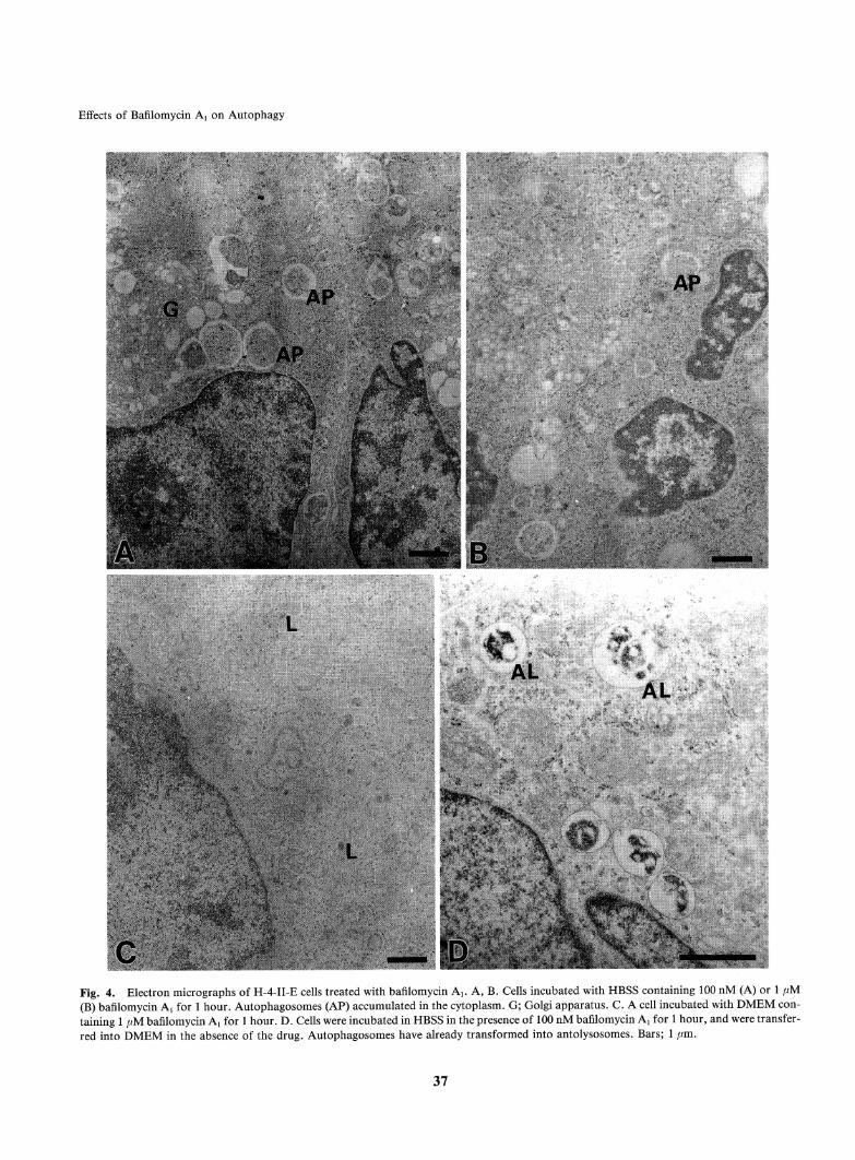
Ultrastructure of H-4-II-E cells treated with bafilomy-cinA!When H-4-II-E cells were incubated in HBSSin the pre-sence of 100 nM (Fig. 4A) or 1 fiM (Fig. 4B) bafilomy-cin Ai for 1 hour, numerous autophagosomes appearedin the cytoplasm, but few autolysosomes were seen.Fewchanges took place in other organelles except forthe swelling of Golgi cisterna especially in trans-Golgi
©
13o+*
o
1
Incubation time (h)Fig. 3. Inhibition of endogenous protein degradation by bafilomy-cin Ai. H-4-II-E cells were labelled in DMEMcontaining 12% FCSand 1.25 /iCi/ml of 14C-leucine for 24 hour. Cells were then incubatedwith HBSS containing 0.1% BSA and 40 mMHepes-NaOH at pH 7.4in the presence of 100nM bafilomycin A! (#), 10mMNH4C1(A)or only \% DMSOas control (O). Release of degradation productsduring incubation was measured. Proteins were precipitated by 10%TCA, and TCAsoluble radioactivity was measured by liquid scintilla-tion counter. The vertical axis shows the percentage of the total cellcount. Meansof two experiments are shown.
36
Effects of Bafilomycin Ax on Autophagy
Fig. 4. Electron micrographs of H-4-II-E cells treated with bafilomycin Ax. A, B. Cells incubated with HBSScontaining 100 nM (A) or 1 pM(B) bafilomycin Ax for 1 hour. Autophagosomes (AP) accumulated in the cytoplasm. G; Golgi apparatus. C. A cell incubated with DMEMcon-taining 1 fM bafilomycin A{ for 1 hour. D. Cells were incubated in HBSSin the presence of 100 nM bafilomycin Ai for 1 hour, and were transfer-red into DMEMin the absence of the drug. Autophagosomes have already transformed into antolysosomes. Bars; 1 /mi.
37

A. Yamamotoet al.
region as observed in GH3cells treated with bafilomycinAi (15). In DMEM,however, the addition of bafilomy-cin Ax did not cause any morphological changes in H-4-II-E cells (Fig. 4C). 3-methyl adenine is well known as aspecific inhibitor of autophagy (31). Addition of 10 mM3-methyl adenine to HBSSinhibited the appearance ofautophagosomes and autolysosomes almost completelyboth in the presence and in the absence of 1 /jM bafilo-mycin Ax (data not shown). These results suggested thatthe accumulation of autophagosomes by bafilomycinAx treatment results from the inhibition of maturationof autophagosomesto autolysosomes rather than the ac-celeration of the formation of autophagosomes.To determine whether the effect of bafilomycin Ax isreversible or not, we incubated H-4-II-E cells in HBSSin the presence of 100nM bafilomycin Ax for 1 hour,and then transferred the cells into DMEMin the ab-sence of the drug. In DMEM,formation of new phago-somes is strongly suppressed as described above. As a re-sult, the autophagosomes which accumulated in H-4-II-E cells by bafilomycin Ax treatment transformed into au-tolysosomes, showing that the effect of bafilomycin Ax
yà"? y' y3 " /
"a / "a /2 / S /w / w /o ^» o /
^ I/ >^^^^\ 2 /4> / / \. « /
Sk / / >o Oh /
o/ 1 1 oi-à" à" i
0 1 2 0 1 2
Incubation time (h) Incubation time (h)
Fig. 5. Kinetics of the appearance of autophagosomesand autolyso-somes. H-4-II-E cells were transferred from DMEMcontaining 12%FCS into HBSSin the absence (Left, only \% DMSOwas added) orin the presence of 100 nMbafilomycin A{ (Right). The area of profilesof autophagosomes (O) or autolysosomes (#) was measured on elec-tron micrographs.
Fig. 6. Cytochemical demonstration of acid phosphatase activity. A. A H-4-II-E cell incubated with HBSScontaining \% DMSOfor 1 hour.Acid phosphatase negative autophagosomes (AP) and acid phosphatase positive autolysosomes (AL) are seen. B. A H-4-II-E cell incubated for 1hour in HBSScontaining 1 jjM bafilomycin A! for 1 hour. Acid phosphatase activity was not detected in the autophagosomes (AP) which accu-mulated in the cytoplasm. Bars; 1 //m.
38

Effects of Bafilomycin A{ on Autophagy
is reversible (Fig. 4D).
Kinetics of the appearance of autophagosomesand au-tolysosomes in bafilomycin A! treated and untreatedcellsFig. 5 shows the kinetics of the appearance of autophag-osomes and autolysosomes in H-4-II-E cells after serumand amino acids deprivation. For this purpose, the areaof autophagosomesor autolysosomes was measured onthe electron micrographs. In the absence of bafilomycinAi (Fig. 5, Left), autophagosomes appeared first aftertransfer into HBSS, and reached plateau at 30 min. Onthe other hand, autolysosomes increased linearly, andoccupied about 6% of the cytoplasmic area after 2hours of incubation. In the presence of 100 nMbafilo-mycin Ai (Fig. 5, Right), autophagosomes increased lin-early, and occupied about 1% of the cytoplasm after2 hours of incubation, whereas autolysosomes scarcelyappeared within 1 hour of incubation. These resultsshowedthat the conversion of autophagosomes to auto-lysosome was markedly suppressed in the presence ofbafilomycin Ax.
Cytochemical and histochemical characterization of au-tophagosomes accumulated in the presence of bafilomy-cinAxFirst of all, we examined whether autophagosomes accu-mulated by bafilomycin Artreatment lack lysosomal en-zymes or not. For this purpose, we demonstrated acidphosphatase activity at electron microscopic level. Fig.6A shows a H-4-II-E cell incubated in HBSSfor 1 hour.Acid phosphatase activity is seen in autolysosomes butnot in autophagosomes. Fig. 6B shows a H-4-II-E cellincubated in HBSS in the presence of 1 //M bafilomycinAi for 1 hour. Autophagosomes that accumulated inthe cytoplasm did not show any acid phosphatase activi-ty. These results suggest that fusion between autophago-somes and lysosomes was inhibited in the presence ofbafilomycin Ai.Convergence of autophagic and endocytic pathways
has been reported by several investigators (10, 12, 19,35). Therefore, we examined whether bafilomycin Ai in-hibits this convergence process. For this purpose, H-4-II-E cells were incubated for 1 hour in HBSScontainingHRP.HRPwas detected in autolysosomes in the ab-
Fig. 7. Localization of HRP internalized by H-4-II-E cells. A. A H-4-II-E cell incubated in HBSS containing 10 mg/ml HRP and 1% DMSOfor 1 hour. The reaction products showing the presence of HRPare seen in autolysosomes (AL), but not in autophagosomes (AP). B. A H-4-II-Ecell incubated for 1 hour in HBSS containing 10 mg/ml HRPand 1 fiM bafilomycin A{ for 1 hour. Few reaction products are seen in autophago-somes (AP). The arrow shows tubular profiles of HRPlabeled endosomes. Bars; 500 nm.
39
A. Yamamotoet al.
sence of bafilomycin Ai (Fig. 7A). However, in the pre-sence of 1 fJtM bafilomycin Als HRP was not detected inthe autophagosomes (Fig. 7B). Tubular profiles ofHRPlabeled endosomes were occasionally found in thebafilomycin Ax treated cells as reported by Clague et al.(6).
DISCUSSION
Bafilomycin Ai is a potent and specific inhibitor of V-ATPase. Unlike NH4C1 (29, 30) or monensin (34) whichcauses vacuolalization of lysosomes or Golgi apparatus,this antibiotic causes very few side effects. That is whythis drug is now widely used as a powerful tool to studythe role of V-ATPaseand acidic compartments.In this study, we studied the effects of bafilomycin Aion the process of autophagy induced in H-4-II-E cellsby serum and amino acids deprivation, and found thatbafilomycin Ai prevents maturation of autophago-
somes into autolysosomes by inhibiting fusion betweenautophagosomes and lysosomes, and inhibits endoge-nous protein degradation effectively. These results sug-gest that the acidification of the lumenal space of auto-phagosomes or lysosomes by V-ATPase may be very im-portant for the fusion between them.Interestingly, there are many reports showing that de-acidification of acidic compartments by bafilomycin Axor another V-ATPase inhibitor, concanamycin (17), in-hibits or perturbs vesicular transport of the proteins intrans-Golgi region (15, 39), to lysosomes (24), to vacu-ole in yeast (18), in recycling of transferrin receptor(25), in recycling to the trans-Golgi network from theplasma membrane(27), from early endosomes to lateendosomes (6), and from late endosomes to lysosomes(37).
Gruenberg and his colleagues investigated the inhibi-tion of protein transport from early endosomesto lateendosomes by bafilomycin Ax (1, 6). They showed that/3-COP and £-COPassociate to endosomes, and this as-sociation as well as endosomecarrier vesicles formationdepends on the lumenal acidity, and suggested the exist-ence of a trans-membrane pH sensor (1). It has beenshown that autophagosomes becomeacidic prior to theacquisition of hydrolytic enzymes (2, ll, 12). There-
fore, it is possible that fusion between autophagosomesand lysosomes is regulated by the trans-membrane pHsensor which is activated after the luminal pH of auto-phagosomes is sufficiently lowered. Recently, it was re-ported that a syntaxin homologue, Vam3pfunctions infusion of autophagosomes with the vacuole in yeastcells (8). It will be interesting to investigate whetherdocking and fusion by the SNAREproteins are regu-lated by lumenal pH in the endosome-lysosomal sys-tem.
The results obtained in this study are inconsistent
with those obtained in the case of yeast cells. In yeastcells, treatment with bafilomycin Ai (33) or the destruc-tion of V-ATPase gene (23) caused inhibition of degra-dation in the vacuoles, but did not block fusion be-tween autophagosomesand the vacuole. The differencessuggest that the fusion of autophagosomes with lyso-somes/vacuoles may be regulated by different mecha-nisms in mammaliancells and yeast cells.The results obtained in this study are also inconsis-tent with those obtained by Punnonen et al. (26). Theyreported that the inhibition of acidification by monen-sin did not prevent the delivery of cathepsin L to auto-phagosomesin rat fibroblast cells. The reason for suchdifference is unknown, though it is possible that itmight have been caused by the difference in drugs, ba-filomycin Ai vs monensin; or the difference in cells,hepatoma H-4-II-E cells vs fibroblast. H-4-II-E cellsscarcely show autophagic activity in the presence of se-rum and amino acids, probably because these cells havehigh insulin sensitivity (13, 16). Therefore this culture
cell system is suitable for studying the early processes ofautophagy such as the formation of autophagosomes oracquisition of hydrolytic enzymes after the induction ofautophagy by serum and amino acid deprivation.Convergence of autophagic and endocytic pathwayshas been reported by several groups (10, 12, 19, 35). Ba-filomycin Ai also prevented the appearance of endocy-tosed HRPin autophagic vacuoles. It remains to besolved whether this resulted from the inhibition of fu-sion between autophagosomesand endosomesor fromthe inhibition of transport of HRPin endosomes.Acknowledgments. We would like to thank to Dr. Soltan A. Salehifor English editing. This work was supported in part by a Grant-in-Aid for Scientific Research from the Ministry of Education, Science,and Culture of Japan.
REFEREN CES
Aniento, F., Gu, F., Parton, R.G., and Gruenberg, J.1996. An endosomal ^-COP is involved in the pH-dependentformation of transport vesicles destined for late endosomes. /.Cell BioL, 133: 29-41.Aplin, A., Jasioanowski, T., Tuttle, D.L., Lenk, S.E., andDunn, W.A. 1992. Cytoskeletal elements are required for theformation and maturation of autophagic vacuoles. /. Cell Phys-
iol, 152: 458-466.
Ballard, F.J., Wong, S.S.C., Knowles, S.E., Partridge,
N.C., Martin, T.J., Wood, CM., and Gunn, J.M. 1980. In-sulin inhibition of protein degradation in cell monolayers. /.Cell Physiol. y 105: 335-346.
Blommaart, E.F.C., Luiken, J.J.F.P., and Meijer, A.J.
1997. Autophagic proteolysis: control and specificity. Histo-chem. /., 29: 365-385.
Bowman, E.M., Siebers, A., and Altendorf, K. 1988. Bafilo-mycins: A class of inhibitors of membraneATPase from micro-organisms, animal cells, and plant cells. Proc. NatI. Acad. Sci.
40

Effects of Bafilomycin A: on Autophagy
USA, 85: 7972-7976.
6. Clague, M.J., Urbe, S., Aniento, F., and Gruenberg, J.1994. Vacuolar ATPase activity is required for endosomal car-rier vesicles formation. J. BioL Chem., 269: 21-24.
7. Crider, B.P., Xie, X.-S., and Stone, D.K. 1994. Bafilomycininhibits proton flow through the H+channel of vacuolar protonpumps. /. BioL Chem., 269: 17379-17381.
8. Darsow, T., Rieder, S.E., andEmr, S.D. 1997. Amultispecifi-ty syntaxin homologue, Vam3p, essential for autophagic and
biosynthetic protein transport to the vacuole. /. Cell BioL , 138:517-529.
9. Deguchi, J., Yamamoto, A., Yoshimori, T., Sugasawa, K.,
Moriyama, Y., Futai, M., Suzuki, T., Kato, K., Uyama, M.,and Tashiro, Y. 1994. Acidification of phagosomes and degra-dation of rod outer segments in rat retinal pigment epithelium.
Invest. Ophthalomol. Vis. Sci., 35: 568-579.
10. Dunn, W.A. 1990. Studies on the mechanisms of autophagy:Maturation of the autophagic vacuole. /. CellBioL, 110: 1935-
1945.
ll. Dunn, W.A. 1994. Autophagy and related mechanisms oflyso-some-mediated protein degradation. Trends Cell BioL , 4: 139-
143.
12. Gordon, P.B. and Seglen, P.O. 1988. Prelysosomal conver-gence of autophagic and endocytic pathways. Biochem. Bio-phys. Res. CommunL, 151: 40-47.
13. Gunn, J.M., Clark, M.G., Knowles, S.E., Hopgood, M.F.,and Ballard, F.J. 1977. Reduced rates of proteolysis in trans-formed cells. Nature, 266: 58-60.
14. Hanada, H., Moriyama, Y., Maeda, M., and Futai, M.
1990. Kinetic studies of chromamn granule H+-ATPase andeffects of bafilomycin A^ Biochem. Biophys. Res. Commun. ,
170: 873-878.15. Henomatsu, N., Yoshimori, T., Yamamoto, A., Moriyama,
Y., and Tashiro, Y. 1993. Inhibition of intracellular transportof newly synthesized prolactin by bafilomycin A: in a pituitary
tumor cell line, GH3 cells. Eur. J. CellBioL, 62: 127-139.16. Iwamoto, Y., Wong, K.Y., and Gold fine, I.D. 1981. Insulin
action in cultured HTCand H35 rat hepatoma cells: Receptorbinding and hormone sensitivity. Endocrinology, 108: 44-51.
17. Kinashi, H., Someno, K., and Sakaguchi. 1984. Isolation andcharacterization of concanamycin A, B and C. /. Antibiot. , 37:
1333-1343.
18. Klionsky, D.J. and Emr, S.D. 1989. Membrane protein sort-ing, biosynthesis, transport and processing of yeast vacuolar al-
kaline phosphatase. EMBOJ., 8: 2241-2250.19. Liou, W., Geuze, H.J., Geelen, M.J.H., and Slot, J.W.
1997. The autophagic and endocytic pathways converge at thenascent autophagic vacuoles. J. Cell BioL , 136: 61-70.
20. Manabe, T., Yoshimori, T., Henomatsu, N., and Tashiro, Y.1993. Inhibitors of vacuolar-type H+-ATPase suppress prolif-eration of cultured cells. /. Cell PhysioL, 157: 445-452.
21. Marsh, M., Griffiths, G., Dean, G.E., Mellman, I., and
Helenius, A. 1986. Three-dimensional structure of the endo-somes in BHK-21 cells. Proc. Natl. Acad. Sci. USA, 83: 2899-
2903.
22. Mellman, I., Fuchs, R., and Helenius, A. 1986. Acidificationof the endocytic and exocytic pathways. Ann. Rev. Biochem.,
55: 663-700.
23. Nakamura, N., Matsuura, A., Wada, Y., and Ohsumi, Y.1997. Acidification of vacuoles is required for autophagic deg-
radation in the yeast, Saccharomyces cerevisiae. /. Biochem.,121: 338-344.
24. Oda, K., Nishimura, Y., Ikehara, Y., andKato, K. 1991. Ba-
filomycin Ai inhibits the targeting of lysosomal acid hydrolasesin cultured hepatocytes. Biochem. Biophys. Res. Commun.,178: 369-377.
Presley, J.F., Mayor, S., McGraw, T.E., Dunn, K.W., andMaxfield, F.R. 1997. Bafilomycin A2 treatment retards transfer-rin receptor recycling more than bulk membranerecycling. /.
Biol. Chem., 272: 13929-13936.
Punnonen, E.-L., Autio, S., Marjomaki, V.S., and
Reunanen, H. 1992. Autophagy, cathepsin L, transport, andacidification in cultured rat fibroblast. /. Histochem. Cyto-chem., 40: 1579-1587.
Reaves, B. and Banting, G. 1994. Vacuolar ATPase inactiva-tion blocks recycling to the trans-Golgi network from the plas-ma membrane. FEBSLett., 345: 61-66.Robinson, J.M. and Karnovsky, M.J. 1983. Ultrastructurallocalization of several phosphatases with cerium. /. Histochem.
Cytochem., 31: 1197-1208.
Seglen, P.O. and Reith, A. 1976. Ammonia inhibition of pro-tein degradation in isolated rat hepatocytes. Quantitative ultra-structural alterations in the lysosomal system. Exp. Cell Res. ,
100: 276-280.
Seglen, P.O., Grinde, B., and Solheim, A.E. 1979. Inhibi-
tion of the lysosomal pathway of protein degradation in isolatedrat hepatocytes by ammonia, methylamine, chloroquine and leu-
peptin. Eur. J. Biochem., 95: 215-225.
Seglen, P.O. and Gordon, P.B. 1982. 3-methyl adenine: spe-cific inhibitor of autophagic/lysosomal protein degradation inisolated rat hepatocytes. Proc. Natl. Acad. Sci. USA, 79: 1889-
1892.
Seglen, P.O. and Bohley, P. 1992. Autophagy and other vacu-olar protein degradation mechanisms. Experimentia, 48: 158-
172.
Takeshige, K., Baba, M., Tuboi, S., Noda, T., and Ohsumi,Y. 1992. Autophagy in yeast demonstrated with proteinase-defi-cient mutants and conditions for its induction. /. Cell Biol.,
119: 301-311.
Tougard, C, Picart, R., Mortin, A., and Tixer-Vidal, A.1983. Effect of monensin on secretory pathway in GH3 prolac-tin cells. /. Histochem. Cytochem., 31: 745-754.
TOOZE, J., HOLLINSHEAD, M., LUDWIG, T., HOWELL, K.,
Hoflack, B., and Kern, H. 1990. In exocrine pancreas, the ba-solateral endocytosis pathway converges with the autophagic
pathway immediately after the early endosome. /. Cell Biol.,Ill: 329-345.
Umata, T., Moriyama, Y., Futai, M., and Mekada, E. 1990.The cytotoxic action of diphtheria toxin and its degradation inintact vero cells are inhibited by bafilomycin Au a specific inhibi-tor of vacuolar-type H+-ATPase. /. Biol. Chem., 265: 21940-
21945.
van Weert, A.W.M., Dunn, K.W., Geuze, H.J., Maxfield,F.R., and Stoorvogel, W. 1995. Transport from late endo-somes to lysosomes, but not sorting of integral membrane pro-
teins in endosomes, depends on vacuolar proton pump. /. CellBiol., 130: 821-834.
Werner, G., Hagenmaier, H., Drautz, H., Baumgartner,A., and Zahner, H. 1984. Metabolic products of microorgan-
isms. Bafilomycins, a new group of macrolide antibiotics. Pro-duction, isolation, chemical structure and biological activity. /.
Antibot., 37: 110-117.
Yilla, M., Tan, A., Ito, K., Miwa, K., and Ploegh, L. 1993.Involvement of the vacuolar H+-ATPasein the secretory path-way of HepG2 cells. /. Biol. Chem., 268: 19092-19100.Yokota, S., Himeno, M., and Kato, K. 1995. Formation ofau-
41

A. Yamamotoet al.
tophagosomes during degradation of excess peroxisomes in-duced by di-(2-ethylhexyl)-phthalate treatment. III. Fusion ofearly autophagosomes with lysosomal compartments. Eur. J.
Cell BioL, 66: 15-24.Yoshimori, T., Yamamoto, A., Moriyama, Y., Futai, M., andTashiro, Y. 1991. Bafilomycin Au a specific inhibitor of vacuo-lar-type H+-ATPase, inhibits acidification and protein degrada-tion in lysosomes of cultured cells. /. Biol. Chem., 266: 17707-
17712.
42. Zhang, J., Feng, Y., and Forgac, M. 1994. Proton conduc-tion and bafilomycin binding by the Vo domain of the coatedvesicle V-ATPase. /. Biol Chem., 269: 23518-23523.
(Received for publication, November 20, 1997and accepted, December 26, 1997)
42