basal gangila cahnges
TRANSCRIPT
8/6/2019 Basal Gangila Cahnges
http://slidepdf.com/reader/full/basal-gangila-cahnges 1/12
Subacute Sclerosing Panencephalitis: Evaluation with CT and MR
Jan Brismar, Generoso G. Gascon, Kristina Vult von Steyern, and Saeed Bohlega
PURPOSE: To evaluate the progression of CT and MR changes of the brain in subacute sclerosing
panencephalitis (SSPE) as a basis for assessing the effects of different types of therapy. METH-
ODS: Fifty-two patients with SSPE were examined, 44 with MR imaging and 42 with CT of the brain
on one or more occasions. A total of 92 MR and 67 CT studies were performed. RESULTS:
Correlation between the clinical status and the MR findings on admission was poor. Of 20 patients
with clinically advanced disease, only 8 had marked MR abnormalities; 6 had normal or almost
normal findings on MR examinations. Two of 4 patients with clinically mild disease had advanced
MR changes. The progression of the MR findings appeared to follow a constant pattern. The earliest
pathologic finding was focal, high-T2-intensity white matter changes; later atrophic changes
followed. The atrophy lagged behind the white matter changes and was thus mild when white
matter changes were moderate or severe. In the most advanced stage, when the patient was in a
neurovegetative state, an almost total loss of white matter had usually taken place. At this stage,
the corpus callosum was also thin. Basal ganglia changes, usually involving the putamina, were
seen in one third of patients and cortical gray matter changes were seen in one fourth of patients
examined with MR imaging. In 2 of 20 patients, MR changes regressed in parallel with clinical
improvement following therapy, but in 5 patients clinical improvement was accompanied by
progression of MR changes. CONCLUSION: The progress of MR abnormalities seen in patients with
SSPE seems to follow a constant pattern, but the severity of MR changes does not always correlate
well with the clinical findings. Caution must therefore be used when evaluating the effects of
therapy.
Index terms: Brain, computed tomography; Brain, magnetic resonance; Encephalitis
AJNR Am J Neuroradiol 17:761–772, April 1996
Subacute sclerosing panencephalitis (SSPE)is an invariably fatal neurodegenerative disease,developing as a sequel to early childhood mea-sles infection. Following the original measlesinfection, the virus becomes altered and re-mains dormant intracellularly, only to manifestas SSPE a decade or so later. The disease wasoriginally described by Dawson in 1933 (1), anda major review article in 1964 (2) reported only30 patients in North America. Increased aware-
ness led to the identification of more cases, andin 1980, 575 cases were registered in the UnitedStates (3). Measles vaccination has now all buteradicated the disease in developed countries,but SSPE is still endemic in many developingcountries, where measles vaccination in earlyinfancy has not yet reached the World HealthOrganization’s goal of greater than 80% cover-age. The disease also is of interest as a modelfor persistent viral infection of the brain. Thehistopathologic manifestations in the brain areindistinguishable from those of acquired immu-nodeficiency syndrome (AIDS) and from thoseseen in parainfectious and postinfectious en-cephalomyelitis (4).
The purpose of this study of 52 patients wasto determine the time course for the develop-ment of neuroradiologic findings and to chartthe distribution of lesions in order to establish abasis for the evaluation of possible effects of different types of therapy.
Received May 23, 1995; accepted after revision October 16.
From the Departments of Radiology (J.B., K.V.v.S.), Pediatrics
(G.G.G.), and Medicine (S.B.), King Faisal Specialist Hospital and Re-
search Centre, Riyadh, Saudi Arabia.
Address reprint requests to Jan Brismar, MD, PhD, Department of
Radiology, MBC 28, King Faisal Specialist Hospital and Research Centre,
PO Box 3354, Riyadh 11211, Saudi Arabia.
AJNR 17:761–772, Apr 1996 0195-6108/96/1704–0761
American Society of Neuroradiology
761
8/6/2019 Basal Gangila Cahnges
http://slidepdf.com/reader/full/basal-gangila-cahnges 2/12
Subjects and Methods
During the last 8 years, we examined the brains of 52
patients with SSPE using magnetic resonance (MR) imag-ing and/or computed tomography (CT). Forty-two of thepatients were male and 10 were female. The findings in 21of these patients have been reported previously (5); theinterpretation of the imaging findings in these patients hassince been revised and a different scheme for clinical stag-ing has been used. The age of the patients and the durationof their symptoms at the time of the initial CT or MR studyare given in Figures 1 and 2. The age at the time of theoriginal measles infection was known in only a few pa-tients. All patients fulfilled generally accepted criteria forthe diagnosis of SSPE (6) (ie, elevated serum and cere-
brospinal fluid [CSF] antimeasles antibody titers and two of
the following four criteria: typical clinical presentation,
typical electroencephalographic [EEG] pattern, IgG in CSF
greater than 20% of total protein, and typical findings on
brain biopsy specimens). No brain biopsy was performed
in this series. Information on the clinical stage of the dis-
ease (as defined in the Table) at presentation was avail-
able in 49 patients and appears in Figure 2. In the “Dis-
cussion,” the clinical stages suggested by Jabbour et al (6)
will be denoted as J:I to J:IV; the stages identified in our
series (7) are designated G:I to G:IV. The relationship
between the 2 clinical staging schemes is presented in the
Table.
For the CT studies, 10-mm-thick contiguous sections
were obtained. For contrast enhancement, iopromide, 2
mL/kg of body weight, was used. The MR studies were
performed on a 1.5-T scanner using T1-weighted, 600–700/20/2 (repetition time/echo time/excitations), and du-
al-echo T2-weighted, 1800 –2000/30– 40,80/1–2, axial
sections with 0- to 2.5-mm section gaps (depending on
head size) and sagittal T1-weighted images. Gado-
pentetate dimeglumine, 0.2 mL/kg of body weight, was
given in one patient. The CT and MR studies were evalu-
ated retrospectively for enlargement of the CSF spaces
and for white matter changes, and graded subjectively in a
nonblinded manner as absent (0), mild (), moderate
(), or marked/severe ().
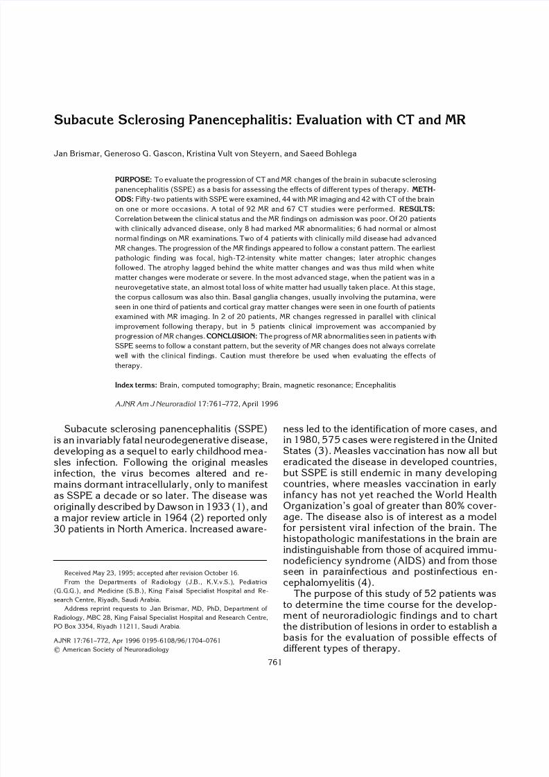
Fig 2. Clinical stage and duration of signs and symptoms at
initial CT or MR study in 49 patients with SSPE (information was
not available in 3 of a total of 52 patients). Note logarithmic time
scale.
Fig 1. Sex and age of52 patientswith SSPEat initial CTor MR
study.
Two schemes for the clinical staging of subacute sclerosing panen-
cephalitis (SSPE)
SSPE stages according to Jabbour
J:I. Cerebral signs (mental, behavioral)
J:II. Convulsive motor signs, myoclonus, incoordination,
choreoathetosis, tremorsJ:III. Coma, opistotonus, decerebrate rigidity; no responsiveness
to any stimulus
J:IV. Mutism, loss of cerebral cortex function, less frequent
myoclonus, diminished hypertonia
SSPE stages according to Gascon
G:IA. Behavioral, cognitive, and personality changes, walking
G:IB. Aperiodic, myoclonic spasms
G:IIA. Further mental deterioriation, periodic generalized
myoclonic spasms, possibly no walking because of drop spells
G:IIB. Language difficulties, spasticity, ataxia, walking with
assistance
G:IIIA. Speaking less, visual difficulties; sitting up independently,
possible standing, but no independent ambulation; frequent
myoclonic spasms, possible seizures
G:IIIB. No speech, poor comprehension, possible blindness,
confinement to bed, dysphagia, possible need of tubal feeding,
possible choreoathetosis
G:IV. Neurovegetative stage, no spasms, very low background
EEG activity
Note.—Gascon stage IA and IB are both included in Jabbour stage
I; Gascon stage IIA, IIB, and IIIA are all included in Jabbour stage II;
Gascon stage IIIB approximately equals Jabbour stage III; and Gascon
stage IV approximately equals Jabbour stage IV.
762 BRISMAR AJNR: 17, April 1996
8/6/2019 Basal Gangila Cahnges
http://slidepdf.com/reader/full/basal-gangila-cahnges 3/12
In total, 92 MR examinations were performed in 44patients and 67 CT examinations were performed in 42patients. Contrast medium was used in 41 CT examina-tions (27 patients) and in one MR study. In 25 patients, CTand MR studies were performed less than a week apart.
Results
The radiologic findings in our series includedatrophy, white matter changes (ie, lesions of high T2 intensity and low CT attentuation), graymatter changes (high T2 and decreased T1 in-tensity), and basal ganglia involvement. Radio-logic staging of SSPE was as follows:
stage 0: no atrophy or white matterchanges
stage 1: () white matter changes or atro-
phystage 2: () white matter changes and at-
rophystage 3: () white matter changes, 0 to
atrophy, or vice versastage 4: () white matter changes and
atrophystage 5: () white matter changes, 0 to
atrophy, or vice versastage 6: () white matter changes and
atrophy
The results from radiologic staging using CTinformation as compared with MR informationwas evaluated in 25 patients who were exam-ined with both techniques within 1 week. Asexpected, a marked understaging was achievedwhen CT examinations were used to stage thedisease, mainly because white matter changesare difficult to see on CT scans. Thus, the ra-diologic staging was thereafter based on MRresults alone (ie, MR stage). Enhancement fol-lowing administration of contrast medium wasnever observed.
The severity of atrophy was correlated with
the severity of white matter changes—bothgraded as 0, , , or —as determinedfrom all 92 MR studies. In 49 studies the atrophyand white matter changes were graded as beingof equal severity. In only 3 studies (2 patients)did the atrophy seem slightly more pronouncedthan the white matter changes, whereas in 30studies the white matter changes appearedworse than the atrophy. In 9 studies withmarked white matter changes no atrophy wasseen.
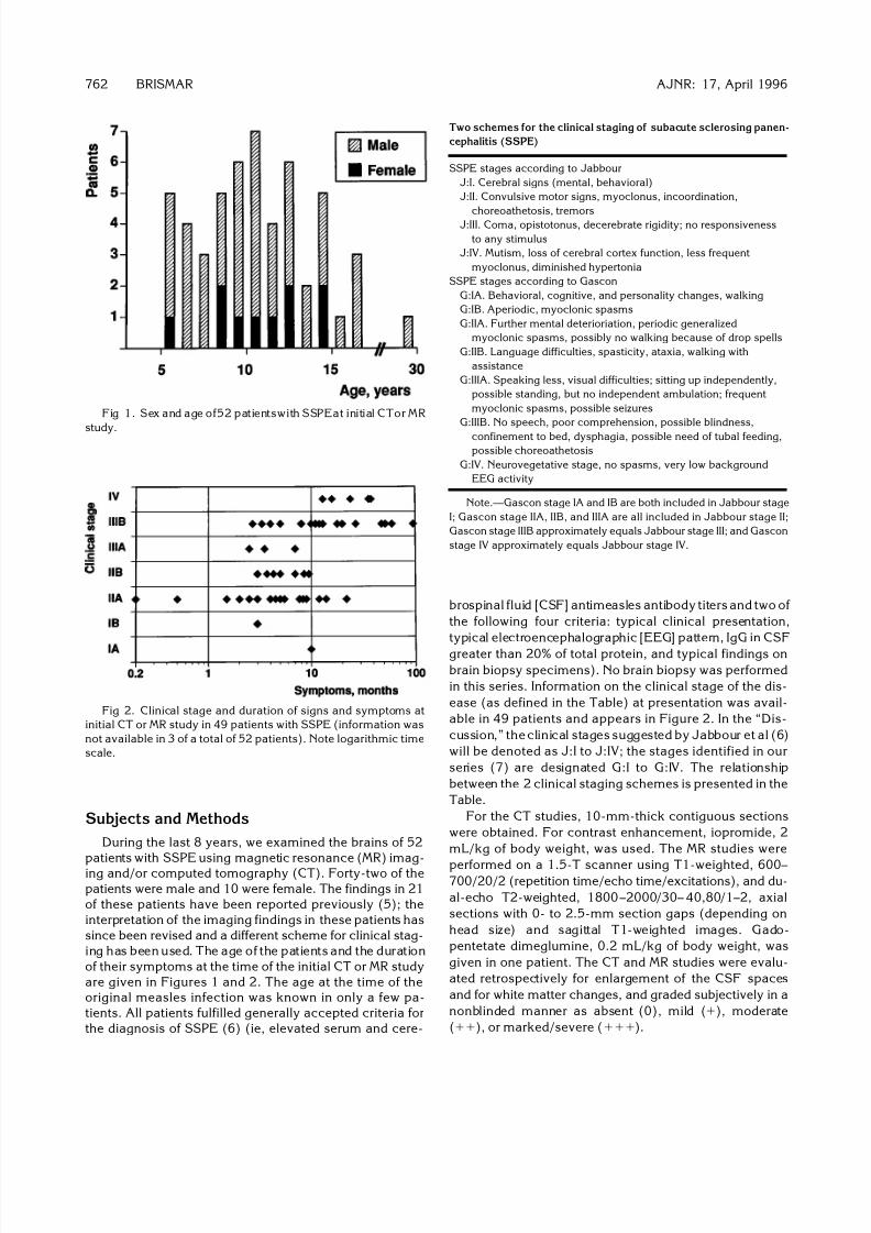
Figure 3 plots the correlation between theinitial MR stage and the duration of the symp-toms. Although, as expected, there was a cor-relation between the duration of the disease andthe MR stage, large variations did appear. Thus,one patient with only a 1-week history of symp-toms already had moderate white matter
changes, whereas another child with more than4 years’ history of symptoms still had normalMR findings.
The correlation between the clinical stageand the severity of the MR changes was alsoquite weak (Fig 4). Thus, several patients withsevere clinical findings (stages G:IIIA, G:IIIB, oreven G:IV) still had normal or almost normal MRfindings (Fig 5). On the other hand, in two pa-tients with mild clinical disease, the MR findingswere markedly abnormal (Fig 6).
Fig 4. Correlation between clinical stage and MR stage at the
first MR study in 44 patients with SSPE.
Fig 3. MR stage at the initial MR study in 44 patients with
SSPE in relation to duration of symptoms (logarithmic scale).
AJNR: 17, April 1996 PANENCEPHALITIS 763

8/6/2019 Basal Gangila Cahnges
http://slidepdf.com/reader/full/basal-gangila-cahnges 4/12
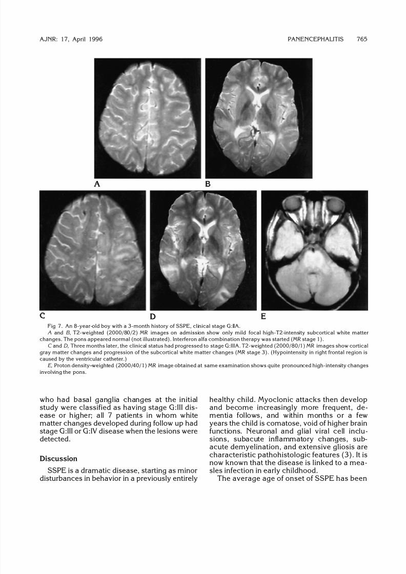
Cortical gray matter was involved in 11 of the44 patients examined by MR imaging (Figs 7–9)(involvement was also seen in one patient stud-ied only by CT). In 3 of the patients studied byMR imaging, the changes were minor; they weremoderate in 7 and severe in 1. In 1 of these
patients, the pons was also swollen and of highT2 intensity (Fig 7); in a second patient, high-T2-intensity lesions were seen in the left thala-mus (Fig 8). In an additional patient with nor-mal-appearing cortical gray matter, severehigh-T2-intensity changes were present withinthe pons, brain stem, and cerebral peduncles.The gray matter changes were present on theinitial MR study in 4 patients; in 8 patients graymatter changes developed between MR studies(Figs 7 and 9). No obvious correlation was
found between presence of gray matter changesand either the duration of the disease or theseverity of other MR changes, but gray matterwas usually affected only in patients with clini-cally advanced disease. Thus, in 3 of 4 patientswith gray matter changes initially and in 6 of 8
patients in whom gray matter lesions developedduring follow up, the disease was classified asstage G:III or G:IV.
The basal ganglia were involved in 18 pa-tients, including 15 of 44 patients who wereexamined with MR imaging (Figs 10 and 11).Although evenly distributed among the differentMR stages, and not correlated with the length of the clinical history, basal ganglia changes wereseen mainly in patients with clinical stage G:IIIAdisease or higher. Thus, all but 3 of 11 patients
Fig 6. A 14-year-old boy with a 52-
month history of SSPE, clinical stage G:IB
(3 years after this MR study the patient is
still in clinical stage G:IB). Axial T1-
weighted (600/20/2) and T2-weighted
(2000/80/2) MR images show marked at-
rophy and moderately advanced white
matter changes (MR stage 5). Corpus cal-
losum was thin (not shown).
Fig 5. A 13-year-old boy with an 18-
month history of SSPE, clinical stage G:IV.
Despite the advanced disease, T2-
weighted MR images (2000/80/2)show no
atrophy and only mild white matter
changes (MR stage 1). (Decreased signal
over right posterior and left anterior part of the brain in both images is due to coil
properties.)
764 BRISMAR AJNR: 17, April 1996
8/6/2019 Basal Gangila Cahnges
http://slidepdf.com/reader/full/basal-gangila-cahnges 5/12

8/6/2019 Basal Gangila Cahnges
http://slidepdf.com/reader/full/basal-gangila-cahnges 6/12
reported to vary from 9 to 13 years (3, 8); in ourseries it was 9 years. More than 90% of ourpatients were between 4 and 14 years old at theonset of symptoms, which is similar to otherreports (8). Information about the date of thepreceding measles infection was available foronly a few of our patients. Other investigatorshave had such information available in 65% to75% of cases (3, 8); in those series the averageage at infection was between 2 and 3 years andthe interval from the measles infection to theonset of SSPE symptoms averaged from 8 to 11years.
It is still not known how the measles virusmanages to survive clinically dormant for manyyears and why it becomes active again andcauses SSPE. Possibly the immature immunesystem fails to destroy the virus completely, and
the partially degraded virus remains in the cen-tral nervous system (CNS) (3, 9). Perhaps asimultaneous infection with another virus, suchas Epstein-Barr virus, parainfluenza type 1 vi-rus, or toxoplasmosis, might be involved inchanging the properties of the measles virus (9)into those of a slow virus. Virus mutations mayalter the surface antigen of the virus and therebymake it invisible to the immune response whileit at the same time retaining the ability to repro-duce and spread from cell to cell (10). Perhaps
nonproductive, cell-associated forms of themeasles virus occur naturally during a measlesinfection but are kept passive by the hosts’ de-fense mechanisms, such as interferon. If sup-pression then fails, for instance from develop-
ment of viral forms less sensitive to interferon orfrom a too low production of interferon, the virusmay reproduce and spread within the CNS,causing SSPE (11). This hypothesis receivessome support from the finding that intraventric-ular interferon seems to induce a clinical im-provement, or at least it temporarily arrests thedisease in more than half the cases (7, 11, 12).
The initial symptoms of SSPE are usually be-havioral changes, such as irritability, impairedschool performance, disobedience, inappropri-ate affection, and withdrawal. These symptomsmay be ongoing for several years (13), and may
be recognized only in retrospect. Myoclonicspasms then appear, often seen as drop at-tacks. These are initially sporadic but may lateroccur at intervals of only a few seconds, and willeventually prevent the patient from ambulating.The mental deterioration progresses, and acharacteristic EEG pattern develops with gen-eralized brief, bilaterally synchronous bursts of spike-wave and/or slow-wave complexes.Spasticity and ataxia become prominent andmay later be followed by choreoathetosis. Lan-guage difficulties progress, patients speak lessand have poor verbal comprehension; visualproblems may proceed to cortical blindness.Seizures follow; the patient becomes bedridden,and may need tubal feeding. The spasticityprogresses to opisthotonus and the patientlapses into a coma. Terminally, the muscularhypertonia decreases, myoclonus disappears,and the patient passes into a neurovegetativestate and eventually dies (2, 6, 8, 14).
Several different staging schemes have beensuggested (2, 6, 13, 15) with different numbersof intermediate stages between the first stage,which includes only behavioral symptoms and
perhaps mild myoclonic attacks, and the finalstage of neurovegetation. The clinical stagingsystem used to classify disease in our patients(Table) (7) was slightly modified from previ-ously published schemes (2, 6) to separate themiddle stages better.
In a few patients, positron emission tomogra-phy was performed (16–19). In one patient withrapidly progressing SSPE, the glucose metabo-lism of the cortical gray matter was markedlyreduced; in a patient with slowly developing
Fig 8. A 9-year-old boy with a 5-month history of SSPE,
clinical stage G:IIA. An MR study obtained at admission (not
shown) revealed only mild focal subcortical high-T2 white matter
changes. Four months later, with the patient at clinical stage
G:IIIB, T2-weighted (2000/80/1) MR image shows progress of
high-intensity white matter changes, slight atrophy (MR stage 3),
and extensive gray matter changes involving the left temporal
(not shown) and parietooccipital regions. High T2 signal changes
were also present in the left thalamus.
766 BRISMAR AJNR: 17, April 1996
8/6/2019 Basal Gangila Cahnges
http://slidepdf.com/reader/full/basal-gangila-cahnges 7/12
disease, the PET findings were normal (16).One boy with stage J:II disease showed luxuryperfusion in the anterior half of the cerebrumand a decrease in cerebral blood flow and oxy-gen metabolism in the right frontal watershedzone, where CT scans showed a low-densitylesion. In another boy with stage J:III disease, amarked decrease of oxygen metabolism and
cerebral blood flow was found in all regionsexcept the occipital lobe (18). Huber et al (17,19) hypothesized, on the basis of their resultsfrom seven PET studies in four patients and onpreviously reported results (16, 18), that in-flammation with hypermetabolism in the basalganglia inhibits the connection between frontal,temporal, and parietal areas, thus causing thesymptoms of stage J:II. When the basal ganglialater become defective, the inhibition decreasesand cortical activity increases. When the dis-
ease eventually progresses to involve midlinestructures and the brain stem, hypermetabolismin these structures causes decline of corticalfunctions and impairment of consciousness,which progress to decerebrate rigidity and stageJ:III disease.
The time course of SSPE is variable (Figs 2,9, 12). In 1969 a review of 274 published cases
(2) concluded that only 31% of patients sur-vived for more than 1 year after onset of symp-toms, and that only 7 of 274 showed a remis-sion. In contrast, in a large series of 118 casesfrom the Middle East, noteworthy improve-ments and plateaus occurred in more than half the patients (13), and in 6 patients substantialspontaneous long-term improvement tookplace (8, 20); in all 6 cases, previously bedrid-den patients, incapable of self-care, becameambulatory and were able to tend to their basic
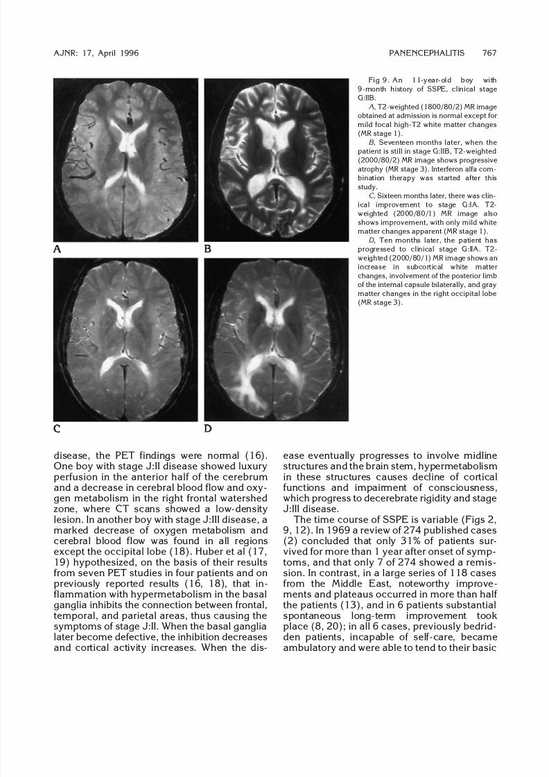
Fig 9. An 11-year-old boy with
9-month history of SSPE, clinical stage
G:IIB.
A, T2-weighted (1800/80/2) MR image
obtained at admission is normal except for
mild focal high-T2 white matter changes
(MR stage 1).B , Seventeen months later, when the
patient is still in stage G:IIB, T2-weighted
(2000/80/2) MR image shows progressive
atrophy (MR stage 3). Interferon alfa com-
bination therapy was started after this
study.
C , Sixteen months later, there was clin-
ical improvement to stage G:IA. T2-
weighted (2000/80/1) MR image also
shows improvement, with only mild white
matter changes apparent (MR stage 1).
D , Ten months later, the patient has
progressed to clinical stage G:IIA. T2-
weighted (2000/80/1) MR image shows an
increase in subcortical white matter
changes, involvement of the posterior limb
of the internal capsule bilaterally, and gray
matter changes in the right occipital lobe
(MR stage 3).
AJNR: 17, April 1996 PANENCEPHALITIS 767
8/6/2019 Basal Gangila Cahnges
http://slidepdf.com/reader/full/basal-gangila-cahnges 8/12
needs. Similar cases of spontaneous long-termimprovement (21–23) and of patients survivingfor more than 10 years after diagnosis (like oneof our patients) have been reported (20, 24–26). This variation in the natural course of thedisease makes it difficult to evaluate the effectsof therapy in small series (2, 27).
The uniformity of the mode of clinicalprogress in SSPE suggests a constant pattern of involvement of the CNS. Neuropathologic find-ings suggest that the disease initially affects theoccipital regions of the cortex, progresses to theanterior parts of the cerebrum, and finallyspreads to involve the subcortical structures,brain stem, and spinal cord (28). Involvementof cortical gray matter should then be respon-sible for the nonspecific symptoms of stage J:I.Others have claimed that an intact cerebral cor-tex is required to explain the appearance of thecharacteristic EEG changes in stage J:II disease
(29); these EEG changes could then be triggedfrom affected centers in the brain stem andreach the cortex through intact pathways. Thiswould be consistent with the normal findings of cortical biopsies seen early in the disease (28).Few patients (only one in our series) have beenexamined with MR imaging at this stage, andearly gray matter changes have not been re-ported (the early gray matter involvement couldof course be on a biochemical level, and notdepictable with MR imaging).
In the next stage of the disease (J:II or G:IB),at the onset of myoclonic spasms, CT in 3 of 3children showed abnormally small lateral ven-tricles (30). This finding was supported in aseries of 15 patients (31) in which cerebral
edema and diffuse low-attenuation white matterwas found in 6 of 8 patients with J:II disease, buthas been refuted in other reports (24, 32, 33).Thus, 7 patients examined during the first 4months after onset of symptoms all had normalCT findings, without any sign of brain swelling(32); no evidence of brain swelling was found inany of 5 patients studied during the first yearafter onset of SSPE (only 1 of these patients wasstudied during the first 6 months) (24). In ourseries, none of 21 patients who were examinedwithin 6 months of onset of symptoms (19 withMR imaging) showed signs of brain swelling.
Later subcortical white matter is involved withlesions identical to those of other slow virusinfections of the brain, such as subacute AIDSencephalomyelitis. Immune complexes depos-ited in the walls of cerebral blood vessels arebelieved to cause damage to the blood brainbarrier. The leakage of fluid and lymphocytescreates a perivascular edema and inflammationand is followed by demyelination (4). Radio-logic evidence of barrier lesions has been doc-umented in a few reports (24, 27, 34). Onepatient with rapidly progressing disease showedsigns of acute white matter inflammation withmultiple areas of contrast enhancement (24).Contrast enhancement has also been shownduring an acute relapse of the disease (27). Intwo patients, contrast-enhanced CT scansshowed normal findings, whereas radionucleidebrain scans showed multiple lesions. The au-thors concluded that the latter technique wassuperior in detecting acute SSPE (34). No en-hancement was observed in our series.
An alternative hypothesis would be that SSPEis primarily a subcortical disease. The wholeclinical picture fits better with what neurologists
would call a subcortical dementia (like Hunting-ton disease, Parkinson disease, and so on)rather than a cortical dementia (like Alzheimerdisease). The early MR changes primarily in-volve white matter and are more subcortical-occipital than frontal (Figs 7 and 9). It may bethat the oligodendroglia (responsible for form-ing the myelin in the CNS) are first affected andthen subsequently the basal ganglia and brainstem neurons. The myoclonic spasms are abrain stem myoclonus and occur early (stage
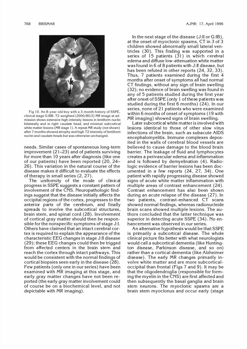
Fig 10. An 8-year-old boy with a 3-month history of SSPE,
clinical stage G:IIIB. T2-weighted (2000/80/2) MR image at ad-
mission shows extensive high-intensity lesions in lentiform nuclei
bilaterally and in right caudate head, and minimal subcortical
white matter lesions (MR stage 1). A repeat MR study (not shown)
after 7 months showed atrophy and high T2 intensity of lentiform
nuclei and caudate heads but was otherwise unchanged.
768 BRISMAR AJNR: 17, April 1996
8/6/2019 Basal Gangila Cahnges
http://slidepdf.com/reader/full/basal-gangila-cahnges 9/12
G:IB or G:IIA); the cortical atrophy occurs late.Most early behavioral changes can be explainedby subcortical white matter involvement of as-sociated areas (apraxia, agnosia) and brainstem reticular (thalamocortical) mechanisms(attention and concentration difficulties).
As the disease progresses, there is an in-creasing loss of white matter, and atrophy be-comes a more prominent feature (24, 35, 36)(Fig 12). CT findings are often negative untilthis late stage. Thus, in a series of 76 patients,only 22 had abnormal CT findings (37): CTfindings were normal in 25 of 33 patients withstage J:III disease, in 26 of 40 patients withstage J:II disease, and in all 3 patients withstage J:I disease. In another series, 11 of 14patients (1 stage J:I, 9 stage J:II, and 1 stage
J:III) initially had normal CT findings (27). In thefinal stage of SSPE, most white matter is lost,the ventricles and extracerebral CSF spaces areseverely widened, the corpus callosum is verythin, and posterior fossa structures are mark-edly atrophic (Fig 13).
Reported experiences with MR imaging inSSPE are limited (9, 25, 33, 38–44). MR imag-ing has major advantages in demonstratingwhite matter changes in SSPE (25, 33, 38, 39,42). This was obvious from our comparison of the results of CT and MR examinations per-formed less than 1 week apart in 25 patients. In11 of these patients, white matter changes wereseen only on MR images.
On the initial MR study (Fig 3), all but 11 of our 44 patients had white matter changes. In 22
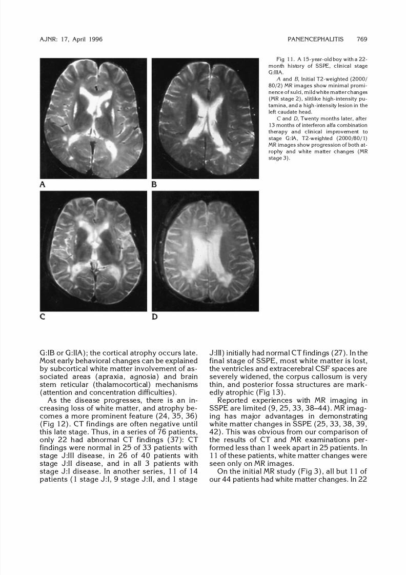
Fig 11. A 15-year-old boy with a 22-
month history of SSPE, clinical stage
G:IIIA.
A and B , Initial T2-weighted (2000/
80/2) MR images show minimal promi-
nence of sulci, mild white matter changes
(MR stage 2), slitlike high-intensity pu-tamina, and a high-intensity lesion in the
left caudate head.
C and D , Twenty months later, after
13 months of interferon alfa combination
therapy and clinical improvement to
stage G:IA, T2-weighted (2000/80/1)
MR images show progression of both at-
rophy and white matter changes (MR
stage 3).
AJNR: 17, April 1996 PANENCEPHALITIS 769

8/6/2019 Basal Gangila Cahnges
http://slidepdf.com/reader/full/basal-gangila-cahnges 10/12
examinations with mild to moderate white mat-ter changes, no atrophy was seen. In only 3examinations (in 2 patients) was the atrophymore striking than the white matter disease.
In most patients the gray matter still appearsnormal on MR images, even in the most ad-vanced clinical and MR stages; thus, in our se-ries, signal intensity changes within the graymatter were seen in only 8 of 23 patients whowere followed up with MR imaging to stageG:IIIB or G:IV disease. The gray matter lesionswere not correlated with the severity of whitematter changes or with atrophy (Figs 7 and 9).In 2 of our patients, central gray matter struc-tures were involved; in 1 in addition to cortical
gray matter (Fig 7) and in the second the cor-tical gray matter appeared normal but extensivechanges were found in the brain stem and pons.Similar findings have previously been reported.One patient with J:II disease seen 3 monthsafter onset of symptoms had normal CT findingswhile MR showed extensive increased-T2-inten-sity white matter lesions involving the supraten-torial white matter, the cerebellum, and thepons (42). Six months later these white matterchanges had markedly improved and the pos-terior fossa changes had normalized, but pu-taminal lesions had developed.
Basal ganglia lesions are not infrequent inSSPE, and were eventually seen in 18 of our 52patients. Such lesions were already present onthe initial imaging study in 11 patients. Theywere not related to the severity of other MRchanges (39, 41) (Figs 10 and 11), but oc-curred predominantly in patients whose diseasewas of longer duration and more clinically ad-vanced.
The correlation between the clinical stage of SSPE disease and the MR findings is often poor(33, 43). Thus, patients bedridden with severe
disease may still have normal findings at MRexaminations (Fig 9). Improvement of MR find-ings despite progress of the disease has alsobeen reported (44, 45). In several of our pa-tients, different treatment regimens were tried;in later years this included combined oral iso-prinosine–intraventricular interferon alfa ther-apy (7). In 8 of 20 patients on this latter regimenan arrest of disease or even an improvement of the clinical status was seen. In 2 patients thisimprovement was associated with a slight but
Fig 13. A 5-year-old boy with a 10-month history of SSPE,
clinical stage G:IIIB. A CT scan at admission (not shown) revealed
moderate atrophy and white matter changes. This T1-weighted
(700/20/2) MR image obtained 44 months later shows severe loss
of brain tissue (MR stage 6).
Fig 12. A 12-year-old boy with a
5-month history of SSPE, clinical stage
G:IIB.
A, CT scan at admission was normal,
but 2 weeks later, a CT scan (not shown)
revealed marked gray and white matter
changes.B , Three months later (clinical stage
G:IV), there is further progression, with
severe loss of brain tissue.
770 BRISMAR AJNR: 17, April 1996
8/6/2019 Basal Gangila Cahnges
http://slidepdf.com/reader/full/basal-gangila-cahnges 11/12
clear temporary improvement in the MR find-ings (Fig 9). In 1 patient the MR findings re-mained unchanged, in 5 patients the clinicalimprovement was accompanied by a progres-sion of MR abnormalities (Fig 11), and in an-
other 2 patients a slight improvement of MRfindings was accompanied by clinical impair-ment. In view of the large variability in the nat-ural course of SSPE, the results of therapy musttherefore be considered questionable. It is mostlikely that, in order to alter significantly thecourse of the disease, therapy must be initiatedat an early stage. Presently, the diagnosis of SSPE is usually not made until major, perma-nent brain damage has taken place. An in-creased awareness of the possibility of SSPE asthe cause of behavioral changes in a patient
with normal findings on CT or MR examinationsof the brain is probably one of many prerequi-sites for better therapeutic results. It is hopedthat an increased use of measles vaccine indeveloping countries will eventually decreasethe risk of SSPE.
References
1. Dawson JR Jr. Cellular inclusions in cerebral lesions of lethargic
encephalitis. Arch Neurol Psychiatry 1934;31:685–700
2. Freeman JM. The clinical spectrum and early diagnosis of Daw-
son’s encephalitis: with preliminary notes on treatment. J Pediatr
1969;75:590–603
3. Dyken PR. Subacute sclerosing panencephalitis: current status.Neurol Clin 1985;3:179–196
4. Poser CM. Notes on the pathogenesis of subacute sclerosing pa-
nencephalitis. J Neurol Sci 1990;95:219–224
5. Bohlega S, Al-Kawi MZ. Subacute sclerosing panencephalitis: im-
aging and clinical correlation. J Neuroimag 1994;4:71–76
6. Jabbour JT, Garcia JH, Lemmi H, et al. Subacute sclerosing
panencephalitis: a multidiciplinary study of eight cases. JAMA
1969;207:2248–2254
7. Gascon G, Yamani S, Crowell J, et al.Combined oral isoprinosine-
intraventricular -interferon therapy for subacute sclerosing pan-
encephalitis. Brain Dev (Tokyo) 1993;15:346–355
8. Haddad FS, Risk WS, Jabbour JT. Subacute sclerosing panen-
cephalitis in the Middle East: report of 99 cases. Ann Neurol
1977;1:211–217
9. Case records of the Massachusetts General Hospital: weekly clin-icopathological exercises. N Eng J Med 1986;314:1689–1700.
Case 25-1986
10. Cattaneo R, Schmid A, Rebmann G, et al. Accumulated measles
virus mutations in a case of subacute sclerosing panencephalitis:
interrupted matrix protein reading frame and transcription alter-
ation. Virology 1986;154:97–107
11. Carrigan DR, Kabacoff CM. Identification of a nonreproductive,
cell-associated form of measles virus by itsresistance to inhibition
by recombinant human interferon. J Virol 1987;61:1919–1926
12. Yalaz K, Anlar B, Oktem F, et al. Intraventricular interferon and
oral inosiplex in the treatment of subacute sclerosing panen-
cephalitis. Neurology 1992;42:488–491
13. Risk WS, Haddad FS. The variable natural history of subacute
sclerosing panencephalitis: a study of 118 cases from the Middle
East. Arch Neurol 1979;36:610–614
14. Chao D. Subacute inclusion body encephalitis: report of 3 cases.
J Pediatr 1962;61:501–510
15. Foley J, Williams D. Incussion encephalitis and its relation to
subacute sclerosing-leuco-encephalitis. Q J Med 1953;22:157–194
16. Yanai K, Iinuma K, Tada K, et al. Regional cerebral metabolic rate
for glucose in subacute sclerosing panencephalitis.Eur J Pediatr
1987;146:288–289
17. Huber M, Herholz K, Pawlik G, Szelies B, Jurgens R, Heiss W-D.
Cerebral glucose metabolism in the course of subacute sclerosing
panenceephalitis. Arch Neurol 1989;46:97–100
18. Yoshikawa H, Fueki N, Yoneyama H, Ogawa M, Sakuragawa N.
Positron emission tomography demonstrated localized luxury per-
fusion in subacute sclerosing panencephalitis. J Child Neurol
1990;5:311–315
19. Huber M, Pawlik G, Bamborschke S, et al. Changing pattern of
glucose metabolism during the course of subacute sclerosing
panencephalitis as measured with 18FDG-positron-emission to-
mography. J Neurol 1992;239:157–161
20. Risk WS, Haddad FS, Chemali R. Substantial long-term improve-
ment in subacute sclerosing panencephalitis: six cases from the
MiddleEast and a review of the literature. Arch Neurol 1978;35:
494–502
21. Resnick JS, Engel WK, Sever JL. Subacute sclerosing panen-
cephalitis: spontaneous improvement in a patient with elevated
measles antibody in blood and spinal fluid. N Engl J Med 1968;
279:126–129
22. Cobb WA, Marshall J, Scaravilli F. Long survival in subacute
sclerosing panencephalitis. J Neurol Neurosurg Psychiatry 1984;
47:176–183
23. Furby A, Vallee L, Rosseaux M, Nuyts JP, Destee A. Remissions
prolongees dan s la panencephalitis sclerosante subaigu e: 2 cas.
Rev Neurol (Paris) 1990;146:191–195
24. Krawiecki NS, Dyken PR, Gammal T, DuRant RH, Swift A. Com-
puted tomography in subacute sclerosing panencephalitis. Ann Neurol 1984;15:489–493
25. Lum GB, Williams JP, Dyken PR, et al. Magnetic resonance and
CT imaging correlated with clinical status in SSPE. Pediatr Neurol
1986;2:75–79
26. Boucebci M, Bouchefra A, Abbad D, Taright F. Panencephalite
sclerosante subaigue. A propos de 8 cas d ont 1 evoluant depuis
13 ans. Encephale 1987;13:279–284
27. Mahdi AH, Familusi JB. Subacute sclerosing panencephalitis in
Riyadh, Saudi Arabia. Ann Trop Med 1992;12:95–104
28. Ohya T, Martinez AJ, Jabbour JT, Lemmi H, Duenas DA. Sub-
acute sclerosing panencephalitis: correlation of clinical, neuro-
physiologic and neuropathologic findings. Neurology 1974;24:
211–218
29. Cobb W. The periodic events of subacute sclerosing panencepha-
litis. Electroencephalogr Clin Neurophysiol 1966;21:278–29430. Pedersen H, Wulff CH. Computed tomographic findings of early
subacute sclerosing panencephalitis. Neuroradiology
1982;23:31–32
31. Jayakumar PN, Taly AB, Arya BYT, Nagaraj D. Computed to-
mography in subcute sclerosing panencephalitis: report of 15
cases. Acta Neurol Scand 1988;77:328–330
32. Begeer JH, Haaxma R, Snoek JW, Boonstra S, le Coultre R. Signs
of focal posterior cerebral abnormality in early subacute scleros-
ing panencephalitis. Ann Neurol 1986;19:200–202
AJNR: 17, April 1996 PANENCEPHALITIS 771
8/6/2019 Basal Gangila Cahnges
http://slidepdf.com/reader/full/basal-gangila-cahnges 12/12
33. Miller DH, Robb SA, Ormerod IEC, et al. Magnetic resonance
imaging of inflammatory and demyelinating white-matter dis-
eases of childhood. Dev Med Child Neurol 1990;32:97–107
34. Dodson WE, Prensky AL, Siegel BA. Radionuclide imaging in
subacute sclerosing panencephalitis. Neurology 1979;29:749–
752
35. Duda EE, Huttenlocher PR, Patronas NJ. CT of subacute scleros-ing panencephalitis. AJNR Am J Neuroradiol 1980;1:35–38
36. Manabe Y, Ono Y, Okuno T, et al. Serial CT scans in subacute
sclerosing panencephalitis. Comput Tomogr 1981;5:25–30
37. Anlar B, Yalaz K, Ustacelebi S. Symptomes et signes cliniques,
donnees du laboratoire dans 80 cas de panencephalite sclero-
sante subaig ue. Rev Neurol (Paris) 1988;144:829–832
38. Takemoto K, Koizumi Y, Kogame S, et al. Magnetic resonance
imaging of subacute sclerosing panencephalitis.JpnJ Clin Radiol
1986;31:999–1004
39. Murata R, Matsuoka O, Nakajima S, et al. Serial magnetic reso-
nance imaging in subacute sclerosing panencephalitis. Jpn J
Psychiatr Neurol 1987;41:277–282
40. Geller TJ, Vern BA, Sarwar M. Focal MRI findings in early SSPE.
Pediatr Neurol 1987;3:310–312
41. Woodward KG, Weinberg PE, Lipton HL. Basal ganglia involve-
ment in subacute sclerosing panencephalitis: CT and MR demon-
stration. J Comput Assist Tomogr 1988;12:489–451
42. Tsuchiya K, Yamauchi T, Furui S. MR imaging vs CT in subacute
sclerosing panencephalitis. AJNR Am J Neuroradiol 1988;9:943–946
43. Dietrich RB, Vining EP, Taira RK, Hall TR, Phillipart M. Myelin
disorders of childhood: correlation of MR findings and severity of
neurological impairment. J Comput Assist Tomogr 1990;14:693–
698
44. Winer JB, Pires M, Kermode A, Ginsberg L, Rossor M. Resolving
MRI abnormalities with progression of subacute sclerosing pan-
encephalitis. Neuroradiology 1991;33:178–180
45. Noetzel MJ, Dodson WE. Progressive CT abnormalities despite
clinical improvement in SSPE treated with inosiplex. Ann Neurol
1983;13:457–460
772 BRISMAR AJNR: 17, April 1996