biochimica et biophysica acta - esalq.usp.br · review the mechanism of rotating proton pumping...
TRANSCRIPT

Biochimica et Biophysica Acta 1797 (2010) 1343–1352
Contents lists available at ScienceDirect
Biochimica et Biophysica Acta
j ourna l homepage: www.e lsev ie r.com/ locate /bbab io
Review
The mechanism of rotating proton pumping ATPases
Mayumi Nakanishi-Matsui a,⁎, Mizuki Sekiya a, Robert K. Nakamoto b, Masamitsu Futai a
a Department of Biochemistry, Faculty of Pharmaceutical Sciences, Iwate Medical University, Yahaba, Iwate 028-3694, Japanb Department of Molecular Physiology and Biological Physics, University of Virginia, Charlottesville, VA 22908-0886, USA
Abbreviations: F-ATPase, ATP synthase; V-ATPase,fluorescence resonance energy transfer; rps, revolution⁎ Corresponding author. Nishitokuta 2-1-1, Yahaba,
Tel./fax: +81 19 698 1843.E-mail address: [email protected] (M. Naka
0005-2728/$ – see front matter © 2010 Elsevier B.V. Adoi:10.1016/j.bbabio.2010.02.014
a b s t r a c t
a r t i c l e i n f oArticle history:Received 6 October 2009Received in revised form 8 February 2010Accepted 11 February 2010Available online 17 February 2010
Keywords:ATP synthaseF-ATPaseV-ATPaseSubunit rotationSingle molecule observationThermodynamic analysis
Two proton pumps, the F-ATPase (ATP synthase, FoF1) and the V-ATPase (endomembrane proton pump),have different physiological functions, but are similar in subunit structure and mechanism. They arecomposed of a membrane extrinsic (F1 or V1) and a membrane intrinsic (Fo or Vo) sector, and couple catalysisof ATP synthesis or hydrolysis to proton transport by a rotational mechanism. The mechanism of rotation hasbeen extensively studied by kinetic, thermodynamic and physiological approaches. Techniques for observingsubunit rotation have been developed. Observations of micron-length actin filaments, or polystyrene or goldbeads attached to rotor subunits have been highly informative of the rotational behavior of ATP hydrolysis-driven rotation. Single molecule FRET experiments between fluorescent probes attached to rotor and statorsubunits have been used effectively in monitoring proton motive force-driven rotation in the ATP synthesisreaction. By using small gold beads with diameters of 40–60 nm, the E. coli F1 sector was found to rotateat surprisingly high speeds (N400 rps). This experimental system was used to assess the kinetics andthermodynamics of mutant enzymes. The results revealed that the enzymatic reaction steps and the timingof the domain interactions among the β subunits, or between the β and γ subunits, are coordinated in amanner that lowers the activation energy for all steps and avoids deep energy wells through the rotationally-coupled steady-state reaction. In this review, we focus on the mechanism of steady-state F1-ATPase rotation,which maximizes the coupling efficiency between catalysis and rotation.
vacuolar-type ATPase; FRET,per secondShiwa, Iwate 028-3694, Japan.
nishi-Matsui).
ll rights reserved.
© 2010 Elsevier B.V. All rights reserved.
1. Introduction
Two proton pumping ATPases, the F- and V-ATPases, are structurallyand mechanistically similar, but have different physiological roles (forreviews, see [1–11]). They are entirely different from the gastric andyeast plasma membrane proton pumping P-type ATPases (P-ATPases),which form an aspartyl–phosphate intermediate during the course ofthe reaction cycle [6,12,13]. The F-ATPase is a ubiquitouswell conservedcomplex found inmitochondria, chloroplasts, and bacterialmembranes,and plays a central role in energy transduction [6]. This enzymesynthesizes ATP coupled to the electrochemical proton gradient formedby electron transfer chains. In contrast, the V-ATPase located onendomembrane organelles or plasma membrane is involved inacidification of organellar lumens or extracellular compartments [4,9].A characteristic feature of the V-ATPase is its subunit diversity: sixsubunits have two to four isoforms specific for different organelles orcell types [7,9,10]. The presence of a specific V-ATPase is important fororganellar function as well as targeting of this enzyme to a properdestination.
Despite their different physiological roles, the F- and V-ATPasesare similar in subunit structure and mechanism [3,6,7,9,10]. They arereversible ATP hydrolases/synthases coupled to proton transport. Insome situations, V-ATPases synthesize ATP when an electrochemicalproton gradient is applied [14,15]. In both enzymes, the three catalyticsites are not independent. The steady-state ATP synthesis andhydrolysis reactions are highly cooperative mechanisms and involveall three catalytic sites in each β subunit. Boyer proposed the basiccatalytic scheme, called the binding change mechanism, whichpredicted that the F-ATPase utilizes a rotational mechanism [16](the body of biochemical evidence supporting rotational catalysis issummarized in [1]). The physical features of the bovine F1 discoveredin the first X-ray structure clearly showed that a rotation mechanismwas feasible [17]. Quickly, investigators produced indirect biochem-ical evidence for the rotation of the γ subunit: polarized adsorptionrelaxation after photobleaching of eosin attached to the carboxylterminus of the γ subunit showed ATP-dependent changes in orien-tation [18]; ATP hydrolysis-dependent changes of chemical cross-linking between the β and γ subunits [19]; and images captured bycryo-electron microscopy showed positional changes of subunits [20].Finally, rotation was directly observed by attaching a fluorescently-tagged actin filament to the γ subunit of the Bacillus PS3 F1 enzymecomplex and watching the actin filament rotate in an ATP hydrolysis-dependent manner relative to the rest of the F1 complex [21]. Rotationof the entire rotor group, εγc10, relative to the complete F-ATPase

1344 M. Nakanishi-Matsui et al. / Biochimica et Biophysica Acta 1797 (2010) 1343–1352
complex was demonstrated by observations of actin probes attachedto each of the subunits [22–25]. Since the initial single moleculeobservation, the rotational mechanism of F-ATPase has been exten-sively studied using improved techniques as will be described in thisreview.
In this article, we concentrate on the rotational catalyticmechanism revealed by single molecule experiments using probescausing little to no viscous drag. Unfortunately, due to spacelimitations, we are unable to discuss several important findingsdirectly related to our main topic, but refer the reader to severalexcellent recent reviews [11,26,27].
2. The ATPase structures are designed for rotation
2.1. Catalytic sector and stalks
The F-ATPases and V-ATPases have some common structuralfeatures that investigators have found to be important for arotational mechanism. The basic subunit structure of F-ATPaseincluding that of E. coli is α3β3γδεab2c10, and that of yeast V-ATPaseis A3B3CDE2FG2Hac4c'c"d (no c' has been found in mammals). Boththe membrane extrinsic catalytic sectors (F1 and V1), and themembranous proton translocation sectors (Fo and Vo) have rota-tional mechanisms [3,6,7,9–11] (Fig. 1). The nucleotide bindingsubunits (α3β3 in F1 or A3B3 in V1) and the proton translocationsubunits in the membrane (ac10 or ac4c'c") are connected by thecentral (γε or DFd) and peripheral (δb2 or CE2G2Ha) stalks. The F1rotor subunit γ is located at the center of the α3β3 pseudo-hexamer[17] and together, α3β3γ, defines the minimal complex required forrotational catalysis. γ and ε subunits directly connect to the protontranslocating c subunit ring [28].
Mutagenesis studies helped to identify the F-ATPase amino acidsthat are involved in catalysis [6,29–33]. βLys155 and βArg182 in the βsubunit were shown to be required for binding of the γ-phosphatemoiety of ATP [34–38], while the hydroxyl moiety of βThr156 isessential for Mg2+ binding [35,39]. βGlu181 is a critical catalyticresidue which forms a hydrogen bond with a water molecule locatednear the ATP γ-phosphate [17,36]. The βGlu185 residue may beinvolved in promotion of catalysis to steady-state turnover [40]. Allthese residues are conserved in the V-ATPase, suggesting that it hasthe same catalytic mechanism as the F-ATPase [6]. Similarly, genetic,biochemical, kinetic and thermodynamic analyses were used to
Fig. 1. Schematic models of the F-ATPase (E. coli) and V-ATPase (mammalian). The membranand membrane domains (FO and VO) are shown. Red arrows represent the direction of theDiverse isoforms of V-ATPase are shown with their specific localization [7,9]. Those not ind
identify the roles of γ subunit residues in catalysis and energycoupling [6,41–46].
On the other hand, there are distinct structural differencesbetween the F- and V-ATPases. The subunit composition of theperipheral stalk of V-ATPase is more complex than that of F-ATPasebecause the V-ATPase C, E, G, H, and a subunits have two to fourisoforms each [7]. These subunits may be related to the reversibledisassembly/reassembly regulatory mechanism specific to the V-ATPase [9].
2.2. Proton-transporting sector
In the F-ATPase, ATP hydrolysis in the β subunits couples torotation of the γεc10 complex against the stator (α3β3δab2), whichdrives uphill proton transport. Themechanism of proton translocationoccurs at the interface between a and c subunits and specificallyinvolves proton binding residues aArg210 and the Asp61 of therotating c ring [5,47–49]. In the reverse or physiological condition, theelectrochemical gradient of protons drives rotation of the c ring, whichcouples to ATP synthesis in F1. The number of c subunits in a complexappears to be dependent on the source. The ATP synthases of yeastmitochondria, E. coli, and Bacillus PS3 have 10 subunits in their c rings[50–52]. In contrast, those of Ilyobacter tartaricus and Propionigeniummodestum have 11 c subunits [53,54], while that of chloroplasts has 14subunits [55,56], and cyanobacterium Spirulina platensis contains 15 csubunits [57]. Assuming one proton per c subunit, the number ofc subunits in the ring complex implies the number of protonstransported during the course of a 360° rotation, 10–15 protons inthe above mentioned examples, or 3.3–5 protons per ATP synthesizedor hydrolyzed. The proton/ATP coupling ratio of ATP synthases areprobably optimized for their specific physiological condition.
The similar subunit structure of the V-ATPase suggests the DFc4c'c"complex along with perhaps the d subunit acts as the rotor [9].Because a single proton-transporting Glu residue is found in each ofthe c, c' and c" subunits which form the transmembrane rotor ring,rotation of the c4c'c" ring likely translocates only six protons per 360°rotation. Therefore, two protons are pumped per ATP hydrolyzedimplying that the V-ATPase requires a much higher proton motiveforce to drive the reverse ATP synthesis direction [58].
The direction of the chemical reaction (ATP hydrolysis orsynthesis), which is directly coupled to the direction of protontransport, and therefore the direction of subunit rotation, appears to
e extrinsic (F1 and V1) sectors, catalytic hexamers (α3β3 and A3B3, respectively), stalks,chemical reaction, subunit rotation, and proton transport in physiological conditions.icated are ubiquitous.

1345M. Nakanishi-Matsui et al. / Biochimica et Biophysica Acta 1797 (2010) 1343–1352
be regulated by the energy state of the cell. The energetic balance ofthe ATP/ADP concentration ratio and the electrochemical protongradient exclusively define whether the enzyme operates as an ATPsynthase or an ATPase [6,7]. Similar to the reversibility of the F-typeATP synthase to hydrolyze ATP, the V-ATPase synthesizes ATP in somecases [14,15]. Finally,wenote that the basic structure of V-ATPase is verysimilar and mechanically the same as that of the F-ATPase. Commonstructural features of these ATPases are exquisitely designed forcoupling the rotational catalytic and proton transport mechanisms.
An interesting question is which sector of ATPase rotatesphysiologically because the rotor or stator has been defined byin vitro single molecule studies. In this regard, Holliday et al. and Lu etal. found that the B and E subunits of V-ATPase bind actin and aldolase,respectively [59,60]. The interaction with actin likely immobilizes theA3B3 hexamer, and thereby causes the A3B3 hexamer to act as a stator.A similar protein interaction is unknown for F-ATPase.
3. Rotational catalysis in the F-ATPase
3.1. Observation of subunit rotation
In 1979, Paul Boyer [16] suggested that the F-ATPase catalyticmechanism involved rotation of an F1 subunit relative to the threecatalytic sites. The crystal structure of the α3β3γ complex reported in1994 by Walker and colleagues [17] rekindled this idea. Severalinvestigators set out to design experiments that would prove such amechanism. Indirect biochemical approaches led to results that wereconsistent with ATP-dependent γ subunit rotation (described above).Finally, rotation was conclusively shown by direct observation insingle molecule experiments. Noji et al. reported the ATP-dependentrotation of a fluorescently-labeled actin filament attached to the γsubunit of Bacillus PS3 F1 [21]. Using similar methods, rotation of theE. coli γ subunit was also shown to rotate indicating that rotation wasa general mechanism of the F1 enzyme [61].
Subsequently, rotation in the Fo transport sector was shown byfixing the detergent-solubilized FoF1 complex on a glass surface viathe α subunit with a fluorescent probe attached to the rotor c subunitring [23], or the FoF1 fixed to the surface via the c ring with the probeattached to the β subunits [24,62]. The results demonstrate that therotor and stator are interchangeable. Rotation of membrane-boundFoF1 has also been shown experimentally [25], establishing that thec ring rotates relative to the a and β subunits. Rates of ATP-driven γsubunit rotation in F1 and ATP-driven γεc10 rotation in FoF1 are verysimilar in the two complexes with a calculated frictional torque of∼40 pN nm. The rotation of γεc10 was consistent with results of inter-subunit cross-linking experiments [63,64] where a disulfide bondbetween γ and c subunits had little or no effect on ATP hydrolysis orsynthesis, while cross-links between γ and β, or γ and α, resulted inloss of steady-state ATPase activity. In this regard, Scanlon et al. [65]showed by pre-steady-state analysis that γ–β cross-linked enzymecan hydrolyze the first ATP but not other bound ATP. Other singlemolecule FRET experiments also observed that the γ and ε subunitsrotate relative to the b subunits in membrane-bound FoF1 [66,67].
While ATP hydrolysis-dependent rotation of γεc10 is alwaysobserved in one direction, one expects that ATP synthesis driven byproton flow through the Fo should rotate in the reverse direction. Thishypothesis was proven by showing rotation in the expected directionduring ATP synthesis in FoF1 reconstituted in liposomes. In this case,FRET was used to report the real time changes in distances betweenprobes attached to b and γ subunits, or ε and b subunits [66–68]. Aspredicted, the direction of rotation during ATP synthesis was reversedto that of ATP hydrolysis. Similarly, artificially forced rotation of γ inthe same direction was shown to drive ATP synthesis. Itoh et al. [69]and Rondelez et al. [70] observed a low but significant level of ATPsynthesis when a bead firmly attached to γwas forced to rotate. Theseresults established beyond the shadow of any doubt the mechano-
chemical coupling between ATP synthesis/hydrolysis and the elec-trochemical gradient of protons.
3.2. Mechanical rotation and ATPase reaction
3.2.1. Can rotation of the γ subunit be resolved into steps that are relatedto the chemical reaction kinetics?
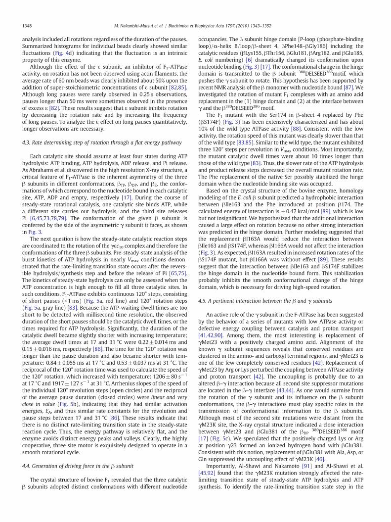
The binding change mechanism indirectly predicts that a rotarymechanism is intimately involved in the mechanism of ATP synthesisand hydrolysis [16]. Using the thermophilic Bacillus PS3 F1 at ATPconcentrations well below the Km, γ subunit rotation was found todwell in distinct 120° steps [71,72] (Fig. 2). Because the duration ofthe pause depends upon the ATP concentration, the pause wasattributed to the enzyme waiting for an ATP binding event (ATP-waiting dwell) (Fig. 2, blue line). Experimental set-ups with bettertime resolution using 40-nm diameter gold beads revealed that the120° steps were divided into 80° and 40° substeps [73]. The durationof the pause before the 80° substep became shorterwith increasing ATPconcentration, once again suggesting the ATP-waiting dwell (Fig. 2, blueline). In contrast, the pause duration before the 40° substep wassignificantly increased by the slow hydrolysis rate when ATPγS wasused as a substrate, or by the catalytic site mutation βE190D (Bacillusnumbering) [74]. Together these data indicate that the pause durationbefore the 40° substep is the catalytic dwell (Fig. 2, red line), or the timeduring reversible ATP–ADP hydrolysis/synthesis step [75].
3.2.2. Does ATP bind to all catalytic sites at the same time?Nadanaciva et al. suggested that two out of three catalytic sites
have the capability of forming a transition state between ATP andADP+Pi simultaneously. Their hypothesis was based on theobservation that two sites were titrated with fluoroscandium(ScFx) plus ADP, the complex of which, F1-ADP–ScFx, mimics acatalytic transition state [76]. Consistent with this result, analysis ofthe F1 crystal structure found that the three catalytic sites wereoccupied by ATP, ADP, or devoid of nucleotide, and thus thecorresponding conformations were designated βTP, βDP, and βE,respectively [17] (Fig. 3). Simultaneous observation of rotation andCy3–ATP bound to the catalytic site suggested that one ATP (ADP)molecule remained bound to a catalytic site for at least 240° of γsubunit rotation. This ATP appeared not hydrolyzed upon bindingbut rather hydrolyzed after the first 120° revolution [77]. Theseresults indicated that the reactions at the three catalytic sites werenot simultaneous but rather occurred sequentially in a highlycooperative fashion: for each 120° step, ATP binds to one β subunit,ADP is released from the second β subunit, ATP is hydrolyzed in thethird β subunit, and Pi is released from the second β [78,79].
In contrast, a different kinetic pathway was proposed based onpre-steady-state kinetic analysis using millisecond mixing experi-ments. Scanlon et al. proposed a model where ATP binds to one βsubunit after ADP release, ATP in the second β is hydrolyzed, and Piand ADP in the third β are released within one 120° step [75]. In thismodel, all three catalytic sites should bind nucleotides transiently, asshown in the kinetic scheme in Fig. 2. Consistent with this model,crystals grown in the presence of ADP and aluminum fluoride showedthe F1 enzyme in a conformation with nucleotides bound to all three βsubunits [80].
3.2.3. Does Bacillus PS3 F1 exhibit the same rotational scheme at itsphysiological temperature?
All experiments on the thermophilic Bacillus were carried outbelow ∼24 °C, whereas the organism optimally grows at 75 °C. Thusfar, few single molecule experiments have addressed this question,although F1 was recently studied at 4–50 °C [81]. In this regard, theadvantage of studying the mesophilic enzyme from E. coli should benoted because it is easily studied in its physiological temperaturerange between 25 and 37 °C.

Fig. 2. Scheme of F1 rotational catalysis. A model of the relationship of the chemical reaction to the β subunit conformation, and the position of the γ subunit at ATP concentrationsbelow the Km are shown. The ATP-waiting dwell time is the time waiting for ATP binding (blue line). The catalytic dwell time is the time for ATP hydrolysis and Pi release (red line).Note that the γ-β subunit interactions are different for each β subunit. βTP, βDP, βE, and βHC are the β subunits with ATP or ADP bound, empty, or half closed, respectively.Modified from Sekiya et al. [86].
1346 M. Nakanishi-Matsui et al. / Biochimica et Biophysica Acta 1797 (2010) 1343–1352
4. Toward understanding the rotation mechanism
4.1. Subunit rotation and steady-state ATPase
Discussion in this section is focused mainly on the E. coli F-ATPase.Extensive genetic and biochemical analyses provide many tools fordissecting the mechanism of catalysis coupled to transport [6]. Similarto the case of the thermophilic enzyme, the rotation speed of an actin
Fig. 3. The structures of the bovine β and γ subunits (PDB ID: 2JDI) [99]. Yellow and greeconformationswith different nucleotide occupancies. The expanded figure (orange) shows thethe catalytic residues (βLys155, βThr156, βGlu181, and βArg182, in black with E. coli numberi
filament attached to the γ subunit was ∼10 rps, which was muchslower than the 30 rps expected based on steady-state ATPase rates[61]. Obviously, the high viscous drag generated by actin (∼1–2 μm inlength) would decrease the speed. In addition, the long length of thefilament also caused relatively large deviations in the data, whichmade kinetic analyses difficult to interpret.
These considerations prompted us to use small gold beads asprobes for observation of subunit rotation, which allowed the enzyme
n represent the β and γ subunits, respectively. The catalytic β subunits adopt differentβ subunit hinge domain (P-Loop/α-HelixB/Loop/β-Sheet4, βPhe148–βGly186) includingng). The conformation of this domain changes dramatically upon nucleotide binding.
1347M. Nakanishi-Matsui et al. / Biochimica et Biophysica Acta 1797 (2010) 1343–1352
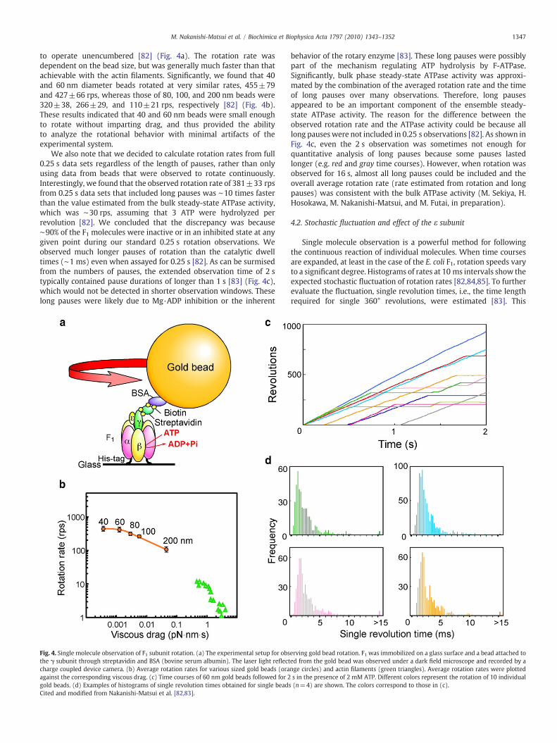
to operate unencumbered [82] (Fig. 4a). The rotation rate wasdependent on the bead size, but was generally much faster than thatachievable with the actin filaments. Significantly, we found that 40and 60 nm diameter beads rotated at very similar rates, 455±79and 427±66 rps, whereas those of 80, 100, and 200 nm beads were320±38, 266±29, and 110±21 rps, respectively [82] (Fig. 4b).These results indicated that 40 and 60 nm beads were small enoughto rotate without imparting drag, and thus provided the abilityto analyze the rotational behavior with minimal artifacts of theexperimental system.
We also note that we decided to calculate rotation rates from full0.25 s data sets regardless of the length of pauses, rather than onlyusing data from beads that were observed to rotate continuously.Interestingly, we found that the observed rotation rate of 381±33 rpsfrom 0.25 s data sets that included long pauses was ∼10 times fasterthan the value estimated from the bulk steady-state ATPase activity,which was ∼30 rps, assuming that 3 ATP were hydrolyzed perrevolution [82]. We concluded that the discrepancy was because∼90% of the F1 molecules were inactive or in an inhibited state at anygiven point during our standard 0.25 s rotation observations. Weobserved much longer pauses of rotation than the catalytic dwelltimes (∼1 ms) even when assayed for 0.25 s [82]. As can be surmisedfrom the numbers of pauses, the extended observation time of 2 stypically contained pause durations of longer than 1 s [83] (Fig. 4c),which would not be detected in shorter observation windows. Theselong pauses were likely due to Mg·ADP inhibition or the inherent
Fig. 4. Single molecule observation of F1 subunit rotation. (a) The experimental setup for obsthe γ subunit through streptavidin and BSA (bovine serum albumin). The laser light reflectcharge coupled device camera. (b) Average rotation rates for various sized gold beads (oraagainst the corresponding viscous drag. (c) Time courses of 60 nm gold beads followed for 2gold beads. (d) Examples of histograms of single revolution times obtained for single beadCited and modified from Nakanishi-Matsui et al. [82,83].
behavior of the rotary enzyme [83]. These long pauses were possiblypart of the mechanism regulating ATP hydrolysis by F-ATPase.Significantly, bulk phase steady-state ATPase activity was approxi-mated by the combination of the averaged rotation rate and the timeof long pauses over many observations. Therefore, long pausesappeared to be an important component of the ensemble steady-state ATPase activity. The reason for the difference between theobserved rotation rate and the ATPase activity could be because alllong pauseswere not included in 0.25 s observations [82]. As shown inFig. 4c, even the 2 s observation was sometimes not enough forquantitative analysis of long pauses because some pauses lastedlonger (e.g. red and gray time courses). However, when rotation wasobserved for 16 s, almost all long pauses could be included and theoverall average rotation rate (rate estimated from rotation and longpauses) was consistent with the bulk ATPase activity (M. Sekiya, H.Hosokawa, M. Nakanishi-Matsui, and M. Futai, in preparation).
4.2. Stochastic fluctuation and effect of the ε subunit
Single molecule observation is a powerful method for followingthe continuous reaction of individual molecules. When time coursesare expanded, at least in the case of the E. coli F1, rotation speeds varyto a significant degree. Histograms of rates at 10 ms intervals show theexpected stochastic fluctuation of rotation rates [82,84,85]. To furtherevaluate the fluctuation, single revolution times, i.e., the time lengthrequired for single 360° revolutions, were estimated [83]. This
erving gold bead rotation. F1 was immobilized on a glass surface and a bead attached toed from the gold bead was observed under a dark field microscope and recorded by ange circles) and actin filaments (green triangles). Average rotation rates were plotteds in the presence of 2 mM ATP. Different colors represent the rotation of 10 individual
s (n=4) are shown. The colors correspond to those in (c).
1348 M. Nakanishi-Matsui et al. / Biochimica et Biophysica Acta 1797 (2010) 1343–1352
analysis included all rotations regardless of the duration of the pauses.Summarized histograms for individual beads clearly showed similarfluctuations (Fig. 4d) indicating that the fluctuation is an intrinsicproperty of this enzyme.
Although the effect of the ε subunit, an inhibitor of F1-ATPaseactivity, on rotation has not been observed using actin filaments, theaverage rate of 60 nm beads was clearly inhibited about 50% upon theaddition of super-stoichiometric concentrations of ε subunit [82,85].Although long pauses were rarely observed in 0.25 s observations,pauses longer than 50 ms were sometimes observed in the presenceof excess ε [82]. These results suggest that ε subunit inhibits rotationby decreasing the rotation rate and by increasing the frequencyof long pauses. To analyze the ε effect on long pauses quantitatively,longer observations are necessary.
4.3. Rate determining step of rotation through a flat energy pathway
Each catalytic site should assume at least four states during ATPhydrolysis: ATP binding, ATP hydrolysis, ADP release, and Pi release.As Abrahams et al. discovered in the high resolution X-ray structure, acritical feature of F1-ATPase is the inherent asymmetry of the threeβ subunits in different conformations, βTP, βDP, and βE, the confor-mations of which correspond to the nucleotide bound in each catalyticsite, ATP, ADP and empty, respectively [17]. During the course ofsteady-state rotational catalysis, one catalytic site binds ATP, whilea different site carries out hydrolysis, and the third site releasesPi [6,45,73,78,79]. The conformation of the given β subunit isconferred by the side of the asymmetric γ subunit it faces, as shownin Fig. 3.
The next question is how the steady-state catalytic reaction stepsare coordinated to the rotation of the γεc10 complex and therefore theconformations of the three β subunits. Pre-steady-state analysis of theburst kinetics of ATP hydrolysis in nearly Vmax conditions demon-strated that the rate-limiting transition state occurs after the revers-ible hydrolysis/synthesis step and before the release of Pi [65,75].The kinetics of steady-state hydrolysis can only be assessed when theATP concentration is high enough to fill all three catalytic sites. Insuch conditions, F1-ATPase exhibits continuous 120° steps, consistingof short pauses (b1 ms) (Fig. 5a, red line) and 120° rotation steps(Fig. 5a, gray line) [83]. Because the ATP-waiting dwell times are tooshort to be detected with millisecond time resolution, the observedduration of the short pauses should be the catalytic dwell times, or thetimes required for ATP hydrolysis. Significantly, the duration of thecatalytic dwell became slightly shorter with increasing temperature;the average dwell times at 17 and 31 °C were 0.22±0.014 ms and0.15±0.016 ms, respectively [86]. The time for the 120° rotation waslonger than the pause duration and also became shorter with tem-perature: 0.84±0.055 ms at 17 °C and 0.53±0.037 ms at 31 °C. Thereciprocal of the 120° rotation time was used to calculate the speed ofthe 120° rotation, which increased with temperature: 1206±80 s−1
at 17 °C and 1917±127 s−1 at 31 °C. Arrhenius slopes of the speed ofthe individual 120° revolution steps (open circles) and the reciprocalof the average pause duration (closed circles) were linear and veryclose in value (Fig. 5b), indicating that they had similar activationenergies, EA, and thus similar rate constants for the revolution andpause steps between 17 and 31 °C [86]. These results indicate thatthere is no distinct rate-limiting transition state in the steady-statereaction cycle. Thus, the energy pathway is relatively flat, and theenzyme avoids distinct energy peaks and valleys. Clearly, the highlycooperative, three site motor is exquisitely designed to operate in asmooth rotational cycle.
4.4. Generation of driving force in the β subunit
The crystal structure of bovine F1 revealed that the three catalyticβ subunits adopted distinct conformations with different nucleotide
occupancies. The β subunit hinge domain [P-loop (phosphate-bindingloop)/α-helix B/loop/β-sheet 4, βPhe148–βGly186] including thecatalytic residues (βLys155, βThr156, βGlu181, βArg182, and βGlu185,E. coli numbering) [6] dramatically changed its conformation uponnucleotide binding (Fig. 3) [17]. The conformational change in the hingedomain is transmitted to the β subunit 380DELSEED386motif, whichpushes the γ subunit to rotate. This hypothesis has been supported byrecent NMR analysis of the βmonomer with nucleotide bound [87]. Weinvestigated the rotation of mutant F1 complexes with an amino acidreplacement in the (1) hinge domain and (2) at the interface betweenγ and the β380DELSEED386 motif.
The F1 mutant with the Ser174 in β-sheet 4 replaced by Phe(βS174F) (Fig. 3) has been extensively characterized and has about10% of the wild type ATPase activity [88]. Consistent with the lowactivity, the rotation speed of this mutant was clearly slower than thatof the wild type [83,85]. Similar to the wild type, the mutant exhibitedthree 120° steps per revolution in Vmax conditions. Most importantly,the mutant catalytic dwell times were about 10 times longer thanthose of the wild type [83]. Thus, the slower rate of the ATP hydrolysisand product release steps decreased the overall mutant rotation rate.The Phe replacement of the native Ser possibly stabilized the hingedomain when the nucleotide binding site was occupied.
Based on the crystal structure of the bovine enzyme, homologymodeling of the E. coli β subunit predicted a hydrophobic interactionbetween βIle163 and the Phe introduced at position β174. Thecalculated energy of interaction is −0.47 kcal/mol [89], which is lowbut not insignificant. We hypothesized that the additional interactioncaused a large effect on rotation because no other strong interactionwas predicted in the hinge domain. Further modeling suggested thatthe replacement βI163A would reduce the interaction betweenβIle163 and βS174F, whereas βI166A would not affect the interaction(Fig. 3). As expected, βI163A resulted in increased rotation rates of theβS174F mutant, but βI166A was without effect [89]. These resultssuggest that the interaction between βIle163 and βS174F stabilizesthe hinge domain in the nucleotide bound form. This stabilizationprobably inhibits the smooth conformational change of the hingedomain, which is necessary for driving high-speed rotation.
4.5. A pertinent interaction between the β and γ subunits
An active role of the γ subunit in the F-ATPase has been suggestedby the behavior of a series of mutants with low ATPase activity ordefective energy coupling between catalysis and proton transport[41,42,90]. Among them, the most interesting is replacement ofγMet23 with a positively charged amino acid. Alignment of theknown γ subunit sequences reveals that conserved residues areclustered in the amino- and carboxyl terminal regions, and γMet23 isone of the few completely conserved residues [42]. Replacement ofγMet23 by Arg or Lys perturbed the coupling between ATPase activityand proton transport [42]. The uncoupling is probably due to analtered β–γ interaction because all second site suppressor mutationsare located in the β–γ interface [43,44]. As one would surmise fromthe rotation of the γ subunit and its influence on the β subunitconformations, the β–γ interactions must play specific roles in thetransmission of conformational information to the β subunits.Although most of the second site mutations were distant from theγM23K site, the X-ray crystal structure indicated a close interactionbetween γMet23 and βGlu381 of the βDP
380DELSEED386 motif[17] (Fig. 5c). We speculated that the positively charged Lys or Argat position γ23 formed an ionized hydrogen bond with βGlu381.Consistent with this notion, replacement of βGlu381 with Ala, Asp, orGln suppressed the uncoupling effect of γM23K [46].
Importantly, Al-Shawi and Nakamoto [91] and Al-Shawi et al.[45,92] found that the γM23K mutation strongly affected the rate-limiting transition state of steady-state ATP hydrolysis and ATPsynthesis. To identify the rate-limiting transition state step in the
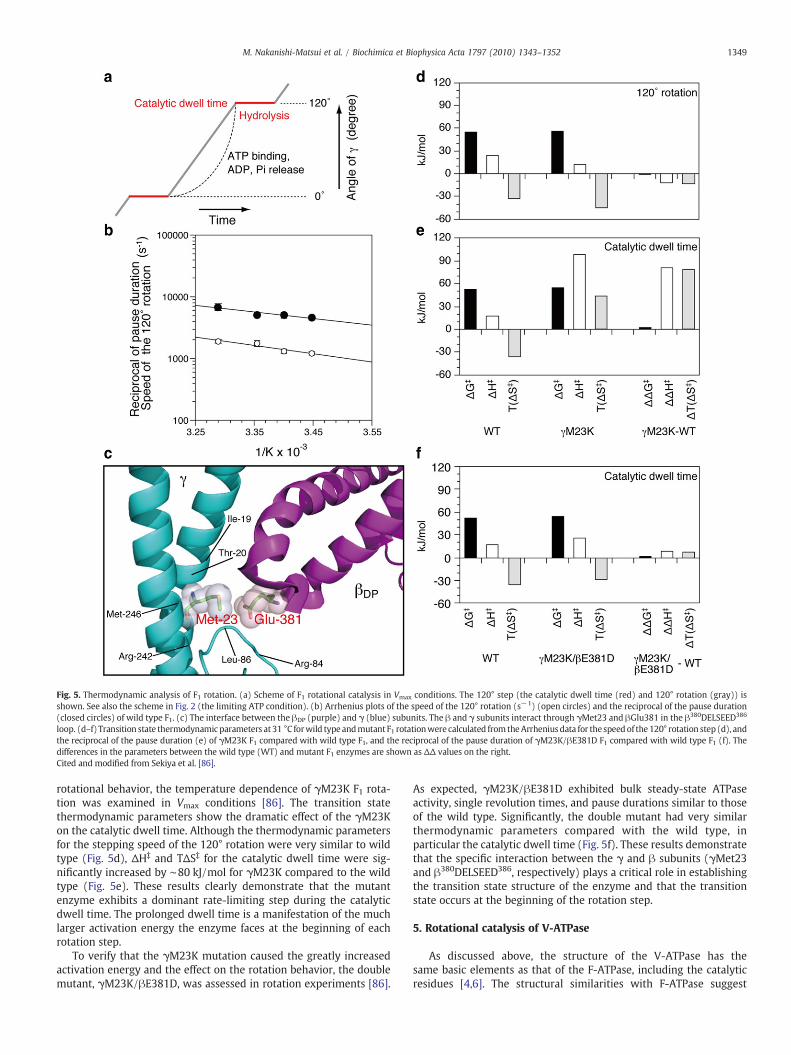
Fig. 5. Thermodynamic analysis of F1 rotation. (a) Scheme of F1 rotational catalysis in Vmax conditions. The 120° step (the catalytic dwell time (red) and 120° rotation (gray)) isshown. See also the scheme in Fig. 2 (the limiting ATP condition). (b) Arrhenius plots of the speed of the 120° rotation (s−1) (open circles) and the reciprocal of the pause duration(closed circles) of wild type F1. (c) The interface between the βDP (purple) and γ (blue) subunits. The β and γ subunits interact through γMet23 and βGlu381 in the β380DELSEED386
loop. (d–f) Transition state thermodynamic parameters at 31 °C forwild type andmutant F1 rotationwere calculated from theArrheniusdata for the speedof the 120° rotation step (d), andthe reciprocal of the pause duration (e) of γM23K F1 compared with wild type F1, and the reciprocal of the pause duration of γM23K/βE381D F1 compared with wild type F1 (f). Thedifferences in the parameters between the wild type (WT) and mutant F1 enzymes are shown as ΔΔ values on the right.Cited and modified from Sekiya et al. [86].
1349M. Nakanishi-Matsui et al. / Biochimica et Biophysica Acta 1797 (2010) 1343–1352
rotational behavior, the temperature dependence of γM23K F1 rota-tion was examined in Vmax conditions [86]. The transition statethermodynamic parameters show the dramatic effect of the γM23Kon the catalytic dwell time. Although the thermodynamic parametersfor the stepping speed of the 120° rotation were very similar to wildtype (Fig. 5d), ΔH‡ and TΔS‡ for the catalytic dwell time were sig-nificantly increased by ∼80 kJ/mol for γM23K compared to the wildtype (Fig. 5e). These results clearly demonstrate that the mutantenzyme exhibits a dominant rate-limiting step during the catalyticdwell time. The prolonged dwell time is a manifestation of the muchlarger activation energy the enzyme faces at the beginning of eachrotation step.
To verify that the γM23K mutation caused the greatly increasedactivation energy and the effect on the rotation behavior, the doublemutant, γM23K/βE381D, was assessed in rotation experiments [86].
As expected, γM23K/βE381D exhibited bulk steady-state ATPaseactivity, single revolution times, and pause durations similar to thoseof the wild type. Significantly, the double mutant had very similarthermodynamic parameters compared with the wild type, inparticular the catalytic dwell time (Fig. 5f). These results demonstratethat the specific interaction between the γ and β subunits (γMet23and β380DELSEED386, respectively) plays a critical role in establishingthe transition state structure of the enzyme and that the transitionstate occurs at the beginning of the rotation step.
5. Rotational catalysis of V-ATPase
As discussed above, the structure of the V-ATPase has thesame basic elements as that of the F-ATPase, including the catalyticresidues [4,6]. The structural similarities with F-ATPase suggest
1350 M. Nakanishi-Matsui et al. / Biochimica et Biophysica Acta 1797 (2010) 1343–1352
catalysis by V-ATPase occurs by a rotational mechanism, although ithas been studied less extensively. An actin filament attached to theG subunit of the yeast V-ATPase rotated upon ATP hydrolysis [93]. It isnoteworthy that this experiment showed rotation of the peripheralstalk against the c ring immobilized on a glass surface. A bacterialhomologue of the V-ATPase also exhibited rotation as subunit rotationof the T. thermophilus V1 sector was observed relative to the A3B3hexamer. In these experiments, a 560 nm bead was attached to the Dor F subunit, which were considered to be the functional homologuesof F1 γ or ε subunits [94]. Rotation of T. thermophilus VoV1 fixedthrough the A3B3 complex was also observed using a bead attached tothe c4c'c" ring [95]. In both cases, the torque generated during rotationwas ∼35 pN nm, slightly lower than that measured for the F-ATPase.
To determine if V1 has a different rotation scheme from F1, thesubsteps of the V1 were recorded with a duplex of 209 nm beadsattached to the V1 D subunit [96]. Upon hydrolysis of ATPγS, the beadsshowed the expected 120° stepping rotation. After changing reactionbuffer to one containing a low concentration of ATP, the same beadduplex showed 120° steps at the same orientation of the D subunit asthat of ATPγS. This result indicated that catalysis and ATP bindingoccurred at the same D subunit orientation, whereas in F-ATPase,catalysis and ATP binding occurred at different orientations. As de-scribed above, catalysis occurs in the F1 enzyme at the 80° positionadvanced from that of ATP binding. Based on these results, couplingbetween the mechanical rotation and chemistry in V1 is likely to bedifferent from that of F1. The scheme comprising 40° and 80° substepsmay not be an intrinsic property for rotational catalysis by all protonpumping ATPases. To elucidate the rotational mechanism of V-ATPasein more detail, the crystal structure of V1 is needed as well as anexperimental system for observing the rotation of a probe thatimparts low viscous drag.
6. Perspectives
“Seeing is believing” is as true in bioenergetics as in any other field.The single molecule approach and direct observation of rotationprovided a new method for answering questions regarding thecomplicated F- and V-ATPase enzymes. The fast rotation of the E. coliF1 was unexpected (∼430 rps at 24 °C) [82] because the Bacillus F1rotated much more slowly (∼130 rps at 24 °C) [73]. The E. coli F1 hasan intrinsic mechanism for rotation at such a high speed. Studiesof rotation are pertinent to understanding the mechanism, thermo-dynamics, and physiological roles. Initial probes used for observingrotation were actin filaments (∼1 μm in length), but great progresshas been made in the past decade using more sophisticated probes.Gold beads of 40–60 nm enabled us to analyze rotation withoutthe artifact of viscous drag. High-speed stepped rotation, stochasticfluctuation of rotation rates, an inhibitory effect of the ε subunit, theprofile of the energy pathway, and pertinent interactions in theβ hinge domain or between β and γ have been demonstrated usingsuch beads [82,83,86,89].
The progress of understanding the F- and V-ATPases raises furtherquestions:
(1) The detailed mechanism of energy coupling between catalysisand proton transport through mechanical rotation remainsunknown. As discussed above, the γ subunit rotates in F1through 120° steps, which is consistent with the three catalyticsites. In the FoF1, the integration of the catalytic 120° steps tothe proton transport via the 10 proton-transporting subunit cAsp residues is not understood. The ratio of the number ofprotons transported to ATP synthesized or hydrolyzed appearsnot to be an integer quantity. Alternatively, the number ofprotons transported in each catalytic 120° step may vary toaccommodate the non-integer H+/ATP ratio: for example,three protons in two catalytic 120° steps and four in one 120°
step may be transported in a complex with 10 c subunits. Asimilar mechanism may be expected for other organisms thathave 11, 13, or 14 c subunits in the c ring [50,55,97]. Whateverthe case, an elastic energy storage mechanism is most likely inthe coupling of proton transport and catalysis through rotation.
(2) The rotation mechanism during ATP synthesis should beexamined to confirm whether or not ATP synthesis is thecomplete reverse of ATP hydrolysis. It is challenging to assayrotation and chemistry driven by a proton gradient at the sametime.
(3) The question remains whether the F-ATPase rotates throughthe same mechanism in physiological conditions. Althoughrotational catalysis has been studied extensively using thethermophilic Bacillus enzyme, the experiments were mostlyperformed at room temperature, i.e., far from the physiologicalcondition of the bacterium. In this regard, further analysis usingthe E. coli enzyme is critical because it can be studied in itsphysiological temperature range. The next challenge is todevelop the techniques to observe rotation in situ.
(4) Many questions about the V-ATPase remain to be answered. Asmentioned above, some V-ATPase subunits have two to fourisoforms that have organellar or tissue-specific distribution[6,7,9,10]. The different isoforms are involved in localizationand in different biological roles. In this regard, the two E sub-unit isoforms, the acrosome-specific E1 and the ubiquitous E2,have different effects on Vo and V1 assembly [98]. It is notknown if E1 confers differences in the rotation behavior com-pared to E2. Observation of the rotation behavior may be themost straightforward method for determining the effectsof different isoforms. It is likely that the different behaviorswill be reflected in the biological functions including determi-nation of organellar pH. An experimental system for observingV-ATPase rotation with a low viscous drag probe is essential foranswering these questions. Many interesting questions remainto be addressed and single molecule experiments on protonpumping ATPases will continue to fascinate us in the nextdecade.
Acknowledgements
Our studies described in this article were supported by CREST(Core Research for Evolutional Science and Technology), JST (JapanScience and Technology Agency), and the Japanese Ministry of Educa-tion, Culture, and Science. Our studies were also supported by fundsfrom the Terumo Life Science Foundation and the Japan Foundationfor Applied Enzymology (to M. N.-M.).
References
[1] P.D. Boyer, The ATP synthase—a splendidmolecular machine, Annu. Rev. Biochem.66 (1997) 717–749.
[2] J. Weber, A.E. Senior, Catalytic mechanism of F1-ATPase, Biochim. Biophys. Acta1319 (1997) 19–58.
[3] D. Stock, C. Gibbons, I. Arechaga, A.G. Leslie, J.E. Walker, The rotary mechanism ofATP synthase, Curr. Opin. Struct. Biol. 10 (2000) 672–679.
[4] T. Nishi, M. Forgac, The vacuolar (H+)-ATPases—nature's most versatile protonpumps, Nat. Rev. Mol. Cell Biol. 3 (2002) 94–103.
[5] R.H. Fillingame, C.M. Angevine, O.Y. Dmitriev, Mechanics of coupling protonmovements to c-ring rotation in ATP synthase, FEBS Lett. 555 (2003) 29–34.
[6] M. Futai, G.H. Sun-Wada, Y.Wada, Proton transporting ATPases: introducing uniqueenzymes coupling catalysis and proton translocation through mechanical rotation,in: M. Futai, Y. Wada, J. Kaplan (Eds.), Handbook of ATPases: Biochemistry, CellBiology, Pathophysiology, 2004, pp. 237–260.
[7] G.H. Sun-Wada, Y. Wada, M. Futai, Diverse and essential roles of mammalianvacuolar-type proton pump ATPase: toward the physiological understanding ofinside acidic compartments, Biochim. Biophys. Acta 1658 (2004) 106–114.
[8] K.W. Beyenbach, H. Wieczorek, The V-type H+ ATPase: molecular structure andfunction, physiological roles and regulation, J. Exp. Biol. 209 (2006) 577–589.
[9] M. Forgac, Vacuolar ATPases: rotary proton pumps in physiology and pathophys-iology, Nat. Rev. Mol. Cell Biol. 8 (2007) 917–929.

1351M. Nakanishi-Matsui et al. / Biochimica et Biophysica Acta 1797 (2010) 1343–1352
[10] V. Marshansky, M. Futai, The V-type H+-ATPase in vesicular trafficking: targeting,regulation and function, Curr. Opin. Cell Biol. 20 (2008) 415–426.
[11] C. von Ballmoos, A. Wiedenmann, P. Dimroth, Essentials for ATP synthesis by F1F0ATP synthases, Annu. Rev. Biochem. 78 (2009) 649–672.
[12] J.M. Shin, O. Vagin, K.Munson, G. Sachs, Gastric H+, K+-ATPase, in:M. Futai, Y.Wada,J. Kaplan (Eds.), Handbook of ATPases : Biochemistry, Cell Biology, Pathophysiology,2004, pp. 179–210.
[13] S. Lecchi, C.W. Slayman, Yeast plasma-membrane H+-ATPase: model system forstudies of structure, function, biogenetics, and regulation, in: M. Futai, Y. Wada,J. Kaplan (Eds.), Handbook of ATPases : Biochemistry, Cell Biology, Pathophys-iology, 2004, pp. 3–24.
[14] T. Hirata, N. Nakamura, H. Omote, Y. Wada, M. Futai, Regulation and reversibilityof vacuolar H+-ATPase, J. Biol. Chem. 275 (2000) 386–389.
[15] M. Nakano, H. Imamura, M. Toei, M. Tamakoshi, M. Yoshida, K. Yokoyama, ATPhydrolysis and synthesis of a rotary motor V-ATPase from Thermus thermophilus,J. Biol. Chem. 283 (2008) 20789–20796.
[16] P.D. Boyer, The binding-changemechanism of ATP synthesis, in: C.P. Lee, G. Schatz,L. Ernster (Eds.), Membrane Bioenergetics, Addison-Wesley, Reading, MA, 1979,pp. 461–479.
[17] J.P. Abrahams, A.G. Leslie, R. Lutter, J.E. Walker, Structure at 2.8 Å resolution ofF1-ATPase from bovine heart mitochondria, Nature 370 (1994) 621–628.
[18] D. Sabbert, S. Engelbrecht, W. Junge, Intersubunit rotation in active F-ATPase,Nature 381 (1996) 623–625.
[19] T.M. Duncan, V.V. Bulygin, Y. Zhou, M.L. Hutcheon, R.L. Cross, Rotation of subunitsduring catalysis by Escherichia coli F1-ATPase, Proc. Natl. Acad. Sci. USA 92 (1995)10964–10968.
[20] E.P. Gogol, E. Johnston, R. Aggeler, R.A. Capaldi, Ligand-dependent structuralvariations in Escherichia coli F1 ATPase revealed by cryoelectron microscopy, Proc.Natl. Acad. Sci. USA 87 (1990) 9585–9589.
[21] H. Noji, R. Yasuda, M. Yoshida, K. Kinosita Jr., Direct observation of the rotation ofF1-ATPase, Nature 386 (1997) 299–302.
[22] Y. Kato-Yamada, H. Noji, R. Yasuda, K. Kinosita Jr., M. Yoshida, Direct observation ofthe rotation of epsilon subunit in F1-ATPase, J. Biol. Chem. 273 (1998) 19375–19377.
[23] Y. Sambongi, Y. Iko, M. Tanabe, H. Omote, A. Iwamoto-Kihara, I. Ueda, T. Yanagida,Y. Wada, M. Futai, Mechanical rotation of the c subunit oligomer in ATP synthase(F0F1): direct observation, Science 286 (1999) 1722–1724.
[24] M. Tanabe, K. Nishio, Y. Iko, Y. Sambongi, A. Iwamoto-Kihara, Y. Wada, M. Futai,Rotation of a complex of the γ subunit and c ring of Escherichia coli ATP synthase.The rotor and stator are interchangeable, J. Biol. Chem. 276 (2001) 15269–15274.
[25] K. Nishio, A. Iwamoto-Kihara, A. Yamamoto, Y. Wada, M. Futai, Subunit rotationof ATP synthase embedded in membranes: α or β subunit rotation relative to thec subunit ring, Proc. Natl. Acad. Sci. USA 99 (2002) 13448–13452.
[26] W. Junge, H. Sielaff, S. Engelbrecht, Torque generation and elastic powertransmission in the rotary FOF1-ATPase, Nature 459 (2009) 364–370.
[27] R.K. Nakamoto, J.A. Baylis Scanlon, M.K. Al-Shawi, The rotary mechanism of theATP synthase, Arch. Biochem. Biophys. 476 (2008) 43–50.
[28] S.D. Watts, Y. Zhang, R.H. Fillingame, R.A. Capaldi, The γ subunit in the Escherichiacoli ATP synthase complex (ECF1F0) extends through the stalk and contacts thec subunits of the F0 part, FEBS Lett. 368 (1995) 235–238.
[29] H. Omote, M. Futai, Mutational analysis of F1F0 ATPase: catalysis and energycoupling, Acta Physiol. Scand. Suppl. 643 (1998) 177–183.
[30] M. Futai, H. Omote, Y. Sambongi, Y. Wada, Synthase (H+ ATPase): couplingbetween catalysis, mechanical work, and proton translocation, Biochim. Biophys.Acta 1458 (2000) 276–288.
[31] H. Ren, W.S. Allison, On what makes the γ subunit spin during ATP hydrolysis byF1, Biochim. Biophys. Acta 1458 (2000) 221–233.
[32] A.E. Senior, S. Nadanaciva, J. Weber, Rate acceleration of ATP hydrolysis by F1Fo-ATPsynthase, J. Exp. Biol. 203 (2000) 35–40.
[33] A.E. Senior, S. Nadanaciva, J. Weber, Themolecular mechanism of ATP synthesis byF1F0-ATP synthase, Biochim. Biophys. Acta 1553 (2002) 188–211.
[34] K. Ida, T. Noumi, M. Maeda, T. Fukui, M. Futai, Catalytic site of F1-ATPaseof Escherichia coli. Lys-155 and Lys-201 of the β subunit are located near theγ-phosphate group of ATP in the presence of Mg2+, J. Biol. Chem. 266 (1991)5424–5429.
[35] H. Omote, M. Maeda, M. Futai, Effects of mutations of conserved Lys-155and Thr-156 residues in the phosphate-binding glycine-rich sequence of theF1-ATPase β subunit of Escherichia coli, J. Biol. Chem. 267 (1992) 20571–20576.
[36] M.Y. Park, H. Omote, M. Maeda, M. Futai, Conserved Glu-181 and Arg-182 residuesof Escherichia coli H+-ATPase (ATP synthase) β subunit are essential for catalysis:properties of 33 mutants between βGlu-161 and βLys-201 residues, J. Biochem.116 (1994) 1139–1145.
[37] S. Löbau, J. Weber, S. Wilke-Mounts, A.E. Senior, F1-ATPase, roles of three catalyticsite residues, J. Biol. Chem. 272 (1997) 3648–3656.
[38] S. Nadanaciva, J. Weber, A.E. Senior, The role of β-Arg-182, an essential catalyticsite residue in Escherichia coli F1-ATPase, Biochemistry 38 (1999) 7670–7677.
[39] A.E. Senior, M.K. Al-Shawi, Further examination of seventeen mutations inEscherichia coli F1-ATPase β-subunit, J. Biol. Chem. 267 (1992) 21471–21478.
[40] H. Omote, N.P. Le, M.Y. Park, M. Maeda, M. Futai, β subunit Glu-185 of Escherichiacoli H+-ATPase (ATP synthase) is an essential residue for cooperative catalysis,J. Biol. Chem. 270 (1995) 25656–25660.
[41] A. Iwamoto, J. Miki, M. Maeda, M. Futai, H+-ATPase γ subunit of Escherichia coli.Role of the conserved carboxyl-terminal region, J. Biol. Chem. 265 (1990)5043–5048.
[42] K. Shin, R.K. Nakamoto, M. Maeda, M. Futai, F0F1-ATPase γ subunit mutationsperturb the coupling between catalysis and transport, J. Biol. Chem. 267 (1992)20835–20839.
[43] R.K. Nakamoto, M. Maeda, M. Futai, The γ subunit of the Escherichia coli ATPsynthase. Mutations in the carboxyl-terminal region restore energy coupling tothe amino-terminal mutant γMet-23–NLys, J. Biol. Chem. 268 (1993) 867–872.
[44] R.K. Nakamoto, M.K. Al-Shawi, M. Futai, The ATP synthase γ subunit. Suppressormutagenesis reveals three helical regions involved in energy coupling, J. Biol.Chem. 270 (1995) 14042–14046.
[45] M.K. Al-Shawi, C.J. Ketchum, R.K. Nakamoto, The Escherichia coli FOF1 γM23Kuncoupling mutant has a higher K0.5 for Pi. Transition state analysis of this mutantand others reveals that synthesis and hydrolysis utilize the same kinetic pathway,Biochemistry 36 (1997) 12961–12969.
[46] C.J. Ketchum, M.K. Al-Shawi, R.K. Nakamoto, Intergenic suppression of the γM23Kuncoupling mutation in F0F1 ATP synthase by βGlu-381 substitutions: the role ofthe β380DELSEED386 segment in energy coupling, Biochem. J. 330 (Pt 2) (1998)707–712.
[47] R.N. Lightowlers, S.M. Howitt, L. Hatch, F. Gibson, G.B. Cox, The proton pore inthe Escherichia coli FoF1-ATPase: a requirement for arginine at position 210 of thea subunit, Biochim. Biophys. Acta 894 (1987) 399–406.
[48] B.D. Cain, R.D. Simoni, Proton translocation by the F1Fo ATPase of Escherichia coli:mutagenic analysis of the α subunit, J. Biol. Chem. 264 (1989) 3292–3300.
[49] S. Eya, M. Maeda, M. Futai, Role of the carboxyl terminal region of H+-ATPase(FOF1) a subunit from Escherichia coli, Arch. Biochem. Biophys. 284 (1991)71–77.
[50] D. Stock, A.G.W. Leslie, J.E. Walker, Molecular architecture of the rotary motor inATP synthase, Science 286 (1999) 1700–1705.
[51] W. Jiang, J. Hermolin, R.H. Fillingame, The preferred stoichiometry of c subunits inthe rotary motor sector of Escherichia coli ATP synthase is 10, Proc. Natl. Acad. Sci.USA 98 (2001) 4966–4971.
[52] N. Mitome, T. Suzuki, S. Hayashi, M. Yoshida, Thermophilic ATP synthase has adecamer c-ring: indication of noninteger 10:3 H+/ATP ratio and permissiveelastic coupling, Proc. Natl. Acad. Sci. USA 101 (2004) 12159–12164.
[53] H. Stahlberg,D.J.Muller, K. Suda,D. Fotiadis, A. Engel, T.Meier,U.Matthey, P. Dimroth,BacterialNa+–ATP synthasehas anundecameric rotor, EMBORep. 2 (2001)229–233.
[54] T. Meier, U. Matthey, C. von Ballmoos, J. Vonck, T. Krug von Nidda, W. Kuhlbrandt,P. Dimroth, Evidence for structural integrity in the undecameric c-rings isolatedfrom sodium ATP synthases, J. Mol. Biol. 325 (2003) 389–397.
[55] H. Seelert, A. Poetsch, N.A. Dencher, A. Engel, H. Stahlberg, D.J. Muller, Structuralbiology. Proton-powered turbine of a plant motor, Nature 405 (2000) 418–419.
[56] P. Turina, D. Samoray, P. Gräber, H+/ATP ratio of proton transport-coupled ATPsynthesis and hydrolysis catalysed by CF0F1-liposomes, EMBO J. 22 (2003)418–426.
[57] D. Pogoryelov, J. Yu, T. Meier, J. Vonck, P. Dimroth, D.J. Muller, The c15 ring of theSpirulina platensis F-ATP synthase: F1/F0 symmetry mismatch is not obligatory,EMBO Rep. 6 (2005) 1040–1044.
[58] R.L. Cross, V. Muller, The evolution of A-, F-, and V-type ATP synthases andATPases: reversals in function and changes in the H+/ATP coupling ratio, FEBSLett. 576 (2004) 1–4.
[59] L.S. Holliday, M. Lu, B.S. Lee, R.D. Nelson, S. Solivan, L. Zhang, S.L. Gluck, The amino-terminal domain of the B subunit of vacuolar H+-ATPase contains a filamentousactin binding site, J. Biol. Chem. 275 (2000) 32331–32337.
[60] M. Lu, L.S. Holliday, L. Zhang, W.A. Dunn Jr., S.L. Gluck, Interaction betweenaldolase and vacuolar H+-ATPase: evidence for direct coupling of glycolysis to theATP-hydrolyzing proton pump, J. Biol. Chem. 276 (2001) 30407–30413.
[61] H. Omote, N. Sambonmatsu, K. Saito, Y. Sambongi, A. Iwamoto-Kihara, T. Yanagida,Y. Wada, M. Futai, The γ-subunit rotation and torque generation in F1-ATPasefrom wild-type or uncoupled mutant Escherichia coli, Proc. Natl. Acad. Sci. USA 96(1999) 7780–7784.
[62] H.Hosokawa,M.Nakanishi-Matsui, S. Kashiwagi, I. Fujii-Taira, K.Hayashi, A. Iwamoto-Kihara, Y.Wada, M. Futai, ATP-dependent rotation ofmutant ATP synthases defectivein proton transport, J. Biol. Chem. 280 (2005) 23797–23801.
[63] S.P. Tsunoda, R. Aggeler, M. Yoshida, R.A. Capaldi, Rotation of the c subunitoligomer in fully functional F1FO ATP synthase, Proc. Natl. Acad. Sci. USA 98 (2001)898–902.
[64] P.C. Jones, J. Hermolin, R.H. Fillingame, Mutations in single hairpin units of ge-netically fused subunit c provide support for a rotary catalytic mechanism in FOF1ATP synthase, J. Biol. Chem. 275 (2000) 11355–11360.
[65] J.A. Scanlon, M.K. Al-Shawi, R.K. Nakamoto, A rotor-stator cross-link in theF1-ATPase blocks the rate-limiting step of rotational catalysis, J. Biol. Chem.283 (2008) 26228–26240.
[66] M. Diez, B. Zimmermann, M. Börsch, M. König, E. Schweinberger, S. Steigmiller,R. Reuter, S. Felekyan, V. Kudryavtsev, C.A. Seidel, P. Gräber, Proton-poweredsubunit rotation in single membrane-bound F0F1-ATP synthase, Nat. Struct. Mol.Biol. 11 (2004) 135–141.
[67] B.Zimmermann,M.Diez,N. Zarrabi, P. Gräber,M.Börsch,Movements of the ε-subunitduring catalysis and activation in singlemembrane-boundH+-ATP synthase, EMBO J.24 (2005) 2053–2063.
[68] M. Börsch, M. Diez, B. Zimmermann, R. Reuter, P. Gräber, Stepwise rotation of theγ-subunit of EF0F1-ATP synthase observed by intramolecular single-moleculefluorescence resonance energy transfer, FEBS Lett. 527 (2002) 147–152.
[69] H. Itoh, A. Takahashi, K. Adachi, H. Noji, R. Yasuda, M. Yoshida, K. Kinosita,Mechanically driven ATP synthesis by F1-ATPase, Nature 427 (2004) 465–468.
[70] Y. Rondelez, G. Tresset, T. Nakashima, Y. Kato-Yamada, H. Fujita, S. Takeuchi, H. Noji,Highly coupled ATP synthesis by F1-ATPase single molecules, Nature 433 (2005)773–777.
[71] R. Yasuda, H. Noji, K. Kinosita Jr., M. Yoshida, F1-ATPase is a highly efficientmolecular motor that rotates with discrete 120° steps, Cell 93 (1998)1117–1124.
1352 M. Nakanishi-Matsui et al. / Biochimica et Biophysica Acta 1797 (2010) 1343–1352
[72] K. Adachi, R. Yasuda, H. Noji, H. Itoh, Y. Harada, M. Yoshida, K. Kinosita Jr., Steppingrotation of F1-ATPase visualized through angle-resolved single-fluorophoreimaging, Proc. Natl. Acad. Sci. USA 97 (2000) 7243–7247.
[73] R. Yasuda, H. Noji, M. Yoshida, K. Kinosita Jr., H. Itoh, Resolution of distinctrotational substeps by submillisecond kinetic analysis of F1-ATPase, Nature 410(2001) 898–904.
[74] K. Shimabukuro, R. Yasuda, E. Muneyuki, K.Y. Hara, K. Kinosita Jr., M. Yoshida,Catalysis and rotation of F1 motor: cleavage of ATP at the catalytic site occursin 1 ms before 40° substep rotation, Proc. Natl. Acad. Sci. USA 100 (2003)14731–14736.
[75] J.A. Scanlon, M.K. Al-Shawi, N.P. Le, R.K. Nakamoto, Determination of the partialreactions of rotational catalysis in F1-ATPase, Biochemistry 46 (2007) 8785–8797.
[76] S. Nadanaciva, J. Weber, A.E. Senior, New probes of the F1-ATPase catalytictransition state reveal that two of the three catalytic sites can assume a transitionstate conformation simultaneously, Biochemistry 39 (2000) 9583–9590.
[77] T. Nishizaka, K. Oiwa, H. Noji, S. Kimura, E. Muneyuki, M. Yoshida, K. Kinosita Jr.,Chemomechanical coupling in F1-ATPase revealed by simultaneous observation ofnucleotide kinetics and rotation, Nat. Struct. Mol. Biol. 11 (2004) 142–148.
[78] K. Adachi, K. Oiwa, T. Nishizaka, S. Furuike, H. Noji, H. Itoh, M. Yoshida, K. KinositaJr., Coupling of rotation and catalysis in F1-ATPase revealed by single-moleculeimaging and manipulation, Cell 130 (2007) 309–321.
[79] R. Watanabe, R. Iino, K. Shimabukuro, M. Yoshida, H. Noji, Temperature-sensitivereaction intermediate of F1-ATPase, EMBO Rep. 9 (2008) 84–90.
[80] R.I. Menz, J.E. Walker, A.G. Leslie, Structure of bovine mitochondrial F1-ATPasewith nucleotide bound to all three catalytic sites: implications for the mechanismof rotary catalysis, Cell 106 (2001) 331–341.
[81] S. Furuike, K. Adachi, N. Sakaki, R. Shimo-Kon, H. Itoh, E. Muneyuki, M. Yoshida,K. Kinosita Jr., Temperature dependence of the rotation and hydrolysis activitiesof F1-ATPase, Biophys. J. 95 (2008) 761–770.
[82] M. Nakanishi-Matsui, S. Kashiwagi, H. Hosokawa, D.J. Cipriano, S.D. Dunn, Y. Wada,M. Futai, Stochastic high-speed rotation of Escherichia coliATP synthase F1 sector: theε subunit-sensitive rotation, J. Biol. Chem. 281 (2006) 4126–4131.
[83] M.Nakanishi-Matsui, S. Kashiwagi, T.Ubukata, A. Iwamoto-Kihara, Y.Wada,M. Futai,Rotational catalysis of Escherichia coli ATP synthase F1 sector. Stochastic fluctuationand a key domain of the β subunit, J. Biol. Chem. 282 (2007) 20698–20704.
[84] M. Nakanishi-Matsui, M. Futai, Stochastic proton pumping ATPases: from singlemolecules to diverse physiological roles, IUBMB Life 58 (2006) 318–322.
[85] M. Nakanishi-Matsui, M. Futai, Stochastic rotational catalysis of proton pumpingF-ATPase, Philos. Trans. R. Soc. Lond. B Biol. Sci. 363 (2008) 2135–2142.
[86] M. Sekiya, R.K. Nakamoto, M.K. Al-Shawi, M. Nakanishi-Matsui, M. Futai,Temperature dependence of single molecule rotation of the Escherichia coli ATP
synthase F1 sector reveals the importance of γ–β subunit interactions in thecatalytic dwell, J. Biol. Chem. 284 (2009) 22401–22410.
[87] H. Yagi, N. Kajiwara, T. Iwabuchi, K. Izumi, M. Yoshida, H. Akutsu, Stepwisepropagation of the ATP-induced conformational change of the F1-ATPase βsubunit revealed by NMR, J. Biol. Chem. 284 (2009) 2374–2382.
[88] A. Iwamoto, H. Omote, H. Hanada, N. Tomioka, A. Itai, M. Maeda, M. Futai,Mutations in Ser174 and the glycine-rich sequence (Gly149, Gly150, and Thr156)in the β subunit of Escherichia coli H+-ATPase, J. Biol. Chem. 266 (1991)16350–16355.
[89] S. Kashiwagi, A. Iwamoto-Kihara, M. Kojima, T. Nonaka, M. Futai, M. Nakanishi-Matsui, Effects of mutations in the β subunit hinge domain on ATP synthase F1sector rotation: interaction between Ser 174 and Ile 163, Biochem. Biophys. Res.Commun. 365 (2008) 227–231.
[90] H. Kanazawa, H. Hama, B.P. Rosen, M. Futai, Deletion of seven amino acid residuesfrom the γ subunit of Escherichia coli H+-ATPase causes total loss of F1 assemblyon membranes, Arch. Biochem. Biophys. 241 (1985) 364–370.
[91] M.K. Al-Shawi, R.K. Nakamoto, Mechanism of energy coupling in the FOF1-ATPsynthase: the uncoupling mutation, γM23K, disrupts the use of binding energy todrive catalysis, Biochemistry 36 (1997) 12954–12960.
[92] M.K. Al-Shawi, C.J. Ketchum, R.K. Nakamoto, Energy coupling, turnover, andstability of the F0F1 ATP synthase are dependent on the energy of interactionbetween γ and β subunits, J. Biol. Chem. 272 (1997) 2300–2306.
[93] T. Hirata, A. Iwamoto-Kihara, G.H. Sun-Wada, T. Okajima, Y. Wada, M. Futai,Subunit rotation of vacuolar-type proton pumping ATPase: relative rotation of theG and C subunits, J. Biol. Chem. 278 (2003) 23714–23719.
[94] H. Imamura, M. Nakano, H. Noji, E. Muneyuki, S. Ohkuma, M. Yoshida, K. Yokoyama,Evidence for rotation of V1-ATPase, Proc. Natl. Acad. Sci. USA 100 (2003)2312–2315.
[95] K. Yokoyama, M. Nakano, H. Imamura, M. Yoshida, M. Tamakoshi, Rotation of theproteolipid ring in the V-ATPase, J. Biol. Chem. 278 (2003) 24255–24258.
[96] H. Imamura, M. Takeda, S. Funamoto, K. Shimabukuro, M. Yoshida, K. Yokoyama,Rotation scheme of V1-motor is different from that of F1-motor, Proc. Natl. Acad.Sci. USA 102 (2005) 17929–17933.
[97] T. Meier, P. Polzer, K. Diederichs, W. Welte, P. Dimroth, Structure of the rotor ringof F-type Na+-ATPase from Ilyobacter tartaricus, Science 308 (2005) 659–662.
[98] K. Hayashi, G.H. Sun-Wada, Y. Wada, M. Nakanishi-Matsui, M. Futai, Defectiveassembly of a hybrid vacuolar H+-ATPase containing the mouse testis-specific E1isoform and yeast subunits, Biochim. Biophys. Acta 1777 (2008) 1370–1377.
[99] M.W. Bowler, M.G. Montgomery, A.G. Leslie, J.E. Walker, Ground state structure ofF1-ATPase from bovine heart mitochondria at 1.9 Å resolution, J. Biol. Chem. 282(2007) 14238–14242.