boott, c., leitao, e., hayward, d., laine, r., mahou, p ... › files › 168725315 ›...
TRANSCRIPT
Boott, C., Leitao, E., Hayward, D., Laine, R., Mahou, P., Guerin, G., Winnik,M., Richardson, R., Kaminski, C., Whittell, G., & Manners, I. (2018).Probing the Growth Kinetics for the Formation of Uniform 1D BlockCopolymer Nanoparticles by Living Crystallization-Driven Self-Assembly.ACS Nano, 12(9), 8920-8933. https://doi.org/10.1021/acsnano.8b01353
Peer reviewed version
Link to published version (if available):10.1021/acsnano.8b01353
Link to publication record in Explore Bristol ResearchPDF-document
This is the author accepted manuscript (AAM). The final published version (version of record) is available onlinevia ACS at https://pubs.acs.org/doi/10.1021/acsnano.8b01353 . Please refer to any applicable terms of use ofthe publisher.
University of Bristol - Explore Bristol ResearchGeneral rights
This document is made available in accordance with publisher policies. Please cite only the publishedversion using the reference above. Full terms of use are available: http://www.bristol.ac.uk/pure/user-guides/explore-bristol-research/ebr-terms/
1
Probing the Growth Kinetics for the Formation of Uniform 1D Block
Copolymer Nanoparticles by Living Crystallization-Driven Self-Assembly
Charlotte E. Boott,†,ɸ Erin M. Leitao,†,ɸ, Dominic W. Hayward, † Romain F. Laine,§ Pierre Mahou,§
Gerald Guerin,ψ Mitchell A. Winnik,ψ Robert M. Richardson,‡,* Clemens F. Kaminski,§,* George R.
Whittell, †,* and Ian Manners†,*
†School of Chemistry, University of Bristol, Cantock’s Close, Bristol, BS8 1TS, U.K.
‡School of Physics, University of Bristol, Tyndall Avenue, Bristol, BS8 1TL, U.K.
§Department of Chemical Engineering and Biotechnology, University of Cambridge, Philippa
Fawcett Drive, Cambridge, CB3 0AS, U.K.
ψ Chemistry Department, University of Toronto, 80 St. George Street, Toronto, M5S 3H6, Canada
School of Chemical Sciences, University of Auckland, 23 Symonds Street, Auckland, 1010, New
Zealand
Please address correspondence to [email protected], [email protected],
[email protected] and [email protected]
ɸThese authors contributed equally
Abstract
Living crystallization-driven self-assembly (CDSA) is a seeded growth method for crystallizable
block copolymers (BCPs) and related amphiphiles in solution and has recently emerged as a highly
promising and versatile route to uniform core-shell nanoparticles (micelles) with control of
dimensions and architecture. However, the factors that influence the rate of nanoparticle growth have
not been systematically studied. Using transmission electron microscopy (TEM), small- and wide-
angle X-ray scattering (SAXS and WAXS), together with the super-resolution fluorescence
microscopy techniques, we have investigated the kinetics of the seeded growth of
poly(ferrocenyldimethylsilane)-b-(polydimethylsiloxane) (PFS-b-PDMS), as a model living CDSA
system for those employing, for example, crystallizable emissive and biocompatible polymers. By

2
altering various self-assembly parameters including concentration, temperature, solvent, and BCP
composition our results have established that the time taken to prepare fiber-like micelles via the
living CDSA method can be reduced by decreasing temperature, by employing solvents that are
poorer for the crystallizable PFS core-forming block, and by increasing the length of the PFS core-
forming block. These results are of general importance for the future optimization of a wide variety of
living CDSA systems. Our studies also demonstrate that the growth kinetics for living CDSA do not
exhibit the first order dependence of growth rate on unimer concentration anticipated by analogy with
living covalent polymerizations of molecular monomers. This difference may be caused by the
combined influence of chain conformational effects of the BCP on addition to the seed termini, and
chain length dispersity.
Keywords: block copolymers, self-assembly, nanofibers, reaction kinetics, laser scanning confocal
microscopy.
A key scientific challenge of widespread current interest involves the creation of well-defined
functional materials based on soft matter that exist on a length-scale of nanometers to microns.1–5 One
key strategy employed to achieve this goal is the solution processing of block copolymers (BCPs)
with amorphous core-forming segments, which produces core-shell nanoparticles (micelles) with a
range of morphologies and functionalities.6,7 This has resulted in a variety of actual and potential
applications from drug delivery to composite reinforcement.8–11 Nevertheless, a major problem in this
field is the limited ability to create uniform samples of micellar nanoparticles with predictable and
anisotropic shape, tunable dimensions, and controllable spatially-confined core and/or corona
chemistries.
The solution self-assembly of BCPs with a crystallizable core-forming block has recently
emerged as a powerful route to core-shell nanoparticles with high levels of precision and control.
Upon addition of a selective solvent for the corona-forming block to a molecularly dissolved BCP
(unimer), both crystallization of the core-forming block and amphiphilicity influence micelle
formation, with low curvature morphologies, such as cylinders and platelets, normally being favoured.

3
This process has now been studied for a wide range of BCPs with different crystallizable core-
forming segments and has been termed crystallization-driven self-assembly (CDSA).12–35
Length control can be achieved using a CDSA approach through the application of seeded
growth36 techniques to produce low dispersity micelles. For example, previous work has shown that
the exposed crystal faces at the termini of pre-existing cylindrical micelles with a crystalline
poly(ferrocenyldimethylsilane) (PFS)37 core remain active to further growth via epitaxy.38 This
technique can then be used to prepare monodisperse micelles of controlled length by the addition of a
known amount of unimer to a colloidal solution of small crystallite seeds, generated by the sonication
of multimicron long cylindrical micelles (Scheme 1).36 The control of length through variation of the
unimer-to-seed ratio can be considered to be analogous to molecular weight control in a living
covalent chain growth polymerization, and the process has thus been termed living CDSA. Using this
seeded growth approach, the sequential addition of BCPs with a common crystallizable core-forming
block,38 or different core blocks with a small lattice mismatch,39 results in the formation of a range of
multicompartment block comicelles with nanosegregated functionality.40,41 A variety of BCPs with
different crystallizable core-forming blocks has been shown to undergo living CDSA32,37,39,42–44 and
the process has been extended to amphiphiles that form supramolecular polymers.45–56 For the BCPs,
those with a PFS core-forming block have been the most widely studied to date. Living CDSA of PFS
BCPs has recently been shown to be scalable57 and to enable the preparation of a wide range of BCP
nanoparticles, including 2D platelets,58,59 non-centrosymmetric60 and branched cylinders61 and
hierarchical structures, such as supermicelles62,63 and superlattices.64 Furthermore, a key feature of the
resulting crystalline BCP assemblies is that a high barrier exists to unimer exchange between micelles
at near ambient temperatures, which prevents equilibration, and the consequential loss of uniformity
and complexity. PFS BCP-based nanoparticles also have advantages for many studies as staining is
unnecessary for TEM analysis, as a result of the electron-dense Fe-rich PFS core and coronal
derivatization (e.g. for functionalization by fluorescent dyes) is synthetically well-developed.37
4
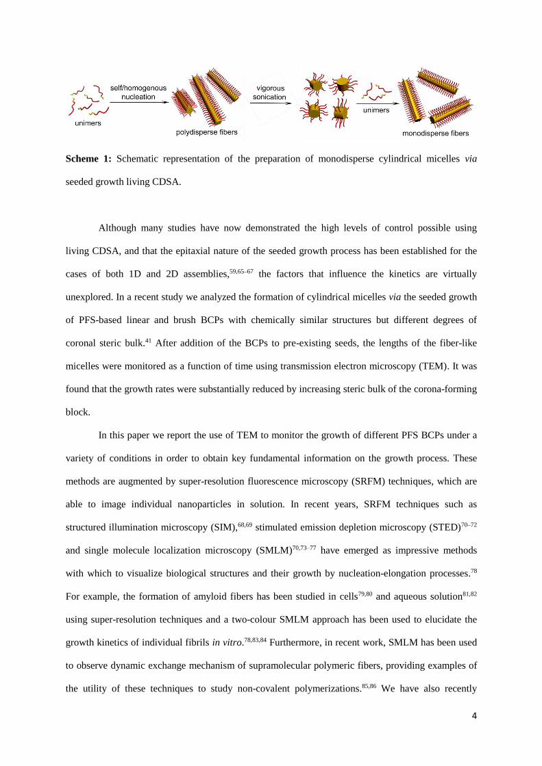
Scheme 1: Schematic representation of the preparation of monodisperse cylindrical micelles via
seeded growth living CDSA.
Although many studies have now demonstrated the high levels of control possible using
living CDSA, and that the epitaxial nature of the seeded growth process has been established for the
cases of both 1D and 2D assemblies,59,65–67 the factors that influence the kinetics are virtually
unexplored. In a recent study we analyzed the formation of cylindrical micelles via the seeded growth
of PFS-based linear and brush BCPs with chemically similar structures but different degrees of
coronal steric bulk.41 After addition of the BCPs to pre-existing seeds, the lengths of the fiber-like
micelles were monitored as a function of time using transmission electron microscopy (TEM). It was
found that the growth rates were substantially reduced by increasing steric bulk of the corona-forming
block.
In this paper we report the use of TEM to monitor the growth of different PFS BCPs under a
variety of conditions in order to obtain key fundamental information on the growth process. These
methods are augmented by super-resolution fluorescence microscopy (SRFM) techniques, which are
able to image individual nanoparticles in solution. In recent years, SRFM techniques such as
structured illumination microscopy (SIM),68,69 stimulated emission depletion microscopy (STED)70–72
and single molecule localization microscopy (SMLM)70,73–77 have emerged as impressive methods
with which to visualize biological structures and their growth by nucleation-elongation processes.78
For example, the formation of amyloid fibers has been studied in cells79,80 and aqueous solution81,82
using super-resolution techniques and a two-colour SMLM approach has been used to elucidate the
growth kinetics of individual fibrils in vitro.78,83,84 Furthermore, in recent work, SMLM has been used
to observe dynamic exchange mechanism of supramolecular polymeric fibers, providing examples of
the utility of these techniques to study non-covalent polymerizations.85,86 We have also recently
5
demonstrated the use of STED and two-colour SMLM to image PFS-based BCP micelles in organic
media, which demonstrated that the length data obtained by TEM analysis on dried samples is
representative of the micelles in their native environment.87 Based on these considerations, as
complementary experiments to TEM, the SRFM techniques, STED and two-colour SMLM were
employed to study the growth by living CDSA in solution. In addition, our micelle growth data were
fitted to kinetic models, which enabled rate constants to be extracted quantitatively and detailed
information on the living CDSA growth process to be obtained.
Results
Monitoring the growth of PFS63-b-PDMS513 one-dimensional micelles by TEM
In a typical living CDSA experiment, a PFS63-b-PDMS51388 [PDMS = polydimethylsiloxane,
subscripts indicate the number average degree of polymerization (DPn)] unimer solution in THF was
added to a solution of crystallite seed micelles, prepared by sonication of longer cylindrical micelles,
in a selective solvent for the corona-forming block. A range of parameters was subsequently varied
including concentration, temperature, selective solvent and percentage common solvent, and BCP
composition. To monitor the effect of each of these parameters on the growth of PFS63-b-PDMS513
BCP micelles, aliquots were taken from the reaction mixtures at set time points, placed on carbon-
coated copper grids and analyzed by TEM. This enabled the micelle length at different time points
throughout the growth process to be determined.
The crystalline core structure of PFS BCP micelles
The key feature of PFS-based BCP micelles from the perspective of living CDSA is the
crystalline PFS core. Previous studies using wide-angle X-ray scattering (WAXS) of PI637-b-PFS53
cylindrical micelles (PI = polyisoprene) have shown that the PFS chains extend perpendicular to the
long axis of the micelle and pack with pseudo hexagonal symmetry.65 Further studies involving
micelles from a range of different PFS-containing BCPs suggest that the core cross-section can be
either rectangular, elliptical or near circular, depending on the degree of polymerization of the core-
forming block.89,90 This change in core structure results from different numbers of chain-folds within

6
the cross-sectional area, while the spacing between chains remains the same. The majority of
experiments discussed herein involve the use of the same PFS-containing BCP, namely, PFS63-b-
PDMS513. This enabled us to investigate whether the factors that influence the micelle growth rate
also had a simultaneous effect on the micelle cross-section. For the BCP used in these experiments,
both short (Ln = 92 nm, PDI = Lw / Ln = 1.07, where Ln and Lw represent the number/weight average
contour length,) and long (Ln = 1757 nm, PDI = 1.01) low length dispersity micelles were prepared by
seeded growth, and the micelle structures were analyzed by small- and wide-angle X-ray scattering
(SAXS and WAXS) and TEM (Figure S1). The SAXS data for both sets of micelles was best fitted by
a model for infinite rigid rods with a rectangular cross-section. This afforded core dimensions of 8.6 ±
1.8 x 7.6 ± 2.8 nm and 9.4 ± 0.8 x 7.4 ± 0.1 nm for the short and long micelles, respectively (Table
S1). The model also incorporated a corona extending from the narrow side of the core, which refined
as 18.5 ± 3.4 nm for the short micelles and 17.4 ± 0.5 nm for the long examples. It is noteworthy that
the polydispersity in the wide dimension of the core was much larger than in the other, which may
suggest a deviation from a purely rectangular cross-section. Analysis by high-resolution TEM
revealed number average contour widths, Wn, of 11 nm for both samples, which we view to
predominantly reflect the core width. These data are therefore in reasonable agreement with wider
core dimension as determined by SAXS, and together demonstrate that the micellar structure does not
change with contour length. The linear aggregation numbers (i.e. the number of PFS BCP molecules
per unit length) were 3.5 ± 1.5 and 3.7 ± 0.3 chains/nm for the short and long micelles, respectively
(see Supporting Information page 4), which are the same within error.91
Varying the growth conditions for PFS63-b-PDMS513 one-dimensional micelles
Effect of initial unimer concentration
The overall length of fiber-like micelles formed in living CDSA seeded growth experiments is
proportional to the unimer-to-seed ratio. This is analogous to living covalent polymerizations in which
the chain length is dependent on the ratio of monomer to initiator. It was therefore of interest to
investigate whether this analogy also applied to the reaction kinetics. In a living covalent
polymerization the reaction is first order in both monomer and initiator concentrations,92 and this

7
would translate to the reaction being first order in both unimer, [U], and seed, [S], concentrations for
living CDSA. As the latter remains constant during the course of the reaction, and initiation occurs
from both ends of the seed, [S] should be substituted by 2[S]0 (the concentration at t = 0) in the
differential form of the rate equation (Eq. 1).
𝑑[𝑈]
𝑑𝑡= −𝑘[𝑈]2[𝑆]0 Eq. 1
Integrating Eq. 1 then affords an expression for the concentration of unimer as a function of time,
[U(t)], in terms of the initial concentrations (Eq. 2).
[𝑈(𝑡)] = [𝑈]0𝑒−2𝑘[𝑆]0𝑡 Eq. 2
Determining the concentrations of micelle and unimeric BCP in solution, however, is problematic.
We therefore chose to monitor the reaction kinetics by determining the average length, L(t), of the
individual micelles formed, which could be measured microscopically. The total length grown over all
micelles, Ltot(t), is related to the concentrations of unimer, [U(t)] and U0, the volume of solution, V,
and the linear aggregation number (number of BCP molecules per unit length), Nagg/L (Eq. 3). We have
shown (vide supra) that the latter is constant with micelle length, under the growth conditions used.
[𝑈(𝑡)] = [𝑈]0 −𝐿𝑡𝑜𝑡(𝑡)𝑁𝑎𝑔𝑔/𝐿
𝑁𝐴𝑉 Eq. 3
Rearranging Eq. 3 to make Ltot(t) the subject and dividing by the number of micelles, which for a
living process equals the number of seeds, Nseed, then affords an expression for the length grown
Lgrown(t) for each micelle (Eq. 4).
𝐿𝑔𝑟𝑜𝑤𝑛(𝑡) =1
𝑁𝑠𝑒𝑒𝑑
𝑁𝐴𝑉
𝑁𝑎𝑔𝑔/𝐿[𝑈]0(1 − 𝑒−2𝑘[𝑆]0𝑡) Eq. 4
Nseed is related to [S]0 (Eq. 5), and this leads to Eq.6.
𝑁𝑠𝑒𝑒𝑑 = [𝑆]0𝑁𝐴𝑉 Eq. 5
𝐿𝑔𝑟𝑜𝑤𝑛(𝑡) =1
𝑁𝑎𝑔𝑔/𝐿
[𝑈]0
[𝑆]0(1 − 𝑒−2𝑘[𝑆]0𝑡) Eq. 6
The experimentally determined length, L(t), however, also includes that of the seed, Lseed, and this
needs to be taken into account in the final model (Eq. 7).

8
𝐿(𝑡) =1
𝑁𝑎𝑔𝑔/𝐿
[𝑈]0
[𝑆]0(1 − 𝑒−2𝑘[𝑆]0𝑡) + 𝐿𝑠𝑒𝑒𝑑 Eq. 7
We investigated the effect of initial unimer concentration on the growth of PFS63-b-PDMS513
fiber-like micelles. Varying the initial unimer concentration (from 10 to 30 mg/mL) while keeping the
concentration of common solvent and seed concentration constant, resulted in different unimer-to-
seed ratios and therefore different final micelle lengths. These (Ln = 613 ± 106, 982 ± 139 and 1798 ±
180 nm for [U]0 = of 10, 20 and 30 mg/mL, respectively) were the same to the 3 confidence level as
the theoretically predicted lengths (Ln = 516, 973, 1431 nm for [U]0 = 10, 20 and 30 mg/mL,
respectively), and thus demonstrated the appropriateness of Eq. 7 in this regard. Despite having
different final lengths we expected all micelles to have the same linear aggregation number based on
the SAXS/WAXS data for the short and long cylindrical micelles discussed above. The time required
for the micelles to reach their final length, however, was approximately the same for all 3 experiments
(Figure 1 and 2a).
If Eq. 7 correctly describes the micellar length as a function of time, then a plot of the
logarithm of L∞ - L(t), which approximates to Lfinal - L(t), versus t should generate a straight line. This,
however, was not found to be the case (r-squared = 0.61-0.82) and neither did the data afford a
straight-line plot for 1/(Lfinal - L(t)) versus t (r-squared = 0.86-0.94), which would be characteristic of a
second order reaction (Figure 1). Non-integer reaction orders can be evaluated by the initial rates
method, which involves analysis of the linear region of the Lfinal - L(t) vs. t plot (Figure S2). A log-log
plot of these rates vs. [U]0 then yields the reaction order as the gradient of the line (Figure S3). This
method gave an order of 1.16 ± 0.25 with respect to the concentration of unimer, which contrary to
the ln(Lfinal - L(t)) vs. t plot suggests that the reaction is first-order. This discrepancy, however, is most
likely a result of the reduced data set used in the initial rates method, which does not capture the
larger deviation from first-order behaviour that occurs at later time points. We therefore explored
other data fitting approaches to extract rate information from our experimental data.

9
Figure 1: Lengths of PFS63-b-PDMS513 micelles as a function of time, monitored over a period of
24 h after the addition of unimer (16 L of 10, 20 or 30 mg/mL of PFS63-b-PDMS513 THF solution,
respectively) to 200 L of a 0.1 mg/mL PFS63-b-PDMS513 seed micelles in EtOAc, (Ln = 57 nm, Lw/Ln
= 1.11), a) first order plot of ln(Lfinal - L(t)) versus time for different unimer concentrations b) second
order plot of 1/(Lfinal - L(t)) versus time for different unimer concentrations. Error bars propagated
from ±σ (standard deviation) of the micelle length distribution.
Effect of seed concentration
We investigated the effect of seed concentration (1.0 ×10⁻², 6.7 ×10⁻³ and 3.3 ×10⁻³ mg/mL)
while keeping the initial concentration of unimer and common solvent constant. This resulted in
different unimer-to-seed ratios and therefore different final micelle lengths (Ln = 613 ± 106, 919 ± 101
and 1664 ± 13 nm for [S]0 = 1.0 ×10⁻², 6.7 ×10⁻³ and 3.3 ×10⁻³ mg/mL, respectively), in a similar
manner to varying the initial unimer concentration. As before, these values were within 3 of the
theoretically predicted lengths (Ln = 516, 746 and 1429 nm for [S]0 = 1.0 ×10⁻², 6.7 ×10⁻³ and 3.3 ×10-
3 mg/mL, respectively). The time required for the micelles to reach their final length, however, was
slightly different for the 3 concentrations, and the precise form of this data will be discussed in detail
in the Data Fitting Section.
Eq. 1 indicates that the rate of unimer consumption should be first order in [S], if the reaction
kinetics are analogous to those of a living covalent polymerisation. With regards to the rate of micelle
growth, however, the situation is slightly different. Differentiating Eq. 7 and examining the initial rate
(2k[S]0t ≪ 1) demonstrates that dL/dt ≃ 2[U]0k/Nagg/L, which is independent of [S]0. The condition of
0
0.005
0.01
0.015
0.02
0 500 1000 1500
1/(L f
inal
-L(t
))
Time (min)
10 mg/mL
20 mg/mL
30 mg/mL
3
5
7
9
0 500 1000 1500
ln(L
fin
al-L
(t))
Time (min)
10 mg/mL
20 mg/mL
30 mg/mL
a) b)
10
constant [U]0 required to probe the effect of varying [S]0, however, is also only true at low extents of
growth. We therefore determined initial rates at each seed concentration (Figure S4), and represented
the data as a function of [S]0 in the form of a log-log plot (Figures S5). This afforded a reaction order
of -0.75 ± 0.05 in the concentration of seeds, which is not consistent with the expected zero-order
kinetics of micelle growth. The method of initial rates can have limitations in that reaction
complexities may not be reflected over the short time-frame that the initial rate can be extracted. We
therefore sought a model that could fit the data over the whole course of the reaction.
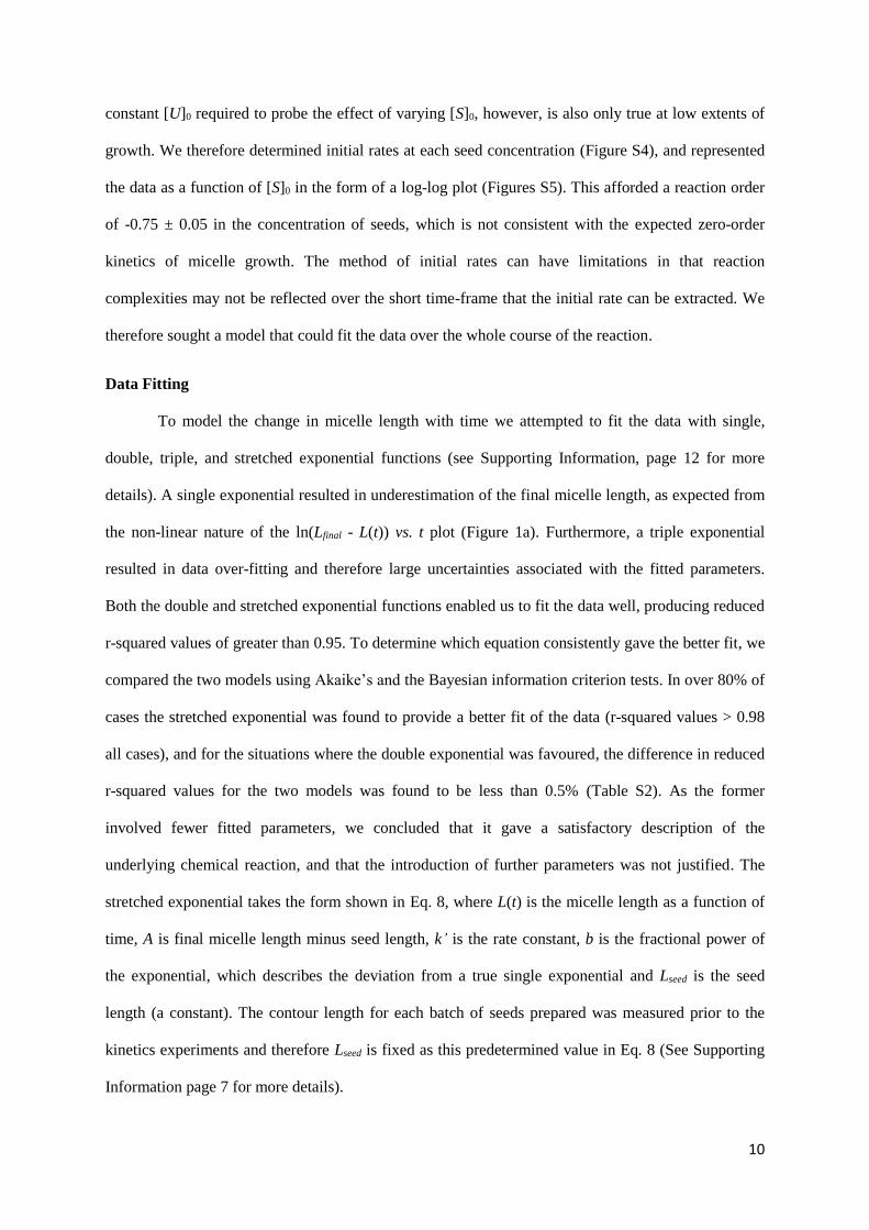
Data Fitting
To model the change in micelle length with time we attempted to fit the data with single,
double, triple, and stretched exponential functions (see Supporting Information, page 12 for more
details). A single exponential resulted in underestimation of the final micelle length, as expected from
the non-linear nature of the ln(Lfinal - L(t)) vs. t plot (Figure 1a). Furthermore, a triple exponential
resulted in data over-fitting and therefore large uncertainties associated with the fitted parameters.
Both the double and stretched exponential functions enabled us to fit the data well, producing reduced
r-squared values of greater than 0.95. To determine which equation consistently gave the better fit, we
compared the two models using Akaike’s and the Bayesian information criterion tests. In over 80% of
cases the stretched exponential was found to provide a better fit of the data (r-squared values > 0.98
all cases), and for the situations where the double exponential was favoured, the difference in reduced
r-squared values for the two models was found to be less than 0.5% (Table S2). As the former
involved fewer fitted parameters, we concluded that it gave a satisfactory description of the
underlying chemical reaction, and that the introduction of further parameters was not justified. The
stretched exponential takes the form shown in Eq. 8, where L(t) is the micelle length as a function of
time, A is final micelle length minus seed length, k’ is the rate constant, b is the fractional power of
the exponential, which describes the deviation from a true single exponential and Lseed is the seed
length (a constant). The contour length for each batch of seeds prepared was measured prior to the
kinetics experiments and therefore Lseed is fixed as this predetermined value in Eq. 8 (See Supporting
Information page 7 for more details).

11
𝐿(𝑡) = 𝐴 (1 − 𝑒−(𝑘′𝑡)𝑏) + 𝐿𝑠𝑒𝑒𝑑 Eq. 8
Employing Eq. 8 to model the change in length as of function time for different unimer
concentrations (Figure 2a, c) affords k’ values of 1.81 ± 0.19×10⁻⁴, 2.13 ± 0.23×10⁻⁴ and 1.91 ±
0.15×10⁻⁴ s-1 for [U]0 values of 10, 20 and 30 mg/mL, respectively. These rate constants are the same
within experimental error. It is noteworthy that the b values, which are 0.57, 0.60 and 0.58 for 10, 20
and 30 mg/mL, respectively, are also the same to within experimental error.
For the initial seed concentration data (Figure 2b, d), a fit of Eq. 8 produces k’ values of 1.81
± 0.19×10⁻⁴, 1.80 ± 0.26×10⁻⁴ and 1.26 ± 0.14×10⁻⁴ s-1 for [S]0 values of 1.0×10⁻², 6.7×10⁻³ and 3.3×10-
3 mg/mL. Although the rate constants at the highest 2 concentrations are almost identical when the
experimental errors are considered, that at the lowest concentration is significantly lower. Comparison
of Eqs. 7 and 8, however, show that k’ = 2k[S]0, and therefore, unlike the case with [U]0, k’ is not
independent of [S]0. Nonetheless, when k’ is converted to the [S]0 independent rate constant, k, values
of 0.009 ± 0.001, 0.013 ± 0.002 and 0.019 ± 0.002 mL/mg.s are obtained for initial concentrations of
1.0×10⁻², 6.7×10⁻³ and 3.3×10⁻³ mg/mL, respectively. The reasons for this behaviour are currently
unclear, although the inverse relationship between k and [S]0 is not what would be expected if the
process were diffusion limited, nor if there were systematic error in [S]0. The fits also afford values of
0.57 ± 0.02, 0.62 ± 0.03 and 0.62 ± 0.02 for b, of which the former 2 are the same within experimental
error with b at the lowest concentration marginally larger than that at the highest.
0
500
1000
1500
2000
0 500 1000 1500 2000 2500 3000
Len
gth
(n
m)
Time (min)
10 mg/mL Fit
20 mg/mL Fit
30 mg/mL Fit0
500
1000
1500
2000
0 500 1000 1500 2000 2500 3000
Len
gth
(n
m)
Time (min)
0.01 mg/mL Fit
0.0067 mg/mL Fit
0.0033 mg/mL Fit
Unimerconcentration A (nm)
Error (nm) k' (s-1) Error (s-1) b Error
10 mg/mL 528 13 1.81×10⁻⁴ 1.9×10⁻⁵ 0.57 0.02
20 mg/mL 888 23 2.13×10⁻⁴ 2.3×10⁻⁵ 0.60 0.02
30 mg/mL 1696 34 1.91×10⁻⁴ 1.5×10⁻⁵ 0.58 0.02
a) b)
c) d)
Seed concentration A (nm)
Error(nm) k' (s-1) Error (s-1) b Error
1.0×10⁻² mg/mL 528 13 1.81×10⁻⁴ 1.9×10⁻⁵ 0.57 0.02
6.7×10⁻³ mg/mL 826 61 1.80×10⁻⁴ 2.6×10⁻⁵ 0.62 0.03
3.3×10⁻³ mg/mL 1553 49 1.26×10⁻⁴ 1.4×10⁻⁵ 0.62 0.02

12
Figure 2: Lengths of PFS63-b-PDMS513 micelles as a function of time, monitored over a period of 2
days after the addition of unimer to PFS63-b-PDMS513 seed micelles, (Ln = 57 nm, Lw/Ln = 1.11) for a)
variable unimer concentration (16 L of 10, 20 or 30 mg/mL PFS63-b-PDMS513 THF solution,
respectively and 200 L of 0.1 mg/mL PFS63-b-PDMS513 seed micelles in EtOAc), and b) variable
seed concentration. (16 L of 10 mg/mL PFS63-b-PDMS513 THF solution, and 67, 134 and 201 L of
0.1 mg/mL PFS63-b-PDMS513 seed micelles in EtOAc, respectively), c, d) Table of kinetic data for a)
variable unimer concentration, b) variable seed concentration, respectively, A (nm) = final micelle
length minus seed side. a, b) Error bars = ±σ (standard deviation) of the micelle length distribution. c,
d) Standard errors for the values A, k’, and b obtained from the fit of Eq.8 to data.
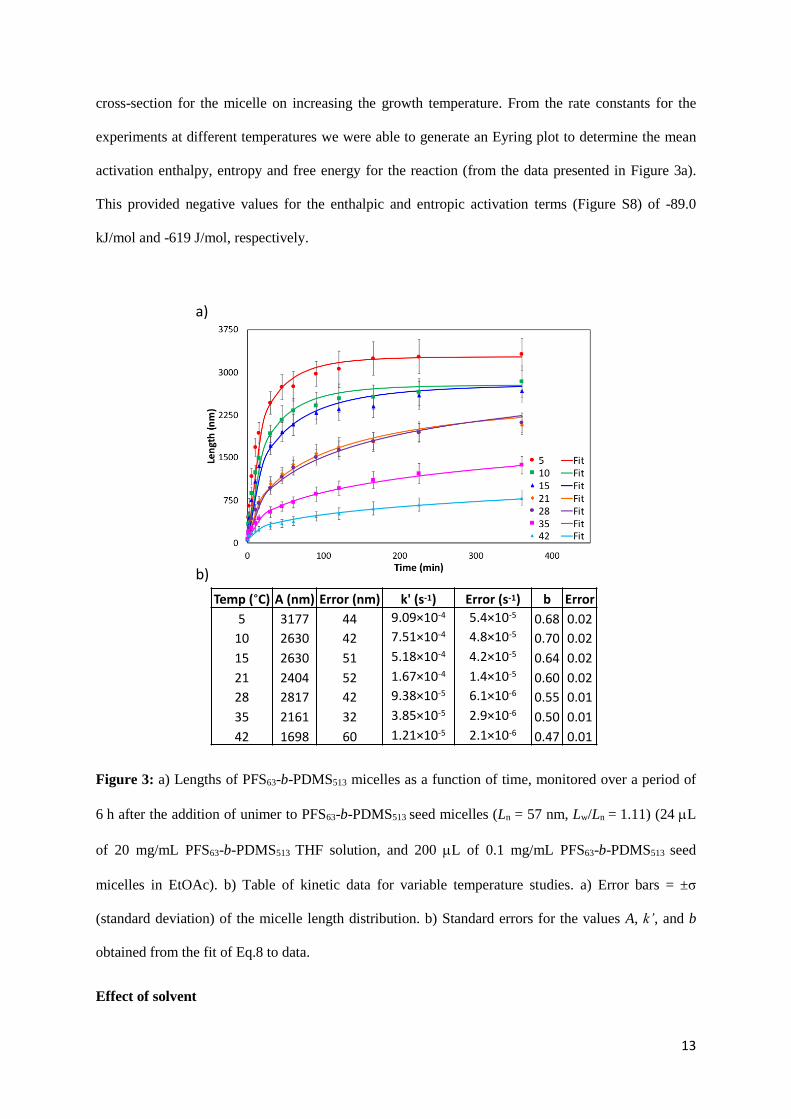
Effect of temperature
To explore the effect of temperature on the growth process, living CDSA experiments
involving the addition of PFS63-b-PDMS513 to crystallite seeds of the same BCP (Ln = 57 nm) were
performed at 7 different temperatures; 5, 10, 15, 21, 28, 35 and 42 °C. Elevating the self-assembly
temperature increased the length of time required for the micelles to reach their final lengths (Figure
3a and S6) with concomitant reductions in k’ from 9.09×10⁻⁴ to 1.21×10⁻⁵ s-1 (Figure 3b). There was
also a general trend for a reduction in the b values (0.68 to 0.47) and final micelle length achieved
with increasing temperature. Specifically, the final micelle length decreased from 3314 ± 279 nm to
1764 ± 209 nm, as the temperature was increased from 5 to 42 °C. The most likely cause of this
change would be an increase in micellar width and, consequently, in Nagg/L with temperature. This is
similar to the reported thickening in the chain axis direction of homo- and random copolymer crystals
on thermal annealing.93,94 Indeed, SAXS on the final micelle solutions revealed an increase in core
cross-sectional area from 41 to 76 nm2 on increasing the temperature from 5 to 42 °C (Figure S7 and
Table S3). It would be expected for this to scale with the decrease in L, if change in cross-sectional
area were the sole contributor. This was not the case, however, with the 47% decrease in length being
associated with an 85% increase in cross-sectional area. Furthermore, WAXS indicated that the inter-
chain spacing within the cores of the two different micellar samples was the same (Figure S7c). A
possible explanation for this discrepancy could be the transition from a rectangular to a more elliptical
13
cross-section for the micelle on increasing the growth temperature. From the rate constants for the
experiments at different temperatures we were able to generate an Eyring plot to determine the mean
activation enthalpy, entropy and free energy for the reaction (from the data presented in Figure 3a).
This provided negative values for the enthalpic and entropic activation terms (Figure S8) of -89.0
kJ/mol and -619 J/mol, respectively.
Figure 3: a) Lengths of PFS63-b-PDMS513 micelles as a function of time, monitored over a period of
6 h after the addition of unimer to PFS63-b-PDMS513 seed micelles (Ln = 57 nm, Lw/Ln = 1.11) (24 L
of 20 mg/mL PFS63-b-PDMS513 THF solution, and 200 L of 0.1 mg/mL PFS63-b-PDMS513 seed
micelles in EtOAc). b) Table of kinetic data for variable temperature studies. a) Error bars = ±σ
(standard deviation) of the micelle length distribution. b) Standard errors for the values A, k’, and b
obtained from the fit of Eq.8 to data.
Effect of solvent
Temp (°C) A (nm) Error (nm) k' (s-1) Error (s-1) b Error
5 3177 44 9.09×10⁻⁴ 5.4×10⁻⁵ 0.68 0.02
10 2630 42 7.51×10⁻⁴ 4.8×10⁻⁵ 0.70 0.02
15 2630 51 5.18×10⁻⁴ 4.2×10⁻⁵ 0.64 0.02
21 2404 52 1.67×10⁻⁴ 1.4×10⁻⁵ 0.60 0.02
28 2817 42 9.38×10⁻⁵ 6.1×10⁻⁶ 0.55 0.01
35 2161 32 3.85×10⁻⁵ 2.9×10⁻⁶ 0.50 0.01
42 1698 60 1.21×10⁻⁵ 2.1×10⁻⁶ 0.47 0.01
a)
b)

14
To investigate the effect of common solvent on the growth kinetics, 3 different THF
compositions were examined, namely 2.3, 4.8 and 9.7% v/v THF in n-hexane. As the THF content
increased, the growth rate of the micelles slowed as evidenced by k’ = 1.56×10⁻², 3.04×10⁻³ and
4.57×10⁻⁴ s-1 for 2.3, 4.8 and 9.7% v/v THF, respectively (Figure 4a, c and S9 and S10). This
reduction in rate constant, however, is accompanied by an increase in b, which is the opposite of the
trend observed for the experiments performed at different temperatures. It is noteworthy, however,
that although all experiments showed convergence to a constant final L, this is around the 3
confidence limit above the length predicted from the seed size and unimer to seed ratio (628 nm).
In order to probe the role of the selective solvent on the rate of the self-assembly process,
solvents with differing solubility parameters were chosen: n-hexane (14.9 MPa1/2), n-heptane (15.3
MPa1/2), and ethyl acetate (18.2 MPa1/2).95 Other solvents were also explored, but it proved difficult to
analyze the resulting micelles by TEM, due to severe aggregation or fragmentation upon drying (see
Supporting Information, page 19 for more details). The rate constants decreased on exchanging n-
hexane for n-heptane and then ethyl acetate (k’ = 6.32×10⁻³, 3.45×10⁻³ and 1.75×10⁻⁵ s-1, respectively),
whilst maintaining a constant THF composition of 4.8% v/v. These experiments demonstrated that as
the medium becomes a less poor solvent for the insoluble core-forming PFS block (18.7 MPa1/2,
PDMS = 14.9 MPa1/2),96 the rate constant decreases (Figure 4b, d and S11). Although the series: n-
hexane, n-heptane, and ethyl acetate became concomitantly poorer for the PDMS corona-forming
block, there was no evidence of reverse micelle formation or any decrease in the colloidal stability of
the resulting micelles. This suggests that the effect of the solvent on the crystallization of the core is
more significant in determining the rate constant than the change in amphiphilicity of the copolymer.
A trend of increasing b as the solvent medium became better for PFS was also observed, similar to
that for the experiments with different concentrations of common solvent (THF). Once again, the
similar final micelle lengths (Ln = 837 ± 79, 876 ± 89 and 956 ± 116 nm for n-hexane, n-heptane and
EtOAc, respectively) were comparable and within 3 of the predicted length (628 nm), which
suggested that there was no significant change in linear aggregation number on changing the selective
solvent. It is noteworthy that the micelles grown in ethyl acetate were slightly shorter than those

15
grown in n-heptane and n-hexane after 24 h (1440 min, Figure 4b), but after 1 month the final micelle
lengths were the same within error (Figure S11).
Figure 4: Lengths of PFS63-b-PDMS513 micelles as a function of time, monitored over a period of a) 6
h and b) 24 h after the addition of unimer to PFS63-b-PDMS513 seed micelles, (Ln = 25 nm, Lw/Ln =
1.25) for a) variable percentage THF (48 L of 10 mg/mL PFS63-b-PDMS513 THF solution, and
200L of 0.1 mg/mL PFS63-b-PDMS513 seed micelles in n-hexane), and b) variable selective solvent
(48 L of 10 mg/mL PFS63-b-PDMS513 THF solution, and 200L of 0.1 mg/mL PFS63-b-PDMS513
seed micelles in n-hexane). c), d) Table of kinetic data for a) variable percentage THF, b) variable
selective solvent, respectively. a, b) Error bars = ±σ (standard deviation) of the micelle length
distribution. c, d) Standard errors for the values A, k’, and b obtained from the fit of Eq.8 to data.
Effect of PFS degree of polymerization
To investigate the effect the degree of polymerization of the PFS core has on the micelle growth rate,
kinetic experiments were also performed with the BCPs, PFS63-b-PDMS513 and PFS49-b-PDMS504
(block ratios 1:8 and 1:10, respectively). These BCPs were selected as they have very similar degrees
of polymerization for the corona-forming block (difference < 2 %), but PFS49-b-PDMS504 has a core-
forming PFS block which has a degree of polymerization 22% lower than for PFS63-b-PDMS513.
Reducing the PFS degree of polymerization not only reduced the micelle growth rate (from k’ = 1.16
Solvent A (nm)Error (nm) k' (s-1) Error (s-1) b Error
n-hexane 734 8 6.32×10⁻³ 5.0×10⁻⁴ 0.49 0.03
n-heptane 748 16 3.45×10⁻³ 3.4×10⁻⁴ 0.54 0.06
EtOAc 928 32 1.75×10⁻⁵ 2.7×10⁻⁶ 0.62 0.03
THF (%)
A (nm)
Error(nm) k' (s-1) Error (s-1) b Error
2.3 790 12 1.56×10⁻² 3.3×10⁻³ 0.31 0.04
4.8 832 14 3.04×10⁻³ 3.9×10⁻⁴ 0.35 0.02
9.7 855 24 4.57×10⁻⁴ 5.9×10⁻⁴ 0.55 0.03
a) b)
c) d)
0
250
500
750
1000
0 100 200 300 400
Len
gth
(n
m)
Time (min)
2.3% THF Fit
4.8% THF Fit
9.7% THF Fit

16
± 0.08×10⁻⁴ s-1 to k’ = 4.6 ± 0.7×10⁻⁶ s-1), but also resulted in much shorter micelles being formed (Ln
= 2690 ± 253 nm and 1426 ± 134 nm for PFS63-b-PDMS513 and PFS49-b-PDMS504, respectively)
(Figure 5). We viewed this as a likely consequence of the linear aggregation number being different
for the two types of micelle on account of the different BCPs employed. To investigate this hypothesis
further, solution SAXS/WAXS was performed on the final micelles grown from PFS63-b-PDMS513
and PFS49-b-PDMS504. These revealed a likely increase in core cross-sectional area from 70 ± 6 to 78
± 9 nm2 on reducing the PFS length from DPn = 63 to 49 (Figure S12 and Table S4) with no
significant change in the associated core structure. The linear aggregation numbers are therefore 3.7 ±
0.3 and 5.4 ± 0.6 chains/nm for PFS63-b-PDMS513 and PFS49-b-PDMS504, respectively, and this
increase of 46% almost fully accounts for the observed 47% reduction in final micelle length. More
strikingly, the kinetic data demonstrate that reducing the PFS core length by an average of 14 repeat
units caused the rate constant to be reduced by a factor of 25 from a k’ value of 1.16 ± 0.08×10⁻⁴ s-1 for
PFS63-b-PDMS513 to 4.6 ± 0.7×10⁻⁶ s-1 for PFS49-b-PDMS504. It is also noteworthy that different b
values were obtained from the fits (b = 0.60 ± 0.01 and 0.53 ± 0.02 for PFS63-b-PDMS513 and PFS49-b-
PDMS504, respectively).
PFS Length A (nm) Error (nm) k' (s-1) Error (s-1) b Error
PFS63-b-PDMS513 2520 42 1.16×10⁻⁴ 7.8×10⁻⁶ 0.60 0.01
PFS49-b-PDMS504 1376 45 4.59×10⁻⁶ 6.7×10⁻⁷ 0.53 0.02
a)
b)0
1000
2000
3000
0 10000 20000 30000 40000
Len
gth
(n
m)
Time (min)
PFS63
Fit
PFS49
Fit
PFS63-b-PDMS513
PFS49-b-PDMS504

17
Figure 5: a) Lengths of PFS-b-PDMS micelles as a function of time, monitored over a period of 4
weeks after the addition of unimer to PFS63-b-PDMS513 seed micelles (Ln = 57 nm, Lw/Ln = 1.11), (48
L of 10 mg/mL PFS63-b-PDMS513 THF solution, or 48 L of 9.25 mg/mL PFS49-b-PDMS504 THF
solution, and 200L of 0.1 mg/mL PFS63-b-PDMS513 seed micelles in EtOAc). b) Table of kinetic data
for changing PFS core length. a) Error bars = ±σ (standard deviation) of the micelle length
distribution. b) Standard errors for the values A, k’, and b obtained from the fit of Eq.8 to data.
Monitoring micelle growth using super-resolution fluorescence microscopy
It is important to verify that the data we have obtained from the TEM analysis of dried
samples characterizing the growth of PFS63-b-PDMS513 cylindrical micelles is representative of the
CDSA process that occurs in solution. It has already been established for fully assembled micelles
that the super-resolution techniques STED and SMLM provide comparable results to TEM.87
However, for the kinetics experiments all techniques were performed with high concentrations of
unassembled unimer at early time points, which could, in principle, add to the micelle termini during
the drying process. We therefore followed the micelle growth using SRFM as a solution-based
technique to validate the trends obtained from TEM experiments. We have recently established
protocols for use in organic media with the BCP PFS56-b-PDMS775/DYE20.87 For both techniques,
short fluorescent seed micelles were prepared from BCPs 3% labelled with the appropriate dye,
STAR635 (STED) and CAGE635 (SMLM). The fluorescent seed micelles were longer (Ln = ca. 600
nm) than those typically used (Ln = 25-57 nm) to aid visualization of the structures during the
experiments, particularly at the early time points where the unimer concentration remains relatively
high, which limits the signal to background ratio. Fluorescently labelled BCP unimer (dye =
STAR635 single colour STED, CAGE500 for dual-colour SMLM) was added to the short fluorescent
seed micelles and aliquots were taken from the solution and analyzed by STED, SMLM and TEM
(Figure 6 and S13).

18
Figure 6: a) Plot of micelle length against time for TEM run 1 and 2 and STED run 1 and 2,
monitored over a period of 6 h, 6.5 L of 10 mg/mL PFS56-b-PDMS775/STAR63520 THF solution and
200 L of 0.15 mg/mL short STAR635-fluorescent micelles (Ln = 577 nm, Lw/Ln = 1.03) in EtOAc. b)
representative STED images at time points 1 min, 45 min and 240 min. Scale bars = 4.8 μm. c) Table
of kinetic data for STAR635 BCP analyzed by STED and TEM. a) Error bars = ±σ (standard
deviation) of the micelle length distribution. c) Standard errors for the values A, k’, and b obtained
from the fit of Eq.8 to data.
For the STED/TEM kinetics experiments, all k’ values are within error of each other,
however, the b values obtained from the data fitting for these experiments show quite significant
discrepancies between the runs analyzed using STED and those employing TEM. The b values from
the STED runs are self-consistent (ca. 0.8), but do not agree with the TEM experiments, which
provide much larger b values (1.0 - 1.1). Furthermore, these contradict the b values obtained (0.57 –
0.6) for the self-assembly of the non-dye-labelled BCP (Data Fitting Section). Inspection of the raw
TEM data obtained for both the unlabelled and labelled PFS-b-PDMS experiments indicate that
PFS56-b-PDMS775/STAR63520 produces micelles with higher dispersities (PDI = 1.2-1.3) than PFS63-
b-PDMS513 (PDI = 1.1-1.2). This increase in polydispersity increases the error associated with the
length at each time point in the series, and therefore also the uncertainty in the fit. Repeating the fit of
the data, but without weighting for the standard deviation of the micelle lengths produced b values
which were all 0.9 or lower. This indicates the significant role that the errors play in determining the b
250
750
1250
1750
2250
2750
0 100 200 300 400
Len
gth
(n
m)
Time (min)
TEM1 Fit
TEM2 Fit
STED1 Fit
STED2 Fit
Run A (nm) Error (nm) k' (s-1) Error (s-1) b Error
STED1 1489 93 5.04×10⁻⁴ 9.9×10⁻⁵ 0.82 0.09
STED2 1585 57 4.53×10⁻⁴ 5.3×10⁻⁵ 0.80 0.05
TEM1 1579 62 3.82×10⁻⁴ 4.1×10⁻⁵ 0.99 0.08
TEM2 1601 66 4.52×10⁻⁴ 4.8×10⁻⁵ 1.17 0.11
t = 1 min t = 45 min t = 240 mina) b)
c)

19
values obtained, especially given the modest change in dispersity between samples. Nonetheless, even
when the error weighting is removed, the STED and TEM b values are still not in agreement, and this
could be due to the lower resolution of the former effecting the accuracy of the measurement at early
time points (i.e. shorter lengths). We also studied the micelle growth kinetics using two-colour SMLM
(Figure S13 and S14), however, like the STED/TEM results, the techniques were inconsistent with
each other, and the TEM data also differed from that obtained with unlabelled BCP. Although
consideration of the STED data alone confirmed the non-first order behaviour of CDSA with respect
to unimer (b ≠ 1), as determined by TEM analysis, the necessity for dye-labelled BCP (albeit at only
3%) clearly complicates the self-assembly process. The exact role of the dye is currently unclear, but
it evidently increases the rate of addition (k’ = 4.53 ± 0.53 x 10⁻⁴ and 1.81 ± 0.19 x 10⁻⁴ s-1 for with
and without dye, respectively), and also the length dispersity of the sample.
Discussion
An investigation of the proposed differential rate equation for living CDSA (Eq. 1), by the
method of initial rates, yielded reaction orders for [U] and [S] of 1.16 ± 0.25 and -0.75 ± 0.05,
respectively. Although the estimate of the former was consistent with the first-order behaviour
expected by analogy to a living covalent polymerization, the zero-order dependence on [S] expected
for L(t) was not observed. L(t), however, exhibited a more complex relationship to both [U] and [S]
when the whole duration of the reaction was considered. The length of PFS63-b-PDMS513 micelles
observed during the self-assembly process as a function of time was therefore best modelled by a
stretched exponential function (Eq. 8). In nature, stretched exponential functions have been
successfully used to model processes that required a certain protein conformation to be obtained, but
which may be achieved from any number of non-functional alternatives. This gives rise to a
distribution of activation energies, and thus also to a distribution of kinetic rates, where the parameter
b (Eq. 8) is related to the width of the distribution. The crystallization of unimer onto the termini of
the growing micelle would require the polymer in solution, and at the surface of the micellar core, to
adopt a limited number of all the possible conformations, many of which will be energetically similar
and accessible at ambient temperature. This situation would give rise to the distribution of activation

20
energies required for the observed stretched exponential relationship of L with respect to t. It should
be noted that there is no requirement for molecular weight polydispersity in this explanation, only that
the molecular weight is high enough for energetically similar conformations to be available. Indeed,
the supramolecular polymerization of porphyrin-based monomers, where both these complexities are
necessarily absent, is first-order with respect to monomer concentration.49
It is evident from the experiments performed at different temperatures that increasing the
temperature results in a reduced rate constant (k’). This is consistent with reports that the linear
growth rate of homopolymer crystals decreased upon lowering the extent of supercooling,97 and may
also be due to increased solubility of the core-forming block on increasing the temperature. However,
the different temperature kinetic experiments also revealed that altering the self-assembly temperature
effects the dimensions of the final cylindrical micelles in a similar manner to that observed for
homopolymer crystals.98 Cylindrical micelles grown at higher temperatures were shorter in length
with larger core radii, and with a higher linear aggregation number (2.2 chains/nm at 5 °C compared
to 4.1 chains/nm at 42 °C). It is noteworthy that studies of the annealing of PI637-b-PFS53 cylindrical
micelles in solution revealed no thickening of the micellar core at temperatures below 60 °C.99 These
experiments therefore demonstrate that it is only when growth occurs at elevated temperatures that the
micelle structure changes, and that it is temperature rather than the core radius of the seed that
determines the radius of the newly grown section. We suggest that the change in Nagg/L occurs in the
early stages of growth, so as to give a different core cross-sectional area by SAXS, and not a
distribution of areas bounded at the low end by that of the seed (scattering from the seed is dominated
by that from the newly grown portion on account of the relative amounts). It is noteworthy that no
step change in width is seen by TEM, although the ca. 1 nm change is at the resolution limit of the
instrument.
The kinetic experiments at different temperatures also allowed us to obtain information
regarding the thermodynamics of the activation parameters involved for the living CDSA of PFS63-b-
PDMS513 micelles through an Eyring plot. Both the entropic and enthalpic terms were negative, which
is expected due to the increased ordering of the core-forming PFS BCP chains upon crystallization
and the exothermic nature of crystallization, respectively.
21
The solvent composition plays an important role in the rate of micelle growth. Altering the
solvent composition so that it is more favourable for the PFS core-forming block, either by increasing
the amount of common solvent, or through using a selective solvent that has a solubility parameter
closer to that of PFS (e.g. using EtOAc instead of n-hexane) causes a reduction in the rate constant.
This trend underscores how any parameter that reduces the propensity for the PFS segment to
precipitate from solution and crystallize on the termini of the growing micelle reduces the micellar
growth rate.
It has been reported that altering the overall composition of PFS-containing micelles effects
the dimensions of the resulting micellar structure,89 but no comment was made as to how this might
affect the rate of growth. In this experiment, it was found that reducing the number average degree of
polymerization of the PFS segment by 22% (14 monomer units) from PFS63 to PFS49, whilst keeping
the length of the corona block approximately the same (< 2% decrease), resulted in a rate constant (k’)
25 times smaller. It is known that the solubility of polymer chains decrease as the degree of
polymerization increases and, therefore, it is expected that the PFS block will be less soluble in n-
hexane in the BCP with the longer PFS core-forming segment than that with the shorter block.
Furthermore, the equilibrium melting point, Tm, will increase with molecular weight, leading to a
larger degree of supercooling for the BCP with the longer PFS block, and this is reported to increase
the linear crystal growth rate.97 Increasing the length of the PFS segment has also been shown to
decrease the linear aggregation number, and hence increase the final length of the resulting micelles.
An implication of chains of different lengths adding at different rates would be the establishment of a
continuum of rates over the molecular weight distribution of the sample, which could give rise to the
observed stretched exponential behaviour. We therefore modelled micelle growth, L(t), using an
initial Zimm-Schulz distribution of molecular weights, and a scaling factor relating the change in k’
per unit degree of polymerization (see Supporting Information, page 24 for more details). Using the
experimentally determined PDI for the PFS block (1.04) only afforded the typically observed b value
(ca. 0.6) for scaling powers between 4 and 5, and these are far less than the value of 12 observed
between the change in DPn for the core-forming block and corresponding change in k’. However, the
range of molecular weights constituting the majority of each copolymer sample [σ(Mn) = 2373 and

22
3051 g mol-1 for PFS49 and PFS63, respectively] is narrower than the separation between PFS49-b-
PDMS504 and PFS63-b-PDMS513 [ΔMn(PFS) = 3390 g mol-1]. This may mean that more subtle changes
in k’(DPn) exist within a relatively monodisperse copolymer sample, and this is lost on studying two
more disparate examples. Nonetheless, this analysis clearly cannot discount a role for core-forming
block polydispersity as part of the reason for stretched exponential growth kinetics. It should also be
noted that PFS63-b-PDMS513 with the longer PFS segment added at a faster rate than PFS49-b-
PDMS504, which has a shorter PFS segment, which is the opposite to what would be expected if this
were a diffusion limited process.
Previous work has demonstrated how the seed structure at the low length limit (ca. 20 nm)
differs from that of the resulting micelle, and even the parent micelle from which it is derived.100 This
suggests that it is possibly simplistic to expect totally symmetric growth from either side of the seed,
and growth from seeds of all sizes to be the same. The seed size, however, appears to have modest
influence on the growth rate with k’ (9.8 ± 1.5 × 10⁻⁵ s-1) for the longest seed size (915 nm) decreasing
below the 3 confidence limit of that of the shortest (k’ = 4.61 ± 0.65 × 10⁻⁴ s-1 for Ln(seed) = 25 nm).
Nonetheless, the similarity of k’ values (6.10 ± 0.42 × 10⁻⁴ and 3.22 ± 0.56 × 10⁻⁴ s-1) when equal
amounts of unimer were added to seeds of Ln = 25 and 493 nm, respectively, suggests that the short
seeds are not unusual from a kinetics standpoint (see Supporting Information Figures S15-17 and
Table S2).
The in situ experiments performed using SRFM techniques demonstrated how the stretched
exponential also characterizes the observed growth process in solution, validating the data obtained
from the TEM experiments. In these experiments, despite only being present on 3% of the corona, the
nature of the dye has an effect on the micellar growth rate. The STAR635 dye-labelled BCP has a
faster growth rate than the CAGE500-labelled BCP for the SMLM experiments. This effect of dye
structure on the growth rate had been ascribed to the required modification of the corona-forming
block altering amphiphilicity of the BCP.101 Finally, for the SRFM experiments the sample
preparation methods prevented the addition of any remaining unimer present in the sample to the
micelle termini, and therefore effectively quenched the self-assembly reaction. This was convenient as

23
image acquisition times are typically between 2-10 minutes, depending on the SRFM technique, and it
is therefore important that growth is halted prior to analysis.
Conclusion
In summary, we have probed the growth kinetics of living CDSA using PFS -b-PDMS BCPs
as a model system. We discovered that a range of parameters affect the micellar growth rate including
concentration, temperature, the nature of the solvent medium, and BCP composition. It is noteworthy
that the rate of self-assembly can be increased by either lowering the temperature, using poorer
solvents for the crystalline PFS core-forming block, or by employing BCPs with longer core-forming
PFS segments. Data fitting also enabled us to determine that the analogy of living CDSA to a living
covalent polymerization of molecular monomers does not apply to the reaction kinetics, and that the
growth rate is neither first order in unimer nor in seed concentration. The deviation from first order
kinetics could be modelled using a stretched exponential function and we postulate that this behaviour
results from a combination of the influences of polymer chain conformation and molecular weight
polydispersity on the addition of unimer to the micelle termini. These two factors arise as a natural
consequence of the increased complexity of synthetic BCP chains compared to their monodisperse,
small molecule counterparts.
Overall, these results provide important additional insight into living CDSA processes and
also provide guidelines for optimizing the efficiency of experimental protocols that should be of
considerable significance for work with a growing variety of BCPs with crystalline core-forming
blocks32,39,42-44 and analogous amphiphiles45-56 that can be used for seeded supramolecular
polymerizations. In addition, an informative comparison is enabled with analogous biological and
synthetic supramolecular systems that undergo fiber formation via nucleation-elongation processes.
For example, the low length polydispersities and kinetic data obtained for living CDSA underscore
the virtual absence of heterogeneous growth rates where some 1D nanoparticles grow substantially
faster than others, which has been observed for the case of certain protein fibers and has been
attributed to fiber polymorphism.44
Methods

24
General Experimental Considerations
Anionic polymerizations were carried out in an argon atmosphere glovebox. All other manipulations
were carried out under an open atmosphere unless otherwise stated. All block copolymers were
prepared by the sequential living anionic polymerization of dimethylsila[1]ferrocenophane and
hexamethylcyclotrisiloxane, as previously reported.87,88 All reagents were purchased from Sigma-
Aldrich unless otherwise stated. Monomer purifications were performed under an atmosphere of
purified N2. THF was distilled from Na/benzophenone immediately before use. The dyes STAR635
NHS ester and CAGE635 NHS ester were purchased from Abberior GmbH. Photoirradiation
experiments were carried out with Pyrex-glass filtered emission from a 125 W medium-pressure
mercury lamp (Photochemical Reactors Ltd.). An ethylene glycol/water bath in conjunction with a
thermostat was used to maintain constant temperatures of 20 °C during the photoirradiation
experiments. 1H and 13C NMR spectra were recorded using Jeol Eclipse 400 MHz or Varian VNMR
400 MHz spectrometers.
Polymer Characterization
Gel permeation chromatography was carried out using a Viscotek VE 2001 Triple-Detector Gel
Permeation Chromatograph equipped with an automatic sampler, a pump, an injector, an inline
degasser, and a column oven (30 °C). The elution columns consist of styrene/divinylbenzene gels with
pore sizes between 500 Å and 100,000 Å. Detection was conducted by means of a VE 3580
refractometer, a four-capillary differential viscometer, and 90° and low angle (7°) laser light (λ0= 670
nm) scattering detectors, VE 3210 & VE 270. THF (Fisher) was used as the eluent, with a flow rate of
1.0 mL/min. Samples were dissolved in the eluent (2 mg/mL) and filtered with a Ministart SRP 15
filter (polytetrafluoroethylene membrane of 0.45 μm pore size) before analysis. The calibration was
conducted using a PolyCALTM polystyrene standard (PS115K) from Viscotek. Matrix-assisted laser
desorption/ionization time of flight (MALDI-TOF) mass spectrometry measurements of
polyferrocenyldimethylsilane (PFDMS) were performed using a Bruker Ultraflextreme running in
linear mode. Samples were prepared using a trans-2-[3-(4-tert-butylphenyl)-2-methyl-2-
propenylidene]malononitrile matrix (20 mg/mL in THF) and the polymer sample (2 mg/mL in THF),
25
mixed in a 10:1 (v/v) ratio. Approximately 1 µL of the mixed solution was deposited onto a MALDI
sample plate and allowed to dry in air. The molecular weights of the diblock copolymers were then
determined by combining the molecular weight Mn of the first block from MALDI-TOF
measurements with the block ratio of the diblock copolymer obtained by integrating the 1H NMR
spectroscopic signal intensities of the respective blocks.
Transmission electron microscopy (TEM)
Copper grids from Agar Scientific, mesh 400, were coated with a carbon film. Carbon coating was
done using an Agar TEM Turbo Carbon Coater where carbon was sputtered onto mica sheets before
deposition on the copper grids via flotation on water. Bright field TEM micrographs were obtained on
a JEOL1200EX II microscope operating at 120 kV and equipped with an SIS MegaViewIII digital
camera.
Stimulated emission depletion (STED) microscopy
STED imaging was performed on a home-built pulsed STED microscope described in detail in the
following reference.102 Briefly, the excitation and STED beam are obtained from a single titanium-
sapphire oscillator centered at λSTED = 765 nm (Ti:S, Mai Tai HP, Spectraphysics). The excitation
beam, centered at λex = 640 nm, was extracted from a supercontinuum source (FemtoWhite, NKT
Photonics) by a bandpass filter (637/7 BrightLine HC, Semrock) and coupled into a polarization
maintaining single-mode fiber (PM630-HP, Thorlabs). The pulse duration of the STED beam was
stretched to approximately 100-200 ps thanks to a 50 cm glass block of SF66 (IC Optical Systems,
United Kingdom) and 100 m long polarization maintaining single-mode fiber (PM-S630-HP,
Thorlabs). Additionally the STED beam was shaped into a donut beam by a spatial light modulator
(X10468−02, Hamamatsu). The excitation and STED beam were recombined with a dichroic mirror
(T735spxr, Chroma) and sent to a commercial point-scanning microscope (Abberior Instruments)
comprising: a microscope frame (IX83, Olympus), a set of galvanometer mirrors (Quad scanner,
Abberior Instruments) and a detection unit. The beams were focused onto the sample by a 100×/1.4
NA oil immersion objective lens (UPLSAPO 100XO, Olympus) and images were acquired by raster
scanning the beams across the sample using the Inspector Image Acquisition software (Andreas

26
Schönle, Abberior Instruments GmbH, Göttingen, Germany). Typically a field of view of 8080 m2
was used with a pixel size of 2020 nm2 and pixel dwell times of 30 µs and 50 µs respectively.
Moreover, each line was scanned successively five times with a pixel dwell time of 10 µs per line-
scanning. Fluorescence photons emerging from the sample were collected by the microscope
objective lens, de-scanned by the galvanometer mirrors, focused onto a pinhole and sent to an
avalanche photodiode (SPCM-AQRH, Excelitas Technologies). Laser powers, measured at the
objective back aperture, were ∼20-30 μW for the excitation beam and ∼100-150 mW for the STED
beam.
Acknowledgements
C.E.B. thanks the Bristol Chemical Synthesis Centre for Doctoral Training, funded by the EPSRC for
the provision of a PhD studentship. E. M. L acknowledges the EU for a Marie Curie postdoctoral
fellowship. D.W.H. was supported by the EPSRC Bristol Cente for Functional Nanomaterials doctoral
training grant [EP/G036780/1]. R. F. L., P. M. and C.F.K. acknowledge grants from the EPSRC, UK
(grant EP/H018301/1, EP/L015889/1) and the Medical Research Council (grant MR/K015850/1).
I.M. thanks the EU for an ERC Advanced Investigator Grant. TEM studies were carried out in the
Chemistry Imaging Facility at UoB with equipment funded by UoB and EPSRC (EP/K035746/1). The
authors would like to thank David J. Lunn, John R. Finnegan, and Jieshu Qian for helpful discussions
and Dr. Torben Gädt for preliminary experiments on the micelle growth kinetics.
Author Contributions
C. E. B. and E. M. L. performed all experiments, data analysis and wrote the paper together with R. F.
L., G. R. W., C. F. K. and I. M. and additional scientific input was provided by D.W.H., G. G., and M.
A. W. D. W. H. performed the X-ray analysis and assisted with the data fitting along with R. F. L., P.
M. and R. M. R. The STED and SMLM was performed by P. M. and R. F. L., respectively. C. F. K.
and I. M. supervised the project.

27
References
(1) De Greef, T. F. A.; Smulders, M. M. J.; Wolffs, M.; Schenning, A. P. H. J.; Sijbesma, R. P.;
Meijer, E. W. Supramolecular Polymerization. Chem. Rev. 2009, 109, 5687–5754.
(2) Whitesides, G. M. Self-Assembly at All Scales. Science 2002, 295, 2418–2421.
(3) Schacher, F. H.; Rupar, P. A.; Manners, I. Functional Block Copolymers: Nanostructured
Materials with Emerging Applications. Angew. Chem. Int. Ed. 2012, 51, 7898–7921.
(4) Mai, Y.; Eisenberg, A. Self-Assembly of Block Copolymers. Chem. Soc. Rev. 2012, 41, 5969–
5985.
(5) Wang, J.; Liu, K.; Xing, R.; Yan, X. Peptide Self-Assembly: Thermodynamics and Kinetics.
Chem. Soc. Rev. 2016, 45, 5589–5604.
(6) Hayward, R. C.; Pochan, D. J. Tailored Assemblies of Block Copolymers in Solution: It Is All
about the Process. Macromolecules 2010, 43, 3577–3584.
(7) Gröschel, A. H.; Walther, A.; Löbling, T. I.; Schacher, F. H.; Schmalz, H.; Müller, A. H. E.
Guided Hierarchical Co-Assembly of Soft Patchy Nanoparticles. Nature 2013, 503, 247-251.
(8) Blanazs, A.; Armes, S. P.; Ryan, A. J. Self-Assembled Block Copolymer Aggregates: From
Micelles to Vesicles and Their Biological Applications. Macromol. Rapid Commun. 2009, 30,
267–277.
(9) Gröschel, A. H.; Müller, A. H. E. Self-Assembly Concepts for Multicompartment
Nanostructures. Nanoscale 2015, 7, 11841–11876.
(10) Elsabahy, M.; Wooley, K. L. Design of Polymeric Nanoparticles for Biomedical Delivery
Applications. Chem. Soc. Rev. 2012, 41, 2545–2561.
(11) Ge, Z.; Liu, S. Functional Block Copolymer Assemblies Responsive to Tumor and
Intracellular Microenvironments for Site-Specific Drug Delivery and Enhanced Imaging
Performance. Chem. Soc. Rev. 2013, 42, 7289.
(12) Lazzari, M.; Scalarone, D.; Vazquez-Vazquez, C.; López-Quintela, M. A. Cylindrical Micelles
from the Self-Assembly of Polyacrylonitrile-Based Diblock Copolymers in Nonpolar Selective
Solvents. Macromol. Rapid Commun. 2008, 29, 352–357.
(13) Du, Z. X.; Xu, J. T.; Fan, Z. Q. Micellar Morphologies of Poly(ε-caprolactone)-b-
Poly(ethylene oxide) Block Copolymers in Water with a Crystalline Core. Macromolecules
2007, 40, 7633–7637.
(14) He, W. N.; Zhou, B.; Xu, J. T.; Du, B. Y.; Fan, Z. Q. Two Growth Modes of Semicrystalline
Cylindrical Poly(ε-caprolactone)-b-Poly(ethylene oxide) Micelles. Macromolecules 2012, 45,
9768–9778.
(15) Zhang, J.; Wang, L. Q.; Wang, H.; Tu, K. Micellization Phenomena of Amphiphilic Block
Copolymers Based on Methoxy Poly(ethylene glycol) and Either Crystalline or Amorphous
Poly(caprolactone-b-lactide). Biomacromolecules 2006, 7, 2492–2500.
(16) Schmalz, H.; Schmelz, J.; Drechsler, M.; Yuan, J.; Walther, A.; Schweimer, K.; Mihut, A. M.
Thermo-Reversible Formation of Wormlike Micelles with a Microphase-Separated Corona
from a Semicrystalline Triblock Terpolymer. Macromolecules 2008, 41, 3235–3242.
(17) Yin, L.; Lodge, T. P.; Hillmyer, M. A. A Stepwise “Micellization-Crystallization” Route to
Oblate Ellipsoidal, Cylindrical, and Bilayer Micelles with Polyethylene Cores in Water.
Macromolecules 2012, 45, 9460–9467.
(18) Schmelz, J.; Karg, M.; Hellweg, T.; Schmalz, H. General Pathway toward Crystalline-Core
Micelles with Tunable Morphology and Corona Segregation. ACS Nano 2011, 5, 9523–9534.
(19) Mihut, A. M.; Drechsler, M.; Möller, M.; Ballauff, M. Sphere-to-Rod Transition of Micelles
Formed by the Semicrystalline Polybutadiene-Block-Poly(ethylene oxide) Block Copolymer in
28
a Selective Solvent. Macromol. Rapid Commun. 2010, 31, 449–453.
(20) Gilroy, J. B.; Lunn, D. J.; Patra, S. K.; Whittell, G. R.; Winnik, M. A.; Manners, I. Fiber-like
Micelles via the Crystallization-Driven Solution Self-Assembly of poly(3-Hexylthiophene)-
Block-Poly(methyl methacrylate) Copolymers. Macromolecules 2012, 45, 5806–5815.
(21) Massey, J. A.; Temple, K.; Cao, L.; Rharbi, Y.; Raez, J.; Winnik, M. A.; Manners, I. Self-
Assembly of Organometallic Block Copolymers: The Role of Crystallinity of the Core-
Forming Polyferrocene Block in the Micellar Morphologies Formed by Poly(ferrocenylsilane-
b-dimethylsiloxane) in n-Alkane Solvents. J. Am. Chem. Soc. 2000, 122, 11577–11584.
(22) Kynaston, E. L.; Gould, O. E. C.; Gwyther, J.; Whittell, G. R.; Winnik, M. A.; Manners, I.
Fiber-Like Micelles from the Crystallization-Driven Self-Assembly of Poly(3-
Heptylselenophene)-Block-Polystyrene. Macromol. Chem. Phys. 2015, 216, 685–695.
(23) Legros, C.; De Pauw-Gillet, M.-C.; Tam, K. C.; Taton, D.; Lecommandoux, S. Crystallisation-
Driven Self-Assembly of Poly(2-isopropyl-2-oxazoline)-Block-Poly(2-methyl-2-oxazoline)
above the LCST. Soft Matter 2015, 11, 3354–3359.
(24) Yu, B.; Jiang, X.; Yin, J. Size-Tunable Nanosheets by the Crystallization-Driven 2D Self-
Assembly of Hyperbranched Poly(ether amine) (hPEA). Macromolecules 2014, 47, 4761–
4768.
(25) Tong, Z.; Li, Y.; Xu, H.; Chen, H.; Yu, W.; Zhuo, W.; Zhang, R.; Jiang, G. Corona Liquid
Crystalline Order Helps to Form Single Crystals When Self-Assembly Takes Place in the
Crystalline/Liquid Crystalline Block Copolymers. ACS Macro Lett. 2016, 5, 867–872.
(26) Wu, J.; Weng, L.-T.; Qin, W.; Liang, G.; Tang, B. Z. Crystallization-Induced Redox-Active
Nanoribbons of Organometallic Polymers. ACS Macro Lett. 2015, 4, 593–597.
(27) Rizis, G.; van de Ven, T. G. M.; Eisenberg, A. Homopolymers as Structure-Driving Agents in
Semicrystalline Block Copolymer Micelles. ACS Nano 2015, 9, 3627–3640.
(28) Lee, I.-H.; Amaladass, P.; Yoon, K.-Y.; Shin, S.; Kim, Y.-J.; Kim, I.; Lee, E.; Choi, T.-L.
Nanostar and Nanonetwork Crystals Fabricated by In Situ Nanoparticlization of Fully
Conjugated Polythiophene Diblock Copolymers. J. Am. Chem. Soc. 2013, 135, 17695–17698.
(29) Presa-Soto, D.; Carriedo, G. A.; de la Campa, R.; Presa Soto, A. Formation and Reversible
Morphological Transition of Bicontinuous Nanospheres and Toroidal Micelles by the Self-
Assembly of a Crystalline-b-Coil Diblock Copolymer. Angew. Chem. Int. Ed. 2016, 55,
10102–10107.
(30) Lee, C.-U.; Smart, T. P.; Guo, L.; Epps, T. H.; Zhang, D. Synthesis and Characterization of
Amphiphilic Cyclic Diblock Copolypeptoids from N-Heterocyclic Carbene-Mediated
Zwitterionic Polymerization of N-Substituted N-Carboxyanhydride. Macromolecules 2011, 44,
9574–9585.
(31) Yang, S.; Shin, S.; Choi, I.; Lee, J.; Choi, T.-L. Direct Formation of Large-Area 2D
Nanosheets from Fluorescent Semiconducting Homopolymer with Orthorhombic Crystalline
Orientation. J. Am. Chem. Soc. 2017, 139, 3082–3088.
(32) Tao, D.; Feng, C.; Cui, Y.; Yang, X.; Manners, I.; Winnik, M. A.; Huang, X. Monodisperse
Fiber-like Micelles of Controlled Length and Composition with an Oligo(p-
Phenylenevinylene) Core via “Living” Crystallization-Driven Self-Assembly. J. Am. Chem.
Soc. 2017, 139, 7136–7139.
(33) Arno, M. C.; Inam, M.; Coe, Z.; Cambridge, G.; Macdougall, L. J.; Keogh, R.; Dove, A. P.;
O’Reilly, R. K. Precision Epitaxy for Aqueous 1D and 2D Poly(ε-caprolactone) Assemblies. J.
Am. Chem. Soc. 2017, 139, 16980–16985.
(34) Sun, L.; Pitto-Barry, A.; Kirby, N.; Schiller, T. L.; Sanchez, A. M.; Dyson, M. A.; Sloan, J.;
Wilson, N. R.; O’Reilly, R. K.; Dove, A. P. Structural Reorganization of Cylindrical
Nanoparticles Triggered by Polylactide Stereocomplexation. Nat. Commun. 2014, 5, 5746.
29
(35) Ganda, S.; Dulle, M.; Drechsler, M.; Förster, B.; Förster, S.; Stenzel, M. H. Two-Dimensional
Self-Assembled Structures of Highly Ordered Bioactive Crystalline-Based Block Copolymers.
Macromolecules 2017, 50, 8544–8553.
(36) Gilroy, J. B.; Gädt, T.; Whittell, G. R.; Chabanne, L.; Mitchels, J. M.; Richardson, R. M.;
Winnik, M. A.; Manners, I. Monodisperse Cylindrical Micelles by Crystallization-Driven
Living Self-Assembly. Nat. Chem. 2010, 2, 566–570.
(37) Hailes, R. L. N.; Oliver, A. M.; Gwyther, J.; Whittell, G. R.; Manners, I.
Polyferrocenylsilanes: Synthesis, Properties, and Applications. Chem. Soc. Rev. 2016, 45,
5358–5407.
(38) Wang, X.; Guerin, G.; Wang, H.; Wang, Y.; Manners, I.; Winnik, M. A. Cylindrical Block
Copolymer Micelles and Co-Micelles of Controlled Length and Architecture. Science 2007,
317, 644–647.
(39) Gädt, T.; Ieong, N. S.; Cambridge, G.; Winnik, M. A.; Manners, I. Complex and Hierarchical
Micelle Architectures from Diblock Copolymers Using Living, Crystallization-Driven
Polymerizations. Nat. Mater. 2009, 8, 144–150.
(40) Hudson, Z. M.; Lunn, D. J.; Winnik, M. A.; Manners, I. Colour-Tunable Fluorescent
Multiblock Micelles. Nat. Commun. 2014, 5, 3372–3379.
(41) Finnegan, J. R.; Lunn, D. J.; Gould, O. E. C.; Hudson, Z. M.; Whittell, G. R.; Winnik, M. A.;
Manners, I. Gradient Crystallization-Driven Self-Assembly: Cylindrical Micelles with
“Patchy” Segmented Coronas via the Coassembly of Linear and Brush Block Copolymers. J.
Am. Chem. Soc. 2014, 136, 13835–13844.
(42) Petzetakis, N.; Dove, A. P.; O’Reilly, R. K. Cylindrical Micelles from the Living
Crystallization-Driven Self-Assembly of Poly(lactide)-Containing Block Copolymers. Chem.
Sci. 2011, 2, 955–960.
(43) Schmelz, J.; Schedl, A. E.; Steinlein, C.; Manners, I.; Schmalz, H. Length Control and Block-
Type Architectures in Worm-like Micelles with Polyethylene Cores. J. Am. Chem. Soc. 2012,
134, 14217–14225.
(44) Gwyther, J.; Gilroy, J. B.; Rupar, P. A.; Lunn, D. J.; Kynaston, E.; Patra, S. K.; Whittell, G.
R.; Winnik, M. A.; Manners, I. Dimensional Control of Block Copolymer Nanofibers with a π-
Conjugated Core: Crystallization-Driven Solution Self-Assembly of Amphiphilic Poly(3-
Hexylthiophene)-B-poly(2-Vinylpyridine). Chem. - Eur. J. 2013, 19, 9186–9197.
(45) Ogi, S.; Stepanenko, V.; Sugiyasu, K.; Takeuchi, M.; Würthner, F. Mechanism of Self-
Assembly Process and Seeded Supramolecular Polymerization of Perylene Bisimide
Organogelator. J. Am. Chem. Soc. 2015, 137, 3300–3307.
(46) Zhang, W.; Jin, W.; Fukushima, T.; Saeki, A.; Seki, S.; Aida, T. Supramolecular Linear
Heterojunction Composed of Graphite-Like Semiconducting Nanotubular Segments. Science
2011, 334, 340–343.
(47) Robinson, M. E.; Lunn, D. J.; Nazemi, A.; Whittell, G. R.; De Cola, L.; Manners, I. Length
Control of Supramolecular Polymeric Nanofibers Based on Stacked Planar Platinum(II)
Complexes by Seeded-Growth. Chem. Commun. 2015, 51, 15921–15924.
(48) Bu, L.; Dawson, T. J.; Hayward, R. C. Tailoring Ultrasound-Induced Growth of Perylene
Diimide Nanowire Crystals from Solution by Modification with Poly(3-hexylthiophene). ACS
Nano 2015, 9, 1878–1885.
(49) Ogi, S.; Sugiyasu, K.; Manna, S.; Samitsu, S.; Takeuchi, M. Living Supramolecular
Polymerization Realized through a Biomimetic Approach. Nat. Chem. 2014, 6, 188–195.
(50) Pal, A.; Malakoutikhah, M.; Leonetti, G.; Tezcan, M.; Colomb-Delsuc, M.; Nguyen, V. D.;
van der Gucht, J.; Otto, S. Controlling the Structure and Length of Self-Synthesizing
Supramolecular Polymers through Nucleated Growth and Disassembly. Angew. Chem. Int. Ed.
2015, 54, 7852–7856.

30
(51) Ma, X.; Zhang, Y.; Zhang, Y.; Liu, Y.; Che, Y.; Zhao, J. Fabrication of Chiral-Selective
Nanotubular Heterojunctions through Living Supramolecular Polymerization. Angew. Chem.
Int. Ed. 2016, 55, 9539–9543.
(52) Fukui, T.; Kawai, S.; Fujinuma, S.; Matsushita, Y.; Yasuda, T.; Sakurai, T.; Seki, S.;
Takeuchi, M.; Sugiyasu, K. Control over Differentiation of a Metastable Supramolecular
Assembly in One and Two Dimensions. Nat. Chem. 2016, 9, 493–499.
(53) Greciano, E. E.; Sánchez, L. Seeded Supramolecular Polymerization in a Three-Domain Self-
Assembly of an N-Annulated Perylenetetracarboxamide. Chem. - Eur. J. 2016, 22, 13724–
13730.
(54) He, X.; Hsiao, M.-S.; Boott, C. E.; Harniman, R. L.; Nazemi, A.; Li, X.; Winnik, M. A.;
Manners, I. Two-Dimensional Assemblies from Crystallizable Homopolymers with Charged
Termini. Nat. Mater. 2017, 16, 481–488.
(55) Görl, D.; Zhang, X.; Stepanenko, V.; Würthner, F. Supramolecular Block Copolymers by
Kinetically Controlled Co-Self-Assembly of Planar and Core-Twisted Perylene Bisimides.
Nat. Commun. 2015, 6, 7009.
(56) Zhang, W.; Jin, W.; Fukushima, T.; Mori, T.; Aida, T. Helix Sense-Selective Supramolecular
Polymerization Seeded by a One-Handed Helical Polymeric Assembly. J. Am. Chem. Soc.
2015, 137, 13792–13795.
(57) Boott, C. E.; Gwyther, J.; Harniman, R. L.; Hayward, D. W.; Manners, I. Scalable and
Uniform 1D Nanoparticles by Synchronous Polymerization, Crystallization and Self-
Assembly. Nat. Chem. 2017, 9, 785–792.
(58) Hudson, Z. M.; Boott, C. E.; Robinson, M. E.; Rupar, P. A.; Winnik, M. A.; Manners, I.
Tailored Hierarchical Micelle Architectures Using Living Crystallization-Driven Self-
Assembly in Two Dimensions. Nat. Chem. 2014, 6, 893–898.
(59) Qiu, H.; Gao, Y.; Boott, C. E.; Gould, O. E. C.; Harniman, R. L.; Miles, M. J.; Webb, S. E. D.;
Winnik, M. A.; Manners, I. Uniform Patchy and Hollow Rectangular Platelet Micelles from
Crystallizable Polymer Blends. Science 2016, 352, 697–701.
(60) Rupar, P. A.; Chabanne, L.; Winnik, M. A.; Manners, I. Non-Centrosymmetric Cylindrical
Micelles by Unidirectional Growth. Science 2012, 337, 559–562.
(61) Qiu, H.; Du, V. A.; Winnik, M. A.; Manners, I. Branched Cylindrical Micelles via
Crystallization-Driven Self-Assembly. J. Am. Chem. Soc. 2013, 135, 17739–17742.
(62) Qiu, H.; Cambridge, G.; Winnik, M. A.; Manners, I. Multi-Armed Micelles and Block Co-
Micelles via Crystallization-Driven Self-Assembly with Homopolymer Nanocrystals as
Initiators. J. Am. Chem. Soc. 2013, 135, 12180–12183.
(63) Qiu, H.; Russo, G.; Rupar, P. A.; Chabanne, L.; Winnik, M. A.; Manners, I. Tunable
Supermicelle Architectures from the Hierarchical Self-Assembly of Amphiphilic Cylindrical
B-A-B Triblock Co-Micelles. Angew. Chem. Int. Ed. 2012, 51, 11882–11885.
(64) Qiu, H.; Hudson, Z. M.; Winnik, M. A.; Manners, I. Multidimensional Hierarchical Self-
Assembly of Amphiphilic Cylindrical Block Comicelles. Science 2015, 347, 1329–1332.
(65) Gilroy, J. B.; Rupar, P. A.; Whittell, G. R.; Chabanne, L.; Terrill, N. J.; Winnik, M. A.;
Manners, I.; Richardson, R. M. Probing the Structure of the Crystalline Core of Field-Aligned,
Monodisperse, Cylindrical Polyisoprene- Block -Polyferrocenylsilane Micelles in Solution
Using Synchrotron Small- and Wide-Angle X-Ray Scattering. J. Am. Chem. Soc. 2011, 133,
17056–17062.
(66) Hsiao, M.-S.; Yusoff, S. F. M.; Winnik, M. A.; Manners, I. Crystallization-Driven Self-
Assembly of Block Copolymers with a Short Crystallizable Core-Forming Segment:
Controlling Micelle Morphology through the Influence of Molar Mass and Solvent Selectivity.
Macromolecules 2014, 47, 2361–2372.

31
(67) Nazemi, A.; He, X.; MacFarlane, L. R.; Harniman, R. L.; Hsiao, M.-S.; Winnik, M. A.; Faul,
C. F. J.; Manners, I. Uniform “Patchy” Platelets by Seeded Heteroepitaxial Growth of
Crystallizable Polymer Blends in Two Dimensions. J. Am. Chem. Soc. 2017, 139, 4409–4417.
(68) Gustafsson, M. G. L.; Shao, L.; Carlton, P. M.; Wang, C. J. R.; Golubovskaya, I. N.; Cande,
W. Z.; Agard, D. A.; Sedat, J. W. Three-Dimensional Resolution Doubling in Wide-Field
Fluorescence Microscopy by Structured Illumination. Biophys. J. 2008, 94, 4957–4970.
(69) Ströhl, F.; Kaminski, C. F. Frontiers in Structured Illumination Microscopy. Optica 2016, 3,
667.
(70) Hell, S. W.; Wichmann, J. Breaking the Diffraction Resolution Limit by Stimulated Emission:
Stimulated-Emission-Depletion Fluorescence Microscopy. Opt. Lett. 1994, 19, 780–782.
(71) Klar, T. A.; Jakobs, S.; Dyba, M.; Egner, A.; Hell, S. W. Fluorescence Microscopy with
Diffraction Resolution Barrier Broken by Stimulated Emission. Proc. Natl. Acad. Sci. U.S.A.
2000, 97, 8206–8210.
(72) Nespovitaya, N.; Mahou, P.; Laine, R. F.; Schierle, G. S. K.; Kaminski, C. F. Heparin Acts as
a Structural Component of β-Endorphin Amyloid Fibrils rather than a Simple Aggregation
Promoter. Chem. Commun. 2017, 53, 1273–1276.
(73) Rust, M. J.; Bates, M.; Zhuang, X. Sub-Diffraction-Limit Imaging by Stochastic Optical
Reconstruction Microscopy (STORM). Nat. Methods 2006, 3, 793–795.
(74) Hess Girirajan T, Mason M, S. Ultra-High Resoluion Imaging by Fluorescence
Photoactivation Localization Microscopy. Biophys. J. 2006, 91, 4258–4272.
(75) Betzig, E.; Patterson, G. H.; Sougrat, R.; Lindwasser, O. W.; Olenych, S.; Bonifacino, J. S.;
Davidson, M. W.; Lippincott-Schwartz, J.; Hess, H. F. Imaging Intracellular Fluorescent
Proteins at Nanometer Resolution. Science 2006, 313, 1642–1645.
(76) Heilemann, M.; Van De Linde, S.; Schüttpelz, M.; Kasper, R.; Seefeldt, B.; Mukherjee, A.;
Tinnefeld, P.; Sauer, M. Subdiffraction-Resolution Fluorescence Imaging with Conventional
Fluorescent Probes. Angew. Chem. Int. Ed. 2008, 47, 6172–6176.
(77) Huang, B.; Wang, W.; Bates, M.; Zhuang, X. Three-Dimensional Super-Resolution Imaging
by Stochastic Optical Reconstruction Microscopy. Science 2008, 319, 810–813.
(78) Pinotsi, D.; Buell, A. K.; Galvagnion, C.; Dobson, C. M.; Kaminski Schierle, G. S.; Kaminski,
C. F. Direct Observation of Heterogeneous Amyloid Fibril Growth Kinetics via Two-Color
Super-Resolution Microscopy. Nano Lett. 2014, 14, 339–345.
(79) Kaminski Schierle, G. S.; Van De Linde, S.; Erdelyi, M.; Esbjörner, E. K.; Klein, T.; Rees, E.;
Bertoncini, C. W.; Dobson, C. M.; Sauer, M.; Kaminski, C. F. In Situ Measurements of the
Formation and Morphology of Intracellular β-Amyloid Fibrils by Super-Resolution
Fluorescence Imaging. J. Am. Chem. Soc. 2011, 133, 12902–12905.
(80) Roberti, M. J.; Fölling, J.; Celej, M. S.; Bossi, M.; Jovin, T. M.; Jares-Erijman, E. A. Imaging
Nanometer-Sized α-Synuclein Aggregates by Superresolution Fluorescence Localization
Microscopy. Biophys. J. 2012, 102, 1598–1607.
(81) Duim, W. C.; Chen, B.; Frydman, J.; Moerner, W. E. Sub-Diffraction Imaging of Huntingtin
Protein Aggregates by Fluorescence Blink-Microscopy and Atomic Force Microscopy.
ChemPhysChem 2011, 12, 2387–2390.
(82) Ries, J.; Udayar, V.; Soragni, A.; Hornemann, S.; Nilsson, K. P. R.; Riek, R.; Hock, C.; Ewers,
H.; Aguzzi, A. A.; Rajendran, L. Superresolution Imaging of Amyloid Fibrils with Binding-
Activated Probes. ACS Chem. Neurosci. 2013, 4, 1057–1061.
(83) Kaminski, C. F.; Kaminski Schierle, G. S. Probing Amyloid Protein Aggregation with Optical
Superresolution Methods: From the Test Tube to Models of Disease. Neurophotonics 2016, 3,
41807.
(84) Gaspar, R.; Meisl, G.; Buell, A. K.; Young, L.; Kaminski, C. F.; Knowles, T. P. J.; Sparr, E.;

32
Linse, S. Secondary Nucleation of Monomers on Fibril Surface Dominates α-Synuclein
Aggregation and Provides Autocatalytic Amyloid Amplification. Q. Rev. Biophys. 2017, 50,
e6.
(85) Albertazzi, L.; van der Zwaag, D.; Leenders, C. M. A.; Fitzner, R.; van der Hofstad, R. W.;
Meijer, E. W. Probing Exchange Pathways in One-Dimensional Aggregates with Super-
Resolution Microscopy. Science 2014, 344, 491–495.
(86) Adelizzi, B.; Aloi, A.; Van Zee, N. J.; Palmans, A. R. A.; Meijer E. W.; Voets, I. K. Painting
Supramolecular Polymers in Organic Solvents by Super-resolution Microscopy ACS Nano
2018, 12, 4431–4439.
(87) Boott, C. E.; Laine, R. F.; Mahou, P.; Finnegan, J. R.; Leitao, E. M.; Webb, S. E. D.;
Kaminski, C. F.; Manners, I. In Situ Visualization of Block Copolymer Self-Assembly in
Organic Media by Super-Resolution Fluorescence Microscopy. Chem. - Eur. J. 2015, 21,
18539–18542.
(88) Ni, Y.; Rulkens, R.; Manners, I. Transition Metal-Based Polymers with Controlled
Architectures: Well-Defined Poly(ferrocenylsilane) Homopolymers and Multiblock
Copolymers via the Living Anionic Ring-Opening Polymerization of Silicon-Bridged
[1]Ferrocenophanes. J. Am. Chem. Soc. 1996, 118, 4102–4114.
(89) Hayward, D. W.; Gilroy, J. B.; Rupar, P. A.; Chabanne, L.; Pizzey, C.; Winnik, M. A.;
Whittell, G. R.; Manners, I.; Richardson, R. M. Liquid Crystalline Phase Behavior of Well-
Defined Cylindrical Block Copolymer Micelles Using Synchrotron Small-Angle X-Ray
Scattering. Macromolecules 2015, 48, 1579–1591.
(90) Qiu, H.; Gao, Y.; Du, V. A.; Harniman, R.; Winnik, M. A.; Manners, I. Branched Micelles by
Living Crystallization-Driven Block Copolymer Self-Assembly under Kinetic Control. J. Am.
Chem. Soc. 2015, 137, 2375–2385.
(91) The linear aggregation number can also be determined by static light scattering. Using this
method a linear aggregation number of 2.5 chain/nm was also obtained for the comparable
BCP, PFS50-b-PDMS285. Cambridge, G.; Guerin, G.; Manners, I.; Winnik, M. A. Fiberlike
Micelles Formed by Living Epitaxial Growth from Blends of Polyferrocenylsilane Block
Copolymers. Macromol. Rapid Commun. 2010, 31, 934–938.
(92) Odian, G. Principles of Polymerization, 4th ed.; John Wiley and Sons, 2004.
(93) Sanchez, I. C.; Colson, J. P.; Eby, R. K. Theory and Observations of Polymer Crystal
Thickening. J. Appl. Phys. 1973, 44, 4332–4339.
(94) Doye, J. The Effect of Temperature Jumps during Polymer Crystallization. Polymer 2000, 41,
1519–1528.
(95) Barton, A. F. M. CRC Handbook of Solubility Parameters and Other Cohesion Parameters,
Second.; CRC Press, 1991.
(96) Kulbaba, K.; MacLachlan, M. J.; Evans, C. E. B.; Manners, I. Organometallic Gels:
Characterization and Electrochemical Studies of Swellable, Thermally Crosslinked
Poly(ferrocenylsilane)s. Macromol. Chem. Phys. 2001, 202, 1768–1775.
(97) Zhou, Y.; Hu, W. Kinetic Analysis of Quasi-One-Dimensional Growth of Polymer Lamellar
Crystals in Dilute Solutions. J. Phys. Chem. B 2013, 117, 3047–3053.
(98) Xu, J.; Ma, Y.; Hu, W.; Rehahn, M.; Reiter, G. Cloning Polymer Single Crystals through Self-
Seeding. Nat. Mater. 2009, 8, 348–353.
(99) Guerin, G.; Rupar, P.; Molev, G.; Manners, I.; Jinnai, H.; Winnik, M. A. Lateral Growth of 1D
Core-Crystalline Micelles upon Annealing in Solution. Macromolecules 2016, 49, 7004–7014.
(100) The height by AFM of the seed micelles is significantly less than that for the parent micelles
(See Ref. 13, Figure S2 and S1, respectively). This suggests that some structural
reorganization of the micelle occurs after prolonged ultrasound, but without any reduction in

33
the fidelity of the seed for further growth.
(101) McDonough, R.; Cueto, R.; Phillies, G. D. J.; Russo, P. S.; Dorman, D.; Streletzky, K. A.
Fluorescent Labeling Can Alter Polymer Solution Dynamics. Macromolecules 2015, 48,
7245–7255.
(102) Mahou, P.; Curry, N.; Pinotsi, D.; Kaminski Schierle, G.; Kaminski, C. Stimulated Emission
Depletion Microscopy to Study Amyloid Fibril Formation; Enderlein, J., Gregor, I.,
Gryczynski, Z. K., Erdmann, R., Koberling, F., Eds.; 2015; p 93310U.
TOC Image:
0
500
1000
1500
2000
0 500 1000 1500
Len
gth
(n
m)
Time(m in)
crystalliteseedsmonodisperse
cylindricalfibres
2 μm
1m in 45m in
165m in 1d ayPFS-b-PDMS