case report- percutaneous intervention
TRANSCRIPT

Case Presentation-A 57 y/o male with chest tightness for 2 months
Intern吳易儒

Basic data
Chart number:9180565
Age/Sex: 57/Male
Education: University
Occupation: Business
Marital Status: Married

Past medical history
Hypertension- regularly f/u in MMH
DM- regularly f/u in MMH
Hyperlipidemia-recently diagnosed

Present illness
This 56 y/o male with past history of hypertension and diabetes mellitus type 2 for 10 years under our OPD medical control was admitted here due to frequent chest tightness for two months.
chest tightness often woke him up at night and could be relieved soon by taking NTG. It located around 4th intercostal area at left anterior chest. The pain didn’t last more than 5 minutes.
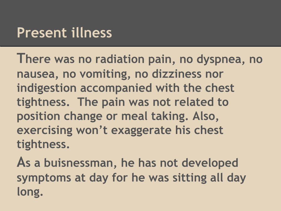
Present illness
There was no radiation pain, no dyspnea, no nausea, no vomiting, no dizziness nor indigestion accompanied with the chest tightness. The pain was not related to position change or meal taking. Also, exercising won’t exaggerate his chest tightness.
As a buisnessman, he has not developed symptoms at day for he was sitting all day long.

Present illness
He went to our OPD for help, where treadmill was done 2 months ago and revealed ST-segment depression.
Heart echo found EF=57.1%, mild AR and mild MR.
Thallium scan showed suspected large extent of myocardial ischemia in the anteroseptal, apical anterior, apical, inferior and inferiolateral walls of left ventricle.

Present illness
Then he was admitted to receive CAG 2 weeks ago. CAG reported CAD with TVD.
This time, he was admitted to our ward to receive PCI for RCA and LCX.

Social History
Allergy: none
Alcohol drinking: social drinking
Betalnuts chewing: no
Cigarette smoking: previously 1 PPD, quitted for 2 months(due to chest pain)

Family History
Father died of Hypertension.
Mother died of Leukemia
One of his brother living with previous stroke. (four elder sisters and two elder brothers)
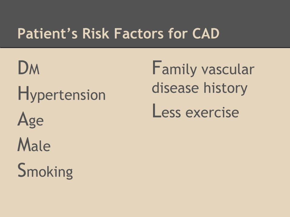
Patient’s Risk Factors for CAD
DM
Hypertension
Age
Male
Smoking
Family vascular disease history
Less exercise

Review of systems
General:Recent weight change:-Fever/Chills:- Change in appetite:- Fatigue:- Generalized weakness:-
Cardiovascular:Heart trouble:+ Chest pain:+ Dyspnea on exertion:- Orthopnea:- Palpitation:- Intermittent claudicate:-Varicose veins:+ Edema:-

Review of systems
Musculoskeletal:arthralgia:- Myalgia:- Back pain:-
Neurological:Syncope:- Seizures:- Focal weakness or paralysis:- Numbness/paresthesias:-Tremor:-
Psychiatric:Irritability:- Difficulty concentrating:- Memory loss:- Depression:- Anxiety:-

Physical Examination
Height:166 cm; Weight:72 kg
Vital signs: T/P/R : 36.9/79/17Blood Pressure:132/77
General Appearance:
Consciousness: clear, alert, Ill-looking
Chest:Symmetric expansionNo focal tendernessNo chest wall lesionBS: clear, no wheezing or crackles
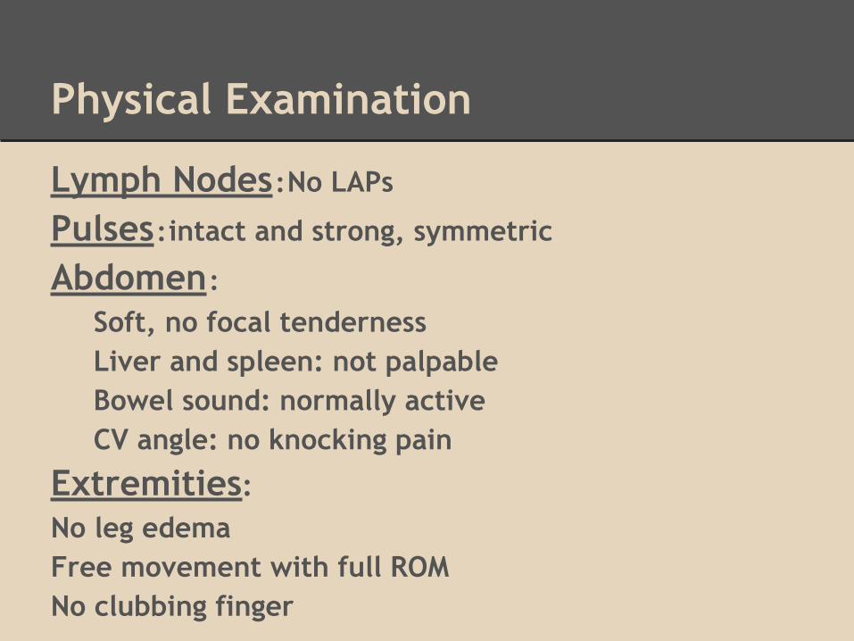
Physical Examination
Lymph Nodes:No LAPs
Pulses:intact and strong, symmetric
Abdomen:
Soft, no focal tendernessLiver and spleen: not palpableBowel sound: normally activeCV angle: no knocking pain
Extremities:No leg edemaFree movement with full ROMNo clubbing finger
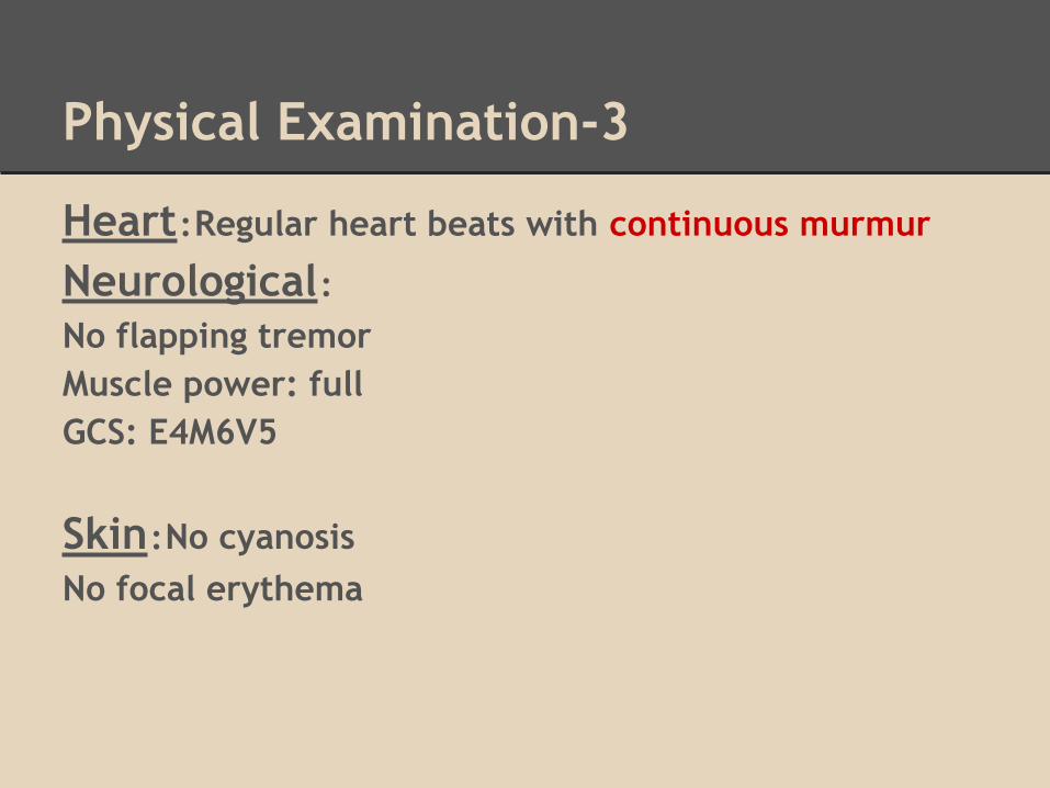
Physical Examination-3
Heart:Regular heart beats with continuous murmur
Neurological:No flapping tremorMuscle power: fullGCS: E4M6V5
Skin:No cyanosisNo focal erythema

EKG

Treadmill
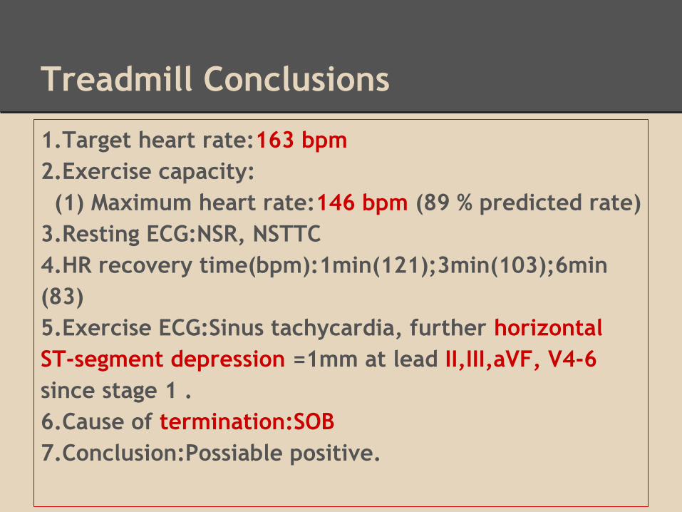
Treadmill Conclusions
1.Target heart rate:163 bpm2.Exercise capacity: (1) Maximum heart rate:146 bpm (89 % predicted rate)3.Resting ECG:NSR, NSTTC4.HR recovery time(bpm):1min(121);3min(103);6min(83)5.Exercise ECG:Sinus tachycardia, further horizontal ST-segment depression =1mm at lead II,III,aVF, V4-6 since stage 1 .6.Cause of termination:SOB7.Conclusion:Possiable positive.
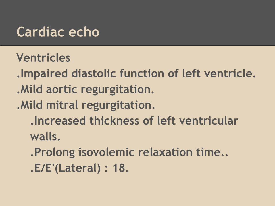
Cardiac echo
Ventricles.Impaired diastolic function of left ventricle..Mild aortic regurgitation..Mild mitral regurgitation.
.Increased thickness of left ventricular walls..Prolong isovolemic relaxation time...E/E'(Lateral) : 18.

Cardiac echo (05/05)
Valves.MR jet area is <20% of LA area..AR jet width is <25% of LVOT diameter.

Thalium scan

CAG report
CAG showed CAD with TVD. LM-d and LAD-p 95% stenosis with calcificationLAD-d 70% stenosisLCX-m 70% stenosisLCX-d 100% stenosisRCA-p 99% stenosisRCA-m 95% stenosisPLV 85% stenosis.
LVG demostrated preserved LV systolic function without significant regional wall motion abnormality . MR,AR,LV aneurysm was found.
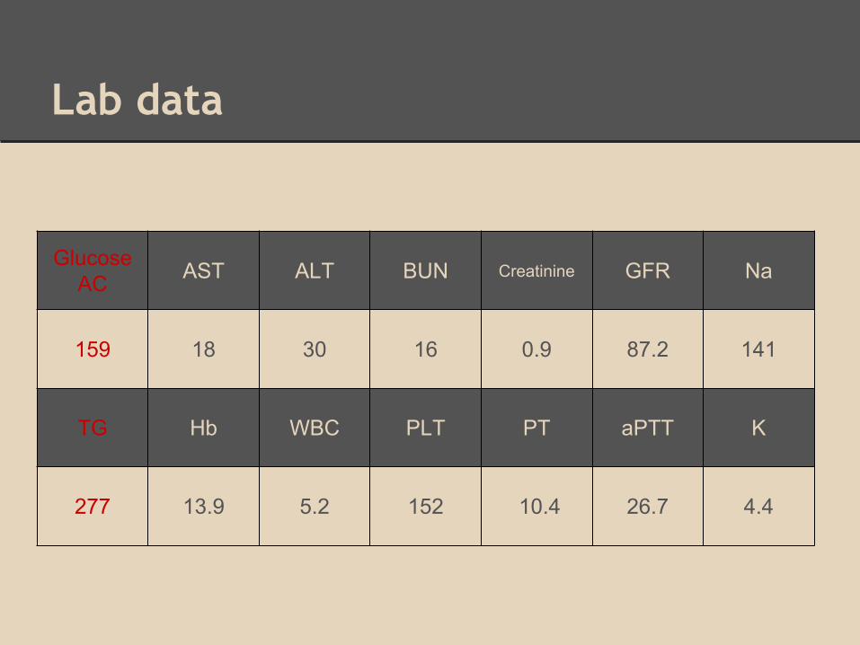
Lab data
Glucose AC AST ALT BUN Creatinine GFR Na
159 18 30 16 0.9 87.2 141
TG Hb WBC PLT PT aPTT K
277 13.9 5.2 152 10.4 26.7 4.4

Diagnosis
CAD with TVD s/p CAG on 2013/5/22HyperlipidemiaHypertensionDiabetes mellitus type 2

Plan
Plan for PTCA on 6/9
Lifestyle control, quit smoking
Under Aspirin, NTG and Colpidogrel control

PTCA on 6/9
1.At first
RCA-P-M-PLV lesion was dilated with Hiryu 2.5*15 mm balloon at 6-18 atm2.then
Resolute 3.0*38mm stent was placed at the RCA-P lesion across RCA-M vessel and deployed at 14-16 atm
In RCA-P-M stent post dilatation with nc Sapphire 3.75*12mm balloon at 10-25 atm
-> The lesion was dilated with the same balloon
-> Final coronary angiography showed successful stent implantation