caused by neisseria meningitidis, streptococcus pneumoniae ... · caused by neisseria meningitidis,...
TRANSCRIPT
August, 1998
Centers for Disease Controland Prevention
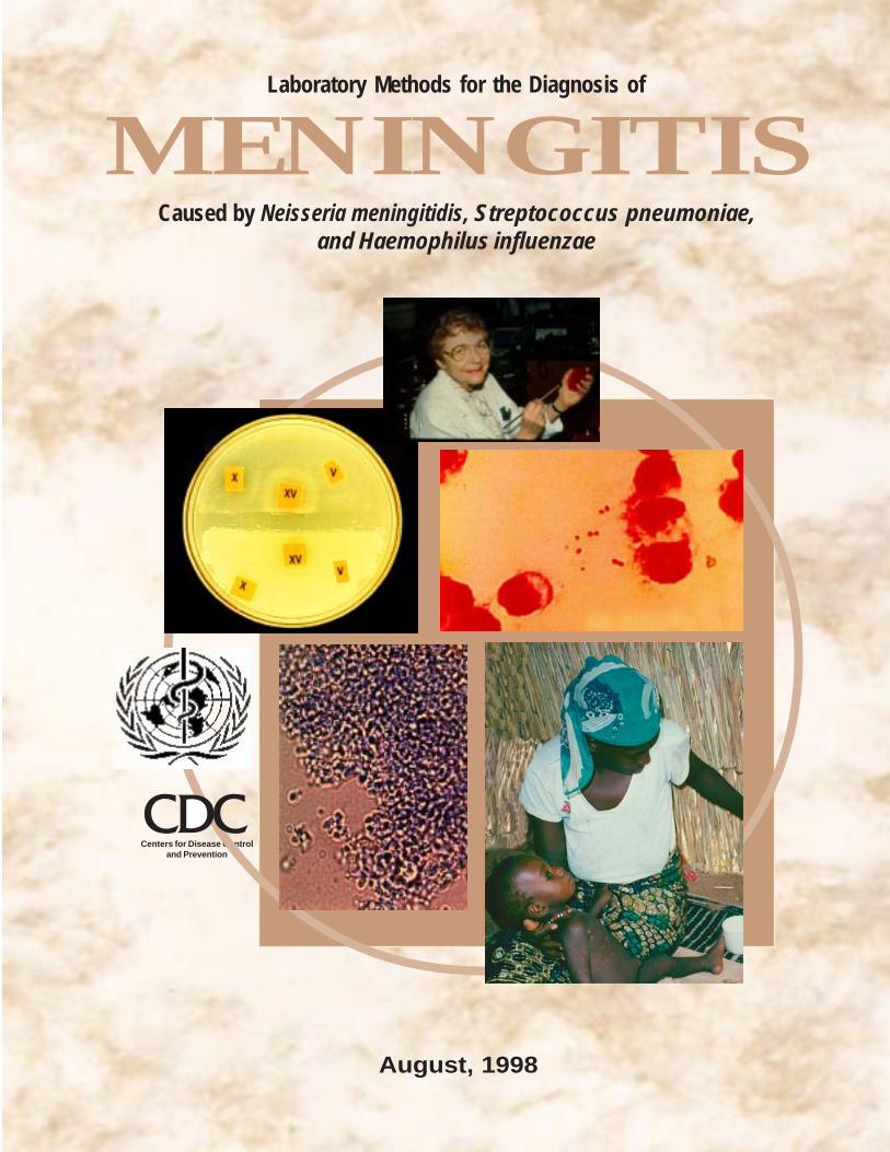
MENINGITISLaboratory Methods for the Diagnosis of
Caused by Neisseria meningitidis, Streptococcus pneumoniae,and Haemophilus influenzae
Laboratory Methods for the Diagnosis of MeningitisCaused by Neisseria meningitidis, Streptococcus pneumoniae,
and Haemophilus influenzae
Table of Contents
Introduction………………………………………………………………………………… 1
Acknowledgments ……………………………………………………………………….. 2
I. Epidemiology of Meningitis Caused by Neisseria meningitidis, Haemophilusinfluenzae and Streptococcus pneumoniae,…………………………………………… 3
II. General Considerations ......................................................................................................... 5A. Record Keeping ................................................................................................................... 5
III. Collection and Transport of Clinical Specimens ................................................................... 6A. Collection of Cerebrospinal Fluid (CSF)............................................................................... 6
A1. Lumbar Puncture ................................................................................................... 6B. Collection of Blood .............................................................................................................. 7
B1. Precautions ............................................................................................................ 7B2. Sensitivity of Blood Cultures ................................................................................. 7B3. Venipuncture ......................................................................................................... 8
C. Transport of Clinical Specimens ........................................................................................... 8C1. CSF ....................................................................................................................... 8C2. Blood..................................................................................................................... 9
IV. Primary Culture, Subculture and Presumptive Identification ............................................. 10A. Inoculation of Primary Culture Media .................................................................................. 10
A1. CSF ....................................................................................................................... 101.1. Gram Stain Procedure for CSF (Hucker Modification)............................... 101.2. General Methods for Performing Latex Agglutination Tests ...................... 11
A2. Blood..................................................................................................................... 12B. Subculture ........................................................................................................................ 12
B1. Blood Culture Bottle .............................................................................................. 12B2. T-I Medium ........................................................................................................... 12
C. Macroscopic Examination of Colonies.................................................................................. 12
V. Identification of N. meningitidis ............................................................................................. 14A. Kovac’s Oxidase Test........................................................................................................... 14B. Identification of the N. meningitidis Serogroup ..................................................................... 14C. Carbohydrate Utilization by N. meningitidis – Cystine Trypticase Agar Method ................... 15D. Commercial Identification Kits............................................................................................. 16
VI. Identification of S. pneumoniae ............................................................................................. 18A. Susceptibility to Optochin .................................................................................................... 18B. Bile Solubility Test............................................................................................................... 18C. Slide Agglutination Test ....................................................................................................... 19
VII. Identification of H. influenzae ............................................................................................... 20A. Identification of the H. influenzae Serotype .......................................................................... 20

B. Identification of X and V Factor Requirements ..................................................................... 20B1. X, V and XV Paper Disks or Strips ........................................................................ 20B2. Haemophilus ID “Quad” Plates .............................................................................. 21
VIII. Preservation and Transport of N. meningitidis, S. pneumoniae, and H. influenzae ............. 24A. Short-Term Storage .............................................................................................................. 24B. Long-Term Storage .............................................................................................................. 24
B1. Preservation by Lyophilization............................................................................... 24B2. Preservation by Freezing........................................................................................ 25
C. Transportation of Cultures .................................................................................................... 25C1. Transport in Silica Gel Packages ............................................................................ 25
IX. Bibliography ........................................................................................................................... 27
X. Annexes .................................................................................................................................. 28
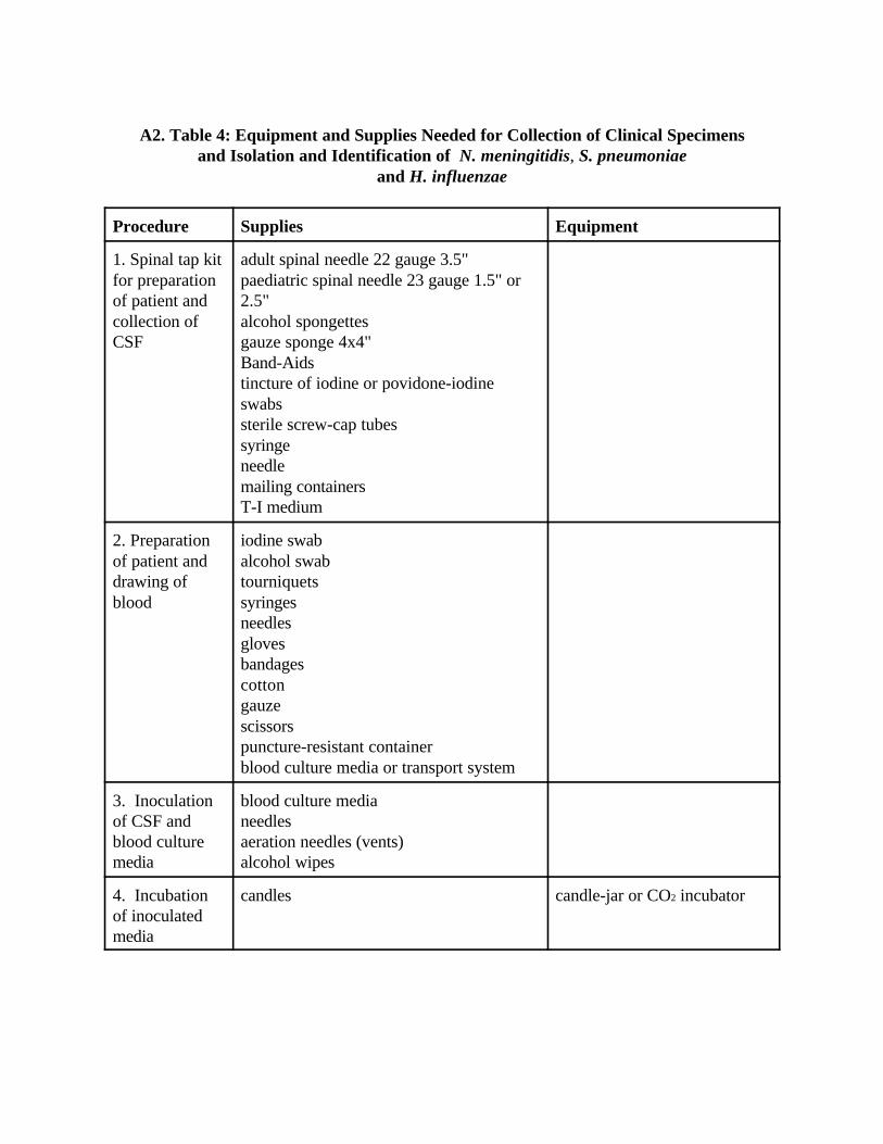
A. Basic Requirements, Supplies and Equipment ...................................................................... 28A1. Table 3: Basic Requirements and Supplies for Microbiology Laboratory................ 29A2. Table 4: Equipment and Supplies Needed for Collection of Clinical
Specimes and Isolation and Identification of N. meningitidis, S.pneumoniae, and H. influenzae.................................................................. 30
B. Media and Reagents ............................................................................................................. 33B1. Table 5: Media and Reagents Necessary for Isolation and Identification of
N. meningitidis, S. pneumoniae, and H. influenzae....................................... 34B2. Table 6: Commercially Available Tests for Latex Agglutination,
Coagglutination and Serogrouping/Serotyping of N. meningitidis, S.pneumoniae, and H. influenzae.................................................................. 39
B3. Table 7: Control Strains for Quality Assurance Testing of Media andReagents ................................................................................................... 43
B4. Manufacturers’ Addresses ...................................................................................... 44C. Preparation of Media and Reagents....................................................................................... 48
C1. Quality Control of Media ....................................................................................... 48C2. Routine Agar and Broth Media............................................................................... 48
Heart Infusion Agar (HIA) and Trypticase Soy Agar (TSA) ............................. 48Blood Agar Plate (BAP): TSA with 5% Sheep Blood....................................... 48Heart Infusion Rabbit Blood Agar Plate (HIA - Rabbit Blood) ......................... 49Horse Blood Agar (Blood Agar Base) .............................................................. 49Trypticase Soy Broth (TSB)............................................................................. 49Blood Culture Broths ....................................................................................... 50
C3. Special Media ........................................................................................................ 50Chocolate Agar Plate (CAP) ............................................................................ 50Chocolate Agar with TSA and Growth Supplement.......................................... 51Chocolate Agar with Gonococcus Medium (GC) Base andGrowth Supplement ......................................................................................... 51Levinthal’s Medium for Haemophilus .............................................................. 52
C4. Transport and Storage Media ................................................................................. 52Trans-Isolate (T-I) Medium.............................................................................. 52Greaves Solution for Preservation of Strains by Freezing ................................. 53
C5. Miscellaneous Reagents .............................................................................................Reagents for Gram Stain (Hucker Modification) .............................................. 54McFarland Density Standard............................................................................ 55Skin Antiseptic ................................................................................................ 55

D. Serotyping of S. pneumoniae ................................................................................................ 56D1. Quellung Typing of S. pneumoniae ........................................................................ 56
D2. Typing and/or Grouping of S. pneumoniae ......................................................................... 57E. Biotyping of H. influenzae .................................................................................................... 59
XII. List of Figures ............................................................................................................61
XII. Abbreviations … …………………………………………………………… 63
Introduction
This manual summarizes laboratory techniques used in the isolation andidentification of Neisseria meningitidis (the meningococcus), Streptococcus pneumoniae(the pneumococcus) and Haemophilus influenzae from the cerebrospinal fluid and bloodof patients with clinical meningitis. The procedures described here are not new; mosthave been used for many years. Even though they require an array of laboratorycapabilities, these procedures were selected because of their utility, ease of performance,and ability to give reproducible results. The diversity of laboratory capabilities, theavailability of materials and supplies, and their cost, were taken into account. In additionto these basic procedures, methods for subtyping and biotyping of these organisms areincluded for reference laboratories that have the facilities, the trained personnel, and thedesire to perform them.
Acknowledgments
This manual was prepared in collaboration with the following WHO CollaboratingCentres: WHO Collaborating Center for Prevention and Control of Epidemic Meningitis,Centers for Disease Control and Prevention, Atlanta, GA, USA; WHO CollaboratingCentre for Reference and Research on Meningococci, National Institute of Public Health,Oslo, Norway; and WHO Collaborating Centre for Reference and Research onMeningococci, Institut de Médecine Tropicale du Service de Santé des Armées,Marseilles, France.
Coordinators
Tanja Popovic, Gloria W. Ajello, Richard R. Facklam, National Center forInfectious Diseases, Centers for Disease Control and Prevention, Atlanta, GA, USA.
Contributors
Dominique A. Caugant, National Institute of Public Health, Oslo, NorwayPierre Nicolas, Institut de Médecine Tropicale du Service de Santé des Armées,Marseilles, FranceBrad Perkins, Nancy Rosenstein, Orin Levine, Anne Schuchat, Bradford A. Kay, MariaLucia Tondella, National Center for Infectious Diseases, Centers for Diseases Controland Prevention, Atlanta, GA, USADr David Heymann, EMC/WHO/HQDr Eugene Tikhomirov, EMC/WHO/HQDr André Ndikuyeze, WHO/AFRDr Oyewale Tomori, WHO/AFRProfessor B. Koumare, WHO/AFRDr D. Barakamfitiye, WHO/AFRDr Z. Hallaj, WHO/EMRDr B. Sadrizadeh, WHO/EMREllen Jo Baron, Stanford University Hospital, Stanford, CA, USAJohn Robbins, National Institutes of Health, Bethesda, MD, USARick Nolte, Emory University, Atlanta, GA, USADiana Martin, Institute of Environmental Science and Research Limited, Porirua, New Zealand
Technical Support
Christopher Jambois, Ruth Thornberg, Anne Mather, Erica Pearson, National Centerfor Infectious Diseases, Centers for Disease Control and Prevention, Atlanta, GA, USA.

I. Epidemiology of Meningitis Caused by Neisseria meningitidis,Streptococcus pneumoniae and Haemophilus influenzae
Bacterial menigitis, an infection of the membranes (meninges) and cerebrospinalfluid (CSF) surrounding the brain and spinal cord, is a major cause of death and disabilityworld-wide. Beyond the perinatal period, three organisms, transmitted from person toperson through the exchange of respiratory secretions, are responsible for most cases ofbacterial meningitis: Neisseria meningitidis, Haemophilus influenzae, and Streptococcuspneumoniae. The etiology of bacterial meningitis varies by age group and region of theworld. Worldwide, without epidemics one million cases of bacterial meningitis areestimated to occur and 200,000 of these die annually. Case-fatality rates vary with age atthe time of illness and the species of bacterium causing infection, but typically rangefrom 3 to 19% in developed countries. Higher case-fatality rates (37-60%) have beenreported in developing countries. Up to 54% of survivors are left with disability due tobacterial meningitis, including deafness, mental retardation, and neurological sequelae.
Two clinically overlapping syndromes – meningitis and bloodstream infection(meningococcaemia) - are caused by infection with N. meningitidis (meningococcaldisease). While the two syndromes may occur simultaneously, meningitis alone occursmost frequently. N. meningitidis is classified into serogroups based on the immunologicalreactivity of the capsular polysaccharide. Although 13 serogroups have been identified,the three serogroups A, B and C account for over 90% of meningococcal disease.Meningococcal disease differs from other leading causes of bacterial meningitis becauseof its potential to cause large-scale epidemics. A region of sub-Saharan Africa extendingfrom Ethiopia in the East to The Gambia in the West and containing fifteen countries andover 260 million people is known as the “meningitis belt” because of its high endemicrate of disease with superimposed, periodic, large epidemics caused by serogroup A, andto a lesser extent, serogroup C. During epidemics, children and young adults are mostcommonly affected, with attack rates as high as 1,000/100,000 population, or 100 timesthe rate of sporadic disease. The highest rates of endemic or sporadic disease occur inchildren less than 2 years of age. In developed countries, endemic disease is generallycaused by serogroups B and C. Epidemics in developed countries are typically caused byserogroup C although epidemics due to serogroup B have also occurred in Brazil, Chile,Cuba, Norway and more recently in New Zealand.
Meningitis caused by H. influenzae occurs mostly in children under the age of 5years, and most cases are caused by organisms with the type b polysaccharide capsule(H. influenzae type b, Hib). While most children are colonized with a species ofH. influenzae, only 2-15% harbour Hib. The organism is acquired through the respiratoryroute. It adheres to the upper respiratory tract epithelial cells and colonizes thenasopharynx. Following acquisition of Hib, illness results when the organism is able topenetrate the respiratory mucosa and enters the blood stream. This is the result of acombination of factors, and subsequently the organism gains access to the CSF, whereinfection is established and inflammation occurs. An essential virulence factor whichplays a major role in determining the invasive potential of an organism is thepolysaccharide capsule of Hib. Meningitis is the most severe form of Hib disease; in mostcountries, however more cases and deaths are due to pneumonia than to meningitis.
Meningitis in individuals at the extremes of age infants, young children and theelderly is commonly caused by S. pneumoniae. Younger adults with anatomic orfunctional asplenia, haemoglobinopathies, such as sickle cell disease, or who areotherwise immunocompromised, also have an increased susceptibility to S. pneumoniaeinfection. S. pneumoniae, like Hib, is acquired through the respiratory route. Followingthe establishment of nasopharyngeal colonization, illness results once bacteria evade themucosal defences, thus accessing the bloodstream, and eventually reaching the meningesand CSF. As is the case with Hib, many more cases and deaths are due to pneumococcalpneumonia, even though pneumococcal meningitis is the more severe presentation ofpneumococcal disease.
The risk of secondary cases of meningococcal disease among close contacts (i.e.household members, day-care centre contacts, or anyone directly exposed to the patient’soral secretions) is high. Antimicrobial chemoprophylaxis with a short course of oralrifampin, a single oral dose of ciprofloxacin, or a single injection of ceftriaxone iseffective in eradicating nasopharyngeal carriage of N. meningitidis. Although veryeffective in preventing secondary cases, antimicrobial chemoprophylaxis is not aneffective intervention for altering the course of an outbreak. In epidemics, masschemoprophylaxis is not recommended.
Vaccines have an important role in the control and prevention of bacterialmeningitis. Vaccines against N. meningitidis, H. influenzae, and S. pneumoniae arecurrently available, but the protection afforded by each vaccine is specific to eachbacterium and restricted to some of the serogroups or serotypes of each bacterium. Forexample, vaccines are currently available to prevent H. influenzae infections due toserotype b (Hib) but not those infections due to other serotypes or unencapsulatedorganisms (i.e. nontypeable H. influenzae). In addition to establishing a diagnosis, animportant role for the laboratory, therefore, is to determine the bacteria andserogroups/serotypes that are causing meningitis in a community.
In industrialized countries, routine use of polysaccharide-protein Hib conjugatevaccines for immunization of infants has almost eliminated Hib meningitis and otherforms of severe Hib disease. Several studies in developing countries have corroboratedthese finding. Pneumococcal polysaccharide vaccines have been used to prevent diseasein the elderly and in persons with chronic illnesses that may impair their naturalimmunity to pneumococcal disease. Meningococcal polysaccharide vaccines aregenerally used in response to epidemics and for the prevention of disease in overseastravellers although other uses are currently under investigation.
In addition to the existing armamentarium of vaccines, new generation vaccinesagainst meningococcal and pneumococcal disease are under development and evaluation.These vaccines may provide a high degree of protection and broad coverage in all agegroups. Until these vaccines become widely available, the current vaccines should beused appropriately and efficiently. Use of any of these vaccines will require laboratoryidentification of the agents causing disease in addition to epidemiological informationabout the age and risk groups that are most affected.
II. General Considerations
This manual provides recommendations for the use of media and reagents in theisolation and identification of N. meningitidis, S. pneumoniae, and H. influenzae.Alternatively, other media and reagents not listed in this manual may be substitutedwhere appropriate. When making changes outside of these recommendations, it may behelpful for the laboratory staff to consult the authors of this manual.
All of the assays described in this manual should be conducted at Biosafety Level 2.Please refer to the World Health Organization (WHO) Laboratory Biosafety Manual fordetailed descriptions of safety precautions.
Antimicrobial susceptibility testing of these organisms is not addressed in thismanual, and readers are referred to special textbooks and manuals (i.e. the Manual ofClinical Microbiology, the Clinical Microbiology Procedures Handbook, or the WHOManual for the National Surveillance of Antimicrobial Resistance of S. pneumoniae andH. influenzae).
A. Record Keeping
It is recommended that all clinical samples be recorded on a form that provides thefollowing information:
1. Patient’s name2. Patient’s age and sex3. Patient’s hospital room number or address4. Physician’s name and address or where the physician can be located5. Anatomical site of specimen collection6. Date and time of specimen collection7. Clinical diagnosis and relevant patient history8. Antimicrobial agents, if any, that the patient is receiving
In addition, each specimen should have a label firmly attached to the specimencontainer and bearing the following information:
Patient’s name:_________________________________
Hospital:______________________________________
Room: _______________________________________
Physician: ____________________________________
Specimen type:_________________________________
Date and time of collection:_______________________
Test requested: _________________________________
A registry at the laboratory should include a bound record book listing each clinicalsample received, specifying the date received, the referring hospital or physician, theidentification of the organism, results of assays performed, and whether the isolate was
sent to the reference laboratory for further studies. In addition, basic information aboutthe patient should also be noted (age, sex, name, address).
III. Collection and Transport of Clinical Specimens
The collection of clinical specimens is important in the isolation and identificationof bacterial agents that cause meningitis. It is recommended that clinical specimens beobtained before antimicrobial therapy is begun to avoid loss of viability of the etiologicalagents. Treatment of the patient, however, should not be delayed while awaitingcollection of specimens. CSF and blood should be processed in a bacteriology laboratoryas soon as possible.
A. Collection of Cerebrospinal Fluid (CSF)
The collection of CSF is an invasive technique and should be performed byexperienced personnel under aseptic conditions. If meningitis is suspected, CSF is thebest clinical specimen to use for isolating and identifying the etiological agents. Thecollection of CSF should be performed for diagnosis only. CSF should be inoculateddirectly onto both a supplemented chocolate agar plate (CAP) and a blood agar plate(BAP).
A1. Lumbar Puncture
Usually, 3 tubes of CSF are collected for chemistry, microbiology, and cytology. Ifonly one tube of fluid is available, it should be given to the microbiology laboratory. Ifmore than one tube (1 ml each) is available, the second or third tube should go to themicrobiology laboratory.
The kit for collection of CSF (Figure 1) should contain:1. skin disinfectant2. sterile gauze and Band-Aid3. lumbar puncture needles: 22 gauge/3.5"for adults; 23 gauge/2.5" for children4. sterile screw-cap tubes5. syringe and needle6. transport container7. Trans-Isolate (T-I) medium (if CSF cannot be analysed in the microbiological
laboratory immediately)
The patient should be kept motionless, either sitting up or laying on the side, withhis or her back arched forward so that the head almost touches the knees during theprocedure (Figure 2). Disinfect the skin along a line drawn between the crests of the twoilia with 70 % alcohol to clean the surface and remove debris and oils. Then applytincture of iodine or povidone-iodine. Let dry. The needle is introduced, and the drops offluid (1 ml minimum, 3-4 ml if possible) are collected into sterile, screw-cap tubes. Labelthe specimen as described earlier. Do not refrigerate the specimen. Hand carry it(whenever feasible) to the laboratory as soon as possible. Avoid exposure to excessiveheat or sunlight.
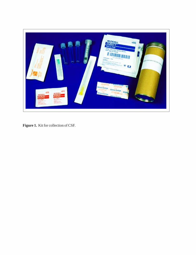
Figure 1. Kit for collection of CSF.

Illustration and procedure reprinted with permission of J. B. Lippincott Company fromKoneman, Elmer w. et al. (1992). Diagnostic Microbiology, Page 90.
The patient lies on his side with knees flexed and back arched to separate the lumbar vertebrae. The patient is surgically draped, and an area overlying the lumbar spine is disinfected.
The space between lumbar vertebrae L3 and L4 is palpated with the sterilely gloved forefinger.
The spinal needle is carefully directed between the spinous processes, through the intraspinousprocesses, through the intraspinous ligaments into the spinal canal.
A.
B.
C.
Figure 2. Collection of CSF by lumbar puncture.

B. Collection of Blood
For the diagnosis of bacterial meningitis, blood should be collected when a spinaltap is contraindicated or cannot be performed for technical reasons.
B1. Precautions
Infection may be transmitted from patient to staff and from staff to patient during theblood-taking procedure. Viral agents are the greatest hazard and in some instances arepotentially lethal. Of particular importance are the viruses causing hepatitis and acquiredimmunodeficiency syndrome. To decrease the risk of transmission of these viral agents,the recommendations below should be followed:
(a) Wear latex or vinyl gloves impermeable to liquids.(b) Change gloves between patients.(c) Inoculate blood into blood culture media immediately to prevent the blood from
clotting in the syringe. Syringes and needles should be disposed of in a puncture-resistant, autoclavable container. No attempt should be made to re-cap the needle. Anew syringe and needle must be used for each patient.
(d) Wipe the surface of the blood culture bottle and the gloves with a disinfectant.(e) Label the bottle.(f) For the transport to the microbiology laboratory, place the blood culture medium in a
container that can be securely sealed.(g) Specimen containers should be individually and conspicuously labelled. Any
containers with blood on the outside should be wiped thoroughly. Such containersshould be transported in individual plastic envelopes.
(h) Remove gloves and discard in an autoclavable container.(i) Wash hands with soap and water immediately after removing gloves.(j) Transport the specimen to the microbiology laboratory or, if that facility is closed,
store the specimen in an approved location.(k) In the event of a needle-stick injury or other skin puncture or wound, wash the
wound liberally with soap and water. Encourage bleeding.(l) Report any contamination of the hands or body with blood, or any puncture wound
or cut to the supervisor and the health service for treatment.
B2. Sensitivity of Blood Cultures
Several variables affect the sensitivity of blood cultures: the number of collections,the volume of each collection, and the steps taken to inhibit or neutralize bactericidalproperties of blood may vary with the age of the patient. It may be difficult to collect alarge amount of blood from a child; 1-3 ml is usually sufficient. Blood cultures fromyoung children should be diluted to 1-2 ml of blood in 20 ml of broth (1:10 to 1:20). Blood cultures fromadults should be diluted to 5-10 ml of blood in 50 ml of broth (1:5 to 1:10).
Blood should be cultured in trypticase soy broth (TSB) or brain heart infusion with agrowth supplement (such as IsoVitaleX or Vitox) to support growth of organisms such asH. influenzae. Neutralization of normal bactericidal properties of blood and potentialantimicrobial agents is accomplished by adding chemical inhibitors such as 0.025%sodium polyanetholesulfonate (SPS) to culture media and by diluting the blood. SPS,

which has anticoagulant, antiphagocytic, anticomplementary, and antilysozymal activity,may be inhibitory if used in higher concentrations.
B3. Venipuncture
An outline of the proper method for collecting blood from the arm is shown inFigure 3.
(a) Gather everything needed to complete the process: gloves, syringe, needle,tourniquet, gauze squares, cotton balls, Band-Aid, puncture resistant container,culture medium and antiseptic; iodine tincture or povidone-iodine is preferred, but70% alcohol is an acceptable alternative. However, alcohol with concentrationsgreater than 70% should not be used because the increased concentrations result indecreased antibactericidal activity. The size of the needle will depend on thecollection site and the size of the vein. A 23-gauge needle that is 20-25 mm in lengthor a butterfly needle is generally used for children.
(b) Select an arm and apply a tourniquet to restrict the flow of venous blood. The largeveins of the forearm are illustrated in Figure 3. The most prominent vein is usuallychosen.
(c) Vigorously wipe the skin with the 70% alcohol, and swab with the iodine tincture orpovidone-iodine. Rub over the selected area. Allow to dry. If the vein is palpatedagain, repeat the skin disinfection.
(d) After the disinfectant has dried, insert the needle into the vein with the bevel of theneedle face up. Once the vein is entered, withdraw the blood by pulling back thebarrel of the syringe in a slow, steady manner. Air must not be pumped into a vein.After the desired amount of blood is obtained, release the tourniquet and place asterile cotton ball over the insertion site while holding the needle in place. Withdrawthe needle and have patient hold the cotton-ball firmly in place until the wound hasstopped bleeding. Inoculate the culture medium. Put the Band-Aid on the wound.
(e) Vacutainer tubes should be used for blood collection, if available.
C. Transport of Clinical Specimens
S. pneumoniae, H. influenzae and N. meningitidis are fastidious and fragile bacteria.They are more reliably isolated if the clinical material is examined as soon as possibleafter collection.
C1. CSF
As soon as the CSF has been collected, it should be transported to the microbiologylaboratory, where it should be examined as soon as possible (within one hour from thetime of collection) (Figure 4). Do not expose the CSF to sunlight or extreme heat or cold.If N. meningitidis is suspected to be the cause of the illness, and a delay of several hoursin processing specimens is anticipated, incubating the CSF (with screw-caps loosened) at35 •C in a 5% CO
2 atmosphere (or candle-jar) may improve bacterial survival. If same-day transport to the laboratory is not possible, CSF should be inoculated aseptically into aTrans-Isolate (T-I) medium with a syringe and held overnight at, or close to, 35 •C. T-I isa biphasic medium that is useful for the primary culture of meningococci and other

2
1
3
Figure 3. Collection of blood from an arm.
1. Apply the tourniquet
2. Select a vein
3. Plan proposed puncture site
etiological agents of bacterial meningitis from CSF and blood samples (Figure 5). It canbe used as a growth medium as well as a holding and transport medium. The preparationof the T-I medium is described in Annex C (C4).
(a) The T-I bottle septum should be disinfected with alcohol and iodine and allowed todry before inoculation. Inoculate 1 ml of CSF into the T-I medium, which has eitherbeen pre-warmed in the incubator (35 •C-37 •C) or kept at room temperature(25 •C).Keep the remaining CSF in the container or syringe in which it was collected. Donot refrigerate, but hold at room temperature before Gram staining.
(b) After inoculation, T-I bottles are incubated at 35 •C overnight. Label the T-I bottleappropriately with the patient identification, and date and time of CSF inoculation.Incubate the T-I medium at 35 •C for up to 7 days. Venting the bottle with a ventingneedle, or a sterile cotton-plugged hypodermic needle after the initial 24-hincubation, or as soon as possible after transportation has been completed,encourages growth and survival. If transport is delayed, vented bottles can be heldfor days at moderate to warm room temperatures (25 •C-30 •C). The vents must beremoved before shipment. It is essential to obtain specimens aseptically and to avoidcontamination when inoculating or sampling the bottles.
C2. Blood
(a) Blood cannot be transported before being placed in broth because the collectionprocedure does not use an anticoagulant. Blood should be injected into the brothculture medium within one minute of collection. If the blood culture bottle containsa diaphragm, clean the diaphragm with 70% alcohol and povidone-iodine beforeinoculating the medium. Swirl the bottle several times. Discard the needle andsyringe in a puncture-resistant container. Do not re-cap the needle. Clean thediaphragm of the blood culture bottle, if necessary. Then label it appropriately withpatient identification and the date and time of blood collection. The preparation ofblood culture media is described in Annex C (C2).
(b) The inoculated medium can be held at room temperature (20 •C-25 •C) for 4 to 6hours before incubation at 35 •C. Inoculated or uninoculated blood-culture mediummust not be placed in a refrigerator. A portable incubator may be used (temperaturerange 25 •C-35 •C).
(c) Immediately transport the inoculated media to the laboratory. All inoculated blood-culture media should be received by the laboratory within 12 to 18 hours forsubculture and should be protected from temperature extremes (less than 18 •C,more than 37 •C) with a transport carrier such as Styrofoam, which can keep thesamples at moderate temperature.

Transport to the Transport to the laboratory laboratory < than 1 hour > than 1 hour
Cerebrospinal Fluid
Figure 4. Processing of CSF.
Latex agglutination
Centrifuge at 2000 rpm/20 min.
Supernatant Sediment
< than 1 hour > than 1 hour
Gram Primary plating stain (CAP and BAP)
Inoculate T-I
Incubate O/N(35 C in CO
2)
Subculture(CAP and BAP)

Figure 5. Trans-Isolate medium.

IV. Primary Culture, Subculture, and Presumptive Identification
A. Inoculation of Primary Culture Media
A1. CSF
Once the CSF has arrived at the microbiology laboratory, centrifuge it for 20minutes at 2000 rpm. Draw off the supernatant with a Pasteur pipette. When antigendetection by latex agglutination is planned, save the supernatant. Sediment must be eithervigorously vortexed or well mixed. Use one or two drops of sediment to prepare theGram stain and use 1 drop to streak the primary culture media (Figure 4).
Note: If less than 1 ml of CSF is available, do not centrifuge. Use it for the Gramstain and plate it directly.
The best medium for S. pneumoniae is a BAP, which is a trypticase soy agar (TSA)plate containing sheep or horse blood (5%). Human blood is not an acceptable substitute.For H. influenzae, a CAP supplemented with haemin and a growth supplement such asIsoVitaleX, supplement B, or Vitox should be used. An acceptable alternative to achievegrowth of H. influenzae on BAP is cross-streaking the medium with Staphylococcusaureus or applying a filter paper/disk saturated with X and V factors to the surface of themedium, after the medium had been inoculated. H. influenzae forms satellite coloniesalong the length of the staphylococcal growth or produces a halo of growth around theXV strip/disk. N. meningitidis grows on both BAP and CAP, as will S. pneumoniae. Ifonly one type of plate is available, choose CAP, because all three etiological agents cangrow on it. A bacteriological loop is used to streak or thin the bacteria into singlecolonies. BAP that have been properly streaked are shown in Figures 6 and 7, andproperly streaked CAP is shown in Figure 8. The agar plates should be incubated in a 5%CO
2 incubator or candle-jar. A backup broth (e.g. brain heart infusion broth) should beinoculated with some of the pellet and also incubated.
When T-I medium was used for transport, after 24 hours of incubation, with a sterileneedle and syringe, transfer 100 µl of the liquid portion of T-I onto BAP and CAP, andstreak for isolation.
1.1. Gram Stain Procedure for CSF (Hucker Modification)
A presumptive diagnosis of bacterial meningitis caused by H. influenzae, S.pneumoniae, and N. meningitidis can be made by Gram stain of the CSF sediment or bydetection of specific antigens in the CSF by a latex agglutination test. Positive results ofeither or both tests can provide evidence of infection, even if cultures fail to grow.
(a) Centrifuge the CSF for 20 minutes at 2000 rpm.(b) Prepare smear by placing 1 or 2 drops of sediment on an alcohol-rinsed and dried
slide, allowing drop(s) to form one large drop. Do not spread fluid nor use too heavya concentration of sediment.
(c) Air dry the slide in a biosafety cabinet, if available.(d) Pass the slide quickly through a flame three times to fix the smear. Do not flame
until dry. Alternatively, fixation by methanol (95%-100%) can be used.(e) Flood the smear with ammonium oxalate-crystal violet and let stand for 1 minute.
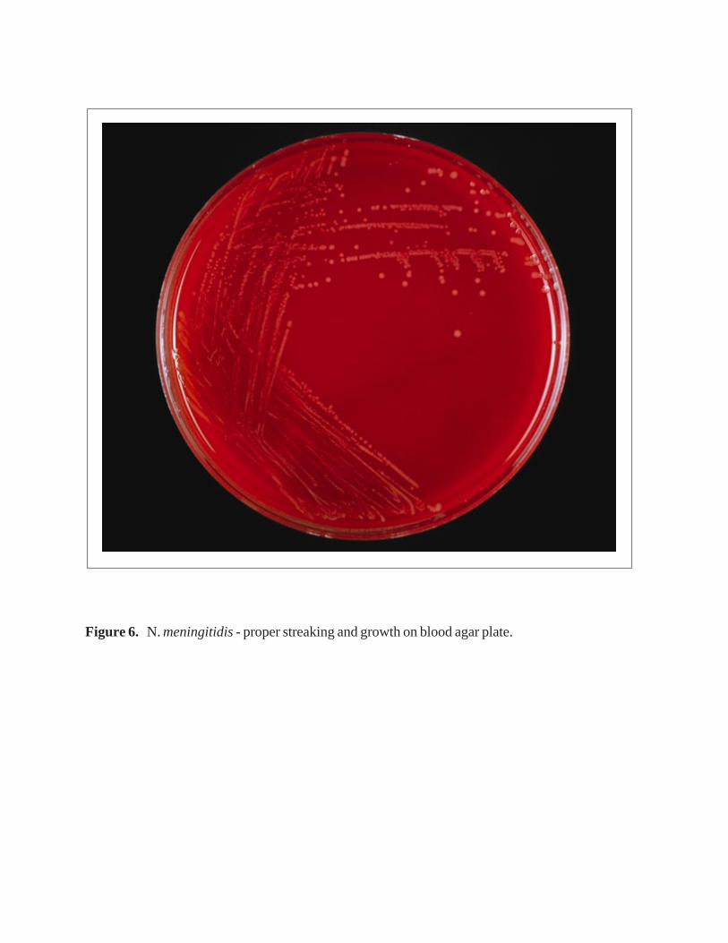
Figure 6. N. meningitidis - proper streaking and growth on blood agar plate.
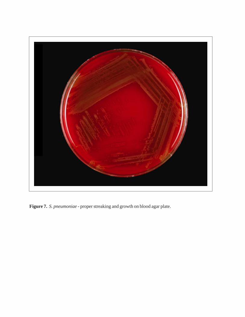
Figure 7. S. pneumoniae - proper streaking and growth on blood agar plate.
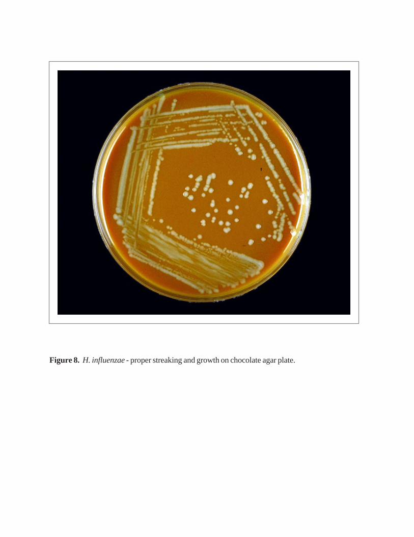
Figure 8. H. influenzae - proper streaking and growth on chocolate agar plate.
(f) Rinse gently with tap water. Drain off excess water.(g) Flood the smear with Gram’s iodine solution and let stand for 1 minute.(h) Rinse gently with tap water and drain.(i) Decolorize with 95% ethyl alcohol (5-10 seconds may be enough).(j) Counterstain with safranin for 20-30 seconds, or with carbol-fuchsin for 10-15
seconds.(k) Rinse the slide with tap water and blot dry.(l) Examine the stained smear microscopically, using a bright-field condenser and an
oil-immersion lens.
N. meningitidis may occur intra- or extra-cellularly in the polymorphonuclearleukocytes and will appear as Gram-negative, coffee-bean-shaped diplococci (Figure 9a). S. pneumoniae are lanceolate, Gram-positive diplococci sometimes occurring in shortchains (Figure 9b). H. influenzae are small, pleomorphic Gram-negative rods orcoccobacilli with random arrangements (Figure 9c). Other manuals should be consultedfor Gram stain reactions of other bacteria.
1.2. General Method for Performing Latex Agglutination Tests
Several commercial kits are available. Follow the manufacturer’s instructionsprecisely when using these tests. General recommendations and instructions typical forthe detection of soluble bacterial antigens are provided here. For best results, test thesupernatant of the centrifuged CSF sample as soon as possible. If immediate testing is notpossible, the sample can be refrigerated (between 2 •C and 8 •C) up to several hours, orfrozen at –20 •C for longer periods. Reagents should be kept refrigerated between 2 •Cand 8 •C when not in use. Product deterioration occurs at higher temperatures, especiallyin tropical climates, and test results may become unreliable before the expiration date ofthe kit. Latex suspensions should never be frozen.
Performance of the Test
(a) Heat the supernatant of the CSF in a boiling water bath for 5 minutes.(b) Shake the latex suspension gently until homogenous.(c) Place one drop of each latex suspension on a ringed glass slide or a disposable card.(d) Add 30-50 µl of the CSF to each suspension.(e) Rotate by hand for 2-10 minutes. Mechanical rotation at 100 rpm, if available, is
recommended.
Reading the Test Results
Read under a bright light without magnification.Negative reaction: the suspension remains homogenous and slightly milky inappearance.Positive reaction: agglutination (or visible clumping) of the latex particles occurs within2 minutes,.
Note: Counter immunoelectrophoresis may also be used for direct antigen detectionin CSF.A2. Blood
The blood culture bottles are inoculated directly with blood and should be ventedbefore incubation at 35 •C-37 •C. This is accomplished by inserting a sterile cotton-plugged needle into the rubber part of the blood culture bottle.
B. Subculture
Since the primary purpose of this manual is to aid in the identification ofN. meningitidis, S. pneumoniae, and H. influenzae, the methods described here will notallow for identifying other isolates that may be of clinical importance but more rarelyencountered. Microbiologists should refer to clinical microbiology manuals, such as theAmerican Society for Microbiology’s Manual of Clinical Microbiology, for procedures toidentify other bacteria.
B1. Blood Culture Bottle
Examine the blood culture bottles at 14 to 17 hours and then every day for up to 7days. Any turbidity or lysis of the erythrocytes may be indicative of growth, andsubcultures should be made immediately. Since these three organisms are fragile,subcultures should be performed after 14 to 17 hours of incubation, again at 48 hours,and at day 7, regardless of the appearance of the blood culture bottles, since theabsence of turbidity does not always correlate with the absence of bacterial growth.Before subculturing, swirl the bottle to mix the contents.
Subcultures are made by first disinfecting the surface of the blood culture bottlerubber stopper with alcohol and a povidone-iodine swab, and then aspirating a smallvolume (0.5 ml) with a syringe and needle from the blood culture bottle and inoculatingthe agar media with the fluid. Ordinarily, both CAP and BAP are used for subculture.When only one agar plate is used, it should be CAP, since CAP contains growth factorsneeded for H. influenzae whereas BAP does not. The agar plates should be streaked asshown in Figures 6, 7 and 8, and incubated for up to 48 hours. When bacterial growth hasbeen confirmed by subculture of the blood culture bottle, there is no need to continue toincubate the bottle. The bottle should be disposed of according to safety procedures.
B2. T-I Medium
(a) After 24 hours of incubation, using a sterile needle and syringe, transfer 100 µl of theliquid portion of T-I onto BAP and CAP, streak the plate, and incubate at 35 • C in aCO
2 atmosphere for up to 48 hours.(b) Check for purity of the growth by performing Gram stain.(c) If no growth occurs, subculture the T-I medium at 3 days and again at 7 days.
C. Macroscopic Examination of Colonies
N. meningitidis grows on BAP, but H. influenzae does not. When grown on CAP, H.influenzae and N. meningitidis look similar, but the two can be distinguished: H.influenzae gives off a pungent smell of indol, while N. meningitidis does not.

Figure 9a. Gram stain of cerebrospinal fluid: N. meningitidis: intra-cellular, gram-negative diplococci.

Figure 9b. Gram stain of cerebrospinal fluid: S. pneumoniae: gram-positive diplococci. Note that this slide represents a case where an exceptionally large number of organisms are present.
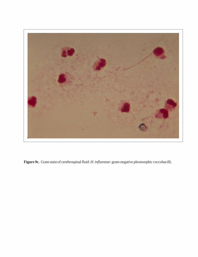
Figure 9c. Gram stain of cerebrospinal fluid: H. influenzae: gram-negative pleomorphic coccobacilli.
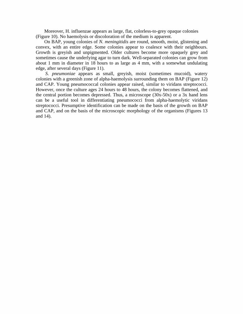
Moreover, H. influenzae appears as large, flat, colorless-to-grey opaque colonies(Figure 10). No haemolysis or discoloration of the medium is apparent.
On BAP, young colonies of N. meningitidis are round, smooth, moist, glistening andconvex, with an entire edge. Some colonies appear to coalesce with their neighbours.Growth is greyish and unpigmented. Older cultures become more opaquely grey andsometimes cause the underlying agar to turn dark. Well-separated colonies can grow fromabout 1 mm in diameter in 18 hours to as large as 4 mm, with a somewhat undulatingedge, after several days (Figure 11).
S. pneumoniae appears as small, greyish, moist (sometimes mucoid), waterycolonies with a greenish zone of alpha-haemolysis surrounding them on BAP (Figure 12)and CAP. Young pneumococcal colonies appear raised, similar to viridans streptococci.However, once the culture ages 24 hours to 48 hours, the colony becomes flattened, andthe central portion becomes depressed. Thus, a microscope (30x-50x) or a 3x hand lenscan be a useful tool in differentiating pneumococci from alpha-haemolytic viridansstreptococci. Presumptive identification can be made on the basis of the growth on BAPand CAP, and on the basis of the microscopic morphology of the organisms (Figures 13and 14).
Figure 10. On chocolate agar plate, H. influenzae appear as large colorless to gray, opaque colonies with no discoloration of the surrounding medium.

Figure 11. Overnight growth of N. meningitidis on blood agar plate appears as round, moist,
glistening and convex colonies.

Figure 12. S. pneumoniae appear as small grayish mucoid (watery) colonies with a greenish zone of alpha-hemolysis surrounding them on the blood agar plate.

Figure 13. Presumptive identification of N. meningitidis, S. pneumoniae, and H. influenzae.
Growth on Gram stain Presumptiveidentification CAP BAP
(Sheep)
+ Gram-negative H. influenzaepleomorphiccoccobacilli
+ + Gram-negative N. meningitidis
diplococci
+ + Gram-positive S. pneumoniaediplococci

Figure 14. N. meningitidis (left), S. pneumoniae (right), and H. influenzae (top): (a) growth on blood agar plate and (b) growth on chocolate agar plate.
(a) Growth on blood agar plate.
(b) Growth on chocolate agar plate.
V. Identification of N. meningitidis
The following steps are recommended to confirm the identity of cultures thatmorphologically appear to be N. meningitidis (Figure 15). The best results are obtainedwith day-old cultures. Always check for purity of the growth by performing a Gram stain(N. meningitidis is a Gram-negative, coffee-bean-shaped diplococcus). When necessary,make subcultures to ensure purity. From growth on a BAP, perform Kovac’s oxidase test,and then identify the serogroup. Finally, confirm the results with carbohydrate (sugar)reactions.
A. Kovac’s Oxidase Test
The oxidase test determines the presence of cytochrome oxidase. The reagenttetramethyl-p-phenylenediamine hydrochloride is turned into a purple compound byorganisms containing cytochrome c as part of their respiratory chain.
Preparation of 1% Oxidase Reagent from Powder
To prevent deterioration of stock oxidase-reagent powder, store in a tightly closedbottle in a desiccator kept in a cool dark area. Prepare 10 ml of a 1.0% tetramethyl-p-phenylenediamine hydrochloride solution in distilled water. Dispense the reagent in 1-mlaliquots and store frozen at –20 •C. For use, thaw a 1-ml vial and wet as many strips offilter paper as possible on a nonporous surface (i.e. Petri dish, glass plate). Let the stripsdry in air. When the strips are completely dry, place them in a tightly capped tube/bottleand refrigerate at 4 •C. Use as needed.
Note: Oxidase reagent is intended only for in vitro diagnostic use. Avoid contactwith the eyes and skin. It can cause irritation. In case of contact, immediately flush eyesor skin with plenty of water for at least 15 minutes.
Performance of the Test
Using a platinum inoculating loop, a disposable plastic loop, or a wooden applicatorstick, pick a portion of the colony to be tested and rub it onto a treated strip of filter paper(Figure 16). Do not use a Nichrome loop, since it may produce a false-positive reaction.
Reading Test Results
Positive reactions will develop within 10 seconds in the form of a purple colour.Delayed reactions are unlikely with N. meningitidis. This test aids in the recognition of N.meningitidis, but other members of the genus Neisseria, as well as unrelated bacterialspecies, may also give a positive reaction.
B. Identification of the N. meningitidis Serogroup
Twelve serogroups, based on capsular polysaccharides, are currently recognized: A,B, C, H, I, K, L, W135, X, Y, Z, and Z’ (29E).
Note: serogroup D is no longer recognized.
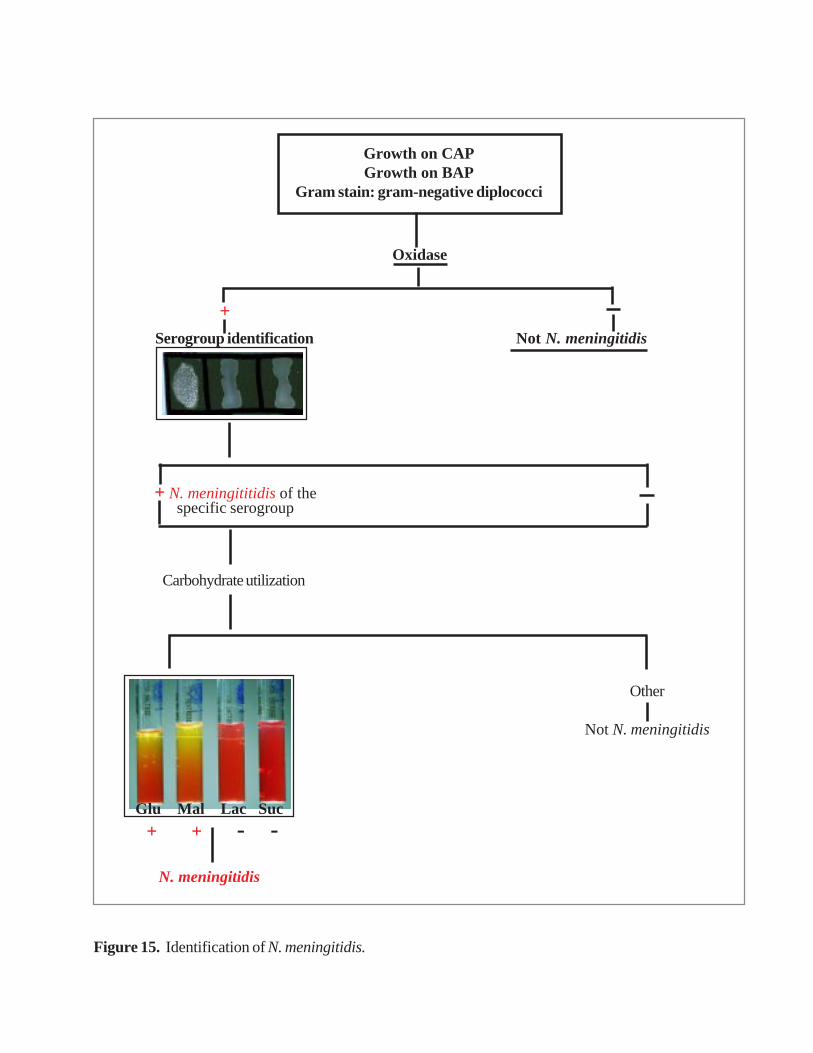
Figure 15. Identification of N. meningitidis.
+Serogroup identification Not N. meningitidis
Carbohydrate utilization
Other
Not N. meningitidis
Glu Mal Lac Suc
N. meningitidis
+ + - -
Growth on CAPGrowth on BAP
Gram stain: gram-negative diplococci
Oxidase
+ N. meningititidis of thespecific serogroup
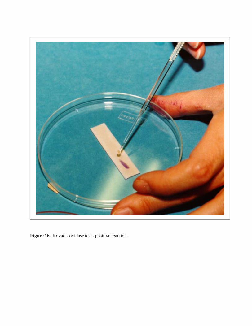
Figure 16. Kovac’s oxidase test - positive reaction.
Groups A, B, C, W135 and Y are the most common causes of meningitis. SerogroupA is the most common cause of epidemics in Africa and Asia, followed by serogroup C.Grouping antisera are available commercially.
Performance of the Test
(a) Clean a glass slide with alcohol. Divide the slide into three 10x4-mm sections with awax pencil or other marker.
(b) Place 10 µl of 0.5% formalinized saline near the bottom of each of three sections.Collect a portion of the growth from the surface of the BAP with a sterileinoculating loop, needle, sterile applicator stick or toothpick. Make a moderatelymilky suspension of the culture being tested in each drop of saline.Note: For safety reasons, it is recommended to use formalin-killed meningococcalsuspensions rather than saline suspensions of living organisms. However, formalinis a carcinogen and must be stored and handled with great care. Alternatively, workunder a safety hood.
(c) In the upper portion of each glass section, add 10 µl each of the antiserum of choiceand saline/phosphate-buffered saline (PBS) (unformalinized).Note: In Africa, testing with A and C antisera with a saline control to detectnonspecific autoagglutination should be adequate to characterize most isolatesserologically. Strains reacting negatively with A and C antisera should then betested with other available antisera, particularly Y, W135 and B.
(d) Mix each antiserum and saline with the respective culture suspension and rock theslide for 1 to 2 minutes (time may vary depending on the manufacturer of theantisera) under a bright light and over a black background.
Reading the Test Results
Agglutination should occur with only one of the antisera and not with saline(Figure 17). Agglutination in saline would characterize the culture as autoagglutinable.Agglutination with several serum samples in the absence of agglutination in saline wouldcharacterize the culture as rough. No agglutination with any of the antisera or the salinewould characterize the strain as non-groupable. These results are rare with fresh caseisolates, but they do happen occasionally. Store antisera and saline/PBS in the refrigeratorat 4 •C when not in use.
C. Carbohydrate Utilization by N. meningitidis - Cystine Trypticase Agar Method
Carbohydrate utilization tests are used to further validate the identification of astrain as N. meningitidis. Various carbohydrates are added to the cystine trypticase agarbase to a final concentration of 1%. To confirm a culture as N. meningitidis, a set of fourtubes, each containing a sugar (glucose, maltose, lactose, and sucrose) is used. Membersof Neisseria spp. produce acid from carbohydrates by oxidation, not fermentation. N.meningitidis oxidizes glucose and maltose, but not lactose and sucrose. A phenol redindicator is included in the medium; it is a sensitive indicator that develops a yellowcolour in the presence of acid, at a pH of 6.8 or less.
Performance of the Test
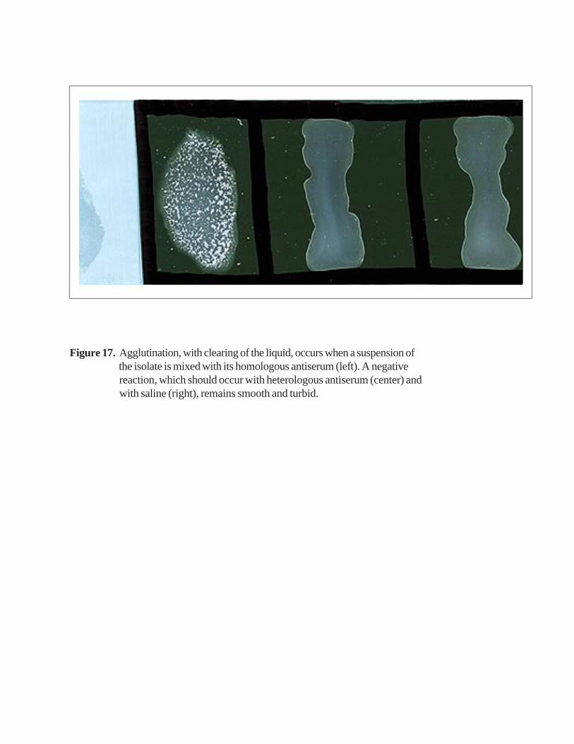
Figure 17. Agglutination, with clearing of the liquid, occurs when a suspension of the isolate is mixed with its homologous antiserum (left). A negative reaction, which should occur with heterologous antiserum (center) and with saline (right), remains smooth and turbid.
(a) With an inoculating needle, take up a small amount of growth from an overnightculture on BAP or CAP.
(b) Stab the inoculum several times into the upper 10 mm of medium. Use a separateneedle, or flame the needle, before inoculating each carbohydrate to be tested.
(c) Fasten caps of tubes tightly and place in a 35 •C incubator (without CO2). Incubatefor at least 72 hours before discarding as negative.
Reading the Test Results
Development of visible turbidity and a yellow colour in the upper portion of themedium indicates growth and the production of acid and is interpreted as a positive test(Figure 18). Although reactions may occur as early as 24 hours after inoculation, somereactions are delayed, and negative results should not be interpreted before 72 hours ofincubation (Table 1).
D. Commercial Identification Kits
Several commercial identification systems that use biochemical or enzymaticsubstrates are available for identification of Neisseria spp. These systems mayoccasionally require supplemental tests, and additional characteristics, such asmicroscopic and colony morphology, must be considered. Generally, each system is self-contained, but addition of one or more reagents to complete certain reactions may benecessary. Follow the manufacturer’s instructions precisely when using these kits. Fordetailed instructions and use of appropriate control strains also consult the ClinicalMicrobiology Procedures Handbook. Alternatively, the rapid sugar utilization tests maybe used.

Figure 18. Cystine trypticase agar-sugar reactions differentiating N. meningitidis from other Neisseria species. Acid production (yellow color) shows oxidative utilization of dextrose and maltose with no utilization of lactose and sucrose.
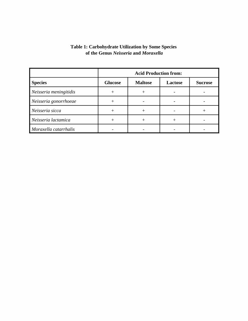
Table 1: Carbohydrate Utilization by Some Species of the Genus Neisseria and Moraxella
Acid Production from:
Species Glucose Maltose Lactose Sucrose
Neisseria meningitidis + + - -
Neisseria gonorrhoeae + - - -
Neisseria sicca + + - +
Neisseria lactamica + + + -
Moraxella catarrhalis - - - -

VI. Identification of S. pneumoniae
S. pneumoniae appear as small, greyish, mucoid (watery) colonies surrounded by agreenish zone of alpha-haemolysis on BAP and CAP. A 3x hand lens or a microscope(30x-50x) is a useful aid in differentiating pneumococci from alpha-haemolytic viridansstreptococci. Both types of colonies appear raised when young. However, after 24-48hours, the centre of pneumococcal colonies becomes depressed whereas viridansstreptococcalcolonies retain their raised appearance. Differentiation between S.pneumoniae and viridans streptococci is accomplished by optochin and bile solubilitytesting (Figure19). For optimal results it is recommended that plates for all assays beincubated in a CO
2 atmosphere.
A. Susceptibility to Optochin
Presumptive identification of S. pneumoniae is made by determining thesusceptibility of the strain to optochin.
Performance of the Test
(a) Touch the suspect alpha-haemolytic colony with a sterile bacteriological loop andstreak onto a BAP, as shown in Figure 7.
(b) Aseptically place an optochin or “p” disk with a diameter of 6 mm (containing 5 µg ethylhydrocupreine) on the end of the streak where the wire loop was first placed.Three to four colonies may be tested on the same plate.
(c) Incubate the plates in a CO2 incubator or candle-jar at 35 •C for 18-24 hours.
Reading the Test Results
In Figure 20, the strain in the top streak is resistant to optochin and, therefore, is nota pneumococcus. The strains in the centre and lower streaks are susceptible to optochinand appear to be pneumococci. Alpha-haemolytic strains with a zone of inhibition ofgrowth greater than 14 mm in diameter are pneumococci. Alpha-haemolytic strains withno zones of inhibition are viridans streptococci. Alpha-haemolytic strains with zones ofinhibition ranging between 9 and 13 mm should be tested for bile solubility.
B. Bile Solubility Test
Performance of the Tests
(a) Take a loop of the strain from the growth on a BAP and make a suspension in 0.5 mlof sterile saline. If there is sufficient growth on the optochin test plate, thesuspension can be made with the cells taken from the single streak. When there isinsufficient growth to make a satisfactory suspension, a fresh culture on blood agarwill have to be made. The suspension of cells should be equal to that of a 0.5McFarland density standard (Annex C5).
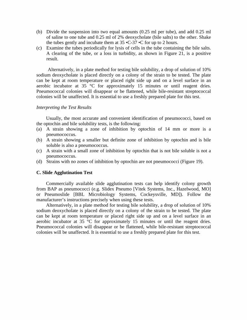
(b) Divide the suspension into two equal amounts (0.25 ml per tube), and add 0.25 mlof saline to one tube and 0.25 ml of 2% deoxycholate (bile salts) to the other. Shakethe tubes gently and incubate them at 35 •C-37 •C for up to 2 hours.
(c) Examine the tubes periodically for lysis of cells in the tube containing the bile salts.A clearing of the tube, or a loss in turbidity, as shown in Figure 21, is a positiveresult.
Alternatively, in a plate method for testing bile solubility, a drop of solution of 10%sodium deoxycholate is placed directly on a colony of the strain to be tested. The platecan be kept at room temperature or placed right side up and on a level surface in anaerobic incubator at 35 °C for approximately 15 minutes or until reagent dries.Pneumococcal colonies will disappear or be flattened, while bile-resistant streptococcalcolonies will be unaffected. It is essential to use a freshly prepared plate for this test.
Interpreting the Test Results
Usually, the most accurate and convenient identification of pneumococci, based onthe optochin and bile solubility tests, is the following:(a) A strain showing a zone of inhibition by optochin of 14 mm or more is a
pneumococcus.(b) A strain showing a smaller but definite zone of inhibition by optochin and is bile
soluble is also a pneumococcus.(c) A strain with a small zone of inhibition by optochin that is not bile soluble is not a
pneumococcus.(d) Strains with no zones of inhibition by optochin are not pneumococci (Figure 19).
C. Slide Agglutination Test
Commercially available slide agglutination tests can help identify colony growthfrom BAP as pneumococci (e.g. Slidex Pneumo [Vitek Systems, Inc., Hazelwood, MO]or Pneumoslide [BBL Microbiology Systems, Cockeysville, MD]). Follow themanufacturer’s instructions precisely when using these tests.
Alternatively, in a plate method for testing bile solubility, a drop of solution of 10%sodium deoxycholate is placed directly on a colony of the strain to be tested. The platecan be kept at room temperature or placed right side up and on a level surface in anaerobic incubator at 35 °C for approximately 15 minutes or until the reagent dries.Pneumococcal colonies will disappear or be flattened, while bile-resistant streptococcalcolonies will be unaffected. It is essential to use a freshly prepared plate for this test.

Figure 19. Identification of S. pneumoniae.
Growth on CAP Growth on BAP
Gram stain: gram-positive diplococci
Optochin susceptibilityand
Bile solubility tests
Optochinsusceptibility
(mm)
ZI > 14*14 >ZI > 814 >ZI > 8
ZI = 0
Bilesolubility
Interpretation
+
-
S. pneumoniaeS. pneumoniae
Not S. pneumoniaeNot S. pneumoniae
Note: ZI = Zone of inhibition for BBL optochin disks. When using any other brand of optochin disks,
follow the instructions for the interpretation of zones of inhibition, as specified by that manufacturer.
*95% of clinical S. pneumoniae isolates react in this manner
-+

Figure 20. Optochin susceptibility test for identification of S. pneumoniae. The strain in the top streak is resistant to optochin and, therefore, is not a pneumococcus.
The strains in the center and lower streaks are susceptible to optochin and appear to be pneumococci.
Figure 21. The bile solubility test for two different strains. Strain 1 is not S. pneumoniae as both
tubes are turbid. There is a slight decrease in turbidity in the tube containing bile salts
for strain 1 (2nd tube from the left) but the tube is almost as turbid as the control tube
(1st tube on the left). Strain 2 is S. pneumoniae. Note that the tube on the far right is
clear, all the turbidity due to the cells has disappeared and the cells have lysed; by
contrast the control tube (3rd tube from the left) is still very turbid.

VII. Identification of H. influenzae
Small, Gram-negative bacilli or coccobacilli which require X and V factors, grow onCAP and not on BAP, have a pungent indol smell, and do not group with meningococcalantisera, may be H. influenzae (Figure 22). In the absence of vaccination, almost all casesof meningitis caused by H. influenzae are serotype b.
A. Identification of the H. influenzae Serotype
Performance of the test is similar to that described for N. meningitidis except for thechoice of antisera. Suspected Hib isolates should be tested against H. influenzae type bantiserum, an antiserum to one of the other groups, and saline. A strongly positivereaction with b antiserum and no reactivity with an antiserum to one of the other groupsand saline is presumptive evidence of Hib. If the isolate is non-reactive with the bantiserum, test it with a polyvalent antiserum. If positive, the isolate must be tested withthere maining antisera (a, c, d, e, f) to determine its serotype. If negative, it is probablynon-typable. Determination of X and V factor requirements is necessary to confirm theidentity of the isolate as H. influenzae or another Haemophilus species.
Performance of the Test
(a) Make a milky suspension of cells from an overnight culture on the CAP or theHaemophilus ID plate in 10 µl of 0.5% formalinized saline.
(b) For the agglutination reaction, transfer a loopful (5 µl) of the cell suspension to anethanol-washed slide divided into three sections.
(c) To each section, respectively, add an equal volume of Hib antiserum, a differenttype specific antiserum, and saline.
(d) Mix with a toothpick and gently rock the slide for up to a minute.
Reading of Test Results
Only strong agglutination reactions are read as positive. In a strong reaction, all thebacterial cells will be clumped and the suspension fluid will appear clear. When a strainreacts with more than one antiserum, the result is recorded as non-typable.
Note: Haemophilus can also be serotyped by using the Quellung reaction, asdescribed for the serotyping of S. pneumoniae (Annex D).
B. Identification of X and V Factor Requirements
B1. X, V and XV Paper Disks and Strips
H. influenzae is a fastidious organism requiring media containing haemin (X factor)and nicotinamide adenine dinucleotide (NAD, V factor). Growth occurs on CAP becauseof haemin released during the heating process in the preparation of chocolate agar.Haemin is available from non-haemolyzed as well as haemolyzed cells. Alternatively,NAD is added as an IsoVitalex or Vitox component. H. influenzae is identified on thebasis of its growth requirements for X and V factors (Table 2).

Figure 22. Identification of H. influenzae.
H. influenzae ofspecific serotype
Growth on CAPNo growth on BAP
Gram stain: gram-negative pleomorphic coccobacilli
Serotype
+
Requirements for X and V Factors
Lack of requirementsfor either factor
Not H. infuenzae
Both factors required for growth
H. influenzae

Performance of the Test
(a) Prepare a heavy suspension of cells (No. 1 McFarland) from a primary isolationplate in a suitable broth (trypticase soy, heart infusion, or peptone water). If theprimary isolation plate contains insufficient growth or is contaminated, make asubculture on a CAP. Caution should be used in preparation of the broth to avoidtransfer of media to the broth; even the smallest sample of agar will affect the testand may lead to misidentification of the bacteria.
(b) Inoculate a heart infusion or tryptic soy agar plate. A sterile swab of the suspensionis streaked over one-half of the plate, and paper strips or disks containing X, V, andXV factors are placed on the inoculated plate after the inoculum has dried. Whentwo bacterial strains are tested on the same plate, as shown in Figure 23, the disksmust be placed in the exact manner shown.
Reading the Test Results
H. influenzae will grow only around the disk containing both X and V factors, asshown in Figure 23 on the upper half of the plate.
Alternatively, the porphyrin test of Kilian can be used. This determines theX-factor requirement of the isolate while avoiding the problem of X-factor carryoverfrom primary culture media and X-factor contamination of test media (Manual of ClinicalMicrobiology).
B2. Haemophilus ID “Quad” Plates
These plates are another, although more expensive, method for determining factorrequirements of Haemophilus isolates (Figure 24). This agar plate is divided into fourcompartments. One quadrant includes medium containing haemin (X factor) (upper left).One quadrant includes medium containing NAD (V factor) (lower left). Another quadrantcontains medium that includes both X and V factors (upper right). The fourth quadrantcontains heart infusion agar or blood agar base with 5% horse blood (lower right) fordifferentiatingH. haemolyticus, an oral species requiring X and V factors, from H. influenzae.
Performance of the Test
(a) Inoculate the Haemophilus ID plate by suspending the growth from a young, pureculture of suspected Haemophilus in TSB or distilled water to approximately theturbidity of 0.5 McFarland standard. Using a bacteriological loop, streak one loopfulof this suspension on each quadrant of the plate, beginning with the V quadrant andending with the blood quadrant. Streak the entire quadrant, starting at the peripheryand streaking toward the centre of the plate. Stab into the blood agar for detection ofweak haemolysis.
(b) Incubate at 35 •C in a candle-jar or CO2 incubator for 18-24 hours.
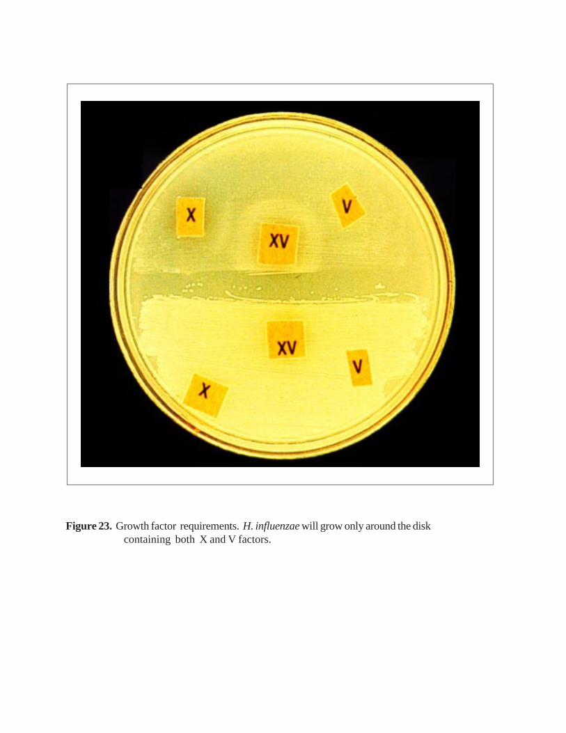
Figure 23. Growth factor requirements. H. influenzae will grow only around the disk containing both X and V factors.
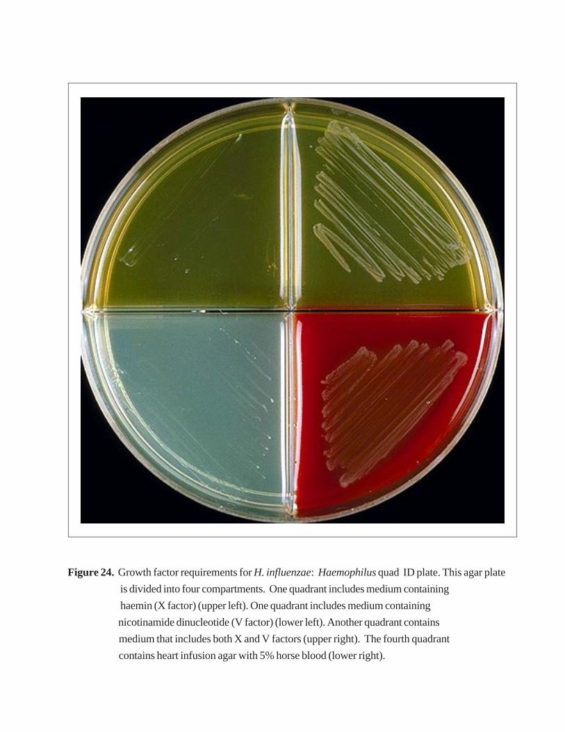
Figure 24. Growth factor requirements for H. influenzae: Haemophilus quad ID plate. This agar plate
is divided into four compartments. One quadrant includes medium containing
haemin (X factor) (upper left). One quadrant includes medium containing
nicotinamide dinucleotide (V factor) (lower left). Another quadrant contains
medium that includes both X and V factors (upper right). The fourth quadrant
contains heart infusion agar with 5% horse blood (lower right).
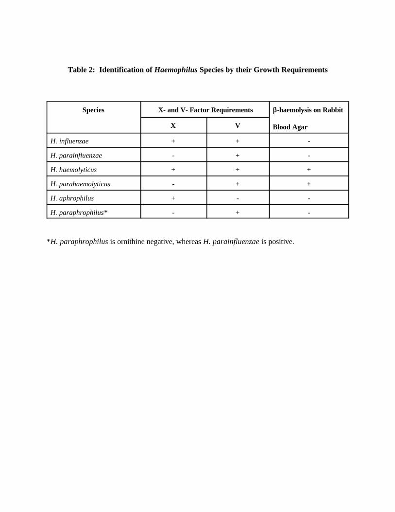
Table 2: Identification of Haemophilus Species by their Growth Requirements
Species X- and V- Factor Requirements $$-haemolysis on Rabbit
Blood AgarX V
H. influenzae + + -
H. parainfluenzae - + -
H. haemolyticus + + +
H. parahaemolyticus - + +
H. aphrophilus + - -
H. paraphrophilus* - + -
*H. paraphrophilus is ornithine negative, whereas H. parainfluenzae is positive.

Reading the Test Results
After incubation, examine the blood section for haemolysis and the other sectionsfor growth (Figure 24). An organism growing on both the V and X quadrant requiresneither, and thus is not a Haemophilus. Very light background growth on the V sectionmay be seen with some X- and V-requiring Haemophilus strains. This should bedisregarded.
VIII. Preservation and Transport of N. meningitidis, S. pneumoniae andH. influenzae
To confirm the identification and to test antimicrobial susceptibility of bacteriaisolated from cases of meningitis, it may be necessary to preserve and transport thestrains to national, and/or WHO Reference Laboratories. N. meningitidis, S. pneumoniaeand H. influenzae are fragile bacteria, and care must be employed to preserve andtransport them.
A. Short-Term Storage
Viability during short-term storage (one week or less) is best if S. pneumoniae and H. influenzae areinoculated onto chocolate agar slants (with screw-capped tubes), incubated overnight at 35 •C, and thenmaintained at 4 •C. These bacterial species do not survive well in broth and survive only 3-4 days onprimary agar plates. For N. meningitidis, solid screw-caps should be loosened during storage butpermeable membrane screw-caps, which allow for an exchange of gases and are available commercially(Biomedical Polymers, Leominster, MA) should be used. An overlay of TSB may also be helpful, andmay increase viability to 14 days. N. meningitidis slants should not be refrigerated.
B. Long-Term Storage
Long-term storage is best accomplished by either lyophilization or freezing.Lyophilization is the most convenient method of storage because lyophilized bacteria canbe stored for long periods at 4 •C or –20 •C and should not be adversely affected bytransport.
B1. Preservation by Lyophilization
(a) Grow the H. influenzae on CAP and the S. pneumoniae and N. meningitidis on BAPor CAP. Incubate the plates in a CO
2 incubator or candle-jar for 18-20 hours at35 •C. Inspect the plate for purity.
(b) Harvest the growth from the plate with l-2 ml of sterile skim milk. Place about 0.5ml of suspension into a sterile ampoule or lyophilization vial. Several vials can beprepared from a single plate, if desired. Maintain sterility at all times during thepreparation of the vial.
(c) The cell suspension should be shell-frozen on the walls of the lyophilization vial.This is accomplished by one of two methods. Keep the lyophilization vial at –70 •Cuntil just before the cell suspension is added. Add the cell suspension, and rapidlyrotate the vial to freeze the suspension to the wall. Return the vial to the – 70 •Cfreezer until ready to attach to the lyophilizer. If a –70 •C freezer is not available, amixture of alcohol (95% ethanol) and dry ice can be prepared and used to shell-freeze the cell suspensions. Shell-freezing is accomplished by placing the cellsuspension in the lyophilization vial and rotating the vial at a 45o to 60o angle in thealcohol-dry ice mixture.
(d) Attach the vials to the lyophilizer. Follow the manufacturer’s instructions since eachinstrument uses different types of apparatus. The time of lyophilization will depend
upon the number of vials being lyophilized and the capacity of the instrument. Onan average machine, 4-5 hours are required to completely dry 10 to 20 small vials.
(e) The vials are sealed at the end of the run. Seal the vials with a torch while they arestill attached to the lyophilizer and under vacuum. The vials can be stored at 4 •C orat freezer temperatures after being sealed.
B2. Preservation by Freezing
(a) Grow the H. influenzae on CAP and the S. pneumoniae and N. meningitidis on BAPor CAP. Incubate the plates in a CO
2 incubator or candle-jar for 18-20 hours at35 •C. Inspect the plates for purity.
(b) Harvest all of the growth from a plate with a sterile swab.(c) Dispense the growth in a 2-ml screw-capped (externally threaded) cryogenic vial,
containing 1 ml of sterile defibrinated blood, by twirling the swab to release theorganisms. Defibrinated sheep, horse, or rabbit blood can be used for all of theseorganisms. Human blood should not be used. Alternatives such as TSB with 15%-20% reagent grade glycerol, or Greaves solution can also be used. The vials shouldbe cryogenic (able to withstand extreme cold).
(d) Squeeze the excess blood from the swab by rotating it against the sides of the vialbefore carefully withdrawing it.
(e) Discard the swab in disinfectant.(f) If possible, rapidly freeze the suspension in a bath of 95% alcohol and dry-ice
pellets.(g) Place the vials in a nitrogen freezer (-120 •C) or a –70 •C freezer. A –20 •C freezer
can be used, but some loss of viability can be expected. Freezers with automaticdefrosters should never be used.
C. Transportation of Cultures
Lyophilized cultures and chocolate agar slant cultures can be transported withoutrefrigeration. The cultures should be packaged according to the regulations specified inthe WHO Laboratory Biosafety Manual. Each vial or tube should be individuallywrapped before being enclosed in a metal container including absorbent material. Thiscontainer is then inclosed in a cardboard shipping container. An address label and anetiological agent-warning label (EA label) are attached to the shipping container. Nomore than 50 ml of culture should be shipped in one package.
Individually wrapped, lyophilized culture vials can be transported in double-mailingcontainers. Cultures stored in the frozen state must be recultured, inoculated ontochocolate agar slants in screw-capped tubes, incubated overnight, and then packaged fortransport as indicated above. H. influenzae, N. meningitidis, and S. pneumoniae willusually survive for at least a week under these conditions.
C1. Transport in Silica Gel Packages
N. meningitidis, S. pneumoniae and H. influenzae can be successfully transported insilica gel packages, which are easy to handle and ship (Figure 25).
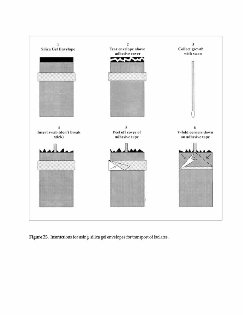
Figure 25. Instructions for using silica gel envelopes for transport of isolates.
growth
(a) Grow the bacteria on BAP or CAP (H. influenzae). Incubate the plates in a CO2 orcandle-jar for 18-20 hours at 35 •C. Inspect the plate for purity.
(b) Harvest all of the growth from a plate with a sterile swab.(c) Cut open the silica gel package and place the swab in it. Fold the corners and seal
the silica gel package with the tape. Label the silica gel package with the appropriateidentification number.
(d) When the swabs in silica gel packages are kept at 4 •C, N. meningitidis can surviveat least 3 weeks, and S. pneumoniae and H. influenzae at least 2 weeks. At roomtemp-erature, N. meningitidis, H. influenzae, and S. pneumoniae can survive 2weeks.

IX. Bibliography
1. Ajello GW, Feeley JC, Hayes PS, Reingold AL, Bolan G, Broome CV, andPhillips CJ. Trans-Isolate medium: a new medium for primary culturing andtransport of Neisseria meningitidis, Streptococcus pneumoniae, and Haemophilusinfluenzae. J. Clin. Microbiol., 1984, 20:55-58.
2. Austrian, R. The Quellung reaction, a neglected microbiologic technique.Mt. Sinai J. Med., 1976, 43:699-703.
3. Campos, JM. Haemophilus. In: Manual of Clinical Microbiology, 6th ed.PR Murray, EJ Baron, MA Pfaller, FC Tenover, RH Yolken (eds.),1995:556-565.American Society for Microbiology, Washington, DC, USA.
4. Facklam, RR, and JA Washington II. Streptococcus and related catalase negativegram-positive cocci. In: Manual of Clinical Microbiology, 5th ed. A Balows, WJHausler, KL Herrmann, HD Isenberg, and HJ Shadomy (eds.), 1991:238-257.American Society for Microbiology, Washington, DC, USA.
5. Isenberg, HD, ed. Clinical Microbiology Procedures Handbook 1992, Volume #1.American Society for Microbiology, Washington, DC.
6. Knapp, JS, and RJ Rice. Neisseria and Branhamella. In: Manual of ClinicalMicrobiology, 6th ed. PR Murray, EJ Baron, MA Pfaller, FC Tenover, RH Yolken(eds.), 1995: 324-340. American Society for Microbiology, Washington, DC, USA.
7. Miller, MJ. A guide to specimen management in clinical microbiology.Microbiology technical services, Dunwoody, GA, and Diagnostic MicrobiologySection, Hospital Infections Program, Centers for Disease Control and Prevention, Atlanta, GA,USA, 1996.
8. Schwartz, B, and RR Facklam. Manual for the National Surveillance ofAntimicrobial Resistance of S. pneumoniae and H. influenzae: Epidemiological andMicrobiological Methods. World Health Organization, Geneva, Switzerland, andCenters for Disease Control and Prevention, Atlanta, GA, USA, 1994.
9. Sørensen, UBS. Typing of pneumococci by using 12 pooled antisera. J. Clin.Microbiol., 1993, 31:2097-2100.
10. Van der Ende, A, IGA Schuurman, CTP Hopman, CAP Fijen, and J Dankert.Comparison of commercial diagnostic tests for identification of serogroup antigensof Neisseria meningitidis. J. Clin. Microbiol., 1995, 33: 3326-7.
11. Control of Epidemic Meningococcal Disease: WHO Practical Guidelines, 2nd
edition. World Health Organization, Geneva, Switzerland, 1998.12. Murray CJL, Lopez AD. Global Health Statistics: a compendium of incidence,
prevalence, and mortality estimates for over 200 conditions, 1996: 283-309. TheHarvard School of Public Health on behalf of the World Health Organization andthe World Bank, Boston, MA, USA.

X. Annexes
Annex A. Basic Requirements, Supplies and Equipment

A1. Table 3: Basic Requirements and Supplies for a Microbiology Laboratory
Basic LaboratoryRequirements
Supplies Equipment
good lightingelectricitygas supply for burnersbench space (4-6 ft/person)distilled water supplydesk lampssink with running water
laboratory coatsglovesdisinfecting fluid (Amphyl)sharp disposal box (needles)containers for waste disposallabelsmarking pensrecord bookstowelshand soapinoculating loopsscissorsforcepscandle jars3 x hand lensspatulas or spoonsrulers or callipertest tube racksrequired mediathermometerslens paper lens cleanerimmersion oilpH paperstandard buffersautoclave tapePasteur pipettesmicroscope slides
autoclaverefrigeratorincubatorweight balancepH metertimersBunsen or alcohol burnersmicroscope with immersion lenswater bathfreezersafety hood
A2. Table 4: Equipment and Supplies Needed for Collection of Clinical Specimensand Isolation and Identification of N. meningitidis, S. pneumoniae
and H. influenzae
Procedure Supplies Equipment
1. Spinal tap kitfor preparationof patient andcollection ofCSF
adult spinal needle 22 gauge 3.5"paediatric spinal needle 23 gauge 1.5" or2.5"alcohol spongettesgauze sponge 4x4"Band-Aidstincture of iodine or povidone-iodineswabssterile screw-cap tubes syringeneedlemailing containersT-I medium
2. Preparationof patient anddrawing ofblood
iodine swabalcohol swabtourniquetssyringesneedlesglovesbandagescottongauzescissorspuncture-resistant containerblood culture media or transport system
3. Inoculationof CSF andblood culturemedia
blood culture medianeedlesaeration needles (vents)alcohol wipes
4. Incubationof inoculatedmedia
candles candle-jar or CO2 incubator

5. Subculturingof inoculatedmedia andblood culturebottles
candlesalcohol wipesinoculating loops
Bunsen or alcohol burnerscandle-jar or CO2 incubator
6. Transportand culture ofmedia
silica gel packagespolyester-tipped swabspacking materialslabelsStyrofoam containers
7. Sterilizationof disposableandcontaminatedmaterials
containers for autoclave autoclave
8. Identification
chromatography paper such as Whatman3MMimmersion oilinoculating loopinoculating needlepipettespipette bulbcapillary pipettepipette tipspipette manglass slidescotton swabs0.5 McFarland turbidity standardsforcepstest tube racksscissors
microscope (oil immersion)magnifying glass or broad field microscope

9. Permanentstorage ofstrains
vials or ampoules -70 °C or N2 freezer-20 EC freezer for shorterstoragelyophilizer
10. Mediapreparation
Petri dishessterile cotton swabstest tubesErlenmeyer flasks (250 ml, 500 ml, 1 l, 2 l)beakers (100 ml, 250 ml, 500 ml)weighing papersholdersdistilled waterglass beadspipettespipette bulbsspatulassterilization filter unitscylinders (50 ml, 100 ml, 250 ml, 500 ml,1 l)
weighing balancerefrigeratorincubatormagnetic stirrerautoclavepH metertimer

Annex B. Media and Reagents
Note : The following list of the manufacturers of the commonly used media and reagentsdoes not indicate special endorsement of these products and/or manufacturers by WHO.Please follow closely the manufacturer’s instructions when using these media andreagents.
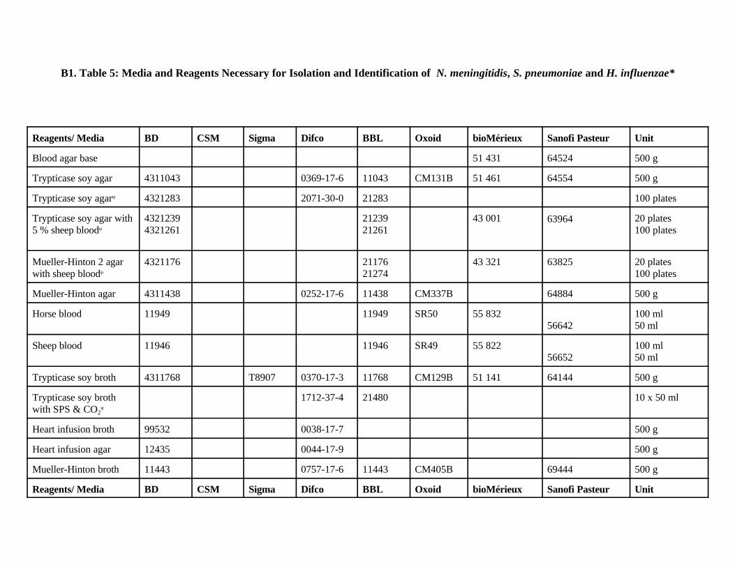
B1. Table 5: Media and Reagents Necessary for Isolation and Identification of N. meningitidis, S. pneumoniae and H. influenzae*
Reagents/ Media BD CSM Sigma Difco BBL Oxoid bioMérieux Sanofi Pasteur Unit
Blood agar base 51 431 64524 500 g
Trypticase soy agar 4311043 0369-17-6 11043 CM131B 51 461 64554 500 g
Trypticase soy agarψ 4321283 2071-30-0 21283 100 plates
Trypticase soy agar with5 % sheep bloodψ
43212394321261
2123921261
43 001 63964 20 plates100 plates
Mueller-Hinton 2 agarwith sheep bloodψ
4321176 2117621274
43 321 63825 20 plates100 plates
Mueller-Hinton agar 4311438 0252-17-6 11438 CM337B 64884 500 g
Horse blood 11949 11949 SR50 55 83256642
100 ml50 ml
Sheep blood 11946 11946 SR49 55 82256652
100 ml50 ml
Trypticase soy broth 4311768 T8907 0370-17-3 11768 CM129B 51 141 64144 500 g
Trypticase soy brothwith SPS & CO2
ψ
1712-37-4 21480 10 x 50 ml
Heart infusion broth 99532 0038-17-7 500 g
Heart infusion agar 12435 0044-17-9 500 g
Mueller-Hinton broth 11443 0757-17-6 11443 CM405B 69444 500 g
Reagents/ Media BD CSM Sigma Difco BBL Oxoid bioMérieux Sanofi Pasteur Unit

Trypticase soy brothψ 4321716 21716 100 x 5 ml
GC agar 4311275 0289-17-3 11275 CM367B 500 g
Chocolate agar platesψ 4321169 21169 43 101 63934 20 plates100 plates
Chocolate agar slantsψ 4321348 21348 10 slants
Haemoglobin, driedbovine
11871 11871 LP053B 100 g
Haemoglobin (2% sol)ψ 11874 3248-73-8 B11874 10 x 100 ml
IsoVitaleX or Vitoxψ 11876 11876 SR090A 55 652 55592 5 x 10 ml
Sodium polyanetholesulfonate
P2008
Haematin, bovine H3505
Haematin, chloride H2250
Dextroseψ 02-065-02
21634 100 plates
Lactoseψ 02-066-02
100 plates
Maltoseψ 02-067-02
21638 100 tubes
Sucroseψ 02-068-02
21646 100 tubes

Reagents/ Media BD CSM Sigma Difco BBL Oxoid bioMérieux Sanofi Pasteur Unit
Crystal violet C0775
Safranin S8884
Iodine I3380
Potassium iodine P8256
Crystal violetψ 12293 12293 55 545 4 x 250 ml
Iodineψ 12294 12294 55 546 4 x 250 ml
Acetone-alcoholψ 12295 12295 55 547 4 x 250 ml
Safraninψ 12296 12296 55 548 4 x 250 ml
Ethanol
Acetone
Barium chloride B6394
1 N sulfuric acid 84728
1 N sodium hydroxide 930-65
Sodium hydroxidepellets
S0899
Dextrose 11863 0155-17-4 11863 6403462242
500 g(30%) 5 ml
Maltose 12029 0168-17-9 1202962361
500 g(30%) 5ml

Reagents/ Media BD CSM Sigma Difco BBL Oxoid bioMérieux Sanofi Pasteur Unit
Lactose 11881 0156-17-3 1188162272
500 g(25%) 10 ml
Sucrose 11916 0176-17-9 1191662571
500 g(30%) 10 ml
1N hydrochloric acid 920-1
Kovac’s oxidasereagent
T3134
V-factor disksψ 31108 1694-90-0 DD004A 50 discs
X-factor disksψ 31006 1695-90-9 DD003A 50 discs
XV-factor disksψ 31104 1696-90-8 DD005A 50 discs
Haemophilus quad ID 4397890
Optochin p-discs 31046 1632-35-3 31046 DD001A 53852 30-50 discs
Nicotinamide adeninedinucleotide
N7004

Reagents/ Media BD CSM Sigma Difco BBL Oxoid bioMérieux Sanofi Pasteur Unit
T-I medium -supplement B -activated charcoal -soluble starch -agar -trypticase soy broth -gelatin -3 LN- morpholino propanesulfonic acid (MOPS)bovine albumin
4311768
C5510S9765A5306T8907
M5162
A9647
0276-60-4
0140-01-00370-17-30143-17-9
11768 64144
6 x 10 ml
1 lb500 g500 g
*BD - Becton Dickinson and Co., CSM - Carr-Scarborough Microbiologicals, Inc., Sigma - Sigma Chemical Co., Difco - Difco Laboratories, BBL -
BBL Microbiology Systems. For the manufacturers’ addresses and contact numbers, please see section B4.
ψ Although commercially prepared media and reagents are more expensive than media or reagents that can be prepared locally, there will be times when
commercially available items will be preferable. It may be desirable to purchase the supply of media and reagents to perform short-term studies rather
than attempt formulation. The media and reagents designated with (ψ) are available in ready-to-use form in most parts of the world. It is essential that
each medium lot have a satisfactory expiry date and that this date and lot number be recorded in the laboratory, and noted for quality and control testing.
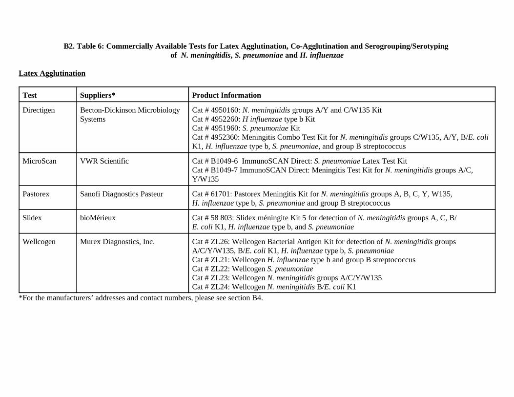
B2. Table 6: Commercially Available Tests for Latex Agglutination, Co-Agglutination and Serogrouping/Serotyping of N. meningitidis, S. pneumoniae and H. influenzae
Latex Agglutination
Test Suppliers* Product Information
Directigen Becton-Dickinson MicrobiologySystems
Cat # 4950160: N. meningitidis groups A/Y and C/W135 KitCat # 4952260: H influenzae type b KitCat # 4951960: S. pneumoniae KitCat # 4952360: Meningitis Combo Test Kit for N. meningitidis groups C/W135, A/Y, B/E. coliK1, H. influenzae type b, S. pneumoniae, and group B streptococcus
MicroScan VWR Scientific Cat # B1049-6 ImmunoSCAN Direct: S. pneumoniae Latex Test KitCat # B1049-7 ImmunoSCAN Direct: Meningitis Test Kit for N. meningitidis groups A/C,Y/W135
Pastorex Sanofi Diagnostics Pasteur Cat # 61701: Pastorex Meningitis Kit for N. meningitidis groups A, B, C, Y, W135, H. influenzae type b, S. pneumoniae and group B streptococcus
Slidex bioMérieux Cat # 58 803: Slidex méningite Kit 5 for detection of N. meningitidis groups A, C, B/E. coli K1, H. influenzae type b, and S. pneumoniae
Wellcogen Murex Diagnostics, Inc. Cat # ZL26: Wellcogen Bacterial Antigen Kit for detection of N. meningitidis groupsA/C/Y/W135, B/E. coli K1, H. influenzae type b, S. pneumoniaeCat # ZL21: Wellcogen H. influenzae type b and group B streptococcusCat # ZL22: Wellcogen S. pneumoniaeCat # ZL23: Wellcogen N. meningitidis groups A/C/Y/W135Cat # ZL24: Wellcogen N. meningitidis B/E. coli K1
*For the manufacturers’ addresses and contact numbers, please see section B4.

Co-Agglutination
Test Supplier* Product Information
Phadebact Pharmacia Diagnostics(US distributor REMEL)
Cat # 60-401: CSF test for N. meningitidis groups A/B/C/Y/W135, H. influenzae type b, S.pneumoniae, and group B streptococcus; CSF pneumococcus test; CSF Haemophilus test
*For the manufacturers’ addresses and contact numbers, please see section B4.
Serogrouping/Serotyping Antisera
Test Supplier* Product Information
Serogroupingantisera
Murex Diagnostics, Inc. Cat # ZM 33: Polyvalent set 1 of N. meningitidis agglutinating antiseraCat # ZM 34: Polyvalent set 2 of N. meningitidis agglutinating antiseraCat # ZM 37: N. meningitidis antiserum group ACat # ZM 51: N. meningitidis antiserum group BCat # ZM 39: N. meningitidis antiserum group CCat # ZM 44: N. meningitidis antiserum group W135Cat # ZM 41: N. meningitidis antiserum group XCat # ZM 42: N. meningitidis antiserum group YCat # ZM 43: N. meningitidis antiserum group Z
Serogroupingantisera
Difco Laboratories Cat # 2228-50-1: N. meningitidis antiserum group ACat # 2229-50-0: N. meningitidis antiserum group BCat # 2230-50-7: N. meningitidis antiserum group CCat # 2253-50-9: N. meningitidis antiserum group W135Cat # 2880-50-0: N. meningitidis antiserum group XCat # 2281-50-9: N. meningitidis antiserum group YCat # 2891-50-7: N. meningitidis antiserum group ZCat # 2252-50-5: N. meningitidis antiserum group Z’Cat # 2232-50-5: Antiserum Poly - polyvalent antiserum for N. meningitidis A/B/C/DCat # 2910-50-4: Antiserum Poly 2 - polyvalent antiserum for N. meningitidis X/Y/Z

Slidex pneumo-kit
bio-Mérieux Cat # 58821: S. pneumoniae
Pneumoslide Becton-Dickinson MicrobiologySystems
Cat # 40840: S. pneumoniae
Serotypingantisera
Difco Laboratories Cat # 2237-50-0: polyvalent H. influenzae antiseraCat # 2250-50-2: H. influenzae antisera type aCat # 2236-50-1: H. influenzae antisera type bCat # 2789-50-2: H. influenzae antisera type cCat # 2790-50-9: H. influenzae antisera type dCat # 2791-50-8: H. influenzae antisera type eCat # 2792-50-7: H. influenzae antisera type f
Serotypingantisera
Murex Diagnostics, Inc. Cat # ZM 20: H. influenzae antisera type aCat # ZM 21: H. influenzae antisera type bCat # ZM 22: H. influenzae antisera type cCat # ZM 23: H. influenzae antisera type dCat # ZM 24: H. influenzae antisera type eCat # ZM 25: H. influenzae antisera type f
*For the manufacturers’ addresses and contact numbers, please see section B4. Note: Slashes (/) indicate polyvalent diagnostic reagents and commas (,) indicate monovalent diagnostic reagents.
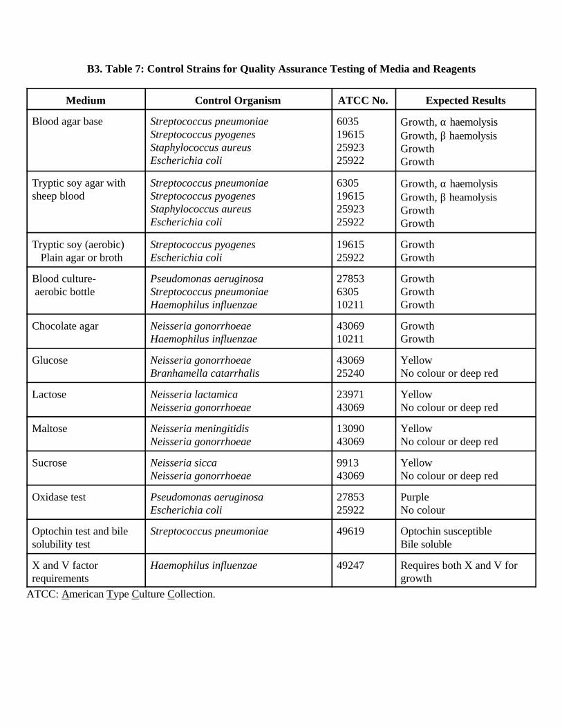
B3. Table 7: Control Strains for Quality Assurance Testing of Media and Reagents
Medium Control Organism ATCC No. Expected Results
Blood agar base Streptococcus pneumoniaeStreptococcus pyogenesStaphylococcus aureusEscherichia coli
6035196152592325922
Growth, α haemolysisGrowth, β haemolysisGrowthGrowth
Tryptic soy agar with sheep blood
Streptococcus pneumoniaeStreptococcus pyogenesStaphylococcus aureusEscherichia coli
6305196152592325922
Growth, α haemolysisGrowth, β heamolysisGrowthGrowth
Tryptic soy (aerobic) Plain agar or broth
Streptococcus pyogenesEscherichia coli
1961525922
GrowthGrowth
Blood culture- aerobic bottle
Pseudomonas aeruginosaStreptococcus pneumoniaeHaemophilus influenzae
27853630510211
GrowthGrowthGrowth
Chocolate agar
Neisseria gonorrhoeaeHaemophilus influenzae
4306910211
GrowthGrowth
Glucose Neisseria gonorrhoeaeBranhamella catarrhalis
4306925240
YellowNo colour or deep red
Lactose Neisseria lactamicaNeisseria gonorrhoeae
2397143069
YellowNo colour or deep red
Maltose Neisseria meningitidisNeisseria gonorrhoeae
1309043069
YellowNo colour or deep red
Sucrose Neisseria siccaNeisseria gonorrhoeae
991343069
YellowNo colour or deep red
Oxidase test Pseudomonas aeruginosaEscherichia coli
2785325922
PurpleNo colour
Optochin test and bilesolubility test
Streptococcus pneumoniae 49619 Optochin susceptibleBile soluble
X and V factorrequirements
Haemophilus influenzae 49247 Requires both X and V forgrowth
ATCC: American Type Culture Collection.

B4. Manufacturers’ Addresses:
Becton-Dickinson
250 Schilling CircleCockeysville, MD 21030, USAPhone: (1) 800 638-8663FAX: (1) 410 584 81 29BBL Microbiology Systems (orders through Fisher Scientific Co.)
PO Box 4829Norcross, GA 30091, USAPhone: (1) 800 766-7000FAX: (1) 800 926-1166
bioMérieux
bioMérieux Vitek, Inc.595 Anglum DriveHazelwood, MO 63042-2395, USAPhone: (1) 314 731-8500FAX: (1) 314 731-8700
bioMérieux s.a.69280 Marcy-l’Etoile, FrancePhone: (33) 4 78 87 20 00FAX: (33) 4 78 87 20 90Telex: 330967Carr-Scarborough Microbiologicals, Inc. (orders through Remel)
12076 Santa Fe Trail Dr.Lenexa, KA 66215, USAPhone: (1) 800 255-6730 (orders) (1) 800 447-3641 (customer service)FAX: (1) 800 621-8251
Difco Laboratories
PO Box 331058Detroit, MI 48232-7058, USAPhone: (1) 800 521-0851
(1) 313 462-8500FAX: (1) 313 462-8517
Fisher Scientific Co.
PO Box 4829Norcross, GA 30091, USA

Phone: (1) 800 766-7000FAX: (1) 800 926-1166
Europe/Middle East/Africa HeadquartersFisher Scientific Overseas Marketing, Inc.46 Queen Anne StreetLondon W1M 9LA, United KingdomPhone: (44) 171 935-4440FAX: (44) 171 935-5758
MicroScan
Travenol Laboratories, Inc.West Sacramento, CA 95691Mahwah, NJ 07430, USABellevue, WA 98004, USAPhone: (1) 800 631-7216
Murex Diagnostics, Inc.
Central Temple HillDartford, Kent DA1 5LR, United KingdomPhone: (44) 132 227-7711FAX: (44) 132 227-3288
Customer Services Department3075 Northwoods CircleNorcross, GA 30071, USAPhone: (1) 404 662-0660FAX: (1) 404 447-4989
Oxoid
Oxoid s.a.6 route de Paisy, B.P. 1369572 Dardilly Cedex, FrancePhone: (33) 4 78 35 17 31FAX: (33) 4 78 66 03 76
Unipath LimitedWade RoadBasingstoke, Hampshire RG24, United KingdomPhone: (44) 1256 694 364FAX: (44) 1256 479 525
Unipath Co.Oxoid DivisionPO Box 691

Ogdensburg, NY 13669, USAPhone: (1) 800 567-8378
Pastorex
Sanofi Diagnostics Pasteur3, Bld Raymond Poincaré - BP 392430 Marnes-la-Coquette, FrancePhone: (33) 1 47 95 60 00FAX: (33) 1 47 41 91 33Telex: 631293F
Pharmacia Diagnostics
Uppsala, SwedenDistributed in the USA by: Remel Laboratories
Remel Laboratories
12076 Santa Fe Trail Dr.Lenexa, KA 66215, USAPhone: (1) 800 255-6730 (orders) (1) 800 447-3641 (customer service)FAX: (1) 800 621-8251
Sigma Chemical Co.
PO Box 14508St. Louis, MO 63178, USAPhone: (1) 800 325-3010 (orders)
(1) 800 325-8070 (customer service)
Fancy Road, PooleDorset, BH17 7NH, United KingdomPhone: (44) 0800 373 731FAX: (44) 0800 378 785
Sigma-Aldrich Chimie S.a.r.l.L’Isle d’Abeau ChesnesB.P. 701, 38297 St. Quentin Fallavier Cedex, FrancePhone: (33) 05 21 14 08FAX: (33) 05 03 10 52
Wellcome Diagnostics
Laboratories Wellcome S.A.Division Diagnostics159 Rue Nationale,

75640Paris Cedex 13, FrancePhone: (33) 1 46 12 49 12FAX: (33) 1 46 54 55 47
Distributed in the USA by: Murex Diagnostics, Inc.
Annex C. Preparation of Media and Reagents
C1. Quality Control of All Media
Immediately after preparation, each medium should be tested with a reference strainof N. meningitidis, S. pneumoniae and H. influenzae for proper growth characteristics asdescribed for each medium. Keep a record of all media preparation or purchase dates andquality control test results. Note any unusual characteristic such as colour of the mediumor slow growth of test bacteria.
C2. Routine Agar and Broth Media
All agar media can be dispensed into 15x100-mm Petri dishes (15 to 20 ml per dish).After pouring, leave the plates at room temperature for several hours to prevent excesscondensation from forming on the covers of the dishes. Place the plates and store them ina plastic bag in an inverted position at 4 •C.
Heart Infusion Agar (HIA) and Trypticase Soy Agar (TSA)
These media are general purpose media used with or without blood for isolating andcultivating a number of microorganisms. The media should appear straw colored (ayellowish to gold colouring). HIA and TSA are also used for determining the X- and V-factor requirements of H. influenzae. Each freshly prepared or purchased batch of HIA orTSA should be quality control tested by determining the X and V requirements ofH. influenzae. A fresh plate is inoculated with a control strain; X, V, and XV disks shouldbe placed on the inoculated plate identical to that shown in Figure 23. H. influenzaeshould grow only around the XV disk.
(a) Prepare the HIA or TSA according to the instructions on the label of the dehydratedmedium. Prepare the volume needed in flasks. These media should be fullydissolved with no powder on the walls of the vessel, before autoclaving. Autoclaveat 121 •C for 20 minutes.
(b) Cool to 50 •C and pour into 15x100-mm Petri dishes.(c) Allow to solidify and condensation to dry out before placing in plastic bags and
storing at 4 •C until used.
Blood Agar Plate (BAP): TSA + 5% Sheep Blood
BAP is used as a general blood agar medium (sheep blood agar plate). It is also usedfor the optochin test and subcultures of S. pneumoniae. The plate should appear a brightred colour. If the plates appear dark red, the blood has been added when the agar was toohot. If this happens, discard the medium and prepare another batch. Test each new,freshly prepared or purchased batch of BAP for growth and haemolytic reaction with astrain of S. pneumoniae. The colonies are small and should appear grey to grey-greensurrounded by a distinct greenish halo in the agar similar to that shown in Figure 12.
(a) Prepare TSA according to the instructions given on the label of the dehydratedpowder. It is convenient to prepare 500 ml of molten agar in a l-litre flask. Add 20 gof agar into 500 ml of water. Heat to dissolve.
(b) Autoclave at 121 °C for 20 minutes. Cool to 60 •C.(c) Add 5% sterile, defibrinated sheep blood (25 ml sheep blood is added to 500 ml of
agar). If a different volume of basal medium is prepared, the amount of blood addedmust be adjusted accordingly to 5% (e.g. 50 ml of blood per litre of medium).
(d) Dispense 20 ml into 15x100-mm Petri dishes. Allow to solidify, dry out, place in aplastic bag, and store at 4 •C.
Heart Infusion Rabbit Blood Agar Plate (HIA - Rabbit Blood)
HIA-rabbit blood is used for determining the haemolytic reaction of Haemophilusspecies. This medium should appear bright red and look very similar to BAP. If themedium is dark red, discard and prepare a new batch. Horse blood may be substituted forrabbit blood in this medium. A strain of H. haemolyticus should be used to quality controlthe proper growth and haemolytic reactions of the medium. H. haemolyticus should growwell and be surrounded by a distinct zone of complete haemolysis which appears as aclear halo surrounding the colonies.
(a) Prepare the HIA according to the instructions on the label of the dehydratedmedium. Prepare the volume needed in flasks and autoclave at 121 •C for 20minutes. Cool to 50 •C in a water bath.
(b) Add 5% sterile, defibrinated rabbit blood (5 ml/100 ml of medium) and dispenseinto 15x100-mm Petri dishes. Allow to solidify and dry for a few hours. Then, placein a plastic bag and store at 4 •C.
Horse Blood Agar (Blood Agar Base)
This highly nutritive medium may be used for the primary isolation of H. influenzaeand for the determination of the haemolysis with H. haemolyticus or other bacteria. Thequality control testing of this medium should be the same as that described for HIA-rabbitblood.
(a) Prepare blood agar base according to the instructions on the label of the dehydratedmedium. Oxoid number 2 base is best, but other blood agar bases may be substituted.
(b) Autoclave at 121 •C for 15 minutes, and cool to 50 •C in a water bath.(c) Add horse blood (5 ml/100 ml of medium).(d) Mix well, dispense in 15x100-mm Petri dishes. Allow to solidify and dry out excess
moisture before placing in plastic bags and storing at 4 •C.
Trypticase Soy Broth (TSB)
TSB is used for making suspensions of H. influenzae prior to testing for X- and V-factor requirements. Heart infusion broth, sterile saline or PBS may be substituted forTSB. H. influenzae does not grow in TSB but the medium should not be toxic to otherbacteria. Therefore, S. pneumoniae should be used to quality control for toxicity.Inoculate a tube of medium with a loop of freshly growing strain of S. pneumoniae;

incubate overnight at 35 •C. The broth should be turbid the next day. Subculture the brothto test for proper growth characteristics of S. pneumoniae (use a BAP).
(a) Prepare the TSB according to the instructions on the label of the dehydratedmedium.
(b) Dispense 5 ml into 15x125-mm tubes, autoclave at 121• C for 20 minutes, cool, andstore at 4 •C.
Blood Culture Broths
Each new batch of freshly prepared or purchased blood culture medium should betested for supporting the growth of N. meningitidis, S. pneumoniae and H. influenzae.Add 1 to 3 ml of sterile rabbit, horse, or human blood to three bottles of freshly preparedblood culture medium. A fresh culture of each of the three bacteria should be inoculatedinto separate blood culture bottles. Make a dilute suspension of growth from an agar platein broth by collecting a loopful of the growth and suspending it in 1-2 ml of broth.Inoculate this suspension into the blood culture broth to be tested. Incubate the broths at35 •C for 7 days, observe for growth and subculture at 14 hours and 48 hours. All threebacteria should be recovered on subculture after 14 and 48 hours.
(a) Follow the instructions of the manufacturer on the label of each bottle of dehydratedTSB.
(b) Add 0.25 g SPS per litre of medium. SPS is especially important for recovery ofH. influenzae.
(c) Dispense in 20-ml (paediatric blood culture bottle) and 50-ml (adult blood culturebottle) amounts into suitable containers (tubes or bottles) with screw-caps withrubber diaphragms. The amount of liquid in the containers should make up at leasttwo-thirds of the total volume of the container.
(d) Sterilize by autoclaving at 121 •C for 15 minutes. Cool and store medium at roomtemperature.
C3. Special Media
Chocolate Agar Plate (CAP)
CAP with growth supplements is a medium that supports the special growthrequirements needed for the isolation of fastidious organisms, such as H. influenzae andN. meningitidis when incubated in a 5% CO
2 atmosphere. CAP has reduced concentrationof agar, which increases the moisture content of the medium.
Supplemented chocolate agar should support the growth of N. meningitidis,H. influenzae, and S. pneumoniae. Chocolate agar slants for transport and short-termstorage can be prepared in the same manner as that described for agar plates, except thatthe medium is dispensed in 16x125-mm screw-cap tubes and slanted before solidifying.All freshly prepared or purchased chocolate agar media should be tested with the threespecies of bacteria to determine the medium’s capacity to support their growth. Chocolateagar slants should look brown to brownish-red in colour. N. meningitidis and H.influenzae should appear as a greyish, almost translucent film on the slant’s surface with
no discolouring of the medium after 24 hours of incubation. S. pneumoniae should appearas small grey to grey-green colonies with a very distinct greenish discolouring of themedium (see Figures 7 and 12 for these reactions). If the medium does not support thegrowth of one or all of the bacteria, discard and prepare or purchase a fresh batch. If H.influenzae does not grow, the IsoVitaleX or its equivalent may have been inadvertently omitted.
Chocolate Agar with TSA and Growth Supplement
(a) Use TSA as the basal medium. Prepare double strength (20 g in 250 ml distilledwater). Autoclave, and cool to 50 •C. Use the thermometer to verify the coolingtemperature.
(b) Haemoglobin solution: Prepare a solution of 2% haemoglobin (5 g in 250 mldistilled water). Mix the haemoglobin in 5-6 ml of the distilled water to form asmooth paste. Continue mixing as the rest of the water is added. Autoclave, and coolto 50 •C.
(c) Add the haemoglobin solution to the double-strength TSA and continue to hold at 50 •C.
Alternative to steps a-c: If a haemoglobin solution is unavailable, an alternative isto add 5% sterile defibrinated sheep, rabbit, guinea pig, or horse blood (5 ml blood per100 ml agar) to full-strength TSA agar base (20 g in 500 ml distilled water). DO NOT usehuman blood. After the base medium has been autoclaved and cooled to 50 •C, add theblood and place in a hot water bath at no more than 80 •C for 15 minutes or until achocolate colour is achieved. Then cool to 50 •C.
(d) Supplement: after the haemoglobin solution or the defibrinated blood has beenadded to the base medium and the medium has cooled to 50 •C, add growthsupplement (such as IsoVitaleX or Vitox) to a final concentration of 1%. Mix theingredients by gently swirling the flask; avoid forming bubbles.
(e) Dispense 15 to 20 ml in each 15x100-mm Petri dish.
Chocolate Agar with Gonococcus Medium (GC) Base and Growth Supplement
(a) Suspend 7.2 g of GC agar base in 100 ml of distilled water. Mix thoroughly, heat with frequentagitation, and bring to a boil for 1 minute to completely dissolve the powder.
(b) Autoclave the bottle at 121 •C for 15 minutes. Cool to 50 •C.(c) Add 100 ml of warm distilled water to 2 g of soluble haemoglobin powder. Mix the
powder with 5-10 ml of water until a smooth paste is achieved. Gradually add thebalance of water until the solution is homogenous. Continually stir the solutionduring the addition of water. Autoclave the solution at 121 •C for 15 minutes. Coolto 50 •C. Alternatively, use 100 ml ready-made 2% sterile haemoglobin solution, warmed to 50 •C.
(d) Reconstitute the growth supplement (e.g. IsoVitaleX or Vitox): reconstitute eachlyophilized vial by aseptically transferring with a sterile needle and syringe 10 ml ofthe accompanying diluent. Shake to assure complete solution. After reconstitution,use immediately, or store at 4 •C and use within 2 weeks.
(e) Aseptically add 100 ml sterile haemoglobin solution and growth supplement to 100ml of GC agar base solution (2 ml of IsoVitaleX or 4 ml of Vitox). Mix gently, butthoroughly, to avoid air bubbles in agar.
(f). Dispense 15 to 20 ml into sterile 15x100-mm Petri dishes.
(g) Allow the medium to stay at room temperature for several hours. Place plates in aplastic bag and store at 4 •C.
Cystine Trypticase Agar (CTA) with 1% Carbohydrate (a semi-solid medium)
(a) Suspend 28 g of CTA medium (BBL) or 29.5 g (Difco) in 900 ml of distilled water.Mix thoroughly, heat with frequent agitation, and bring to a boil for 1 minute tocompletely dissolve the powder.
(b) Autoclave the flask at 121 °C for 15 minutes. Cool to 50 °C.(c) Prepare 10% glucose solution using 10 g glucose in 100 ml of distilled water. Filter
sterilize using a 0.22 micron filter.(d) Aseptically add this entire solution (100 ml of 10% glucose solution) to the 900 ml
of the CTA medium to obtain a final 1% concentration of the glucose.(e) Dispense 7 ml in 16 x 125 mm glass tubes with screw caps.(f) Store at 4 °C.(g) Repeat this procedure for the remaining 3 carbohydrates: maltose, lactose and
sucrose.
Levinthal’s Medium for Haemophilus spp.
Solution A: Levinthal’s stock solution(a) Suspend 37 g of brain heart infusion broth base into 450 ml of endotoxin-free water.
Mix thoroughly and heat to boiling to dissolve powder.(b) Add 50 ml of defibrinated rabbit blood to the hot mixture and mix well.(c) Cool and filter through gauze or glass wool.(d) Centrifuge the liquid portion at 12,000 revolutions per minute for 50 minutes.(e) Filter sterilize, and dispense in 100-ml amounts.
Solution B: agar(a) Add 7.5 g of agar to 500 ml of endotoxin-free water. Heat to dissolve.(b) Autoclave at 121 •C for 20 minutes.
The final agar medium is prepared by adding 500 ml of filter sterilized Levinthal’ssolution to 500 ml of sterile agar base at 55 •C. Aseptically dispense into sterile Petridishes.
C4. Transport and Storage Media
Trans-Isolate (T-I) Medium
T-I medium is a biphasic medium that is useful for the primary culture ofN. meningitidis, S. pneumoniae, and H. influenzae from CSF and blood samples. It canbe used as a growth medium as well as a holding and transport medium.
(a) Use glass serum bottles with flange-type, slotted rubber plugs and aluminum crimpseals. Any size from 20- or 30-ml capacity or greater will do, provided that thecombined volume of solid and liquid phases equals approximately one-half thecapacity of the bottle.
Diluent for solid and liquid phases:a 3- (N-morpholino) propanesulfonic acid (MOPS) buffer
0.1 M, pH 7.2 20.93 gb Distilled water up to 1000 ml after adjusting to pH 7.2 with NaOH
(b) Solid phase:Activated charcoal 2.0 gSoluble starch 2.5 gAgar (Difco) 10.0 g
Suspend in 500 ml of MOPS buffer and add a magnetic bar to the flask. Heat on amagnetic stirrer-heater to dissolve the starch and the agar. While stirring to keep thecharcoal in suspension, dispense 5.0 ml to each 20-ml serum bottle. Cap each bottlewith a piece of aluminium foil and autoclave in metal baskets at 121 °C for 20minutes. Remove from the autoclave and slant the baskets until the bottles cool, sothat the apex of the agar reaches the shoulder of each bottle.
(c) Liquid phase:Tryptic soy broth (Difco) 30.0 gGelatin (Difco) 10.0 gMOPS buffer 500.0 ml
. Heat the medium to dissolve the gelatin and avoid coagulation. Then autoclave at121 °C for 15 minutes. To permit use of the medium for blood culture, addaseptically when cool:
SPS 250.0 mgDissolve by mixing vigorously in 5 ml or less of MOPS buffer.Pass through a 0.22-µ membrane filter to sterilize before adding to the medium.
Optional additive: Ten ml of the growth supplement (Vitox or IsoVitaleX) to helpsupport growth of H. influenzae can be added aseptically to cooled medium.Alternatively, add 0.1 ml of the supplement to an individual T-I bottle (l% of the volumeof both phases) or to a limited number of bottles, as needed.
(d) Dispense 5 ml of the broth aseptically into each of the bottles containing the solid-phase slants. Seal with sterile rubber stoppers and aluminum caps. Use a hand-crimping tool to fasten the aluminium caps if an automated system is not available.T-I bottles can be stored and used for at least 2 years if tightly capped and stored at4 °C. In the refrigerator, the liquid phase becomes gelatinous but re-liquefies atroom temperature. Before use, check several inoculated bottles for their ability tosupport meningococcal growth as well as some uninoculated bottles for sterility at35 °C. Before inoculation, the bottles should be pre-warmed in the incubator (35 °C-37 °C) or allowed to reach room temperature (25 °C-30 °C).
Greaves solution for preservation of strains by freezing
Albumin, bovine, fraction V 10.0 g

L-glutamic acid, sodium salt (Fluka, Buchs, Switzerland, 49621) 10.0 gGlycerol 20.0 mlDistilled water 200.0 ml
Mix all ingredients and let them dissolve for 2-3 hours. Sterile filter the solution. Transferthe solution to a sterile tube. Incubate for 2 days at 35 °C-37 °C to control the sterility ofthe medium. Store at 4 °C.
C5. Miscellaneous Reagents
Reagents for Gram Stain (Hucker Modification)
(a) Ammonium oxalate-crystal violetSolution a.
Crystal violet (certified) 2.0 gDissolve in 95% ethyl alcohol 20.0 ml
Solution b.Ammonium oxalate 0.8 gDistilled water 80.0 ml
Mix solutions a. and b. Let stand overnight. Filter through coarse filter paper beforeuse.
(b) Gram’s iodine (protect solution from light)Iodine (crystalline) 1.0 gPotassium 2.0 gDistilled water 300.0 ml
Grinding the dry chemicals in a mortar with small additions of distilled water maybe helpful in preparing the solution.
(c) Decolorizer: 95% ethyl alcohol
(d) Counterstaina. SafraninStock solution:
Safranin-O (certified) 2.5 g95% ethyl alcohol 100.0 ml
Working solution:Safranin stock solution 10.0 mlDistilled water 90.0 ml
b. Ziehl-Nielsen carbol-fuchsin (considered by many to be a more effectivecounter-stain than safranin)
Basic fuchsin 0.3 g95% ethyl alcohol 10.0 mlPhenol crystals, melted 5.0 mlDistilled water 95.0 ml
Dissolve fuchsin in alcohol. Add the 5% phenol solution. Let stand overnight. Filterthrough coarse filter paper.Note: This solution can be used as described or diluted 1:10. Alternatively,counterstain can be prepared as a 0.3-0.5 aqueous solution of the basic fuchsin.
Gram-stain kits or individual reagents are available commercially from severallaboratory supply companies.
McFarland Turbidity Standards
Prepare a 1% solution of anhydrous BaCl2 (barium chloride) and a 1% solution of
H2SO4 (sulfuric acid). The McFarland standards consist of mixtures of the two solutionsin various proportions to form a turbid suspension of BaSO4. A 0.5 McFarland standard isprepared by mixing 0.05 ml of the 1% barium chloride solution with 9.95 ml of 1%sulfuric acid solution, while 1.0 McFarland standard is prepared by mixing 0.1 ml of the1% barium chloride solution with 9.9 ml of 1% sulfuric acid solution. The resultingmixture is placed in a tube identical to that used for preparing the dilution used for thebile solubility test and the Haemophilus ID plate. The same size tube (screw-capped) andvolume of liquid must be used. Store in the dark at room temperature when not in use.Prepare a fresh standard solution every 6 months. McFarland standard density solutionwill precipitate and clump over time, and it needs vigorous vortexing before each use.Mark the tube to indicate the level of liquid, and check before use to be sure thatevaporation has not occurred. If it has, prepare a fresh standard.
Skin Antiseptic
Iodine tincture: add 100 ml of 70% isopropyl alcohol to 1 g of iodine.
Annex D. Serotyping of S. pneumoniae
Typing of the pneumococci isolated from patients with various clinical syndromes,such as sporadic cases of meningitis, is not usually necessary. However, in some studies,where the study protocols focus on evaluation of vaccine efficacy and transmission oforganisms, it will be necessary to serogroup and serotype the pneumococci. Thecheckerboard typing system will sufficiently identify the serotypes of pneumococci inmost cases. There will be studies that will require complete testing for all pneumococcaltypes and the isolates will have to be sent to a reference laboratory for identification of all90 serotypes. The availability of Omniserum (Statens Seruminstitut, Copenhagen, Denmark), a pooledpneumococcal serum that reacts with all types, provides clinical microbiologylaboratories with an invaluable reagent for rapid identification of pneumococci.
D1. Quellung Typing of S. pneumoniae
A Quellung reaction results when a type-specific antibody is bound to thepneumococcal capsular polysaccharide and causes a change in the refractive index of thecapsule so that it appears “swollen” and more visible. The pneumococcal cell stains darkblue and is surrounded by a sharply demarcated halo, which represents the outer edge ofthe capsule (Figure 26). The light transmitted through the capsule appears brighter thaneither the pneumococcal cell or the background. Single cells, pairs, chains, and evenclumps of cells may have Quellung reactions.
The Quellung reaction is traditionally used for the typing of pneumococci. This isthe method of choice because it is easy, fast, accurate, and economical. Most laboratoriesdo not type pneumococcal isolates because of the large number of diagnostic antiserarequired for typing; a total of 90 different pneumococcal types have been described.Types that exhibit close serological cross-reactivity are grouped together. Of the 90 types,58 belong to 20 groups containing from 2 to 4 types (Table 8); a total of 46 differentpneumococcal types or groups are currently known. Monovalent factor sera (renderedspecific by multiple absorptions or by induction of immunological tolerance to cross-reacting types previous to immunization) for identification of types within groups will notbe discussed in this manual.
In most parts of the world, about 90% of all pneumococcal strains isolated fromblood or CSF belong to one of the 23 different types or groups represented in the 23-valent pneumococcal vaccine. A total of 7 pooled sera in addition to 21 type or groupsera are needed in order to type or group these strains by the use of the traditionalpneumococcal diagnostic antisera. However, this manual describes a simplecheckerboard typing system, based on 12 pooled sera, intended for typing and/orgrouping most pneumococci isolated from CSF or blood.
Antigen Preparation and Typing
The type and condition of a culture that is received in the laboratory will determine theprocedure used to prepare a suitable cell suspension for observation of the Quellungreaction.
Performance of the Test

Figure 26. Quellung reaction for identification of S. pneumoniae.

(a) Inoculate a BAP with an inoculating loop (always using a freshly prepared BAP).Inoculate about one-third of the plate heavily and streak the remainder of the platefor isolated colonies. Place the agar plate in a candle-jar or a carbon dioxideincubator for 18-24 hours at 35 •C.
(b) Using a sterile loop, sweep across the surface of the 18-24 hours plate for inoculumand prepare a cell suspension equal to a 0.5 McFarland density standard in 0.5 ml ofsaline (0.85% sodium chloride). Optimum Quellung can be observed when there are25 to 50 cells in a microscopic field.
(c) With a loop or a micropipette, dispense equal amounts (5 to 10 µl) of the cellsuspension and pneumococcal pool antiserum on a microscope slide. Add a smallamount (1 loop) of 0.3% aqueous methylene blue and mix.
(d) Cover the mixture with a 22-mm2 coverslip and incubate at room temperature for 30minutes.
Reading the Test Results
All positive Quellung reactions are shown in Figure 26. Note the clear area aroundthe dark cell and the area outside the clear area (the capsule) in the dark background.
Non-Reactive Strains
(a) If a Quellung reaction is not observed in one of the pools with the cell suspensionfrom an agar plate, inoculate a 1.0 ml Todd-Hewitt broth, which has beensupplemented with 2-3 drops of defibrinated sheep blood.
(b) Incubate at 35 •C for 1-3 hours or until the broth above the blood is turbid.(c) Use one or two loops of the broth culture and test as in steps (c)-(d) above.(d) If a Quellung reaction is not observed in any of the pools, the identification of the
strain asS. pneumoniae should be confirmed by re-testing for optochin susceptibility and bilesolubility.
D2. Typing and/or Grouping of Pneumococci
The capsular reaction test is first carried out with, successively, the nine traditionalpools (A to I) until a positive reaction is observed. Ordinarily, typing then proceeds bytesting the strain in question with antisera against those individual types or groups thatare included in the serum pool that gave a positive reaction. However, the checkerboardmethod described here proceeds by testing for a positive reaction with the serum pools (Pto T). The type or group is then established from the reaction pattern by the use of a tablewith the types and groups entered in a rectangular checkerboard arrangement (Table 8).
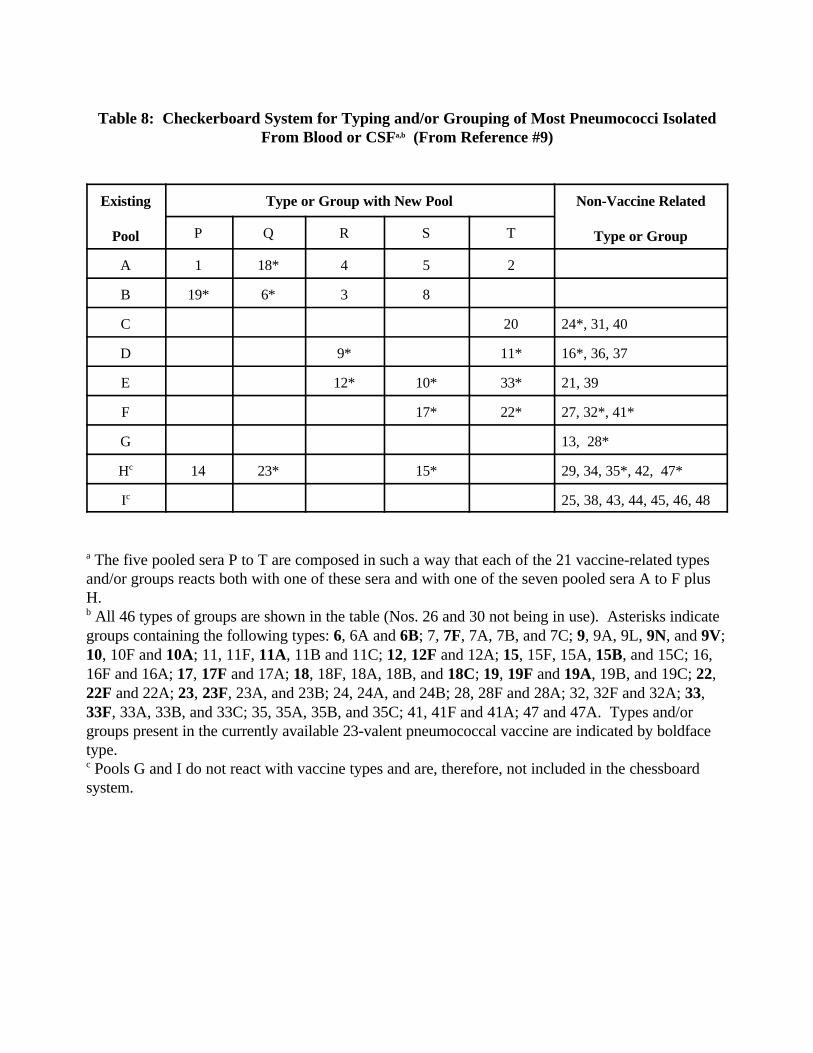
Table 8: Checkerboard System for Typing and/or Grouping of Most Pneumococci IsolatedFrom Blood or CSFa,b (From Reference #9)
Existing
Pool
Type or Group with New Pool Non-Vaccine Related
Type or GroupP Q R S T
A 1 18* 4 5 2
B 19* 6* 3 8
C 20 24*, 31, 40
D 9* 11* 16*, 36, 37
E 12* 10* 33* 21, 39
F 17* 22* 27, 32*, 41*
G 13, 28*
Hc 14 23* 15* 29, 34, 35*, 42, 47*
Ic 25, 38, 43, 44, 45, 46, 48
a The five pooled sera P to T are composed in such a way that each of the 21 vaccine-related typesand/or groups reacts both with one of these sera and with one of the seven pooled sera A to F plusH.b All 46 types of groups are shown in the table (Nos. 26 and 30 not being in use). Asterisks indicategroups containing the following types: 6, 6A and 6B; 7, 7F, 7A, 7B, and 7C; 9, 9A, 9L, 9N, and 9V;10, 10F and 10A; 11, 11F, 11A, 11B and 11C; 12, 12F and 12A; 15, 15F, 15A, 15B, and 15C; 16,16F and 16A; 17, 17F and 17A; 18, 18F, 18A, 18B, and 18C; 19, 19F and 19A, 19B, and 19C; 22,22F and 22A; 23, 23F, 23A, and 23B; 24, 24A, and 24B; 28, 28F and 28A; 32, 32F and 32A; 33,33F, 33A, 33B, and 33C; 35, 35A, 35B, and 35C; 41, 41F and 41A; 47 and 47A. Types and/orgroups present in the currently available 23-valent pneumococcal vaccine are indicated by boldfacetype.c Pools G and I do not react with vaccine types and are, therefore, not included in the chessboardsystem.

Annex E. Biotyping of H. influenzae
In the absence of Hib vaccination, H. influenzae isolated from the CSF of a patientwith clinical meningitis will usually be Hib. Most laboratories will conclude theiridentification at that stage. However, in certain cases, reference laboratories may want tocarry out the biotyping of H. influenzae. Biotyping can be performed irrespective ofwhether or not a serotype antigen was detected. The biotype is determined by the testingfor indole, ornithine decarboxylase, and urease.
(a) Spot indole test: Transfer a heavy inoculum (a 25-µl loopful) to a piece of filterpaper so that the bacteria are contained in a heavy spot. Add one drop of indolereagent (Kovac’s reagent) to the cells. A blue colour will appear within 1 minutewhen the bacteria are producing indole. A red, pink or pinkish-yellow reaction isnegative.Do not read the test after 5 minutes, as all reactions may appear positive.
(b) Ornithine decarboxylase test: Inoculate a tube of ornithine medium with a loopful ofbacterial suspension (0.5 McFairland). This suspension can also be used to inoculatethe Haemophilus ID medium described above. After inoculating the ID medium,cover the ornithine decarboxylase test tube media with sterile mineral oil. Incubate 4hours or overnight at 35 •C. A purple colour is positive for the presence of theornithine decarboxylase enzyme, and a yellow colour or no colour is negative.
(c) Urease test: Inoculate a tube of Christensen’s medium with a loopful of bacteria.Incubate 4 hours or overnight at 35 •C. A pink to red colour is positive, and nochange or a yellow colour is negative.
(d) The biotype is determined by matching the observed reactions to those described inTable 9.
Note: When performing these tests follow the specific instructions provided by themanufacturers as different colour indicators may be employed by differentmanufacturers.
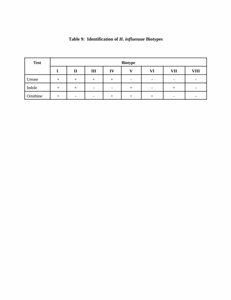
Table 9: Identification of H. influenzae Biotypes
Test Biotype
I II III IV V VI VII VIII
Urease + + + + - - - -
Indole + + - - + - + -
Ornithine + - - + + + - -
XI. List of Figures
1. Kit for the collection of CSF. 2. Collection of CSF by lumbar puncture.
A. The patient lies on his side with knee flexed and back arched to separate thelumbar vertebrae. The patient is surgically draped, and an area overlaying thelumbar spine is disinfected.B. The space between lumbar vertebrae L3 and L4 is palpated with the sterilelygloved forefinger.C. The spinal needle is carefully directed between the spinous processes, throughthe intraspinous ligaments into the spinal canal.
3. Collection of blood from an arm.1. Apply the tourniquet.2. Select a vein.3. Plan proposed puncture site.
4. Processing of CSF. 5. Trans-Isolate medium everywhere. 6. N. meningitidis -- proper streaking and growth on blood agar plate. 7. S. pneumoniae -- proper streaking and growth on blood agar plate. 8. H. influenzae -- proper streaking and growth on chocolate agar plate. 9. Gram stain of CSF:
a N. meningitidis: intra-cellular, Gram-negative diplococci,b S. pneumoniae: Gram-positive diplococci, andc H. influenzae: Gram-negative pleomorphic coccobacilli.
10. On chocolate agar plate, H. influenzae appear as large colourless to grey opaquecolonies with no discoloration of the surrounding medium.
11. Overnight growth of N. meningitidis on blood agar plate appears as round, moist,glistening and convex colonies.
12. S. pneumoniae appear as small greyish mucoid (watery) colonies with a greenishzone of alpha-haemolysis surrounding them on the blood agar plate.
13. Presumptive identification of N. meningitidis, S. pneumoniae, and H. influenzae.14 N. meningitidis (left), S. pneumoniae (right), and H. influenzae (top):
(a) growth on blood agar plate and (b) growth on chocolate agar plate.15. Identification of N. meningitidis.16. Kovac’s oxidase test -- positive reaction.17. Agglutination, with clearing of the liquid, occurs when a suspension of the isolate
is mixed with its homologous antiserum (left). A negative reaction, as in the caseof heterologous antiserum (centre) and with saline (right), remains smooth andturbid.
18. Cystine trypticase agar-sugar reactions differentiating N. meningitidis from otherNeisseria species. Acid production (yellow colour) shows oxidative utilization ofdextrose and maltose with no utilization of lactose and sucrose.
19. Identification of S. pneumoniae.20. Optochin susceptibility test for identification of S. pneumoniae. The strain in the
top streak is resistant to optochin and, therefore, is not a pneumococcus. Thestrains in the centre and lower streaks are susceptible to optochin and appear to bepneumococci.
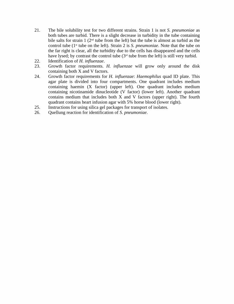
21. The bile solubility test for two different strains. Strain 1 is not S. pneumoniae asboth tubes are turbid. There is a slight decrease in turbidity in the tube containingbile salts for strain 1 (2nd tube from the left) but the tube is almost as turbid as thecontrol tube (1st tube on the left). Strain 2 is S. pneumoniae. Note that the tube onthe far right is clear, all the turbidity due to the cells has disappeared and the cellshave lysed; by contrast the control tube (3rd tube from the left) is still very turbid.
22. Identification of H. influenzae.23. Growth factor requirements. H. influenzae will grow only around the disk
containing both X and V factors.24. Growth factor requirements for H. influenzae: Haemophilus quad ID plate. This
agar plate is divided into four compartments. One quadrant includes mediumcontaining haemin (X factor) (upper left). One quadrant includes mediumcontaining nicotinamide dinucleotide (V factor) (lower left). Another quadrantcontains medium that includes both X and V factors (upper right). The fourthquadrant contains heart infusion agar with 5% horse blood (lower right).
25. Instructions for using silica gel packages for transport of isolates.26. Quellung reaction for identification of S. pneumoniae.
XII. Abbreviations
ATCC American type culture collectionBAP Blood agar plateCAP Chocolate agar plateCDC Centers for Disease Control and PreventionCSF Cerebrospinal fluidGC Gonococcus mediumHIA Heart infusion agarHIA-rabbit blood Heart infusion rabbit blood agar plateHib Haemophilus influenzae type bMOPS 3- (N-morpholino) propanesulfonic acidNAD Nicotinamide adenine dinucleotideO/N OvernightPBS Phosphate-buffered salinerpm revolutions per minuteSPS Sodium polyanetholesulfonateT-I Trans-Isolate mediumTSA Trypticase soy agarTSB Trypticase soy brothWHO World Health Organization