ヒト角層細胞のcell envelopeの発生とその機能障害を来す病態の …
TRANSCRIPT

− 62−
帝京大学医学部附属市原病院皮膚科学教室
秋 山 真 志
Terminally differentiated stratified squamous epithelium forms a lining of the plasma membrane called the cornified cell envelope (CCE), a thick layer of several covalently cross-linked precursor proteins including involucrin, small proline-rich proteins (SPRPs) and loricrin. Their cross-linking isodipeptide bonds are formed by epidermal transglutaminases (TGases) 1, 2 and 3. The formation of CCE and sequential expression of major CCE precursor proteins, TGases, and 25-kD lamellar granule-associated protein (LGP) were studied in human embryonic and fetal skin. Ultrastructurally, membrane thickening has already started in periderm cells of the two-layered epidermis and an electron dense, thickened cell envelope similar to CCE in adult epidermis is observed in periderm cells at the three-layered and later stages of skin development. In the two-layered epidermis (49-65 days EGA), immunoreactivities of involucrin, SPRPs, all the TGases and LGP were present only in the periderm. In the three-layered epidermis and thereafter (66-160 days EGA), loricrin became positive in the periderm cells, TGases extended to the entire epidermis, and LGP was detected in intermediate cells as well as periderm cells. Immunoelectron microscopy demonstrated that all three major CCE precursor proteins, involucrin, SPRPs and loricrin, were restricted to the CCE in periderm cells at this stage of development. After 160 days EGA, the periderm had disappeared and CCE proteins and LGP were expressed in the spinous, granular and cornified cells and TGases were detected in the entire epidermis. These findings indicate that CCE precursor proteins, TGases, and LGP are expressed in coordination in periderm cells during human epidermal development and suggest that periderm cells form CCE in the process of regression.
As a next step, five patients with non-bullous congenital ichthyosiform erythroderma (NBCIE), one patient with lamellar icthyosis and two patients with mutilating palmoplantar keratoderma (MPPK) were studied.
In one patient out of five NBCIE cases, expression of TGase 1 molecule was markedly reduced in the patient’ s epidermis. Electron microscopy revealed incomplete thickening of CCE during keratinization in the epidermis. Sequencing of the entire exons and exon-intron borders of TGase 1 gene (TGM1) revealed that the proband was a compound heterozygote for two novel mutations, 9008delA and R388H. These findings indicate that the compound heterozygote for missense mutation (R388H) in the core domain and the frameshift mutation (9008delA) resulting in a premature termination codon at the tail of the TGase 1 peptide leads to the NBCIE phenotype. TGM1 mutations were also found in the lamellar ichthyosis patient.
In the patients with MPPK, ultrastructural, immunohistochemical, and immunoelectron microscopic analyses revealed malformed CCE and reduced deposition of loricrin to CCE. Sequencing of the entire exons and exon-intron borders of loricrin gene of the patients excluded a mutation in loricrin DNA sequence. These data confirm the presence of MPPK phenotype with abnormal loricrin cross-linking at the final stage of CCE formation that is not caused by mutations in the epidermal differentiation complex region.
Studies on the developmental processes of human cornified cell envelope and the pathogenesis of its dysfunctionMasashi AkiyamaDepartment of Dermatology, Teikyo University School of Medicine,Ichihara Hospital
ヒト角層細胞の cell envelope の発生とその機能障害を来す病態の解析
1.緒 言
皮膚の角層は,角化細胞と呼ばれる高度に分化した細胞より成っているが,角化細胞は,cornified cell envelopeという非常に厚い膜状物質に覆われている1)。この cornified cell envelope は,角化細胞のバリアー機能,相互接着,剥離において重要な役割を果たすと考えられており,魚鱗癬等、角化細胞の剥離の障害によって起こる過角化を主症状とする疾患の発症に関与している可能性が示唆されている。さらに,“肌荒れ” を来す角層のバリアー機能の障害にも密接に関与しているものと思われる。近年,生化学的,分
子生物学的解析により,cornified cell envelope は表皮細胞が分化し角化する際に,細胞膜に loricrin, small proline-rich proteins, involucrin 等の複数の cornified cell envelope 前駆蛋白が結合して形成されることが明らかになってきた。この前駆蛋白間の結合は表皮に発現している transglutaminases
(TGases)によって形成される2)3)。 ヒトの胎生期皮膚は、胎生 40 日以前では、表皮に特徴的な形態的所見を有さない単層の上皮細胞層として認められる。胎生 40 日を過ぎると、表皮は最上層の periderm とその下の基底細胞層の2層性となる4)。periderm は、胎生期の皮膚にのみ認められる細胞層であり、periderm を形成する細胞は、細胞質内小器官に乏しく、ケラチン線維束を小量しか持たず、細胞辺縁には大小の多数の突起を有するなど、他の表皮細胞とは異なった形態的特徴を示す2)3)。胎生 65 日を過ぎる頃より、periderm と基底細胞層の間に、intermediate cell layer が認められるようになり、表皮は3層性となる。胎生 95 日を経ると、表皮は4層以上となり、periderm は、次第に退縮傾向を示すようになる。それにつ

ヒト角層細胞の cell envelope の発生とその機能障害を来す病態の解析
− 63−
れて、下方の細胞層は、成人の表皮細胞に近い形態を示すようになり、胎生 160 日を過ぎると、periderm は消失し、それと同時に、下層の細胞層は角化する。この時期の表皮には、既に成人の表皮に認められる基底層、有棘層、顆粒層、角層の各成分がそろっている。 periderm について多くの研究がなされてきたが、いまだに、この細胞層の本態は明らかにされておらず、その機能、および、この細胞層の退縮、消失するメカニズムも不明である。この periderm について、明らかなことは、はじめは、単層上皮として生じ、細胞表面、および、細胞内小器官の一連の形態学的変化をへて、表皮の角化が始まるのと時を同じくして消失することである4)。 loricrin, small proline-rich proteins, involucrin 等のcornified cell envelope 構成蛋白それぞれの,ヒト皮膚における発現様式,および,超微形態学的分布には,未だ不明な点が多く,ヒト皮膚における cornified cell envelopeの機能の解析,その機能障害を来す病態の解明に際して大きな障害となっている。今回の研究の第1の目的は,ヒト胎生期皮膚の periderm を含む表皮における cornified cell envelope の形成のメカニズムを明らかにすることである。 さらに、最近、葉状魚鱗癬の患者家系において、分子遺伝学的に、表皮の transglutaminase の異常が報告されたが5)
6)、この表皮の transglutaminase は、cornified cell envelope の構成蛋白を表皮細胞膜へ結合させる際に働くことが知られている。以上の事実より、葉状魚鱗癬の発症に、 cornified cell envelope の構成蛋白の細胞膜への結合障害を含めた、 cornified cell envelope の形成異常が関与している可能性が強く示唆されている。さらに、非水疱型先 天 性 魚 鱗 癬 様 紅 皮 症(non-bullous congenital ichthyosiform erythroderma)の症例においても、表皮のtransglutaminase の活性の低下が報告されている。以上の事実より、cornified cell envelope の形成異常が葉状魚鱗癬、非水疱型先天性魚鱗癬様紅皮症の発症において、重要な役割を果たしている可能性が考えられる。 本研究の第2の目的は、先天性魚鱗癬などの角化異常症の病変部皮膚における cornified cell envelope 構成蛋白の発現および分布様式を解析することにより,involucrin, loricrin, small proline-rich proteins 等の cornified cell envelope 構成蛋白がヒト角層細胞の cell envelope の機能維持に果たす役割を解明することである。
2.実 験
2. 1 ヒト胎児皮膚検体: 本研究に用いられたヒト胎児組織は全て,Central Laboratory of Human Embryology,University of Washington(Seattle)より,Human Subjects Review Board の認可のもと,United States DHEW policies に従
い,供与されたものである。今回は,胎生 49 日齢から163 日齢までの,皮膚疾患以外の障害により死亡,または,妊娠中絶されたヒト胎児よりの皮膚組織が用いられた。
2. 2 形態学的観察: ヒト胎児皮膚組織を2%グルタ−ルアルデハイドにて固定、オスミウム酸にて後固定、型通り脱水後エポキシ樹脂中に包埋、光顕観察用に厚さ1µm の切片、電顕観察用には、厚さ 70nm の切片を作製観察した。
2. 3 抗体: 本研究に用いた一次抗体は、以下のものである:モノクロ−ナル抗ヒト SPRPs 抗体(mouse)7)、ポリクロ−ナル抗ヒト involucrin 抗体(rabbit)(Biomedical Technologies, Inc.)、ポリクロ−ナル抗ヒト loricrin 抗体(rabbit)8)、ポリクロ−ナル抗ヒト TGase 1 抗体(goat)9)、ポリクロ−ナル抗ヒト TGase 2 抗体 (rabbit) 10)、ポリクロ−ナル抗ヒト TGase 3 抗体(rabbit)10)、 モノクロ−ナル抗ヒト LGP抗体、AE17(mouse)11)。
2. 4 免疫蛍光抗体法、免疫電顕法: 免疫蛍光抗体法は,皮膚組織より未固定の厚さ6µm の凍結切片を作成、FITC 飽合二次抗体を用いて染色した。必要に応じて propidium iodide にて核染色を施行した.免疫電顕用には、皮膚組織を液体プロパンを用いて凍結固定、エタノ−ルによって凍結置換し、Lowicryl K11M 中に包埋したブロックを用いた。超薄切片を作製後、免疫反応を施行、1nm ゴ−ルド粒子飽合二次抗体を用い、silver enhancement 法を行ない一次抗体の沈着部位を観察した。
2. 5 先天性角化異常症患者: 非水疱型魚鱗癬様紅皮症5例(4家系)、葉状魚鱗癬1例(1家系)、mutilating palmoplantar keratoderma2例(1家系)について、完全な informed consent に基づき、皮膚生検を施行した。遺伝子変異の検索のため、末梢血を採取、genomic DNA を抽出した。同時に、家系内の健常者からも、可及的に DNA を得た。
2. 6 遺伝子変異の同定 患者ならびに家族の末梢血より得られた genomic DNAについて、上記の形態学的検討の結果、発現、分布に異常のみられた蛋白(今回は、transglutaminase 1 および loricrin)について遺伝子変異の検索を行った。genomic DNA を鋳型として、PCR にてその遺伝子のすべての exon ならびにexon-intron 境界部分を増幅し、自動シークエンサーにて解析した。遺伝子変異が見い出されたものについては、enzyme digestion 法あるいは allele specific oligonucleotide
− 64−
hybridization 法により、遺伝子変異を確認した。
3.結 果
3. 1 ヒト胎児表皮における cornified cell envelopeの発生 12)
3. 1. 1 2層性の表皮(胎生 40 ー 65 日): 2層性の表皮(胎生 40 − 65 日)の時期のヒト胎児皮膚は最表層の periderm と、その下方の基底膜上の基底細胞層の2層からなっている(図1)。この期間の初期には、電顕上、Periderm の細胞は、厚さ 7-9nm の平坦な細胞膜を有していた(図2)。後期に入ると、Periderm の細胞の細胞膜が次第に厚くなってくるが、同時期の基底細胞の細胞膜には肥厚は認められなかった。免疫組織学的には、involucrin、および、SPRPs が、periderm の細胞に陽性であり、細胞辺縁により多く存在していた。他方、この時期、involucrin、SPRPs ともに、基底細胞は、発現していなかった。TGases 1 , 2 , 3 は、periderm に陽性であり、基底細胞にも、弱い発現が認められた。loricrin はこの時期、periderm の細胞、基底細胞ともに陰性であった。層板顆粒の成分蛋白も、periderm の細胞のみに、発現がみとめられた。
3. 1. 2 3層性の表皮(胎生 66 ー 95 日): 3層性の表皮(胎生 66 − 95 日)になると、peridermの下層には、基底細胞と periderm の間の intermediate cell が見られる(図1)。この時期、電顕的には peridermの細胞膜はさらにその厚さと電子密度を増しいた(図2)。免疫蛍光抗体法では、TGases 1 , 2 , 3、involucrin、SPRPs、および、loricrin は、全て periderm に陽性であった。それらの蛋白は、すでにこの時期に、蛍光顕微鏡レベルでも、 periderm の細胞辺縁に分布していることが分かった。cell envelope の構成蛋白の periderm の細胞膜への局在は、免疫電顕によって確認された。periderm より下方の基底細胞、intermediate cell でも、TGase の発現は認められるものの、periderm と比較すると非常に弱い発現であった。
loricrin、SPRPs などの、cell envelope の構成蛋白は、intermediate cell、基底細胞には、まだ、発現が見られなかった。層板顆粒の成分蛋白は periderm と intermediate cell に発現がみられ、特に、強い線状の分布が、peridermと intermediate cell が接する部分にみられた。
3. 1. 3 4層性、および、それ以降の表皮(胎生 96 −160 日):
4層性、さらに、それ以上の多層性の表皮(胎生 96 −160 日)になると(図1)、電顕的には periderm の細胞膜は3層性の時期よりさらにその厚さと電子密度を増し、この時期の後期の periderm の細胞膜は成人の角化細胞のcell envelope と同様、約 15nm の厚さの高電子密度の層になった(図2)。3層性の表皮から引き続いて、この時期の早期から、periderm には、免疫蛍光抗体法では、TGases 1 , 2 , 3、involucrin、SPRPs、および、loricrin は、全て陽性であった。この periderm における TGases 1 , 2 , 3、involucrin、SPRPs、および、loricrin の発現は、表皮の角化にともない periderm が消退するまで、常に認められた。 cell envelope の構成蛋白が、実際に、periderm の細胞膜近傍で cell envelope を、形成しているのかどうかを知るため、免疫電顕を用いて調べると、平坦な部分、細胞突起を形成している部分ともに、involucrin, SPRPs、および、loricrin は、periderm の細胞膜の厚くなっている部分に局在して認められた(図3)。periderm 直下の intermediate cell では、involucrin の発現が見られるものの、その他の
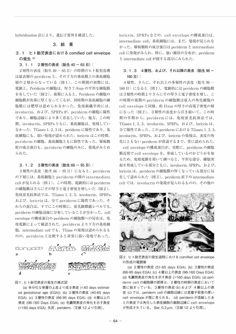
図1. ヒト胎児表皮の発生の模式図 (a) 未分化な単層の上皮より成る表皮 (<40 days estimat-
ed gestational age (EGA)). (b) 2層性の表皮 (40-65 days EGA). (c) 3層性の表皮 (66-95 days EGA). (d) 4層以上の表皮 (96-160 Days EGA). (e) 毛嚢間表皮の角化を示す表皮 (>160 days EGA). 矢尻 , periderm.(文献 12 より引用)。
図2. ヒト胎児表皮の発生過程における cornified cell envelopeの形成の電顕像
(a) 2層性の表皮 (51-65 days EGA). (b) 3層性の表皮 (66-95 days EGA). (c) 4層以上の表皮 (96-160 Days EGA). (d) 毛嚢間表皮の角化を示す表皮 (>160 days EGA). (a) peri-derm cell の細胞膜の肥厚は、2層性の時期の表皮において既に始まっている。3層性の表皮 (b) および 4層以上の表皮 (c) では、 periderm cell の細胞辺縁には高電子密度の厚いcell envelope が既に見られる。 (d) periderm が退縮したあとの表皮では角化した表皮細胞の細胞辺縁に cell envelopeが形成されている。 Bar; 0.3μm.(文献 12 より引用)。

ヒト角層細胞の cell envelope の発生とその機能障害を来す病態の解析
− 65−
cell envelope の構成蛋白(loricrin、SPRPs など)は、基底細胞、および、intermediate cell では陰性であった。TGase の発現はこの時期の表皮の全層に認められるが、角化において最も重要な TGase の subtype である TGase 1 の発現は、基底細胞層では弱かった。層板顆粒の成分蛋白は、3層性の表皮におけるのと同様に、periderm とintermediate cell に認められた。periderm とその直下のintermediate cell とが、接する面にみられる強い線状の発現は、3層性の表皮の時期より、さらに、強調されてきていた。これは、periderm の下床の細胞層が、この後の時期に、最初の角化を始める予兆とも言える。
3. 1. 4 Periderm 退縮後の角化を示す表皮(胎生 161 日以降):
毛嚢間表皮が角化を示す胎生 160 日以降の表皮では、periderm は完全に消失している(図1)。最上層の角化し
始めた細胞は、厚さおよそ 15nm の、成人の皮膚の角層のcell envelope と類似の厚く、電子密度の高い細胞膜を形成していた(図2)。cell envelope の構成蛋白、involucrin, SPRPs, loricrin, ならびに層板顆粒の成分蛋白のすべてが、この時期の顆粒細胞層および角化細胞層に陽性であった。層板顆粒の成分蛋白は,角層と顆粒層の間に特に強く線状に分布していた.TGase1 , 2 , 3、の発現は,表皮全層にみられ,TGase1 および 3 の分布は細胞辺縁に膜状に認められた.これらの所見は、成人の皮膚で見られるものと同様であった。
3. 2 先天性角化異常症患者における cornified cell envelope の形成 3. 2. 1 非水疱型先天性魚鱗癬様紅皮症 5例中4例の非水疱型先天性魚鱗癬様紅皮症患者において、involucrin, loricrin, TGases 1, 2, 3の分布は正常であった 13)。1例の非水疱型先天性魚鱗癬様紅皮症患者においてのみ、TGase 1の発現に低下が見られた 14)。超微形態学的には、前者4例では、cornified cell envelope の正常な形成が認められたが、TGase 1の発現に異常がみられた1例では、cornified cell envelope の形成不全が観察された(図4)14)。
3. 2. 2 葉状魚鱗癬 今 回 検 索 し た 1 例 の 葉 状 魚 鱗 癬 患 者 に お い て、involucrin, loricrin, TGases 2, 3の分布は正常であったが、TGase 1蛋白の発現に低下が見られ、TGases 1活性も低下していた。超微形態学的にも、cornified cell envelopeの形成不全が確認された。
図3. 免疫電顕像:ヒト胎児表皮の periderm における cornified cell envelope の形成
Colloidal gold を用いた immunoelectron microscopy にて、periderm の細胞膜に involucrin (b,c)、および、loricrin (d) が結合していることが示された。(a) は、正常成人皮膚における角層細胞の細胞膜への involucrin の沈着(文献 12 より引用)。
図4. TGase 1 活性の低下を認めた非水疱型魚鱗癬様紅皮症症例における皮膚の病理組織像、および電顕像
(a) 著明な hyperkeratosis と中等度の acanthosis が患者皮膚には見られた (toluidine blue stain)。(b) 電顕的には顆粒細胞内の keratin clump や脂肪滴は認められなかった。(c) 顆粒層の細胞では、薄い cell envelope ( 矢尻 ) が正常に形成されていた。(d) 角層では、cornified cell envelope ( 矢尻 ) は、患者皮膚では著明に薄く、低形成を認めた。 (e) 正常皮膚における cornified cell envelope ( 矢尻 )。Bars, 50μm(a), 1μm(b), 0.3μm(c-e).(データは文献 14 より引用)。

− 66−
3.2.3 Mutilating palmoplantar keratoderma15)
2例(1家系)の患者病変部皮膚において免疫蛍光抗体法では、involucrin および SPRPs は、病変部では正常皮膚に比して、より広い範囲で、正常の顆粒層の部分より下方においても発現していた。光顕レベルでも involucrin および SPRPs の顆粒細胞における細胞膜への局在が認められた.loricrin の発現は,病変部皮膚において著明に減弱しており,顆粒細胞においても細胞質内に弱い染色性が認められるのみであり,細胞膜への沈着は認められなかった.超微形態学的には角化細胞内に異常な脂肪滴が認められた。移行細胞から上方において、 cornified cell envelope の形成がみられたが、正常ヒト皮膚で認められる顆粒細胞から角化細胞にかけての,電子密度の高い物質の沈着による cornified cell envelope の肥厚が見られなかった(図5).免疫電顕法では、病変部皮膚においても、正常皮膚と同様に、involucrinおよび SPRPs は cornified cell envelope の形成に伴い cornified cell envelope 部分に局在していた。しかし、loricrin の cornified cell envelope への沈着は正常皮膚と比べて減少していた(図6).
3. 3 先天性角化異常症患者における遺伝子解 析結果 3. 3. 1 非水疱型先天性魚鱗癬様紅皮症: TGase 1の発現に異常を認めた1例で、direct sequencing の 結 果、TGase 1 gene(TGM1) に、heterozygous な2つの変異、9008delA (paternal) とR388H(maternal)、が認められた(図7)。これらの変異は、コントロールとした正常日本人 50 人のDNA には、認められなかった。 TGM1 のすべてのexons、exon-intron borders に つ い て direct sequencing を行ったが、この変異の他には、病因となり得る変異は見られなかった。
3. 3. 2 葉状魚鱗癬: 今回検索した1例で、direct sequencing の結果、TGase 1 gene (TGM1) に、heterozygous な2つのミスセンス変異、L204Q と R306W、が認められた。これらの変異は、コントロールとした正常日本人 50人の DNA には、認められなかった。GM1 のすべての exons、exon-intron borders について direct sequencing を行ったが、この変異の他には、病因となり得る変異は見られなかった。
3. 3. 3 Mutilating palmoplantar keratoderma: loricrin gene の2つの exons には、調べ得たかぎ
図5. mutilating palmoplantar keratoderma 病変部皮膚の電顕像 (a) 顆粒層の細胞は、正常なケラチン束とケラトヒアリン顆粒を形
成していた。(b) 正常な、薄い cornified cell envelope ( 矢尻 ) が、顆粒層の細胞には見られた。(c) 患者病変部の角層では cornified cell en-velope ( 矢尻 ) の低形成があり、cornified cell envelope ( 矢尻 ) は正常皮膚 (d) におけるものとくらべて著明に薄かった。薄い cornified cell envelope と desmosome からなるリング状の構造が、顆粒層上部の細胞に見られた (e)。矢尻 , cornified cell envelope; 矢印 , lipid droplets。Scale bars, 2μm (a), 0.2μm (b-d), 0.5μm (e).(文献 15 より引用)。
図6. 免疫電顕像:mutilating palmoplantar keratoderma の症例における cornified cell envelope の形成不全
患者皮膚 (a) では、正常人皮膚 (b) と比べて loricrin の細胞膜への結合が少ない。他方、患者皮膚 (c) での involucrin のラベルは、正常人皮膚 (d) より強い(文献 15 より引用)。
図7.TGase 1 活性の低下を認めた非水疱型魚鱗癬様紅皮症症例に見られた TGase 1 遺伝子の変異、9008delA および R388H
本家系の父方の変異、9008delA は、 TGase 1 蛋白の c 末に 置する β-barrel 2 domain 内にある。母方の変異、 R388H は、 TGase 1蛋白の酵素活性部位を含むcore domain内にある(文献14より引用)。

ヒト角層細胞の cell envelope の発生とその機能障害を来す病態の解析
− 67−
り、mutation は発見されなかった。
4.考 察
本研究の結果,主な cornified cell envelope 前駆蛋白である involucrin, SPRPs,および,loricrin は,ヒト皮膚の発生過程において periderm の細胞に存在していることが明らかになった。さらに,胎生期に,TGases 1 , 2 , 3,LPG が,やはり,periderm の細胞に発現していることが示された。これらの免疫蛍光所見と,超微形態学的にperiderm の細胞膜に肥厚が認められることとを考え合わせると,ヒト胎児の発生過程にある表皮の periderm は,その退縮過程において,成人皮膚の角化細胞が形成するものと類似の cornified cell envelope を形成することが推測される。免疫電顕による検討の結果,主な cornified cell envelope 前駆蛋白である involucrin, SPRPs,および,loricrin は,ヒト皮膚 periderm の肥厚した細胞膜に結合した形で存在していることが確認された。これは,上記仮説,すなわち,periderm が cornified cell envelope を形成するという考えを支持するものである。以上の今回得られたデータより, cornified cell envelope の形成という点からみると,periderm の退縮過程は,成人皮膚表皮のケラチノサイトの角化過程と類似したものであるといえるであろう。 periderm の細胞は、胎芽から胎児へと発達する時期から、毛嚢間表皮が角化を始める時期までの間に、顕著な形態学的変化を示す。この periderm の細胞の形態的変化のなかで、後期(退縮期)の変化と、表皮細胞の角化との間には、類似点が見られる。periderm の細胞も、退縮期には、中間径繊維をのこして、細胞内小器官を失い、核は凝集してくる。これらの所見から、periderm は、「普通とは異なる角化様式を示す角化細胞層」とも考えられるわけである。periderm の退縮から表皮の角化への、角化様式の転換は、羊水中から出生後の環境へと、急激に変化する外部環境に、胎児が適応するように、胎児の体表面を変えることになる。periderm の存在意義,及び,その退縮のメカニズムを解明するためには,さらなる今後の研究が必要と思われる。 近年、cell envelope 前駆蛋白のなかで重要なものであるloricrin の 異 常 に よ る cell envelope の 形 成 不 全 が、Vohwinkel syndrome16)や erythrokeratoderma17)等の遺伝性角化異常症の病因であることが明らかになり、また、loricrin 分子の異常以外にも、cell envelope の異常が掌蹠角化症等の原因となり得ることが示唆されている 15)。葉状魚鱗癬の一部の家系でも、TGase 1 の異常による cell envelope の形成不全がその病因であることが報告されている5)6)。今回の研究の結果からは、検索した1例の日本人葉状魚鱗癬患者において、TGase 1 遺伝子に変異が認められた。さらに、非水疱型先天性魚鱗癬様紅皮症でも、
TGase 1 の遺伝子変異が原因である可能性が示された。また、本研究の対象となった mutilating palmoplantar keratoderma の症例では cornified cell envelope の形成不全がありながら、既知の cornified cell envelope 関連蛋白遺伝子に変異がある可能性は否定的であった。このことから、今回の mutilating palmoplantar keratoderma の症例については、その病因として、他のまだ明らかにされていない cornified cell envelope 関連蛋白等が想定された。 出生時に最も重篤な症状を呈する葉状魚鱗癬などの場合、病像の形成に胎生期の cell envelope の形成異常などが関与している可能性が考えられ 18)19)、ヒト胎生期表皮のperiderm の退縮、ケラチノサイトの角化などの過程は、表皮細胞の発達、分化のメカニズムを理解するという意味のみでなく、重症型魚鱗癬の病態の解明という点からも今後の興味深い研究対象である。 近年、腫瘍発生のメカニズム、遺伝性皮膚疾患とその出生前診断、遺伝子治療と関連した幹細胞の生物学、各種の分化、増殖の制御因子の研究等、様々な分野で発生学的なアプローチは、盛んに行われている。今後、ますます、皮膚発生学が皮膚科学、皮膚の生物学の領域で占める役割は大きくなるものと考えられる。この発生学的アプローチと角化異常症の患者皮膚を対象とした研究によって、さらに他の cell envelope 構成蛋白の分布,役割が明らかなり、他の cornified cell envelope 関連蛋白と角化症の病因の関係、さらには、cornified cell envelope 本体、ならびに個々の構成蛋白の機能が明らかになることが期待される。
(参考文献)1) Hohl D: Cornified cell envelope. Dermatologica 180 :201-
211 , 19902) Gre enbe rg CS , B i r c kb i c h l e r P J , R i c e RH :
Transglutaminases: multifunctional cross-linking enzyme that stabilizes tissues. FASEB J 5 :3071-3077 , 1991
3) Steinert PM, Marekov LN: The proteins elafin, filaggrin, keratin intermediate filaments, loricrin, and small proline-rich proteins 1 and 2 are isodipeptide cross-linked components of the human epidermal cornified cell envelope. J Biol Chem 270 :17702-17711 , 1995
4) Holbrook KA, Odland GF: The fine structure of developing human epidermis: light, scanning, and transmission electron microscopy of the periderm. J Invest Dermatol 65 :16-38 , 1975
5) Huber M, Rettler I, Bernasconi K, et al.: Mutations of keratinocyte transglutaminase in lamellar ichthyosis. Science 267 :525-528 , 1995
6) Russell LJ, DiGiovanna JJ, Rogers GR, et al:: Mutations in the gene for transglutaminase 1 in autosomal recessive
− 68−
lamellar ichthyosis, Nature Genet 9 :279-283 , 19957) Hohl D, de Viragh PA, Amiguet-Barras F, et al:: The
small proline-rich proteins constitute a multigene family of differentially regulated cornified cell envelope precursor proteins. J Invest Dermatol 104 :902-909 , 1995
8) Mehrel T, Hohl D, Rothnagel JA, et al:: Identification of a major keratinocyte cell envelope protein, loricrin. Cell 61 :1103-1112 , 1990
9) Kim I-G, McBride OW, Wang M, et al:: Structure and organization of the human transglutaminase 1 gene. J Biol Chem 267 :7710-7717 , 1992
10) Tarcsa E, Marekov LN, Andreoli J, et al:: The fate of trichohyalin: sequential post-translational modifications by peptidyl-arginine deiminase and transglutaminases. J Biol Chem 272 :27893-27901 , 1997
11) O'Guin WM, Manabe M, Sun T-T: Association of a basic 25K protein with membrane coating granules of human epidermis. J Cell Biol 109 :2313-232 , 1989
12) Akiyama M, Smith LT, Yoneda K, et al:: Periderm cells form cornified cell envelope in their regression process during human epidermal development. J Invest Dermatol 112 :903-909 , 1999
13) Akiyama M, Shimizu H, Yoneda K, et al:: Collodion baby: ultrastructure and distribution of cornified cell envelope proteins and keratins. Dermatology 195 :164-
168 , 199714) Akiyama M, Takizawa Y, Kokaji T, et al.: Novel
mutations of TGM1 in a chi ld with congenital ichthyosiform erythroderma. Br J Dermatol 144 :401-407 , 2001
15) Akiyama M, Christiano AM, Yoneda K, et al:: Abnormal cornified cell envelope formation in mutilating palmoplantar keratoderma unrelated to epidermal differentiation complex. J Invest Dermatol 111 :133-138 , 1998
16) Maestrini E, Monaco AP, McGrath JA, et al:: A molecular defect in loricrin, the major component of the cornified cell envelope, underlies Vohwinkel syndrome. Nature Genet 13 :70-77 , 1996
17) Ishida-Yamamoto A, McGrath JA, Lam H-M, et al:: The m o l e c u l a r b a s i s o f a u t o s o m a l d o m i n a n t erythrokeratoderma: a frameshift mutation in the loricrin gene. Am J Hum Genet 61 :581-589 , 1997
18) Akiyama M: Severe congenital ichthyosis of the neonate. Int J Dermatol 37 :722-728 , 1998
19) Akiyama M: The pathogenesis of severe congenital ichthyosis of the neonate. J Dermatol Sci 21 :96-104 , 1999