ch 6 - neoplasia · 2019-12-08 · origin of neoplasm §epithelial ... – benign tumors show...
TRANSCRIPT
Dr. Marwan Qubaja / Pathology Neoplasia
1
Neoplasia
Dr. Marwan Qubaja
Al-Quds University
Faculty of Medicine
Pathology Department 1
•• NeoplasiaNeoplasia: new growth
•• OncologyOncology: Onco: tumor, logoslogos: study of
• Neoplasm is benign and malignant
•• CancerCancer is a malignant neoplasmis a malignant neoplasm
•• InvasiveInvasive: tumor capable of destroying
structures
•• MetastasisMetastasis: spread to distant sites
Definitions
2

Dr. Marwan Qubaja / Pathology Neoplasia
2
Definitions
•• GrowthGrowth: Increase in size due to synthesis of
tissue components
•• ProliferatationProliferatation: Cell division
•• DifferentiationDifferentiation: Functional and structural
maturity of cells
•• TumorTumor: Swelling / new growth / mass3
Controls of Growth
•• Growth factorsGrowth factors – PDGF, FGF
•• Growth InhibitorsGrowth Inhibitors.
•• CyclinsCyclins, Cyclin dependent kinases (CDK).
•• Cancer suppressor genesCancer suppressor genes – p53
•• OncogenesOncogenes – c-onc, p-onc, v-onc etc.
4

Dr. Marwan Qubaja / Pathology Neoplasia
3
Non-Neoplastic Proliferation
Controlled & ReversibleControlled & Reversible
• Hypertrophy – increase in cell size
• Hyperplasia – increase in cell number
• Metaplasia – change of cells type
• Dysplasia – disordered cells
5
Neoplastic ProliferationUncontrolled & IrreversibleUncontrolled & Irreversible
• Progressive, purposeless, pathologic,
proliferation of cells characterized by loss
of control over cell division.
• DNA damage at growth control genes is
central to development of neoplasm.
6

Dr. Marwan Qubaja / Pathology Neoplasia
4
How do we get cancer• Damage to genetic material
• Affects different sites in the genome
• Evolution of more aggressive clones
Carcinogens (Chemical, radiation, viruses)
DNA damage
Neoplasm 7
“an abnormal mass of tissue, the growth
of which exceeds and is uncoordinated
with that of the normal tissues, and
persists in the same excessive manner
after cessation of the stimuli which
evoked the change”
Neoplasm definition:
8

Dr. Marwan Qubaja / Pathology Neoplasia
5
Neoplasm Properties
•• Normal properties loss:Normal properties loss:
– Increased proliferation
– Decreased cell death
– Failure to differentiate
•• Abnormal properties develop:Abnormal properties develop:
– Invasion
– Metastasis 9
Tumors have two basic components:
11)) Parenchyma:Parenchyma:• made up of neoplastic cells
• from which the tumor derives its name
22) ) StromaStroma::• supporting, host-derived, non-neoplastic
• made up of connective tissue and blood vessels
• provides support for the growth of parenchyma
• crucial to the growth of the neoplasm10

Dr. Marwan Qubaja / Pathology Neoplasia
6
1. Behaviour- benign and malignant
2. Extent of spread- Primary and secondary
3. The cell of origin or histogenesis:§ epithelial
§ mesodermal/connective tissue
§ hematopoietic
§ nervous system
§ germ cells
§ embryonic tissue
The classification of neoplasm
11
Neoplastic Behaviour• The terms “benign” and “malignant” describe
the biologic or clinical behavior of a tumor.
•• Benign:Benign:
– Localized, non-invasive, patient usually
survives
•• Malignant (Cancer):Malignant (Cancer):– Spreading, Invasive, may result in early death
of the patient12

Dr. Marwan Qubaja / Pathology Neoplasia
7
The main distinguishing features of benign and malignant neoplasms
Suffix “oma” eg. Fibroma. Suffix “Carcinoma” or “Sarcoma”13
Extent of spread
•• Primary tumourPrimary tumour gives rise to secondary secondary
tumourstumours
• Tumours invade lymphatics, blood vessels or
through peritoneal or other surfaces to form
secondaries (metastases)
• Always think the tumour might have a
primary tumour elsewhere14

Dr. Marwan Qubaja / Pathology Neoplasia
8
Origin of Neoplasm
§ Epithelial
§Mesodermal/connective tissue
§ hematopoietic
§ nervous system
§ germ cells
§ embryonic tissue
15
Tissue of OriginTissue of Origin BenignBenign MalignantMalignant
Epithelial:Epithelial:• Squamous Papilloma Squamous cell carcinoma• Transitional Papilloma Transitional cell carcinoma• Glandular Adenoma Adenocarcinoma
Connective Tissue/mesenchymal:Connective Tissue/mesenchymal:• Fibrous tissue Fibroma Fibrosarcoma• Fat Lipoma Liposarcoma• Bone Osteoma Osteosarcoma• Cartilage Chrondroma Chrondrosarcoma• Smooth muscle Leiomyoma Leiomyosarcoma• Striated muscle Rhabdomyoma Rhabdomyosarcoma• Blood vessels Hemangioma Angiosarcoma
Nomenclature: Cell of origin + Suffix
16

Dr. Marwan Qubaja / Pathology Neoplasia
9
PapillomaPapilloma is a benign neoplasm growing on any surface, composed of epithelial cells forming finger like projections.
Papilloma in esophagus17
AdenomaAdenoma::
• Applied to benign epithelial neoplasms producing gland
patterns.
E.g. Surface epithelium (stomach, small intestine & colon)
• Applied to benign neoplasms derived from glands but not
necessarily exhibiting gland patterns.
E.g. Solid glandular epithelium (endocrine and exocrine)
and ducts (Thyroid, kidney, liver)
18

Dr. Marwan Qubaja / Pathology Neoplasia
10
Examples of malignant tumors
•• Sarcoma (mesenchymal derivation):Sarcoma (mesenchymal derivation):
– fibrosarcoma
– chondrosarcoma
•• Carcinoma (epithelial derivation):Carcinoma (epithelial derivation):
– adenocarcinoma,
– squamous cell carcinoma
19
Hematopoietic§ Leukemias
§ Lymphomas
Brain tumors
§ Glioma
ExceptionsExceptions in Terminology
Germ cell tumor
§ Teratoma
§ Seminoma
Pediatric tumors
§ Hepatoblastoma
§ Nephroblastoma
§ Retinoblastoma
Malignant tumors:Malignant tumors:
20

Dr. Marwan Qubaja / Pathology Neoplasia
11
Characteristics of benign & malignant neoplasms
Tumors can be distinguished by:Tumors can be distinguished by:
1. Differentiation and anaplasia
2. Rate of growth
3. Local invasion
4. Metastasis 21
1. Differentiation & AnaplasiaDifferentiationDifferentiation:: extent to which parenchymal
neoplastic cells resemble normal cells morphologically and functionally, while stroma does not aid in the separation of benign from malignant.
•• WellWell--differentiated tumors:differentiated tumors:
contain cells that resemble the normal cells of origin and retains its functional capacity
•• PoorlyPoorly--differentiated or undifferentiated tumors:differentiated or undifferentiated tumors:contain cells that do not resemble their normal cells of origin 22

Dr. Marwan Qubaja / Pathology Neoplasia
12
•• Benign tumorsBenign tumors are composed of well-differentiated
cells.
•• Malignant tumorsMalignant tumors are characterized by a wide
range of cellular differentiation (well, moderate,
poor, undifferentiated).
•• AnaplasiaAnaplasia: malignant neoplasms that are
composed of undifferentiated cells
•• UndifferentiatedUndifferentiated: loss of the structural and
functional differentiation of normal cells.
Differentiation & Anaplasia
23
Anaplasia:Anaplasia: lack of differentiationHistological Features of anaplasia:Histological Features of anaplasia:
§ Cellular pleomorphism
§ Giant cells
§ Hyperchromatic nuclei
§ Nuclear- cytoplasmic ratio may approach 1:1
§ Nuclear pleomorphism
§ Mitosis is numerous and/or atypical
§ Loss of cell polarity 24

Dr. Marwan Qubaja / Pathology Neoplasia
13
Dysplasia: “disordered growth”• Dysplasia is a non-neoplastic proliferation
• Dysplasia is an abnormal type of excessive cell proliferation
characterized by loss of normal tissue arrangement and cell
structure in epithelium.
• Dysplasia may or may not progress to cancer.
•• In epitheliaIn epithelia, represents a state between hyperplasia and
carcinoma in situ (pre-invasive neoplasia)
• Pleomorphism & mitoses are more prominent than in the
normal 25
2. Rate of growth
• Benign and well-differentiated malignant tumors
have a slower rate of growth than moderately-
differentiated and poorly-differentiated malignant
tumors.
• There are exceptions.
• Malignant tumors sometimes grow slowly for years
and suddenly enter a phase of rapid growth26

Dr. Marwan Qubaja / Pathology Neoplasia
14
Rate of Growth• Factors that can affect
the growth rateof tumor:– Blood supply
– Site
– hormonal stimulation
• Example:
LeiomyomasLeiomyomas: influenced by estrogen, increase during pregnancy, and cease growing or atrophy after menopause 27
3. InvasionInvasion•• Benign tumorsBenign tumors
– usually grow by slow expansion
– usually encapsulated
•• Malignant tumorsMalignant tumors (cancer)
– usually infiltrate and destroy surrounding tissue
– do not develop well-defined capsules
– Some induce formation of dense fibrous stroma
(desmoplasia)
– Pathologists carefully examine the margin of resected
specimens (clean margins). 28

Dr. Marwan Qubaja / Pathology Neoplasia
15
•• DefinitionDefinition: the development of secondary implants
(metastases) discontinuous with the primary tumor,
possibly in distant tissues
• Next to metastases, local invasiveness is the most
reliable feature that distinguishes malignant from
benign tumors.
• Not all cancers have equivalent ability to
metastasize, e.g Basal cell carcinoma
4. Metastasis
29
Basal cell carcinoma Basal cell carcinoma (BCC):(BCC): skin cancer that is common and slow growing. Grossly, the tumor begins as papules with rolled margins (top photo), but can ulcerate and locally invade underlying structures and bone (bottom photo). Hence, the name "rodent ulcer."
Malignant tumorsMalignant tumors: Invasion
30

Dr. Marwan Qubaja / Pathology Neoplasia
16
4. Metastasis• Metastasis indicates
malignancy
• ~ 30% of newly diagnosed patients with solid tumors present with clinically evident metastases.
• In general, the more anaplastic and the larger the primary neoplasm, the more likely is metastatic spread; however, with exceptions. 31
Methods of spread1. Direct Spread
2. Seeding of body cavities: ovarian tumors
3. Hematogenous spread: favored pathway for
sarcomas. The liver and lung are the most
frequently involved secondary sites.
4. Lymphatic spread: is more typical of
carcinomas32

Dr. Marwan Qubaja / Pathology Neoplasia
17
Biology of Invasion and Metastasis§ Invasion of the basement membrane
§ Movement through extracellular matrix
§ Penetration of vascular or lymphatic channels
§ Survival and arrest within the circulating blood
or lymph
§ Exit from the circulation into new site
§ Survival and growth as a metastasis 33
Seeding of body cavities
• Occurs when neoplasms invade a natural
body cavity.
•• ExamplesExamples:– Carcinoma of the colon may penetrate the
wall of the gut and reimplant at distant sites in
the peritoneal cavity.
– Lung cancers in the pleural cavities.
– Cancers of the ovary in the peritoneal cavity. 34
Dr. Marwan Qubaja / Pathology Neoplasia
18
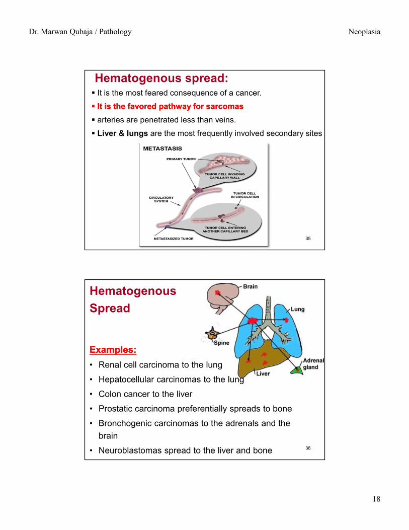
§ It is the most feared consequence of a cancer.
§§ It is the favored pathway for sarcomasIt is the favored pathway for sarcomas§ arteries are penetrated less than veins.
§ Liver & lungs are the most frequently involved secondary sites
Hematogenous spread:
35
Hematogenous Spread
Examples:Examples:• Renal cell carcinoma to the lung
• Hepatocellular carcinomas to the lung
• Colon cancer to the liver
• Prostatic carcinoma preferentially spreads to bone
• Bronchogenic carcinomas to the adrenals and the brain
• Neuroblastomas spread to the liver and bone 36

Dr. Marwan Qubaja / Pathology Neoplasia
19
Lymphatic spread• It is more typical of carcinomas
• Example: Lung and breast carcinoma
• Skip metastases:– The cancer cells seem to traverse the lymphatic channels
within the immediately proximate nodes to be trapped in subsequent lymph nodes
– The cells may traverse all of the lymph nodes to reach the blood via the thoracic duct.
• The necrotic products of the neoplasm and tumor antigens often evoke reactive changes in the nodes:
– Lymphadenitis
– Sinus histiocytosis 37
Grading & Staging of Tumor
•• GradingGrading (Microscopic)
– Indicates how aggressive it is
– How much different it looks from the tissue of
origin
•• StagingStaging (clinical)
– How advanced the cancer is
– How far it has spread38

Dr. Marwan Qubaja / Pathology Neoplasia
20
Grading & Staging
•• GradingGrading is based on the microscopic features of the cells which compose a tumor and is specific for the tumor type.
•• StagingStaging is based on clinical, radiological, and surgical criteria, such as, tumor size, involvement of regional lymph nodes, and presence of metastases. Staging usually has prognostic value.
39
•• Malignant Malignant neoplasmsneoplasms
–– Low gradeLow grade, is relatively non-aggressive
–– high gradehigh grade, likely to grow and spread quickly.
•• Features used to grade malignant Features used to grade malignant neoplasmsneoplasms::
1. Degree of tissue differentiation.
2. Number of mitoses.
3. Host response in terms of lymphocytic infiltration.
4. Invasive margin of the tumor.
5. Degree of nuclear pleomorphism
Grading of TumorGrading of Tumor
40

Dr. Marwan Qubaja / Pathology Neoplasia
21
Grading of tumoursGRADE1 Well differentiated
2 Moderately differentiated
3 Poorly differentiated
4 Undifferentiated (anaplastic)
Near normal Undifferentiated41
Why do we grade & stage cancer?• To estimate the prognosis
– i.e. how long the patient may survive
• To decide how to treat the tumor– More advanced/ aggressive tumors are given
more radical treatment
• To compare treatments or prognostic factors in research– Do males die earlier of a certain tumour than
females?42

Dr. Marwan Qubaja / Pathology Neoplasia
22
Tumours are staged using TNM system
• Each organ has a different system
• Three components are included:
– T Extent of primary TTumour
– N Regional lymph NNode metastasis
– M Distant MMetastases
• An overall stage is allocated I to IV43
T staging of breast cancer
T stage
Tis In situ disease
T1 < 2cm
T2 2 – 5 cm
T3 > 5cm
T4 Involving skin or chest wall
44
Dr. Marwan Qubaja / Pathology Neoplasia
23
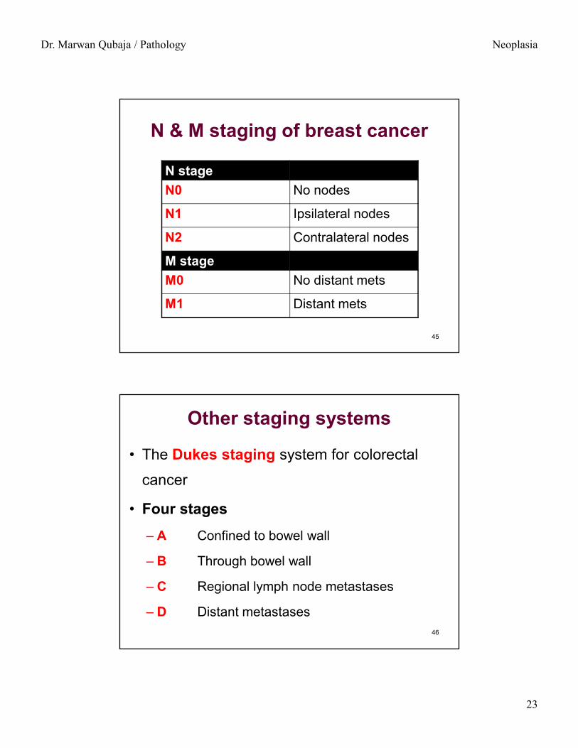
N & M staging of breast cancer
N stageN0 No nodes
N1 Ipsilateral nodes
N2 Contralateral nodes
M stageM0 No distant mets
M1 Distant mets
45
Other staging systems
• The Dukes staging system for colorectal
cancer
• Four stages – A Confined to bowel wall
– B Through bowel wall
– C Regional lymph node metastases
– D Distant metastases46

Dr. Marwan Qubaja / Pathology Neoplasia
24
Epidemiology
•• CauseCause: contributes to understanding the association
of cancer with certain causastive agents (smoking),
or with races (stomach cancer in Japan)
•• GeographyGeography: comparison of colon cancer incidence
between Western world and Africa led to recognition
of the role of diet (colon cancer)
•• PreventionPrevention: underscores the importance of
screening in controlling cancer (cervix, breast, colon) 47
Some causative factors associated with cancer at various sites
Smoking Mouth, pharynx, oesophagus, lip,
larynx, lung, bladder
Alcohol Mouth, pharynx, larynx,
oesophagus, colorectal
Iatrogenic:• Estrogens Endometrium, vagina, breast
• Androgens Prostate
• Radiotherapy carcinoma of breast & Bronchus48

Dr. Marwan Qubaja / Pathology Neoplasia
25
Some causative factors associated with cancer at various sites
High-fat diet Breast cancer
Hepatitis B virus Liver (hepatocellular carcinoma)
Hepatitis C virus Liver (hepatocellular carcinoma)
Epstein–Barr virus Burkitt’s lymphoma
Hodgkin’s lymphoma
Helicobacter pylori Stomach (gastric cancer)
49
Environmental Carcinogens• Drugs: immune suppressing etc..
• Organic chemicals: Insecticides, herbicides, etc..
• Cigarette Smoke
• Ethanol
• Heavy Metals
• Sexually transmitted viruses: Herpes simplex, Human papilloma virus
• Radiation: Ultraviolet light
50

Dr. Marwan Qubaja / Pathology Neoplasia
26
Heredity and Cancer
Inherited Cancer Syndromes:
• Autosomal Dominant
• Autosomal Recessive Syndromes
• Familial Cancers
51
(40% are familial, usually bilaterally)
52

Dr. Marwan Qubaja / Pathology Neoplasia
27
Familial Cancers§ Examples: carcinomas of colon, breast, ovary, and brain.
§ Features that characterize familial cancers include:
• early age at onset
• tumors arising in two or more close relatives
• sometimes multiple or bilateral tumors
§ Familial cancers are not associated with specific marker
phenotypes, e.g. The transmission pattern is not clear.
§ certain familial cancers can be linked to the inheritance of
mutant genes. Examples include linkage of BRCA1 and
BRCA2 genes to familial breast and ovarian cancers. 53
Acquired Preneoplastic Disorders
•• RegenerativeRegenerative (e.g. hepatocellular carcinoma in
cirrhosis)
•• HyperplasticHyperplastic (e.g. endometrial carcinoma in
endometrial hyperplasia)
•• DysplasticDysplastic (e.g. Lung cancer in bronchial dysplasia)
•• AtrophicAtrophic (e.g. gastric carcinoma in atrophic gastritis)
•• UlcerativeUlcerative (e.g. colorectal carcinoma in ulcerative
colitis) 54

Dr. Marwan Qubaja / Pathology Neoplasia
28
The prognosis of a patient with any type of neoplasm depends on:
• rate of growth of the tumor
• tumor size
• tumor site
• cell type and degree of differentiation
• presence of metastasis
• responsiveness to therapy
• general health of the patient
Clinical Features of Neoplasia
55
Clinical Features of Neoplasia
§ Local effect
• Pressure effect
• Functional activity (e.g. hormone synthesis)
• Bleeding
• Infection
§ Cachexia (wasting)
§ Paraneoplastic syndromes 56
Dr. Marwan Qubaja / Pathology Neoplasia
29
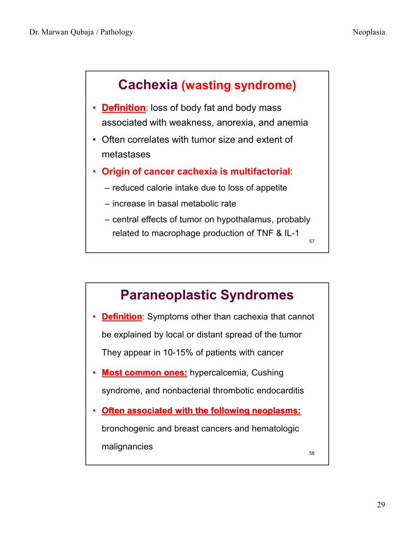
Cachexia (wasting syndrome)
•• DefinitionDefinition: loss of body fat and body mass associated with weakness, anorexia, and anemia
• Often correlates with tumor size and extent of metastases
• Origin of cancer cachexia is multifactorial:– reduced calorie intake due to loss of appetite
– increase in basal metabolic rate
– central effects of tumor on hypothalamus, probably related to macrophage production of TNF & IL-1
57
Paraneoplastic Syndromes•• DefinitionDefinition: Symptoms other than cachexia that cannot
be explained by local or distant spread of the tumor
They appear in 10-15% of patients with cancer
•• Most common ones:Most common ones: hypercalcemia, Cushing
syndrome, and nonbacterial thrombotic endocarditis
•• Often associated with the following neoplasms:Often associated with the following neoplasms:
bronchogenic and breast cancers and hematologic
malignancies58

Dr. Marwan Qubaja / Pathology Neoplasia
30
Paraneoplastic Syndromes
§Endocrinopathies
§Neuromyopathies
§Osteochondral Disorders
§Vascular Phenomena
§Fever
§Nephrotic Syndrome
59
Paraneoplastic syndromes Syndrome Mechanism Example
Cushing's Syndrome ACTH-like substance Lung (oat cell) carcinoma
Hypercalcemia PTH-like substance Lung (squamous cell) carcinoma
Hyponatremia Inappropriate ADH secretion Lung (oat cell) carcinoma
Polycythemia Erythropoietin-like substance Renal cell carcinoma
Trousseau's Syndrome Hypercoagulable state Various carcinomas
Hypoglycemia Insulin-like substance Various carcinomas and sarcomas
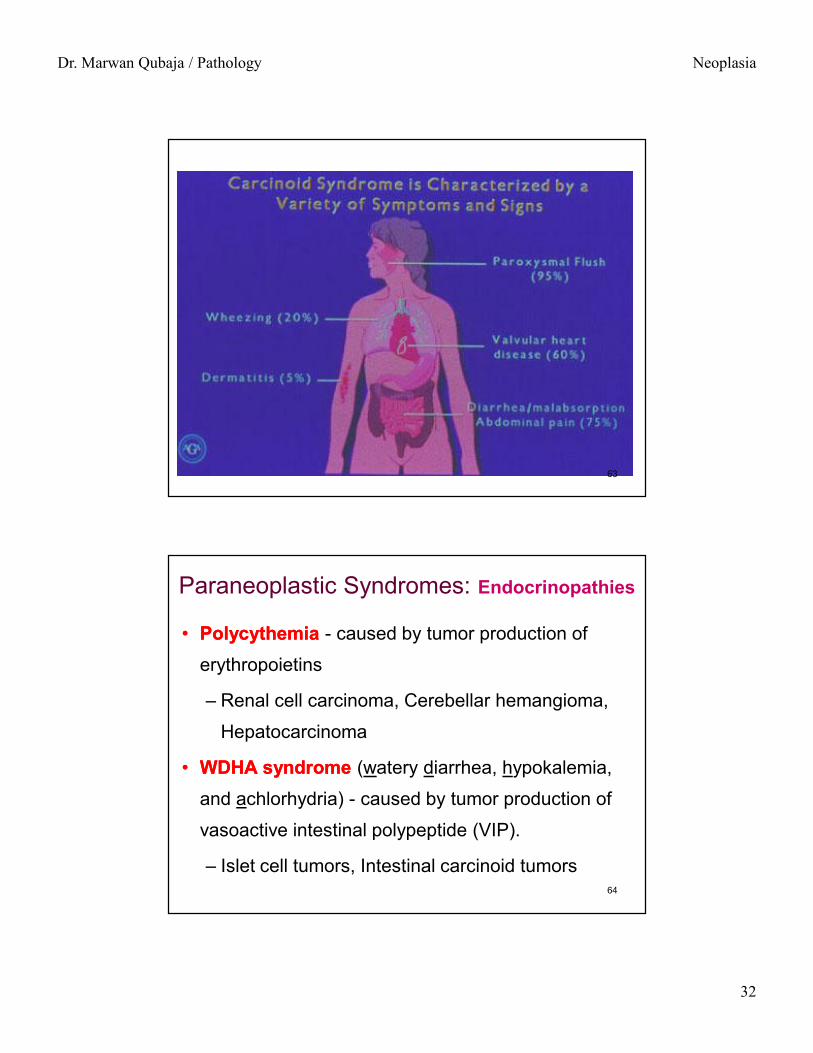
Carcinoid Syndrome Seretonin, bradykinin Metastatic malignant
carcinoid tumors 60

Dr. Marwan Qubaja / Pathology Neoplasia
31
•• HypercalcemiaHypercalcemia (Cancer is the most common cause of hypercalcemia
•• Causes of hypercalcemia in cancer:Causes of hypercalcemia in cancer:
•• HormonalHormonal (e.g. PTHrP synthesis in squamous cell lung carcinomas)
•• Osteolytic metastatic diseaseOsteolytic metastatic disease of bone (e.g. metastatic breast carcinoma)
•• TumorTumor--derived factorsderived factors (e.g. TGF-α, that activates osteoclasts and the active form of vitamin D)
Paraneoplastic Syndromes: Endocrinopathies
61
•• HypoglycemiaHypoglycemia - caused by tumor over-production
of insulin or insulin like activities
– Fibrosarcoma, Cerebellar hemangioma,
Hepatocarcinoma
•• Carcinoid syndromeCarcinoid syndrome - Caused by serotonin and
bradykinin produced by the tumor
– Bronchial carcinoids, Pancreatic carcinoma,
Carcinoid tumors of the bowel
Paraneoplastic Syndromes: Endocrinopathies
62
Dr. Marwan Qubaja / Pathology Neoplasia
32
63
•• PolycythemiaPolycythemia - caused by tumor production of
erythropoietins
– Renal cell carcinoma, Cerebellar hemangioma,
Hepatocarcinoma
•• WDHA syndromeWDHA syndrome (watery diarrhea, hypokalemia,
and achlorhydria) - caused by tumor production of
vasoactive intestinal polypeptide (VIP).
– Islet cell tumors, Intestinal carcinoid tumors
Paraneoplastic Syndromes: Endocrinopathies
64
Dr. Marwan Qubaja / Pathology Neoplasia
33
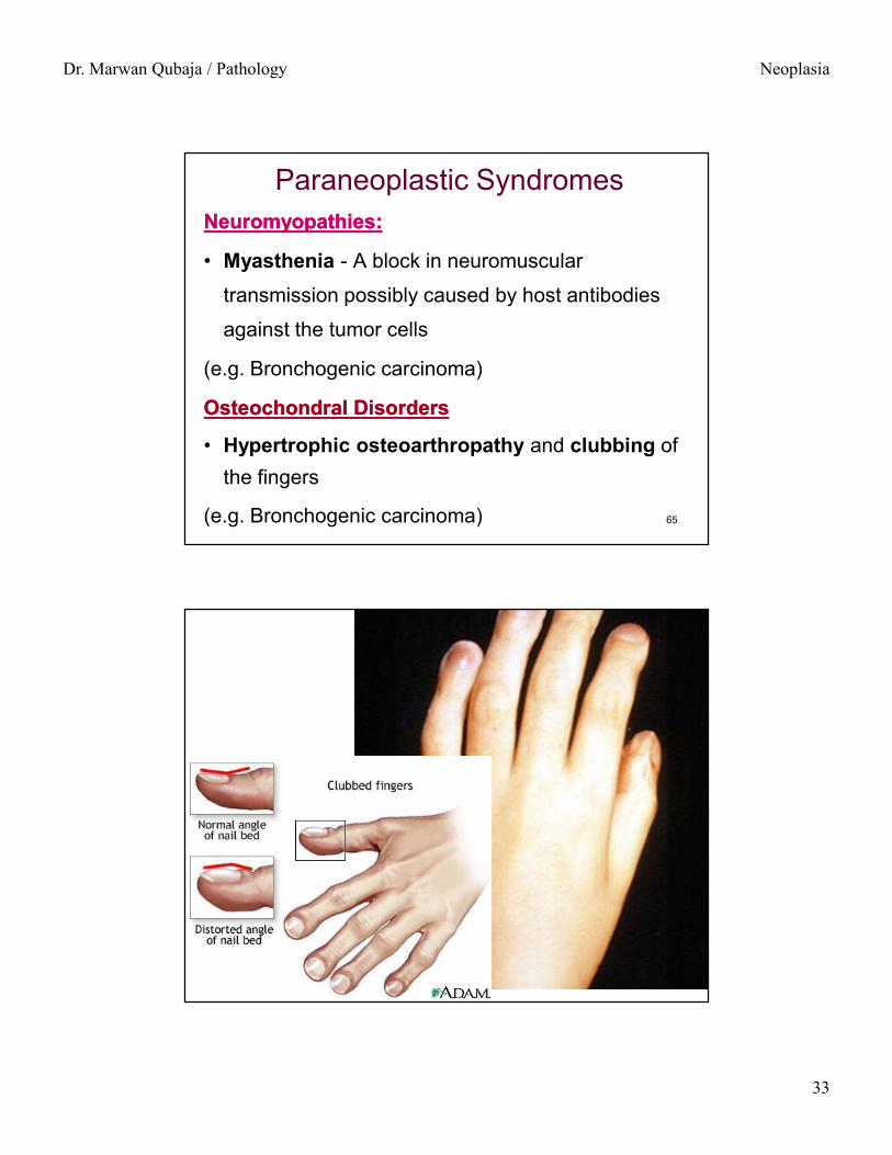
Paraneoplastic SyndromesNeuromyopathies:Neuromyopathies:
• Myasthenia - A block in neuromuscular
transmission possibly caused by host antibodies
against the tumor cells
(e.g. Bronchogenic carcinoma)
Osteochondral DisordersOsteochondral Disorders
• Hypertrophic osteoarthropathy and clubbing of the fingers
(e.g. Bronchogenic carcinoma) 65
66

Dr. Marwan Qubaja / Pathology Neoplasia
34
Paraneoplastic SyndromesVascular & hematoligical changesVascular & hematoligical changes
•• HyperHyper--coagulabilitycoagulability leading to:
–– venous thrombosisvenous thrombosis (Trousseau’s phenomenon)
e.g.Pancreatic and bronchogenic carcinomas
–– nonbacterial thrombotic endocarditisnonbacterial thrombotic endocarditis
e.g. sterile vegetations on valves that occur with
advanced carcinomas.
•• AnemiaAnemia (e.g. Thymic neoplasms)67
Paraneoplastic Syndromes
FeverFever• Associated with bacterial infections
– Common where blockage of drainage occurs
• Not associated with infection– Likely caused by response to necrotic tumor cells and/or
immune response to necrotic tumor proteins.
Nephrotic SyndromeNephrotic Syndrome• probably caused by damage to renal glomeruli by
tumor antigen-antibody complexes.68

Dr. Marwan Qubaja / Pathology Neoplasia
35
What Are The Final Complications Of Malignancy (Causes Of Death)
• metastases
• cachexia
• severe anemia, throbocytopeina
• hypercoagulability
• rupture into major vessels e.g. bleeding
• compression of vital organs
• organ failure e.g. renal failure
• infection e.g. pneumonia 69
Tumor Diagnosis
• History and Clinical examination
• Imaging - X-Ray, US, CT, MRI
• Tumor markers- laboratory analysis
• Cytology –Pap smear, FNAB
• Biopsy - Histopathology.
• Molecular Tech – Gene detection. 70

Dr. Marwan Qubaja / Pathology Neoplasia
36
• Immunohistochemistry
• Flow cytometry
• Electron microscopy
• In Situ Hybridization
Tumor Diagnosis
71
Tumor markers: sometimes diagnostic or prognostic
Some serological markers associated with malignant tumors:
hCG choriocarcinoma
AFP hepatocellular ca
calcitonin thyroid medullary ca
prolactin pituitary adenomas
CA 125 ovarian carcinoma
PSA prostate carcinoma 72

Dr. Marwan Qubaja / Pathology Neoplasia
37
• Four classes of genes are targets of
genetic damage:
1. The growth promoting proto-oncogenes
2. The growth-inhibiting tumor-suppressor genes (antianti--
oncogenesoncogenes)
3. Genes that regulate apoptosis
4. The DNA repair genes
Molecular Basis of Cancer
73
Mechanisms of carcinogenesis
Cancer as a multistep process:
1. Initiation: DNA alteration or cell change
2. Tumor-promotion: from single mutated cell to
formation of tumor
3. Tumor-progression: development of
malignancy
From: Brooks, Chap. 7
74

Dr. Marwan Qubaja / Pathology Neoplasia
38
Step 1: Initiation
• Results from mutation of DNA to activate a proto-
oncogene or inactivate a tumor suppressor gene.
• Initiation alone does not result in tumors.
From: Brooks, Chap. 7
Some initiators can
subsequently act as
promoters (are “complete
carcinogens”).75
Step 2: Promotion•• PromotorsPromotors are usually substances that produce
cell activation and proliferation.
• Effects of promotors are reversible.
•• Promotors cannot induce neoplasia:Promotors cannot induce neoplasia:
i) alone
ii) if applied before initiator
iii) if applied in too small amount
iv) if too much time elapses between applications 76
Dr. Marwan Qubaja / Pathology Neoplasia
39
Step 2: Promotion
From: Brooks, Chap. 7
The promoter does not cause mutation, but it leads to
clonal expansion of the initiated (mutated) cells.
PromotersPromoters: e.g. hormones (estrogen), growth factors
77
Step 3: Progression
• Karyotypic instability:
– Increased growth rates
– Increased invasiveness
– Increased hormonal response
– Anaplasia
78

Dr. Marwan Qubaja / Pathology Neoplasia
40
• Every Cancer analyzed reveals multiple genetic
alterations involving activation of several
oncogenes and loss of two or more cancer
suppressor genes.
• The good example is colon cancer:
– Colon epithelial hyperplasia
– Formation of adenomas
– Malignant transformation
Step 3: Progression
79
Karyotypic Changes in Tumors
• Point mutation: e.g. ras oncogene
• Balanced translocations
• Gene amplification
• Deletions
• Whole chromosomes may be gained or
lost. 80

Dr. Marwan Qubaja / Pathology Neoplasia
41
Molecular Basis of Multistep Mechanism
Special order of the mutations is important
• Genes that regulate entry of cells into the multi-step
carcinogenesis are called gatekeepersgatekeepers– E.g. mutations of Rb, NF-1, VHL, or APC gives rise to
retinoblastoma, schwannomas, renal cell cancer, and
colon cancer
• In contrast to gatekeeper genes, those that affect
genomic stability are called caretakercaretaker genes
– E.g. the DNA repair genes81
CarcinogensGenetic damage may be:Genetic damage may be:
1. Inherited:
2. Acquired: a) Chemical agents
• Direct agents
• Indirect agents
b) Radiations
c) Microbial agents (viruses) 82

Dr. Marwan Qubaja / Pathology Neoplasia
42
A) Chemical Carcinogens•• Direct agents:Direct agents:
They require no metabolic conversion to become carcinogenic
•• Indirect agents:Indirect agents:They become active after metabolic conversion
• These agents react with RNA, cellular proteins, and DNA
• Some chemical carcinogens are augmented by certain agents that are called promoters 83
MAJOR CHEMICAL CARCINOGENS
Direct-Acting Carcinogens
1. Alkylating agents
Anticancer drugs (cyclophosphamide , chlorambucil ,
nitrosoureas, and others)
2. Acylating agents
1-Acetyl-imidazole
Dimethylcarbamyl chloride
84

Dr. Marwan Qubaja / Pathology Neoplasia
43
Polycyclic and heterocyclic aromatic hydrocarbons
Benz[a]anthracene
Benzo[a]pyrene
Aromatic amines, amides, azo dyes
2-Naphthylamine (β-naphthylamine)
2-Acetylaminofluorene
Dimethylaminoazobenzene (butter yellow)
Natural plant and microbial products
Aflatoxin B1
Griseofulvin
Betel nuts
Others
Nitrosamine and amides
Asbestos, nickel, chromium
Insecticides, fungicides, Vinyl chloride, Arsenic
Polychlorinated biphenyls (PCBs)
Indirect Acting Agents
bladder cancer in workers exposed to bladder cancer in workers exposed to aniline dye & rubber industryaniline dye & rubber industry
hepatocellular carcinomahepatocellular carcinoma
lung cancerlung cancer
85
B) Radiation Carcinogenesis1.1. UV radiation of sunlight:UV radiation of sunlight:
– can cause skin cancers (melanomas, squamouscell carcinomas, and basal cell carcinomas).
– At greatest risk are fair-skinned people
– Due to inability to repair environmentally induced DNA damage.
– E.g. Xeroderma pigmentosum
2.2. Ionizing radiation: Ionizing radiation: – (e.g. X-rays, nuclear bombs) 86

Dr. Marwan Qubaja / Pathology Neoplasia
44
C) Viral & Microbial Carcinogenesis1.1. RNA viruses (retroviruses):RNA viruses (retroviruses):
– Human T-cell leukemia virus-1 (HTLV-1):
2.2. DNA viruses:DNA viruses:– Human Papillomavirus (HPV)
– Epstein-Barr virus (EBV)
– Human Herpesvirus 8 (HHV-8)
– Hepatitis B virus (HBV)
3.3. Helicobacter Pylori:Helicobacter Pylori:– Associated with gastric carcinoma & gastric lymphoma 87
Human Human PapillomaPapilloma Virus (HPV):Virus (HPV):
•• HPV (types HPV (types 11, , 22, , 44, , 77):): cause benign squamous papillomas
(warts in human)
•• HPV (types HPV (types 1616, , 1818, , 3131):): involved in the genesis of
oropharyngeal, cervical, anal, perianal, and penile cancers
EpsteinEpstein--Barr Virus:Barr Virus:
• Involved in the pathogenesis of several human tumors:
– Burkitt lymphoma
– AIDS-related lymphomas
– Nasopharyngeal carcinoma88

Dr. Marwan Qubaja / Pathology Neoplasia
45
Hepatitis B Virus:Hepatitis B Virus:
• associated with hepatocellular carcinomahepatocellular carcinoma
• HCV is also linked to hepatocellular carcinoma
Helicobacter PyloriHelicobacter Pylori
• gram negative spiral bacteria
• associated with 90% of duodenal ulcers, and 70-90%
of gastric ulcers
• the likely cause for gastric carcinoma & lymphoma
(MALToma: marginal zone associated lymphoma)89
Four classes of genes are targets of genetic damage
1. The growth promoting proto-oncogenes
2. The growth-inhibiting tumor-suppressor genes
(anti-oncogenes)
3. Genes that regulate apoptosis
4. The DNA repair genes
90

Dr. Marwan Qubaja / Pathology Neoplasia
46
Six fundamental changes in cell physiology that together dictate malignant phenotype
1. Self-sufficiency in growth signals (oncogenes)
2. Insensitivity to growth-inhibitory signals (Cancer Cancer Suppressor Genes)Suppressor Genes)
3. Evasion of apoptosis
4. Limitless replicative potential
5. Sustained angiogenesis
6. Ability to invade and metastasize
91
Oncogenes:Oncogenes: Genes that promote
autonomous (uncontrolled) cell
growth in cancer cells.
Protooncogenes:Protooncogenes: Genes that
promote normal growth and
differentiation
OncogenesNormal cellNormal cell Tumor cellTumor cell
92

Dr. Marwan Qubaja / Pathology Neoplasia
47
Oncogenes• They are derived by mutations in proto-oncogenes
• characterized by the ability to promote cell growth in the
absence of normal growth-promoting signals.
• Their products, called oncoproteinsoncoproteins, resemble the normal
products of proto-oncogenes except that oncoproteins are
devoid of important regulatory elements
• Encode oncoproteins as growth factors, receptors, signal
transducers, transcription factors, and cell-cycle components
•• HistoryHistory: Varmus & Bishop first recognized their presence
within the genome of transforming retroviruses (vv--onconc) 93
Growth Factors:Growth Factors:
• All normal cells require stimulation by growth
factors to undergo proliferation.
• Many cancer cells acquire growth self-sufficiency,
however, by acquiring the ability to synthesize the
same growth factors to which they are responsive.
Self-Sufficiency in Growth Signals
94

Dr. Marwan Qubaja / Pathology Neoplasia
48
Growth Factor Receptors:Growth Factor Receptors:• Mutations and pathologic overexpression of normal forms of
growth factor receptors have been detected in several tumors.
• e.g. the epidermal growth factor (EGF) receptor is overexpressed in 80% of squamous cell carcinomas of the lung.
Self-Sufficiency in Growth Signals
95
Cancer Suppressor Genes• Both normal alleles of the Rb gene must be
inactivated (two hits) for the development of
retinoblastoma
Knudson (two hit theory):
• In hereditary retinoblastoma, children are born with
one normal and one defective copy of the Rb gene
(first hitfirst hit).
• They lose the normal copy by some somatic mutation
(second hitsecond hit). 96

Dr. Marwan Qubaja / Pathology Neoplasia
49
Tumor Suppressor Genes: p53
• located on chromosome 17
• It’s the most common Tumor Suppressor Gene target
for genetic alterations in human tumors
• More than 50% of human tumors contain mutations in
this gene
• Familial loss causes LiLi--FraumeniFraumeni syndromesyndrome (multiple
tumors) includes sarcomas, breast cancer, leukemia,
brain tumors, and carcinomas of the adrenal cortex97
Tumor Angiogenesis•• Angiogenesis is required for:Angiogenesis is required for:
1. continued tumor growth
2. metastasis
• Tumors can not enlarge beyond 1-2 mm in diameter or
thickness unless they are vascularized
• The 1-2 mm zone represents the maximal distance across
which oxygen and nutrients can diffuse from blood vessels.
Beyond this distance the tumor fails to enlarge without
vascularization. Hypoxia will induce apoptosis by activation
of p53 98

Dr. Marwan Qubaja / Pathology Neoplasia
50
How Do Growing Tumor Develop a Blood Supply
• Tumor growth is controlled by the balance between
angiogenic factors, & antiangiogenesis molecules.
•• Examples of angiogenic factors:Examples of angiogenic factors:
– Vascular endothelial growth factor (VEGF)
– Basic fibroblast growth factor (bFGF)
•• Examples of antiangiogenesis are:Examples of antiangiogenesis are:
– thrombospondin-1, angiostatin, endostatin, &
vasculostatin. 99
Angiogenesis• Hypoxia within the growing tumor favors
angiogenesis by release of hypoxia-inducibale factor–1 (HIF-1).
• HIF-1 controls transcription of VEGF
• Transcription of VEGF is under the control of Ras oncogene
• Ras oncogene activation upregulates the production of VEGF
• Proteases are involved in regulating the balance between angiogenic and antiangiogenic factors 100
Dr. Marwan Qubaja / Pathology Neoplasia
51
Angiogenesis
• P53 inhibits angiogenesis by inducing the synthesis of
the antiangiogenic molecule thrombospondin-1
• With mutational inactivation of both p53 alleles, the level
of thrombospondin-1 drop markedly tilting the balance in
favor of angiogenic factors
• Some success in the treatment of cancer has been
achieved through the use of angiogenesis inhibitors
such as endostatin101
Mechanism of Invasion & Metastasis
• Metastatic process can be divided into two
phases:
1. Invasion of the extracellular matrix
2. Vascular dissemination and homing of tumor cells
• E-cadherin acts as intercellular glue that bind cells
together.
• E-cadherin function is lost in almost all epithelial cancer102

Dr. Marwan Qubaja / Pathology Neoplasia
52
Sequential steps involved in the hematogenous spread of a tumor
•• Invasion of the cellular matrix in Invasion of the cellular matrix in
4 4 steps:steps:
1. Detachment of tumor cells from
each other
2. Attachment of tumor cells to
matrix components
3. Degradation of ECM
4. Migration of tumor cells 103
• Attachment of tumor cells to ECM proteins such as lamininand fibronectin
• Local degradation of the basement membrane and interstitial connective tissue
– Tumor cells either secrete proteolytic enzymes themselves or induce the host cells fibroblasts to elaborate proteases
– Several of metalloproteinases including gelatinases, collagenases, and stromelysins, cathepsin-D are involved
– Benign tumors show little type IV type IV collagenasecollagenase activity,whereas their malignant counterpart overexpresses this enzyme
Invasion of the extracellular matrixInvasion of the extracellular matrix
104

Dr. Marwan Qubaja / Pathology Neoplasia
53
Summary•• NeoplasiaNeoplasia-- an abnormal mass of tissue
which has lost its responsiveness to growth controls
Benign neoplasms:Benign neoplasms:• slow-growing
• well-differentiated tumors
• lack the ability to metastasize
• remain localized
• amenable to surgery 105
Summary
Malignant neoplasmsMalignant neoplasms
• fast-growing lesions
• invade normal structures
• vary in the degree of differentiation
• some show anaplasia
• capable of metastasis106