chondromyxoid fibroma of the frontal bone - ajnr · chondromyxoid fibroma of the frontal bone...
TRANSCRIPT
Chondromyxoid Fibroma of the Frontal Bone
Tatsuo Morimura, 1·2 Atsuhisa Nakano, 1 Tsuyoshi Matsumoto, 1 and Eiichi Tani
1
Summary: Chondromyxoid fibroma of frontal bone is a rare lesion. Plain skull films showed a round radiolucent mass with a sclerotic margin. It was dense on plain CT scan and showed
no convincing contrast enhancement. MR imaging showed low signal intensity relative to gray matter on Tl-weighted image
A
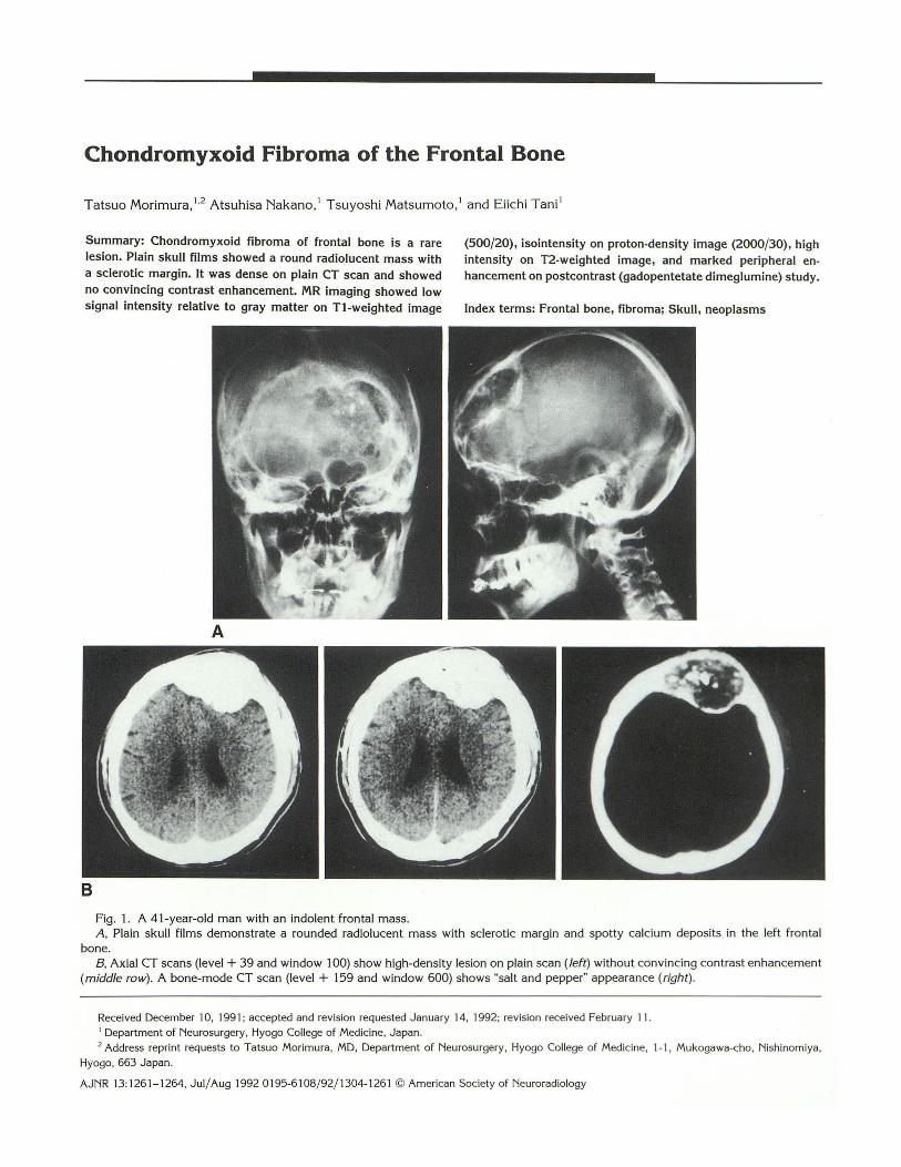
Fig. 1. A 41-year-old man with an indolent frontal mass.
(500/20), isointensity on proton-density image (2000/30), high intensity on T2-weighted image, and marked peripheral enhancement on postcontrast (gadopentetate dimeglumine) study.
Index terms: Frontal bone, fibroma; Skull, neoplasms
A, Plain skull films demonstrate a rounded radiolucent mass with sclerotic margin and spotty calcium deposits in the left frontal bone.
8, Axial CT scans (level + 39 and window 1 00) show high-density lesion on plain scan (left) without convincing contrast enhancement (middle row) . A bone-mode CT scan (level + 159 and window 600) shows "salt and pepper" appearance (right) .
Received December 10, 1991 ; accepted and revision requested January 14, 1992; revision received February 11. 1 Department of Neurosurgery, Hyogo College of Medicine, Japan. 2 Address reprint requests to Tatsuo Morimura, MD, Department of Neurosurgery, Hyogo College of Medicine, 1-1, Mukogawa-cho, Nishinomiya,
Hyogo, 663 Japan.
AJNR 13:1261-1264, Jui/Aug 1992 0195-6108/92/ 1304-1261 © American Society of Neuroradiology
1262
Chondromyxoid fibromas (CMF) are benign bone tumors composed of chondroid, myxoid, and fibrous elements. The majority of these tumors arise from the long bones and their overall incidence comprises less than 1 % of all bone tumors (1). Those arising from the skull are rare. We have identified 13 cases reported since Jaffe and Lichtenstein established it as a histopathologic entity in 1948 (1-13). We describe a case of CMF of the frontal bone in a 41-year-old man and present the neuroradiologic findings.
Case Report
This 41-year-old man was admitted with a complaint of right-hand hypesthesia for 2 months. His past medical and
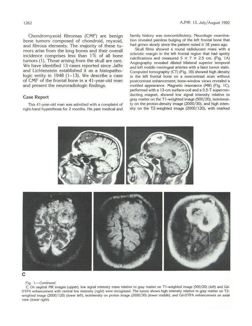
c Fig. 1-Continued.
AJNR : 13, July/ August 1992
family history was noncontributory. Neurologic examination revealed painless bulging of the left frontal bone that had grown slowly since the patient noted it 18 years ago.
Skull films showed a round radiolucent mass with a sclerotic margin in the left frontal region that had spotty calcifications and measured 5 X 7 X 2.5 em. (Fig. 1A) Angiography revealed dilated bilateral superior temporal and left middle meningeal arteries with a faint tumor stain. Computed tomography (CT) (Fig. 1 B) showed high density in the left frontal bone on a noncontrast scan without postcontrast enhancement; bone-window views revealed a mottled appearance. Magnetic resonance (MR) (Fig. 1 C), performed with a 13-cm surface-coil and a 0.5-T superconducting magnet, showed low signal intensity relative to gray matter on the T1-weighted image (500/20), isointensity on the proton-density image (2000/30), and high intensity on the T2-weighted image (2000/120), with marked
C, On sagittal MR images (upper), low signal intensity mass relative to gray matter on Tl-weighted image (500/ 20) (left) and GdDTPA enhancement with central low intensity (right) were recognized. The tumor shows high intensity relative to gray matter on T2-weighted image (2000/ 120) (lower left), isointensity on proton image (2000/ 30) (lower middle), and Gd-DTPA enhancement on axial view (lower right) .
AJNR: 13, July/ August 1992
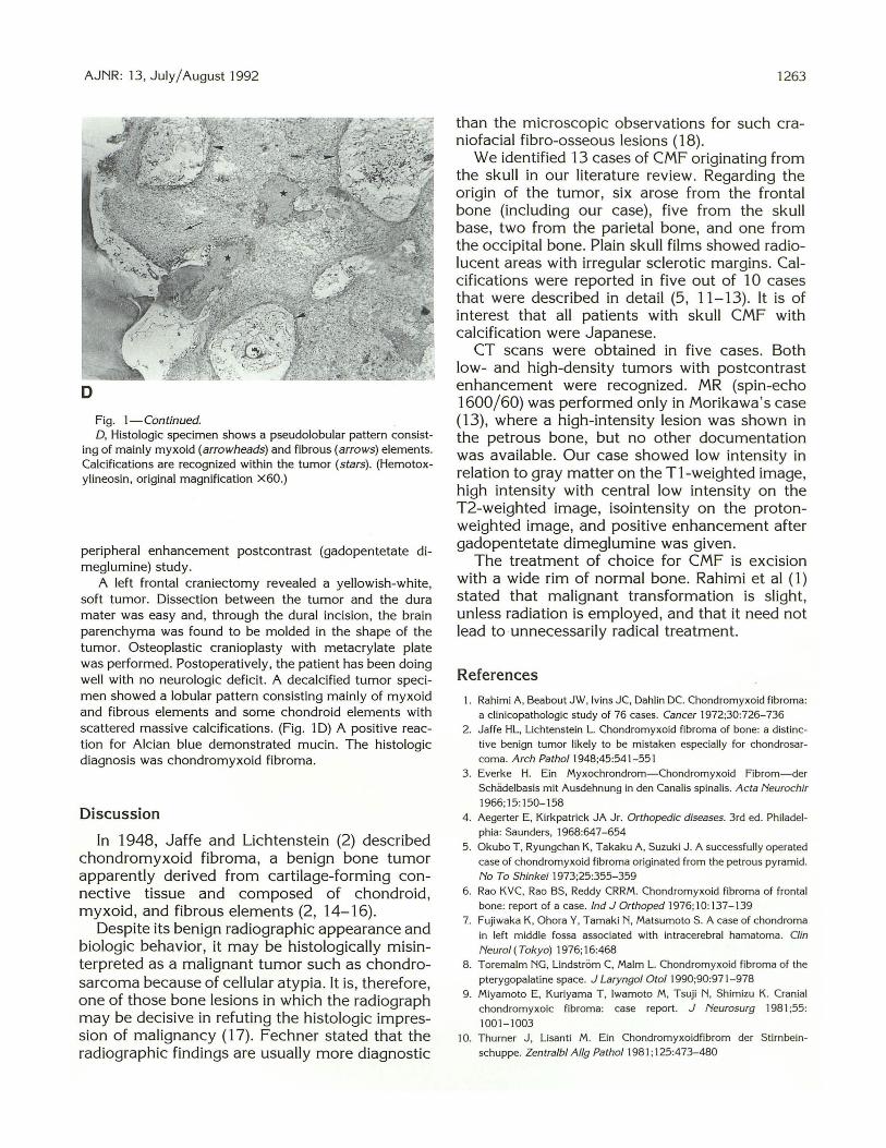
D
Fig. 1- Continued. D, Histologic specimen shows a pseudolobular pattern consist
ing of mainly myxoid (arrowheads) and fibrous (arrows) elements. Calcifications are recognized within the tumor (stars). (Hemotoxylineosin, original magnification X60.)
peripheral enhancement postcontrast (gadopentetate dimeglumine) study.
A left frontal craniectomy revealed a yellowish-white, soft tumor. Dissection between the tumor and the dura mater was easy and, through the dural incision, the brain parenchyma was found to be molded in the shape of the tumor. Osteoplastic cranioplasty with metacrylate plate was performed. Postoperatively, the patient has been doing well with no neurologic deficit. A decalcified tumor specimen showed a lobular pattern consisting mainly of myxoid and fibrous elements and some chondroid elements with scattered massive calcifications. (Fig. 1 D) A positive reaction for Alcian blue demonstrated mucin. The histologic diagnosis was chondromyxoid fibroma.
Discussion
In 1948, Jaffe and Lichtenstein (2) described chondromyxoid fibroma, a benign bone tumor apparently derived from cartilage-forming connective tissue and composed of chondroid, myxoid, and fibrous elements (2, 14-16).
Despite its benign radiographic appearance and biologic behavior, it may be histologically misinterpreted as a malignant tumor such as chondrosarcoma because of cellular atypia. It is, therefore, one of those bone lesions in which the radiograph may be decisive in refuting the histologic impression of malignancy (17). Fechner stated that the radiographic findings are usually more diagnostic
1263
than the microscopic observations for such craniofacial fibro-osseous lesions (18).
We identified 13 cases of CMF originating from the skull in our literature review. Regarding the origin of the tumor, six arose from the frontal bone (including our case), five from the skull base, two from the parietal bone, and one from the occipital bone. Plain skull films showed radiolucent areas with irregular sclerotic margins. Calcifications were reported in five out of 1 0 cases that were described in detail (5, 11-13). It is of interest that all patients with skull CMF with calcification were Japanese.
CT scans were obtained in five cases. Both low- and high-density tumors with postcontrast enhancement were recognized. MR (spin-echo 1600/60) was performed only in Morikawa's case (13), where a high-intensity lesion was shown in the petrous bone, but no other documentation was available. Our case showed low intensity in relation to gray matter on the T1-weighted image, high intensity with central low intensity on the T2-weighted image, isointensity on the protonweighted image, and positive enhancement after gadopentetate dimeglumine was given.
The treatment of choice for CMF is excision with a wide rim of normal bone. Rahimi et al (1) stated that malignant transformation is slight, unless radiation is employed, and that it need not lead to unnecessarily radical treatment.
References
1. Rahimi A, Beabout JW, Ivins JC, Dahlin DC. Chondromyxoid fibroma: a clin icopathologic study of 76 cases. Cancer 1972;30:726-736
2. Jaffe HL, Lichtenstein L. Chondromyxoid fibroma of bone: a distinctive benign tumor likely to be mistaken especially for chondrosar
coma. Arch Patho/1948;45:541-551 3. Everke H. Ein Myxochrondrom-Chondromyxoid Fibrom-der
Schiidelbasis mit Ausdehnung in den Canalis spinalis. Acta Neurochir
1966;15:150-158 4. Aegerter E, Kirkpatrick JA Jr. Orthopedic diseases. 3rd ed. Philadel
phia: Saunders, 1968:647-654
5. Okubo T, Ryungchan K, Takaku A, Suzuki J. A successfully operated case of chondromyxoid fibroma originated from the petrous pyramid. No To Shinkei 1973;25:355-359
6. Rao KVC, Rao BS, Reddy CRRM. Chondromyxoid fibroma of frontal bone: report of a case. lnd J Orthoped 1976; 10:137-139
7. Fujiwaka K, Ohora Y, Tamaki N, Matsumoto S. A case of chondroma in left middle fossa associated with intracerebral hamatoma. Clin
Neural (Tokyo) 1976; 16:468 8. Toremalm NG, Lindstrom C, Maim L. Chondromyxoid fibroma of the
pterygopalatine space. J Laryngol Otol 1990;90:971-978 9. Miyamoto E, Kuriyama T, Iwamoto M, Tsuji N, Shimizu K. Cranial
chondromyxoic fibroma: case report. J Neurosurg 1981 ;55:
1001-1003 10. Thurner J, Lisanti M. Ein Chondromyxoidfibrom der Stirnbein
schuppe. Zentralbl Allg Patho/1981 ;125:473-480
1264
11 . Kanemaru R, Gonda M , Hirahara K, Hamada H, Mihara T, Asakura
T. A giant chondromyxoid fibroma originated from the right orbital roof: a -case report. Neurosurgery ( Tokyo) 1982; 10:731-736
12. Watanabe Y, Goto T , Sasaki T , Yamao N, Tanji H, Kodama N. A case of chondromyxoid fibroma of the fron tal bone. Neurosurgery (Tokyo) 1985; 13:167-172
13. Morikawa E, Sasaki T , Basugi N, Hashimoto K, Iwata J. Chondro
myxoid fibroma of the skull base ex tending from middle fossa to
posterior fossa : case report. Neurosurgery (Tokyo) 1987; 15: 1233-1238
14. Dahlin DC. Chondromyxoid fibroma of bone, with emphasis on its
AJNR: 13, July/ August 1992
morphological relationship to benign chondroblastoma. Cancer 1956; 9:159-203
15. Iwata S, Coley BL. Report of six cases of chondromyxoid f ibroma of bone. Surg Gynecol Obstet 1958;101 :571-576
16. Schajowics F, Gallardo H. Chondromyxoid fibroma (fibromyxoid chondroma) of bone: a clinico-pathological study of thirty-two cases. J Bone Joint Surg 1971 ;538: 198-216
17. Feldman F, Hecht HL, Johnston AD. Chondromyxoid fibroma of
bone. Radiology 1970;94:249-260 18. Fechner RE. Problematic lesions of the craniofacial bones. Am J Surg
Patho/1989;13:17-30