clinical application of silent period for the …cdn.intechweb.org/pdfs/25859.pdf · evaluation of...
TRANSCRIPT

10
Clinical Application of Silent Period for the Evaluation of Neuro-Muscular Function in the
Field of the Sports Medicine and Rehabilitation
Shinichi Daikuya1, Atsuko Ono1, Toshiaki Suzuki2, Tetsuji Fujiwara3 and Kyonosuke Yabe4
1Kishiwada Eishinkai Hospital 2Kansai University of Health Sciences
3Kyoto University 4Nagoya University
Japan
1. Introduction
A transient suppression of muscle activation was produced by electric stimulation to the innervating nerve during continued effort. This period of electrical inactivity, designated the mixed nerve silent period, results from several physiologic mechanisms (Kimura, 2001). On the other hand, the silent period during tonic muscle contraction demonstrated on electromyography is due to the rapid voluntary movement during tonic and mild muscle contraction (Ikai, 1955) and is elicited by cutaneous electrical stimulation of supplying nerve during muscle contraction (Higgins & Lieberman, 1968). In many previous studies about silent period, it was generally classified into three categories: (1) the quiet period of bursting wave activity on electromyography, recorded before the rapid motion in response to visual, auditory, light and/or sound stimulation (Yabe, 1976); (2) a transient suppression of muscle activation following electric stimulation of the mixed nerve innervating that muscle during continued effort (Kimura, 2001); (3) a pause of the muscle activity following the motor potential elicited by cortical magnetic stimulation during voluntary target muscle contraction (Calancie et al, 1987). In this study, we used the second category, according to which the silent period consists of several waves, including M wave, F wave and Long Latency Reflex (LLR). The silent period in this article, which classified in the second category, is the duration of the inhibitory period of muscle contraction detected on surface electromyography, which is due to electrical stimulation at the innervating nerve during tonic muscle contraction (Figure 1). We have considered that the silent period of lower extremity is the total circuit time from the peripheral nerve stimulus point to the central nervous system (i.e., brainstem or motor cortex), because the M wave, F wave and LLR are included in the silent period on evoked electromyography. M wave is affected by the conductive condition of peripheral nerve and the muscle state (i.e. rest or contraction, length, volume and so on) (Fuglevand et al, 1993, Cupido et al, 1992, Behm & St-Pierre, 1997). F wave is influenced by the excitability of the spinal motor neuron function (Suzuki et al, 1993). LLR in the lower extremities is affected by
www.intechopen.com

EMG Methods for Evaluating Muscle and Nerve Function 188
the excitability of the spinal cord, brainstem or motor cortex (Roby-Brami & Bussel, 1987, Upton et al, 1971, Kuroiwa, 1986). Therefore, it has been thought that a variation of the silent period may be able to become an index of the degree of facilitation of the brainstem or motor cortex. So, the origin of silent period that we used in this study is thought to be influenced by muscle spindle function, afferent inhibited impulse from Golgi tendon organ or Ia fiber, recurrent inhibition in the spinal cord, inhibitive mechanism of cerebral cortex (Higgins & Lieberman, 1968, Upton et al, 1971, Anastasijevic & Vuco, 1980). And we have tried to apply the silent period to clarify the magnitude of facilitation of the brainstem or motor cortex in the field of physical therapy. In previous study, we had studied the neuromuscular function of healthy subjects in various conditions (i.e. control of degree of muscle contraction and alteration of postural condition) with a silent period using an electro-physiological method (Daikuya et al, 2003.). Though LLR may be included in silent period, LLR was usually distinguished under the supra-maximal stimulation, which was used to record the silent period. In addition, if the fluctuation of silent period was large, it was thought that it shown the possibility of having arrived in the level where electric stimulus differs every stimulus.
Fig. 1. Schematic Silent Period.
While, the state of improvement process after reconstruction of anterior cruciate ligament of the knee (ACL), following parameters were usually used as the index of recovery of physical function; muscle strength, range of motion (ROM), quality of sports performance, and so on. However, it is thought that results of their index and ability or disability of sports competition have little correlation, because we have experienced some cases with instability and/or a strange sensation of the knee during sports activity, although their muscle strength and ROM were good enough. Whereas, we have also experienced some cases without any problems, although their muscle strength and ROM were not enough. Therefore, we considered that as the index of recovery degree after reconstruction of ACL, only their parameters were not sufficient. And, we emphasized the necessity of evaluation of neuromuscular function after reconstruction of ACL. So, we had considered the neuromuscular function of healthy subjects in various conditions (i.e. control of degree of muscle contraction and alteration of postural condition.) with a mixed nerve silent period using an electro-physiological method (Daikuya et al, 2003.) and have attempted to apply the alteration of silent period aspects for the evaluation of the neuromuscular function of a lower extremity after reconstruction of ACL.
www.intechopen.com
Clinical Application of Silent Period for the Evaluation of Neuro-Muscular Function in the Field of the Sports Medicine and Rehabilitation 189
2. Silent period in healthy subjects; its aspects in relation to strength of tonic muscle contraction and postural alteration
Silent period aspects in healthy subjects were displayed as preliminary findings, which demonstrated the tendency of the change of the duration of silent period with an alteration of the strength of tonic muscle contraction, and the relationship between the duration of silent period and postural change in upper and lower extremity for application in the field of sports medicine.
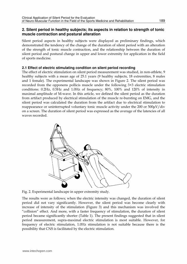
2.1 Effect of electric stimulating condition on silent period recording The effect of electric stimulation on silent period measurement was studied, in non-athlete, 9 healthy subjects with a mean age of 25.1 years (9 healthy subjects, 18 extremities, 8 males and 1 female). The experimental landscape was shown in Figure 2. The silent period was recorded from the opponens pollicis muscle under the following 3×3 electric stimulation conditions: 0.2Hz, 0.5Hz and 1.0Hz of frequency; 80%, 100% and 120% of intensity in maximal amplitude of M-wave. In this article, we defined the silent period as the duration from artifact produced by electrical stimulation of the muscle re-bursting on EMG, and the silent period was calculated the duration from the artifact due to electrical stimulation to reappearance or uninterrupted voluntary tonic muscle activity under the 200 or 500μV/div on a screen. The duration of silent period was expressed as the average of the latencies of all waves recorded.
Fig. 2. Experimental landscape in upper extremity study.
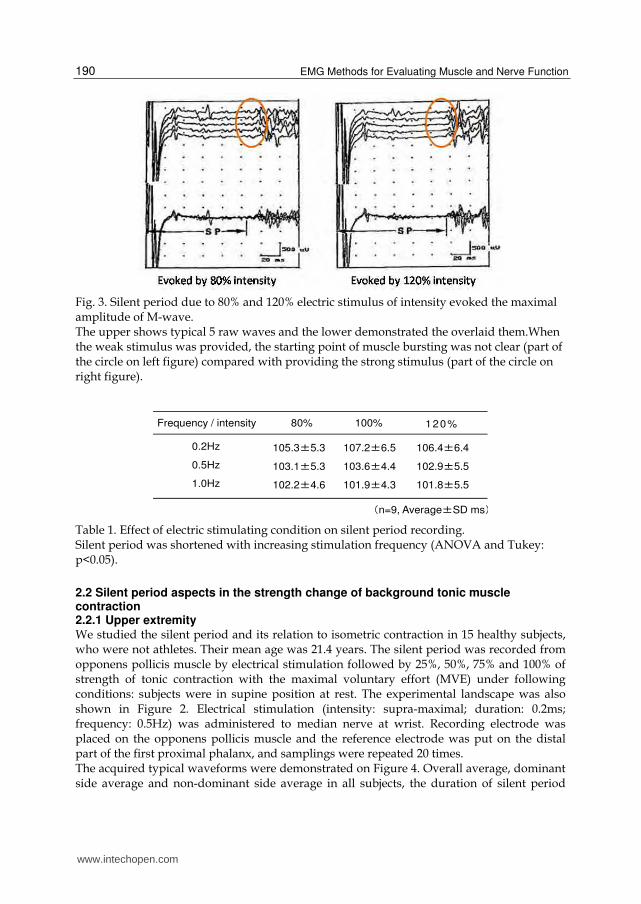
The results were as follows; when the electric intensity was changed, the duration of silent period did not vary significantly. However, the silent period was become clearly with increase of intensity of the stimulation (Figure 3) and this mechanism was involved the “collision” effect. And more, with a faster frequency of stimulation, the duration of silent period became significantly shorter (Table 1). The present findings suggested that in silent period measurement, supra-maximal electric stimulation is most suitable. However, for frequency of electric stimulation, 1.0Hz stimulation is not suitable because there is the possibility that CNS is facilitated by the electric stimulation.
www.intechopen.com
EMG Methods for Evaluating Muscle and Nerve Function 190
Fig. 3. Silent period due to 80% and 120% electric stimulus of intensity evoked the maximal amplitude of M-wave. The upper shows typical 5 raw waves and the lower demonstrated the overlaid them.When the weak stimulus was provided, the starting point of muscle bursting was not clear (part of the circle on left figure) compared with providing the strong stimulus (part of the circle on right figure).
Table 1. Effect of electric stimulating condition on silent period recording. Silent period was shortened with increasing stimulation frequency (ANOVA and Tukey: p<0.05).
2.2 Silent period aspects in the strength change of background tonic muscle contraction 2.2.1 Upper extremity We studied the silent period and its relation to isometric contraction in 15 healthy subjects, who were not athletes. Their mean age was 21.4 years. The silent period was recorded from opponens pollicis muscle by electrical stimulation followed by 25%, 50%, 75% and 100% of strength of tonic contraction with the maximal voluntary effort (MVE) under following conditions: subjects were in supine position at rest. The experimental landscape was also shown in Figure 2. Electrical stimulation (intensity: supra-maximal; duration: 0.2ms; frequency: 0.5Hz) was administered to median nerve at wrist. Recording electrode was placed on the opponens pollicis muscle and the reference electrode was put on the distal part of the first proximal phalanx, and samplings were repeated 20 times. The acquired typical waveforms were demonstrated on Figure 4. Overall average, dominant side average and non-dominant side average in all subjects, the duration of silent period
105.3±5.3
103.1±5.3
102.2±4.6
Frequency / intensity 80% 100% 120%
膅n=9, Average±SD ms䐢
0.2Hz
0.5Hz
1.0Hz
107.2±6.5
103.6±4.4
101.9±4.3
106.4±6.4
102.9±5.5
101.8±5.5
www.intechopen.com

Clinical Application of Silent Period for the Evaluation of Neuro-Muscular Function in the Field of the Sports Medicine and Rehabilitation 191
showed significant differences in all combinations. In particular, the duration of silent period at 75%MVE was shortest (paired t-test; p<0.05, Table 2). This suggests that CNS may be more facilitated during 75%MVE of isometric contraction compared to other levels, and that the 75% level may be most suitable for preparing some types of movements.
Fig. 4. Typical waveforms of silent period in upper extremity study. The left figure shows typical 20 raw waves and the right figure demonstrates the overlaid them. Silent period on this figure was recorded at 25%MVE (Maximal Voluntary Effort).
Table 2. The duration of silent period in the strength change of background tonic muscle contraction in upper extremity.
2.2.2 Lower extremity The study was performed on the soleus muscles of 10 healthy non-athletes (20 lower extremities of healthy 8 males and 2 females, their mean age 25.2±2.7 years). The silent period was recorded from soleus muscle with same conditions of muscle contraction and sampling numbers as upper extremity. Subjects were in supine position with the hip and knee joints of the recorded side flexed at 45 degrees, and with the ankle joint kept 0 degree (Figure 5). Electrical stimulation was administered to tibial nerve at popliteal fossa with same condition as upper extremity. Recording electrode was placed on the soleus muscle and the reference electrode was put on the ipsilateral Achilles tendon. The acquired typical waveforms were demonstrated on Figure 6. Similar to the results of upper extremity, the results indicated that the duration of silent period was the shortest during 75% of maximal effort (paired t-test; p<0.05, Table 3). It was considered that the
108.8±6.4
110.1±5.9
107.5±6.6
Contraction level 25%MVE 50%MVE 75%MVE
Average±SD msMVE: Maximal Voluntary Effort
Bilateral
Dominant
Non-dominant
106.9±7.2
106.7±6.2
107.1±8.1
105.8±6.6
105.0±6.5
106.7±6.6
100%MVE
107.6±8.0
107.4±7.6
107.8±8.4
www.intechopen.com

EMG Methods for Evaluating Muscle and Nerve Function 192
supra-spinal inhibition was remarkably released during sub-maximal effort level; in other words, CNS was most facilitated during sub-maximal effort.
Fig. 5. Experimental landscape in lower extremity study.
Fig. 6. Typical waveforms of silent period in lower extremity study. The left figure shows typical 20 raw waves and the right figure demonstrates the overlaid them. Silent period on this figure was recorded at 25%MVE (Maximal Voluntary Effort).
Table 3. The duration of silent period in the strength change of background tonic muscle contraction in lower extremity.
153.0±15.4
150.3±14.5
155.7±16.5
Contraction level 25%MVE 50%MVE 75%MVE
Average±SD msMVE: Maximal Voluntary Effort
Bilateral
Dominant
Non-dominant
142.3±14.2
141.2±15.6
143.4±13.5
127.6±14.5
127.6±17.5
127.6±11.7
100%MVE
144.3±13.7
144.1±13.6
144.5±14.6
Silent Period
www.intechopen.com

Clinical Application of Silent Period for the Evaluation of Neuro-Muscular Function in the Field of the Sports Medicine and Rehabilitation 193
2.3 Silent period aspects in relation to postural change 2.3.1 Creeping posture We measured the silent period in 10 healthy right-handed subjects (9 males and 1female), who were not athletes. Their mean age was 24.9±2.6 (range; 21-31) years and mean height was 169.7±7.8 (range; 152-182) cm. The silent period was recorded in the abductor pollicis brevis muscles (APB) of the dominant side. The subjects were asked to adapt 3 positions as follows in a shielded room. The 3 positions were all fours with bilateral elbows not bent (position 1), all fours with bilateral elbows bent at 45 degrees (position 2) and all fours with using the only dominant upper extremity (position 3).
Fig. 7. Typical waveforms of silent period in relation to creeping posture. The left figure shows the schematic three kinds of creeping postures and the right figures demonstrates the overlaid waveforms of the silent period on each posture.
And we confirmed that amplitudes of background EMG of APB were almost same in all positions. The silent period was recorded at APB with electrical stimulation of the median nerve in each position. The position sequence was randomized, and we let subjects take sufficient inter-test intervals to exclude the influence of contraction and electric stimulation of the previous test or muscle fatigue. Monopolar electrodes were used to measure surface EMG on median nerve stimulation at the wrist, and the sweep lime was 200ms. Electrodes were attached to the APB (recording), and the distal part of the proximal phalanx 1 (reference). The stimulus conditions were as follows; intensity: 1.2 times that which evoked maximum M wave, frequency: 0.5Hz, duration: 0.2ms, and number of recordings: 20 limes. A representative pattern of the silent period is shown in Figure 7. Table 4 summarizes the duration of silent period data in each position. The duration of silent period changed with alterations in posture. The duration of silent period in position 3 was the shortest. This finding indicates that the amount of weight bearing has an effect on the silent period and CNS function, because the amount of weight borne by the upper-extremity was the most in position 3.
www.intechopen.com

EMG Methods for Evaluating Muscle and Nerve Function 194
Table 4. The duration of silent period from APB in the alteration of creeping postures. The duration of silent period in position 3 was the shortest. And the one in position 2 is shorter than in position 1 and longer than in position 3 (Paired t-test, p<0.05).
2.3.2 Standing posture Subjects were 8 healthy males, with a mean age of 23.5±2.2 (21-27) years and a mean height of 170.8±2.4 (167-175) cm. Every subject's dominant leg was the right leg. The silent period by single stimulation to tibial nerve at the popliteal fossa was recorded from the dominant side soleus and gastrocnemius muscles during ten kinds of standing postures regulated by visual information, supporting or not by a finger and a width of base of support (Figure 8). And the silent period of each muscle was recorded during above ten kinds of standings under the following stimulus and recording conditions. The stimulating condition for
Fig. 8. Ten kinds of standing postures.
124.3±15.2
Position 1 Position 2 Position 3
Duration of
silent period119.6±13.7 112.0±13.9
(Average±SD ms)
www.intechopen.com

Clinical Application of Silent Period for the Evaluation of Neuro-Muscular Function in the Field of the Sports Medicine and Rehabilitation 195
recording the silent period was as follows; intensity of supra-maximum, duration of 0.2ms, frequency of 0.5Hz and numbers of 30 times. As to the recording conditions of silent period, recording electrodes were placed on soleus and gastrocnemius (lateral head) muscles and reference electrode was put on the ipsilateral Achilles tendon. Sweep time on recording was 200ms. The raw data were amplified with a band pass between 20Hz and 2000Hz and averaged 30 times by a Nicolet Viking IIe. The silent period was calculated the duration from the artifact due to electrical stimulation to reappearance of uninterrupted voluntary tonic muscle activity under the 100μV or 200 μV/div on a screen. The difference of soleus and gastrocnemius silent periods was compared with the postural alteration as the ten kinds of standings. And a one-way analysis of variance (one-way ANOVA) was used as the statistical method to compare the data.
Fig. 9. Typical waveform of silent period on natural standing (posture 1). Each arrow shows the re-bursting of the muscle activity. Each silent period was calculated the duration from starting of a waveform to a point demonstrated by arrow (22 y.o., male. Gain of amplitude: 100μⅤ/div, Sweep: 200ms, Averaged: 30 times.).
Typical wave forms on natural standing of soleus and gastrocnemius silent periods were demonstrated on Figure 9. The duration of silent period on each standing was demonstrated on the Table 5. Both soleus and gastrocnemius silent periods did not change among 10 kinds of standing (One-way ANOVA, F = 1.797, F = 1.786). It is thought that a variation of the duration of silent period may reflect the magnitude of facilitation or disinhibition of the CNS including spinal, brainstem or motor cortex. As the result of this study in healthy persons, it was suggested that the degree of facilitation or disinhibition of CNS related to silent period from soleus and gastrocnemius was not different on ten kinds of standings regulated by visual information and a width of base of support.
3. Silent period application in the field of sports medicine - Silent period from soleus muscle as an index in a neuro-muscular function after reconstruction of anterior cruciate ligament
To clarify that silent period from soleus muscle may become an index expressed neuro-muscular function after reconstruction of anterior cruciate ligament (ACL), we studied the alteration of silent period from the soleus muscle in the patient with ACL reconstruction. Subjects of this study were three patients with anterior cruciate ligament (ACL) reconstruction, with two male athletes (case A and B) and one female sport instructor (case C). They have consented to be performed an electrophysiological study and to report their
www.intechopen.com

EMG Methods for Evaluating Muscle and Nerve Function 196
Table 5. The duration of silent period on each standing.
own data in this study. Case A was a college soccer player for competition level. In
rehabilitation process, he remarkably brought his nonl0Perative side lower extremity into
the practice of running and cutting. Case B was a high school basketball player for
competition level. Much instruction was need to alter his incorrect motion image related to
sports activity on time, when his activity was growing in his rehabilitation process. Both
case A and B was performed the left side ACL reconstruction immediately after the injury,
and they reached to competition level in 6 months after operation. Case C was a sports
instructor, and ACL reconstruction was provided to bilateral knee followed a conservative
therapy for a few years after ACL tear. Rehabilitation after the operation was focused to
acquired ability of her daily life, and she had no trouble when she returned to her
occupation.
Rehabilitation protocol after ACL reconstruction was following (Figure 10). Hard type knee
brace have been used for two weeks. Four weeks after, full weight bearing was permitted.
Following the permission of full weight bearing, such various activities were trained as
ambulation, jogging, running, cutting, splinting, jumping, and landing and so on with an
adequate graded acquisition of sports activities. For the time of acquisition of sports
activities, adequate grading was very important and was emphasized from the point of a
dynamic energy and/or an Injury mechanism. Concretely, we have set the rehabilitation
protocol as follows; stepping exercise for all direction and slow and fast stamping without
sway for up and/or down and left or right were introduced at post eight weeks. From three
to four months later, jogging and running without neither starting dash nor sudden stop,
and side step cutting were started. At five months, exercises for jumping and landing were
introduced. Post six months, which was final stage, following activities were acquired; a
pursuit, a flight, a hopping and a contact under a therapist's control.
165.9±26.7
144.0±28.3
143.8±18.8
157.8±17.8
135.6±20.9
164.9±19.5
148.5±24.5
129.7±14.3
157.7±21.7
147.1±23.9
160.5±27.3
135.5±28.9
129.3±18.9
146.5±21.3
130.1±20.4
157.0±22.2
136.7±21.8
121.3±15.9
147.1±25.1
137.1±23.3
Gastrocnemius Soleus
Position 1
2
3
4
5
6
7
8
9
10
(Average±SD ms)
www.intechopen.com

Clinical Application of Silent Period for the Evaluation of Neuro-Muscular Function in the Field of the Sports Medicine and Rehabilitation 197
Fig. 10. Rehabilitation protocol after reconstruction of ACL.
Silent period from soleus muscle were recorded at every month after ACL reconstruction
from post one month after operation (see Table 6). Silent period from soleus muscle were
evoked by single stimulation to tibial nerve at the popliteal fossa on prone position with a
tonic slight voluntary contraction of ankle plantar flexion. The stimulating conditions for
recording the silent period were as follows; Intensity of supra-maximum, duration of 0.2 ms,
frequency of 0.5 Hz and numbers of 16 times. As to the recording conditions of silent period,
recording electrodes were placed on soleus muscle (leg medial and 4 or 5cm upper from
ankle joint) and reference electrode was put on the ipsilateral attachment of Achilles tendon
(calcaneus). Sweep time on recording was 200 ms. The raw data were amplified with a band
pass between 20 Hz and 2000 Hz by a Nicolet Viking quest. And we determined a
coefficient of variation of the duration of silent period in each recording.
Table 6. Recording sessions of silent period in Case A, B and C.
Silent period was recorded for each subject at monthly intervals after ACL reconstruction as
frequently as possible from one to six months post-surgery.
All subjects were able to return to sporting competition or her occupation at six months after
their operations. (m: months)
www.intechopen.com

EMG Methods for Evaluating Muscle and Nerve Function 198
Acquired raw waveforms, and the duration and the coefficient of variation of silent period in every subject were demonstrated in Figure 11-16. With case A, silent period of non-operative side shortened and a coefficient of variation of silent period increased in time of 2 months and 4months after the ACL reconstruction. In case B, bilateral silent period had no tendency in the mean value, however as for a coefficient of variation of silent period, it was little on non-operative side and it was large on operative side. Also, on operative side from one month to four months after ACL reconstruction, the fluctuation of silent period became large, and after four months it became the same as one of non-operative side. Furthermore, from one month after the operation, the long latency reflex (LLR) appeared in silent period
Fig. 11. Typical waveform of silent periods in Case A (Upper: non-operative side, Lower: operative side). In non-operative side, a shortening of the duration and an increase in the coefficient of variation of the silent period were observed at 2 and 4 months post-surgery, when his overuse of the non-operative lower extremity was detected during sporting activity. In operative side, no remarkable finding was observed and it was clarified that his neural functions related to soleus on the operative side did not change during the course of rehabilitation (Arrows show re-bursting points of voluntary muscle activation. The silent period was identified by the duration after the arrow).
www.intechopen.com

Clinical Application of Silent Period for the Evaluation of Neuro-Muscular Function in the Field of the Sports Medicine and Rehabilitation 199
recording, and it became most remarkable in four months and disappeared in six months after ACL reconstruction. With case C, remarkable and characteristic finding was not obtained in both duration and coefficient of variation of silent period.
Fig. 12. The duration and coefficient of variation of silent period in Case A. Coefficient of variation of the silent period increased at two and four months after ACL reconstruction.
ゲケケ
ゲコケ
ゲ4ケ
ゲ6ケ
ゲ8ケ
ケ
コ
4
6
8
ゲケ
ゲコ
ゲ4
ゲ6
ゲ8
coefficient of variation of SP, operative side
Operative side
Non-operative side
Coefficient of variation of SP, non-operative
side
Du
ratio
n o
f sile
nt
pe
riod
(m
s)
Co
effic
ien
t o
f varia
tio
n o
f
sile
nt pe
rio
d
2 months 3 months 4 months 5 months
www.intechopen.com

EMG Methods for Evaluating Muscle and Nerve Function 200
Fig. 13. Typical waveform of silent periods in Case B (Upper: non-operative side, Lower: operative side). In non-operative side, no remarkable finding of the silent period was not observed. In operative side, the appearance of LLR was the most clearest at 4 months post-surgery. After 4 months was the period when the patient had to re-acquire various activities at a higher rate and involved rapid step cutting in various directions. Large silent period variations indicated that this activated various neural functions (i.e., polysynaptic reflex on spinal and/or supra-spinal nervous system). Arrows show re-bursting points of voluntary muscle activation. The silent period was identified by the duration after the arrow.
www.intechopen.com

Clinical Application of Silent Period for the Evaluation of Neuro-Muscular Function in the Field of the Sports Medicine and Rehabilitation 201
Fig. 14. The duration and coefficient of variation of silent period in Case B. The large variation in the silent period from one to five months after operation needed to activate various neural functions (i.e., polysynaptic reflex on spinal and/or supra-spinal nervous system) for ankle plantar flexion in the prone position.
50
60
70
80
90
100
110
120
130
0
2
4
6
8
10
12
14
16
18
Du
ratio
n o
f sile
nt
pe
rio
d (
ms)
Co
effic
ien
t o
f va
ria
tio
n o
f sile
nt p
eri
od
Non-operative side
Operative side
Coefficient of variation of SP, non-operative side
Coefficient of variation of SP, operative side
1 month 2 months 3 months 4 months 5 months 6 months
www.intechopen.com

EMG Methods for Evaluating Muscle and Nerve Function 202
Fig. 15. Typical waveform of silent periods in Case C (bilateral reconstruction, Upper: right side, Lower: left side). No remarkable findings of the silent period were observed on the non-operative side (Arrows show re-bursting points of voluntary muscle activation. The silent period was identified by the duration after the arrow.)
www.intechopen.com

Clinical Application of Silent Period for the Evaluation of Neuro-Muscular Function in the Field of the Sports Medicine and Rehabilitation 203
Fig. 16. The duration and coefficient of variation of the silent period in Case C. There was no typical finding.
60
70
80
90
100
110
120
130
140
0
1
2
3
4
5
6
7
8
9
Dura
tion o
f si
lent
peri
od (
ms)
Co
effic
ien
t of va
ria
tion o
f si
lent peri
od
Left side
Right side
Left sideRight side
1 month 3 months 4 months 6 months
www.intechopen.com

EMG Methods for Evaluating Muscle and Nerve Function 204
From a result of this experiment, non-operative side silent period in case A presented a
shortening of the duration and an increase of coefficient of variation of silent period when
we could see his overusing activity of non-operative lower extremity in the sports activity.
From the results of the shortening of the duration of silent period in case A, following
speculation about neuro-muscular function of case A was acquired. When his overusing
activity of non-operative side was observed, the excitability of spinal neural function has
increased and supra-spinal neural function also has affected by his overuse. In operative
side of case B, the appearance of LLR and the increase of coefficient of variation of silent
period in simultaneous period were observed when much guidance was required in order
to correct motion image in operative side. The appearance of LLR in operative side was
most remarkably four months after the operation. As for the rehabilitation for the patient
with ACL reconstruction, from operation to four months later, various activities has to be
acquired; i.e., from no to full weight-bearing ambulation, squatting, jogging, and cutting.
Therefore, they have to acquire the various activities rapidly within four months after the
operation. With case B, his large silent period variation within four months after the
operation indicated that it needed to activate various neural functions (i.e., polysynaptic
reflex on spinal and/or supra-spinal nervous system) for ankle plantar flexion on prone
position, which is easy and simple task. In case C, remarkable problem related to acquire the
activity of daily life and to return her occupation did not appear, and she did not have any
clinical findings in silent period. As for this, it was thought that neurological function
related to silent period from soleus muscle did not affect with ACL reconstruction due to a
long term conservative therapy and a sequential reconstruction in both side. Like above, it
has been verified that silent period from soleus muscle has the possibility of reflecting the
neural function according to recovery situation after the ACL reconstruction from three
cases findings. Concretely, it has been cleared that silent period can become an index
expressed neuro-muscular function during a process of acquisition the various activity, the
motor learning and the adaptation after ACL reconstruction with a recovery state from
injury and operation.
4. Conclusion
4.1 Clinical findings from healthy subject
From the examination in the healthy person, following findings were existed;
1. The duration of silent period was changed by strength of tonic muscle contraction.
2. The duration of silent period was also altered by intensity of muscle contraction of
remote parts.
3. The duration of silent period was affected with amount of load of weight to extremity
including recording muscle.
4. The duration of silent period was also affected with amount of load of weight to remote
part.
4.2 Clinical application of silent period in the field of sports medicine
About the clinical application of silent period for the evaluation of neuro-muscular function
in the field of the sports medicine and rehabilitation, we mention like below based on the
experiment aimed at healthy subjects and its application to the patient with ACL
reconstruction.
www.intechopen.com

Clinical Application of Silent Period for the Evaluation of Neuro-Muscular Function in the Field of the Sports Medicine and Rehabilitation 205
The silent period from soleus muscle has become an index expressed neuro-muscular function of lower extremity and supra-spinal function in the patient after reconstruction of ACL. From the examination in cases after the reconstruction of ACL, following findings were existed; 1. The duration and aspects of silent period have become the proof of observation view of
case’s sports activity and were able to become the index of the neuro-muscular function in the recovery phase after ACL reconstruction.
2. Silent period can apply to the evaluation of CNS function in the field of sports science, because the fluctuation of silent period was useful to search the adequate condition of readiness of sports activity and was able to clear the clinical and observational impression about athletes’ sports activity.
5. References
Anastasijevic R & Vuco J (1980): Renshaw cell discharge at the beginning of muscular contraction and its relation to the silent period. Experimental Neurology, Vol.69, No.3, (Sep 1980), pp.589-598, ISSN 0014-4886.
Behm DG & St-Pierre DM (1997): Effects of fatigue duration and muscle type on voluntary and evoked contractive properties. Journal of Applied Physiology, Vol. 82, No.5, (May 1997), pp.1654-1661, ISSN 8750-7587.
Calancie B.; Nordin M, Wallin U & Hagbarth KE (1987): Motor-unit responses in human wrist flexor and extensor muscles to transcranial cortical stimuli. Journal of Neurophysiology, Vol.58, No.5, (Nov 1987), pp.1168-1185, ISSN 0022-3077.
Cupido CM.; Hicks AL & Martin J (1992): Neuromuscular fatigue during repetitive stimulation in elderly and young adults. European Journal of Applied Physiology and Occupational Physiology Vol.65, No.6, (1992), pp.567-572, ISSN 0301-5548.
Daikuya S.; Tanino Y, Nishimori T, Takasaki K & Suzuki T (2003): The silent period from soleus and gastrocnemius muscles in relation to conditions of standing. Electromyography and Clinical Neurophysiology, Vol.43, No.4, (June 2003), pp.217-222, ISSN 0301-150X
Fuglevand AJ.; Zackowski KM, Huey KA & Enoka RM (1993): Impairment of neuromuscular propagation during human fatiguing contraction at submaximal force. Journal of Physiology, Vol.460, (Jan 1993), pp.549-572, ISSN 0022-3751.
Higgins DC & Lieberman JS (1968): The muscle silent period and spindle function in man. Electroencephalography and Clinical Neurophysiology, Vol.25, No.3, (Sep 1968), pp.238-243, ISSN 1388-2457.
Ikai M (1955): Inhibition as an accompaniment of rapid voluntary act (Summary in English). Nippon Seirigaku Zasshi (Journal of the Physiological Society of Japan), Vol. 17, (1955), pp.292-298, ISSN 0031-9341.
Kimura J (2001): Electrodiagnosis in diseases of nerve and muscle: principles and practice. Ed 3., Oxford university press, ISBN 0-19-512977-6, NY, USA.
Kuroiwa Y (1986): Long-loop reflex. Rinsho Nouha (Clinical electroencephalography), Vol.28, (1986), pp.353-362, ISSN 0485-1447. (Abstract in English)
Roby-Brami A & Bussel B (1987): Long-latency spinal reflex in man after flexor reflex afferent stimulation. Brain, Vol.110, No.Pt3, (Jun 1987), pp.707-725, ISSN 0006-8950.
www.intechopen.com

EMG Methods for Evaluating Muscle and Nerve Function 206
Suzuki T.; Fujiwara T & Takeda I (1993): Excitability of the spinal motor neuron pool and F-waves during isometric ipsilateral and contralateral contraction. Physiotherapy Theory and Practice, Vol.9, (1993), pp.19-24, ISSN 0959-3985.
Upton AR.; McComas AJ & Sica RE (1971): Potentiation of ‘late’ responses evoked in muscles during effort. Journal of Neurology, Neurosurgery & Psychiatry, Vol.34, No.6, (Dec 1971), pp.699-711, ISSN 0022-3050.
Yabe K (1976): Premotion silent period in rapid voluntary movement. Journal of Applied Physiology, Vol.41, No.4, (Oct 1976), pp.470-473, ISSN 8750-7587.
www.intechopen.com

EMG Methods for Evaluating Muscle and Nerve FunctionEdited by Mr. Mark Schwartz
ISBN 978-953-307-793-2Hard cover, 532 pagesPublisher InTechPublished online 11, January, 2012Published in print edition January, 2012
InTech EuropeUniversity Campus STeP Ri Slavka Krautzeka 83/A 51000 Rijeka, Croatia Phone: +385 (51) 770 447 Fax: +385 (51) 686 166www.intechopen.com
InTech ChinaUnit 405, Office Block, Hotel Equatorial Shanghai No.65, Yan An Road (West), Shanghai, 200040, China
Phone: +86-21-62489820 Fax: +86-21-62489821
This first of two volumes on EMG (Electromyography) covers a wide range of subjects, from Principles andMethods, Signal Processing, Diagnostics, Evoked Potentials, to EMG in combination with other technologiesand New Frontiers in Research and Technology. The authors vary in their approach to their subjects, fromreviews of the field, to experimental studies with exciting new findings. The authors review the literature relatedto the use of surface electromyography (SEMG) parameters for measuring muscle function and fatigue to thelimitations of different analysis and processing techniques. The final section on new frontiers in research andtechnology describes new applications where electromyography is employed as a means for humans tocontrol electromechanical systems, water surface electromyography, scanning electromyography, EMGmeasures in orthodontic appliances, and in the ophthalmological field. These original approaches to the use ofEMG measurement provide a bridge to the second volume on clinical applications of EMG.
How to referenceIn order to correctly reference this scholarly work, feel free to copy and paste the following:
Shinichi Daikuya, Atsuko Ono, Toshiaki Suzuki, Tetsuji Fujiwara and Kyonosuke Yabe (2012). ClinicalApplication of Silent Period for the Evaluation of Neuro-Muscular Function in the Field of the Sports Medicineand Rehabilitation, EMG Methods for Evaluating Muscle and Nerve Function, Mr. Mark Schwartz (Ed.), ISBN:978-953-307-793-2, InTech, Available from: http://www.intechopen.com/books/emg-methods-for-evaluating-muscle-and-nerve-function/clinical-application-of-silent-period-for-the-evaluation-of-neuro-muscular-function-in-the-field-of-