ct and mri of pleural lesionsct and mri findings of the pleura were reviewed, mainl y focusin g on...
TRANSCRIPT
6-(6) 断層映像研究会雑誌第26巻第1号
原著 胸膜病変のCT、 MRI
戸津 和人
長崎大学医学部放射線科
CT and MRI of Pleural Lesions
Kazuto Ashizawa
Department of Radiology, Nagasaki University School of Medicine
Abstract
CT and MRI findings of the pleura were reviewed, mainly focusing on the normal anatomy of the pleura and chest wall ,
differential diagnosis of pleural disease, and diagnosis of chest wall invasion by lung cancer. It is very important for
radiologists to understand the normal anatomy of the pleura when they interpret CT and MRI. Differential diagnosis of
pleural disease includes differentiation of pleural 巴xudates and transudates and distinction betwe巴n benign and
malignant Parietal pleural thickening and increased ex汀apleural fat always indicate the presence of a pleural exudate. CT
findings suggesting malignant pleural diseas巴 are: (1) circumf巴rential pl巴ural thickening including mediastinal and / or fiss町剖
pleural involv巴ment, (2) diffuse nodular pleural thickening, and (3) parietal pleural thick巴ning more than 1 cm
Although many CT and MRI features have been evaluated for the diagnosis of chest wall invasion by lung cancer,
definite findings are limited. D巴finite positive findings include chest wall mass, rib destruction, and fixation of th巴 tumor
during respiration, while definite negative findings are visibility of extrapleural fat layer and free movement of the
tumor during respiration.
はじめに
胸膜病変の画像診断法としては、胸部単純写真、
CT、 MRI、 超音波などがある。 胸部単純写真は第
に行われる検査法であるが、胸膜病変の存在すら指
摘できないことがある。 また 、 病変の肺内、肺外の鑑
別や、肺外病変と診断できてもそれが胸膜の病変か
胸壁の病変かの鑑別は困難なことが多い。
方、 横断{象が得られるCTの出現によりこの領域
の診断能は著しく向上した。 CTの有用性としては、少
量の胸水や気胸の検出、軽度の胸膜肥厚や石灰化の
検出、 胸水と胸膜肥厚の鑑別、 膿胸と肺膿蕩に代表
される胸膜病変と肺実質病変との鑑別などである。 血
胸や脂肪l匝の診断も容易である。 従って、 CTは胸部
単純写真に引き続いて行われるべき検査法である。
MRIは高いコントラスト分解能を持ち、 矢状断像や
冠状断{象が得られるので、 特に肺尖部やi横隔膜近傍
の病変、 胸壁病変の診断に有用である。 現時点では、
必要に応じてCTに追加すべき検査法と考えられる。
本稿では、 1) 画像の読影に際して認識しておくべ
き胸膜、胸壁の正常解剖、 2) 胸膜病変の鑑別診断お
よび3) 原発性肺癌の胸膜、胸壁浸潤の診断の3点に
ついて、 CT、 MRIを 中心に述べる。
胸膜、胸壁の正常解剖
図 1 に正常胸壁のCT像を示した。 肋間では、肺に
接して→のような1-2mmの厚さの線状構造が認められ
るが、これは正常構造物であり胸膜肥厚と誤らないよ
うにしなければならない。 このためには、 胸膜、胸壁
の正常解剖を十分に理解しておく必要がある。
図2に胸膜、胸壁の模式図を示す1) 。 肋間では、 内
側より臓側胸膜、 壁倶Ij胸膜、胸膜外脂肪があり、 さら
に胸内筋膜、 最内肋間筋、肋間筋問脂肪、 内外肋間
筋が存在する。 CT上、正常で、は胸膜外脂肪は同定で
きない場合が多く、肋間では胸膜、胸内筋膜および、最
内肋間筋が合さって前述の1-2mmの線状構造として
認められる(主に最内肋間筋を反映)。一方、 肋骨下
では、胸膜、胸膜外脂肪および胸内筋膜のみが存在
する。 胸膜外脂肪はIJ市とのコントラストが小さく通常同
定できないが、 たとえ認められでも胸膜は同定できな
いのが正常である。 胸膜外脂肪は胸膜肥厚や胸水が
別刷請求先 : 〒852-0000 長崎県長崎市坂本1-7-1 長崎大学医学部放射線医学教室 芦滞和人
Tel : 095-849-7355 Fax: 095-849-7357

1999ifô6月 30EI
図 1 正常胸壁のCT像:肋間では、 1 -2mmの厚さの線状
構造が認められる (→) 。
図3 びまん性胸膜肥厚 (→) と胸膜外脂肪の増加 (*) が
みられる。 図2と対比されたい。
あると同定しやすくなる。
図 3は胸膜肥厚および胸膜外脂肪の増加がみられ
た陳旧性胸膜炎の症例で、 胸壁の解剖がよく理解さ
れる。 正常解剖の認識で、胸膜外腔の血腫(図 4) 2)
や空気の存在 (図 5 ) も指摘可能である。
胸膜病変の鑑別診断
1.胸水の溶出液か漏出液かの識別
臨床的に、 胸水が渉出液か漏出液かを識別するこ
とは重要であり、特に感染症や悪性疾患を持った患
者においてはその治療法の選択などに関して重要で
ある。 一般には、胸水の性状で、もって診断される。
CT上は、 胸水のCT値や壁側胸膜の肥厚の有無、
胸膜外脂肪の増加の有無での鑑別が試みられてきた。
しかし、胸水のCT値では血性胸水の診断は可能で
あるが、 一般に渉出液と漏出液の識別は困難である。
一方、壁側胸膜の肥厚や胸膜外脂肪の増加の有無は、
両者の鑑別に有用である。
7-(7)
臓側胸膜および壁側胸膜胸膜外脂肪層
胸内筋膜および最内肋間筋
肋間筋間脂肪層内肋間筋
外肋間筋
図2 胸膜、胸壁解剖の模式図 (文献1 ) より改変)
図 4 52歳、男性胸部外傷 :胸膜外脂肪層の偏位 (→)
により、胸膜外腔にも血腫が.存在することが理解できる(.....) 。
Aq山10ら3) は、 壁側胸膜の肥厚があるものを穆出液と
診断すると、 そのsensitivity, spec泊city, positive predicュ
tive value (PPV) は、それぞれ61%、 96%、 97%であ
ったという。 我々の検討でもPPVは97%であり4) 、 壁側
胸膜の肥厚が認められたら、胸水は渉出液である可
能性が高いといえる (図 6) 。 しかし、胸膜の肥厚がな
いからといって惨出性胸水や悪性胸水を否定すること
はできない。
胸膜外脂肪の厚さは2mm未満が正常とされるが1) 、
2mm以上あるものを穆出液と診断すると、その
sensitivity, specificity, PPVは、それぞれ36%、 96%、
95%で、あったとAquinoらは報告しているの。 我々の検
討でもPPVは96%であり4) 、 胸膜外脂肪の増加も胸水が
穆出液であることを示唆する所見と考えられる (図 6 ) 。
2. 胸膜病変の良悪性の鑑別
胸膜肥厚が認められたら、次に胸膜病変の良悪性
の鑑別が臨床的に重要となる。 そのためには、 胸膜
肥厚の範囲(限局性かびまん性か) 、 肥厚の性状 (平
8-(8)
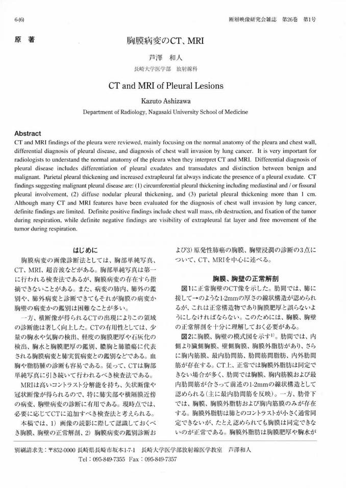
図 5 69歳、男性 気管切開後:縦隔気腫、皮下気腫に
加えて胸膜外腔にも空気がみられる (→) 。
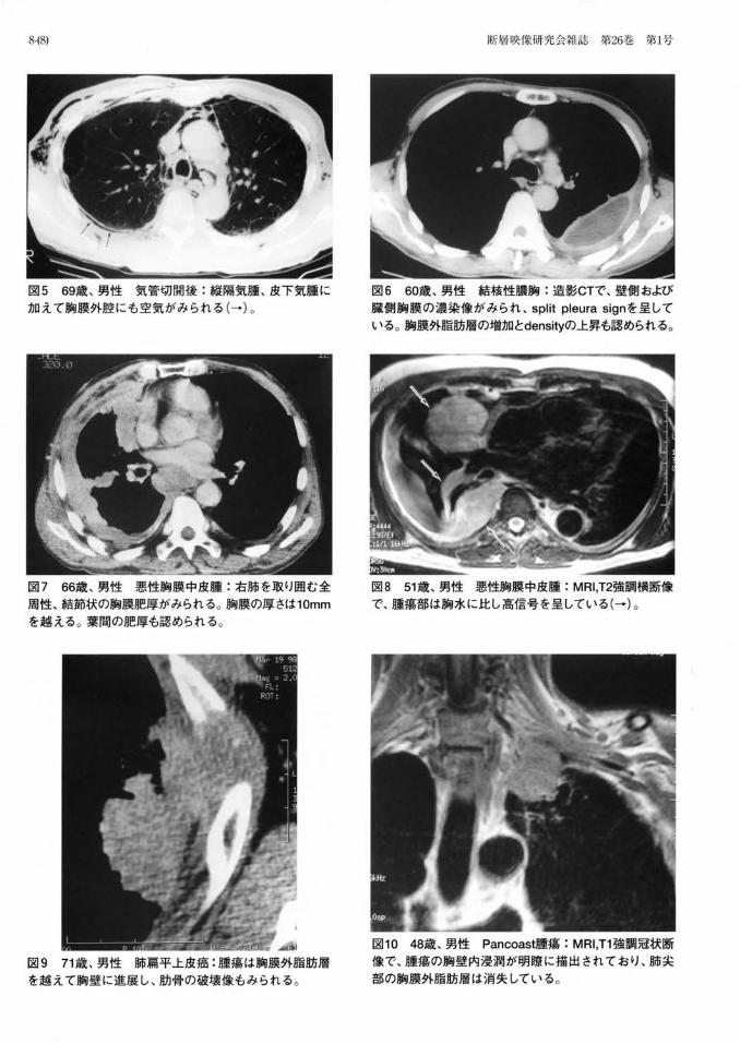
図 7 66歳、 男性 悪性胸膜中皮腫:右肺を取り囲む全
周性、結節状の胸膜肥厚がみられる。 胸膜の厚さは10mm
を越える。 薬聞の肥厚も認められる。
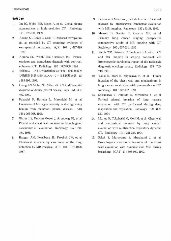
図 9 71歳、男性肺扇平上皮癌 :腫嬉は胸膜外脂肪層
を越えて胸壁に進展し、 肋骨の破壊像もみられる。
断層l映像研究会雑誌 第26巻第I号
図6 60歳、男性 結核性膿胸:造影CTで、壁側および
臓側胸膜の濃染像がみられ、 split pleura signを呈している。 胸膜外脂肪層の増加とdensityの上昇も認められる。
図8 51歳、男性悪性胸膜中皮腫: MRI,T2強調横断像
で、腫蕩部は胸水に比し高信号を呈している(→) 。
図10 48歳、男性 Pancoast腫蕩 : MRI ,T1強調冠状断
像で、腫蕩の胸壁内浸潤が明瞭に描出されており、肺尖
部の胸膜外脂肪層は消失している。

1999年6月 30 日 9-(9)
図11 ・A 図11 ・B
図11 79歳、男性肺腺癌
A:腫蕩は胸壁に接し胸膜外脂肪層も同定し難く、胸壁浸潤は否定できない。
B :CTガイド下生検後に生じた気胸で腫痕は胸Eまから離れ、壁側胸膜への浸潤はないことが明らかとなった。
滑か結節状か) 、肥厚の程度 (胸膜の厚さ)、肥厚の部
位(胸壁側か、縦隔側や葉聞の肥厚があるか)、胸膜
の石灰化の有無などに注目してCTを読影すべきで、る。
胸膜の石灰化は良性病変を示唆する所見である。
一方、 我々4) やLeungら5) の検討では、悪性を強く示
唆する所見として、 1) 縦隔側や葉聞を含めた肺を取り
囲む全周性の胸膜肥厚、 2) びまん性結節状の胸膜肥
厚、 3) 1cm以上の胸膜肥厚、が挙げられる。 これらの
所見は転移性胸膜腫蕩、悪性中皮腫(図 7) ともにみら
れ、両者を画像上鑑別することは困難である。 また前
述したように、悪性胸水の初期には胸膜肥厚はみられ
ないこともあり注意が必要で、ある。
MRIは、胸膜病変の良悪性の鑑別のためだけに行
われることは少なく 、 主にCTで悪性病変が疑われた
ケースで、その病変の頭尾側方向の拡がりゃ縦隔、胸
壁への進展などの把握に用いられる。 ただし、 T2強
調像での胸膜の信号強度が良悪性の鑑別で有用で
(図 8) 、 低信号を呈するものは良性病変である可能性
が高いとの報告もみられる6) 。
原発性肺癌の胸膜、胸壁浸潤の診断
近年、胸壁を含めたT3肺癌症例に対しても外科的
切除術が施行されるようになり、治癒切除例では長期
生存も可能となってきた。 拡大手術に伴う死亡率や合
併症を増やすことなく、また非治癒切除を避けるため
にも、術前の胸壁浸潤の有無や範囲の評価は重要と
考えられる。
1. 通常のC丁、 MRIによる診断
胸壁浸潤の評価のために、これまで、数多くのCT所
見が検討されてきたが、胸壁内の腫癒と肋骨の破壊
像 (図 9) を除いては確実なものはみられない。 Glazer
ら 7) は、 1) 腫癌と胸壁のなす角度が鈍角、 2) 胸壁と
の接触範囲が3cm以上、 3) 胸膜肥厚像、 の3項目の
2つ以上を満たす症例を浸潤ありとすると、 sensitivity
は87%だが、 specificity : 59%、 accuracy : 68%だ、った
と報告している 。 また 、 胸膜外脂肪層の消失や
densityの上昇、腫癌と胸壁の接触範囲と腫癒径との
比なども信頼性の高い所見ではない。 胸膜肥厚や胸
膜外脂肪層のdensity上昇は、 腫蕩浸潤だけでなく炎
症性変化や線維化でもみられ、 浸潤がなくても胸膜外
脂肪層が同定できないことはしばしば経験される。
MRIでも、胸壁内の腫痛や胸壁の肥厚、造影剤に
よる胸壁内の造影効果の有無、 胸膜外脂肪層の消失
などの所見で検討がなされてきた8) 9L 一般に、胸壁

10-(10)
図12・A
図12・B
浸潤におけるMRIの診断能はCTとかわらないとの報
告が多い10) 11L しかし、冠状断、 矢状断像が得られる
MRIは、 j肺尖や横隔膜における胸壁浸潤の評価では
CTより有用であることが多い8) 11) (図10) 。
2. CT、 MRIによる新しい診断法
最近CT、 MRIを用いた新しい診断法が試みられて
きた。 一つは、通常のCT検査において人工的に気胸
をつくり、腫療の壁側胸膜への浸潤の有無-を評価する
もので (図11) 、 高いaccuracyと 100%のnegative PV
(NPV) が報告されている 12) 。 すなわち、 浸潤がない
症例の診断は確実である。 しかし、腫蕩の線維性癒着
による偽陽性例があることは注意すべきであり 、 また、
u - 句園内
図12・C
断層映像研究会雑誌第26巻第l号
図12 75歳、男性肺薦平上皮癌
A:腫痕は胸壁に接し、一部胸膜外
脂肪層に断裂があるようにみえる。
胸壁浸潤の可能性があると恩われ
る。
B C : Dynamic cine MRI画像から
の深吸気および深呼気画像
呼吸によって腫蕩か.胸壁に対し移動
しており、胸壁浸潤がないと判定で
号 きる。
侵襲性の高い検査法で気胸ができない症例もあるこ
とが欠点である。
もう一つは、ヘリカ lレCTや超高速CT、 MRIを用い
て、 呼吸下の腫蕩の胸壁に対する可動性を評価する
ものである 13) -15) 。 気胸CT同様、 高いaccuracyと
100%のNPVが報告されており、 有効な検査法と思わ
れる (図12) 。 ただし、 真の腫蕩浸潤と線維性癒着(偽
陽性)との鑑別はできず、 呼吸による動きが少ない上
葉の腫蕩の評価は困難で、ある。
本論文の要旨は、 第27回断層映像研究会で教育講
演として発表した。
1999年6月 30 日
参考文献
l. 1m ]G. Webb WR. Rosen A. et al. Costal pleura:
appearances at high-resolution CT. Radiology
171 : 125-131. 1989.
2. Aquino SL. Chiles C, Oaks T. Displaced ex回pleural
fat as revealed by CT scanning: evidence of
extrapleural hematoma. A]R 169 : 687-689,
1997.
3. Aquino SL. Webb WR. Gushiken B]. Pleural
exudates and 仕加sudate芯 diagnωis 叩出 contrast
enhanced CT. Radiology 192 : 803-808, 1994.
4. 芦津和人 びまん性胸膜病変のCT像一特に胸膜及
び胸膜外脂肪の変化について一日本医放会誌 53
: 283-296. 1993
5. Le凹g AN. Muller NL. Miller RR CT 凶 differential
diagnosis of diffuse pleural disease, A]R 154: 487-
492, 1990.
6. Falaschi F , Battolla L, Mascalchi M. et al.
Usefulness of MR signal intensity in distinguishing
benign from malignant pleural disease, A]R
166 : 963-968. 1996
7. Glazer HS, Duncan-Meyer J. Aronberg D], et al.
Pleural and chest wall invasion in bronchogenic
carcinoma: CT evaluation, Radiology 157: 191
194.1985.
8. Haggar AM. Pearlberg JL, Froelich ]W, et al.
Chest-wall invasion by carcinoma of the lung:
detection by MR imaging, A]R 148: 1075-1078,
1987.
11-(11)
9. Padovani B, Mouroux ] , Seksik L, et al. Chest wall
invasion by bronchogenic carcinoma: evaluation
with MR imaging, Radiology 187: 33-38, 1993.
10. Musset D. Grenier P, Carette MF, et al.
Primary lung cancer staging: prospective
comparative study of MR imaging with CT,
Radiology 160: 607-611, 1986
1l. Webb WR. Gatsonis C, Zerhouni EA. et al. CT
and MR imaging in staging non-small cell
bronchogenic carcinoma: report of the radiologic
diagnostic oncology group, Radiology 178: 705-
713, 1991.
12. Y okoi K, Mori K, Miyazawa N, et al. Tumor
invasion of the chest wall and mediastinum in
lung cancer: evaluation with pneumothorax CT,
Radiology 181: 147-152. 1991.
13. Shirakawa T. Fukuda K, Miyamoto Y. et al
Parietal pleural invasion of lung masses:
evaluation with CT performed during deep
inspiration and expiration. Radiology 192: 809-
811. 1994.
14. Murata K, T池山ashi M. Mori M. et al. Chest wall
and mediastinal invasion by lung cancer:
evaluation with multisection expiratory dynamic
CT. Radiology 191: 251-255. 1994.
15. Sakai S. Murayama S. Murakami J. et al.
Bronchogenic carcinoma invasion of the chest
wall: evaluation with dynamic cine MRI during
breathing, ]CA T 21: 595-600. 1997.
ダウンロードされた論文は私的利用のみが許諾されています。公衆への再配布については下記をご覧下さい。
複写をご希望の方へ
断層映像研究会は、本誌掲載著作物の複写に関する権利を一般社団法人学術著作権協会に委託してお
ります。
本誌に掲載された著作物の複写をご希望の方は、(社)学術著作権協会より許諾を受けて下さい。但
し、企業等法人による社内利用目的の複写については、当該企業等法人が社団法人日本複写権センタ
ー((社)学術著作権協会が社内利用目的複写に関する権利を再委託している団体)と包括複写許諾
契約を締結している場合にあっては、その必要はございません(社外頒布目的の複写については、許
諾が必要です)。
権利委託先 一般社団法人学術著作権協会
〒107-0052 東京都港区赤坂9-6-41 乃木坂ビル3F FAX:03-3475-5619 E-mail:[email protected]
複写以外の許諾(著作物の引用、転載、翻訳等)に関しては、(社)学術著作権協会に委託致してお
りません。
直接、断層映像研究会へお問い合わせください
Reprographic Reproduction outside Japan
One of the following procedures is required to copy this work.
1. If you apply for license for copying in a country or region in which JAACC has concluded a
bilateral agreement with an RRO (Reproduction Rights Organisation), please apply for the license
to the RRO.
Please visit the following URL for the countries and regions in which JAACC has concluded bilateral
agreements.
http://www.jaacc.org/
2. If you apply for license for copying in a country or region in which JAACC has no bilateral
agreement, please apply for the license to JAACC.
For the license for citation, reprint, and/or translation, etc., please contact the right holder directly.
JAACC (Japan Academic Association for Copyright Clearance) is an official member RRO of the
IFRRO (International Federation of Reproduction Rights Organisations).
Japan Academic Association for Copyright Clearance (JAACC)
Address 9-6-41 Akasaka, Minato-ku, Tokyo 107-0052 Japan
E-mail [email protected] Fax: +81-33475-5619