detection of antibody against lymphocystis disease virus ... · 魚病研究 fish...
TRANSCRIPT

魚 病 研 究 Fish Pathology,33(4),207-211,1998.10
Detection of Antibody against Lymphocystis Disease Virus in Japanese
Flounder by Enzyme Linked Immunosorbent Assay
Hiroko Nishida, Mamoru Yoshimizu* and Yoshio Ezura
Laboratory of Microbiology, Faculty of Fisheries, Hokkaido University, Hakodate 041-8611, Japan
(Received March 20, 1998)
Enzyme linked immunosorbent assay (ELISA) was applied to detect the antibody of Japanese flounder
(Paralichthys olivaceus) against lymphocystis disease virus (LCDV). Purified Japanese flounder LCDV was used as a capture antigen of ELISA. Monitoring the immune response of Japanese flounder to LCDV was done using the sera obtained from apparently healthy, LCD-diseased and LCD-recovered fish. ELISA absorbances of these three groups were clearly different. Using the serum with high ELISA absorbance, the optimum ELISA condition was set up. Apparently healthy fish were injected with inactivated LCDV. One to three months after the injection, the antibody against LCDV was detected by ELISA established for LCDV and their ELISA antibody titer increased during that period.
Key words: lymphocystis disease, LCDV, Japanese flounder, antibody, ELISA
Japanese flounder (Paralichthys olivaceus) is economically important fish and recently it's culture is popular in different parts of Japan. In Hokkaido, there are some
hatcheries and fish farms that culture the Japanese floun
der. Almost every year, lymphocystis disease (LCD) occurred in the fish cultured in Hokkaido and the economic loss from this disease is severe to the farms. The
disease is a worldwide in water environments (Anders
and Darai, 1985) and it's infection is widely spreading. It was reported that LCDV infected not only Japanese
flounder Paralichthys olivaceus (Tanaka et al., 1984), more over it spontaneously infected many other fish species, such as sea bass Lateolabrax japonicus (Miyazaki
and Egusa, 1972), yellowtail Seriola quinqueradiata
(Matusato, 1975) and also some other commercial fishes. It was also reported that the cultured black rockfish
Sebastes schlegeli was infected by LCDV in Hokkaido
(Tanaka et al., 1984). The causative agent is the fish lymphocystis disease virus which induces some charac
teristic nodules on the body and fins of some fish spe
cies (Nigrelli and Ruggieri, 1965). Infected fish showed poor growth and anemia, in the worst case die
of hunger. But most of the infected fish were able to recover from the disease and would not be infected again in the next year. It has been reported that seroprevalence
and antibody activity of the fish increased with age
(Lorenzen and Dixon, 1991). Therefore, protective immunization to LCDV in flounder may be effective. For the immunization, it is necessary to monitor the
immune response against the antigen in the fish. The purpose of this study is to examine the detection
of antibody against LCDV in cultured Japanese flounder by ELISA and to compare the ELISA antibody titers
between apparently healthy, LCD-diseased, LCD-recovered and immunized fish.
Materials and Methods
Fish Japanese flounder used in this study was cultured at
Kumaishi Fisheries Station in Hokkaido. Firstly, the antibody-monitoring test was done in the spring using the 3 types of each 10 fish cultured in different tanks; apparently healthy, LCD-diseased and recovered fish. Average body weight of these fish was about 40 g. Following this experiment, 5 healthy fish (45 g) were injected with LCDV by the method described below. Two 1+ year-old Japanese flounder, weighting about 800 g, were also injected and detected their ELISA antibody titers against LCDV.
Purification of LCDV antigen Lymphocystis cells were collected from the infected
* Corresponding author.Fax: 0138-40-5569 E-mail: [email protected]
208 H. Nishida, M. Yoshimizu and Y. Ezura
Japanese flounder. The specimens of lesion (10 g)
were washed with PBS (137 mM NaCl, 2.68 mM KCl,
8.10 mM Na2HPO4, and 1.47 mM KH2PO4), homog
enized and suspended to TE (50 mM Tris-HCl, 2 mM
EDTA). The suspension was repeated freezing and
thawing for 3 times, then centrifuged for 20 min at 1,800•~
g. The supernatant was centrifuged for 2 h at 81,000•~
g. The pellet was resuspended in TE, overlaid on 15,
20, 30, 40 and 50% sucrose steps and centrifuged for 2 h
at 81,000•~g. Virus bands were harvested and diluted
in TE. After 2 h centrifugation at 81,000•~g, the pellets
were resuspended in TE.
ELISA to detect the antibody against LCDV in Japanese
flounder Sandwich ELISA based on the method reported by
Yoshimizu et al. (1992, 1997) was used. Briefly,
microtiter plate (Greiner) was coated with 50ƒÊl of
purified LCDV antigen diluted to 10ƒÊg/ml and left
overnight at 4•Ž. Following 3 times washes with PBS
containing 0.05% Tween 20 (T-PBS), the plate was
blocked with 2% skim milk for 1 h at 37•Ž. After 3
times washes with T-PBS, 50ƒÊl of fish serum diluted to
1: 40 in T-PBS were applied and incubated for 2 h at
37•Ž. Followed by the T-PBS wash, 50 1.11 of the
anti-flounder IgM rabbit serum (the 2 nd serum; Suzuki
et al., 1998) diluted to 1:400 in T-PBS was added to
each well and incubated for 1 h at 37•Ž. Washed by
the T-PBS and 50ƒÊl of peroxidase conjugated anti
-rabbit IgG swine serum (the 3 rd serum; Dako) diluted
to 1:500 in T-PBS was added to each well and incu
bated for 30 min at 37•Ž. Followed by the wash with
T-PBS for 5 times, 50ƒÊl of substrate (ƒÍ-phenylenedi
amine) in citric acid buffer (0.2M Na2HPO4 : 0.1M
citric acid = 103:97) was added and incubated for 15
min at room temperature. The reaction was stopped
by adding of 50ƒÊl /well of 2N H2SO4. The absorbance
was read at 492 nm, using a micro plate reader (MTP-120
, Corona).
Selection of the antigen concentration and antisera dilution in ELISA The capture antigen was prepared ranging from 1 ng/ ml to 1,000 ng/ml, anti-Japanese flounder IgM rabbit serum was diluted from 1:100 to 1:16,00, and peroxidase conjugated anti-rabbit IgG swine serum was diluted from 1:100 to 1:1,000 and examined for the ELISA assay.
Absorbed test with LCDV
Fish serum which had high ELISA absorbance was
reacted with homogenized lymphocystis cells in vitro
overnight at 4•Ž. Then the suspension was centrifuged
and the supernatant was collected to use for the ELISA
assay.
Injection of inactivated LCDV to Japanese flounder
Five healthy fish (0+ year-old) that had negligible
ELISA titers were injected with formalin-inactivated
purified virus (250ƒÊg/ml) at pterygiophore region, just
below the dorsal margin and 2 healthy fish (1+ year-old)
were injected at the blood vessel. Fish were tagged for
monitoring individually and 1 and 3 months later, their
ELISA antibody titers were measured. Control fish
were injected with Hanks' BSS by the same method.
Results
Comparison in ELISA absorbances among healthy, LCD diseased and recovered fish
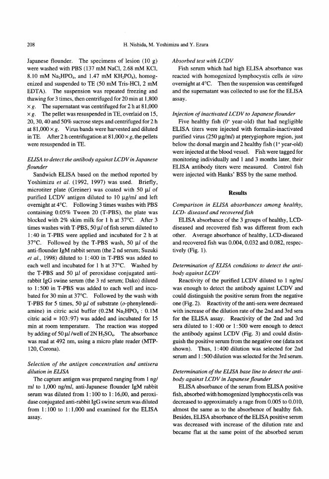
ELISA absorbance of the 3 groups of healthy, LCD-diseased and recovered fish was different from each other. Average absorbance of healthy, LCD-diseased and recovered fish was 0.004, 0.032 and 0.082, respectively (Fig. 1).
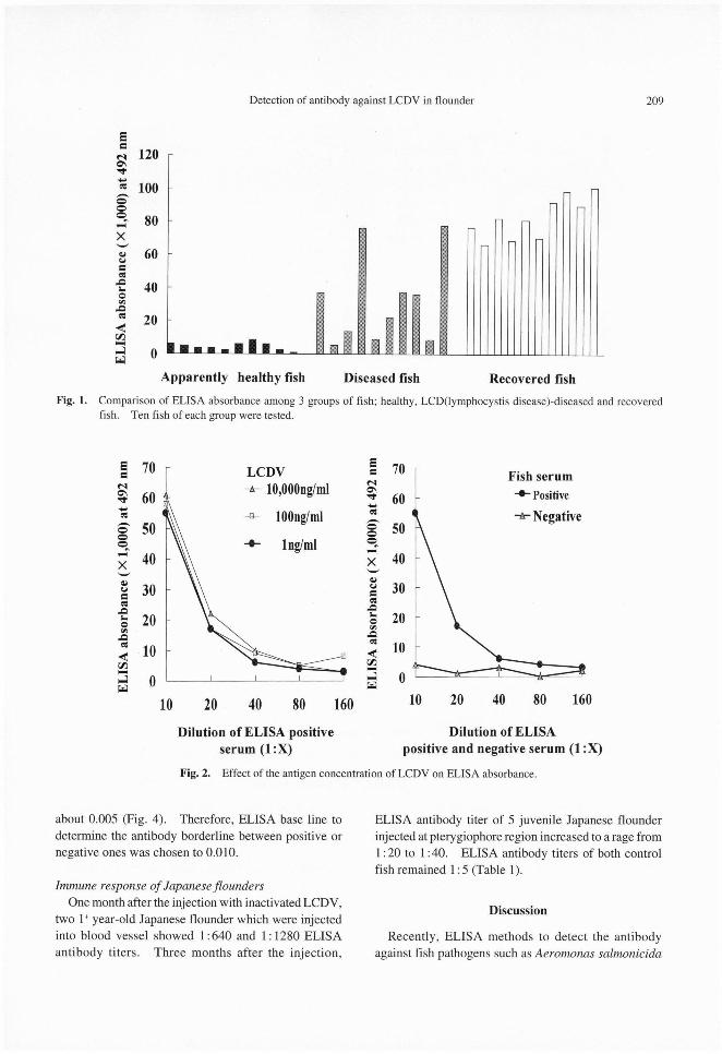
Determination of ELISA conditions to detect the antibody against LCDV Reactivity of the purified LCDV diluted to 1 ng/ml
was enough to detect the antibody against LCDV and
could distinguish the positive serum from the negative
one (Fig. 2). Reactivity of the anti-sera were decreased with increase of the dilution rate of the 2nd and 3rd sera for the ELISA assay. Reactivity of the 2nd and 3rd
sera diluted to 1:400 or 1:500 were enough to detect the antibody against LCDV (Fig. 3) and could distin
guish the positive serum from the negative one (data not shown). Thus, 1:400 dilution was selected for 2nd
serum and 1: 500 dilution was selected for the 3rd serum.
Determination of the ELISA base line to detect the antibody against LCDV in Japanese flounder
ELISA absorbance of the serum from ELISA positive fish, absorbed with homogenized lymphocystis cells was decreased to approximately a rage from 0.005 to 0.010, almost the same as to the absorbence of healthy fish. Besides, ELISA absorbance of the ELISA positive serum was decreased with increase of the dilution rate and became flat at the same point of the absorbed serum
Detection of antibody against LCDV in flounder 209
about 0.005 (Fig. 4). Therefore, ELISA base line todetermine the antibody borderline between positive or
negative ones was chosen to 0.010.
Immune response of Japanese floundersOne month after the injection with inactivated LCDV,
two 1 year-old Japanese flounder which were injectedinto blood vessel showed 1:640 and 1: 1280 ELISAantibody titers. Three months after the injection,
ELISA antibody titer of 5 juvenile Japanese flounderinjected at pterygiophore region increased to a rage from1:20 to 1:40. ELISA antibody titers of both controlfish remained 1: 5 (Table 1).
Discussion
Recently, ELISA methods to detect the antibody
against fish pathogens such as Aeromonas salmonicida
Fig. 1. Comparison of ELISA absorbance among 3 groups of fish; healthy, LCD(lymphocystis disease)-diseased and recovered
fish. Ten fish of each group were tested.
Fig. 2. Effect of the antigen concentration of LCDV on ELISA absorbance.

210 H. Nishida, M. Yoshimizu and Y. Ezura
Fig. 3. Effects of the concentration of anti-Japanese flounder IgM rabbit serum (the 2nd serum) and peroxidase conjugated antirabbit Ig swine serum (the 3rd serum) on ELISA absorbance.
Fig. 4. Comparison of ELISA absorbance of the sera from LCD-recovered (•œ, •£) and healthy (•¡, •Ÿ)fish between non-absorbed
and absorbed with homogenized lymphocystis cell.
(Yoshimizu et al., 1992), striped jack nervous necrosis virus (SJNNV) (Mushiake et al., 1992) and Edwardsiella tarda (Bang et al., 1992) were reported. Determination of the activity responding with cell-mediated immunity is thought to be difficult and then monitoring of the humoral immune response was selected and examined
by ELISA. The ELISA established in this study is
useful to detect the antibody against LCDV in Japanese
flounder without killing the fish. But in general, the
ELISA methods to detect the antibody are not common
to all the pathogens. Detection of the antibody against
Renibacterium salmoninarum that causes bacterial

Detection of antibody against LCDV in flounder 211
Table 1. ELISA antibody titer of Japanese flounder injected with the purified formalin inactivated LCDV 1 month and 3 months after injection
kidney disease has not been regarded as a standard method (Thoesen, 1994). But detection of antibody by
ELISA against Aeromonas salmonicida that causes furunculosis and fish nodavirus that causes viral nervous
necrosis (VNN) were reported to be useful for the detection of the antibody and applied to select the nodavirus
carrying broodstocks of berfin flounder (Veraspermoseri)
(Yoshimizu et al., 1992, 1997). Lymphocystis disease appears in Japanese flounder
in almost every year in Hokkaido and establishment of
the protection method is requested. ELISA absorbance of healthy fish was low and that of the fish recovering
from the disease was high. Most of LCD-diseased fish showed a low level ELISA absorbance but some of the
fish showed high absorbance as the recovered fish. It was thought that the diseased fish with high ELISA
absorbance was going to recover from the disease at that time. And then, the fish immunized with inactivated
LCDV developed the ELISA antibody titer up to 1: 20 to 1:1280 after 1 to 3 months while the control fish
remained 1: 5. These results suggest that the humoral immune response against LCDV reflect the disease
progression of LCD in Japanese flounder. Moreover, vaccination for protecting from LCDV infection will be
effective.
Acknowledgements
The authors wish to express their sincere gratitude to
Mrs. Tukao Tabata, Masaki Kadowaki, Masato Nanbu
and Mamoru Osanai, Kumaishi Fisheries Station,
Hokkaido, Japan.
References
Anders, K. and G. Darai (1985): Genome analysis of fish
lymphocystis disease virus. In "Fish and shell fish pathol
ogy" (ed. by A. E. Ellis). Academic press, London, pp.
301-306.Bang, J. D., S. G. Chun, S. I. Park and Y. J. Choi (1992):
Studies on the biochemical and serological characteristics of Edwadsiella tarda isolated from cultured flounder
(Paralichthys olivaceus). J. Fish Pathol., 5, 29-35.Lorenzen, K. and P. F. Dixon (1991) : Prevalence of antibody
to lymphocystis virus in estuarine flounder Platichthys flesus. Dis. Aquat. Org., 11, 99-103.
Matsusato, T. (1975): On the lymphocystis disease in cultured
yellowtail. Fish Pathol., 10, 90-93.Miyazaki, T. and S. Egusa (1972): On the lymphocystis dis
ease in cultured sea bass (Lateolabrax japonicus, Covier and Valenci). Fish Pathol., 6, 83-89.
Mushiake, K., M. Arimoto, T. Furusawa, I. Furusawa, T. Nakai and K. Muroga (1992): Detection of antibody against striped
jack necrosis virus (SJNNV) from brood stocks of striped jack. Nippon Suisan Gakkaishi, 58, 2351-2356.
Nigrelli, R. F. and G. D. Ruggieri (1965): Studies in viral disease of fishes. Spontaneous and experimentally induced cellu
lar hypertrophy (lymphocystis disease) in fishes of the New York Aquarium with a report of new cases and an annotated
bibliography (1874-1965). Zoologica, 50, 83-96.Nishida, H., T. Enokida, N. Hiramatsu, A. Hara and M.
Yoshimizu (1998): Purification of IgM from Japanese flounder. Bull. Fac. Fish. Hokkaido Univ., 49(3) in press.
Tanaka, M., M. Yoshimizu, M. Kusakari and T. Kimura (1984): Lymphocystis disease in Kurosoi Sebastes schlegeli and
Hirame Paralichthys olivaceus in Hokkaido, Japan. Bull. Japan. Soc. Sci. Fish., 50, 37-42.
Thoesen, J. C. (ed.) (1994): Blue book, Ver. 1. Suggested
procedures for the detection and identification of certain finfish and shellfish pathogens. Fish Health Section,
American Fisheries Society.Yoshimizu, M., S. Direkbsarakom, T. Nomura, Y. Ezura and
T. Kimura (1992): Detection of antibody against Aeromonas salmonicida in the serum of salmonid fish by the enzym
linked immnosorbent assay. Fish Pathol., 27, 73-82.Yoshimizu, M., K. Suzuki, T. Nishizawa, J.R. Winton and Y.
Ezura (1997): Antibody screening for the identification of nervous necrosis virus carriers in a flounder brood stock. In
"New approaches to viral diseases of aquatic animals" (ed. by Y. Inui). NRIA International Workshop, National Re
search Institute of Aquaculture, pp. 124-130.