diabetes and cardiovascular disease: is diabetes a risk ... · diabetes and cardiovascular disease:...
TRANSCRIPT

2013/2014
Rui Alexandre dos Santos Lopes
Diabetes and cardiovascular
disease: is diabetes a risk
equivalent?
março, 2014
Mestrado Integrado em Medicina
Área: Endocrinologia
Trabalho efetuado sob a Orientação de:
Dra. Maria Elisabete Gonçalves Rodrigues
E sob a Coorientação de:
Doutor Davide Maurício Costa Carvalho
Trabalho organizado de acordo com as normas da revista:
Revista Portuguesa de Cardiologia
Rui Alexandre dos Santos Lopes
Diabetes and cardiovascular disease:
is diabetes a risk equivalent?
março, 2014


Ao meu padrinho

1
Diabetes and cardiovascular disease: is diabetes a risk equivalent?
Diabetes e doença cardiovascular: será a diabetes um equivalente de risco?
Rui Santos Lopesa; Elisabete Rodriguesa;b; Davide Carvalhoa;b
aFaculdade de Medicina da Universidade do Porto, Porto, Portugal. bServiço de
Endocrinologia do Centro Hospitalar São João, Porto, Portugal.
Correspondência para autor:
Rui Alexandre dos Santos Lopes
Rua de S. Pedro, nº 101
4405-785 Madalena
Vila Nova de Gaia. Porto. Portugal
Email: [email protected]; [email protected]
Número total de palavras do manuscrito: 4931

2
Resumo
Introdução e objetivos: A doença cardiovascular é causa importante de morbilidade e a principal
causa de mortalidade em doentes diabéticos acarretando um aumento de risco 2 a 3 vezes para
eventos cardiovasculares. O conceito de que a diabetes é um equivalente de risco cardiovascular
surgiu na comunidade médica após a publicação de um estudo de cohort prospectivo, em 1998.
Desde então foram publicados estudos que suportavam e outros que refutavam esta ideia. O
objetivo deste artigo é rever a literatura publicada no últimos 5 anos e discutir o conceito de que
diabetes é um equivalente de risco cardiovascular.
Métodos: Foi realizada uma pesquisa bibliográfica usando a PubMed, a Web of Science e a
Scopus. Os artigos foram restritos aos últimos 5 anos, em inglês e português.
Resultados: Foram encontrados 16801 artigos, obtendo-se 20 artigos que foram usados nesta
revisão.
Conclusões: A Diabetes, por si só, não deve ser considerada um equivalente de risco
cardiovascular. A avaliação de risco deve incluir parâmetros como hemoglobina glicada, glicemia
em jejum e duração da doença. Esta mudança de paradigma tem implicações clínicas,
nomeadamente a restrição da utilização de estatinas nos doentes de risco mais baixo.
Palavras-chave: diabetes, glicemia, glucose, HbA1C, risco cardiovascular, risco de doença
coronária.
3
Abstract
Introduction and Aim: Cardiovascular disease is an important cause of morbidity and the most
important cause of mortality in diabetic patients with a 2-3 fold increased risk of cardiovascular
events. The concept that diabetes is a cardiovascular disease equivalent arose in the medical
community after Haffner’s study in 1998. Since then studies supporting and refuting this principle
were published. The aim of this paper was to review literature in the past 5 years and discuss the
concept of diabetes as a cardiovascular disease equivalent.
Methods: Literature research was performed using PubMed, Web of Science and Scopus.
Articles were restricted to the last 5 years and to English and Portuguese languages.
Results: 16801 articles of were found during research. After exclusion, 20 articles remained and
were used in the conception of this review.
Conclusions: Diabetes per se should not be considered a cardiovascular disease equivalent. Risk
assessment tools should focus on parameters like glycated haemoglobin levels, FPG levels and
duration of the disease. This change of paradigm has clinical implications such as restricting the
use of statins in lower risk groups.
Keywords: diabetes, glycemia, glucose, HbA1C, cardiovascular risk, coronary heart disease risk.
4
Abreviaturas
ADVANCE Action in Diabetes and Vascular Disease:
Preterax and Diamicron MR Controlled
Evaluation
apoB/apoA Apolipoproteina B/apolipoproteina A
CHD Doença coronária
CKD Doença renal crónica
CVD Doença cardiovascular
DM Diabetes Mellitus
FMD% Percentagem de dilatação fluxo-mediada
HbA1c Hemoglobina glicada
IFG Glicemia em jejum anormal
MI Enfarte do miocárdio
NGT Tolerância à glicose
SDDM DM detectada
SRDM DM reportada
T1D DM tipo 1
T2D DM tipo 2
5
Abbreviations
ADVANCE Action in Diabetes and Vascular Disease:
Preterax and Diamicron MR Controlled
Evaluation
apoB/apoA Apolipoprotein B/apolipoprotein A
CHD Coronary heart disease
CKD Chronic kidney disease
CVD Cardiovascular disease
DM Diabetes Mellitus
FMD% Percentage of flow mediated dilatation
HbA1c Glycated haemoglobin
IFG Impaired fasting glucose
MI Myocardial Infartion
NGT Normal glucose tolerance
SDDM Screen-detected DM
SRDM Self-reported DM
T1D Type 1 diabetes mellitus
T2D Type 2 diabetes mellitus

6
Introduction
Cardiovascular disease (CVD) is an important cause of morbidity and the most important
cause of mortality in diabetic patients1, 2 accounting for more than 70% of the deaths2. Studies
have shown that DM is associated with a two to three fold increased risk of cardiovascular events
(CVE)1, 3. In 1998, Haffner et al has shown that people with type 2 DM without a prior myocardial
infarction (MI) have similar risk of coronary heart disease (CHD) as non-diabetics with a previous
MI4, suggesting that DM is a CVD risk equivalent. This study had considerable impact in the
medical community in terms of intensive prevention of CHD in people with DM4. However, the
concept of DM as a CVD equivalent has been questioned and, since then, papers supporting his
findings5, 6 and papers who did not7-9 were published. In the face of this dissent, the aim of this
paper is to review the published literature in the past 5 years and discuss the concept of DM as a
CVD equivalent.
Methods
Search strategy
Literature research was performed using PubMed, Web of Science and Scopus on
September 8th 2013 using the following query: (diabetes AND “cardiovascular risk”) OR
(glycemia AND “cardiovascular risk”) OR (glucose AND “cardiovascular risk”) OR (HbA1c
AND “cardiovascular risk”) OR (diabetes AND “coronary heart disease risk”) OR (glycemia
AND “coronary heart disease risk”) OR (glucose AND “coronary heart disease risk”) OR (HbA1c
AND “coronary heart disease risk”).
PubMed, Web of Science and Scopus researches were limited to the last 5 years with
language restriction to English and Portuguese. Web of Science research was restricted to
cardiovascular system and cardiology, endocrinology and metabolism, nutrition and dietetics,
biochemistry and molecular biology and geriatrics and gerontology subject areas. Scopus research
was restricted to following subject areas: Medicine, Nursing, “Pharmacology, Toxicology and
Pharmaceutics”, “Biochemistry, Genetics and Molecular Biology”. No subject area restriction
was applied to PubMed research.

7
Selection criteria
Articles were included if they: (1) were written in English or Portuguese; (2) contained at
least one of the following keywords: diabetes, glycaemia, glucose, hbA1C; (3) and contained at
least one of the following keywords: cardiovascular risk, coronary heart disease risk.
Articles were excluded if they were not inserted in cardiology, endocrinology nor
metabolism subject areas. Articles whose aim was not cardiovascular or coronary heart disease
risk assessment were excluded.
Data extraction
Titles and abstracts from the literature research were imported to Endnote X7®.
Duplicates were found comparing title, authors and year of publication. After exclusion of
duplicates, titles were reviewed, followed by abstract and full paper analysis. Articles whose
abstracts or full text were not available were excluded.
Results
We found 16801 articles (PubMed: 6244; Web of Science: 8099; Scopus: 2458). After
duplicate exclusion, 13124 articles remained and selection based on title alone was performed
(Fig. 1); 12771 articles were excluded. Abstract selection was performed in 353 articles; 278 were
excluded because abstract did not met the selection criteria and, 7 articles did not presented
abstract at all. Subsequently full text selection was performed on 68 articles; 41 were excluded
because full text did not met the selection criteria, 5 did not present full text available and 2 were
excluded because were editorials. 20 articles remained and were used in the conception of this
review.
Table 1 organizes and overviews the articles included in this review.
8
Discussion
The impact of Haffner’s4 study was undeniable. Despite its limitations and the lack of
power to detect differences between two groups of patients10, the concept that DM as CVD risk
equivalent ingrained into medical community. This led institution of risk modifier drugs, namely
statins, to reduce the risk. However, novel studies were published that questioned or refuted
Haffner et al4, leading to disagreement still today.
In this section, we are going to individually address each study found, dividing them in
the following groups: studies supporting the hypothesis that DM is a CVD equivalent, studies
refuting this hypothesis, studies that give no straight answer, and reviews
Studies supporting the hypothesis that diabetes is a cardiovascular disease equivalent
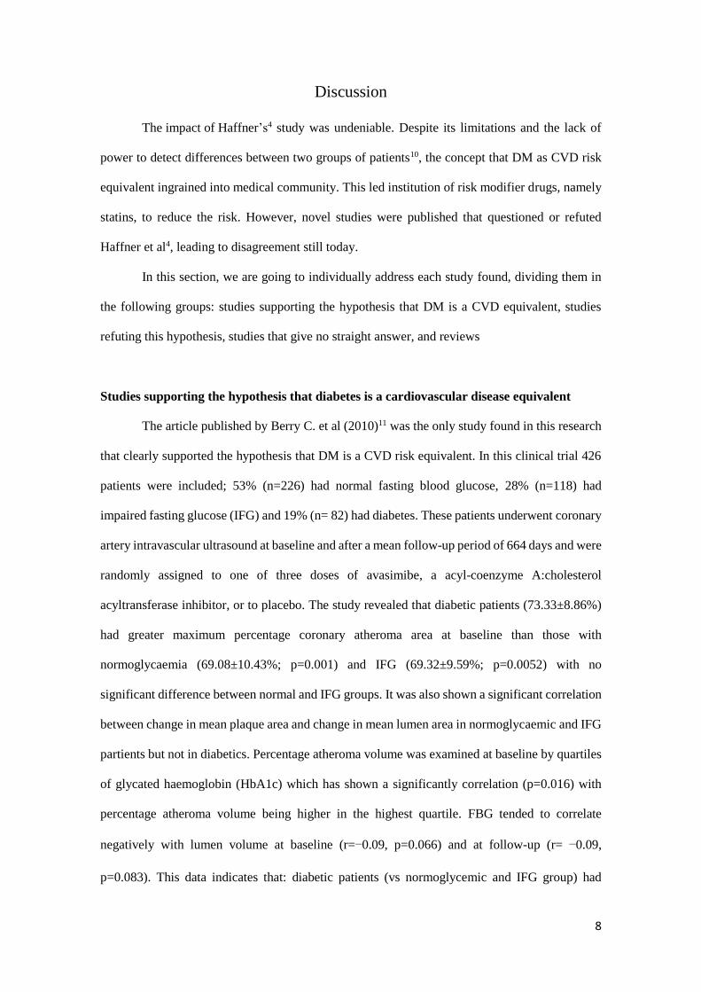
The article published by Berry C. et al (2010)11 was the only study found in this research
that clearly supported the hypothesis that DM is a CVD risk equivalent. In this clinical trial 426
patients were included; 53% (n=226) had normal fasting blood glucose, 28% (n=118) had
impaired fasting glucose (IFG) and 19% (n= 82) had diabetes. These patients underwent coronary
artery intravascular ultrasound at baseline and after a mean follow-up period of 664 days and were
randomly assigned to one of three doses of avasimibe, a acyl-coenzyme A:cholesterol
acyltransferase inhibitor, or to placebo. The study revealed that diabetic patients (73.33±8.86%)
had greater maximum percentage coronary atheroma area at baseline than those with
normoglycaemia (69.08±10.43%; p=0.001) and IFG (69.32±9.59%; p=0.0052) with no
significant difference between normal and IFG groups. It was also shown a significant correlation
between change in mean plaque area and change in mean lumen area in normoglycaemic and IFG
partients but not in diabetics. Percentage atheroma volume was examined at baseline by quartiles
of glycated haemoglobin (HbA1c) which has shown a significantly correlation (p=0.016) with
percentage atheroma volume being higher in the highest quartile. FBG tended to correlate
negatively with lumen volume at baseline (r=−0.09, p=0.066) and at follow-up (r= −0.09,
p=0.083). This data indicates that: diabetic patients (vs normoglycemic and IFG group) had

9
greater coronary atheroma burden; atherosclerosis severity and coronary remodelling differed
according to glycaemic status; and FBG and HbA1c were correlated with plaque burden,
atherosclerosis progression and coronary remodelling. Authors refer that patients with HbA1c
>10% were not included in this study, so the results may under-represent the magnitude of the
diabetes-related effects on CHD.
Studies refuting the hypothesis that diabetes is a cardiovascular disease equivalent
In a 10-year prospective cohort study of 4410 patients, from Catalonia (Spain) Cano et
al12 recruited 2260 patients with type 2 diabetes (T2D) without coronary heart disease (CHD)
and 2150 with first acute MI without diabetes. The authors compared long-term cardiovascular
risk between T2D patients and first MI patients to assess the influence of diabetes duration, type
of treatment, and glycaemic control at baseline. They found that the incidence rate for all cause
death, coronary death, cardiovascular mortality, fatal and non-fatal MI and coronary heart disease
were significantly worse among patients who survived acute, except for stroke death and unstable
angina. These differences held after adjustment for potential confounders. It was shown that
duration of diabetes was a determinant of cardiovascular outcomes (cut point at 8 years of disease
duration) and patients with HbA1c ≥7% had worse prognosis.
Saely CH et al (2010)13 recruited 756 consecutive patients referred to routine coronary
angiography between October 1999 and October 2000 and recorded the vascular events over 4
years to investigate the contribution of baseline coronary atherosclerosis to the risk of diabetic
patients for future CVE. At baseline angiography, 244 had neither T2D nor significant CHD, 50
had T2D but not significant CHD, 342 had significant CHD but no TD2, and 114 had both T2D
and significant CHD. It was verified higher incidence of vascular events among patients with T2D
than among non-diabetics (32.3vs.18.3%; p<0.001) and higher incidence of CVE among patients
with significant CHD compared when compared with those without (29.4vs.8.8%; p<0.001).
Presence of significant CHD at baseline conferred a higher vascular risk (adjusted HR= 3.46;
p<0.001) than the presence of T2D (adjusted HR= 1.55; p=0.021). CVE were similar in T2D
patients without significant CHD vs non-diabetics without significant CHD, but higher in non-

10
diabetics with significant CHD (p<0.001) and highest in patients with both, T2D and CHD
(p<0.001). T2D patients without CHD had a significantly lower event rate than non-diabetic
patients with significant CHD (p=0.008). The authors concluded that baseline coronary artery
state determines vascular risk in patients with T2D patients and diabetes was not a CHD risk
equivalent, though vascular risk was higher in the overall sample of T2D patients than in CHD
patients.
In a prospective cohort study, Paynter NP et al (2011)14 followed a total of 24 674 women
(685 diabetics) and 11 280 men (563 diabetics) aged <80 years. The median follow up were 10.2
years and 11.8 years for women and men, respectively. The aim of this study was to generate
CVD risk models that included HbA1c levels and compare its predictive ability with classification
based on current guidelines for the diabetic participants; it was also examined the effect of a
dichotomous term for diabetes in place of HbA1c levels. The authors demonstrated on both
cohorts of men and women that measurement of HbA1c level in diabetic subjects improved risk
prediction compared with classification of DM as CVD risk equivalent but with improved risk
prediction in the women cohort. The authors also verified that diabetes alone did not confer a 10-
year risk of CVD higher than 20%.
Hernandez D et al (2013)15 conducted a cross sectional study in a representative sample
of 2270 adults, 18 – 80 years, from Malaga (Spain) in order to determine HbA1c cut-off points
for chronic kidney disease (CKD) and CVD. The authors showed that known DM was
significantly associated with CKD, CVD, or both and that HbA1c levels were independently
related to clinical endpoints after adjustment for traditional risk factors. However, when both
known diabetes and HbA1c levels were introduced in the same model, DM was not significantly
associated with CKD nor CVD, suggesting that significant associations between diabetes and
CVD or CKD was mediated by HbA1c concentration, regardless of diabetic status.
Krishnan S et al (2011)16 examined the presence of cardiovascular risk factors in 66
teenagers (13-20 years) with normal weight or overweight, both with and without type 1 diabetes
(T1D). This study hypothesized if teenagers with T1D had a worse CV risk profile than those
without T1D, and if there was a synergic or additive effect of overweight status and T1D on CV

11
risk profile. The authors found that T1D was not associated with higher cardiovascular risk profile
and had consistently and paradoxically higher HDL-C levels (p=0.023) than non-T1D patients
regardless of their overweight status. Also statistically significant adverse effect of diabetes on
arterial compliance was not observed nor interaction between diabetes and overweight status. One
limitation of this study is that only children with controlled T1D were included.
Studies giving no straight answer to the question “is diabetes a cardiovascular disease
equivalent?”
In a case-control study, Deo RK et al (2008)17 looked at the association between HbA1c
levels and CVE, namely MI and stroke. 50 consecutive diabetic patients admitted in wards with
CVE were included (25 with and 25 with stroke); 50 diabetic patients without CVE were taken
as control. The authors showed that among patients with CVE and no CVE, the difference
between levels of HbA1c was statistically significant (p=0.017). For MI, level of HbA1c was
statistically significant (p=0.018) while for stroke, level of HbA1c was not significant. Likewise
mean blood glucose also predicted CVE (p=0.006), MI (p=0.006) but not stroke. Fasting plasma
glucose as well as postprandial plasma glucose also significantly predicted CVE (p=0.024 and
0.019, respectively).
van der Heijden AAWA et al (2009)18 used prospective data from 1482 people (50-75
years), who participated in the Hoorn Study in order to validate and compare results from the
Framingham, SCORE, and UKPDS risk functions in predicting CHD risk of individuals with
normal glucose tolerance (NGT), intermediate hyperglycaemia (impaired glucose tolerance
and/or IFG), and DM (screening-detected and previously known DM). The discriminatory ability
of models was evaluated by calculating area under the receiver operating characteristic curve
(AUROC); discriminatory power was graded low (AUROC 0.5-0.7), moderate (AUROC 0.7-0.9)
or high (AUROC >0.9). The authors verified that Framingham and UKPDS risk functions
overestimated the actual observed CHD incidence rate in all subgroups. The Framingham
algorithm had low ability to discriminate the first CHD in NGT and intermediate hyperglycaemia
groups (AUROC 0.68 and 0.60, respectively) and moderate discrimination ability in screening-

12
detected diabetes group (AUROC 0.74). The UKPDS function had moderate capacity to identify
those with high risk for a first CHD event in NGT, intermediate hyperglycaemia and screening-
detected diabetes subgroups (AUROC 0.71, 0.70and 0.75, respectively) but low ability when
screening-detected and known diabetes were combined (AUROC 0.66). The SCORE algorithm
for prediction of fatal CHD had a moderate ability in all subgroups. It was also verified that
Framingham and UKPDS risk functions - designed to estimate first CHD in the general population
and the diabetic population, respectively - performed better in estimating fatal CHD than the
SCORE risk function.
In a case-control study, Gerstein HC et al (2010)19 evaluated the relationship between
HbA1c levels in MI patients and controls who participated in the INTERHEART study; 15152
MI patients who were admitted within 24 hours of their first acute MI and 14820 controls were
included. The authors observed that a 1% increase in HbA1c was associated with a 40% higher
odds of MI after controlling for age, sex and region alone, and a 19% higher odds after adjusting
for the risk factors used (age, sex and region, diabetes, hypertension, current smoking, physical
activity ≥ 4 hours/week, daily fruit and vegetable intake, alcohol intake, abdominal obesity, BMI,
apoB/apoA). The importance of dysglycaemia as a risk factor for MI in the general population is
further highlighted by an association between an HbA1c ≥5.4% and a 22% higher odds of MI,
after adjusting for the risk factors used this study. It was also observed a 25% higher odds of MI
in people with no previous diabetes and a 18% higher odds in people without previous diabetes
history and an HbA1c<6.5% further emphasising the relevance of these findings to the general
population. The authors concluded that HbA1c was an independent risk factor for MI in the
presence of every other independent cardiovascular risk factor and self-reported diabetes
underestimates the association between dysglycaemia and cardiovascular risk.
In a 15-year prospective cohort study, Wang H et al (2011)20 followed 4549 American
Indian adults, between 45-74 years, recruited from Strong Heart Study (1989–1991). Data from
3,850 individuals with baseline measurements of FPG and HbA1C and no prevalent CVD were
analysed; 1,386 had known diabetes. This study showed that newly diagnosed diabetes via HbA1c
≥6.5% vs. non-diabetic patients was an independent CVD risk factor (HR 1.50) but not with CHD

13
(HR 1.43); previously known DM vs. non-diabetic patients had greater HR (2.52); Newly
diagnosed diabetes via FPG ≥126 mg/dL vs FPG <100 mg/dL was also independent associated to
CVD events (adjusted HR 2.52 [95% CI: 2.06-3.08]). Flat linear relations were observed between
HbA1c and CVD and CHD in individuals without diabetes, with no suggestion of an inflection
point at any HbA1c value. Comparing HbA1c and FPG in prediction models for CVD, HbA1c
and as FPG were significant independent predictors (HR= 1.08 and 1.07, respectively) in subjects
without known diabetes; in known diabetics, neither HbA1c nor FPG were significant
independent predictors. However, no significant increase of CVD risk across HbA1c categories
was shown within the pre-diabetic range after adjustment for known CVD risk factors.
Babar GS et al (2011)21 hypothesized that children with T1D would manifest early signs
of abnormal vascular homeostasis, endothelial dysfunction, increased carotid intima-media
thickness (c-IMT), and elevated circulating markers of inflammation. Endothelial function was
determined by percentage of flow mediated dilatation (FMD%) of the brachial artery. 21 children
with T1D, aged 8.5±0.3 years (diabetes duration: 4.3±0.4 years), recruited from the Children’s
Hospital of Wisconsin Diabetes Clinic were included and compared with a 15 group-matched
healthy siblings (aged 7.6±0.3 years). The authors verified positive correlation between FMD%
and HbA1c (r=0.47, p=0.033) and FMD% and 2 week-glucose variability (r=0.50, p=0.021),
adjusted for diabetes duration. However, no correlation between FMD% and HbA1c and 2 week-
glucose variability among control subjects was found. These data suggest the presence of adverse
changes in vascular homeostasis in preadolescent children with T1D during the earliest stages of
their life.
Bozorgmanesh M et al (2012)22 followed for a mean period of 8.6 years, 6331 patients
with no CVD at baseline, aged >30 years, recruited from Tehran Lipid and Glucose Study, a
population based prospective study. The aim of this study was to quantify CVD burden and all-
cause mortality attributable to self-reported (SRDM) and screen-detected (SDDM) DM. During
the follow-up period 447 CVE were registered (387 CHD events; 209 deaths). Comparing with
non-diabetics, SRDM and SDDM were associated with CVD, CHD and all-cause mortality.
Between SRDM and SDDM there was no significant difference of CVD or all-cause mortality;

14
however SRDM conferred 50% risk increase in CHD, when compared to SDDM (relative hazard
ratio (RRR) 1.48, 95% CI 1.06-2.08). Amongst men, those with SDDM only had increased RRR
for all-cause mortality (RRR 2.72) which translated to a population attributable risk fraction
(PAF) of 10.1%. Amongst women, SDDM was associated with CVD (RRR 2.33) and CHD (RRR
2.31) but not with all-cause mortality (RRR 1.11), which translated to a PAF of 9.3% and 8.8%
for CVD and CHD events.
In this retrospective study, Kato K et al (2012)23 selected 98 patients who underwent 3-
vessel optical coherence tomography (OCT) from the Massachusetts General Hospital OCT
Registry and compared characteristics of non-culprit plaques between DM and non-DM patients.
The authors showed that non-culprit plaques in patients with DM had a wider lipid arc (p=0.001),
a longer lipid length (p=0.001), a larger lipid index (p<0.001), and a higher prevalence of
calcification (p=0.034), and thrombus (p=0.047). DM patients were divided into 2 groups based
on HbA1c level (HbA1c≤7.9% and HbA1c ≥8%), with no significant differences in treatment
modality. When compared to DM with HbA1c ≤ 7.9% and non-DM patients, those with HbA1c
≥8% had non-culprit plaques with higher prevalence of thin-cap fibroatheroma (p=0.043 vs.
HbA1c ≤ 7.9%; p=0.037 vs. non-DM patients) and macrophage infiltration (p=0.024 vs. HbA1c
≤ 7.9%; p=0.042 vs. non-DM patients), and thinner fibrous cap (p=0.035 vs. HbA1c ≤ 7.9%;
p=0.004 vs. non-DM patients). When comparing non-DM patients and diabetics with HbA1c ≤
7.9%, only longer lipid length (p=0.039) and a larger lipid index (p<0.042) were significant
diferent. This results suggest more vulnerability in their coronary plaques in patients with poorly
controlled DM.
Zoungas S et al (2012)24 aimed to investigate the relationship between HbA1c and the
risks of vascular complications and death in T2D patients. 11140 patients, age > 55 years, with at
least one additional risk factor for CVD, were randomised to intensive or standard glucose control
in the ADVANCE trial. In this randomized clinical trial a non-linear relationship between HbA1c
levels and the risk of macrovascular events, all cause death and microvascular events was shown
in overall population. Estimates for these risk associations were: macrovascular disease 6.57, all
cause death 6.54 and microvascular disease 6.14. The authors concluded that HbA1c threshold

15
for macrovascular disease and all cause death is 6.5% to 7.0%, and for microvascular disease
6.0% to 6.5%. Authors also shown that for every 1% increase in HbA1c above these thresholds
there was a 38% higher risk of macrovascular events and all cause death and a 40% higher risk
of microvascular events. Below these thresholds there was no association between HbA1c levels
and these three outcomes. Similar results were found in standard glucose control and intensive
glucose control groups.
Eskesen K et al (2013)25 included 5127 individuals (597 diabetics; 4530 non-diabetics)
from the Danish general population, followed for 10 years in order to investigate the relationships
between HbA1c, CVD, DM and all-cause mortality. During follow-up of up there were 732
deaths, 592 CVE and 61 cases of incident DM. The authors demonstrated that in the non-diabetic
population HbA1c levels were significantly associated with incident fatal and nonfatal CVE in
both univariate (HR 1.38; p=0.004) and multivariate analyses (HR 1.31; p=0.018). In the diabetic
population, there was a non-significant trend towards an association between HbA1c and incident
fatal and nonfatal CVE both in univariate and multivariate analyses. There was no significant
association with development of macrovascular complications or all-cause mortality with HbA1c
in these subjects.
Reviews
Echouffo-Tcheugui JB et al (2011)26 aimed in this review to examine the usefulness of
CVD risk models in patients with DM. The authors reviewed studies comparing the discriminative
power of major cardiovascular risk factors, single or combined, in individuals with and without
DM, for major cardiovascular outcomes. They concluded that CVD risk is not uniformly
distributed in diabetics, rather it follows a gradient, thus the need for estimation of global CVD
risk in these patients with improved and more refined tools to evaluate CVD risk in diabetic
population.
Pistrosch F et al (2011)27 questioned if hyperglycaemia was a cardiovascular risk factor.
In this review the authors exposes the pathophysiological aspects of acute and chronic
hyperglycaemia, their treatment and the relationship between hyperglycaemia and its treatment
16
with CVE. The authors concluded that hyperglycaemia is a CVD risk factor for patients with T2D
and the treatment might reduce CVE and mortality if initiated early, if hypoglycaemia is avoided
and if individualized therapeutic regimens are applied.
Wang CC and Reusch JE (2012)28 in this review exposes the role of diabetes in
atherosclerosis and cardiac dysfunction, as well as the evidence in controlling other CV risk
factors such as high blood pressure, dyslipidaemia and albuminuria. The authors conclude that
though there is a link between glycaemic control and the development of CVD there are numerous
confounding factors, such as dyslipidaemia, obesity and high blood pressure, recommending
multifaced approaches to reduce global cardiovascular risk.
Saely CH and Drexel H (2013)29 questioned if T2D was a CHD risk equivalent. In this
review, the author presented 9 studies that favoured diabetes as a CHD risk equivalent and 8 that
refuted this finding. 8 articles, however, revealed a more complex interaction between DM and
CVD risk, though pointing that DM per se is not a CVD risk equivalent. They concluded that
these differences in literature are due to the fact that patients with diverse clinical background are
often put together in the same group, for example, not all DM patients have history of CHD nor
being asymptomatic excludes subclinical CVD, which can lead to patients being erroneously put
together in the same primary prevention groups.
Sattar N (2013)30 in this review compares Haffner’s hypothesis with the newer findings
and respective clinical implications. He states that: (a) diabetes is not a CHD risk equivalent at
diagnosis nor in those with short duration of disease (<10 years); (b) risk levels approached CHD
risk equivalence after diabetes duration ≥ 10 years or in those with proteinuria or CKD; (c)
diabetics with existing CHD have an excess vascular risk comparing with those with CHD but
without diabetes; (d) statin therapy might not be adequate for some patients.
This literature review from the last 5 years has shown that the paradigm of DM as a CVD
equivalent has been changing, making room for a new perspective: diabetes per se should not be
considered a risk equivalent29. FPG and HbA1C levels and duration of the disease are intrinsic to
DM itself, thus being important confounders. Also patients are not equal and, therefore, not all

17
diabetics have history of CVD nor all patients with CVD suffer from DM.29 Most importantly,
being asymptomatic does not exclude the presence of subclinical CVD29.
Sometimes patients with different CV risk are grouped in the same prevention cohort29,
meaning that some patients are undertreated and others are overtreated with risk modifier drugs,
namely statins30. The use of these drugs should be prudent not to mistreat patients. Large
population based studies are needed to create cut-off points and refine or create novel
individualized risk assessment tools, using FPG and HbA1C levels and duration of disease instead
of presence or absence of DM12, 19, 23, 25.
In addition, more studies are required to investigate T1D population since the majority of
studies cited focus on T2D or use non-discriminatory diabetic populations. The two articles
found16, 21 on T1D are not concordant.
Conclusions
The relationship between DM and CVD risk is a complex and since Haffner et al4 in 1998,
the paradigm that diabetes is a CVD risk equivalent has been changing. DM per se should not be
considered a CVD risk equivalent and risk assessment tools should focus on parameters like
HbA1C levels, FPG levels and duration of disease. This change in the paradigm has clinical
implications, for example, restricting the use of statins in lower risk groups.
Population based studies are required in order to establish cut points for the parameters
presented, ameliorate risk assessment in diabetic patients and evaluate the use of cardiovascular
risk modifying drugs. In addition, more studies are required to study T1D population.
18
Bibliography
1. Howard BV, Best LG, Galloway JM, et al. Coronary heart disease risk
equivalence in diabetes depends on concomitant risk factors. Diabetes Care. 2006;29(2):391-7.
2. Noble E, Melling J, Shoemaker K, et al. Innovation to reduce cardiovascular
complications of diabetes at the intersection of discovery, prevention and knowledge exchange.
Can J Diabetes. 2013;37(5):282-93.
3. Franco OH, Steyerberg EW, Hu FB, et al. Associations of diabetes mellitus with
total life expectancy and life expectancy with and without cardiovascular disease. Arch Intern
Med. 2007;167(11):1145-51.
4. Haffner SM, Lehto S, Ronnemaa T, et al. Mortality from coronary heart disease
in subjects with type 2 diabetes and in nondiabetic subjects with and without prior myocardial
infarction. N Engl J Med. 1998;339(4):229-34.
5. Juutilainen A, Lehto S, Ronnemaa T, et al. Type 2 diabetes as a "coronary heart
disease equivalent": an 18-year prospective population-based study in Finnish subjects. Diabetes
Care. 2005;28(12):2901-7.
6. Whiteley L, Padmanabhan S, Hole D, et al. Should diabetes be considered a
coronary heart disease risk equivalent?: results from 25 years of follow-up in the Renfrew and
Paisley survey. Diabetes Care. 2005;28(7):1588-93.
7. Evans JM, Wang J, Morris AD. Comparison of cardiovascular risk between
patients with type 2 diabetes and those who had had a myocardial infarction: cross sectional and
cohort studies. BMJ (Clinical research ed). 2002;324(7343):939-42.
8. Lee CD, Folsom AR, Pankow JS, et al. Cardiovascular events in diabetic and
nondiabetic adults with or without history of myocardial infarction. Circulation. 2004;109(7):855-
60.
9. Buyken AE, von Eckardstein A, Schulte H, et al. Type 2 diabetes mellitus and
risk of coronary heart disease: results of the 10-year follow-up of the PROCAM study. European
journal of cardiovascular prevention and rehabilitation : official journal of the European Society

19
of Cardiology, Working Groups on Epidemiology & Prevention and Cardiac Rehabilitation and
Exercise Physiology. 2007;14(2):230-6.
10. Bulugahapitiya U, Siyambalapitiya S, Sithole J, et al. Is diabetes a coronary risk
equivalent? Systematic review and meta-analysis. Diabet Med. 2009;26(2):142-8.
11. Berry C, Noble S, Gregoire JC, et al. Glycaemic status influences the nature and
severity of coronary artery disease. Diabetologia. 2010;53(4):652-8.
12. Cano JF, Baena-Diez JM, Franch J, et al. Long-term cardiovascular risk in type
2 diabetic compared with nondiabetic first acute myocardial infarction patients: a population-
based cohort study in southern Europe. Diabetes Care. 2010;33(9):2004-9.
13. Saely CH, Aczel S, Koch L, et al. Diabetes as a coronary artery disease risk
equivalent: before a change of paradigm? European Journal of Cardiovascular Prevention &
Rehabilitation. 2010;17(1):94-9.
14. Paynter NP, Mazer NA, Pradhan AD, et al. Cardiovascular Risk Prediction in
Diabetic Men and Women Using Hemoglobin A(1c) vs Diabetes as a High-Risk Equivalent.
Archives of Internal Medicine. 2011;171(19):1712-8.
15. Hernandez D, Espejo-Gil A, Bernal-Lopez MR, et al. Association of HbA1c and
cardiovascular and renal disease in an adult Mediterranean population. BMC Nephrology.
2013;14(1).
16. Krishnan S, Copeland KC, Bright BC, et al. Impact of Type 1 Diabetes and Body
Weight Status on Cardiovascular Risk Factors in Adolescent Children. Journal of Clinical
Hypertension. 2011;13(5):351-6.
17. Deo RK, Karki P, Sharma SK, et al. Association of cardiovascular events with
glycosylated haemoglobin in diabetic patients. Kathmandu University Medical Journal.
2008;6(24):476-85.
18. van der Heijden AAWA, Ortegon MM, Niessen LW, et al. Prediction of Coronary
Heart Disease Risk in a General, Pre-Diabetic, and Diabetic Population During 10 Years of
Follow-up: Accuracy of the Framingham, SCORE, and UKPDS Risk Functions The Hoorn Study.
Diabetes Care. 2009;32(11):2094-8.

20
19. Gerstein HC, Islam S, Anand S, et al. Dysglycaemia and the risk of acute
myocardial infarction in multiple ethnic groups: An analysis of 15,780 patients from the
INTERHEART study. Diabetologia. 2010;53(12):2509-17.
20. Wang H, Shara NM, Lee ET, et al. Hemoglobin A(1c), Fasting Glucose, and
Cardiovascular Risk in a Population With High Prevalence of Diabetes The Strong Heart Study.
Diabetes Care. 2011;34(9):1952-8.
21. Babar GS, Zidan H, Widlansky ME, et al. Impaired endothelial function in
preadolescent children with type 1 diabetes. Diabetes Care. 2011;34(3):681-5.
22. Bozorgmanesh M, Hadaegh F, Sheikholeslami F, et al. Cardiovascular risk and
all-cause mortality attributable to diabetes: Tehran lipid and glucose study. J Endocrinol Invest.
2012;35(1):14-20.
23. Kato K, Yonetsu T, Kim S-J, et al. Comparison of Nonculprit Coronary Plaque
Characteristics Between Patients With and Without Diabetes A 3-Vessel Optical Coherence
Tomography Study. Jacc-Cardiovascular Interventions. 2012;5(11):1150-8.
24. Zoungas S, Chalmers J, Ninomiya T, et al. Association of HbA 1c levels with
vascular complications and death in patients with type 2 diabetes: Evidence of glycaemic
thresholds. Diabetologia. 2012;55(3):636-43.
25. Eskesen K, Jensen MT, Galatius S, et al. Glycated haemoglobin and the risk of
cardiovascular disease, diabetes and all-cause mortality in the Copenhagen City Heart Study.
Journal of Internal Medicine. 2013;273(1):94-101.
26. Echouffo-Tcheugui JB, Ogunniyi MO, Kengne AP. Estimation of absolute
cardiovascular risk in individuals with diabetes mellitus: rationale and approaches. ISRN
cardiology. 2011;2011:242656.
27. Pistrosch F, Natali A, Hanefeld M. Is Hyperglycemia a Cardiovascular Risk
Factor? Diabetes Care. 2011;34:S128-S31.
28. Wang CC, Reusch JE. Diabetes and cardiovascular disease: changing the focus
from glycemic control to improving long-term survival. The American journal of cardiology.
2012;110(9 Suppl):58B-68B.

21
29. Saely CH, Drexel H. Is type 2 diabetes really a coronary heart disease risk
equivalent? Vascul Pharmacol. 2013;59(1-2):11-8.
30. Sattar N. Revisiting the links between glycaemia, diabetes and cardiovascular
disease. Diabetologia. 2013;56(4):686-95.
Images subtitles
Fig.1 – Flow of identification of included studies.
22
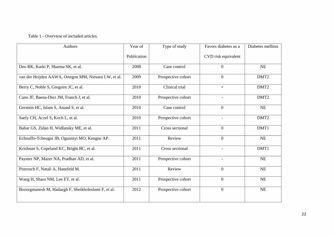
Table 1 - Overview of included articles.
Authors Year of
Publication
Type of study Favors diabetes as a
CVD risk equivalent
Diabetes mellitus
Deo RK, Karki P, Sharma SK, et al. 2008 Case control 0 NE
van der Heijden AAWA, Ortegon MM, Niessen LW, et al. 2009 Prospective cohort 0 DMT2
Berry C, Noble S, Gregoire JC, et al. 2010 Clinical trial + DMT2
Cano JF, Baena-Diez JM, Franch J, et al. 2010 Prospective cohort - DMT2
Gerstein HC, Islam S, Anand S, et al. 2010 Case control 0 NE
Saely CH, Aczel S, Koch L, et al. 2010 Prospective cohort - DMT2
Babar GS, Zidan H, Widlansky ME, et al. 2011 Cross sectional 0 DMT1
Echouffo-Tcheugui JB, Ogunniyi MO, Kengne AP. 2011 Review 0 NE
Krishnan S, Copeland KC, Bright BC, et al. 2011 Cross sectional - DMT1
Paynter NP, Mazer NA, Pradhan AD, et al. 2011 Prospective cohort - NE
Pistrosch F, Natali A, Hanefeld M. 2011 Review 0 NE
Wang H, Shara NM, Lee ET, et al. 2011 Prospective cohort 0 NE
Bozorgmanesh M, Hadaegh F, Sheikholeslami F, et al.
2012 Prospective cohort 0 NE

23
Kato K, Yonetsu T, Kim S-J, et al. 2012 Retrospective 0 NE
Wang CC, Reusch JE. 2012 Review 0 NE
Zoungas S, Chalmers J, Ninomiya T, et al. 2012 Randomized clinical trial 0 DMT2
Eskesen K, Jensen MT, Galatius S, et al. 2013 Prospective cohort 0 NE
Hernandez D, Espejo-Gil A, Bernal-Lopez MR, et al. 2013 Cross sectional - NE
Saely CH, Drexel H. 2013 Review 0 NE
Sattar N. 2013 Review - NE
+ - Suports the hipothesis that diabetes is a cardiovascular disease equivalent. - - Refutes the hipothesis that diabetes is a cardiovascular disease equivalent.
0 – No straight answer is given to the question “is diabetes a cardiovascular disease equivalent?”. DMT1 – Data exclusively from patients with type 1 diabetes
mellitus was quoted in the study or reviewed; DMT2 - Data exclusively from patients with type 2 diabetes mellitus was quoted in the study or reviewed; NE –
It was not explicit if quoted data belonged to patients who suffered from type 1 or type 2 diabetes mellitus.

24

Agradecimentos
Agradeço a todos os que, mesmo não estando envolvidos directamente na elaboração
desta tese, me ajudaram de uma maneira ou de outra: agradeço ao Luís Manuel Ferreira Pinto
por todas as dicas e sugestões dadas; agradeço a todos os meus amigos e colegas que, de uma
forma ou de outra, suportaram os meus devaneios; agradeço aos meus pais, madrinha, e
primos por todo apoio e força dados.

A Revista Portuguesa de Cardiologia, órgão oficial da Sociedade Portuguesa de Cardiologia, é uma publicação científica internacional destinada ao estudo das doenças cardiovasculares.
Publica artigos em português na sua edição em papel e em portu-guês e inglês na sua edição online, sobre todas as áreas da Medicina Cardiovascular. Se os artigos são publicados apenas em inglês, esta versão surgirá simultaneamente em papel e online. Inclui regularmen-te artigos originais sobre investigação clínica ou básica, revisões te-máticas, casos clínicos, imagens em cardiologia, comentários editoriais e cartas ao editor. Para consultar as edições online deverá aceder através do link www.revportcardiol.org.
Todos os artigos são avaliados antes de serem aceites para publi-cação por peritos designados pelos Editores (peer review). A sub-missão de um artigo à Revista Portuguesa de Cardiologia implica que este nunca tenha sido publicado e que não esteja a ser avaliado para publicação noutra revista.
Os trabalhos submetidos para publicação são propriedade da Re-vista Portuguesa de Cardiologia e a sua reprodução total ou parcial deverá ser convenientemente autorizada. Todos os autores deverão enviar a Declaração de Originalidade, conferindo esses direitos à RPC, na altura em que os artigos são aceites para publicação.
Envio de manuscritosOs manuscritos para a Revista Portuguesa de Cardiologia são en-viados através do link http://www.ees.elsevier.com/repc. Para enviar um manuscrito, é apenas necessário aceder ao referido link e seguir todas as instruções que surgem.
Responsabilidades ÉticasOs autores dos artigos aceitam a responsabilidade definida pelo Comité Internacional dos Editores das Revistas Médicas (consultar www.icmje.org).
Os trabalhos submetidos para publicação na Revista Portuguesa de Cardiologia devem respeitar as recomendações internacionais sobre investigação clínica (Declaração de Helsínquia da Associação Médica Mundial, revista recentemente) e com animais de laboratório (So-ciedade Americana de Fisiologia). Os estudos aleatorizados deverão seguir as normas CONSORT.
Informação sobre autorizaçõesA publicação de fotografias ou de dados dos doentes não devem identificar os mesmos. Em todos os casos, os autores devem apre-sentar o consentimento escrito por parte do doente que autorize a sua publicação, reprodução e divulgação em papel e na Revista Portu-guesa de Cardiologia. Do mesmo modo os autores são responsáveis por obter as respectivas autorizações para reproduzir na Revista Portuguesa de Cardiologia todo o material (texto, tabelas ou figuras) previamente publicado. Estas autorizações devem ser solicitadas ao autor e à editora que publicou o referido material.
Conflito de interessesCada um dos autores deverá indicar no seu artigo se existe ou não qualquer tipo de Conflito de Interesses.
Declaração de originalidadeO autor deverá enviar uma declaração de originalidade. Ver anexo IProtecção de dadosOs dados de carácter pessoal que se solicitam vão ser tratados num ficheiro automatizado da Sociedade Portuguesa de Cardiologia (SPC) com a finalidade de gerir a publicação do seu artigo na Revista Por-tuguesa de Cardiologia (RPC). Salvo indique o contrário ao enviar o artigo, fica expressamente autorizado que os dados referentes ao seu nome, apelidos, local de trabalho e correio electrónico sejam publica-dos na RPC, bem como no portal da SPC (www.spc.pt) e no portal online www.revportcardiol.org, com o intuito de dar a conhecer a autoria do artigo e de possibilitar que os leitores possam comunicar com os autores.
INSTRUÇÕES AOS AUTORESTodos os manuscritos deverão ser apresentados de acordo com as normas de publicação. Pressupõe-se que o primeiro autor é o repon-sável pelo cumprimento das normas e que os restantes autores conhe-cem, participam e estão de acordo com o conteúdo do manucrito.
1. Artigos OriginaisApresentação do documento:• Com espaço duplo, margens de 2,5 cm e páginas numeradas.• Não deverão exceder 5.000 palavras, contadas desde a primeira à última página, excluindo as tabelas.• Consta de dois documentos: primeira página e manuscrito• O manuscrito deve seguir sempre a mesma ordem: a) resumo estru-turado em português e palavras-chave; b) resumo estruturado em inglês e palavras-chave; c) quadro de abreviaturas em português e em inglês; d) texto; e) bibliografia; f) legendas das figuras; g) tabelas (opcional) e h) figuras (opcional)-
Primeira páginaTítulo completo (menos de 150 caracteres) em português e em inglês.
Nome e apelido dos autores pela ordem seguinte: nome próprio, seguido do apelido (pode conter dois nomes)
Proveniência (Serviço, Instituição, cidade, país) e financiamento caso haja.
Endereço completo do autor a quem deve ser dirigida a corres-pondência, fax e endereço electrónico.
Faz-se referência ao número total de palavras do manuscrito (ex-cluindo as tabelas).
Resumo estruturadoO resumo, com um máximo de 250 palavras, está dividido em quatro partes: a) Introdução e objectivos; b) Métodos; c) Resultados e d) Conclusões.
Deverá ser elucidativo e não inclui referências bibliográficas nem abreviaturas (excepto as referentes a unidades de medida).
Inclui no final três a dez palavras-chave em português e em inglês. Deverão ser preferencialmente seleccionadas a partir da lista publica-da na Revista Portuguesa de Cardiologia, oriundas do Medical Subject
Normas de publicação da Revista Portuguesa de Cardiologia

Headings (MeSH) da National Libray of Medicine, disponível em: www.nlm.nihgov/mesh/meshhome.html.
O resumo e as palavras-chave em inglês devem ser apresentados da mesma forma.
TextoDeverá conter as seguintes partes devidamente assinaladas: a) In-trodução; b) Métodos; c) Resultados; d) Discussão e e) Conclusões. Poderá utilizar subdivisões adequadamente para organizar cada uma das secções.
As abreviaturas das unidades de medida são as recomendadas pela RPC (ver Anexo II).
Os agradecimentos situam-se no final do texto.
BibliografiaAs referências bibliográficas deverão ser citadas por ordem numérica no formato ‘superscript’, de acordo com a ordem de entrada no texto.
As referências bibliográficas não incluem comunicações pessoais, manuscritos ou qualquer dado não publicado. Todavia podem estar incluídos, entre parêntesis, ao longo do texto.
São citados abstracts com menos de dois anos de publicação, identificando-os com [abstract] colocado depois do título.
As revistas médicas são referenciadas com as abreviaturas utiliza-das pelo Index Medicus: List of Journals Indexed, tal como se publi-cam no número de Janeiro de cada ano. Disponível em: http://www.ncbi.nlm.nih.gov/entrez/citmatch_help.html#JournalLists.
O estilo e a pontuação das referências deverão seguir o modelo Vancouver 3.
Revista médica: Lista de todos os autores. Se o número de autores for superior a três, incluem-se os três primeiros, seguidos da abreviatu-ra latina et al. Exemplo:
17. Sousa PJ, Gonçalves PA, Marques H et al. Radiação na AngioTC cardíaca; preditores de maior dose utilizada e sua redução ao lon-go do tempo. Rev Port cardiol, 2010; 29:1655-65Capítulo em livro: Autores, título do capítulo, editores, título do
livro, cidade, editora e páginas. Exemplo: 23. Nabel EG, Nabel GJ. Gene therapy for cardiovascular disease. En: Haber E, editor. Molecular cardiovascular medicine. New York: Scientific American 1995. P79-96.Livro: Cite as páginas específicas. Exemplo: 30. Cohn PF. Silent myocardial ischemia and infarction. 3rd ed. New York: Mansel Dekker; 1993. P. 33.Material electrónico: Artigo de revista em formato electrónico.
Exemplo: Abood S. Quality improvement initiative in nursing homes: the ANA acts it an advisory role. Am J Nurs. [serie na internet.] 2002 Jun citado 12 Ago 2002:102(6): [aprox. 3] p. Disponível em: http://www.nursingworld.org/AJN/2002/june/Wawatch.htm. A Bibliografia será enviada como texto regular, nunca como nota de rodapé. Não se aceitam códigos específicos dos programas de gestão bibliográfica.
1. FigurasAs figuras correspondentes a gráficos e desenhos são enviadas no for-mato TIFF ou JPEG de preferência, com uma resolução nunca inferior a 300 dpi e utilizando o negro para linhas e texto. São alvo de numera-ção árabe de acordo com a ordem de entrada no texto.• A grafia, símbolos, letras, etc, deverão ser enviados num tamanho que, ao ser reduzido, os mantenha claramente legíveis. Os detalhes especiais deverão ser assinalados com setas contrastantes com a figura.• As legendas das figuras devem ser incluídas numa folha aparte. No final devem ser identificadas as abreviaturas empregues por ordem alfabética.
• As figuras não podem incluir dados que dêem a conhecer a proveniência do trabalho ou a identidade do paciente. As fotogra-fias das pessoas devem ser feitas de maneira que estas não sejam identificadas ou incluir-se-á o consentimento por parte da pessoa fotografada.
TabelasSão identificadas com numeração árabe de acordo com a ordem de entrada no texto.
Cada tabela será escrita a espaço duplo numa folha aparte.• Incluem um título na parte superior e na parte inferior são refe-ridas as abreviaturas por ordem alfabética.• O seu conteúdo é auto-explicativo e os dados que incluem não figuram no texto nem nas figuras.
2. Cartas ao EditorDevem ser enviadas sob esta rubrica e referem-se a artigos publica-dos na Revista. Serão somente consideradas as cartas recebidas no prazo de oito semanas após a publicação do artigo em questão.• Com espaço duplo, com margens de 2,5 cm.• O título (em português e em inglês), os autores (máximo quatro), proveniência, endereço e figuras devem ser especificados de acordo com as normas anteriormente referidas para os artigos originais.• Não podem exceder as 800 palavras.• Podem incluir um número máximo de duas figuras. As tabelas estão excluídas.
3. Casos ClínicosDevem ser enviados sob esta rubrica.• A espaço duplo com margens de 2,5 cm.• O título (em português e em inglês) não deve exceder 10 palavras
Os autores (máximo oito) proveniência, endereço e figuras serão especificados de acordo com as normas anteriormente referidas para os artigos originais.
O texto explicativo não pode exceder 3.000 palavras e contem in-formação de maior relevância. Todos os símbolos que possam constar nas imagens serão adequadamente explicados no texto.
Contêm um número máximo de 4 figuras e pode ser enviado mate-rial suplementar, como por exemplo vídeoclips.
4. Imagens em Cardiologia• A espaço duplo com margens de 2,5 cm.• O título (em português e em inglês) não deve exceder oito palavras• Os autores (máximo seis), proveniência, endereço e figuras serão especificados de acordo com as normas anteriormente referidas pa-ra os artigos originais.• O texto explicativo não pode exceder as 250 palavras e contem informação de maior relevância, sem referências bibliográficas. To-dos os símbolos que possam constar nas imagens serão adequada-mente explicados no texto.• Contêm um número máximo de quatro figuras.
5. Material adicional na WEBA Revista Portuguesa de Cardiologia aceita o envio de material electrónico adicional para apoiar e melhorar a apresentação da sua investigação científica. Contudo, unicamente se considerará para publicação o material electrónico adicional directamente relacionado com o conteúdo do artigo e a sua aceitação final dependerá do critério do Editor. O material adicional aceite não será traduzido e publicar-se-á electronicamente no formato da sua recepção.
Para assegurar que o material tenha o formato apropriado reco-mendamos o seguinte:
Normas de publicação da revista portuguesa de cardiologia
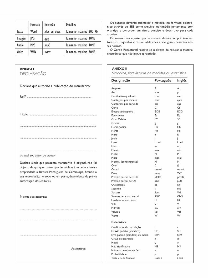
ANEXO IISímbolos, abreviaturas de medidas ou estatística
Formato Extensão Detalhes
Texto Word .doc ou docx Tamanho máximo 300 Kb
Imagem JPG .jpg Tamanho máximo 10MB
Audio MP3 .mp3 Tamanho máximo 10MB
Vídeo WMV .wmv Tamanho máximo 30MB
Os autores deverão submeter o material no formato electró-nico através do EES como arquivo multimédia juntamente com o artigo e conceber um título conciso e descritivo para cada arquivo.
Do mesmo modo, este tipo de material deverá cumprir também todos os requisitos e responsabilidades éticas gerais descritas nes-sas normas.
O Corpo Redactorial reserva-se o direito de recusar o material electrónico que não julgue apropriado.
ANEXO I
DECLARAÇÃO
Declaro que autorizo a publicação do manuscrito:
Ref.ª ........................................................................................
Título ...........................................................................................
........................................................................................................
.........................................................................................................
........................................................................................................
do qual sou autor ou c/autor.
Declaro ainda que presente manuscrito é original, não foi objecto de qualquer outro tipo de publicação e cedo a inteira propriedade à Revista Portuguesa de Cardiologia, ficando a sua reprodução, no todo ou em parte, dependente de prévia autorização dos editores.
Nome dos autores:
.............................................................................................
..................................................................................................
.........................................................................................................
Assinaturas:
Normas de publicação da revista portuguesa de cardiologia
Designação
AmpereAnoCentímetro quadradoContagens por minutoContagens por segundoCurieElectrocardiogramaEquivalenteGrau CelsiusGramaHemoglobinaHertzHoraJouleLitroMetroMinutoMolarMoleNormal (concentração)OhmOsmolPesoPressão parcial de CO2
Pressão parcial de O2
QuilogramaSegundoSemanaSistema nervoso centralUnidade InternacionalVoltMilivoltVolumeWatts
Estatística:
Coeficiente de correlaçãoDesvio padrão (standard)Erro padrão (standard) da médiaGraus de liberdadeMédiaNão significativaNúmero de observaçõesProbabilidadeTeste «t» de Student
Português
Aanocm2
cpmcpsCiECGEq°CgHbHzhJL ou LmminMmolNΩosmolpesopCO2
pO2
kgsSemSNCUIVmVVolW
rDPEPMglχNSnpteste t
Inglês
Ayrcm2
cpmcpsCiECGEq°CgHbHzhJI ou LmminMmolNΩosmolWTpCO2
pO2
kgsecWkCNSIUVmVVolW
rSDSEMdfχNSnpt test