β-GLUCOSIDASE FROM Trichoderma harzianum T12 AS GREEN FUNGICIDE
AGAINST Macrophomina phaseolina IN SOYBEAN ( Glycine max L. )
ELHAM KHALILI
UNIVERSITI TEKNOLOGI MALAYSIA
β-GLUCOSIDASE FROM Trichoderma harzianum T12 AS GREEN FUNGICIDE
AGAINST Macrophomina phaseolina IN SOYBEAN (Glycine max L.)
ELHAM KHALILI
A thesis submitted in fulfilment of the
requirements for the award of the degree of
Doctor of Philosophy (Bioscience)
Faculty of Biosciences and Medical Engineering
Universiti Teknologi Malaysia
OCTOBER 2017

iii
Specially dedicated to:
My family for their endless support and motivation.
iv
ACKNOWLEDGEMENT
First and foremost, I am very much thankful to Almighty Allah for showering
his endless blessings and guidance upon me to obtain this position in my education,
where i can apply the knowledge gained from my supervisor, co-supervisors and
other academic staff for the betterment of my career and country.
My special thanks to my supervisor Dr. Roswanira Abdul Wahab who
throughout my studies gave me great advices and knowledge, without which i would
not be able to complete this degree. She, throughout my stay in the university has
been a very cooperative teacher and mentor. She was always ready to advise me
despite his tough and strict schedule. I would also like to thank my co-supervisor
professor Dr. Muhammad Arshad Javed, who always helped and guided me and also
boosted my confidence at every stage of my ph.D. I would also thank professor Dr.
Fahrul zaman huyop and Dr. Siavosh Rayatpanah for their generous help and
guidance.
In my daily work, I have been blessed with a friendly and cheerful group of
fellow students. Fatin Myra Binti Abd Manan, who was a good companion or
otherwise the laboratory would be exhausting and disappointiting. Alao, big thanks
to Miss Haziqah, for the excellent technical assistance in the laboratory, particularly
for experimental techniques, and kindly answers to my questions. I would like to
thank to Dr. Wafaa Hassan Muslem who always helped me during encouraged me in
the course of my Ph.D. Last but not least, my deepest gratitude goes to my beloved
parents and my lovely brothers. To those who indirectly contributed in this research,
your kindness means a lot to me. Thank you very much.
v
ABSTRACT
Macrophomina phaseolina (Tassi) Goid remains the prevailing causal agent of charcoal rot disease that can significantly suppress yields of a variety of crops. Its wide host range and survivability under arid conditions as well as the ineffectiveness of fungicides have spurred scientific endeavors in search of alternative avenues to control this phytopathogen. The present study is aimed to provide empirical evidence on the efficacy of β-glucosidase from Trichoderma harzianum T12 as a biological control agent against M. phaseolina. In-vitro pathogenicity tests on 60 isolates of M. phaseolina and 30 isolates of T. harzianum, collected from different areas of the Mazandaran province in Iran revealed the isolates, M2 of M. phaseolina and the T12 of T. harzianum were the most virulent and effective in inhibiting growth of M. phaseolina, respectively. The present study showed that biochemical and phylogenetic analyses and BIOLOG results confirmed the fungal antagonists and phytopathogen were T. harzianum (Rifai) and M. phaseolina (Tassi) Goid, respectively. Purified extracellular β-glucosidase of T. harzianum inhibited the growth of M. phaseolina as seen from the large halo zones, indicating its possible application as a green fungicide against M. phaseolina. The β-glucosidase had an optimum pH (7) and temperature 45˚C, respectively, remarkably stable up to 240 min with a half-live of t1/2 = 210 min at 40 ˚C to 60 ˚C. Zn
2+, Mn
2+, and Tween 80
enhanced its activity while was substantially inhibited by Fe3+
. Enzyme activity was the highest when wheat bran and (NH4)2SO4 were used as carbon and nitrogen sources respectively. The kinetic parameters for β-glucosidase T12, Km, Vmax and kcat were estimated as 0.79 mM, 8.45 mM min
-1 mg
-1 protein and 10.69 s
-1, respectively,
to give a turnover number of 10.69 s-1
. Optimization by the Box-Behnken Design (BBD) based on: temperature, carbon sources, inoculum size and pH (7), exhibited the highest β-glucosidase activity (1260 U/mL) at 45˚C, pH 7, using a carbon source 10 % (w/v) and inoculum size of 5 % (w/v). The BBD optimization for the application of the β-glucosidase formulation from T. harzianum to control infestation of M. phaseolina M2 was carried out on soybean plants grown under a greenhouse condition. Under an optimized condition, the lowest plant disease index (PDI) of 4.32% (R
2 = 0.9676) was attained using 10 mM Zn
2+, Tween 80 at 2 % (w/v) an
enzyme concentration at 15 mg/L and an irrigation frequency of 2 times/week. A comparative study showed the developed formulation gave the lowest PDI (4.14 %) (p < 0.05) followed by the antagonist T. harzianum Rifai (26.13 %) and the commercial fungicide, Carbendazim (32.45 %). The assessments cost revealed that the enzyme formulation only costs at USD34/acre as compared to Carbendazim at USD240/acre. Hence, the findings affirmed that the novel use of crude β-glucosidase from the growth supernatant of T. harzianum was efficient in combating charcoal rot disease. Since the enzyme formulation was substantially cheaper and its application combines the practicality of an in-situ spraying for rapid control of M. phaseolina infestation, the technique proposed here was prospectively feasible to control such disease in crops.
vi
ABSTRAK
Macrophomina phaseolina (Tassi) Goid merupakan penyebab penyakit reput arang yang memberi kesan ketara kepada penghasilan pelbagai tanaman. Kepelbagaian perumah dan keupayaan untuk terus hidup di bawah keadaan tandus, ditambah pula dengan ketidakberkesanan fungisid telah merangsang kajian ini untuk mencari alternatif bagi mengawal fitopatogen ini. Kajian ini bertujuan memberi bukti secara empirikal ke atas keupayaan β-glukosida daripada Trichoderma harzianum T12 sebagai agen kawalan biologi terhadap penyakit reput arang. Ujian kepathogenenikan secara in-vitro ke atas 60 isolat M. fhaseolina dan 30 isolat T. harzianum, yang dikutip dari kawasan yang berlainan di wilayah Mazandaran di Iran mendedahkan bahawa isolat M2 M. phaseolina ialah yang paling patogenik manakala isolat T12 T. harzianum paling efektif merencat pertumbuhan M. phaseolina M2. Ujian biokimia, analisa filogeni dan keputusan BIOLOG masing-masing mengesahkan bahawa kulat antagonistik dan patogenik tersebut adalah T. harzianum (Rifai) dan M. phaseolina (Tassi) Goid. β-glukosida ekstrasel T. harzianum didapati menghalang pertumbuhan M. phasaolina seperti yang dilihat dari zon halo yang besar, menunjukkan kemungkinan penggunaannya sebagai fungisida hijau terhadap M. phaseolina. β-glukosida tersebut mempunyai pH dan suhu optimum masing-masing pada pH 7 dan 45˚C, sangat stabil pada 240 minit dengan separuh hayat t1/2 = 210 min pada suhu 40 ˚C to 60 ˚C. Ion Zn
2+, Mn
2+ and surfaktan
Tween 80 didapati dapat meningkatkan aktiviti manakala Fe3+
merencat aktivitinya dengan ketara. Aktiviti enzim adalah pada tahap tertinggi apabila dedak gandum dan amonium sulfida digunakan sebagai sumber karbon dan nitrogen masing-masing. Parameter kinetik Km, Vmax dan kcat β-glukosida T12 yang dijangka masing-masing pada 0.79 mM, 8.45 mM min
-1 mg
-1 protein and 10.69 s
-1, unuk memberi jumlah
perolehan sebanyak 10.69 s-1
. Pengoptimum menggunakan Design Box-Behken (BBD) berdasarkan empat parameter: suhu, sumber karbon, saiz inokulum, pH (7) mendedahkan keadaan optimum. Oleh itu, penghasilan aktiviti β-glukosida (1260 U/mL) adalah pada suhu 45˚C, pH 7, dengan sumber karbon 10 % (w/v) dan saiz inokulum 5 % (w/v). Indeks penyakit tumbuhan terendah (PDI) sebesar 4.32% (R
2 =
0.9676) dicapai dengan menggunakan Zn2+
(10 mM), Tween 80 pada 2% (w/v), 15 mg/L kepekatan enzim dan kekerapan pengairan sekurang-kurangnya 2 kali/minggu. β-glukosida dari T. harzianum digunakan untuk mengawal serangan M. phaseolina M2 dilakukan pada penggunaan pokok soya yang tumbuh di dalam persekitaran rumah hijau. Pengoptimuman BBD untuk penggunaan formula indeks penyakit tumbuhan yang paling rendah (PD1) ialah pada 4.32%. Kajian komparatif menunjukkan formula β-glucosidase memberikan PDI paling rendah (4.14%) (p < 0.05) berbanding antagonis T. harzianum Rifai (26.13%) dan racun kulat komersial, Carbendazim (32.45%). Penilaian kos menunjukkan kos formula yang dibangunkan hanya USD34/ekar berbanding Carbendazim USD 240/ekar. Oleh itu, hasil kajian mengesahkan bahawa β-glucokida yang diperoleh dari supernatant pertumbuhan T. harzianum adalah cekap dalam memerangi penyakit reput arang. Memandangkan formula β-glukosida yang dibangunkan lebih murah dan penggunaannya yang praktikal dengan hanya semburan secara in-situ untuk kawalan segera M. phaseolina. Oleh itu, teknik yang dicadangkan di sini adalah berpotensi dan boleh digunakan untuk mengawal penyakit arang reput pada tanaman.
vii
TABLE OF CONTENTS
CHAPTER TITLE PAGE
DECLARATION ii
DEDICATION iii
ACKNOWLEDGEMENT iv
ABSTRACT v
ABSTRAK vi
TABLE OF CONTENTS vii
LIST OF TABLES xiv
LIST OF FIGURES xvii
LIST OF ABBREVIATIONS xxiii
LIST OF APPENDICES xxvii
1 INTRODUCTION 1
1.1 Background of Study 1
1.2 Problem Statement 4
1.3 Objectives of the Study 5
1.4 Aim of Study 5
1.5 Scope of the Study 6
1.6 Significance of the Study 7
1.7 Operational Framework of the Research 8
2 LITERATURE REVIEW 9
2.1 Macrophomina phaseolina 9
2.1.1 Taxonomy of M. phaseolina 11
2.1.2 M. phaseolina as Plant Pathogen in 12

viii
Various Plants
2.1.3 General Morphological Studies 14
2.1.4 Physiological Properties of M.
phaseolina 17
2.1.5 The Infection Cycle of M. phaseolina 18
2.2 Soybean 20
2.2.1 Biochemistry and Physiological
Characteristics of Soybean
21
2.3 Charcoal Rot Disease 21
2.4 Symptom of Charcoal Rot Disease in Plants 22
2.5 Current techniques in Managing Charcoal Rot
Disease in Crops 24
2.5.1 Irrigation During Drought Season 25
2.5.2 Crop Rotation 27
2.5.3 Tillage 28
2.5.4 Treatments with Fungicides 29
2.5.4.1
Biochemistry and Physiological
Characteristics of Soybean
31
2.5.5 Biological Control 31
2.6 Trichoderma harzianum 34
2.6.1 Taxonomy of T. harzianum 34
2.6.2 Morphology of T. harzianum 36
2.7 T. harzianum as Biological Control Agents M.
phaseolina 37
2.8 Trichoderma harzianum and its Mechanism of
Antagonism 37
2. 9 Trichoderma Species as Biological Control
Agents and Antagonists 38
2.9.1 Competition of Nutrients and Space 39
2.9.2 Production of Bioactive Compounds 39
2.10 Trichoderma as Biological Control Research
on Oil Palm in Malaysia 40
2.11 T. harzianum a Potential Candidate for β-
glucosidases Production 40
2.12 β-glucosidases and its Classification 41
ix
2.12.1 β-glucosidases 41
2.12.2 Classification of β-glucosidases 42
2.13 General Reactions Role of β-glucosidases from
T. harzianum 44
2.14 Conditions for Batch Cultivation of T.
harzianum 44
2.14.1 Effect of Temperature 44
2.14.2 Effect of Carbon Source 45
2.14.3 Effect of Nitrogen Source 46
2.14.4 Effect of Metal Ions 46
2.14.5 Effect of Surfactants 47
2.15 Response Surface Methodology 48
2.16 Box-Behnken Design 48
2.17 Assessment of Plant Disease Index (PDI) 53
3 RESEARCH METHODOLOGY 55
3.1 Introduction 55
3.2 Sample Collection and Isolation of the
Phytopathogen M. phaseolina 55
3.3 Investigation of Sclerotia Population of M.
phaseolina in Soil 57
3.4 Soil Chemical and Physical Properties 58
3.5 Sample Collection and Isolation of Antagonist T.
harzianum 58
3.6 Preparation of Pure Culture of Isolates of M.
phaseolina and T. harzianum 60
3.7 Morphological Identification of M. phaseolina
and T. harzianum 61
3.8 Preparation of Mycelial Mass of the
Phytopathogen M. phaseolina and T. harzianum
antagonist 61
3.9 Molecular Identification of Isolates 62
3.9.1 Extraction of Genomic DNA 62
3.9.2 Polymerase Chain Reactions for ITS
Gene Amplification 63
3.9.3 Gel Electrophoresis 63
3.10 Identification of the Most Pathogenic Isolates of
M. phaseolina 64
x
3.10.1 Determination of Pathogenic variability 64
3.11 Investigation of Antagonistic Mechanism to
Identify Most Effective T. harzianum 65
3.11.1 Laboratory Assessments of Antagonistic
Mechanisms 65
3.11.1.1 Dual Culture Technique 65
3.11.1.2 Volatile Metabolites Production Experiment 66
3.11.1.3 Hyperparasitism Test 67
3.12 Efficacy Assessment of T. harzianum
Antagonists in Soil and Seedling Treatments 67
3.13 Measurement of Stem Length, Root Length and
Seed Weight 68
3.14 Molecular Analysis of M. phaseolina M2 and T.
harzianum T12 69
3.14.1 Sequencing and Analysis of the PCR
Products 69
3.14.2 Phylogenetic Tree Analysis 69
3.15 BiologTM Gen III Micro Plate Identification 70
3.16 Growth Conditions of Fungal Cultures 70
3.17 Sporulation Medium and Inoculum Development 71
3.18 Production and Extraction of β-glucosidase 71
3.19 Quantitative Determination of β-glucosidase
Activity and Protein Determination 72
3.19.1 Determination of β-glucosidase Activity 72
3.19.2 Determination of Protein Content 73
3.20 Purification of β-glucosidase 74
3.21 Determination of Molecular Weight of β-
glucosidase T12 by SDS-PAGE 75
3.22 Amplification of Full Sequence of β-glucosidase
T12 75
3.22.1 Purification of PCR Product 76
3.23 Multiple Sequence Alignment 77
3.24 Optimization of β-glucosidase Activity from T.
harzianum T12 by Using One-Variable-at-A-
Time (OVAT) Method 77
3.24.1 Effect of Temperature 77
3.24.2 Effect of pH 78
3.24.3 Effects of Metal Ions and Surfactants 78
xi
3.24.4 Effect of Natural Carbon Sources 78
3.24.5 Effect of Nitrogen Sources 79
3.24.6 Effect of Inoculum Size 79
3.25 Statistical Analysis 79
3.26 Determination of Kinetic Parameters of β-
glucosidase T12 80
3.27 Plate Inhibition Assay 81
3.28 Experimental Design and Optimization Using
Response Surface Methodology Using Box-
Behnken Design (BBD) and Statistical Analysis 81
3.29 Plate Inhibition Assay Base on RSM 83
3.30 Green House Assessments 83
3.30.1 Experimental Design and Optimization
Using Response Surface Methodology
With a Box-Behnken Design and
Statistical Analysis 83
3.30.2 Determination of PDI 85
3.31 Comparison of β-glucosidase T12 and
Carbendazim Fungicide 86
3.32 Experimental Design and Statistical Analysis 86
4 RESULTS AND DISCUSSION 88
4.1 Symptoms of M. phaseolina on Samples
Soybean 88
4.2 Sclerotia Population of M. phaseolina Pathogen 91
4.3 Morphological Analysis of M. phaseolina 92
4.4 Morphological Analysis of T. harzianum 96
4.5 Identification of M. phaseolina and T. harzanum
Using ITS Gene Analysis 97
4.6 Identification of the Most Pathogenic Isolates of
M. phaseolina 99
4.6.1 Pathogenic Test 99
4.7 Identification of the Most Effective T.
harzianum Isolates 103
4.7.1 Dual Culture Technique 103
4.7.2 Volatile Metabolite Production Test
(VMPT) and Hyperparasitism Test 106
4.8 Measurement of Plant Disease Index (%), Stem 107
xii
Length, Root Length and Seed Weight
4.9 Assessment of Efficacy of Antagonists in Soil
and Seedling Incorporation 110
4.10 Molecular and Biochemical Analysis of Strain
M2 and T12 112
4.10.1 Fungal Genomic DNA Extraction 112
4.10.2 Amplification of ITS Region and
Purification 113
4.11 The ITS Sequence Analysis 115
4.11.1 Sequence Analysis of M. phaseolina M2
and T. harzianum T12 115
4.11.2 Phylogenetic Affiliations of Isolates 118
4.12 Biochemical Test 120
4.13 Quantitative Determination 123
4.13.1 β-glucosidase Activity and Protein
content 123
4.13.2 Production and Purification of β-
glucosidase 123
4.14 Determination of Molecular Mass of β-
glucosidase by SDS-PAGE 124
4.15 Amplification of the Complete Sequence of β-
glucosidase T12 125
4.15.1 Gel Electrophoresis of PCR Product 125
4.15.2 β-glucosidase T12 Sequence and
Analysis 126
4.16 Alignment of β-glucosidases T12 128
4.17 Optimal Conditions for β-glucosidase Activity 132
4.17.1 Effect of Temperature 132
4.17.2 Effect of pH 134
4.17.3 Effect of Metal Ions and Surfactants 137
4.17.4 Effect of Natural Carbon and Nitrogen
Source 139
4.17.5 Effect of Inoculum Size 142
4.18 Statistical Analysis 143
4.19 Determination of Kinetic Parameters of β-
glucosidase T12 143
4.20 Inhibition Assay for the Growth of M.
phaseolina Colonies by β-glucosidase T12 145
xiii
4.21 Experimental Design and Optimization Using
Response Surface Methodology Using Box-
Behnken Design (BBD) and Statistical Analysis 147
4.21.1 Fitting of the Response Models Based
on Result from the OVAT Method 147
4.22 Influence of Experimental Factors on β-
Glucosidase Activity 148
4.23 Effect of Temperature and Carbon Source 154
4.24 Effect of Temperature and Inoculum Size 157
4.25 Effect of Temperature and pH 159
4.26 Effect of Carbon Sources and pH 161
4.27 Verification Experiments for Plate Inhibition
Assay for the Growth of M. Phaseolina M2 by
Β-glucosidase T12 164
4.28 Greenhouse Experiments 168
4.28.1 Regression and Fitting Model
Representation 168
4.29 Influence of the Application Variables on the
PDI 173
4.29.1 Effect of Metal Ions and Surfactant
Concentration 173
4.29.2 Effects of Metal Ions and Irrigation
Frequency 177
4.29.3 Effect of Surfactant Concentration and
Irrigation Frequency 179
4.29.4 Effect of Concentration of β-glucosidase
and Irrigation Frequency 181
4.30 Attaining Optimum Conditions and Model
Verification 183
4.31 Comparison of T. harzianum (Rifai), β-
glucosidase T12 and Carbendazim Fungicide 186
4.32 Cost Assessment of β-glucosidase 189
5 CONCLUSION AND FUTURE WORK 191
5.1 Conclusion 191
5.2 Future Work 193
REFERENCES 194
Appendices A-J 228-239
xiv
LIST OF TABLES
TABLE NO. TITLE PAGE
2.1 Taxonomic Description of M. phaseolina 12
2.2 M. phaseolina morphological observations 16
2.3 Summary of the advantages and disadvantages of the two
irrigation methods 26
2.4 Summary of the advantages and disadvantages of the
biological control method 33
2.5 Coded factor levels for Box-Behnken design of a three-
variable system 51
2.6 Coded factor levels for Box-Behnken designs for an
optimization experiment involving four factors 52
2.7 Example of calculated (PDI) 54
3.1 Isolates of Macrophomina phaseolina collected from
different areas used in this study
56
3.2 Collection of soil samples from different agricultural
regions in Iran for the isolation of the fungal antagonist, T.
harzianum 59
3.3 Components of lysis buffer (Liu et al., 2011) 62
3.4 Universal primers used in the amplification of fungal ITS
gene 63
3.5 Components of nutrient salt 72
3.6 Concentration of pNPG for the standard curve of enzyme
activity 73
3.7 The prepared standard BSA dilution series for the
determination of protein contact 74
3.8 The designed specific oligonucleotide primers used to
amplify the β-glucosidase T12 76
3.9 The model equation was used to estimate the optimum
value and subsequently to describe the interaction between
the factors 82
xv
3.10 Coded and actual values of the variables of the design of
experiment for the overall optimization of PDI 84
4.1 Sclerotia population of M. phaseolina pathogen in
different regions of Mazandaran Province 92
4.2 Soil physical and chemical properties from the
experimental field (Jooybar) 92
4.3 Analysis of variance pathogenicity test of M. phaseolina
on soybean seed 101
4.4 Differential response of soybean cultivar Sahar against
various isolates of M. phaseolina 102
4.5 Inhibition percentage of radial growth of M. phaseolina
M2 colonies affected by T. harzianum isolates in the in
vitro dual culture and volatile production tests 104
4.6 Mean square for the field assessment studies on the
efficacy of T. harzianum isolates as biological control
agents against M. phaseolina charcoal rot pathogen 109
4.7 Evaluation of the efficacy of T. harzianum isolates as
biological control agents against M. phaseolina in the soil
incorporation and seed inoculation techniques 110
4.8 The BLASTn analysis of ITS rDNA of fungal strain M2 117
4.9 The BLASTn analysis of ITS rDNA of fungal strain T12 118
4.10 Extensive biochemical analysis of strain M. phaseolina
M2 using BIOLOGTM
GEN III Microplate 121
4.11 Extensive biochemical analysis of strain T. harzianum T12
using BIOLOGTM
GEN III Microplate 122
4.12 Summary of the purification of β-glucosidase T12 derived
from T. harzianum (Rifai) T12 124
4.13 Comparison of the ITS RNA nucleotide sequence of β-
glucosidase T12 from T. harzianum T12 with other β-
glucosidases 128
4.14 Amino acids residues of β-glucosidases 131
4.15 Kinetic parameters (kcat and Km) of β-glucosidase T12,
which are calculated using non-linear regression analysis
of experimental steady-state data using Michaelis-Menten
plot 144
4.16 Compositions of the various runs of the BBD in coded and
actual terms for the obtained actual and predicted
responses for optimizing β-glucosidase activity for the
batch culture of T. harzianum (Rifai) 150
4.17 Polynomial equations for the estimated coded and
processed factors for β-glucosidase activity used in the
BBD 151
xvi
4.18 ANOVA for the second-order polynomial model of the
BBD 152
4.19 ANOVA for the second order polynomial models and
coefficient values for β-glucosidase activity obtained from
the growth culture supernatant 153
4.20 Comparison of the predicted and experimental values for
β-glucosidase activity obtained from the batch cultures of
T. harzianum (Rifai) 167
4.21 The Box-Behnken design showing the experimental and
predicted values for the PDI of soybean plants 170
4.22 Polynomial equation for the estimated coded and
processed variables for the PDIs obtained from the Box-
Behnken experimental design 171
4.23 Analysis of variance and coefficient values for the PDI 172
4.24 Comparison of the predicted and experimental values for
the PDI 184
4.25 Comparative effects of T. harzianum (Rifai), β-
glucosidase enzyme and Carbendazim fungicide for the
control of M. phaseolina infection in soybean and on
related growth indices under greenhouse conditions 188
4.26 Mean square for different antagonists on the soybean plant
disease index (PDI) in the greenhouse 189
xvii
LIST OF FIGURES
FIGURE NO. TITLE PAGE
1.1 Operational framework of the research methodology 8
2.1 (A) M. phaseolina growing on potato dextrose agar
(PDA) plate (B) cellular morphology of the M.
phaseolina (40x) 10
2.2 The M. phaseolina disease cycle in soybean (Wrather
and Koenning, 2006). 20
2.3 (A) sudden death syndrome symptoms between the
veins (B) the sclerotia under epidermis of infected
soybean stem 24
2.4 (A) sudden death syndrome symptoms between the
veins (B) the sclerotia under epidermis of infected
soybean stem 24
2.5 Enzymatic hydrolysis of cellulose schematic diagram
showing cellulose synergy (Lynd et al., 2002) 42
2.6 Representative structure of 4/7 super family with the
eight fold β/α barrel motif 43
2.7 The cube for BBD and a three interlocking 22 factorial
design; (A) a cube that consists of the central point and
the middle points of the edges, (B) a figure of three
interlocking 22 factorial designs and a central point 50
2.8 Standard area used to estimate plant disease index (A):
stem (B). Leaf of infected plant 53
3.1 Soybean growing location in Mazandaran Province,
Iran; Stars are showing locations of sample collection of
M. phaseolina and T. harzianum isolates. 57
4.1 (A) a healthy soybean field and (B) an infected soybean
field with charcoal rots symptoms (C) an infected
soybean plant with no pod on stem 90
4.2 Appearance of sclerotia under epidermis of infected
soybean stem (A) and root (B) Note: The The circles
shows the sclerotia 91
Plot the antenna radiation pattern

xviii
4.3 White coloured hyphae of M. phaseolina in petri dishes;
(B) hyphae under bright- field photomicrograph; (C)
brown coloured hyphae of M. phaseolina in petri dishes;
(D) hyphae under bright- field photomicrograph (04x) 93
4.4 (A) aerial mycelium of M. phaseolina on PDA; (B)
hyphae of M. phaseolina under bright-field
photomicrograph showing right-angled branching and
constriction at the union point with the mother hyphae
(04x) 94
4.5 (A) fusion between vegetative hyphae starting from a
single hyphae, (B) fusion of adjacent vegetative hyphae
and (C) fusion between vegetative hyphae through
production of connecting hyphae from a vegetative
hypha (04X) 95
4.6 (A) combination of sclerotia together to produce a larger
sized sclerotia that now showed a different shape (40X)
and (B) merged sclerotia (100 X) 95
4.7 Morphological identification of T. harzianum under
microscope (A) the colony of antagonist fungus T.
harzianum (B) conidiophores (C) conidia (D)
chlamydospores (40X) 96
4.8 Amplification of M. phaseolina gene (650 bp) on
electrophoresis gel to identify the M. phaseolina isolates
M: weight marker 1KB; 1-60: Number of M. phaseolina
isolates; NC: Negative control, the amplified the gene in
Aspergillus sp DNA 98
4.9 Amplification of T. harzianum gene (600 bp) on
electrophoresis gel to identify the T. harzianum isolates
M: weight marker 1KB; 1-30: Number of T. harzianum
isolates; NC: negative control Negative control, the
amplified the gene in Aspergillus sp DNA. 99
4.10 Germinated sclerotia of M. phaseolina on the surface of
soybean seed grown in dilute potato dextrose agar. (A)
Sclerotium with single germ hyphae time in hours (B)
Sclerotium showing three germ hypha 100
4.11 (A) healthy soybean seeds (B) soybean seeds with M.
phaseolina M2 103
4.12 Dual culture test (A-C) and volatile metabolites
production test (E-G) on PDA plates showing the
mycelial growth inhibition. Note: M. phaseolina M2 +
T. harzianum (T2) (A); M. phaseolina M2+ T.
harzianum (T10) (B); M. phaseolina M2+ T. harzianum
(T12) (C); M. phaseolina M2 (control) (D); T.
harzianum (T10) with M. phaseolina M2 (E); T.
harzianum (T12) with M. phaseolina M2 (F); T.
harzianum (T2) with M. phaseolina M2 (G). 105
xix
4.13 Mycelial interactions between T. harzianum T12 and M.
phaseolina M2. Note: The circle shows the penetration
sites of the antagonist T. harzianum T12 and lysis of the
pathogen cells. 107
4.14 Results of the field study for the soybean stem (A) root
(B) and seedlings (C) from the plots treated with T12
suspension of T. harzianum. Note: A: soybean stem
(left: T. harzianum (T12) seed treatment; right: control);
B: soybean root (left: T. harzianum (T12) seed
treatment; right: control); C: soybean seed (left: T.
harzianum (T12) seed treatment; right: control) and
control = infected soybean 111
4.15 The extracted genomic DNA of fungal strain with
standard labelled size of DNA marker on gel
electrophoresis. Lane M: DNA ladder Lane 1 and 2:
Genomic DNA of T. harzianum T12 and M. phaseoilina
M2 respectively 113
4.16 The amplified ITS region for Macrophomina strain.
Lane L: DNA ladder; 1: strain M. phaseolina M2; C:
negative control (Fusarium graminearum) 114
4.17 The amplified ITS region for Trichoderma strain. Lane
L: DNA ladder; 1: strain T. harzianum T12; C: negative
control (without genomic DNA) 114
4.18 Partial gene sequence for fungal isolates M. phaseolina
M2 115
4.19 Partial gene sequence for fungal isolate T. harzianum,
T12 116
4.20 Phylogenetic tree of ITS sequence obtained for M.
phaseolina (Tassi) Goid strain M2 (KY549686) with
sequences of other M. phaseolina species and an
outgroup (F. graminearum CS3005) 119
4.21 Phylogenetic tree of ITS sequence obtained for T.
harzianum strain T12 (KY004113) with sequences of
other T. harzianum species and an outgroup (T. viride
strain ITV-VSL34) 120
4.22 The SDS-PAGE of the fractions obtained from the
purification of crude β-glucosidase enzyme T12; M: 1
kB Protein ladder, C: Protein precipitations of crude
extract at various percentage of (NH4)2SO4: F1; 20%
(NH4)2SO4, F2:40 % (NH4)2SO4, F3: 60 % (NH4)2SO4
and (KDa) F4: 80 % (NH4)2SO4 (96 kDa) 125
4.23 Agarose gel (1%) showing β-glucosidase T12 gene
amplification using universal primers. Lane 1: 100 bp
plus Ladder; Lane 2: ~1500 bp amplification using β-
glucosidase T12; Lane 3: Nagative control (without
xx
adding forward primer in the PCR reaction) 126
4.24 Nucleotide alignment for the full sequence of DNA of
the β-glucosidase T12 fragment amplified using
designed primers TFbgl2 and TRbgl2, start and end
124codons are shown in the box 127
4.25 Multiple sequence alignment between the complete
sequence of other β-glucosidaseses T12 with four other
β-glucosidaseses (Table 4.13) with high sequence
identity. The alignment is boxed red with white
characters to highlight conserved amino acids. The stars
( ) indicate the possible residues important to β-
glucosidases T12 130
4.26 Effect of different temperatures on the purified β-
glucosidase T12. (A) enzyme activity and (B)
Thermostability [Assessment condition: up to 240 min,
pH 7.0 at 200 rpm] 133
4.27 Effect of different pH on the activity of the purified β-
glucosidase T12 (A) activity and (B) pH stability
[Assessment condition: Up to 240 min at 45 oC, pH 7.0
at 200 rpm] 136
4.28 Effect of metal ions (10 mM) on the activity of the
purified β-glucosidase T12. The data labels depict the
relative activities of the enzymes in comparison to the
negative control. [Assessment condition: 45˚C, pH 7 at
200 rpm] 137
4.29 Effect of surfactants (10 %, w/v) on the activity of
purified β-glucosidase T12. The data labels depict the
relative activities of the enzyme in comparison to the
negative control. [Assessment condition: 45˚C, pH 7,
Zn2+ (10 mM) at 200 rpm] 138
4.30 Effect of natural carbon sources on the activity of
purified β-glucosidase T12. The data labels depict the
relative activities of the enzyme in comparison to the
control (banana wastes). [Assessment condition: 45˚C,
pH 7, Tween 80 (1 %. v/v), metal ions Zn+2
(10 mM) at
200 rpm]. 140
4.31 Effect of nitrogen sources (1.0 %, w/v) on the activity of
purified β-glucosidase T12. The data labels depict the
relative activity of the enzyme in comparison to the
negative control. [Assessment condition: 45 ˚C, pH 7,
Tween 80 (1 %. v/v), metal ions Zn2+
(10 mM) at 200
rpm] 141
4.32 Effect of inoculum size on the activity of purified β-
glucosidase T12. The data labels depict the relative
activities of the enzyme in comparison to other
xxi
inoculum sizes [Assessment condition: 45 oC, pH 7,
Tween 80 (1 %. v/v), metal ions Zn2+
(10 mM),
(NH4)2SO4 (1.0 %, w/v) at 200 rpm]
142
4.33 Lineweaver-Burk double reciprocal plot of β-
glucosidase T12 activity in the absence of various
concentration of pNPG 145
4.34 (A) the halo zone indicated by the black arrow
illustrates effective anti-fungal activity by the purified
β-glucosidase T12 (right) against fungal phytopathogen.
The halo zone was retained even after 72 h of
incubation. (A), M. phaseolina M2; (B) M. phaseolina
(negative control) 146
4.35 Comparison between the predicted and actual values
obtained for the activity of β-glucosidase obtained from
the growth culture of T. harzianum T12 (Rifai). 148
4.36 The response (a) surface and (b) contour plots showing
the interactive effect of A: Temperature and B: Carbon
source on the activity of β-glucosidase 156
4.37 The response (a) surface and (b) contour plots showing
the interactive effect of A: Temperature and C:
Inoculum size on the activity of β-glucosidase. 158
4.38 The response (a) surface and (b) contour plots showing
the interactive effect of A: Temperature and D: pH on
the activity of β-glucosidase 160
4.39 The response (a) surface and (b) contour plots showing
the interactive effect of B: Carbon sources and D: pH on
the activity of β-glucosidase 163
4.40 Plate inhibition assay showing growth of M. phaseolina
(denoted by dark areas) inhibited by the β-glucosidase
(halo zone) extracted from T. harzianum (Rifai). R1, R2
and R3 were the best conditions that produced the
highest β-glucosidase from the optimized culture broth
(Table 4.18). Data are the means of three replicates 165
4.41 Comparison between the predicted and actual values
obtained for the PDI of soybean plants 173
4.42 The response (a) surface and (b) contour plots showing
the interactive effect of metal ions (A) and surfactants
(B) on the PDI. [Two other variables, β-glucosidase
concentration and irrigation frequency were held
constant at their central values, 15 mg/mL and 2
times/week, respectively] 176
4.43 The response (a) surface and (b) contour plots showing
the interactive effect of metal ions (A) and irrigation
frequency (D) on the PDI. [Two other variables, β-
glucosidase concentration and surfactants were held
xxii
constant at their central values, 15 mg/mL and 2%
(w/v), respectively
178
4.44 The response (a) surface and (b) contour plots showing
the interactive effect of surfactant (B) and irrigation
frequency (D) on the PDI. [Two other variables, β-
glucosidase concentration and metal ions were held
constant at their central values, 15 mg/mL and 10 mM
respectively] 180
4.45 The response (a) surface and (b) contour plots showing
the interactive effect of enzyme concentration (C) and
irrigation frequency (D) on the PDI. [Two other
variables, metal ions and surfactant were held constant
at their central values, 10 mM and 2 % (, w/v),
respectively] 182
4.46 Pots assay base on (A) the three best conditions that
produced the minimum PDI (%) obtained from the
optimized the growth condition plant (Table 4.24). Pots,
A; Protocol 1; B: Protocol 2 and C: Protocol 3. Pots A,
B and C were also grown in the presence of M.
phaseolina M2 and (B) comparison of study for the
control of M. phaseolina infected plants using: H:
Healthy uninfected plant; E: β-glucosidase, T: T.
harzianum T12, F: foliage application of Carbendazim
fungicide 185
xxiii
LIST OF ABBREVIATIONS
˚C - Centigrade
Anm - Absorption Spectroscopy at nm Light Source
ANOVA - Analysis of Variance
APS - Ammonium Per Sulfate
atm - Airborne Thematic Mapper
BLASTn - Basic Local Alignment Search Tool – Nucleotide
bp - Base Pairs
BSA - Bovine Serum Albumin
Ca - Calcium
CaCl2 - Calcium Chloride
CHR - Charcoal Rot
cm - Centimetre
CoCl2 - Cobalt Chloride
CRD - Completely Randomized Design
Cu - Copper
DMRT - Duncan’s Multiple Range Test
DNA - Deoxyribonucleic Acid
dNTPs - Deoxynucleotide Triphosphates
E.q - Equation
ECe - Electrical Conductivity
EDTA - Ethylene Diamine Tetraaceti Cacid
EtBr - Ethidium Bromide
Fe - Iron
FeSO4 - Ferrous Sulfate
xxiv
FeSO4•7H2O - Ferrous Sulfate Heptahydrate
GIP - Growth Inhibition Percentage
h - Hour
H2O - Water
HCl - Hydrogen Chloride
ITS - Internal Transcribed Space
K - Potassium
kDa - kilodalton
kg - Kilogram
KH2PO4 - Assium Dihydrogen Phosphate
KNO3 - Potassium Nitrate
Li - Lithium
M - Molarity (Molar)
MEGA6 - Molecular Evolutionary Genetics Analysis Software
mg - Milligram
Mg - Magnesium
MgCl2 - Magnesium Chloride
MgSO4.H2O - Magnesium Sulfate
MgSO4 - Magnesium Sulfate
min - Minutes
mL - Milliliter
mM - Millimolar
mm - Millimetre
Mn - Manganese
Na - Sodium
NaCl - Sodium Chloride
NaOH - Sodium Hydroxide
NaNO3 - Sodium Nitrate
NCBI - National Center for Biotechnology Information
(NH4)2 SO4 - Ammonium Sulfate
NH4Cl - Ammonium Chloride
xxv
NH4NO3 - Ammonium Nitrate
OVAT - One-Variable-at-A-Time
Pb - Lead
PCR - Polymerase Chain Reaction
PDA - Potato Dextrose Agar
PDB - Potato Dextrose Broth
PDI - Plant Disease Index
pKa - Acid Dissociation Constant
pNPG - P-nitrophenyl-β-D-glucopyranoside
R2 - Coefficient of Determination
R7 - Yellowing of the Leaves and Yellow Pods at 50% Growing
stage
rpm - Revolution Per Minute
rRNA - Ribosomal Ribonucleic Acid
RSM - Response Surface Methodology
s - Second
MnSO4·H2O - Manganese Sulfate Dihydrate
ZnSO4 - Zinc sulfate
SDS- PAGE - Sodium Dodecyl Sulfate Polyacrylamide Gel Electrophoresis
TEMED - Tetra Methyl Ethylene Diamine
TNV - Total Neutralization Value
U - Unite
US - United States Dollars
UV - Ultraviolet
v/v - Volume Percentage per 100mL volume
W - Watt
w/v - Weight per 100mL Volume Percentage
xg - Times Gravity
Zn - Zinc
μ - Mikro
μg - Microgram
μL - Microliter
xxvi
U/ml - Unit Per Millilitre
DNA BASES
A - Adenine
C - Cytosine
G - Guanine
M - Amino; represented by either A or C
N - Any base; A or C or G or T
R - Purine; Represented by Either G or A
T - Thymine
W - Pyrimidine; Represented by Either C or T
AMINO ACIDS
A or Ala - Alanine
C or Cys - Cysteine
D or Asp - Aspartic Acid
E or Glu - Glutamic Acid
F or Phe - Phenylalanine
G or Gly - Glycine
H or His - Histidine
I or Ile - Isoleucine
K or Lys - Lysine
L or Leu - Leucine
N or Asn - Asparagine
P or Pro - Proline
Q or Gln - Glutamine
R or Arg - Arginine
S or Ser - Serine
T or Thr - Threonine
V or Val - Valine
W or Trp - Tryptophan
Y or Tyr - Tyrosine
xxvii
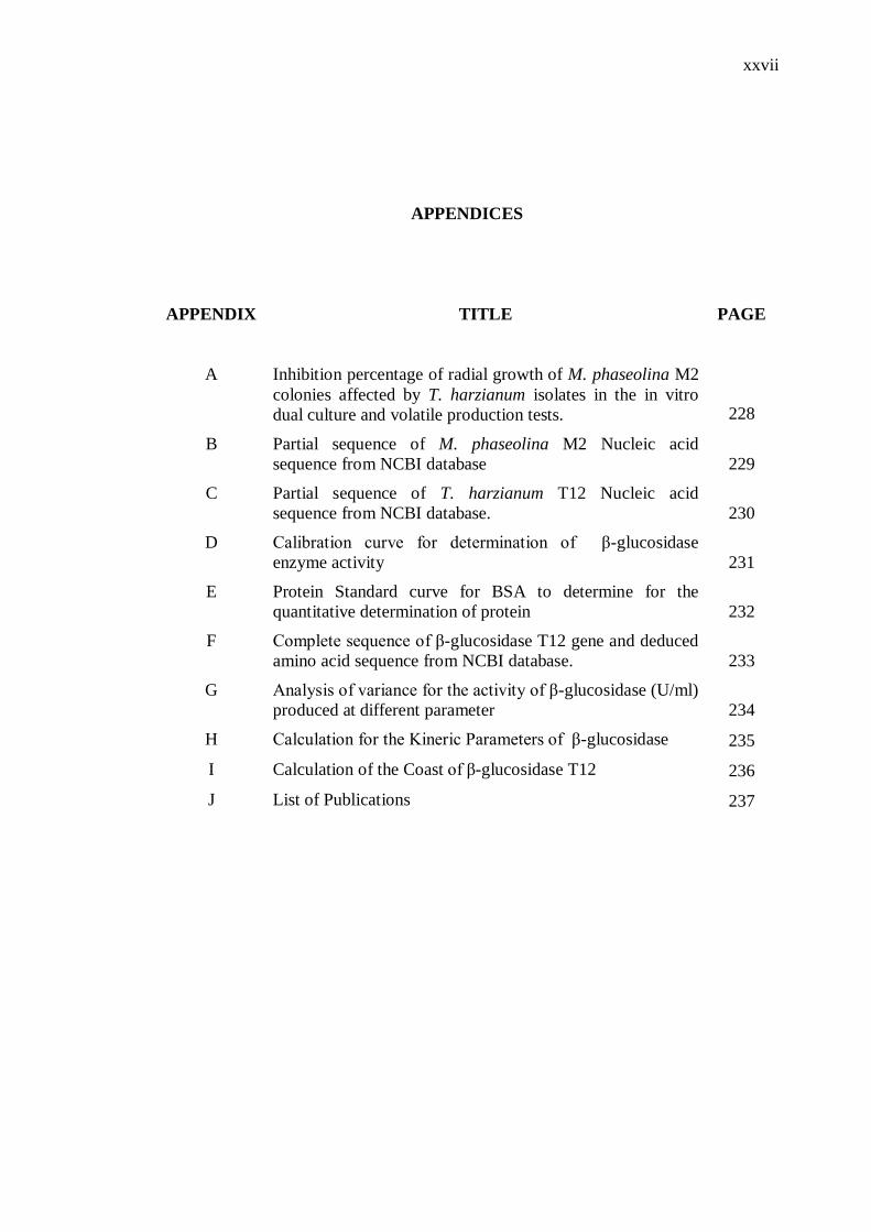
APPENDICES
APPENDIX TITLE PAGE
A Inhibition percentage of radial growth of M. phaseolina M2
colonies affected by T. harzianum isolates in the in vitro
dual culture and volatile production tests. 228
B Partial sequence of M. phaseolina M2 Nucleic acid
sequence from NCBI database 229
C Partial sequence of T. harzianum T12 Nucleic acid
sequence from NCBI database. 230
D Calibration curve for determination of β-glucosidase
enzyme activity 231
E Protein Standard curve for BSA to determine for the
quantitative determination of protein 232
F Complete sequence of β-glucosidase T12 gene and deduced
amino acid sequence from NCBI database. 233
G Analysis of variance for the activity of β-glucosidase (U/ml)
produced at different parameter 234
H Calculation for the Kineric Parameters of β-glucosidase 235
I Calculation of the Coast of β-glucosidase T12 236
J List of Publications 237
CHAPTER 1
INTRODUCTION
1.1 Background of Study
Among the plant diseases, soil-borne diseases are regarded to be more
devastating than air-borne or seed-borne diseases accounting for 10-20% of annual
yield losses (Kumar et al., 2016). Such diseases are mainly caused by
phytopathogenic fungi that constitutes to approximately 10,000 species of such fungi.
Among the phytopathogenic fungi, Macrophomina phaseolina (Tassi) Goid is one of
the most ubiquitous ones which causes the fatal charcoal rot disease and capable of
infecting approximately 700 plant species belonging to over 100 families of
monocots and dicots. Such extraordinary adaptability of M. phaseolina originates in
its significant physiological, morphological and pathogenic diversity that has resulted
in its wide distribution across diverse climatic conditions, ranging from arid to
tropical regions (Ambrosio et al., 2015).
The fungus is also an exceptionally robust soil-borne pathogen that devastate
a myriad of major agricultural crops viz. grains, legumes, oil seeds, jute and cotton
(Ma et al., 2010; Rayatpanah et al., 2012; Sun et al., 2016) as well as some
vegetables and fruits (Ambrosio et al., 2015). The fungus can survive for long
periods in soil, as long as 15 years as resistant structures (sclerotia and
chlamydospores) without relying on a host (Sanchez et al., 2016). Charcoal rot
caused by M. phaseolina is also disseminated by wind, soil and contact with infected
2
plant material (Gaige et al., 2010). Formation of the ‘black leg’ in infected plants is
symptomatic of the charcoal rot disease caused by the formation of sclerotia in the
plant crown. Infected plants generally have weak appearances and decolourized
leaves with fewer black secondary roots (Santos et al., 2016). The infected plants
eventually die from exposure to phytotoxic metabolites released by M. phaseolina,
such as phaseolinone, as well as other complications, such as vascular blockages that
compromise nutrient transport (Santos et al., 2016).
Various types of agricultural management strategies to control plant diseases
caused by M. phaseolina using cultural, biological and chemical techniques have
been investigated for decades. Cultural practices such as using crop rotation, soil
solarization and tolerant cultivars are not sufficiently efficacious on its own and are
usually combined with other controls (biological agents, low doses of pesticides) to
be more effective. For such technique to work, a higher level of technological
support system is needed hence resulting in higher costs of implementation (Patel et
al., 2014). So far, the use of fungicides and fumigants to control M. phaseolina
infestation in crops has been the main choice in the agricultural sector. However,
these approaches have been found ineffective and uneconomical. Recent studies have
shown that pesticide use per hectare, had generally increased more than
proportionally with crop output per hectare. A 1% increase in crop output per hectare
was matched with 1.8 % increase in pesticide use per hectare (Patel et al., 2014).
This is a particularly worrying trend in agricultural practices.
The indiscriminate use of pesticides has incurred serious concerns over the
long term deleterious effects of pesticides on the ecology and human health
(Chamorro et al., 2015; Pastrana et al., 2016). Agricultural run-offs containing
concentrated amounts of pesticides are highly mobile and can pollute nearby water
bodies. These toxic compounds are naturally bioaccumulated through our food chain
and ingested by mammals and bird populations (Chua et al., 2014; Edbeib et al.,
2016). In one hand, biological techniques using biological control agents and agri-
biotechnology i.e. genetically modified crops developed using recombinant DNA
technology appears more promising and ecologically friendlier to combat plant
diseases i.e. charcoal rot. But both techniques have been subjected to much debate,
3
due to concerns over new risks on food security as well as the emergence of
superweeds and superpest. Even so, the former lacks the consistency in the field
studies (Gerbore et al., 2014) which limits its application. Aside from the unknown
long-term and other unintended effects on the environment due to the loss of
biodiversity, the issue pertaining to food allergies has also been mentioned (Maghari
et al., 2011, Arancibia et al., 2013).
In view of the limitations in current management strategies to control
charcoal rot disease in crops caused by M. phaseolina, the study believes the answer
to solving this problem may lie in a simple but effective technique based on a
biological control approach using bioactive secretions produced by a well-known
antagonistic fungus, T. harzianum. For decades, extensive research carried out on
the fungus was solely focused on the isolation and in-situ cultivation of the fungus to
combat M. phaseolina infestation in crops. The antagonistic efficacy of the fungus
against M. phaseolina is attributable to several antagonistic mechanisms such as
nutrient competition, antibiotic production and mycoparasitism (Pastrana et al.,
2016). However, the study believes the need for in-situ propagation of this
antagonistic is rather slow and, possibly inadequately effective to confer a
comprehensive protection on the crops throughout the different growth stages. But
the study notes that the T. harzianum secretes an enzyme via the mycoparasitism
mechanism. It is a well-known fact that the β-glucosidase produced by this fungus
explicitly hydrolyzes the β-linkage of the amorphic β-1, 3-glucan filling material in
the chitin-based cell wall of M. phaseolina (Gajera et al., 2012). This antagonistic
mechanism is well-reported for species of T. harzianum whereby the β-glucosidase
facilitates the penetration of T. harzianum into the cytoplasm of the target pathogenic
fungi (Kavitha et al., 2012; Andersen et al., 2016). This interesting antagonistic
feature of T. harzianum to win the biological war against another competing fungus,
i.e. M. phaseolina seems agronomically useful and scientifically interesting.
4
1.2 Problem Statement
In view of the limitations and ineffectiveness of current management
strategies to control charcoal rot disease caused by the soil borne nature of M.
phaseolina, and the environmental hazards associated with the use of pesticides, the
search for alternative ecologically benign and sustainable strategy, preferably cost-
effective, may prove timely and merits agronomical consideration. Herein, a simple
but possibly effective bio-based technique of in-situ application of the extracellular
lytic enzyme i.e. β-glucosidase secreted by a well-known antagonistic fungus, T.
harzianum is proposed. The technique is justified based on a well-known fact that the
Trichoderma spp. such as T. harzianum has evolved in such a way to specifically
secrete a specific β-glucosidase adapted to hydrolyzing the amorphic β-1, 3-glucan
cell wall components in M. phaseolina. Driven by this fact, the direct spraying of a
formulation containing the growth supernatant of the extracellular β-glucosidase
would instantaneously destroy the M. phaseolina. Hence, further dissemination of the
fungal material by wind, soil or by physical contact could be averted and the charcoal
disease is halted.
It is hypothesized the extracellular enzyme β-glucosidase isolated from the
highly antagonistic T. harzianum T12 strain can be effectively used as the bioactive
ingredient over the chemically formulated ones for an environmentally benign
fungicide formulation. The study strongly believes the enzyme can be formulated as
an effective greener fungicide for in-situ protection against charcoal rot disease due
to M. phaseolina infestation for crops at various growth stages.
Although the direct use of the β-glucosidase would probably be cheaper, a
comprehensive characterization of its vulnerability against other possible additives in
the formulation may prove necessary for this study. This is to ensure its hydrolytic
activity is boosted, hence maximizing the efficacy in inhibiting M. phaseolina
infestation. Furthermore, the specific use of β-glucosidase from T. harzianum as the
active ingredient in fungicide formulation for agricultural management of M.
5
phaseolina infestation has not been reported. The feasibility of this proposed
technique remains to be seen.
1.3 Objectives of Research
This study highlights upon four main research objectives:
1. To collect, isolate and identify the most pathogenic M. phaseolina and most
effective T. harzianum isolates.
2. To purify and characterize the physicho- and biochemical properties of the β-
glucosidase.
3. To optimize the growth conditions of T. harzianum for maximum activity of
the produced β-glucosidase.
4. To optimize the application of the β-glucosidase-loaded formulation for
maximum inhibition on M. phaseolina.
1.4 Aim of Study
The study aimed to prepare a cheaper and greener fungicide using the crude
β-glucosidase from T. harzianum as the bioactive ingredient over the toxic
chemically formulated fungicide for inhibiting the growth of the phytopathogen M.
phaseolina.
6
1.5 Scopes of the Study
This study first isolated and screened for fungal strains of the most effective
T. harzianum and the most pathogenic M. phaseolina done under laboratory settings.
These M. phaseolina strains were isolated from infected plants and T. harzianum
isolates were isolated from soil samples from different agricultural locations in the
Mazandaran province of Iran. The most pathogenic (most prevalent) M. phaseolina
was identified using pathogenic variability test. It was necessary for the study to
isolate the most virulent strain of M. phaseolina as the model. If the T. harzianum
isolate/β-glucosidase was effective against the most virulent strain of the fungal
pathogen, the efficacy of former to inhibit other less virulent ones was highly
possible. Similarly, selection of the most effective T. harzianum isolate was based on
the most prevalent fungus strain found in the soil. This fungus was identified using,
dual culture test, hyper parasitism test, and volatile metabolites production. Both
fungi were then identified using morphological and biochemical tests.
Prior to formulation, the β-glucosidase in the growth supernatant of the T.
harzianum was purified using ammonium sulfate precipitation and dialyzed. The
enzyme was sequenced to ascertain its classification as a β-glucosidase and
subsequently assessed for physicochemical susceptibilities to environmental
conditions and possible additives in the fungicide formulation. The molecular weight
of the enzyme was evaluated using SDS-PAGE and the assessed physicochemical
properties included optimal pH and temperature as well as the effects of metal ions,
surfactants, carbon and nitrogen sources, and inoculum size. Since, this was a novel
enzyme, the kinetic parameters of the purified β-glucosidase was also evaluated.
The study went on to optimizing the physical and nutritional parameters for
batch cultivation of T. harzianum to produce the β-glucosidase using the method of
Response Surface Methodology (RSM) by the Box-Behnken Design (BBD). A cheap
agro-industrial substrate i.e. banana wastes was used as the carbon source for batch
cultivation of the T. harzianum to produce crude β-glucosidase. The response of this
optimization was the highest activity of the harvested β-glucosidase. This was based
7
on the largest inhibition zone surrounding a paper disc soaked with β-glucosidase,
carried out on potato dextrose assay plates.
The study finally optimized the preparation and application of the β-
glucosidase loaded fungicide formulation using soybean plants. The plants were
grown in soil infected with M. phaseolina and the work was evaluated over a period
of 8 weeks under a controlled condition in a greenhouse located within the grounds
of the Faculty of Bioscience and Medical Engineering, UTM. The efficacy of the
crude β-glucosidase to control charcoal rot disease was assessed based on the lowest
plant disease index (PDI). Since this study is a pioneering work using β-glucosidase
as the fungicide, RSM was again used to evaluate the effects of multiple factors viz.
metal ions, surfactants, enzyme concentration and irrigation frequency of the
formulation on the response i.e. PDI. Consequently, a controlled study comparing the
optimum protocol of β-glucosidase, a commonly use pesticide i.e. carbendazim and
the antagonist T. harzianum (Rifai) to result in the lowest PDI in growing soy bean
plants sown on M. phaseolina infected soil was carried out. Cost assessment on the
use of carbendazim and β-glucosidase based on per hectare of cultivated soybean was
also performed.
1.6 Significance of the Study
The strategy proposed here is potentially more rapid and sustainable to
control the spread of charcoal rot disease due to M. phseolina infestation in crops.
The approach used in this study is possibly a more practical and cost-effective means
to avert charcoal rot disease in other agronomically relevant crops, too.
8
1.7 Operational Framework of the Research
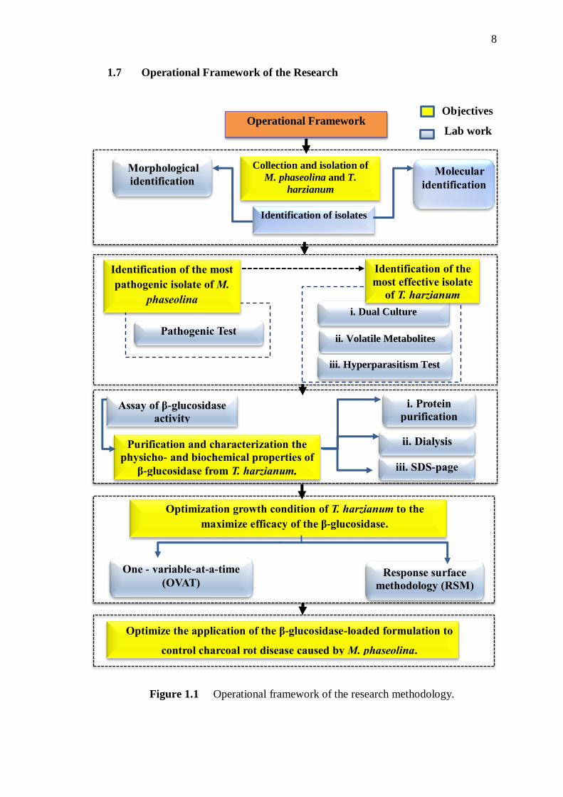
Figure 1.1 Operational framework of the research methodology.
Identification of isolates
Optimization growth condition of T. harzianum to the
maximize efficacy of the β-glucosidase.
Optimize the application of the β-glucosidase-loaded formulation to
control charcoal rot disease caused by M. phaseolina.
Molecular
identification
Morphological
identification
Collection and isolation of
M. phaseolina and T.
harzianum
Identification of the most
pathogenic isolate of M.
phaseolina
Pathogenic Test
i. Dual Culture
ii. Volatile Metabolites
iii. Hyperparasitism Test
iii. SDS-page
ii. Dialysis
i. Protein
purification
Response surface
methodology (RSM)
One - variable-at-a-time
(OVAT)
Assay of β-glucosidase
activity
Purification and characterization the
physicho- and biochemical properties of
β-glucosidase from T. harzianum.
Operational Framework
Objectives
Lab work
Identification of the
most effective isolate
of T. harzianum
REFERENCES
Abdel-Fatah, O. M., Hassan, M. M., Elshafei, A. M., Haroun, B. M., Atta, H. M. and
Othman, A. M. (2012). Physiological studies on carboxymethyl cellulase
formation by Aspergillus terreus DSM 826. Brazilian Journal of
Microbiology. 43(1), 01-11.
Abdella, A., El-Sayed Mazeed, T., Yang, S. T. and F El-Baz, A. (2014). Production
of β-glucosidase by Aspergillus niger on wheat bran and glycerol in
submerged culture: factorial experimental design and process
optimization. Curent Biotechnology. 3(2), 197-206.
Abdulgader Edbeib, M. F., Wahab, R. A. and Huyop, F. (2016). Characterization of
an α-haloalkanoic acid-degrading Pseudomonas aeruginosa MX1 isolated
from contaminated seawater. Bioremediation Journal. 20(2), 89-97.
Abdullah, M. T., Ali, N. Y. and Suleman, P. (2008). Biological control of Sclerotinia
sclerotiorum (Lib.) de Bary with Trichoderma harzianum and Bacillus
amyloliquefaciens. Crop Protection. 27(10), 1354-1359.
Ab-Majid, A. H., Zahran, Z., Rahim, A. H. A., Ismail, N. A., Rahman, W. A.,
Zubairi, K. S. M. and Satho, T. (2015). Morphological and molecular
characterization of fungus isolated from tropical bed bugs in Northern
Peninsular Malaysia, Cimex hemipterus (Hemiptera: Cimicidae). Asian
Pacific Journal of Tropical Biomedicine. 5(9), 707-713.
Aboshosha, S. S., Attaalla, S. I., El-Korany, A. E. and El-Argawy, E. (2007).
Characterization of Macrophomina phaseolina isolates affecting sunflower
growth in El-Behera governorate, Egypt. International Journal of
Agriculture and Biology. 9(6), 807-815.
Adav, S. S., Ravindran, A., Chao, L. T., Tan, L., Singh, S. and Sze, S. K. (2011).
Proteomic analysis of pH and strains dependent protein secretion of
Trichoderma reesei. Journal of Proteome Research. 10(10), 4579-4596.
195
Adekunle, A. T., Ikotun, T., Florini, D. A. and Cardwell, K. F. (2006). Field
evaluation of selected formulations of Trichoderma species as seed
treatment to control damping-off of cowpea caused by Macrophomina
phaseolina. African Journal of Biotechnology. 5(5), 419-424.
Afouda, L. C., Schulz, D., Wolf, G. and Wydra, K. (2012). Biological control of
Macrophomina phaseolina on cowpea (Vigna unguiculata) under dry
conditions by bacterial antagonists. International Journal of Biological
and Chemical Sciences. 6(6), 5068-5077.
Agarwal, V. K. and Sinclair, J. B. (1996). Principles of seed pathology. CRC Press.
253-543.
Ahamed, A. and Vermette, P. (2009). Effect of culture medium composition on
Trichoderma reesei’s morphology and cellulase production. Bioresource
Technology. 100(23), 5979-5987.
Ahmad, S. S. and Dalby, P. A. (2011). Thermodynamic parameters for salt-induced
reversible protein precipitation from automated microscale experiments.
Biotechnology and Bioengineering. 108 (2), 322-332.
Ait-Lahsen, H., Soler, A., Rey, M., de la Cruz, J., Monte, E. and Llobell, A. (2001).
An antifungal exo-α-1, 3-glucanase (AGN13. 1) from the biocontrol
fungus Trichoderma harzianum. Applied Enviromental Microbiology.
67(12), 5833-5839.
Akhtar, S. Shoaib, A., Akhtar, N. and Mehmood, R. (2016). Separate and Combined
Effects of Macrophomina phaseolina and Copper on Growth, Physiology
and Antioxidative Enzymes in Vigna Mungo L. Journal of Animal and
Plant Sciences. 26(5). 1339-1345.
Ali, M. B., Irshad, M., Anwar, Z., Zafar, M. and Imran, M. (2016). Screening and
Statistical Optimization of Physiochemical Parameters for the Production
of Xylanases from Agro-Industrial Wastes. Advances in Enzyme Research.
4(1), 20-23.
Al-Jazairi, M., Abou-Ghorra, S., Bakri, Y. and Mustafa, M. (2015). Optimization of
β-galactosidase production by response surface methodology using locally
isolated Kluyveromyces marxianus. International Food Research Journal.
22(4), 1361-1367.
196
Al-Rajhi, A. M. (2013). Impact of biofertilizer Trichoderma harzianum Rifai and the
biomarker changes in Eruca sativa L. plant grown in metal-polluted soils.
World Applied Sciences Journal. 22(2), 171-180.
Altschul, S. F., Gish, W., Miller, W., Myers, E. W. and Lipman, D. J. (1990). Basic
local alignment search tool. Journal of Molecular Biology. 215(3), 403-
410.
Alvarado-Carrillo, M., Diaz-Franco, A., Delgado-Aguirre, E. and Montes-Garcia, N.
(2010). Impact of corn agronomic management on aflatoxin (Aspergillus
flavus) contamination and charcoal stalk rot (Macrophomina phaseolina)
incidence. Tropical and Subtropical Agroecosystems.12 (3), 575-582.
Alvindia, D. G. and Hirooka, Y. (2011). Identification of Clonostachys and
Trichoderma spp. from banana fruit surfaces by cultural, morphological
and molecular methods. Mycology. 2(2), 109-115.
Ambrosio, M. M., Dantas, A. C., Martinez-Perez, E., Medeiros, A. C., Nunes, G. H.
and Pico, M. B. (2015). Screening a variable germplasm collection of
Cucumis melo L. for seedling resistance to Macrophomina phaseolina.
Euphytica. 206(2), 287-300.
Andersen, B., Poulsen, R. and Hansen, G. H. (2016). Cellulolytic and xylanolytic
activities of common indoor fungi. International Biodeterioration and
Biodegradation. 107, 111-116.
Anderson, P. K., Cunningham, A. A., Patel, N. G., Morales, F. J., Epstein, P. R. and
Daszak, P. (2004). Emerging infectious diseases of plants: pathogen
pollution, climate change and agrotechnology drivers. Trends in Ecology
and Evolution. 19(10), 535-544.
Anees, M., Tronsmo, A., Edel-Hermann, V., Hjeljord, L. G., Heraud, C. and
Steinberg, C. (2010). Characterization of field isolates of Trichoderma
antagonistic against Rhizoctonia solani. Fungal biology. 114(9), 691-701.
Arancibia, R. A., Main, J. L. and Clark, C. A. (2013). Sweetpotato tip rot incidence
is increased by preharvest applications of ethephon and reduced by curing.
Hort Technology. 23(3), 288-293.
Askari, H., Shahbazi, S., Naseripour, T., Moosavi-Nasab, M. and Bakhtiyari, M.
(2014). The impact of extracellular enzymes of Trichoderma viride and
Trichoderma harzianum on succinoglycan produced from Agrobacterium
197
radiobacter. International Journal of Agriculture and Crop Sciences. 7(8),
488-498.
Aviles, M., Castillo, S., Bascon, J., Zea-Bonilla, T., Martin-Sanchez, P. M. and
Perez-Jimenez, R.M. (2008). First report of Macrophomina phaseolina
causing crown and root rot of strawberry in Spain. Plant Pathology. 57(2),
382.
Babalola, O. O. and Glick, B. R. (2012). The use of microbial inoculants in African
agriculture: current practice and future prospects. Journal of Food,
Agriculture and Environment. 10(3-4), 540-549.
Bai, H., Wang, H., Sun, J., Irfan, M., Han, M., Huang, Y. and Yang, Q. (2013).
Production, purification and characterization of novel beta glucosidase
from newly isolated Penicillium simplicissimum H-11 in submerged
fermentation. EXCLI Journal. 12, 528-540.
Baino, O. M., Salazar, S. M., Ramallo, A. C. and Kirsch Baum, D. S. (2016). First
report of Macrophomina phaseolina causing strawberry crown and root rot
in North Western Argentina. Journal of Berry Research Sep. 6(3), 345-
354.
Bas, D. and Boyaci, I. H. (2007). Modeling and optimization I: Usability of response
surface methodology. Journal of food engineering. 78(3), 836-845.
Bashour, I. I. and Sayegh, A. H. (2007). Methods of analysis for soils of arid and
semi-arid regions. Food and Agriculture Organization (FAO) of the United
Nations, Rome. 4,64-65
Batra, J., Beri, D. and Mishra, S. (2014). Response surface methodology based
optimization of β-glucosidase production from Pichia pastoris. Applied
Biochemistry and Biotechnology. 172(1), 380-393.
Beas-Fernandez, R., De Santiago-De Santiago, A., Hernandez-Delgado, S. and
Mayek-Perez, N. (2006). Characterization of Mexican and non-Mexican
isolates of Macrophomina phaseolina based on morphological
characteristics, pathogenicity on bean seeds and endoglucanase genes.
Journal of Plant Pathology. 88(1), 53-60.
Bennett, A. J., Bending, G. D., Chandler, D., Hilton, S. and Mills, P. (2012). Meeting
the demand for crop production: the challenge of yield decline in crops
grown in short rotations. Biological Reviews. 87(1), 52-71.
198
Berbee, M. L. and Taylor, J. W. (1992). Two ascomycete classes based on fruiting-
body characters and ribosomal DNA sequence. Molecular Biology and
Evolution. 9(2), 278-284.
Betiku, E., Okunsolawo, S. S., Ajala, S. O. and Odedele, O. S. (2015). Performance
evaluation of artificial neural network coupled with generic algorithm and
response surface methodology in modeling and optimization of biodiesel
production process parameters from shea tree (Vitellaria paradoxa) nut
butter. Renewable Energy. 76, 408-417.
Bhatti, H. N., Batool, S. and Afzal, N. (2013). Production and Characterization of a
Novel β-glucosidase from Fusarium solani. International Journal of
Agriculture and Biology. 1, 1-15.
Bhiri, F., Chaabouni, S. E., Limam, F., Ghrir, R. and Marzouki, N. (2008).
Purification and biochemical characterization of extracellular β-
glucosidases from the hypercellulolytic Pol6 mutant of Penicillium
occitanis. Applied Biochemistry and Biotechnology. 149(2), 169-182.
Bisby, G. R. (1939). Trichoderma viride Pers. ex Fries, and notes on
Hypocrea. Transactions of the British Mycological Society. 23(2), 149-
168.
Bissett, J. (1991). A revision of the genus Trichoderma. II. Infrageneric
classification. Canadian Journal of Botany. 69(11), 2357-2372.
Biswanger, H. (2014). Enzyme Assays. Perspective in Science.1, 41-55.
Blanco-Canqui, H. (2010). Energy crops and their implications on soil and
environment. Agronomy Journal. 102(2), 403-419.
Box, G. E. and Behnken, D. W. (1960). Some new three level designs for the study
of quantitative variables. Technometrics. 2(4), 455-475.
Bradford, M. M. (1976). A rapid and sensitive method for the quantitation of
microgram quantities of protein utilizing the principle of protein-dye
binding. Analytical Biochemistry. 72(1-2), 248-254.
Bressano, M., Giachero, M.L., Luna, C.M. and Ducasse, D.A. (2010). An in vitro
method for examining infection of soybean roots by Macrophomina
phaseolina. Physiological and Molecular Plant Pathology. 74, 201-204.
Cabanillas, H. E. and Jones, W. A. (2009). Effects of temperature and culture media
on vegetative growth of an entomopathogenic fungus Isaria sp.
199
(Hypocreales: Clavicipitaceae) naturally affecting the whitefly, Bemisia
tabaci in Texas. Mycopathologia.167(5), 263.
Cai, F., Chen, W., Wei, Z., Pang, G., Li, R., Ran, W. and Shen, Q. (2015).
Colonization of Trichoderma harzianum strain SQR-T037 on tomato roots
and its relationship to plant growth, nutrient availability and soil
microflora. Plant and Soil. 388(1-2), 337-350.
Cantarel, B.L., Coutinho, P.M., Rancurel, C., Bernard, T., Lombard, V. and
Henrissat B. (2009). The Carbohydrate-Active Enzymes database (CAZy):
an expert resource for glycogenomics. Nucleic Acids Res. 37, 233-238.
Cardina, J., Herms, C. P. and Doohan, D. J. (2002). Crop rotation and tillage system
effects on weed seedbanks. Weed Science. 50(4), 448-460.
Cardoza, R. E., Malmierca, M. G., Hermosa, M. R., Alexander, N. J., McCormick, S.
P., Proctor, R. H. and Gutiérrez, S. (2011). Identification of loci and
functional characterization of trichothecene biosynthesis genes in
filamentous fungi of the genus Trichoderma. Applied and Environmental
Microbiology. 77(14), 4867-4877.
Chamorro, M., Miranda, L., Dominguez, P., Medina, J. J., Soria, C., Romero, F. and
De los Santos, B. (2015). Evaluation of biosolarization for the control of
charcoal rot disease (Macrophomina phaseolina) in strawberry. Crop
Protection. 67, 279-286.
Chang, K.F., Hwang, S.F., Wang, H.P., Turnbull, G. and Howard, R. (2006).
Etiology and biological control of sclerotinia blight of coneflower using
Trichoderma species. Plant Pathology Journal. 5, 15-19.
Chauhan, B. S., Singh, V. P., Kumar, A. and Johnson, D. E. (2011). Relations of rice
seeding rates to crop and weed growth in aerobic rice. Field Crops
Research. 121(1), 105-115.
Chauhan, P. S., Bharadwaj, A., Puri, N. and Gupta, N. (2014). Optimization of
medium composition for alkali-thermostable mannanase production by
Bacillus nealsonii PN-11 in submerged fermentation. International
Journal of Current Microbiology and Applied Sciences. 3(10), 1033-1045.
Chaverri, P. and Samuels, G. J. (2004). Hypocrea/Trichoderma (Ascomycota,
Hypocreales, Hypocreaceae): species with green ascospores. Studies in
Mycology. 48, 1-36.
200
Chaverri, P., Branco-Rocha, F., Jaklitsch, W., Gazis, R., Degenkolb, T. and
Samuels, G. J. (2015). Systematics of the Trichoderma harzianum species
complex and the re-identification of commercial biocontrol strains.
Mycologia. 107(3), 558-590.
Chen, S. F., Morgan, D., Beede, R. H. and Michailides, T. J. (2015). First report of
Lasiodiplodia theobromae associated with stem canker of almond in
California. Plant Disease. 99(12), 1678-1688.
Chowdhury, S., Basu, A. and Kundu, S. (2014). Green synthesis of protein capped
silver nanoparticles from phytopathogenic fungus Macrophomina
phaseolina (Tassi) Goid with antimicrobial properties against multidrug-
resistant bacteria. Nanoscale Research Letters. 9(1), 1-11.
Chua, E. M., Shimeta, J., Nugegoda, D., Morrison, P. D. and Clarke, B. O. (2014).
Assimilation of polybrominated diphenyl ethers from microplastics by the
marine amphipod, Allorchestes compressa. Environmental Science and
Technology. 48(14), 8127-8134.
Contreras-Cornejo, H. A., Macias-Rodriguez, L., Cortes-Penagos, C. and Lopez-
Bucio, J. (2009). Trichoderma virens, a plant beneficial fungus, enhances
biomass production and promotes lateral root growth through an auxin-
dependent mechanism in Arabidopsis. Plant Physiology. 149(3), 1579-
1592.
Cornish-Bowden, A. (2014). Current IUBMB recommendations on enzyme
nomenclature and kinetics. Perspectives in Science. 1(1), 74-87.
Corpet, F. (1988). Multiple sequence alignment with hierarchical clustering. Nucleic
Acids Research. 16(22), 10881-10890.
Costa, S. S., Matos, K. S., Tessmann, D. J., Seixas, C. D. and Pfenning, L. H. (2016).
Fusarium paranaense sp. nov., a member of the Fusarium solani species
complex causes root rot on soybean in Brazil. Fungal Biology. 120(1), 51-
60.
Crous, P. W., Slippers, B., Wingfield, M. J., Rheeder, J., Marasas, W. F., Philips, A.
J. and Groenewald, J. Z. (2006). Phylogenetic lineages in the
Botryosphaeriaceae. Studies in Mycology. 55, 235-253.
Crucello, A., Sforça, D. A., Horta, M. A. C., dos Santos, C. A., Viana, A. J. C.,
Beloti, L. L. and de Souza, A. P. (2015). Analysis of genomic regions of
201
Trichoderma harzianum IOC-3844 related to biomass degradation. PloS
One. 10(4), 122.
Csodes, I., Cseh, A., Taller, J. and Poczai, P. (2012). Genetic diversity and effect of
temperature and pH on the growth of Macrophomina phaseolina isolates
from sunflower fields in Hungary. Molecular Biology Reports. 39(3),
3259-3269.
Danielson, R. M. and Davey, C. B. (1973). Carbon and nitrogen nutrition of
Trichoderma. Soil Biology and Biochemistry. 5(5), 505-515.
Da-Silva Delabona, P., Pirota, R. D. P. B., Codima, C. A., Tremacoldi, C. R.,
Rodrigues, A. and Farinas, C. S. (2013). Effect of initial moisture content
on two Amazon rainforest Aspergillus strains cultivated on agro-industrial
residues: Biomass-degrading enzymes production and characterization.
Industrial Crops and Product. 42, 236-242.
Daynes, C. N., Zhang, N., Saleeba, J. A. and McGee P.A. (2012). Soil aggregates
formed in vitro by saprotrophic Trichocomaceae have transient water-
stability. Soil Biol Biochem. 48,151-161.
Deka, D., Bhargavi, P., Sharma, A., Goyal, D., Jawed, M. and Goyal, A. (2011).
Enhancement of cellulase activity from a new strain of Bacillus subtilis by
medium optimazation and analysis with various cellulosic substrates.
Enzyme Research. 2011(2011),1-8.
Dexter, A. R. (1988). Advances in characterization of soil structure. Soil and Tillage
Research. 11(3-4), 199-238.
Dhawane, S.H., Kumar. T. and Halder. G. (2015). Central composite design
approach towards optimization of flamboyant pods derived steam
activated carbon for its use as heterogeneous catalyst in transesterification
of Hevea brasiliensis oil. Energy Conversion and Management. 100, 277-
287.
Dhingra, O. D. and Sinclair, J. B. (1978). Biology and pathology of Macrophomina
phaseolina. Universidade Federal de Vicosa. Minas Gerais. 14, 166-279.
Domingues, F.C., Queiroz, J.A., Cabral, J.M. and Fonseca, L.P. (2000). The
influence of culture conditions on mycelial structure and cellulase
production by Trichoderma reesei Rut C-30. Enzyme Microb Technol.
26,394-401.
202
Druzhinina, I. S., Kopchinskiy, A. G., Komon, M., Bissett, J., Szakacs, G. and
Kubicek, C. P. (2005). An oligonucleotide barcode for species
identification in Trichoderma and Hypocrea. Fungal Genetics and
Biology. 42(10), 813-828.
Dube, M. P. (2000). Disorders of glucose metabolism in patients infected with
human immunodeficiency virus. Clinical Infectious Diseases. 31(6), 1467-
1475.
Dubey, R. C., Kumar, H. and Pandey, R. R. (2009). Fungi toxic effect of neem
extracts on growth and sclerotial survival of Macrophomina phaseolina in
vitro. Journal of American Science. 5, 17-24.
El-Fadaly, H. M., El-Kadi, S. M., Hamad, M. N. and Habib, A. A. (2015). Isolation
and Identification of Egyptian Ras Cheese (Romy) Contaminating Fungi
during Ripening Period. Journal of Microbiology Research. 5(1), 1-10.
El-Fiki, A. I. I., Mohamed, F. G., El-Deeb, A. A. and Khalifa, M. M. A. (2004).
Some applicable methods for controlling sesame charcoal rot disease
(Macrophomina phaseolina) under greenhouse conditions. Egyptian
Journal of Phytopathology. 32(1-2), 87-101.
El-Hawary, F. I. and Mostafa, Y. S. (2001). Factors affecting cellulase production by
Trichoderma koningii. Acta Alimentaria. 30(1), 3-13.
Ellis, M. L., Broders, K. D., Paul, P. A. and Dorrance, A. E. (2011). Infection of
soybean seed by Fusarium graminearum and effect of seed treatments on
disease under controlled conditions. Plant Disease. 95(4), 401-407.
Erskine, W., Muehlbauer, F. J. and Short, R. W. (1990). Stages of development in
lentil. Experimental Agriculture. 26(03), 297-302.
Etebarian, H. R. (2006). Evaluation of Trichoderma isolates for biological control of
charcoal stem rot in melon caused by Macrophomina phaseolina. Journal
of Agricultural Science and Technology. 8, 243-250.
Falconer, R. E., Otten, W. and White, N. A. (2015). Chapter One-Toward Modeling
the Resistance and Resilience of “Below-ground” Fungal Communities: A
Mechanistic and Trait-Based Approach. Advances in Applied
Microbiology. 93, 1-44.
Fernandez, F. G. and Hoeft, R. G. (2009). Managing soil pH and crop nutrients.
Illinois Agronomy Handbook. 8, 91-112.
203
Fernando, W. D., Ramarathnam, R., Krishnamoorthy, A. S. and Savchuk, S. C.
(2005). Identification and use of potential bacterial organic antifungal
volatiles in biocontrol. Soil Biology and Biochemistry. 37(5), 955-964.
Ferreira, S. C., Bruns, R. E., Ferreira, H. S., Matos, G. D., David, J. M., Brandao, G.
C. and Dos Santos, W. N. L. (2007). Box-Behnken design: an alternative
for the optimization of analytical methods. Analytica Chimica Acta.
597(2), 179-186.
Fiers, M., Edel-Hermann, V., Chatot, C., Le Hingrat, Y., Alabouvette, C. and
Steinberg, C. (2012). Potato soil-borne diseases. A review. Agronomy for
Sustainable Development. 32(1), 93-132.
Froeliger, E. H., Carpenter, B. E. and Froeliger, E. (1996). NUT1, a major nitrogen
regulatory gene in Magnaporthe grisea, is dispensable for pathogenicity.
Molecular and General Genetics MGG. 251(6), 647-656.
Gaetan, S.A., Fernande, L. and Madia, M. (2006). Ocurrence of charcoal rot caused
by Macrophomina phaseolina on canola in argentina. Plant Disease. 90,
524-524.
Gaige, A. R., Ayella, A. and Shuai, B. (2010). Methyl jasmonate and ethylene induce
partial resistance in Medicago truncatula against the charcoal rot pathogen
Macrophomina phaseolina. Physiological and Molecular Plant Pathology.
74(5), 412-418.
Gajera, H. P., Bambarolia, R. P., Patel, S.V., Khatrani, T. J. and Goalkiya, B. A.
(2012). Antagonism of Trichoderma spp. against Macrophomina
phaseolina: Evaluation of coiling and cell wall degrading enzymatic
activities. Plant Pathology and Microbiology. 3(7), 1-7.
Gams, W. and Bissett, J. (1998). Morphology and identification of
Trichoderma. Trichoderma and Gliocladium. 1, 3-34.
Garcia, N. F. L., Santos, F. R. D. S., Gonçalves, F. A., Paz, M. F. D., Fonseca, G. G.
and Leite, R. S. R. (2015). Production of β-glucosidase on solid-state
fermentation by Lichtheimia ramosa in agroindustrial residues:
Characterization and catalytic properties of the enzymatic extract.
Electronic Journal of Biotechnology. 18(4), 314-319.
Gardes, M., White, T. J., Fortin, J. A., Bruns, T. D. and Taylor, J. W. (1991).
Identification of indigenous and introduced symbiotic fungi in
204
ectomycorrhizae by amplification of nuclear and mitochondrial ribosomal
DNA. Canadian Journal of Botany. 69(1), 180-190.
Gautam, S. P., Bundela, P. S., Pandey, A. K., Awasthi, M. K. and Sarsaiya, S.
(2010). Optimization of the medium for the production of cellulase by the
Trichoderma viride using submerged fermentation. International Journal
of Environmental Sciences. 1(4), 656.
Gemell, L. G., Hartley, E. J. and Herridge, D. F. (2005). Point-of-sale evaluation of
preinoculated and custom-inoculated pasture legume seed. Animal
Production Science. 45(3), 161-169.
Gent, D. H., Mahaffee, W. F., McRoberts, N. and Pfender, W. F. (2013). The use
and role of predictive systems in disease management. Annual Review of
Phytopathology. 51, 267-289.
George, E., Tamerler, C., Martinez, A., Martinez, M. J. and Keshavarz, T. (1999).
Influence of growth medium composition on the lipolytic enzyme activity
of Ophiostoma piliferum (CartapipTM). Journal of Chemical Technology
and Biotechnology. 74(2), 137-140.
Gerbore, J., Benhamou, N., Vallance, J., Le Floch, G., Grizard, D., Regnault-Roger,
C. and Rey, P. (2014). Biological control of plant pathogens: advantages
and limitations seen through the case study of Pythium oligandrum.
Environmental Science and Pollution Research. 21(7), 4847-4860.
Ghangaokar, N. M. and Kshirsagar, A. D. (2013). Study of seed borne fungi of
different legumes. Trends in Life Sciences. 2(1), 32-35.
Ghazanfar, M. U., Hussain, M., Hamid, M. I. and Ansari, S. U. (2016). Utilization
Of Biological Control Agents For The Management Of Postharvest
Pathogens Of Tomato. Pakistan Journal of Botany. 48(5), 2093-2100.
Ghosh, T., Mukherji, N. and Basak, M. (1964). On the occurrence of a new species
of Orbilia Fr. Jute Bulletin. 27, 134-141.
Goujon, M., McWilliam, H., Li, W., Valentin, F., Squizzato, S., Paern, J. and Lopez,
R. (2010). A new bioinformatics analysis tools framework at EMBL-EBI.
Nucleic Acids Research. 38(2), 695-699.
Goyari, S., Devi, S. S., Kalita, M. C. and Talukdar, N. C. (2014). Population,
diversity and characteristics of cellulolytic microorganisms from the Indo-
Burma Biodiversity hotspot. Springer Plus. 3(1), 1-9.
205
Graber, E. R., Frenkel, O., Jaiswal, A. K. and Elad, Y. (2014). How may biochar
influence severity of diseases caused by soilborne pathogens? Carbon
Management. 5(2), 169-183.
Gray, K. A., Zhao, L. and Emptage, M. (2006). Bioethanol. Current Opinion in
Chemical Biology. 10(2), 141-146.
Groenewald, J. Z. and Crous, P. W. (2014). Genetic diversity in Macrophomina
phaseolina, the causal agent of charcoal rot. Phytopathologia
Mediterranea. 53(2), 250-268.
Gupta, G. K., Sharma, S. K. and Ramteke, R. (2012). Biology, epidemiology and
management of the pathogenic fungus Macrophomina phaseolina (Tassi)
Goid with special reference to charcoal rot of soybean (Glycine max (L.)
Merrill). Journal of Phytopathology. 160(4), 167-180.
Gupta, P., Chakraborty, D. and Mittal, R. K. (2015). Anti-fungal activity of
medicinal plants leaf extracts on growth of Macrophomina phaseolina.
Agricultural Science Digest-A Research Journal. 35(3), 211-214.
Haaland, P. D. (1989). Experimental design in biotechnology (Vol. 105). CRC
Press.105, 55-74
Haddadin, M. S., Haddadin, J., Arabiyat, O. I. and Hattar, B. (2009). Biological
conversion of olive pomace into compost by using Trichoderma
harzianum and Phanerochaete chrysosporium. Bioresource Technology.
100(20), 4773-4782.
Hallsworth, J.E. and Magan, N. (1996) Culture age, temperature, and pH affect the
polyol and trehalose contents of fungal propagules. Appl Environ
Microbiol. 62, 2435-2442.
Harighi, M. J., Zamani, M. R. and Motallebi, M. (2007). Evaluation of antifungal
activity of purified chitinase 42 from Trichoderma atroviride PTCC5220.
Biotechnology. 6(1), 28-33.
Harman, G. E., Howell, C. R., Viterbo, A., Chet, I. and Lorito, M. (2004).
Trichoderma species-opportunistic, avirulent plant symbionts. Nature
Reviews Microbiology. 2(1), 43-56.
Hartman, G. L., Bowen, C. R., Haudenshield, J. S., Fox, C. M., Cary, T. R. and
Diers, B. W. (2015). Evaluation of disease and pest damage on soybean
cultivars released from 1923 through 2008 under field conditions in
Central Illinois. Agronomy Journal. 107(6), 2373-2380.
206
Hartman, G. L., Pawlowski, M. L., Herman, T. K. and Eastburn, D. (2016).
Organically grown soybean production in the USA: Constraints and
management of pathogens and insect pests. Agronomy. 6(1), 1-18.
Hartman, G. L., West, E. D. and Herman, T. K. (2011). Crops that feed the World 2.
Soybean-worldwide production, use, and constraints caused by pathogens
and pests. Food Security. 3(1), 5-17.
Hassan, N., Elsharkawy, M. M., Shimizu, M. and Hyakumachi, M. (2014). Control
of root rot and wilt disease of roselle under field conditions. Mycobiology.
42(4), 376-384.
Helali, I., Ferchichi, S., Maaouia, A., Aouni, M. and Harizi, H. (2016). Modulation
of macrophage functionality induced in vitro by chlorpyrifos and
carbendazim pesticides. Journal of Immunotoxicology. 13(5), 745-750.
Hermosa, R., Rubio, M. B., Cardoza, R. E., Nicolas, C., Monte, E. and Gutierrez, S.
(2013). The contribution of Trichoderma to balancing the costs of plant
growth and defense. International Microbiology. 16(2), 69-80.
Hermoso, J., Pignol, D., Kerfelec, B., Crenon, I., Chapus, C. and Fontecilla-Camps,
J. C. (1996). Lipase activation by nonionic detergents the crystal structure
of the porcine lipase-colipase-tetraethylene glycol monooctyl ether
complex. Journal of Biological Chemistry. 271(30), 18007-18016.
Heydari, A. and Pessarakli, M. (2010). A review on biological control of fungal plant
pathogens using microbial antagonists. Journal of Biological Sciences.
10(4), 273-290.
Hmad, I. B., Abdeljalil, S., Saibi, W., Amouri, B. and Gargouri, A. (2014). Medium
initial pH and carbon source stimulate differential alkaline cellulase time
course production in Stachybotrys microspora. Applied Biochemistry and
Biotechnology. 172(5), 2640-2649.
Holliday, P. and Punithalingam, E. (1970). Macrophomina phaseolina. Descriptions
of Fungi and Bacteria. IMI Descriptions of Fungi and Bacteria. 275,1-28.
Hossain, M. T., Hossain, S. M. M., Bakr, M. K., Rahman, A. M. and Uddin, S. N.
(2010). Survey on major diseases of vegetable and fruit crops in
Chittagong region. Bangladesh Journal of Agricultural Research. 35(3),
423-429.
207
Hymowitz, T. and Bernard, R. L. (1991). Origin of the soybean and germplasm
introduction and development in North America. Use of Plant
Introductions in Cultivar Development Part. 1, 147-164.
Ijaz, S., Sadaqat, H. A. and Khan, M. N. (2013). A review of the impact of charcoal
rot (Macrophomina phaseolina) on sunflower. The Journal of Agricultural
Science. 151(02), 222-227.
Iqbal, H. M. N., Asgher, M. and Bhatti, H. N. (2011). Optimization of physical and
nutritional factors for synthesis of lignin degrading enzymes by a novel
strain of Trametes versicolor. Bio Resources. 6(2), 1273-1287.
Iqbal, S., Nguyen, T. H., Nguyen, T. T., Maischberger, T. and Haltrich, D. (2010). β-
Galactosidase from Lactobacillus plantarum WCFS1: biochemical
characterization and formation of prebiotic galacto-
oligosaccharides. Carbohydrate Research. 345(10), 1408-1416.
Iqbal, U. and Mukhtar, T. (2014). Morphological and pathogenic variability among
Macrophomina phaseolina isolates associated with mungbean (Vigna
radiata L.) Wilczek from Pakistan. The Scientific World Journal. 14,1-9.
Iqbal, U., Mukhtar, T. and Iqbal, S. M. (2014). In vitro and in vivo evaluation of
antifungal activities of some antagonistic plants against charcoal rot
causing fungus, Macrophomina phaseolina. Pakistan Journal of
Agricultural Sciences. 51(3), 691-696.
Iriarte, F., Rosskopf, E., Hilf, M., McCollum, G., Albano, J. and Adkins, S. (2007).
First report of Macrophomina phaseolina causing leaf and stem blight of
tropical soda apple in Florida. Plant Health Progress. 10, 1-3.
Irshad, M., Anwar, Z., Ramzan, M., Mahmood, Z. and Nawaz, H. (2013).
Characterization of purified β-glucosidase produced from Trichoderma
viride through bio-processing of orange peel waste. Advances in
Bioscience and Biotechnology. 4(10), 941-944.
Isah, A. A., Mahat, N. A., Jamalis, J., Attan, N., Zakaria, I. I., Huyop, F. and Wahab,
R. A. (2016). Synthesis of geranyl propionate in a solvent-free medium
using Rhizomucor miehei lipase covalently immobilized on chitosan-
graphene oxide beads. Preparative Biochemistry and Biotechnology.
47(2)1-12.
Islam, M. S., Haque, M. S., Islam, M. M., Emdad, E. M., Halim, A., Hossen, Q. M.
M. and Alam, M. M. (2012). Tools to kill: genome of one of the most
208
destructive plant pathogenic fungi Macrophomina phaseolina. Bmc
Genomics. 13 (1), 493.
Jabbour, D., Klippel, B. and Antranikian, G. (2012). A novel thermostable and
glucose-tolerant β-glucosidase from Fervidobacterium islandicum.
Applied Microbiology and Biotechnology. 93, 1947-1956.
Jabeen, K., Javaid, A., Ahmad, E. and Athar, M. (2011). Antifungal compounds
from Melia azedarach leaves for management of Ascochyta rabiei, the
cause of chickpea blight. Natural Product Research. 25(3), 264-276.
Jaspers, M. V., Seyb, A. M., Trought, M. C. T. and Balasubramaniam, R. (2013).
Overwintering grapevine debris as an important source of Botrytis cinerea
inoculum. Plant Pathology. 62(1), 130-138.
Jeya, M., Joo, A. R., Lee, K. M., Tiwari, M. K., Lee, K. M., Kim, S. H. and Lee, J.
K. (2010). Characterization of β-glucosidase from a strain of Penicillium
purpurogenum KJS506. Applied Microbiology and Biotechnology. 86(5),
1473-1484.
Jimenez, D. R. C. (2011). Influence of soils, nutrition, and water relations upon
charcoal rot disease processes in Kansas (Doctoral dissertation, Kansas
State University).1-26.
Jimenez-Diaz, R. M., Blanco-Lopez, M. A. and Sackston, W. E. (1983). Incidence
and distribution of charcoal rot of sunflower caused by Macrophomina
phaseolina in Spain. Plant Disease. 67(9), 1033-1036.
Job, J., Sukumaran , R.K. and Jayachandran, K. (2010). Production of a highly
glucose tolerant β-glucosidase by Paecilomyces variotii MG3:
optimization of fermentation conditions using Plackett-Burman and Box-
Behnken experimental designs. World Journal Microbiol Biotechnol.
26(8),1385-1391.
Jordaan, E. and Van der Waals, J. (2016). With drought comes charcoal rot:
agronomy. Oilseeds Focus. 2(3), 6-7.
Karboune, S., Geraert, P. A. and Kermasha, S. (2008). Characterization of selected
cellulolytic activities of multi-enzymatic complex system from Penicillium
funiculosum. Journal of Agriculture and Food Chemistry. 56(3), 903-909.
Karnchanatat, A., Petsom, A., Sangvanich, P., Piaphukiew, J., Whalley, A. J.,
Reynolds, C. D. and Sihanonth, P. (2007). Purification and biochemical
characterization of an extracellular β-glucosidase from the wood-decaying
209
fungus Daldinia eschscholzii (Ehrenb.: Fr.) Rehm. FEMS Microbiology
Letters. 270(1), 162-170.
Kaur, J., Chadha, B. S., Kumar, B. A., Kaur, G. and Saini, H. S. (2007). Purification
and characterization of ß-glucosidase from Melanocarpus sp. MTCC
3922. Electronic Journal of Biotechnology. 10(2), 260-270.
Kaur, S., Dhillon, G. S., Brar, S. K., Vallad, G. E., Chand, R. and Chauhan, V. B.
(2012). Emerging phytopathogen Macrophomina phaseolina: biology,
economic importance and current diagnostic trends. Critical Reviews in
Microbiology. 38(2), 136-151.
Kavitha, T., Gopalan, A. I., Lee, K. P. and Park, S. Y. (2012). Glucose sensing,
photocatalytic and antibacterial properties of grapheme-ZnO nanoparticle
hybrids. Carbon. 50(8), 2994-3000.
Keswani, C., Mishra, S., Sarma, B. K., Singh, S. P. and Singh, H. B. (2014).
Unraveling the efficient applications of secondary metabolites of various
Trichoderma spp. Applied Microbiology and Biotechnology. 98(2), 533-
544.
Khaledi, N., Taheri, P. and Tarighi, S. (2015). Antifungal activity of various
essential oils against Rhizoctonia solani and Macrophomina phaseolina as
major bean pathogens. Journal of Applied Microbiology.118 (3), 704-717.
Khan, N., Jadhav, S. and Rathod, V. K. (2016). Enzymatic synthesis of n-butyl
palmitate in a solvent-free system: RSM optimization and kinetic
studies. Biocatalysis and Biotransformation. 34(3), 99-109.
Khangura, R. and Aberra, M. (2009). First report of charcoal rot on canola caused by
Macrophomina phaseolina in Western Australia. Plant Disease. 93, 666-
667.
Klein, D. and Eveleigh, D. E. ( 2002). Ecology of Trichoderma. In: Kubicek, C. P.
and Harman, G. E. (eds.). Trichoderma and Gliocladium: Basic Biology,
Taxonomy and Genetics. Taylor and Francis Ltd. 57-69.
Kocher, G., Kalra, K. and Banta, G. (2008). Optimization of cellulase production by
submerged fermentation of rice straw by Trichoderma harzianum Rut-C
8230. International Journal of Medical Microbiology. 5(2)1-7.
Kubicek, C. P., Mach, R. L., Peterbauer, C. K. and Lorito, M. (2001). Trichoderma:
from genes to biocontrol. Journal of Plant Pathology. 1, 11-23.
210
Kumar, A., Kumar, S., Kumar, S. and Kumar, B. (2016). Diseases of Chickpea Crop
and Their Management. In Center rop Diseases and Their Management:
Integrated Approaches Apple Academic Press. 27, 39-55.
Kumar, S. (2016). Diseases of Soybean and Their Management. In Center Crop
Diseases and Their Management: Integrated Approaches . Apple Academic
Press. 27, 95-125
Kusumawati, D. E., Hadiastono, T. and Aini, L. Q. (2012). Leaf Extract of Mirabilis
jalapa L. Induced Defense of Tomato Plant (Lycopersicum esculentum
Mill.) Against Cucumber Mosaic Virus (CMV) Infection. Journal of
Tropical Plant Protection. 1(1), 46-51.
Laemmli, U. K. (1970). Cleavage of structural proteins during the assembly of the
head of bacteriophage T4. Nature. 227, 680-685.
Landa, B.B., Navas-Cortes, J.A. and Jimenez-Diaz, R.M. (2012). Biological control
of Macrophomina phaseolina on cowpea (Vigna unguiculata).
International Journal of Biological and Chemical Sciences. 94, 946-960.
Landmeyer, J. E. (2012). Plant and Groundwater Interactions Under Pristine
Conditions. In Introduction to Phytoremediation of Contaminated
Groundwater. Springer Netherlands. 115-127.
Larralde-Corona, C. P., Santiago-Mena, M. R., Sifuentes-Rincon, A. M., Rodriguez-
Luna, I. C., Rodriguez-Perez, M. A., Shirai, K. and Narvaez-Zapata, J. A.
(2008). Biocontrol potential and polyphasic characterization of novel
native Trichoderma strains against Macrophomina phaseolina isolated
from sorghum and common bean. Applied Microbiology and
biotechnology. 80(1), 167-177.
Latge, J. P. (2010). Tasting the fungal cell wall. Cellular Microbiology. 12(7), 863-
872.
Lee, H., Lee, Y. M., Heo, Y. M., Hong, J. H., Jang, S., Ahn, B. J. and Kim, J. J.
(2017). Optimization of Fungal Enzyme Production by Trichoderma
harzianum KUC1716 through Surfactant-Induced Morphological
Changes. Mycobiology. 45(1), 48-51.
Li, S., Hartman, G. L. and Boykin, D. L. (2010). Aggressiveness of Phomopsis
longicolla and other Phomopsis spp. on soybean. Plant Disease. 94(8),
1035-1040.
211
Li, X. H., Zhang, P., Wang, M. X., Zhou, F., Malik, F. A., Yang, H. J. and Miao, Y.
G. (2011). Expression of Trichoderm aviride endoglucanase III in the
larvae of silkworm, Bombyx mori L. and characteristic analysis of the
recombinant protein. Molecular Biology Reports. 38(6), 3897-3902.
Lieberman, R. L., Wustman, B. A., Huertas, P., Powe Jr, A. C., Pine, C. W., Khanna,
R. and Petsko, G. A. (2007). Structure of acid [beta]-glucosidase with
pharmacological chaperone provides insight into Gaucher disease. Nature
Chemical Biology. 3(2), 101-107.
Lieckfeldt, E., Kuhls, K. and Muthumeenakshi, S. (2002). Molecular taxonomy of
Trichoderma and Gliocladium and their teleomorphs. Trichoderma and
Gliocladium. 1, 35-56.
Liu, K. H., Yeh, Y. L. and Shen, W. C. (2011). Fast preparation of fungal DNA for
PCR screening. Journal of Microbiological Methods. 85(2), 170-172.
Liu, W., Yin, P., Liu, X. and Qu, R. (2014). Design of an effective bifunctional
catalyst organotriphosphonic acid-functionalized ferric alginate (ATMP-
FA) and optimization by Box–Behnken model for biodiesel esterification
synthesis of oleic acid over ATMP-FA. Bioresource Technology. 173,
266-271.
Lopes, F. A. C., Steindorff, A. S., Geraldine, A. M., Brandao, R. S., Monteiro, V. N.,
Júnior, M. L. and Silva, R. N. (2012). Biochemical and metabolic profiles
of Trichoderma strains isolated from common bean crops in the Brazilian
Cerrado, and potential antagonism against Sclerotinia sclerotiorum.
Fungal Biology. 116(7), 815-824.
Lorito, M., Woo, S. L., Harman, G. E. and Monte, E. (2010). Translational research
on Trichoderma: from momics to the field. Annual Review of
Phytopathology. 48, 395-417.
Lopez, M.R., Ros, M. and Pascual, J.A., (2011). Mycoprasitism-related genes
expression of Trichoderma harzianum isolates to evaluate their efficacy as
biocontrol agent. Biological Control .56, 59-66.
Lynd, L. R., Weimer, P. J., Van Zyl, W. H. and Pretorius, I. S. (2002). Microbial
cellulose utilization: fundamentals and biotechnology. Microbiology and
Molecular Biology Reviews. 66(3), 506-577.
212
Ma, J., Hill, C. B. and Hartman, G. L. (2010). Production of Macrophomina
phaseolina conidia by multiple soybean isolates in culture. Plant Disease.
94(9), 1088-1092.
Ma, S. J., Leng, B., Xu, X. Q., Zhu, X. Z., Shi, Y., Tao, Y. M. and Chen, Q. X.
(2011). Purification and characterization of b-1, 4-glucosidase from
Aspergillus glaucus. African Journal of Biotechnology. 10(84), 19607-
19614.
Maghari, B. M. and Ardekani, A. M. (2011). Genetically modified foods and social
concerns. Avicenna Journal of Medical Biotechnology. 3(3), 109-117.
Mahajan, P. M., Desai, K. M. and Lele, S. S. (2012). Production of cell membrane-
bound α-and β-glucosidase by Lactobacillus acidophilus. Food and
Bioprocess Technology. 5(2), 706-718.
Mahapatra, S., Vickram, A. S., Sridharan, T. B., Parameswari, R. and Pathy, M. R.
(2016). Screening, production, optimization and characterization of β-
glucosidase using microbes from shellfish waste. 3 Biotech. 6(213),2-10.
Maki, M., Leung, K. T. and Qin, W. (2009). The prospects of cellulase-producing
bacteria for the bioconversion of lignocellulosic biomass. International
Journal of Biological Sciences. 5(5), 500-516.
Malik, G. and Dawar, S. (2003). Biological control of root infecting fungi with
Trichoderma harzianum. Pakistan Journal of Agricultural Sciences. 35,
971-975.
Manan, F. M. A., Rahman, I. N. A., Marzuki, N. H. C., Mahat, N. A., Huyop, F. and
Wahab, R. A. (2016). Statistical modelling of eugenol benzoate synthesis
using Rhizomucor miehei lipase reinforced nanobioconjugates. Process
Biochemistry. 51(2), 249-262.
Manici, L. M., Bregaglio, S., Fumagalli, D. and Donatelli, M. (2014). Modelling soil
borne fungal pathogens of arable crops under climate change.
International Journal of Biometeorology. 58(10), 2071-2083.
Manici, L. M., Caputo, F. and Cerato, C. (1992). Pathogenic and biological
variability of Macrophomina phaseolina (Tassi) Goid. isolates in different
areas of sunflower coltivation in Italy. Sunflower Conf. 1, 779-784.
Marco, J. L. D. and Felix, C. R. (2007). Purification and characterization of a beta-
Glucanase produced by Trichoderma harzianum showing biocontrol
potential. Brazilian Archives of Biology and Technology. 50(1), 21-29.
213
Martins, I., Garcia, H., Varela, A., Nunez, O., Planchon, S., Galceran, M. T. and
Pereira, C. S. (2014). Investigating Aspergillus nidulans secretome during
colonisation of cork cell walls. Journal of Proteomics. 98, 175-188.
Marzuki, N. H. C., Mahat. N. A., Huyop, F., Aboul-Enein, H. Y. and Wahab, R. A.
(2015). Sustainable production of the emulsifier methyl oleate by Candida
rugosa lipase nanoconjugates. Food Bioproducts and Processing. 96, 211-
220.
Mateos, S. E., Cervantes, C. A. M., Zenteno, E., Slomianny, M. C., Alpuche, J.,
Hernandez-Cruz, P. and Mayoral, L. P. C. (2015). Purification and partial
characterization of β-glucosidase in chayote (Sechium edule). Molecules.
20 (10), 19372-19392.
Mayek-Perez, N., GarcIa-Espinosa, R., LOpez-CastaNeda, C., Acosta-Gallegos, J.
A. and Simpson, J. (2002). Water relations, histopathology and growth of
common bean (Phaseolus vulgaris L.) during pathogenesis of
Macrophomina phaseolina under drought stress. Physiological and
Molecular Plant Pathology. 60(4), 185-195.
Mbarga, J.B., Hoopen, G.M.T., kuate, J., Adiobo, A., Ngonkeu, M.E.L., Ambang, Z.,
Akoa, A., Tondje, P.R. and Begoude, B.A.D., (2012). Trichoderma
asperellum: a potential biocontrol agent for Pythim myriotylum, causal
agent of cocoyam (Xanthosoma sagittifolium) root rot disease in
Cameroon. Crop Protection. 36, 18-22.
McCain, A. H. and Smith, R. S. (1972). Quantitative assay of Macrophomina
phaseoli from soil. Phytopathology. (62), 1062-1980.
Mc-Fadden, A. G. and Sutton, J. C. (1975). Relationship of population of
Trichoderma spp. in soil to disase in maize. Canadian Journal of Plant
Science. 55(2), 579-586.
Medic, J., Atkinson, C. and Hurburgh, C. R. (2014). Current knowledge in soybean
composition. Journal of the American Oil Chemists Society. 91(3), 363-
384.
Meena, A. K. and Meena, A. K. (2016). Characterization and antagonistic effect of
isolated Trichoderma sp. against pathogens under clusterbean (Cyamopsis
tetragonoloba L.). Indian Journal Of Agricultural Research. 50 (3) 249-
253.
214
Mengistu, A., Reddy, K. N., Zablotowicz, R. M. and Wrather, A. J. (2009).
Propagule densities of Macrophomina phaseolina in soybean tissue and
soil as affected by tillage, cover crop, and herbicide. Plant Health
Progress. Online Publication. 30(10), 1-12.
Mergel, O., Gelissen, A. P., W nnemann, P., Boker, A., Simon, U. and Plamper, F.
A. (2014). Selective packaging of ferricyanide within thermoresponsive
microgels. The Journal of Physical Chemistry.118(45), 26199-26211.
Mihail, J.D. (1992). Macrophomina. In Methods for research on soilborne
phytopathogenic fungi. American Phytopathological Society. 134-136.
Mikiashvili, N., Elisashvili, V., Wasser, S. and Nevo, E. (2005). Carbon and nitrogen
sources influence the ligninolytic enzyme activity of Trametes versicolor.
Biotechnology Letters. 27(13), 955-959.
Mirkhani, F. and Alaei, H. (2015). Species diversity of indigenous Trichoderma
from alkaline pistachio soils in Iran. Mycologia Iranica. 2(1), 22-37.
Mohamad, N. R., Buang, N. A., Mahat, N. A., Lok, Y. Y., Huyop, F., Aboul-Enein,
H. Y. and Wahab, R. A. (2015). A facile enzymatic synthesis of geranyl
propionate by physically adsorbed Candida rugosa lipase onto multi-
walled carbon nanotubes. Enzyme and Microbial Technology. 72, 49-55.
Mokhtar, H. and Dehimat, L. (2015). In vitro and In vivo Efficiency of Trichoderma
harzianum against Phoma and Glocladium Soft Rot Occurred on Tomato
Fruits (Lycopersicon esculentum). International Journal of Current
Microbiology and Applied. 4(8), 141-147.
Monis, P. T., Caccio, S. M. and Thompson, R. A. (2009). Variation in Giardia:
towards a taxonomic revision of the genus. Trends in Parasitology. 25(2),
93-100.
Monte, E. and Llobell, A. (2003). Trichoderma in organic agriculture. In Proceeding
V World Avocado Congress. 19, 725-733.
Montealegre, J., Valderrama, L., Herrera, R., Besoain, X. and Perez, L. M. (2009).
Biocontrol capacity of wild and mutant Trichoderma harzianum (Rifai)
strains on Rhizoctonia solani 618: effect of temperature and soil type
during storage. Electronic Journal of Biotechnology. 12(4), 2-3.
Morris, N. L., Miller, P. C. H., Orson, J. H. and Froud-Williams, R. J. (2010). The
adoption of non-inversion tillage systems in the United Kingdom and the
215
agronomic impact on soil, crops and the environment-A review. Soil and
Tillage Research. 108(1), 1-15.
Mueller, D. S., Pierson, W. L. and Wiggs, S. N. (2013). Evaluation of Foliar
Fungicides and Insecticides on Soybeans in Central Iowa. Iowa State
University Research and Demonstration Farms. 30, 13-16.
Mukherjee, D. and Haque, Z. Z. (2015). Antioxidant activity and persistence of
cottonseed protein and oil from two cultivars as determined by their ability
to scavenge peroxyl and alkoxyl radicals. Journal of Agricultural and Life
Sciences. 2, 6-10.
Mukherjee, P. K., Horwitz, B. A., Herrera-Estrella, A., Schmoll, M. and Kenerley,
C. M. (2013). Trichoderma research in the genome era. Annual Review of
Phytopathology. 51, 105-129.
Mwamburi, Lizzy A., Mark D. Laing. and Ray M. Miller. (2015). Effect of
surfactants and temperature on germination and vegetative growth of
Beauveria bassiana. Brazilian Journal of Microbiology. 46 (1) 67-74.
Nakazawa, H., Kawai, T., Ida, N., Shida, Y., Kobayashi, Y., Okada, H. and
Ogasawara, W. (2012). Construction of a recombinant Trichoderma reesei
strain exopessing Aspergillus aculeatus β‐ glucosidase 1 for efficient
biomass conversion. Biotechnology and Bioengineering. 109(1), 92-99.
Naranjo, S. E., Ellsworth, P. C. and Frisvold, G. B. (2015). Economic value of
biological control in integrated pest management of managed plant
systems. Annual Review of Entomology. 60, 621-645.
Narayan, M.S., Thimmaraju, R. and Bhagyalakshmi, N. (2005) Interplay of growth
regulators during solid-state and liquid-state batch cultivation of
anthocyanin producing cell line of Daucus carota. Process Biochem.
40,351-358.
Naseri, B. (2014). Charcoal rot of bean in diverse cropping systems and soil
environments. Journal of Plant Diseases and Protection. 121(1), 20-25.
Ndiaye, M., Sarr, M. P., Cisse, N. and Ndoye, I. (2015). Is the recently described
Macrophomina pseudophaseolina pathogenically different from
Macrophomina phaseolina? African Journal of Microbiology Research.
9(45), 2232-2238.
Newman, J. P. and Molinar, R. H. (2014). Safe and Sustainable Pest Management.
California Master Gardener Handbook. 3382, 219.
216
Nicot, P. C., Blum, B., Kohl, J. and Ruocco, M. (2011). Conclusions and
perspectives for future research-and-development projects on biological
control of plant pests and diseases. Classical and Augmentative Biological
Control Against Diseases and Pests: Critical Status Analysis and Review
of Factors. 1-11.
Niranjane, A. P., Madhou, P. and Stevenson, T. W. (2007). The effect of
carbohydrate carbon sources on the production of cellulase by Phlebia
gigantea. Enzyme and Microbial Technology. 40(6), 1464-1468.
Nitta, M., Furukawa, T., Shida, Y., Mori, K., Kuhara, S., Morikawa, Y. and
Ogasawara, W. (2012). A new Zn (II) 2 Cys 6-type transcription factor
BglR regulates β-glucosidase expression in Trichoderma reesei. Fungal
Genetics and Biology. 49(5), 388-397.
Noyrod, P., Chailapakul, O., Wonsawat, W. and Chuanuwatanakul, S. (2014). The
simultaneous determination of isoproturon and carbendazim pesticides by
single drop analysis using a graphene-based electrochemical
sensor. Journal of Electroanalytical Chemistry. 719, 54-59.
Olajuyigbe, F. M., Nlekerem, C. M. and Ogunyewo, O. A. (2016). Production and
characterization of highly thermostable β-glucosidase during the
biodegradation of methyl cellulose by Fusarium oxysporum. Biochemistry
Research International. 2016, 1-8.
Oliveira-Garcia, E. and Deising, H. B. (2013). Infection Structure–Specific
Expression of β-1, 3-Glucan Synthase Is Essential for Pathogenicity of
Colletotrichum graminicola and Evasion of β-Glucan–Triggered
Immunity in Maize. The Plant Cell. 25(6), 2356-2378.
Opassiri, R., Pomthong, B., Akiyama, T., Nakphaichit, M., Onkoksoong, T., Cairns,
M. K. and Cairns, J. R. K. (2007). A stress-induced rice (Oryza sativa L.)
β-glucosidase represents a new subfamily of glycosyl hydrolase family 5
containing a fascin-like domain. Biochemical Journal. 408(2), 241-249.
Pal, K. K. and Gardener, B. M. (2006). Biological control of plant pathogens. The
Plant Health Instructor. 2(2006), 1117-1142.
Pan, S. Y., Gao, S. H., Lin, R. C., Zhou, S. F., Dong, H. G., Tang, M. K. and Ko, K.
M. (2015). New perspectives on dietary-derived treatments and food
safety-antinomy in a new era. Critical Reviews in Rood Science and
Nutrition. 55(13), 1836-1859.
217
Pardo, A.G. (1996). Effect of surfactants on cellulase production by Nectria
catalinensis. Curr Microbiol. 33(4), 275-278.
Park, A. R., Park, J. H., Ahn, H. J., Jang, J. Y., Yu, B. J., Um, B. H. and Yoon, J. J.
(2015). Enhancement of β-Glucosidase activity from a brown rot fungus
Fpmitopsis pinicola KCTC 6208 by medium optimazation. Mycobiology.
43 (1), 57-62.
Pastrana, A. M., Basallote-Ureba, M. J., Aguado, A., Akdi, K. and Capote, N.
(2016). Biological control of strawberry soil-borne pathogens
Macrophomina phaseolina and Fusarium solani, using Trichoderma
asperellum and Bacillus spp. Phytopathologia Mediterranea. 55(1), 109.
Patel, N., Desai, P., Patel, N., Jha, A. and Gautam, H. K. (2014).
Agronanotechnology for plant fungal disease management: a review.
International Journal Current Microbiology and Applied Sciences.
3(2014), 71-84.
Pearson, C. A. S., Schwenk, F. W., Crowe, F. J. and Kelley, K. (1984). Colonization
of soybean roots by Macrophomina phaseolina. Plant Disease. 68(12),
1086-1088.
Pereira, L. S., Oweis, T. and Zairi, A. (2002). Irrigation management under water
scarcity. Agricultural Water Management. 57(3), 175-206.
Perez-Brandan, C., Arzeno, J. L., Huidobro, J., Grümberg, B., Conforto, C., Hilton,
S. and Vargas-Gil, S. (2012). Long-term effect of tillage systems on soil
microbiological, chemical and physical parameters and the incidence of
charcoal rot by Macrophomina phaseolina (Tassi) Goid in soybean. Crop
Protection. 40(2012), 73-82.
Pratt, R. G. (2006). A direct observation technique for evaluating sclerotium
germination by Macrophomina phaseolina and effects of biocontrol
materials on survival of sclerotia in soil. Mycopathologia. 162(2), 121-
131.
Puglisi, M. P., Sneed, J. M., Sharp, K. H., Ritson-Williams, R. and Paul, V. J.
(2014). Marine chemical ecology in benthic environments. Natural
Product Reports. 31(11), 1510-1553.
Qadir, M., Wichelns, D., Raschid-Sally, L., McCornick, P. G., Drechsel, P., Bahri,
A. and Minhas, P. S. (2010). The challenges of wastewater irrigation in
developing countries. Agricultural Water Management. 97(4), 561-568.
218
Qing, Q,, Yang, B. and Wyman, C,E. (2010). Impact of surfactants on pretreatment
of corn stover. Bioresour Technol. 101(15),5941–5951.
Raaijmakers, J. M., Paulitz, T. C., Steinberg, C., Alabouvette, C. and Moënne-
Loccoz, Y. (2009). The rhizosphere: a playground and battlefield for
soilborne pathogens and beneficial microorganisms. Plant and Soil. 321(1-
2), 341-361.
Radwan, O., Rouhana, L. V., Hartman, G. L. and Korban, S. S. (2014). Genetic
mechanisms of host-pathogen interactions for charcoal rot in soybean.
Plant Molecular Biology Reporter. 32(3), 617-629.
Rafi, H., Dawar, S. and Zaki, M. J. (2015). Seed priming with extracts of Acacia
nilotica (L.) willd. ex Delile and Sapindus mukorossi (L.) plant parts in the
control of root rot fungi and growth of plants. Pakistan Journal of Botany.
47(3), 1129-1135.
Raghunath, M. and Viswanathan, C. L. (2014). Benzimidazole-2-carbamic acid as a
privileged scaffold for antifungal, anthelmintic and antitumor activity: A
Review. International Journal of Pharmacy and Pharmaceutical Sciences.
6(5), 17-25.
Ramamurthy, R. K., Jedlicka, J., Graef, G. L. and Waters, B. M. (2014).
Identification of new QTLs for seed mineral, cysteine, and methionine
concentrations in soybean [Glycine max (L.) Merr.]. Molecular Breeding.
34(2), 431-445.
Ramani, G., Meera, B., Vanitha, C., Rao, M. and Gunasekaran, P. (2012).
Production, purification, and characterization of a β-glucosidase of
Penicillium funiculosum NCL1. Applied Biochemistry and Biotechnology.
167(5), 959-972.
Ramos, A. M., Gally, M., Szapiro, G., Itzcovich, T., Carabajal, M. and Levin, L.
(2016). In vitro growth and cell wall degrading enzyme production by
Argentinean isolates of Macrophomina phaseolina, the causative agent of
charcoal rot in corn. Revista Argentina De Microbiologia. 48(4), 267-273.
Rasheed, T., Mathew, A., Mathew, M., Sukumaran, R. K. and Elyas, K. K. (2016).
Optimization of process parameters for β-glucosidase production by
Byssochlamys fulva in slurry state fermentation. Biotechnological
Research.1(1), 101-107.
219
Rayatpanah, S., Alavi, V. and Arab, G. (2007). Reaction of some Soybean Advanced
Lines to Charcoal Rot Disease. Seed and Plant Improvment Journal.
23(2), 181-189.
Rayatpanah, S., Nanagulyan, S. G. and Alavi, S. V. (2009). Chlorate sensitivity,
temperature responses and pathogenicity of some Iranian isolates of
Macrophomina phaseolina from oilseed plants. Biological Journal of
Armenia. 61 (3), 23-26.
Rayatpanah, S., Nanagulyan, S. G., Alavi, S. V., Razavi, M. and Ghanbari-
Malidarreh, A. (2012). Pathogenic and genetic diversity among Iranian
isolates of Macrophomina phaseolina. Chilean Journal of Agricultural
Research. 72 (1), 40-44.
Razak, M. N. A., Ibrahim, M. F., Yee, P. L., Hassan, M. A. and Abd-Aziz, S. (2012).
Utilization of oil palm decanter cake for cellulase and polyoses
production. Biotechnology and Bioprocess Engineering. 17(3), 547-555.
Reddy, P. P. (2016). Crop Rotation. In Sustainable Intensification of Crop
Production . Springer Singapore. 95-107.
Reichert, I. and Hellinger, E. (1947). On the occurrence, morphology and parasitism
of Sclerotium bataticola. Palestine Journal Botany. 6, 107-147 .
Reicosky, D. C. and Saxton, K. E. (2007). The benefits of no-tillage. No-tillage
Seeding in Conservation Agriculture. 11-20.
Reis, E. M., Boaretto, C. and Danelli, A. L. D. (2014). Macrophomina phaseolina:
density and longevity of microsclerotia in soybean root tissues and free on
the soil, and competitive saprophytic ability. Summa Phytopathologica. 40
(2), 128-133.
Reusche, M., Truskina, J., Thole, K., Nagel, L., Rindfleisch, S., Tran, V. T. and
Lipka, V. (2014). Infections with the vascular pathogens Verticillium
longisporum and Verticillium dahliae induce distinct disease symptoms
and differentially affect drought stress tolerance of Arabidopsis thaliana.
Environmental and Experimental Botany. 31(108), 23-37.
Reyes‐ Franco, M. C., Hernandez‐ Delgado, S., Beas‐ Fernandez, R., Medina‐
Fernandez, M., Simpson, J. and Mayek‐ Perez, N. (2006). Pathogenic and
genetic variability within Macrophomina phaseolina from Mexico and
other countries. Journal of Phytopathology. 154(7‐ 8), 447-453.
220
Rittmann, B. E. and McCarty, P. L. (2012). Environmental biotechnology: principles
and applications. Trends in Biotechnology. 24(6), 261-266.
Rocha, V. A. L., Maeda, R. N., Santa Anna, L. M. M. and Pereira, N. (2013).
Sugarcane bagasse as feedstock for cellulase production by Trichoderma
harzianum in optimized culture medium. Electronic Journal of
Biotechnology. 16(5), 1-13.
Roy, A., Chakraborty, S., Kundu, S. P., Adhikari, B. and Majumder, S. B. (2012).
Adsorption of anionic-azo dye from aqueous solution by lignocellulose-
biomass jute fiber: equilibrium, kinetics, and thermodynamics
study. Industrial and Engineering Chemistry Research. 51(37), 12095-
12106.
Saharan, V., Mehrotra, A., Khatik, R., Rawal, P., Sharma, S. S. and Pal, A. (2013).
Synthesis of chitosan based nanoparticles and their in vitro evaluation
against phytopathogenic fungi. International Journal of Biological
Macromolecules. 62, 677-683.
Saibi, W. and Gargouri, A. (2011). Purification and biochemical characterization of
an atypical β-glucosidase from Stachybotrys microspora. Journal of
Molecular Catalysis B: Enzymatic. 72(3), 107-115.
Sakayaroj, J., Preedanon, S., Supaphon, O., Jones, E. G. and Phongpaichit, S. (2010).
Phylogenetic diversity of endophyte assemblages associated with the
tropical seagrass Enhalus acoroides in Thailand. Fungal Diversity. 42 (1),
27-45.
Sakkas, V. A., Islam, M. A., Stalikas, C. and Albanis, T. A. (2010). Photocatalytic
degradation using design of experiments: a review and example of the
Congo red degradation. Journal of Hazardous Materials. 175(1), 33-44.
Saleh, A. A., Ahmed, H. U., Todd, T. C., Travers, S. E., Zeller, K. A., Leslie, J. F.
and Garrett, K. A. (2010). Relatedness of Macrophomina phaseolina
isolates from tallgrass prairie, maize, soybean and sorghum. Molecular
Ecology. 19(1), 79-91.
Sanchez, S., Gambardella, M., Henríquez, J. L. and Diaz, I. (2016). First report of
crown rot of strawberry caused by Macrophomina phaseolina in Chile.
Journal of Berry Research. 6(3), 345-354.
221
Sangeetha, J. and Thangadurai, D. (2013). Staining techniques and biochemical
methods for the identification of fungi. In Laboratory Protocols in Fungal
Biology. 237-257.
Santos, C. A., Zanphorlin, L. M., Crucello, A., Tonoli, C. C., Ruller, R., Horta, M.
A. and Souza, A. P. (2016). Crystal structure and biochemical
characterization of the recombinant ThBgl, a GH1 β-glucosidase
overexpressed in Trichoderma harzianum under biomass degradation
conditions. Biotechnology for Biofuels. 9(71), 1-11.
Sarkar, T. S., Biswas, P., Ghosh, S. K. and Ghosh, S. (2014). Nitric oxide production
by necrotrophic pathogen Macrophomina phaseolina and the host plant in
charcoal rot disease of jute: Complexity of the interplay between
necrotroph–host plant interactions. Plos One. 9 (9), 107-348.
Schroder, C., Elleuche, S., Blank, S. and Antranikian, G. (2014). Characterization of
a heat-active archaeal β-glucosidase from a hydrothermal spring
metagenome. Enzyme and Microbial Technology. 57, 48-54.
Schuster, A. and Schmoll, M. (2010). Biology and biotechnology of Trichoderma.
Applied Microbiology and Biotechnology. 87(3), 787-799.
Senthilkumar, M. (2005). Studies on the Biocontrol Mechanisms of Soybean
Bacterial Endophytes against Charcoal Rot Fungus-Rhizoctonia Bataticola
(Doctoral dissertation, Indian Agricultural Research Institute, Division Of
Microbiology Indian Agricultural Research Institute: New Delhi). 1-54.
Seshadri, S., Akiyama, T., Opassiri, R., Kuaprasert, B. and Cairns, J. K. (2009).
Structural and enzymatic characterization of Os3BGlu6, a rice β-
glucosidase hydrolyzing hydrophobic glycosides and (1→ 3)-and (1→ 2)-
linked disaccharides. Plant Physiology. 151(1), 47-58
Sexton, Z. F., Hughes, T. J. and Wise, K. A. (2016). Analyzing Isolate Variability of
Macrophomina phaseolina from a regional perspective. Crop Protection.
31(81), 9-13.
Shafawati, S. N. and Siddiquee, S. (2013). Composting of oil palm fibres and
Trichoderma spp. as the biological control agent: A review. International
Biodeterioration and Biodegradation. 85, 243-253.
Shah, S., Nasreen, S. and Sheikh, P.A. (2012). Cultural and morphological
characterization of Trichoderma spp. associated with green mold disease
222
of Pleurotus spp. in Kashmir. Research Journal of Microbiology. 7(2)
,139-144.
Shahidi, F. and Ambigaipalan, P. (2015). Phenolics and polyphenolics in foods,
beverages and spices: Antioxidant activity and health effects-A
review. Journal of Functional Foods. 18, 820-897.
Shahid, A. A., Rao, Q. A., Bakhsh, A. and Husnain, T. (2012). Entomopathogenic
fungi as biological controllers: new insights into their virulence and
pathogenicity. Archives of Biological Sciences. 64(1), 21-42.
Shahzadi, T., Anwar, Z., Iqbal, Z., Anjum, A., Aqil, T., Afzal, B. A. and Irshad, M.
(2014). Induced production of exoglucanase, and β-glucosidase from
fungal co-culture of T. viride and G. lucidum. Advances in Bioscience and
Biotechnology. 5, 426-433.
Sharma, M., Ghosh, R. and Pande, S. (2015). Dry root rot (Rhizoctonia bataticola
(Taub.) Butler): an emerging disease of chickpea–where do we stand?.
Archives of Phytopathology and Plant Protection. 48(13-16), 797-812.
Sharma, R. R., Singh, D. and Singh, R. (2009). Biological control of postharvest
diseases of fruits and vegetables by microbial antagonists: A Review
Biological Control. 50(3), 205-221.
Sharma, S. K. and Srivastavab, S. K. (2014). Fungi Associated with Soybean
[Glycine max (L.) Merrill] Diseases. Fungi from Different Substrates.
27(3), 3-31.
Sharma, S. K., Srivastava, S. K. and Saxena, M. (2016). Integrated management of
fungal diseases of soybean [Glycine max (L.) Merrill] occurring in India.
In Fungi: Applications and Management Strategies. CRC Press. 21, 432-
472.
Shata, H. M., El-Deen, A. M. N., Nawwar, G. A. and Farid M.A. (2014). β-
Glucosidase production by mixed culture using crude hemicellulose from
rice straw black liquor and peat moss as an inert support. Egypt
Pharmaceut Journal. 1(13),121-129.
Short, G. E., Wyllie, T. D. and Bristow, P. R. (1980). Survival of Macrophomina
phaseolina in soil and in residue of soybean. Survival. 1(7), 13-17.
Sinclair, J. B. and Backman, P. A. (1989). Compendium of soybean Diseases. 3edn
St Paul. Minnesota: American Phytopathological Society. 106,101-106.

223
Singhal, L., Bagga, S., Kumar, R. and Chauhan R. (2003). Down-regulation of
humoral immunity in chickens due to carbendazim. Toxicol In Vitro.
17,687-692.
Singhania, R. R., Patel, A. K., Sukumaran, R. K., Larroche, C. and Pandey, A.
(2013). Role and significance of beta-glucosidases in the hydrolysis of
cellulose for bioethanol production. Bioresource Technology. 31(127),
500-507.
Sjaarda, C. P., Abubaker, K. S. and Castle, A. J. (2015). Induction of lcc2 expression
and activity by Agaricus bisporus provides defence against Trichoderma
aggressivum toxic extracts. Microbial Biotechnology. 8(6), 918-929.
Smith, A., Beltran, C. A., Kusunoki, M., Cotes, A. M., Motohashi, K., Kondo, T. and
Deguchi, M. (2013). Diversity of soil-dwelling Trichoderma in Colombia
and their potential as biocontrol agents against the phytopathogenic fungus
Sclerotinia sclerotiorum (Lib.) de Bary. Journal of General Plant
Pathology.79(1), 74-85.
Souza, F. H. M., Nascimento, C. V., Rosa, J. C., Masui, D. C., Leone, F. A., Jorge, J.
A. and Furriel, R. P. M. (2010). Purification and biochemical
characterization of a mycelial glucose-and xylose-stimulated β-
glucosidase from the thermophilic fungus Humicola insolens. Process
Biochemistry. 45(2), 272-278.
Spagnoletti, F. N., Balestrasse, K., Lavado, R. S. and Giacometti, R. (2016).
Arbuscular mycorrhiza detoxifying response against arsenic and
pathogenic fungus in soybean. Ecotoxicology and Environmental
Safety.133(2016), 47-56.
Stanger, T. F. and Lauer, J. G. (2008). Corn grain yield response to crop rotation and
nitrogen over 35 years. Agronomy Journal. 100(3), 643-650.
Steffens, M., Kolbl, A., Totsche, K. U. and Kogel-Knabner, I. (2008). Grazing
effects on soil chemical and physical properties in a semiarid steppe of
Inner Mongolia (PR China). Geoderma. 143(1), 63-72.
Stockley, R. A. Vestbo, J., Hurd, S. S., Agusti, A. G., Jones, P. W., Vogelmeier, C.
and Anzueto, A. (2013). Global strategy for the diagnosis, management,
and prevention of chronic obstructive pulmonary disease: GOLD
executive summary. American Journal of Respiratory and Critical Care
Medicine. 187(4), 347-365.
224
Su, G., Suh, S. O., Schneider, R. W. and Russin, J. S. (2001). Host specialization in
the charcoal rot fungus, Macrophomina phaseolina. Phytopathology.
91(2), 120-126.
Sun, S., Wang, X., Zhu, Z., Wang, B. and Wang, M. (2016). Occurrence of Charcoal
Rot Caused by Macrophomina phaseolina, an Emerging Disease of
Adzuki Bean in China. Journal of Phytopathology. 164 (3), 212-216.
Susanto, A., Sudharto, P. S. and Purba, R. Y. (2005). Enhancing biological control of
basal stem rot disease (Ganoderma boninense) in oil palm
plantations. Mycopathologia. 159(1), 153-157.
Tamura, K., Stecher, G., Peterson, D., Filipski, A. and Kumar, S. (2013). MEGA6:
molecular evolutionary genetics analysis version 6.0. Molecular Biology
and Evolution. 30(12), 2725-2729.
Tan, S. H. (2013). Morphological characterization and sequence analysis of 5.8 s-its
region of Trichoderma species (Doctoral dissertation, UTAR). 17-45.
Tarjuelo, J. M., Rodriguez-Diaz, J. A., Abadia, R., Camacho, E., Rocamora, C. and
Moreno, M. A. (2015). Efficient water and energy use in irrigation
modernization: Lessons from Spanish case studies. Agricultural Water
Management. 162, 67-77.
Terra, V. S., Homer, K. A., Rao, S. G., Andrew, P. W. and Yesilkaya, H. (2010).
Characterization of novel β-galactosidase activity that contributes to
glycoprotein degradation and virulence in Streptococcus pneumoniae.
Infection and Immunity. 78(1). 348-357.
Tiwari, P., Misra, B. N. and Sangwan, N. S. (2013). β-Glucosidases from the fungus
Trichoderma: an efficient cellulase machinery in biotechnological
applications. BioMed Research International. 20, 1-10.
Tonin, R. F. B., Avozani, A., Danelli, A. L. D., Reis, E. M., Zoldan, S. M. and
Garces-Fiallos, F. R. (2013). In vitro mycelial sensitivity of
Macrophomina phaseolina to fungicides. Pesquisa Agropecuária
Tropical. 43(4), 460-466.
Triplett, G. B. and Dick, W. A. (2008). No-tillage crop production: a revolution in
agriculture. Agronomy Journal.100 (3), 153-165.
Troian, R. F., Steindorff, A. S., Ramada, M. H. S., Arruda, W. and Ulhoa, C. J.
(2014). Mycoparasitism studies of Trichoderma harzianum against
225
Sclerotinia sclerotiorum: evaluation of antagonism and expression of cell
wall-degrading enzymes genes. Biotechnology Letters. 36(10), 2095-2101.
Tronsmo, A. and Dennis, C. (1978) Effect of temperature on antagonistic properties
of Trichodermaspecies. Transactions of the British Mycological Society.
71(3), 469-474.
Valiente, C., Diaz, K., Gacitua, S., Martinez, M. and Sanfuentes, E. (2008). Control
of charcoal root rot in Pinus radiata nurseries with antagonistic bacteria.
World Journal of Microbiology and Biotechnology. 24(4), 557-568.
Van Der Plank, J, E. (2012). Disease resistance in plants. Elsevier. 23-76.
Vasebi, A., Bathaee, S. M. T. and Partovibakhsh, M. (2008). Predicting state of
charge of lead-acid batteries for hybrid electric vehicles by extended
Kalman filter. Energy Conversion and Management. 49(1), 75-82.
Vasebi, Y., Safaie, N. and Alizadeh, A. (2013). Biological control of soybean
charcoal root rot disease using bacterial and fungal antagonists In Vitro
and greenhouse condition. Journal of Crop Protection. 2(2), 139-150.
Vikash, K., Irshita, V., Deepak, K. and Sanjay, G. (2013). Cellulase and β-
glucosidase production by Trichoderma viride and Aspergillus wentii in
sub-merged fermentation utilizing pretreated lignocellulosic biomass.
Journal of Microbiology and Biotechnology Research. 3(5), 63-78.
Viktorov, A.V. and Yurkiv, V. A. (2013). Effects of carbendazim on Kupffer cell
functioning. Bulletin of Experimental Biology and Medicine. 154,438-440.
Vinale, F., Sivasithamparam, K., Ghisalberti, E. L., Marra, R., Woo, S. L. and
Lorito, M. (2008). Trichoderma-plant-pathogen interactions. Soil Biology
and Biochemistry. 40(1), 1-10. 568.
Vishunavat, K. (2013). Smut fungi: potential pathogens and biocontrol agents.
Centre of Advanced Faculty Training in Plant Pathology. 11,131-135.
Viterbo, A., Ramot, O., Chernin, L. and Chet, I. (2002). Significance of lytic
enzymes from Trichoderma spp. in the biocontrol of fungal plant
pathogens. Antonie Van Leeuwenhoek. 81(1-4), 549-556.
Vu, V. H., Pham, T. A. and Kim, K. (2011). Improvement of fungal cellulase
production by mutation and optimization of solid state-fermentation.
Mycobiology. 39(1), 20-25.
Wahab, R. A., Basri, M., Rahman, R. N. Z. R. A., Salleh, A. B., Rahman, M. B. A.,
Chaibakhsh, N. and Leow, T. C. (2012). Enzymatic production of a
226
solvent-free menthyl butyrate via response surface methodology catalyzed
by a novel thermostable lipase from Geobacillus zalihae. Biotechnology
and Biotechnological Equipment. 28(6), 1065-1072.
Wang, Y., Xu, Y. and Li, J. (2012). A Novel Extracellular β‐ Glucosidase from
Trichosporon asahii: Yield Prediction, evaluation and application for
aroma enhancement of cabernet sauvignon. Journal of Journal of Food
Science. 77(8), 505-515.
White, T. J., Bruns, T., Lee, S. J. W. T. and Taylor, J. W. (1990). Amplification and
direct sequencing of fungal ribosomal RNA genes for phylogenetics. PCR
Protocols: a Guide to Methods and Applications. 18(1), 315-322.
Wragg, P., Randall, L. and Whatmore, A. M. (2014). Comparison of Biolog GEN III
MicroStation semi-automated bacterial identification system with matrix-
assisted laser desorption ionization-time of flight mass spectrometry and
16S ribosomal RNA gene sequencing for the identification of bacteria of
veterinary interest. Journal of Microbiological Methods. 105(2014), 16-
21.
Wrather, J. A. and Koenning, S. R. (2006). Estimates of disease effects on soybean
yields in the United States 2003 to 2005. Journal of Nematology. 38(2),
173-180.
Xiao, J., Zhang, H., Niu, L., Wang, X. and Lu, X. (2011). Evaluation of
detoxification methods on toxic and antinutritional composition and
nutritional quality of proteins in Jatropha curcas meal. Journal of
Agricultural and Food Chemistry. 59(8), 4040-4044.
Yang, C., Hamel, C., Vujanovic, V. and Gan, Y. (2011). Fungicide: modes of action
and possible impact on nontarget microorganisms. ISRN Ecology. 10, 13-
43.
Yin, W., Zhou, L., Ma, Y., Tian, G., Zhao, J., Yan, L. and Zhao, Y. (2015).
Phytotoxicity, translocation, and biotransformation of NaYF4
upconversion nanoparticles in a soybean plant. Small. 11(36), 4774-4784.
Yun, S. I., Jeong, C. S., Chung, D. and Choi, H. S. (2001). Purification and some
properties of a β-glucosidase from Trichoderma harzianum type C-4.
Biosci Biotechnol Biochem. 65(9): 2028-2032.
Zahoor, S., Javed, M. M., Aftab, S. and Latif, F. (2011). Metabolic engineering and
thermodynamic characterization of an extracellular β-glucosidase
227
produced by Aspergillus niger. African Journal of Biotechnology. 10(41),
8107-8116.
Zain, M. E. (2011). Impact of mycotoxins on humans and animals. Journal of Saudi
Chemical Society. 15(2), 129-144.
Zegada-Lizarazu, W. and Monti, A. (2011). Energy crops in rotation. Biomass and
Bioenergy. 35(1), 12-25.
Zhang, H., Yao, M., Morrison, R. A. and Chong, S. (2003). Commonly used
surfactant, Tween 80, improves absorption of P-glycoprotein substrate,
digoxin, in rats. Archives of Pharmacal Research. 26(9), 768-772.
Zhang, J., Zhao, S., Yin, P., Yan, L., Han, J., Shi, L. and Ma, C. (2014). α-
Glucosidase inhibitory activity of polyphenols from the burs of Castanea
mollissima Blume. Molecules.19(6), 8373-8386.
Zhang, Z., Liu, J. L., Lan, J. Y., Duan, C. J., Ma, Q. S. and Feng, J. X. (2014).
Predominance of Trichoderma and Penicillium in cellulolytic aerobic
filamentous fungi from subtropical and tropical forests in China, and their
use in finding highly efficient β-glucosidase. Biotechnology for Biofuels.
7(107), 1-14.
Zhou, Q., Xu, J., Kou, Y., Lv, X., Zhang, X., Zhao, G. and Liu, W. (2012).
Differential involvement of β-glucosidases from Hypocrea jecorina in
rapid induction of cellulase genes by cellulose and cellobiose. Eukaryotic
Cell. 11(11), 1371-1381.
Zhu, H., Fu, Y., Jiang, R., Yao, J., Xiao, L. and Zeng, G. (2014). Optimization of
copper (II) adsorption onto novel magnetic calcium alginate/maghemite
hydrogel beads using response surface methodology. Industrial and
Engineering Chemistry Research. 53(10), 4059-4066.
Zimbardi, A. L., Sehn, C., Meleiro, L. P., Souza, F. H., Masui, D. C., Nozawa, M. S.
and Furriel, R. P. (2013). Optimization of β-glucosidase, β-xylosidase and
xylanase production by Colletotrichum graminicola under solid-state
fermentation and application in raw sugarcane trash saccharification.
International Journal of Molecular Sciences. 14(2), 2875-2902.


