
29. máj 2013, KLINICKÁ BIOCHÉMIA, s.r.o. Žilina, Medicínske laboratórium Poprad
Jozef Smolka
Beckman Coulter Slovenská republika s.r.o.
Diagnostika onkohematologických ochorení pomocou prietokovej cytometrie

Lymfóm / Leukémia
Leukémia, najrozšírenejší typ rakoviny krvi– Rakovinové leukocyty sa objavujú v krvi a v kostnej dreni
– Lymfatická leukémia– Myeloi(cká)dná leukémia– Môžu byť akútne alebo chronické (ALL, AML, CLL, CML)
Lymfóm: rakovina lymfatického systému– Kancerózne bunky majú tendenciu agregovať a vytvárať solídne tumory v lymfatických tkanivách– Hodgkin`s lymfóm (6 podtypov)– NonHodgkin`s lymfóm (30 podtypov, indolentné, agresívne)
Myelóm– Tumor spôsobený proliferáciou plazmatických buniek– Zvyčajne veľmi vysoká koncentrácia proteínov, spôsobená produkciou protilátok (plazmatické bunky)

Lymfóm / Leukémia - rozdelenie
AML, B‐CLL a lymfómy sú najčastejšie v západnej časti sveta
Ref: NCI 1998, ACS, 2008

Disease Knowledge Evolution
50 Years Ago
Leukemia or Lymphoma40 Years Ago
Chronic LeukemiaAcute LeukemiaPreleukemia
Indolent LymphomaAggressive Lymphoma
60 Years Ago
“Disease of the Blood”
Today
∼38 Leukemia types identified:Acute myeloid leukemia (∼12 types)Acute lymphoblastic leukemia (2 types)Acute promyelocytic leukemia (2 types)Acute monocytic leukemia (2 types)Acute erythroid leukemia (2 types)Acute megakaryoblastic leukemiaAcute myelomonocytic leukemia (2 types)Chronic myeloid leukemiaChronic myeloproliferative disorders (5 types)Myelodysplastic syndromes (6 types)Mixed myeloproliferative/myelodysplastic syndromes (3 types)
∼51 Lymphomas identified:Mature B-cell lymphomas (∼14 types)Mature T-cell lymphomas (15 types)Plasma cell neoplasm (3 types)Immature (precursor) lymphomas (2 types)Hodgkin’s lymphoma (5 types)Immunodeficiency associated lymphomas (∼5 types)Other hematolymphoid neoplasms (∼7 types)
5 yearSurvival
~ 0%
70%
Ries LAG, Eisner MP, Kosary CL, Hankey BF, Miller BA, Clegg L, Mariotto A, Feuer EJ, Edwards BK (eds). SEER Cancer Statistics Review, 1975-2002, National Cancer Institute. Bethesda, MD, http://seer.cancer.gov/csr/1975_2002/, based on Nov 2004 SEER data, posted to the SEER web site 2005.
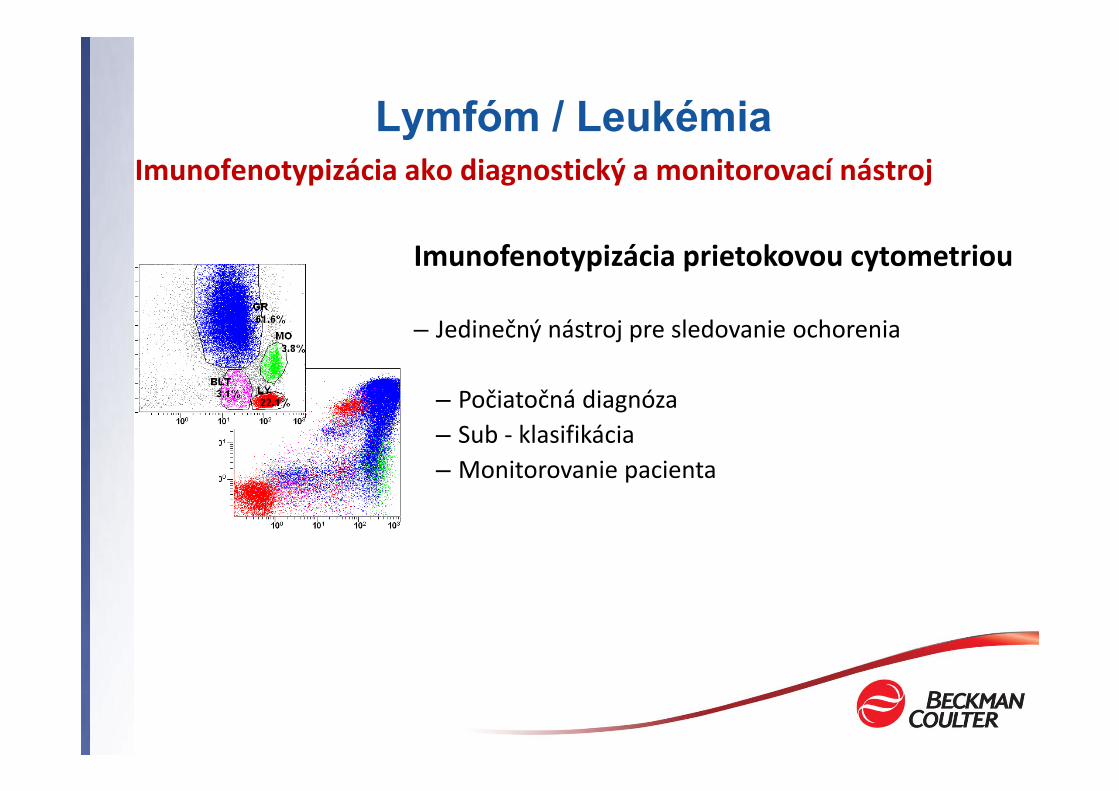
Imunofenotypizácia prietokovou cytometriou
– Jedinečný nástroj pre sledovanie ochorenia
– Počiatočná diagnóza– Sub ‐ klasifikácia– Monitorovanie pacienta
Imunofenotypizácia ako diagnostický a monitorovací nástrojLymfóm / Leukémia

Immunophenotyping in L&L
CHARACTERIZE the leukemic population by describing the antigens expressed
DIFFERENTIATE LEUKEMIC FROM NORMAL CELLS:‐ Normal
‐ Antigens expressed in consistent and reproducible patterns with maturation‐ Myeloid Maturation: Bone Marrow → Peripheral Blood‐ Lymphoid Maturation: Thymus → Peripheral Blood → Lymph Node
‐ Neoplastic – L&L‐ Increased or decreased normal antigens‐ Dysynchronous maturational expression‐ Aberrant antigen expression

L / L klasifikačný systém• FAB, REAL, WHO klasifikácia
Úloha pre prietokovú cytometriu: Štandardizácia, harmonizácia, klasifikačný systém
‐ Cytometrická diagnostika sa vykonáva v mnohých laboratóriách‐ Vysoký počet protokolov a panelov pre typizáciu lymfómov a
leukémií‐ Potrebná harmonizácia metód
‐ Stanovenie diagnózy v rôznych laboratóriách‐ Prognóza a terapia‐ Vývoj a monitoring u nových liekov na základe výsledkov z rôznych
pracovísk‐ (multi‐centre‐studies)

Štandardizácia – rok 2006 a 2008
2006 Bethesda International Consensus Recommendations on the
Immunophenotypic Analysis of Hematolymphoid Neoplasia by Flow
Cytometry
Cytometry 2007 72B:S14‐22
Practical International Consensus Established
WHO 2008 Classification of Tumours of
Hæmatopoietic and Lymphoid Tissues
4th Edition, 2008IARC Press

Conference Recommendations
CombinationStrategy
ConsensusMarkers
Bethesda 2006Consensus
Initial Screening ApproachLineage‐based panels
T‐CellB‐CellMyelo‐monocyticPlasma Cell
Not Defined
Highly expressed antigenson dimmer dyesCD45 as common marker
# of colors per tube (1, 2, 3, 4, 5, …colors)Sample prep & data analysis
Pathway to Standardization in L&L Phenotyping

Markers- descriptionCD2 T cells and most of the NK cellsCD3 Mature T cell (cytoplasmic expression in immature T cells)CD4 Helper/inducer T lymphocytes, monocytes, Immature myeloid cellsCD5 Thymocytes, mature T cells, subpopulation of B cellsCD7 T cells, NK cells, subpopulation of immature myeloid cellsCD8 Cytotoxic/suppressor T cells, subpopulation of NK cellsCD10 Common acute leukemia antigen (CALLA), lymphatic precursor cells, neutrophils,
subpopulation of mature B cellsCD11b Monocytes, macrophages, neutrophils, NK cellsCD13 Myeloid cellsCD14 Monocytes, weak expression on neutrophilsCD15 Neutrophils, weak expression on monocytesCD16 NK cells, neutrophils, subpopulation of monocytesCD19 Precursor and mature B cellsCD20 B cells and a subpopulation of B cell precursorCD33 Monocytes, myeloid precursor cells, weak expression on neutrophilsCD34 Myeloid and lymphoid precursor cellsCD38 Activated lymphocytes, subpopulation of B cells, plasma cellsCD45 All leukocytesCD56 NK cells, subpopulation of T cellsCD117 Myeloid precursor cellsKappa Kappa light chainLambda Lambda light chainHLA‐DR B lymphocytes, activated T lymphocytes, monocytes, precursor cells

Primary Panels – Aid in Initial ClassificationAcute Leukemia – Blasts Present >20%
Myeloid Lineage
CD13+ and/or CD33+ CD7+ and/or CD5+ CD19+
T-Lineage B-Lineage
Myeloid, no or minimal
maturation
CD34+/‐CD117+/‐HLADR+CD56+CD15‐/+
CD11b‐, CD14-
Promyeloid
CD15+/‐CD117+/‐CD34‐HLADR‐
CD11b‐, CD14‐
CD14+CD4+/‐
HLADR+CD15+
CD11b+
Myelomonocytic & Monocytic
Megakaryocytic and erythroid lineages require secondary panels

Acute Leukemias: Initial Lymphocytic Classification
Acute Leukemia – Blasts Present >20%
CD7+ and/or CD5+ CD19+
T-Lineage B-Lineage
CommonThymocyte
MatureThymocyte
CD2+CD5+Dual
CD4+/CD8+sCD3-
CD2+CD5+
CD4+ or CD8+sCD3+
CD2-/+CD5-/+sCD3-CD4-CD8-
TdT+CD10-clg-sig-
Pre-B
CD10+cig+TdT+sig-
B
sig+CD10+/-clg+/-TdT-
Common
TdT+CD10+
clg-sig-
EarlyThymocyte
AAssessment of maturity with primary reagentsRequire secondary panels for sub‐classification
Early B-Precursor

Initial Classification of CLLs and Lymphomas
CLL and Non-Hodgkins Lymphoma
CD2+CD3+
T-Cell Lineage
CD38+CD2-, CD19-
Plasma CellLeukemia/ Myeloma
B-Cell Lineage
CD19+
CD16+ and/or CD56+
CD2+, CD3-
CD5-
CD10-CD10+
Follicular Cell Or
Burkitt’s Lymphoma
SLVLor
Marginal Zone Lymphomaor
Hairy Cell Leukemia
CD5+
B-CLLor
Mantle Cell Lymphoma
Sub‐classification of Lymphomas Required
NK LGLL

Initial Classification of CLLs and Lymphomas
CLL and Non-Hodgkins Lymphoma
CD2+CD3+
T-Cell Lineage
CD38+CD2-, CD19-
Plasma CellLeukemia/ Myeloma
B-Cell Lineage
CD19+
CD16+ and/or CD56+
CD2+, CD3-
CD4+
T-Cell CLLT-Cell Lymphoma
T-PLL
ATLL(Adult T-CellLeukemia)
T-Cell LGLL
CD8+
CD7-
Sezary Syndrome
Sub‐classification of Lymphomas Required
NK LGLL


6 color 2 Laser (5+1)8 color 2 Laser (5+3)10 color 3 Laser (5+3+2)
Prietokové cytometre pre IVD
Cytomics FC 500• 2 konfigurácie
Navios• 3 konfigurácie
5 color 1 Laser 5 color 2 Laser

Navios Flow Cytometer
405nm
638nm
488nm
Available in 3 configurations6 color 2 Laser (5+1)8 color 2 Laser (5+3)
10 color 3 Laser (5+3+2)

Beckman Coulter 5-Color Panel Solutions
• B‐Cell Kit
• T‐Cell Kit
• Myeloid Kit
Aligned with Bethesda Recommendations
Kappa Lambda CD19 CD5 CD45
CD20 CD10 CD19 CD38 CD45
CD2 CD56 CD7 CD5 CD45
CD8 CD4 ⎯ CD3 CD45
CD15 CD11b CD16 CD14 CD45
HLADR CD56 CD34 CD117 CD45
CD7 CD13 CD34 CD33 CD45

NaviosTM / FC500 and SOLASTRATM

Reagent Performance: Target Specifications
• Specimen Types
‐ Peripheral Blood
‐ Bone Marrow
‐ Lymphoid cell suspensions
• EDTA, Heparin
• Specimen age: 24 – 48 hours post‐draw
• Target sensitivity: 1% when 50,000 events are collected
• Working WBC Range: 1000 – 50,000 cells/µL
• Sample preparation using VersaLyse + Fixative Lytic System
B-Lineage KitFITC PE ECD PC5.5 PC7
Kappa Lambda CD19 CD5 CD45
CD20 CD10 CD19 CD38 CD45
Myeloid Lineage KitFITC PE ECD PC5.5 PC7
CD15 CD11b CD16 CD14 CD45
HLADR CD56 CD34 CD117 CD45
CD7 CD13 CD34 CD33 CD45
T-Lineage KitFITC PE ECD PC5.5 PC7
CD2 CD56 CD7 CD5 CD45
CD8 CD4 -- CD3 CD45

Myelomonocytic Lineage Tube 1:
CD15 / CD11b / CD16 / CD14 / CD45
Myelomonocytic Lineage Tube 2:
HLA-DR / CD56 / CD34 / CD117 / CD45
Myelomonocytic Lineage Tube 3:
CD7/ CD13 / CD34 / CD33 / CD45
AML Phenotype:CD34+ 117+ Dim13+ 33+ 15+ 11b-16- 14- 56- 2- 7+
Acute Myeloid Leukemia - AML (BM)

BL1:
Kappa/ Lambda / CD19/ CD5 / CD45
BL2:
CD20/ CD10/ CD19 / CD38 / CD45
Acute Leukocyte Leukemia - ALL (PB)
ALL Phenotype:CD10+ 19+ 34+ 20- sIg- 38+

CLL Phenotype:CD5+ 10- 19+ Dim 20+ 38variable/-Dim Kappa+
Chronic Leukocyte Leukemia - CLL (PB)
BL1:
Kappa/ Lambda / CD19/
CD5 / CD45
BL2:
CD20/ CD10/ CD19 /
CD38 / CD45

Základný panel + ďalšie znaky
PB KrOrange FITC PE ECD PC5.5 PC7 APC APC-A700 APC-A750
Kappa Lambda CD19 CD5CD20 CD10 CD19 CD38 CD45
CD2 CD56 CD7 CD5 CD45CD8 CD4 ⎯ CD3 CD45
CD15 CD11b CD16 CD14 CD45HLADR CD56 CD34 CD117 CD45CD7 CD13 CD34 CD33 CD45

Panel pre orientáciu
405 Excitation 488 Excitation 633 Excitation
Paci
fic
Blue
Kro
me
Ora
nge
FITC
PE ECD
PC5.
5
PC7
APC
APC-
AF7
00
APC-
AF7
50
CD15/CD20 CD45 kappa/CD34 lambda/CD7 CD10 CD4 CD56/CD117 CD3/CD33 CD8 CD19
CD8/CD15/CD20 CD45 kappa/CD4 lambda/CD56 CD7 CD117 CD10 CD34 CD19 CD3/CD33
• Základné protilátky v jednej skúmavke• 10 fluorescencií• 15 monoklonálnych protilátok

Lymphoid Slide Ruler
www.coulterflow.comwww.beckmancoulter.com

Summary
• Role of Flow‐ Correlation of phenotype expression pattern to disease‐ Differentiate leukemic population from normal
• Bethesda Consensus Conference 2006 for Phenotyping L&L‐ Lineage‐Based panels for primary screening‐ Testing driven by patient presentation of medical indications
Beckman Coulter Solution – Lineage Reagent Kits► Aligned with Bethesda Phenotyping Strategy► 5‐Color Combinations for multi‐platform compatibility► Initial characterization as primary screen► Product Flexibility for sub‐classification

Suggested Reading
• Consensus Conference Publications‐ 2006 Bethesda International Consensus Recommendations on the Immunophenotypic Analysis
of Hematolymphoid Neoplasia by Flow Cytometry, Cytometry 2007 72B:S14‐22‐ WHO 2008 Classification of Tumours of Hæmatopoietic and Lymphoid Tissues, 4th Edition,
2008, IARC Press
• Review Articles‐ F.E. Craig & K.A. Foon, Flow Cytometric Immunophenotyping for Hematologic Neoplasms, Blood
Volume 111, Number 8:3941‐3967, 2008.‐ J.W. Vardiman, N.L. Harris, R.D. Brunning, The World Health Organization (WHO) Classification
of the Myeloid Neoplasms, Blood, Volume 100, Number 7:2292‐2302, 2002.

Ďakujem za pozornosť


