experiment posters_asu_emmanuelfonseca
TRANSCRIPT
Chronic stress impairs performance on hippocampal-mediated spatial tasks and alters hippocampal CA3 dendritic complexity in adult male rats (Conrad, 2006).
A post-stress recovery period allows for reversal: spatial ability improves or exceeds that of controls and hippocampal dendritic complexity returns to levels of unstressed controls (Hoffman et al., 2011; Ortiz et al., 2014).
Brain-derived neurotrophic factor (BDNF) is a protein that is required for spatial learning and memory (Radecki et al., 2005) and CA3 dendritic plasticity (Magariños et al., 2011).
We also recently report that hippocampal BDNF is required for the recovery of spatial ability following the end of chronic stress (Ortiz et al., 2014).
We previously found that downregulating hippocampal BDNF prevents the recovery of spatial ability following the end of chronic stress (Ortiz et al., 2014): the current study investigates whether hippocampal dendritic architecture also fails to recover and parallels spatial memory ability.
We hypothesized the hippocampal BDNF mediates the recovery from chronic stress-induced dendritic retraction, which parallels spatial memory deficits.
The RAWM is a task that requires rodents to have an intact hippocampus to optimally navigate to solve. Cues located outside and around the maze help with this “spatial navigation.”
A single platform is located at the end of one of the arms and the location remains constant across all trials for a given rat.
Errors are scored as incorrect entries into non-platformed arms within a given trial.
shRNA against BDNF will prevent recovery of dendritic complexity: stress-induced CA3 apical dendritic atrophy will persist: Str-Imm = Str-Rec-shRNA. Neurons illustrated are of the SS type.
Stress immediate
Stress recovery
Control Viral Infusion
and surgical rehab
2 weeks
No restraint 42 days
Stress 21 days
Stress 21 days
No restraint 21 days
No restraint 21 days
RAWM testing 3 days
8 trials for 2 days 1 trial day 3
Arrival Euthanize
Adult male Sprague-Dawley rats (n = 52) underwent stereotaxic surgery to bilaterally infuse an adeno-associated viral vector (AAV) into the hippocampus. AAV encoded for an shRNA directed against BDNF (shRNA) or a “scrambled” shRNA with no known correspondence to rat mRNA (Scr).
Rats were chronically stressed using wire mesh restraint (6h/d/21d), a procedure known to produce reliable effects on hippocampal morphology and function (McLaughlin, 2007) and then given a 21 day post-stress recovery period (Str-Rec) or not (Str-Imm) and then tested on the radial arm water maze.
The authors greatly acknowledge Dr. Ernest Terwilliger and Dr. Caroline Bass for designing and supplying the viral vectors used in this study. This research was supported in part by funds from the School of Life Sciences Undergraduate Research (SOLUR) Program through the School of Life Sciences at Arizona State University, Tempe Campus, ASU’s College of Liberal Arts and Sciences, the NIH IMSD program (R25GM099650 to Stuart Newfeld for funding Ortiz), and the National Science Foundation Graduate Research Fellowship Program (DGE-1311230, Ortiz).
Conrad CD (2006) What is the functional significance of chronic stress-induced CA3 dendritic retraction within the hippocampus? Behav. Cog, Neurosci. Rev., 5, 41-60.
Hoffman AN, Krigbaum A, Ortiz JB, Mika A, Hutchinson KM, Bimonte-Nelson HA & Conrad CD (2011) Recovery after chronic stress within spatial reference and working memory domains: correspondence with hippocampal morphology. Eur. J. Neurosci., 34, 1023-1030.
Magariños AM, Li C, Toth JG, Bath KG, Jing D, Lee FS & McEwen BS (2011) Effect of brain-derived neurotrophic factor haploinsufficiency on stress-induced remodeling of hippocampal neurons. Hippocampus, 21, 253-264.
McLaughlin KJ, Gomez JL, Baran SE & Conrad CD (2007) The effects of chronic stress on hippocampal morphology and function: an evaluation of chronic restraint paradigms. Brain research, 1161, 56-64.
Ortiz JB, Mathewson CM, Hoffman AN, Hanavan PD, Terwilliger EF & Conrad CD (2014) Hippocampal brain-derived neurotrophic factor mediates recovery from chronic stress-induced spatial reference memory deficits. Eur. J. Neurosci., 40, 3351-3362.
Radecki, DT, LM Brown, J Martinez & TJ Teyler (2005) BDNF protects against stress-induced impairments in spatial learning and memory and LTP. Hippocampus 15, 246
After the single retention trial on day 3 of testing, rats were euthanized and brains extracted for Golgi processing.
Brains were dissected in half, flash frozen in 2-methylbutane, and one hemisphere was processed for Golgi (FD NeuroTechnologies).
The other hemisphere was placed in a -80°C freezer. Our plans include processing this hemisphere for BDNF expression using ELISA (in progress).
With this study, we hope to show that recovery from stress-induced hippocampal CA3 dendritic retraction depends on hippocampal BDNF. • Work is still in progress; approximately five SS and five LS CA3 neurons
have been identified and traced for each rat and 33% have been checked. • We still have neurons from the CA1 and dentate gyrus to identify and draw. • We are working on determining the effectiveness of the shRNA in knocking
down hippocampal BDNF using ELISA (in progress). Uncovering factors, such as BDNF, that are important in mediating plasticity and
facilitating recovery may help identify novel mechanisms that can be targeted to enhance resilience in the face of adversity.
Con-Scr Unstressed Control rats
Str-Imm-Scr Chronically stressed
rats no recovery
Str-Rec-Scr Chronically stressed rats with recovery
Str-Rec-shRNA Chronically stressed rats with
recovery & downregulated hippocampal BDNF
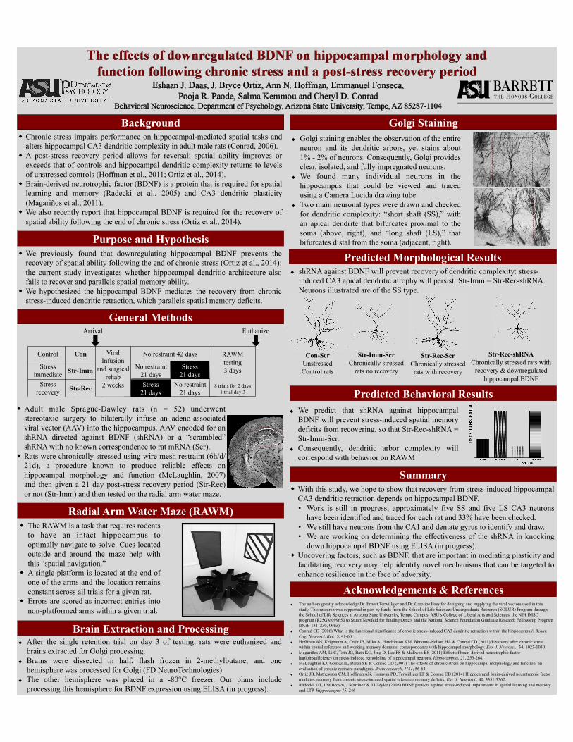
Golgi staining enables the observation of the entire neuron and its dendritic arbors, yet stains about 1% - 2% of neurons. Consequently, Golgi provides clear, isolated, and fully impregnated neurons.
We found many individual neurons in the hippocampus that could be viewed and traced using a Camera Lucida drawing tube.
Two main neuronal types were drawn and checked for dendritic complexity: “short shaft (SS),” with an apical dendrite that bifurcates proximal to the soma (above, right), and “long shaft (LS),” that bifurcates distal from the soma (adjacent, right).
We predict that shRNA against hippocampal BDNF will prevent stress-induced spatial memory deficits from recovering, so that Str-Rec-shRNA = Str-Imm-Scr.
Consequently, dendritic arbor complexity will correspond with behavior on RAWM
Background
Predicted Behavioral Results
Acknowledgements & References
Radial Arm Water Maze (RAWM)
Brain Extraction and Processing
Golgi Staining
Predicted Morphological Results
Summary
Purpose and Hypothesis
General Methods

Chronic Variable Stress Effects on Anxiety and Expression of Organic Cation Transporter 3
1. Amphoux, A., Vialou, V., Drescher, E., Bruss, M., Mannoury La Cour, C., Rochat, C., Millan, M.J., Giros, B., Bonisch, H., Gautron, S., 2006. DifferenFal pharmacological in vitro properFes of organic caFon transporters and regional distribuFon in rat brain. Neuropharmacology 50, 941-‐952.
2. Baganz, N.L., Horton, R.E., Calderon, A.S., Owens, W.A., Munn, J.L., WaUs, L.T., Koldzic-‐Zivanovic, N., Jeske, N.A., Koek, W., Toney, G.M., Daws, L.C., 2008. Organic caFon transporter 3: Keeping the brake on extracellular serotonin in serotonin-‐transporter-‐deficient mice. Proceedings of the NaFonal Academy of Sciences of the United States of America 105, 18976-‐18981.
3. Feng, N., Mo, B., Johnson, P.L., Orchinik, M., Lowry, C.A., Renner, K.J., 2005. Local inhibiFon of organic caFon transporters increases extracellular serotonin in the medial hypothalamus. Brain research 1063, 69-‐76.
4. Feng, N., Telefont, M., Kelly, K. J., Orchinik, M., Forster, G. L., Renner, K. J., & Lowry, C. A. (2009). Local perfusion of corFcosterone in the rat medial hypothalamus potenFates d-‐fenfluramine-‐induced elevaFons of extracellular 5-‐HT concentraFons. Hormones and behavior, 56(1), 149-‐157.
5. Gasser, P.J., Lowry, C.A., Orchinik, M., 2006. CorFcosterone-‐sensiFve monoamine transport in the rat dorsomedial hypothalamus: potenFal role for organic caFon transporter 3 in stress-‐induced modulaFon of monoaminergic neurotransmission. The Journal of neuroscience : the official journal of the Society for Neuroscience 26, 8758-‐8766.
6. Daws, L.C., 2009. Unfaithful neurotransmiUer transporters: focus on serotonin uptake and implicaFons for anFdepressant efficacy. Pharmacol Ther 121, 89-‐99.
7. Marcinkiewcz, C.a., and D.p. Devine, 2015. ModulaFon of OCT3 Expression by Stress, and AnFdepressant-‐like AcFvity of Decynium-‐22 in an Animal Model of Depression. Pharmacology Biochemistry and Behavior 33-‐41.
8. Reber, Stefan O., and Inga D. Neumann, 2008. Defensive Behavioral Strategies and Enhanced State Anxiety during Chronic Subordinate Colony Housing Are Accompanied by Reduced Hypothalamic Vasopressin, But Not Oxytocin, Expression. Annals of the New York Academy of Sciences 184-‐95.
• This project was funded by a grant awarded to MO from the NaFonal Science FoundaFon (NSF 0922085) and BarreU Faculty Support Funds. • This research was supported in part by funds from the School of Life Sciences Undergraduate Research (SOLUR) Program through the School of Life Sciences at Arizona State University, Tempe Campus
• Although there were no significant differences in adrenal weights, the reduced weight gain in the CVS group is consistent with predicFons that these rats were chronically stressed • Contrary to predicFons, there was no difference between CVS and unstressed rats in terms of sucrose preference8 • Also contrary to predicFons, CVS rats showed a non-‐significant tendency for reduced latency in the novel feeding environment. The non-‐significant tendency to consume food in the home environment suggests that CVS rats were not as moFvated or hungry as controls. • Preliminary opFmizaFon of fluorescent in situ hybridizaFon using mouse brains revealed OCT3 mRNA labeling in key limbic regions. • Future experiments will compare OCT3 mRNA and protein expression in CVS vs. control rats • Although there were no significant differences in behavioral tests, we are examining differences in OCT3 expression between CVS and control rats for potenFal paUerns and correlaFons.
• Organic caFon transporters (OCT)s are a family of transporters for monoamines (serotonin (5-‐HT), dopamine, and norepinephrine) in the human and murine brain.1
• OCT3 is believed to help clear extra-‐neuronal monoamines, thereby helping to terminate signaling2-‐5
• One known regulator of OCT3 is corFcosterone (the stress steroid hormone), which can alter 5-‐HT transport, clearance and neurotransmission, as well as HPA axis negaFve feedback and regulaFon5
• Altered expression of OCT3 may play a role in stress coping7
• Numerous basic and clinical studies from the last three to four decades have demonstrated that imbalances in 5-‐HT are linked to certain mood disorders, such as anxiety and depression6
• In previous studies, chronic variable stress (CVS) has produced anxiety and depressive like behaviors as measured by novelty suppressed feeding and sucrose preference tesFng
• Since OCT3 is involved in 5-‐HT clearance and transport, it is likely that OCT3 is influenced by chronic stress • We hypothesized that OCT3 is a mechanisFc link between stress, anxiety, and depression • We predicted that CVS would increase anxiety and depressive-‐like behaviors and increase expression of OCT3
Chronic Variable Stress Protocol
*
a) There was no significant difference in adrenal weights between CVS and control rats b) CVS rats gained less weight than control rats (p < .01) c) No significant difference was found in sucrose preference and total volume drunk between CVS and control rats, although CVS rats appeared to drink more sucrose soluFon and total liquid volume overall. d) No significant difference was found in latency to approach food in bright arena or food eaten in home cage between CVS and control rats, although CVS rats displayed a non-‐significant tendency for reduced latency and feeding in home cage.
Cerebral Aqueduct 400x Bregma -‐3.52
Dorsal Hippocampus CA3 400x Bregma -‐3.52
• 20 Sprague Dawley® rats were used in this study, 10 received unpredictable CVS for 14 days while 10 received no stress (see CVS protocol/Fmeline). • Following last administraFon of CVS, all rats were given both sucrose soluFon and tap water available to them, and the volume of liquid drunk was measured. • Novelty suppressed feeding tesFng measured latency to approach food when rats were placed in a novel, well-‐lit environment, as well as subsequent feeding in the home cage (during the dark cycle when rats are acFve). • Following behavioral tesFng, rats were euthanized, adrenals were removed and weighed, and brains were extracted and flash frozen. • RNA was reverse transcribed and the polymerase chain reacFon (PCR, HotStar Taq Qiagen) was performed using primers that contained the T7 phage promoter sequence. • Riboprobes were in vitro transcribed using a T7 kit (Ambion) containing BioFn-‐16-‐labeled uridine triphosphate • For opFmizaFon of in situ hybridizaFon, 10 µm secFons (Bregma~-‐3.52) of mouse brains were fixed and hybridized with 5µg of the bioFn-‐labeled riboprobe at 80°C overnight. The next day, secFons were washed with increasing stringency, and slides were incubated using Streptavidin-‐AlexaFluor 488 conjugate. • Following 1 hr incubaFon with the conjugate soluFon, slides were incubated in 1% Sudan Black to reduce auto-‐fluorescence .
Piper Boyll1, Pooja R. Paode1,2, Emmanuel Fonseca2, J. Bryce OrFz2, Joshua S. Talboom3,4, Jeremiah Molinaro1, Salma Kemmou2, Eshaan J. Daas1,2, Cheryl D. Conrad2, and Miles Orchinik1
1Arizona State University, School of Life Sciences, Tempe, AZ 85287, 2Arizona State University, Department of Psychology, Tempe AZ 85287 3Banner Sun Health Research InsFtute, Sun City, AZ 85351, 4Arizona Alzheimer’s ConsorFum, Phoenix, AZ 85014
a) b)
c)
d)