expression of inhibin α- and βa-subunit mrna and protein in the fetal sheep ovary throughout...
TRANSCRIPT
ELSEVIER Molecular and Cellular Endocrinology 107 (1995) 141-147
Expression of inhibin a- and PA-subunit mRNA and protein in the fetal sheep ovary throughout gestation
H. Engelhardt aJ, L.M. Harknessa, G.B. Thomasb, A.N. Brook+, A.S. McNeillya-b, D.T. Bairda,*
aDeparfment of Obstetrics and Gynaecology, University of Edinburgh, 37 Chalmers Street, Edinburgh, EH3 9EW. UK bMRC Reproductive Biology Unit, Centre for Reproductive Biology, Universiry of Edinburgh, Edinburgh, UK
Received 25 August 1994; accepted 7 November 1994
Abstract
In adult sheep, inhibin expression in developing follicles appears to be associated with antrum formation. Our objective was to investigate using in situ hybridization and immunohistochemistry whether antral follicles present before birth in the sheep expressed mRNA or peptide for inhibin a- and PA-subunits. At days 70 and 100 when only primordial and primary follicles were present, there was no detectable mRNA or peptide for either inhibin subunit. By days 130 and 140 (term = 145 days), many secondary follicles were present, a proportion of which (-50%) expressed detectable levels of a-subunit mRNA but not peptide. A number of antral follicles were present by this stage, all of which expressed a-subunit mRNA and peptide. Expression of PA-subunit mRNA and peptide was undetectable at all stages of gestation. Our results indicate that even in non-ovulatory follicles present before birth, expression of inhibin, at least the a-subunit, is developmentally linked with antrum formation.
Keywords: Inhibin; Fetus; Ovary; Follicle; Sheep; mRNA
1. Introduction
Inhibin is a glycoprotein composed of two subunits, a and /I. that have been shown to be products of different genes (Mason et al., 1985; Forage et al., 1986). Two re- lated p-subunits have been identified which when com-
bined with the a-subunit give rise to inhibin A (a/,,*) and inhibin B (a/&) (Ling et al., 1985; Mason et al., 1985).
Both forms of inhibin suppress pituitary FSH release
(reviewed by Lincoln et al., 1989), whereas activins,
dimers of the b-subunits stimulate FSH secretion (Ling et al., 1986; Vale et al., 1986). In cycling adult sheep, in- hibin expression has been shown to be highly regulated such that a- and b-subunit mRNA is expressed only by the granulosa cells of selected non-atretic follicles that
have reached the antral stage of development (Engelhardt et al., 1993).
* Corresponding author, Fax: +44 31 229 2408. ’ Present address: Department of Animal and Poultry Science, Uni-
versity of Guelph, Guelph, Ontario, Canada NlG 2Wl. Fax: +l 519 767 0573.
Concentrations of immunoactive FSH and LH in ovine fetal plasma peak at 115-125 days of gestation then de-
cline to low levels by term (145 days) (Sklar et al., 1981).
A regulatory role for inhibin is supported by evidence that injection of steroid-free follicular fluid as a source of crude inhibin suppressed FSH but not LH secretion in
both male and female fetuses (112-l 25 days of gestation)
(Albers et al., 1989b), and infusion of FSH over days
125-130 increased gonadal inhibin content, although the
response in females was variable and not statistically sig-
nificant (Albers et al., 1989a). In the sheep, primordial follicles are present by day 70
(MaulCon, 1969; Riisse, 1983; Smith et al., 1993), and like the human and the cow but in contrast to most other
mammals, antral follicles are present before birth (day 135) (Maulton, 1969; Smith et al., 1993). While expres- sion of inhibin and activin subunits has not been investi- gated in the fetal sheep ovary, both a- and PA-subunit
mRNA has been demonstrated in fetal bovine ovaries by
90 days of gestation (Torney et al., 1990). The objective of the present study was to investigate expression of in- hibin subunit mRNA and peptide throughout ovarian de- velopment in the fetal sheep using in situ hybridization
0303-7207/95/$09.50 0 A995 Elsevier Science Ireland Ltd. All rights reserved SSDI 0303-7207(94)03435-U
142 H. Engelhardt et al. I Molecular and Cellular Endocrinology 107 (1995) 141-147
and immunohistochemistry. More specifically, we ad- dressed the question of whether the antral follicles present
before birth expressed inhibin a- or PA-subunits.
2. Materials and methods
2.1. Animals Pregnant Scottish Blackface ewes with known insemi-
nation dates were killed with an overdose of sodium pentobarbitone anaesthesia on days 70, 100, 130 and 140 of gestation (term = 145 days). Ovaries were removed (all singleton females; n = 2 per stage), cut into two and
placed immediately in freshly prepared paraformaldehyde in PBS (pH 7.4; PFA) overnight. The tissue was then de-
hydrated and embedded in paraffin. Sections (5 pm) were mounted on slides coated with 2% 3arninopropyltri-
ethoxy-silane (TESPA; Sigma, Poole, Dorset, UK), incu- bated overnight at 60°C and then processed for immuno-
histochemistry or in situ hybridization as described be-
low.
2.2. In situ hybridization All in situ hybridization procedures were performed as
previously described in our laboratory (Engelhardt et al., 1993). Briefly, from full-length human a and /IA cDNAs
kindly provided by R.G. Forage, Sydney, Australia (Forage et al., 1986), selected sequence fragments were subcloned into pBluescript SK(&) plasmids and subse- quently transformed into competent XL I blue E. coli. The a-subunit cDNA was 345 bp in length, coding part of
the mature region of the human inhibin a-subunit, corre- sponding to nucleotides 712-1057. The/IA-subunit cDNA was 447 bp in length, coding amino acids in the mature and pro regions of the @*-subunit protein precursors, complementary to nucleotides 670-l 118. 35S-Labelled sense and antisense riboprobes were generated from their cDNA templates by transcription using an RNA tran-
scription kit (Stratagene, La Jolla, CA). Specific activities were 8 x 10s and 6.7 x lo* cpm&g for a and /I,,, antisense
probes, respectively. The in situ hybridization procedure was originally de-
scribed by Wilkinson et al. (1987a,b). Sections were de-
paraffinized, rehydrated through a graded series of etha- nols and postfixed in PFA (20 min). They were then washed, digested with proteinase K (20,@ml; Sigma) in 50 mM Tris, 5 mM EDTA buffer (TE), refixed in PFA, and washed twice in 0.25% acetic anhydride in 0.1 M triethanolamine hydrochloride (TEA; 10 min). The sec- tions were then washed in PBS and saline, dehydrated and
air-dried. Probes were diluted in hybridization mix (50% deionized formamide, 10% dextran sulphate, 20 mM Tris-HCl, 5 mM EDTA, 0.3 M NaCl, 0.02% Ficoll, 0.02% polyvinylpyrroline, 0.02% BSA, 50 mM dithio- threitol (DIT; all from Sigma) to obtain 1 x 10s dpm/ml;
they were then heated at 80°C and cooled on ice before application to sections at lO@lslide. After being overlaid
with cover slips made from Gelbond film (FMC Bio-
products, Rockland, ME), slides were incubated overnight
in a humidified chamber saturated with 5x SSC (standard sodium citrate; single strength = 0.1 M NaCl, 15 mM so- dium citrate) and 50% formamide at 55°C.
After hybridization, slides were washed in 5x SSC, 0.01 M DTT at 55°C for 30 min, then in 2x SSC, 50% formamide, 0.1 M DTT for 20 min at 65°C. The slides
were washed three times in NTE buffer (0.5 M NaCl, 10 mM Tris, 5 n&I EDTA, pH 7.5) for 10 min at 37”C, treated with RNase (4Opglml in NTE; Stratagene) for 30 min at 37°C and washed in NTE at the same tempera- ture. The wash in 2x SSC, 50% formamide, 0.1 M DTT
(20 min, 65°C) was repeated, and the slides were then washed in buffers containing decreasing salt concentra- tions at room temperature (2x and 0.1~ SSC; 3 times,
10 min each). Slides were dehydrated, air-dried, and
dipped in liquid emulsion (Kodak NTB-2 (Rochester, NY) diluted 1: 1 in water immediately before use) at 40-
42°C. Slides were exposed for 4 weeks at 4”C, developed for 4 min in Kodak D-19 developer, washed for 1 min in water and fixed for 4 min (Kodafix/water, 1:4). Slides were stained with haematoxylin (BDH, Glasgow, UK), dehydrated, mounted and examined under bright- and darkfield optics using a Zeiss microscope (Oberkochen,
Germany).
Sense probes were used in parallel with antisense probes on alternate sections to control for non-specific hybridization. No signal was observed with either a or /IA sense probes in any tissue (not shown). Adult sheep ova- ries previously shown to express inhibin a- and PA-
subunit mRNA (Engelhardt et al., 1993) were included in each run as positive controls.
2.3. Zmmunohistochemistry For localization of the a-subunit, the polyclonal anti-
body R146 was raised in a rabbit against the first l-26 amino acids of the N-terminus of the a-subunit of porcine
inhibin (McNeilly et al., 1989). The monoclonal antibody E4 raised against amino acids 84-l 12 of the /IA-subunit of
human inhibin (Groome and Lawrence, 1991) was kindly
donated by N.P. Groome (Oxford Polytechnic, Oxford,
UK). A modified double peroxidase-antiperoxidase (PAP) technique was used to localize inhibin a- and /I,-subunits in the fetal ovaries. Tissue sections were deparaffinized, rehydrated and treated with 3% hydrogen peroxide (BDH Chemicals Ltd., Poole, Dorset UK) in methanol for 30 min to remove endogenous peroxidase activity. The sections were then washed in distilled water for 5 min
before being treated with 0.1% trypsin (Sigma) for 15 min at 37°C followed by 0.5% Triton-X for 15 min. The sections were washed in distilled water for 5 min followed by 0.05 M Tris-HCl buffer (pH 7.6), for 5 min before being incubated with 5% BSA and either 25%
normal pig serum (a) or 25% normal rabbit serum @A) for 30 min to reduce non-specific background staining.
H. Engelhardt et al. I Molecular and Cellular Endocrinology 107 (1995) 141-147 143
The sections were then incubated overnight in a humidi- fied chamber at 4°C with the respective primary antibody (for a-inhibin, 1:600; #I,-inhibin, 1: 1200). Following two washes in 0.05 M Tris-HCl, sections were incubated for 30 min with either swine anti-rabbit or rabbit anti-mouse serum (for a and /IA, respectively; both at 1:30). The sec- tions were washed twice in 0.05 M Tris-HCl and then incubated for 30 min with PAP complex (Dako Ltd., Glostrup, Denmark). These last two incubation steps were repeated before visualization using a freshly prepared solution of 3,3_diaminobenzidine tetrahydrochloride (4 mg/lO ml Tris buffer, Sigma) containing 3% hydrogen peroxide. Sections were stained with haematoxylin, dehy- drated, and mounted as above.
Several criteria were used to test the specificity of the antibodies used. First, control sections were incubated with either normal rabbit serum or normal mouse serum instead of primary antibody.,Sections were also incubated with R146 antiserum which had been pre-absorbed over- night with excess porcine a-inhibin (l-26) peptide (Thomas et al., 1994). As a positive control, sections of adult sheep ovary were included throughout all staining procedures. Finally, serial dilution of both primary anti- bodies resulted in a gradual decrease in overall staining intensity.
3. Results
Because ovaries were not serially sectioned, follicles in a section from a given ovary were considered a sample of those in that ovary. In ovaries containing secondary to antral follicles (days 130 and 140), 36-51 follicles per animal were assessed. For each follicle, diameter was estimated using an eyepiece graticule, and the presence of the antrum or oocyte noted. It is acknowledged that be- cause the plane of section may have missed the antrum and/or the largest cross section of a follicle, these data will provide only an approximation of size and develop- ment. However, in combination with morphological ob- servations, these measures taken for the large number of follicles present during fetal development should provide
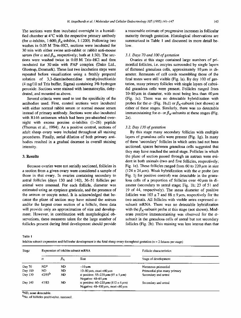
Table 1
a reasonable estimate of progressive increases in follicular maturity through gestation. Histological observations are summarized in Table 1 and discussed in more detail be- low.
3.1. Days 70 and 100 of gestation Ovaries at this stage contained large numbers of pri-
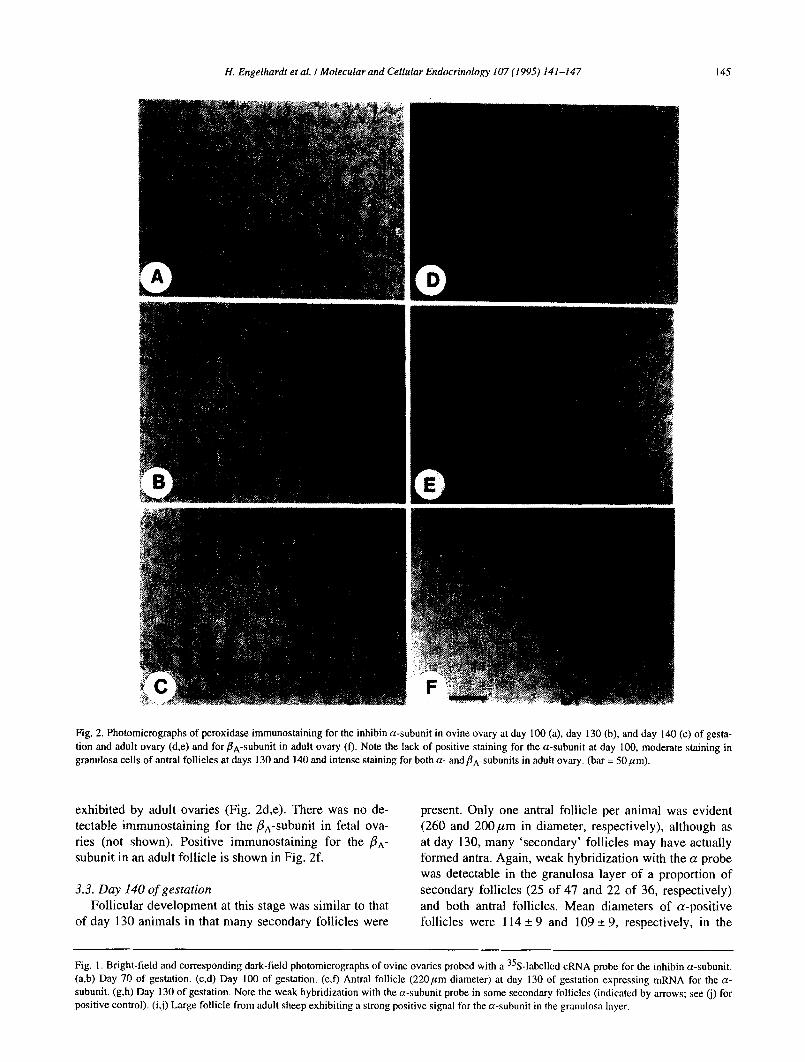
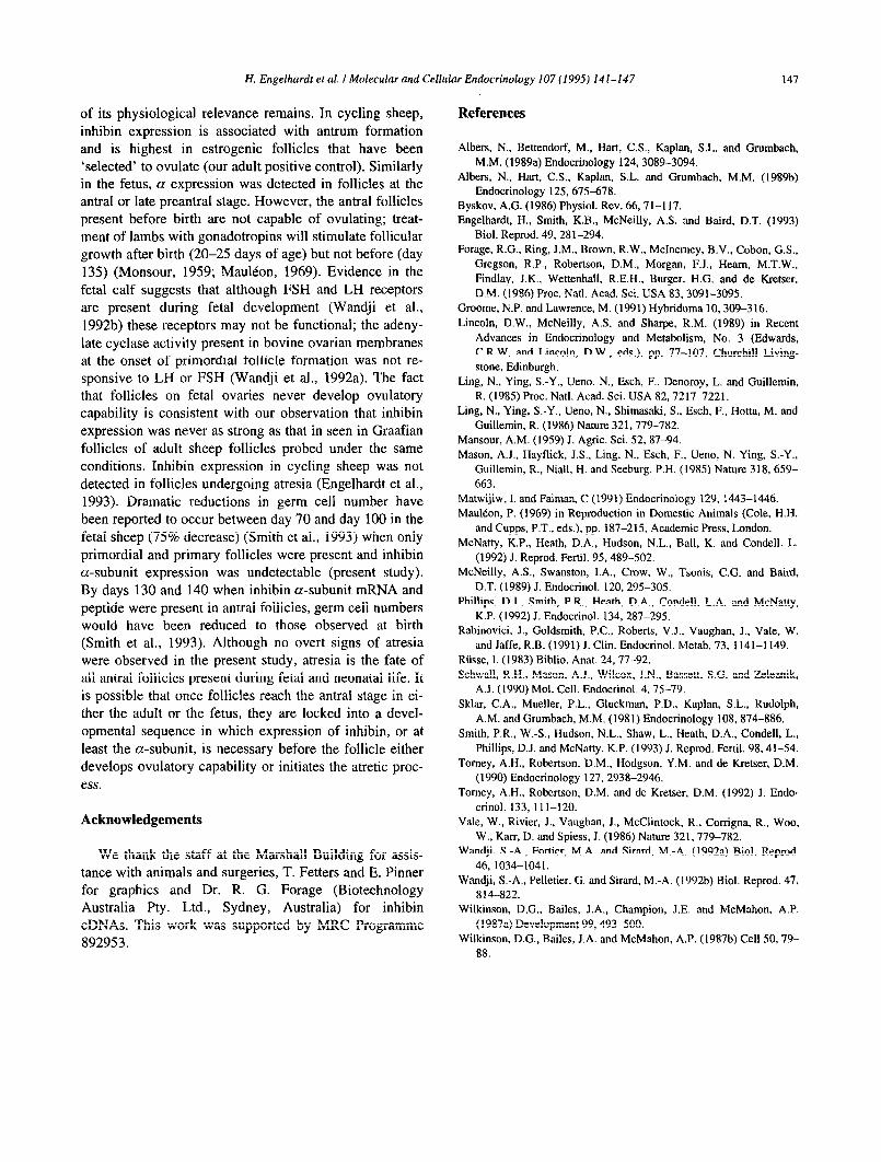
mordial follicles, i.e. oocytes surrounded by single layers of flattened granulosa cells, approximately lO,~m in di- ameter. Remnants of cell cords resembling those of the fetal testes were still visible (Fig. la). By day 100 of ges- tation, many primary follicles with single layers of cuboi- da1 granulosa cells were present. Follicles ranged from lO-80pm in diameter, with most being less than 40pm (Fig. lc). There was no detectable hybridization with probes for the a- (Fig. lb,d) or PA-subunit (not shown) at either of these stages. Similarly, there was no detectable immunostaining for a- or PA-subunits at these stages (Fig. 2a).
3.2. Day 130 of gestation By this stage many secondary follicles with multiple
layers of granulosa cells were present (Fig. lg). In many of these ‘secondary’ follicles in which antra had not been sectioned, spaces between granulosa cells suggested that they may have reached the antral stage. Follicles in which the plane of section passed through an antrum were evi- dent in both animals (two and five follicles, respectively; Fig. le). These follicles ranged from 60 to 220pm in size (124 + 24,~m). Weak hybridization with the a probe (see Fig. lj for positive control) was detectable in the granu- losa cells of a proportion of follicles over 40pm in di- ameter (secondary to antral stage; Fig. lh; 23 of 5 1 and 19 of 44, respectively). The mean diameter of positive follicles was 103 + 7 and 88 +- 9pm, respectively for the two animals. All follicles with visible antra expressed a- subunit mRNA. There was no detectable hybridization with the j3*-subunit probe at this stage (not shown). Mod- erate positive immunostaining was observed for the a- subunit in the granulosa cells of antral but not secondary follicles (Fig. 2b). This staining was less intense than that
Inbibin subunit expression and follicular development in the fetal sheep ovary throughout gestation (n = 2 fetuses per stage)
Stage Expression of inhibin subunit mRNA
a BA Size
Follicle characteristics
Stage of development
Day 70 ND”
Day 100 ND
Day 130 42/95b
Day 140 47183
ND
ND
ND
ND
-1Opm
lO-80pm, most <40pm
a positive: 55-220 pm (97 * 5 pm)
Negative: 40-65 pm
apositive:60-220~m(112+6~m)
Negative: 40-lOOym, most <80,~4m
Numerous primordial
Primordial plus many primary
Secondary and antral
Secondary and antral
aND, none detectable.
bNo. of follicles positive/no. assessed.
144 H. Engelhardt et al. I Molecular and Cellular Endocrinology 107 (1995) 141-147
H. Engelhardt et al. I Molecular and Cellular Endocrinology 107 (1995) 141-147 145
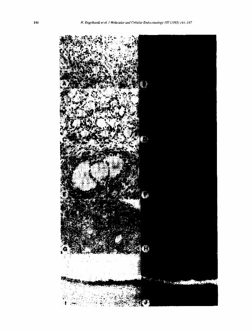
Fig. 2. Photomicrographs of peroxidase immunostaining for the inhibin a-subunit in ovine ovary at day 100 (a), day 130 (b), and day 140 (c) of gesta-
tion and adult ovary (d,e) and for PA-subunit in adult ovary (f). Note the lack of positive staining for the a-subunit at day 100, moderate staining in
granulosa cells of antral follicles at days 130 and 140 and intense staining for both a- and#lA-subunits in adult ovary. (bar = 50pm).
exhibited by adult ovaries (Fig. 2d,e). There was no de- tectable immunostaining for the PA-subunit in fetal ova- ries (not shown). Positive immunostaining for the PA- subunit in an adult follicle is shown in Fig. 2f.
3.3. Day 140 of gestation Follicular development at this stage was similar to that
of day 130 animals in that many secondary follicles were
present. Only one antral follicle per animal was evident (260 and 200pm in diameter, respectively), although as at day 130, many ‘secondary’ follicles may have actually formed antra. Again, weak hybridization with the a probe was detectable in the granulosa layer of a proportion of
secondary follicles (25 of 47 and 22 of 36, respectively) and both antral follicles, Mean diameters of a-positive follicles were 114 rt 9 and 109 + 9, respectively, in the
Fig. 1. Bright-field and corresponding dark-field photomicrographs of ovine ovaries probed with a 35S-labelled cRNA probe for the inhibin a-subunit.
(a,b) Day 70 of gestation. (cd) Day 100 of gestation. (e,f) Antral follicle (220pm diameter) at day 130 of gestation expressing mRNA for the a-
subunit. (g,h) Day 130 of gestation. Note the weak hybridization with the a-subunit probe in some secondary follicles (indicated by arrows; see (j) for
positive control). (ij) Large follicle from adult sheep exhibiting a strong positive signal for the a-subunit in the grnnulosa layer.

146 H. Engelhardt et al. I Molecular and Cellular Endocrinology 107 (1995) 141-147
two animals. Although numbers were limited and assess-
ment of expression qualitative, it appeared that a expres- sion was somewhat weaker at day 140 relative to day 130.
However, it must be pointed out that the a signal in the
fetal ovaries was never as intense as that seen in Graafian follicles of adult sheep ovaries (Fig. lj). As at day 130,
there was no detectable hybridization with the/3,-subunit
probe (not shown) and moderate positive immunostaining was observed for the a- but not the /3,-subunit in the granulosa cells of antral follicles (Fig. 2~).
4. Discussion
At days 70 and 100 of gestation when only primordial and primary follicles were present, there was no detect-
able mRNA or peptide for either of the inhibin subunits.
By days 130 and 140, many secondary to antral follicles
were in evidence. In a proportion of these (approximately 50%), mRNA for the inhibin a-subunit was detectable but
immunostaining for the peptide was not. A number of antral follicles could clearly be identified at these stages,
all of which expressed both a-subunit mRNA and pep- tide. In no case was the signal in fetal ovary as intense as that in adult sheep ovaries. There was no detectable ex-
pression of the PA-subunit mRNA or peptide at any stage of gestation.
ment. Gonadectomy of ovine fetuses increased plasma concentrations of immunoactive FSH in males but not
females (Matwijiw and Faiman, 1991). Concentrations of immunoactive inhibin in male fetal sheep are several-
fold higher than in females (Phillips et al., 1992). Accord- ingly, female sheep fetuses have consistently higher pitui-
tary FSH concentrations and contents than males of comparable age (Phillips et al., 1992). These differences are consistent with the ontogeny of other aspects of gonadal development such as steroidogenesis, synthesis of anti-milllerian hormone, and gonadotropin responsive- ness which begin at the time of sex determination in the male but much later in the female (reviewed by Byskov,
1986).
In the monkey, small antral follicles expressing only
the &,-subunit have been reported (Schwa11 et al., 1990). In human midgestation fetuses, weak immunostaining for the PA-subunit only in a few primary follicles has been
observed (Rabinovici et al. 1991). Because PA- and pa- subunits were co-expressed by inhibin-positive follicles in adult sheep (Engelhardt et al., 1993) we have not probed
for the pa-subunit in subsequent studies. We cannot, therefore, rule out the presence of &-expressing follicles during ovarian development in the fetal sheep, but there was certainly no evidence of PA expression in the current
study.
Previous studies investigating inhibin production by the ovine fetal ovary have been inconclusive; plasma con-
centrations of immunoactive inhibin in female fetuses
were high at day 40 and 55 of gestation at a time when no antral follicles were present (Phillips et al., 1992; Smith et al., 1993). Furthermore, inhibin was not detected in the
ovary, adrenal or mesonephros at this time using the same RIA (Smith et al., 1993). After day 55, plasma inhibin levels declined, reaching levels that were still twofold greater than those in adult ewes by term (McNatty et al.,
1992; Phillips et al., 1992). At later stages of gestation, ovarian inhibin was detectable but at much lower levels
than in adrenals (Smith et al., 1993). In contrast, im-
munostaining for all three subunits was reported in nu-
merous primary and secondary follicles in late gestation monkey ovary (Rabinovici et al., 1991). In the cow, levels of bio- and immunoactive inhibin in fetal ovaries were
reported to increase through gestation (Torney et al., 1990). In the same study, Northern analysis indicated that quantities of mRNA for the a-subunit were low but de- tectable by 90 days, peaked at 180-210 days and declined towards term, while mRNA for the B,-subunit was only detectable at 180-210 days of gestation. Using in situ hybridization, mRNA for both a- and B,-subunits was
localized diffusely over the cortical region of bovine fetal
ovaries at 120-180 days and in the granulosa cells of an- tral follicles at later stages (210-270 days) (Torney et al., 1990).
The failure to detect expression of mRNA or peptide
corresponding to the PA-subunit could be due to limita- tions in the sensitivities of our techniques. In support of this explanation is a report that bioactive inhibin has been
demonstrated in homogenates of fetal ovaries and testes of sheep at 111-143 days of gestation (Albers et al., 1989a). However, the riboprobes used in the current study
give very strong signals in adult sheep ovary (Engelhardt et al., 1993). Although the level of expression of a- subunit mRNA in fetal ovaries was very much lower than in the adult, it was still detectable under our conditions.
Furthermore, in ovine fetal testes at the same stage of
gestation which exhibited a much stronger a signal than the fetal ovaries, /IA expression was still undetectable (our
own unpublished observations). The detection of a- but not PA-subunit raises the question of what these fetal tis-
sues are producing. As mentioned above, although it was possible that they expressed/Is in the complete absence of /l,.+ our findings in the adult sheep suggest that this is un- likely. Alternatively, fetal tissue could be synthesizing biologically inactive free a-subunit. Follicles expressing a-subunit only were found in the adult sheep at all stages of the cycle (Engelhardt et al., 1993). In extracts of bo- vine fetal testes, large amounts of a-subunit precursors
have been identified (Torney et al., 1992) and an excess
of a-subunit mRNA has been demonstrated by both Northern analysis and in situ hybridization (Torney et al., 1990).
Several lines of evidence indicate that inhibin may be If developing follicles of the fetal ovary do indeed
more important in male than in female gonadal develop- synthesize intact inhibin at very low levels, the question
H. Engelhardt et ul. I Molecular and Cellular Endocrinology 107 (1995) 141-147 147
of its physiological relevance remains. In cycling sheep,
inhibin expression is associated with antrum formation
and is highest in estrogenic follicles that have been
‘selected’ to ovulate (our adult positive control). Similarly in the fetus, Q: expression was detected in follicles at the antral or late preantral stage. However, the antral follicles
present before birth are not capable of ovulating; treat- ment of lambs with gonadotropins will stimulate follicular growth after birth (20-25 days of age) but not before (day
135) (Monsour, 1959; Mauleon, 1969). Evidence in the
fetal calf suggests that although FSH and LH receptors
are present during fetal development (Wandji et al.,
199213) these receptors may not be functional; the adeny-
late cyclase activity present in bovine ovarian membranes at the onset of primordial follicle formation was not re- sponsive to LH or FSH (Wandji et al., 1992a). The fact that follicles on fetal ovaries never develop ovulatory
capability is consistent with our observation that inhibin expression was never as strong as that in seen in Graafian
follicles of adult sheep follicles probed under the same
conditions. Inhibin expression in cycling sheep was not
detected in follicles undergoing atresia (Engelhardt et al., 1993). Dramatic reductions in germ cell number have
been reported to occur between day 70 and day 100 in the
fetal sheep (75% decrease) (Smith et al., 1993) when only primordial and primary follicles were present and inhibin
a-subunit expression was undetectable (present study). By days 130 and 140 when inhibin a-subunit mRNA and
peptide were present in antral follicles, germ cell numbers would have been reduced to those observed at birth (Smith et al., 1993). Although no overt signs of atresia were observed in the present study, atresia is the fate of
all antral follicles present during fetal and neonatal life. It
is possible that once follicles reach the antral stage in ei- ther the adult or the fetus, they are locked into a devel-
opmental sequence in which expression of inhibin, or at
least the a-subunit, is necessary before the follicle either
develops ovulatory capability or initiates the atretic proc-
ess.
Acknowledgements
We thank the staff at the Marshall Building for assis- tance with animals and surgeries, T. Fetters and E. Pinner for graphics and Dr. R. G. Forage (Biotechnology Australia Pty. Ltd., Sydney, Australia) for inhibin
cDNAs. This work was supported by MRC Programme 892953.
References
Albers, N., Bettendorf, M., Hart, C.S., Kaplan, S.L. and Grumbach,
M.M. (1989a) Endocrinology 124.3089-3094.
Albers, N., Hart, C.S., Kaplan, S.L. and Grumbach. M.M. (1989b) Endocrinology 125.675-678.
Byskov, A.G. (1986) Physiol. Rev. 66,71-l 17.
Engelhardt, H., Smith, K.B., McNeilly, AS. and Baird, D.T. (1993)
Biol. Reprod. 49, 281-294.
Forage, R.G., Ring, J.M., Brown, R.W., McInemey, B.V., Cobon, G.S.,
Gregson, R.P., Robertson, D.M., Morgan, F.J., Heam, M.T.W.,
Findlay, J.K., Wettenhall, R.E.H., Burger, H.G. and de Kretser,
D.M. (1986) Proc. Natl. Acad. Sci. USA 83,3091-3095.
Groome, N.P. and Lawrence, M. (1991) Hybridoma 10,309-316.
Lincoln, D.W., McNeilly, A.S. and Sharpe, R.M. (1989) in Recent
Advances in Endocrinology and Metabolism, No. 3 (Edwards,
C.R.W. and Lincoln, D.W., eds.), pp. 77-107, Churchill Living-
stone, Edinburgh.
Ling, N., Ying, S-Y., Ueno, N., Esch, F., Denoroy, L. and Guillemin,
R. (198.5) Proc. Natl. Acad. Sci. USA 82.7217-7221.
Ling, N., Ying, S-Y., Ueno, N., Shimasaki, S.. Esch, F., Hotta, M. and
Guillemin, R. (1986) Nature 321.779-782.
Mansour, A.M. (1959) J. Agric. Sci. 52.87-94.
Mason, A.J., Hayflick, J.S.. Ling, N., Esch, F.. Ueno, N. Ying, S.-Y.,
Guillemin, R., Niall, H. and Seeburg, P.H. (1985) Nature 318, 659-
663.
Matwijiw, I. and Faiman, C (1991) Endocrinology 129.1443-1446.
Maulton, P. (1969) in Reproduction in Domestic Animals (Cole, H.H.
and Cupps, P.T., eds.), pp. 187-215, Academic Press, London.
McNatty, K.P., Heath, D.A., Hudson, N.L., Ball, K. and Condell. L.
(1992) J. Reprod. Fertil. 95.489-502.
McNeilly, A.S., Swanston, I.A., Crow, W., Tsonis, C.G. and Baird,
D.T. (1989) J. Endocrinol. 120.295-305.
Phillips, D.J., Smith, P.R., Heath, D.A., Condell, L.A. and McNatty,
K.P. (1992) J. Endocrinol. 134, 287-295.
Rabinovici, J., Goldsmith, P.C., Roberts, V.J., Vaughan, J., Vale, W.
and Jaffe, R.B. (1991) J. Clin. Endocrinol. Metab. 73, 1141-1149.
Rtisse. I. (1983) Biblio. Anat. 24,77-92.
Schwall, R.H., Mason, A.J.. Wilcox, J.N., Bassett, S.G. and Zeleznik,
A.J. (1990) Mol. Cell. Endocrinol. 4,75-79.
Sklar, C.A., Mueller, P.L., Gluckman, P.D., Kaplan, S.L., Rudolph,
A.M. and Grumbach, M.M. (1981) Endocrinology 108,874-886.
Smith, P.R., W.-S., Hudson, N.L., Shaw, L., Heath, D.A., Condell, L.,
Phillips, D.J. and McNatty, K.P. (1993) J. Reprod. Fertil. 98.41-54.
Tomey, A.H., Robertson, D.M., Hodgson, Y.M. and de Kretser, D.M.
(1990) Endocrinology 127.2938-2946.
Tomey, A.H., Robertson, D.M. and de Kretser, D.M. (1992) J. Endo-
crinol. 133, 11 l-120.
Vale, W., Rivier, J., Vaughan, J., McClintock, R., Corrigna, R., Woo.
W., Karr, D. and Spiess. J. (1986) Nature 321, 779-782.
Wandji, S.-A., Fortier, M.A. and Sirard, M.-A. (1992a) Biol. Reprod.
46, 1034-1041.
Wandji, S.-A., Pelletier, G. and Sirard. M.-A. (1992b) Biol. Reprod. 47,
814-822.
Wilkinson, D.G., Bailes, J.A., Champion, J.E. and McMahon, A.P.
(1987a) Development 99,493-500.
Wilkinson, D.G., Bailes, J.A. and McMahon, A.P. (1987b) Cell 50. 79-
88.