ficus benghalensis - nsf digital library: home
TRANSCRIPT

Ficus benghalensis
S . A . D e r a n i y a g a l a R . L . C . W i j e s u n d e r a
N a t i o n a l Sc ience F o u n d a t i o n C o l o m b o
J a n u a r y ' 2 0 0 2

Contents
Page I I n t r o d u c t i o n : The Banyan Tree 1
II P h y t o c h e m i s t r y 5
1 Compounds isolated from the milky juice 5 2 Compounds isolated from the leaves 6 3 Compounds isolated from the heartwood 9 4 Compounds isolated from the fruits and seeds 10 5 Compounds isolated from the stem bark 12
111. P h a r m a c o l o g y 18
1 Anti-tumor activity 18 2 Anti-microbial activity 18 3 Anti-diarrhoeal activity 19 4 Anti-diabetic action and serum insulin raising
effect 21 5 Insulin sparing effect 31 b Hypolipidemic action 34 7 Hffects on glucose-6-phosphate, hexose kinase
and H M G C o A reductase enzyme activity 37 8 Antioxidant effects 38
I V S u m m a r y 41
V . R e f e r e n c e s 44
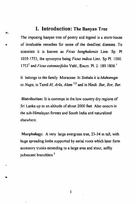
I. Introduction: The Banyan Tree
The imposing banyan tree of poetry and legend is a store-house
of invaluable remedies for some of the deadliest diseases. To
scientists it is known as Ficus benghalensis Linn. Sp. PI
1059.1753, the synonyms being Ficus indica Linn. Sp PI. 1060.
1753 1 and Ficus cotonaeifoliaWM., Enum. PI. 2: 189.1806.'
It belongs to the family Moraceae. In Sinhala it is Mahamiga or Muga, in Tamil Al, Aria, Alam 1 , 2 and in Hindi Bar, Bor, Ber.
Distribution: It is common in the low country dry regions of
Sri Lanka up to an altitude of about 2000 feet. Also occurs in
the sub-Himalayan forests and South India and naturalized
elsewhere.
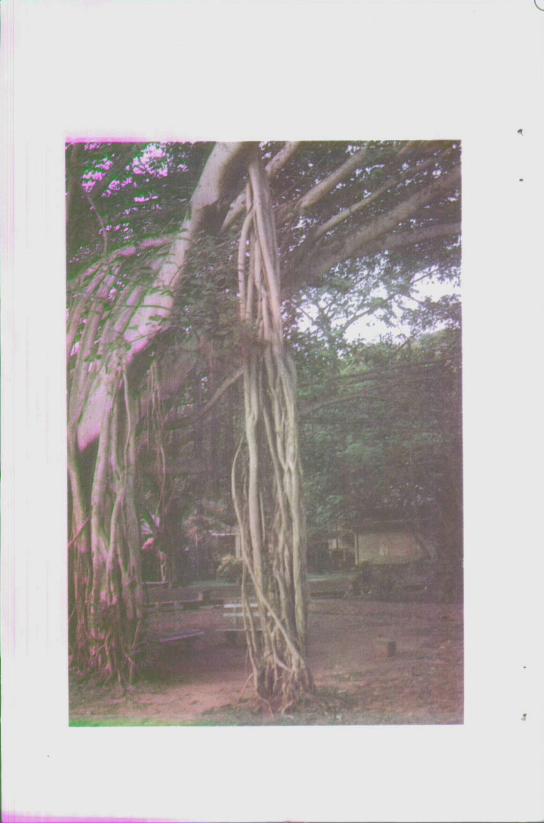
Morphology: A very large evergreen tree, 23-34 m tall, with
huge spreading limbs supported by aerial roots which later form
accessory trunks extending to a large area and stout, softly
pubescent branchlets.2
1

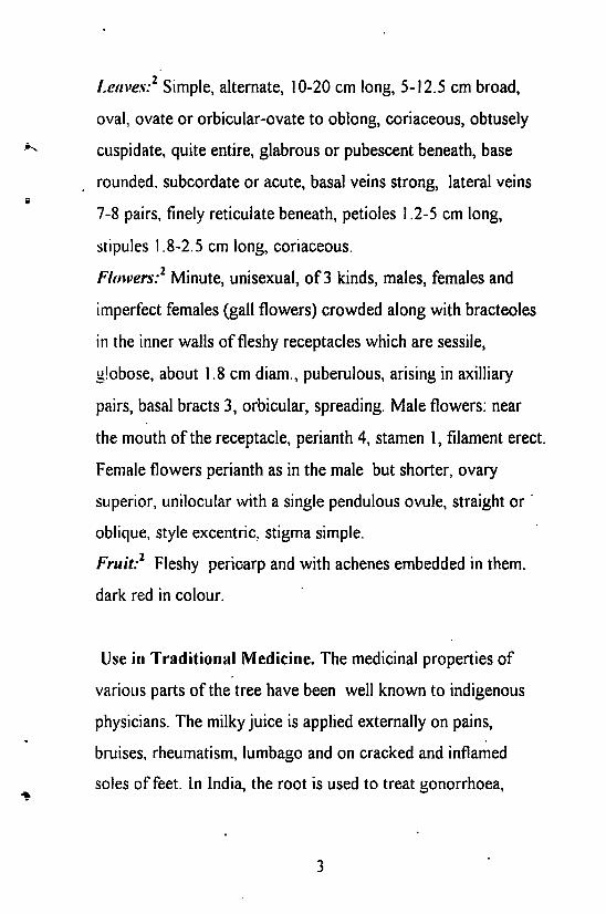
Leaves:2 Simple, alternate, 10-20 cm long, 5-12.5 cm broad,
oval, ovate or orbicular-ovate to oblong, coriaceous, obtusely
cuspidate, quite entire, glabrous or pubescent beneath, base
rounded, subcordate or acute, basal veins strong, lateral veins
7-8 pairs, finely reticulate beneath, petioles 1.2-5 cm long,
stipules 1.8-2.5 cm long, coriaceous.
Flowers:1 Minute, unisexual, of 3 kinds, males, females and
imperfect females (gall flowers) crowded along with bracteoles
in the inner walls of fleshy receptacles which are sessile,
globose, about 1.8 cm diam., puberulous, arising in axilliary
pairs, basal bracts 3, orbicular, spreading. Male flowers: near
the mouth of the receptacle, perianth 4, stamen 1, filament erect.
Female flowers perianth as in the male but shorter, ovary
superior, unilocular with a single pendulous ovule, straight or
oblique, style excentric, stigma simple.
Fruit:1 Fleshy pericarp and with achenes embedded in them,
dark red in colour.
Use in Traditional Medicine. The medicinal properties of
various parts of the tree have been well known to indigenous
physicians. The milky juice is applied externally on pains,
bruises, rheumatism, lumbago and on cracked and inflamed
soles of feet. In India, the root is used to treat gonorrhoea,
3

biliousness, dysenterv and inflammation of the liver The tips of
the aerial roots are also used to relieve persisting vomiting and
dysentery. Infusion of the small branches are used for
heamoptysis The infusion of the bark is supposed to be a
powerful tonic and is considered to have specific properties in
the treatment of diabetes

I I . P h y t o c h e m i s t r y
1 . C o m p o u n d s i s o l a t e d f r o m t h e m i l k y j u i c e
Early work on Ficus benghalensis reports the isolation of
ficosterol from the milky juice 3".
Although, the authors3" refer to the isolation of ficosterol from
the milky juice, the original literature 3 b concerning this does not
assign a structure to ficosterol. It reports the isolation of a
compound (melting point 135°C) from the unsaponifiable
matter of the milky juice of F. benghalensis which has shown
characteristic colour reactions of sterols in Liebermann
Burchard and Salkowski's reaction. According to the iodine
value and the formation of a dibromo compound, ficosterol is
said to have one double bond. The acetyl, benzoyl and the 3,5-
dinitro benzoyl derivatives have been prepared. Cupric oxide
oxidation of ficosterol has yielded a ketone (ficostenone, m.p.
105°C); thus showing the presence of a secondary OH group in
ficosterol. The article also mentions that molecular weight
determination by cryoscopic methods has given a molecular
weight of 410 indicative of a molecular formula C29H50. This
seems to be erroneous since ficosterol has been demonstrated as
having a hydroxyl group.
5

2. Compounds isolated from the leaves
2.1 Hydrocarbons 4
Surface lipids play a vital role in affecting transpiration and
leaf surface properties. The composition of lipids and in
particular hydrocarbon distribution may be a useful parameter
in chemotaxonomy. The predominant occurrence of lower even
numbered alkanes, viz. C M , Ci6, Cjg and C20 from the same
plant of F. benghalensis collected during two different months,
is very striking. The occurrence of such lower hydrocarbons in
greater proportions, from a dicotyledonous plant is unusual.
(The preponderance of odd-numbered alkanes is what is
normally seen in higher plants). The presence of branched chain
alkanes in relatively higher proportion is also noteworthy.
The above analysis has been carried out by GLC (10% SE-30
column with flame ionization detector) of the cold n-hexane
extract of the leaves after initial purification by preparative TLC
with carbon tetrachloride.
2.2 Triterpenoids and Sterols
The triterpene, friedelin (I) has been isolated 5 from the neutral
fraction of the petroleum ether extract of the leaves of the plant
6

following chromatography over alumina with petroleum ether
and benzene as the eluant. The structure is said to have been
proven by elemental analysis, mass spectral data and by mixed
melting point and co-tlc with that of an authentic sample.
P-sitosterol5 (II) has been isolated from the column on elution
with benzene. The compound has been characterized by
elemental analysis, mass spectral data and by mixed melting
point with a pure specimen.
Physical data. Compound \ m.p. 235-238°C, [ a ] D = - 22°
(CHCI3). Pink colour with Liebermann-Burchardt reagent.
Compound II: m.p. 135-137°C, [oc]D= -35° (CHCI3). Positive
test for sterols with Liebermann-Burchardt reagent
Friedelin (I)
HO
P-sitosterol (II)
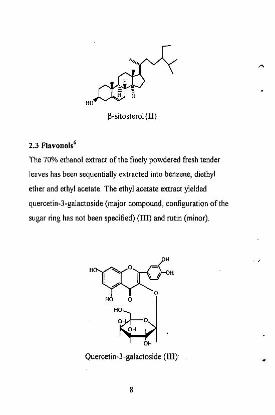
2.3 Flavonols 6
The 70% ethanol extract of the finely powdered fresh tender
leaves has been sequentially extracted into benzene, diethyl
ether and ethyl acetate. The ethyl acetate extract yielded
quercetin-3-galactoside (major compound, configuration of the
sugar ring has not been specified) (HI) and rutin (minor).
8

3. Compounds isolated from the heart wood
3.1 Tiglic acid ester of v|/-taraxasterol (IV) has been isolated 6
from hot benzene extract of the heartwood of the plant. The
compound has been characterized by its infrared (ER) spectra
and by mixed melting point and co-thin layer chromatography
(tic) of the alkaline hydrolysis products with authentic vj/-
taraxasterol and tiglic acid.
Physical data: Compound IV: m.p. 236-238°C, [ o t ] D
2 8 = +88.5°
(CHCI3). IR (cm"1): 2940, 1730, 1455, 1375, 1245, 1025, 980,
873 and 655. Alkaline hydrolysis ( C e H 6 medium) gave v|/-
taraxasterol (m.p. 215-216°C, [ a ] D
2 8 = + 48.6°, acetyl, m.p. 240-
242°C. [ a ] D
2 8 = +53.2°, benzoate 270-274°C) and tiglic acid
(m.p. and mixed m.p. 62-64°)
Taraxasterol (IV)
9

4. Compounds isolated from fruits and seeds Early work reports the isolation of glutathione (V) from
fruits.'"
Glutathione (V)
4.1 Amino acids 7
The free and bound amino acid composition of fruits and seeds
of some medicinal plants are reported. F. benghalensis is
reported as one of the species having the highest level of amino
acids in the fruit proteins. Of the medicinal plants that have
been investigated (among these being Ficus racemosa, Ficus
religiusa and Ficus lacor), cysteine was only found in the
proteins of F. benghalensis and F. religiosa. Amino acids such
as glutamine, methionine and tryptophan have not been
detected in the protein hydrolysates of any of the other plant
species. The essential amino acids arginine and methionine as
well as citrulline and hydroxyproline were not present in the
free state in the fruits of Ficus benghalensis
10
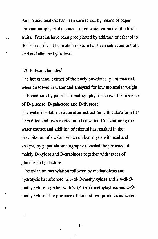
Amino acid analysis has been carried out by means of paper
chromatography of the concentrated water extract of the fresh
fruits. Proteins have been precipitated by addition of ethanol to
the fruit extract. The protein mixture has been subjected to both
acid and alkaline hydrolysis.
4.2 Polysaccharides8
The hot ethanol extract of the finely powdered plant material,
when dissolved in water and analysed for low molecular weight
carbohydrates by paper chromatography has shown the presence
of D-glucose, D-galactose and D-fructose.
The water insoluble residue after extraction with chloroform has
been dried and re-extracted into hot water. Concentrating the
water extract and addition of ethanol has resulted in the
precipitation of a xylan, which on hydrolysis with acid and
analysis by paper chromatography revealed the presence of
mainly D-xylose and D-arabinose together with traces of
glucose and galactose.
The xylan on methylation followed by methanolysis and
hydrolysis has afforded 2,3-di-O-methylxylose and 2,4-di-O-
methylxylose together with 2,3,4-tri-O-methylxylose and 2-0-
methylxylose The presence of the first two products indicated
11

that the xylose units in the xylan are linked by 1,4 and 1,3
bonds. The 2,3,4-tri-O-methylxylose has arisen from the
terminal end groups. The presence of 2-0-methylxylose *
indicated branching.
5. Compounds isolated from the stem bark
5.1 Anthocyanidin derivatives
3',5,7-trimethylether ofleucocyanidin (VI), 3',5,7-trimethyl
ether of delphinidin-3-0-a-L-rhamnoside(VII) and 3',5-
dimethyl ether of leucocyanidin-3-O-P-D-galactosylcellobioside
(VIII) have been isolated from the water insoluble fraction of
the ethanolic extract of the defatted stem bark9. These
compounds have been isolated from a silica gel column on
elution with mixtures of chloroform/rnethanol followed by
methanol itself.
Characteristics. Compound VI: m.p. 165°C (crystallized from
ethyl acetate-petroleum ether), [ a ] D
2 9 = +195°(MeOH).
Compound VII: m.p. 170-171°C (Crystallized form ethyl
acetate-petroleum ether), [ a ] 2 9
D = +232°(MeOH).
Compound VIII: Crystallized from MeOH-ether, m.p. 340°C,
[ a ] 2 9 o = +298° (MeOH)
12
5,7-dimethylether of leucopelargonidin-3-O-a-L-rhamnoside
(IX) has been isolated from the water soluble fraction of the
ethanolic extract of the defatted stem bark after extraction into
ethyl acetate.
Characteristics: Compound IX: m.p. 190°C (Crystallized from
ethanol-ether), [ c t ] 2 9
D = +260°(MeOH).
Starting with 2 kg of stem bark, 350 mg of compound VI, 500
mg of compound VII, 300 mg of compound VIII and 200 mg
of compound IX have been isolated.
These compounds have been characterized by chemical tests
which include demethylation, methylation, acetylation,
degradation methods, use of colour reagents and elemental
analysis and by ultraviolet spectroscopy.
Although, melting points have been recorded no cross reference
has been made 9 to that reported previously in literature for these
compounds. No nmr data is reported. This same article is used
as cross reference by other authors as well, who claim to have
isolated these compounds from the stem bark of F. benghalensis
with the aim of studying its pharmacological activity . However,
they do not give any further data in support of the structures of
the isolated compounds.
13

3',5,7-trimethyl ether of leucocyanidin (VI)
3',5,7-trimethyl ether of delphinidin-3-O-a-L rhamnoside(VII)
5,7-dimethyl ether of leucopelargonidin-3-O-a-L-rhamnoside
(IX)
14

S.2 Other Compounds
(3-sitosterol-a-D-glucose (X), 20-tetratriacontene-2-one (XJ), 6-
heptatriacontene-10-one (XII) and pentatriacontan-5-one(XIII)
have been isolated 3 8 from the hot petroleum ether extract of the
stem bark and meso inositol (XIV) from the hot ethanol extract
of the defatted stem bark.
Compound X was isolated by column chromatography on silica
gel (eluant 1:1 v/v petroleum ether-benzene) of the crystalline
compound that precipitated on standing of unconcentrated
petroleum ether extract. Compounds XI - XIII were isolated by
column chromatography on silica gel of the hot petroleum ether
extract with petroleum ether: benzene 3:1 v/v - 1:1 v/v as
eluant and purified by repeated chromatography with mixtures
of CCU and CHCI3. Compound XIV was obtained by
chromatography of the defatted ethanol extract of the stem bark
on silica gel with methanol as eluant.
Physical data: Compound X: m.p. 283-284°C, the compound
on hydrolysis gave p-sitosterol and a-D-glucose which are said
to have been identified by mixed melting point and co-
chromatography and superimposable IR spectra.
15
1
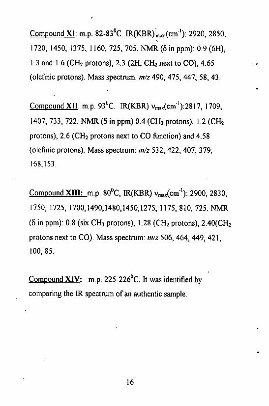
Compound XI: m.p. 82-83°C. IR(KBR) m a x (cm' ' ) : 2920, 2850,
1720, 1450, 1375, 1160, 725, 705. NMR (5 in ppm): 0.9 (6H),
1.3 and 1.6 (CH 2 protons), 2.3 (2H, CH 2 next to CO), 4.65
(olefinic protons). Mass spectrum: m/z 490, 475, 447, 58, 43.
Compound XH: m.p. 93°C. IR(KBR) v ^ c m " 1 ) ^ ^ 1709,
1407, 733, 722. NMR (8 in ppm) 0.4 (CH 3 protons), 1.2 (CH 2
protons), 2.6 (CH 2 protons next to CO function) and 4.58
(olefinic protons). Mass spectrum: m/z 532, 422, 407, 379,
168,153.
Compound XIII: m.p. 80°C, IR(KBR) v ^ c m " 1 ) : 2900, 2830,
1750, 1725, 1700,1490,1480,1450,1275, 1175, 810, 725. NMR
(5 in ppm): 0.8 (six CH 3 protons), 1.28 (CH 2 protons), 2.40(CH 2
protons next to CO). Mass spectrum: m/z 506, 464, 449, 421,
100, 85.
Compound XIV: m.p. 225-226°C. It was identified by
comparing the IR spectrum of an authentic sample.
16

p-Sitosterol-ct-D-glucose (X)
CH 3 -(CH 2 ) 1 2 -CH=CH-(CH2) 1 7 -C-CH3
20-Tetratriacontene-2-one (XI)
? CH3-(CH2)26-C-(CH 2 ) 2 -CH=CH-(CH2 )4 -CH3
6-Heptatriacontene-10-one (XII)
CH 3 - (CH 2 ) 2 9 -C- (CH 2 )3 -CH 3
Pentatriacontan-5-one(XIII)
H o y ^ Y 0 " H c A s ^ O H
OH
/weso-inositol (XIV)
17

III. Pharmacology
1. Anti-tumor activity
The chloroform extract of the fruit of F. benghalensis has
shown toxicity in the brine shrimp (Artemia salina) bioassay
(LC50 < 1000 |!g/ml). It also possessed anti-tumor activity in
the potato disc bioassay (% tumor inhibition >20%).
2. Anti-microbial activity10
The chloroform extract of the fruit of F. benghalensis has also
shown inhibitory activity (0.5 mg/disc) against the bacterium
Micrococcus luleus (18-26 mm diameter inhibition zone),
which was not inhibited by kanamycin (100 u.g/disc),
streptomycin (100 (ig /disc) or penicillin (5 ug/disc).
Streptococcus faecalis and Strep, faecium were also inhibited by
the fruit extract (17-20 mm inhibition zone). Other bacteria
such as Bacillus cereus, B. megaterium, Staphylococcus aureus,
Staph, epidermis, Strep, lactis, Escherichia coli, Klebsiella
pneumonia, Proteus vulgaris and Pseudomonas aeruginosa
were inhibited to a lesser extent (16-19 mm inhibition zone).
The extract did not show any anti-fungal activity.
18

3. Anti-diarrhoeal activity"
The ethanol extract of the hanging roots of Ficus benghalensis
has been evaluated for anti-diarrhoeal activity against different
experimental models of diarrhoea in rats. The extract (400
mg/kg, orally) has shown significant inhibitory activity against
castor oil induced diarrhoea (extract fed rats had 2.21 ±0.27
defecations per animal in 4 hr; control 4.0010.33, P < 0.001)
and P G E 2 induced enteropooling (for extract fed rats the value
reported is 1.25±0.15 in terms of intestinal fluid; control
0.78±0.11, P < 0.02) in rats. The extract has also been
significantly effective in reducing gastrointestinal mobility
(extract fed rats: 50.2±2.7%; control 79.412.76% ,P < 0.001) in
charcoal meal test in rats.
Summary of methodology: The coarse powder of the plant
material was Soxhlet extracted into ethanol. The extract
suspension to be administered to rats was prepared by using
weighed amounts of the ethanol extract in 2%(w/v) aqueous
tragacanth suspension.
Castor oil induced diarrhoea in rats: Albino rats (weighing
between 180- 200 g) were fasted for 18 h. One group was given
the 400 mg/kg dose. Another group received diphenoxylate
(5 mg/kg) orally in the form of a suspension as the standard drug
for comparison. The control group received 2% w/v aqueous
tragacanth suspension only.
19

After I h treatment each animal received I ml castor oil orally
and was observed for defecation. Up to 4 h after the castor oil
treatment the presence of diarrhoeal droplets were noted in the
transparent plastic dishes placed in the individual rat cages.
Gastrointestinal mobility tests: Rats fasted for 18 h were
administered orally with charcoal meal (3% deactivated
charcoal in 10% aqueous tragacanth). Immediately afterwards
one group was orally administered with 400 mg/kg of extract
suspension. Another group received atropine (0.1 mg/kg i.p.
(intra peritoneal)), the standard drug for comparison. A third
group was treated with aqueous tragacanth suspension as the
control. Thirty minutes later each animal was killed and the
intestinal distance moved by the charcoal meal from the pylorus
was cut, measured and expressed as a percentage of the distance
from the pylorus to the caecum for each animal
PGE 2-induced enteropooling: Rats were deprived of food and
water for 18 hours. One group of rats was treated with the
extract at a dose of 400 mg/kg (orally). Another group was
treated with 1 ml of 5% v/v ethanolic normal saline (i.p.) and
aqueous tragacanth solution, which served as a vehicle control.
Immediately after extract administration, P G E 2 was
administered orally to each rat (100 p.g/kg) in the first four
20

groups in 5% v/v ethanol in normal saline. A third group,
treated with P G E 2 as well as tragacanth suspension served as
P G E 2 control group. After 30 mins following administration
each rat was killed and the whole length of the intestine from
the pylorus to caecum dissected out and its contents were
collected in a test tube and the volume was measured.
Each group of rats consisted of ten rats. Statistical significant
tests have been performed by Student's t-test.
4. Anti-diabetic action and insulin raising effect
4.1. Compound VITJ (3',5-dimethylether of leucocyanidin-3-
0-P-D-galactosylcellobioside)1 2
Compound VITJ isolated according to Subramanium and
Misra9 is reported as demonstrating antidiabetic activity. On
oral administration of compound V1H (250 mg/kg body
weight), the blood sugar decreased very significantly (P <
0.001) of both normal (26% fall) and moderately diabetic (19%
fall) rats and serum insulin (39% rise) increased significantly in
the latter at a dosage of 250 mg/kg for a 2 h period. During a
month- treatment of the diabetic rats orally with compound
VIII, at a dosage of 100 mg/kg, there was a significant (P <
0.001) decrease in the blood sugar (58% fall) and the urine
sugar.
21
The mechanism of action has been suggested as the stimulatory
action of compound V D I on the sources of endogenous insulin.
Compound VIU is structurally related to tannin. There are
reports 1 3 that insulin can complex to tannins and remain
biologically active.
Summary of methodology. Short term effect of compound VIU
on normal and on diabetic rats have been determined as follows.
Rats weighing 115-155 g were made diabetic by subcutaneous
injection of an aqueous solution of alloxan (250 mg/kg body
weight). After a week rats having blood sugar in the range 200-
250 mg/lOOml were taken for the study.
Two groups of normal and two groups of diabetic rats (each
group with six rats each) were fed on rat feed. Their fasting
blood sugar was determined according to the method of Asatoor
and King 1 4 from a sample obtained from their eyes (venous
pool). The leucocyanidin derivative (compound VDI)
suspended in saline (10 ml/kg wt of rats) was administered to
the rats as follows: 1.. normal control rats fed saline (10 ml/kg
wt of rats) 2. normal rats treated with compound IV (dose 250
mg/kg), 3. diabetic control rats fed saline 4. diabetic rats treated
with the same doses of compound VIU. Blood samples were
collected at 2 h intervals for blood sugar analysis. All groups
22

were sacrificed and their whole blood was collected for serum
insulin assay. 1 5
The long term effect of the same was determined as follows.
Three groups were used. A normal group of rats was fed saline
(10 ml /kg/day), a diabetic group fed on saline and diabetic
group fed on leucocyanidin derivative (compound VIII) (100
mg/kg/day). Fasting blood sugar 1 4 , urine sugar 1 6 and body
weight of the diabetic rats at weekly intervals and for the
normal rats only their initial and final fasting blood sugar and
body weights were determined.
4.2. Compound IX (5,7-dimethylether of leucopelargonidin-
3-O-a-L-rhamnoside)
a) Compound IX has been isolated from the water soluble
portion of the ethanol extract of the defatted stem bark of F.
benghalensis according to the procedure of Subramanium and
Misra9. The anti-diabetic effect 1 7 of compound IX (250 mg/kg
single dose and lOOmg/kg/day long term study) has been
compared with that of glibenclamide (2 mg/kg and 0.5
mg/kg/day respectively) in moderately diabetic rats. The single
dose glycoside treatment decreased (P < 0.001) fasting blood
glucose by 19% and improved glucose tolerance by 29%. The
corresponding effects of glibenclamide were 25% and 66%
respectively over the control values. On one month treatment,
23

the fasting blood glucose levels had dropped almost to half the
pretreatment levels in both the groups and their glucose
tolerance improved by 4 1 % (P < 0.001) in the glibenclamide
treated group and by 15% (P < 0.02) in the glycoside treated
group. Urine sugar decreased to traces in both groups.
In vitro studies have shown that insulin secretion by beta cells
was more in the presence of compound IX than with compound
VIU (leucocyanadin derivative).
Summary of methodology: Similar methodology to that for
compound VIU in section 4.1
Serum insulin (as determined by radio immunoassay) had
increased 1 8 by 4 1 % (P < 0.01) in the group treated with
compound IX as compared to 51% (P < 0.001) in the
glibenclamide treated group in moderately diabetic rats.
In one month period, while the blood glucose of the diabetic
control group had increased (P < 0.01) by 34%, that of the
glycoside (compound IX) and glibenclamide treated groups
decreased significantly (P < 0.01) by 39% and 53%
respectively. The corresponding serum insulin variations in
these groups were also significant. In the control group it
decreased by 27% and in the glycoside and glibenclamide
treated groups it increased by 43% and 56% respectively (P <
0.01).
24

The body weight of the treated groups has remained steady
while that of the (diabetic) control decreased very significantly
by 27%.
Summary of Methodology: Methodology is given in section 6.1
b) The serum insulin raising and hypoglycemic effects of the
leucopelargonidin derivative (compound IX) has also been
demonstrated 1 9 in normal and alloxan diabetic dogs. A blood
glucose decrease of 15% in normal dogs in the first hour and a
19.5% decrease (P < 0.01) in the second has been reported 1 9.
Corresponding to this there has been a significant increase in
serum insulin by 62% in the first hour and by 76% (P < 0.001)
in the second hour. In the control group there has been no
significant variation in the blood glucose or in the serum insulin
level.
The leucopelargonidin derivative significantly decreased the
blood glucose by 18% in the first hour and by 29% in the
second hour. There was correspondingly an increase of serum
insulin by 60% in the first hour and 100% in the second hour (P
< 0.001).
Acute and chronic administration in single doses of 0.2-1.8
g/kg to different groups of mice and daily administration of
100, 250 and 500 mg/kg to rats for a period of one month
25

respectively had no toxic effect and the compound was not
lethal even at the high dose of 1.8 g/kg in experimental animals.
In the chronic toxicity study, the only significant effect
observed was a 12% blood glucose fall in the 2 . 5 E D 5 0 treated
group and a 14% blood glucose fall in 5ED 5o treated group after
one month treatment. There has been no notable change in the
food intake, body weight gain or in the behaviour as compared
to the control group. Urine analysis has not shown even traces
of sugar, albumin or any abnormality on microscopic analysis.
Haematological studies including Hb content, total WBC count
and differential WBC count, have also not shown any
significant changes in them- Histopathological study of the
intestine, liver and kidney of the 5EDso group has shown that
the chronic administration of the drug did not damage the
organs. There has been no significant changes in the levels of
blood urea and enzymes studied in the chronic test group.
Summary of methodology: Studies on the antidiabetic effect:
Six normal dogs were kept in an animal house and were
maintained on a uniform diet. A pilot study with 100 mg/kg
ED50 dose of the compound on dogs had shown that the
hypoglycemic action attains its peak at 2 h. The same group of
dogs served as the control (placebo fed) and later, after a week,
26

as the test group (fed with leucopelargonidin (compound IX)).
Placebo feed was empty gelatin capsules whereas in the second
week the capsules were filled with compound IX (500-600
mg/capsule). In both the control and the test group the blood of
each group of dogs fasted for 18 h was collected from the veins
of legs and the samples were used to determine the blood
glucose by glucose oxidase 2 0 method and serum insulin by
immunoassay 1 5.
Effects on alloxan diabetic dose. The dogs were rendered
diabetic by intravenous injection of alloxan monohydrate (60
mg/kg). The dogs were maintained on the same diet as before.
After a fortnight, when the dogs were found to be moderately
diabetic (200-300 mg/100 ml glucose levels) and after fasting
them for 18 hours, blood samples were collected. A placebo
was administered to each of them and their blood was again
withdrawn at 1 and 2 h and the blood glucose and serum
insulin levels were determined in all the samples. A ED50 dose
of compound IX (100 mg/kg) was administered to the same
group after a period of one week and the effect of the compound
on blood glucose and serum insulin level has been determined.
Acute Toxicity:. Albino mice weighing between 20-26 g were
divided into 5 groups of 8 each. The drug (compound IX) was
dissolved in normal saline and administered to groups of 18 h
27
fasting mice in doses of 200, 400, 800, 1200 and 1800 mg/kg
body weight. Each group received the drug at these doses in a
volume of 10 ml/kg body weight. The animals were closely
observed for 3 h and the mortality if any was noted during and
after 24 h.
Chronic Toxicity: This was studied on male albino rats
weighing 125-150 g which were divided into four groups of 12
rats each. The control group was fed on normal saline daily
(1 ml/rat) for a period of one month. The drug (compound IX)
was administered orally in 1 ml normal saline daily at a low
dose of 100 mg/kg to the second group and at an intermediary
dose of 250 mg/kg to the third group and at a high dose of 500
mg/kg to the fourth group of rats for a period of one month.
These doses were at E D 5 0 , 2.5EDso, and 5EDso levels. The
fasting blood glucose values and body weight were recorded
initially and again on the 15 t h and 3 0 t h day. Their urine was
collected for detection of albumin, sugar and microscopic
examination on the 3 0 t h day. All the rats were killed the next
day after overnight fasting and blood samples were collected for
estimation of glucose and urea and also haematological
examination. Serum alkaline phosphotase 2 1; glutamate pyruvate
transaminase, glutamate oxaloacetate transaminase 2 2 and serum
albumin: globulin ratio were estimated. Gross pathological
28
study of the liver, spleen, heart, lungs and kidneys have also
been done.
4.3 Compound VII (3',5,7-trimethylether of
leucodelphinidin-3-O-a-L-rhamnoside) 2 4
Compound VII has been isolated by a similar procedure to that
of Subramanium and Misra 9
Compound VII has demonstrated hypoglycemic action at a
dosage of 250 mg/kg given both in normal and alloxan diabetic
rats. Its action was closely similar to that of an effective dose of
glibenclamide (2mg/kg) tested under the same conditions. Both
compound VII and glibenclamide decreased significantly (P <
0.01-0.001) the fasting blood sugar of normal and diabetic rats
by 20 to 24% at two hours. However, compound VII improves
the glucose tolerance in diabetic condition only by 35% whereas
glibenclamide does so by 70% .
Summary of methodology. Rats were made diabetic by a
subcutaneous injection of alloxan monohydrate (160 mg/kg).
After one month, rats with blood glucose range of 200-250
mg/100 ml were selected. Rats were fed on a laboratory diet.
Blood was collected from the venous pool of the eyes of 18 hr
fasting rats for glucose estimation by the glucose oxidase
method 2 0 .
29

Drugs were administered with a stomach tube after the
determination of their fasting blood glucose (FBG).
1. Normal control rats were given saline (10 ml/kg). 2. Normal
rats were administered compound VII (250 mg/kg) 3.Normal
rats were administered glibenclamide (2 mg/kg). 4. Diabetic
rats were given saline (10 ml/kg). 5. Diabetic rats were
administered compound VII (250 mg/kg). 6. Diabetic rats were
administered glibenclamide (2 mg/kg). After two hours of
administration blood glucose was estimated as above .
Effects of Glucose tolerance: The three groups (groups 4,5 and
6) of rats were given a solution of glucose (100 mg/ml) at a
dosage of 3 g/kg. After administration of the drug, blood
glucose was determined every 30 mins for 2.5 hours. The mean
percentage rise in each group was calculated and statistically
analysed.
5. Insulin Sparing Action
5.1. Compound VIII (3',5-dimethylether of leucocyanidin-3-
0-P-D-galactosylcellobioside) 2 5.
Compound VDI in median effective dose (ED50) of lOOmg/kg
30

has demonstrated 11% or more hypoglycemic action in normal
rats. A low dose of insulin in combination with compound VIII
ai ED50 for a long term treatment in alloxan diabetes equaled in
response to the effects brought about by a double dose of insulin
in respect of body weight, urine and blood sugar. It has also
excelled in its amelioration action of serum cholesterol and
triglycerides as compared to that with a high dose of insulin.
Summary of methodology: 1). A control group of diabetic rats
were subcutaneously injected daily with 0.1 ml normal saline.
2) A second group of diabetic rats were similarly injected with a
high dose of plain insulin (10 units/kg). 3) A third group of
diabetic rats were similarly injected with a low dose of plain
insulin (5 units/kg). 4) A fourth group of diabetic rats were
similarly injected with a low dose of plain insulin plus
leucocyanidin (compound VIII) (100 mg/kg/day).
All groups were maintained on a rat feed. After 15 days
treatment, body weight, urine sugar 2 6 and fasting blood
glucose 2 0 for all groups were determined. The treatment on rats
were continued for another two w e e k s , and the above
parameters were determined again.
31
5.2. Compound IX (5,7-dimethylether of leucopelargonidin-
3-0-a-L-rhamnoside) 2 7
Compound IX has shown its median effective dose (ED50) as *
100 mg/kg, in exhibiting about 12% hypoglycemic activity in
normal rats.
The degree of blood glucose control by the high dose of insulin
and low dose of insulin plus compound IX combined therapy on
alloxan treated diabetic rats was not significantly different from
each other at both 15 days and 30 days treatment. However,
such effects brought about by the combined therapy were better
•than those by a low dose of insulin (P < 0.001). Body weight
and urine sugar have also behaved in the same way.
Treatment with insulin and insulin plus leucopelargonidin
(compound IX) controlled very effectively the serum
cholesterol and triglyceride (P < 0.01- 0.001) contents.
In both 15 days and 30 days treatment it has been found that
insulin plus leucopelargonidin combination had a better control
on serum cholesterol and triglycerides (P < 0.01- 0.001) than
that with a high or low dose of insulin. There has been no
significant difference between the effects of a high dose of
insulin and low dose of insulin plus lecupelargonidin
combination on the tissue levels of these parameters.
32
The mechanism of action of this compound is thought to be
partly dependent on its insulin releasing action from beta cells
and insulin complexes and partly on its hypolipidemic and other
pharmacological actions.
Summary of methodology: Similar to that of compound VIQ in
section 5.1
Free hydroxyl groups on carbons 3,5,7,3' and 4' are necessary
for the biological activity of flavonoids and omission of
hydroxyl at C-5 or methylation at C-7 reduces the activity. 1 9
Flavonoids have been reported 2 8 to be potent therapeutic agents
with a wide spectrum of pharmacological action, flavonoids
with more OH groups are feebly toxic. The leuco compounds
contain many OH groups, some of which are methylated.
Further, the methylated leuco compounds isolated from F.
benghalensis do not contain a taxophore group (CO-CH=CH-0-
) characteristic of toxic flavonoids. 2 9 Flavonoids possessing
these groups, such as quercetin (3,3',4',5,7-pentahydroxy
flavone) kaempferol (3,4',5,7-tetrahydroxy flavone) are
powerful mutagens. 3 0
33

6. Hypolipidemic Action
6.1 Compound IX (5,7-dimethylether of leucopelargonidin-
3-O-a-L-rhamnoside)
Compound IX is reported to have produced the maximum
cholesterol lowering effect on kidneys., triglyceride lowering
effect on liver and phospholipid lowering effect on aorta,
whereas in the glibenclamide treated group the maximum
cholesterol lowering effect has been observed in serum and
triglyceride and phospholipid lowering effects in the liver.
The serum fatty acids had also decreased 1 8 very significantly (P
< 0.001) by 39% in the glycoside (compound IX) treated group
and by 52% in the glibenclamide group. In the diabetic
condition the fecal excretion of sterols and bile acids had
decreased by 28% and 33% respectively as compared to
reported normal values. The glycoside significantly enhanced
the fecal excretion of sterols (12%) and bile acids (25%) while
glibenclamide had no such effect even though both controlled
hypercholesteremia.
Summary of methodology: Rats were made moderately diabetic
in the usual way.
34

Blood glucose has been estimated by glucose oxidase method
and the urine has been tested qualitatively. 1 6 Serum insulin has
been estimated by radio immunoassay. 1 5 Blood has been taken
from the tip of the tails of fasting rats for various estimations
before and after the drug administration. Experiment 1: Three
groups of overnight fasting rats containing six in each were
subjected to the study as follows: Group 1 (Control): Rats were
fed normal saline (10 ml/kg). Group 2: Rats were fed with
glycoside in normal saline (250 mg/kg). Group3: Rats were fed
glibenclamide in normal saline (2mg/kg). After 2 hr, their blood
was collected for estimation of blood glucose and serum insulin.
Experiment 2: After a period of 2 weeks the same groups were
used for long term treatment. Their fasting blood glucose, urine
sugar serum insulin and body weight were recorded before the
treatment was carried out usir\g minimal doses of the drug as
follows: The control remained the same, but the rats in group 2
and 3 were fed with 100 mg/kg/day glycoside and 0.5
mg/kg/day glibenclamide respectively.
After 1 month treatment the body weights, urinary sugar, blood
glucose and serum insulin levels were again recorded in fasting
condition for each group. After the last day of treatment, their
feces were collected for 24 hr. The rats were sacrificed and their
serum, liver, heart, aorta, and kidney were collected for
estimation of triglyceride glycerol, cholesterol and
35

phospholipids ' and also for serum insulin and fatty acids
using standard methods. Fecal sterols 3 4 and bile acids 3 5 have
also been determined.
6.2. Compound VDI (3',5-dimethylether of leucocyanidin-
3-0-P-D-gaIactosyIcellobioside)1 2
During the one month treatment of the diabetic rats orally with
compound VDI, cholesterol and triglyceride contents of both
tissues and serum and phospholipids of tissues were found to
increase significantly as compared to normal values 1 2 . On
treatment with compound VDI, with the exception of
phospholipid in serum and cholesterol in heart, the cholesterol,
triglyceride and phospholipid contents of serum and tissues
(liver, aorta, kidney and heart) decreased significantly as
compared to the diabetic controls (Table 1).
After the expiry of one month all the rats were sacrificed and
their blood, liver, heart, intestine and kidney were separated.
Analysis of cholesterol 3 4 , triglyceride glycerols 3 6 and
phospholipid in serum, liver, aorta, kidney and heart were
carried out according to previously published procedures.
36

Table 1. Percentage decrease in cholesterol. triglyceride and
phospholipid content in serum and tissues.
Cholesterol Tiglycerides Phospholipids glycerols
Serum 17% 9%
Aorta 22% 29% 23%
Liver 20% 2 1 % 14%
Kidney 24% 19%* 7.4%
Heart - 25%** 9.7%
Values are significant at P < 0.001 except *P < 0.02 and ** (P < 0.01)
7. Effects on gIucose-6-phosphate, hexose kinase
and HMGCoA reductase enzyme activity12
7.1 Compound VIII (3',5-dimethylether of leucocyanidin-3-OP-D-galactosylcellobioside)
Compound VITJ decreased the glucose-6-phosphatase activity
in the liver by 38%( P < 0.001) as compared to the diabetic
controls 1 2 .
The activity of HMGCoA reductase in the liver (by 12.5%) and
intestine (by 23%) and that of hexose kinase (by 2 9 % ) in the
liver increased significantly (P < 0.01) in diabetic condition. A
higher ratio of HMGCoA/mevalonate was indicative of the
37

lower activity of HMGCoA reductase. On administering
compound VIII these values increased significantly and the
intestinal HMGCoA reductase activity was within the normal
range.
Glucose-6-phosphatase 3 8 and hexose kinase 3 9 in liver, HMG
CoA reductase 4 0 in both liver and intestines have been carried
out according to previously published procedures.
8. Antioxidant effect
8.1 Compound IX (5,7-dimethylether of leucopelargonidin-
3-0-a-L-rhamnoside) and Compound VIII (3',5-dimethyl
ether of leucocyanidin-3-0-(3-D-galactosylcellobioside4 1
Lipid peroxidation has been implicated in the pathogenesis of a
number of diseases such as diabetes mellitus and coronary heart
diseases. Changes in the levels of free radicals have been
suggested as a derogatory condition in atherogenic process,
myocardial infarction, emotional stress, radiation exposure etc.
High fat diet is a risk factor in atherosclerosis and during an
atherogenic process, the production of free radicals increase.
Compounds that can scavenge the activated oxygen species may
therefore be useful as chemoprotective agents against arterial
disease.
38

The antioxidant effects of compound VIII and IX in cholesterol
fed rats have been compared with those of a structurally similar
synthetic flavanoid quercetin with known antioxidant action.
Cholesterol feeding significantly decreased (P < 0.05) the
normal activities of glutathione peroxidase and glutathione
reductase in tissues. Treatment of these rats with compound
VIII, IX and quercetin had significantly elevated these
parameters only in the heart and liver. Other parameters studied
were the malodialdehyde, hydroperoxides, conjugated dienes
and glutathione contents as well as the activities of superoxide
dismutase and catalase. In all these instances quercetin showed
a slightly better effect than compound VITJ or IX.
The decrease in the levels of lipid peroxides observed in
flavonoid treatment of hypolipidemic rats suggests that these
compounds can counteract the deleterious effects of a
cholesterol diet.
The activities of superoxide dismutase and catalase have been
significantly lowered in rats maintained on a cholesterol diet as
compared to those fed on a normal control diet. This was in
agreement with previous reports 4 2 which have suggested that
the feeding of a hyperlipidemic diet to experimental animals
depresses their antioxidant systems resulting in the production
of toxic intermediates. Therefore, the increase in the levels of
39
glutathione on treatment of cholesterol fed rats with
compounds V I E and IX and quercetin is noteworthy.
The slightly better action of quercetin observed on antioxidant
enzymes was thought to be due to its structural advantages over
compounds VIII and IX with relation to the number of
hydroxyl groups. Methylation of the two hydroxyl groups in the
Ficus flavonoids may have slowed down their antioxidant
action but conferred on them a degree of non-toxicity. These
compounds do not contain the toxic group CO-CH=CH-0- as
is the case with quercetin. Quercetin is not advised for human
consumption as it is a toxic flavonoid.
Summary of methodology. Male albino rats were divided into
5 groups of 6 rats each.
Group 1 - Rats fed control diet (normal control)
Group 2- Rats fed 2% cholesterol diet (cholesterol control)
Group 3 - Rats fed 2% cholesterol diet + leucopelargonidin
derivative (compound IX) (100 mg/kg body wt/day)
Group 4 -Rats fed 2% cholesterol diet + leucocyanidin
derivative (compound VDI) (100 mg/kg/ body weight/day)
Group 5- Rats fed 2% cholesterol diet + quercetin (100 mg/kg
body weight/day).
Composition of the control diet (g/100g*diet) was
40

Corn starch-71; Casein - 1 6 ; Groundnut oil-8; salt mixture 4,
vitamin mixture - 1
Composition of the cholesterol diet (g/100 g diet) was
Corn starch-62; Casein - 1 6 ; Coconut oil -15; Cholesterol - 2 ;
salt mixture 4, vitamin mixture - 1
All drugs were administered as suspensions in normal saline
through a gastric tube. The rats were treated for a period of 90
days. At the end of the experimental period, the rats in each
group were deprived of food overnight and sacrificed. Blood
and tissues (liver, heart and kidney) were removed for 'SI 1£ 1*7 AA
estimation of serum l i p i d s " , J 0 , J m a l o n d i a l d e h y d e * 1 ,
hydroperoxides 4 5,and conjugated dienes 4 6 in tissues. The
activities of superoxide dismutase 4 7, catalase 4 8, glutathione
proxidase 4 9 and glutathione reductase 5 0 have been assayed
according to previously published procedures. The
concentration of reduced glutathione 5 1 in the tissues have also
been determined.
IV. Summary It is clear from the literature that the banyan tree contains
compounds with a wide range of biological activities which can
be utilized in the treatment of some common major diseases.
In particular the following have been identified.
41

Activity Part of
the tree
Active Ingredient
Anti-tumour fruits Not isolated,
(chloroform extract)
Anti-microbial fruits Not isolated,
(chloroform extract)
Anti-diarrhoeal hanging
roots
Not isolated,
(ethanol extract)
Anti-diabetic Action and Insulin raising effect 1
stem
bark
3',5-dimethylether of
leucocyanidin-3 -0-P-D-
galactosylcellobioside
—do -do- 5,7-dimethylether of
leucopelargonidin-3 -O-cc-
L-rhamnoside
— d o - — -do- 3',5,7-trimethylether of
leucodelphinidin-3 -O-a-L-
rhamnoside
Insulin sparing action
Stem
bark
3',5-dimethylether of
leucocyanidin-3 -0-P-D-
galactosylcellobioside
— d o — - -do- 5,7-dimethylether of
leucopelargonidin-3-O-a-
L-rhamnoside
42

Hypolipidemic effect Stem
bark
5,7-dimethylether of
leucopelargonidin-3-O-a-
L-rhamnoside
- - d o - -do- 3',5-dimethylether of
leucocyanidin-3-0-&-D-
galactosylcellobioside
Effects on g l u c o s e s -phosphate, hexose kinase and HMGCoA reductase enzyme activity
Stem
bark
3', 5 -dimethylether of
leucocyanidin-3-O-P-D-
galactosylcellobioside
Antioxidant effect Stem
bark
5,7-dimethylether of
leucopelargonidin-3-O-a-
L-rhamnoside
- d o - -do- 3',5-dimethylether of
leucocyanidin-3-0-p-D-
galactosylcellobioside
Further research and careful study of side effects will be
necessary before these compounds are perfected for human use.
It is, nevertherless, satisfying to note that the potential of a
widely accessible tree in the tropics as a treasure-house of
remedies is being explored extensively.
43
V. References 4
1. Dassanayake M. D. and FosbergF.R. (1981). A Revised
Handbook of Sri Lankan Flora HI: pp. 251. Amorial
Publishing Co. Pvt. Ltd. (New Delhi).
2. Jayaweera D. M A . (1982). Medicinal Plants, IV: pp. 91.
National Science Council of Sri Lanka.
3a. Subramanian P.M., MisraG.S. (1978).Chemical
Constituents of Ficus bengalensis. Polish Journal of
Pharmacology and Pharmacy 30: 559-562.
3b. Nath M. C. and Debnath G. R. (1947). Sci. Cult. 12: 599.
4. Bhar P. and Thakur S. (1981). Hydrocarbons from the
Leaves of Ficus hispida Rxob., Ficus bengalensis Linn. &
Ficus infectoria Linn. Indian Journal of Chemistry 20B:
722-723.
5. Chatterjee D. and Chakraborty D.P. (1968). Chemical
Examination of Ficus bengalensis. Journal of the Indian
Chemical Society 45(3): 286.
6. Subramanian S.Sankara and Nair A.G.R. (1970). Sterols and
Flavonols of Ficus bengalensis. Phytochemistry 90:2583.
7. Ali M. and Qadry J. S.(1987). Amino Acid Composition
of Fruits and Seeds of Medicinal Plants. Journal of the
Indian Chemical Society LXIV: 230-231.
44

8. Haq Q.N., Hannan A., Akanda B. K., Rahman J. and Hoque
A. K. M. .(1992). Studies on Polysaccharides from the
* Fruits of Ficus bengalensis Linn. Bangaladesh J. Sci. Ind.
Res. XXVII (3-4): 141-147. e
9. Subramanian P.M and Misra G.S. (1977). Chemical
Constituents of Ficus bengalensis. Indian Journal of
Chemistry 15B: 762-763.
10. Mousa 0., Vuorela P., Kiviranta J., Wahab S. A., Hiltunen
R., Vuorela H. (1994). Bioactivity of Certain Egyptian
Ficus Species. Journal of Ethnopharmacology 41: 71-76.
11. Mukherjee Pulok K, Saha K., Murugesan T., Mandal
S C . , Pal M., Saha B.P. (1998). Screening of Anti-
diarrhoeal Profile of Some Plant Extracts of a Specific
Region of West Bengal, India. Journal of
Ethnopharmacology 60: 85-89.
12. Kumar R. V. and Augusti K.T. (1989). Antidiabetic Effect
of a Leucocyanidin Derivative Isolated from the Bark of
Ficus bengalensis. Indian Journal of Biochemistry and
Biophysics 26: 400-404.
13. Wanda M. K and Irena L. K. (1966). ActaBiochemica
(Polonia), 13: 77-86.
14. Asatoor A. and King E. J. (1954). Biochemistry Journal.
56: 14.
45
15. Pillai M. R. A. and Mani R. S. (1978). Indian J Pharm
Edun. 12(4): 172-183.
16. Hawk P.B., Loser B. and Summerson W. H. (1954). ? 4
Practical Physiological Chemistry, 13 t h edition, pp. 826. Mc
Graw-Hill Book Company INC, N e w York
17. Cherian S., Kumar R V. and Augusti K. T. (1992).
Antidiabetic Effect of a Glycoside of Pelargonidin Isolated
from the Bark of Ficus bengalensis. Indian Journal of
Biochemistry and Biophysics 29: 380-382.
18. Cherian S. and Augusti K.T. (1993). Antidiabetic Effect of
a Glycoside of Leucopelargonidin Isolated from the Bark of
Ficus bengalensis Linn. Indian Journal of Experimental
Biology 31: 26-29.
19. Augusti K T., Daniel R. S., Cherian S., Sheela C.G and
Nair C. R. S. (1994). Effect of Leucopelargonidin
Derivative from Ficus bengalensis Linn, on Diabetic Dogs.
Indian Journal of Medical Research, 99: 82-86.
20. Wootton I. D. P. (1964). Microanalysis in Medical
Biochemistry, pp.96-98. J and A Churchill Ltd, Glouchester
Place, London W l .
21. King J. (1965). Serum Alkaline Phosphatase Assay (using
Folin-Ciocaltex reagent). In: Practical Clinical Enzymology,
pp. 196. Van Nostrand Company Ltd, London.
46

22. Wootton I. D. P. (1964).Colorimetric method for assay
GOT and GPT. In: Microanalysis in Medical Biochemistry,
4 t h edition, pp. 112. J and A Churchill Ltd, London.
23 Wootton l.D.P. (1964). Biuret method. In: Microanalysis
in Medical Biochemistry, 4 t h edition, pp. 138. J and A
Churchill Ltd, London.
24. Geetha B. S., Mathew, B. C. and Augusti K. T. (1994).
Hypoglycemic Effects of Leucodelphidin Derivative
Isolated from Ficus bengalensis (Linn.) Indian Journal of
Physiology and Pharmacology 3 8 ( 3 ) : 220-222.
25. Kumar R. V. and Augusti K.T. (1994). Insulin Sparing
Action of a Leucocyanidin Derivative Isolated from Ficus
bengalensis Linn. Indian Journal of Biochemistry and
Biophysics 3 1 : 73-76.
26. Bernard L.O. (1965). Hawk's Physiological Chemistry
(14 t h edition) pp 1175-1176. McGraw-Hill Book Co., Inc.,
New York
27. Cherian S. and Augusti, K.T. (1995). Insulin Sparing
Action of a Leupelargonidin Derivative Isolated from Ficus
bengalensis Linn. Indian Journal of Experimental Biology
3 3 : 608-611.
28. Beretz A., Anton R. and Stoclet, J. C. (1977). Flavonoid
compounds are potent inhibitors of cyclic AMP
phophodiesterase. Experientia, 34 : 1054.
47

29. Chari N. N. and Seshadari T.R. (1977). Flavonoids and
their toxicity. Proceedings of the Indian Academy of
Sciences A. 21: 1054.
30. Weisburg J.H. and Williams G.M. (1982). Metabolism of
Chemical Carcinogens. In: Cancer -a comprehensive
treatise 2 n d edition ( E d . F. F Becker), pp. 241. Plenum
Press, New York.
, 3 1 . Augusti K. T. (1975). Experientia. 31: 1263.
32. Sodiumu 0 . , Joseph P. K. and Augusti K. T. (1984).
Experientia 40. 78.
33. Falholt K., Lund B. and Falholt W. (1973). Clinical
Chimica Acts, 46: 105.
34. Carr, J J. and Drekter I. J. (1965). Clinical Chimica Acts
35. Snell F. D & Snell C. T. (1961). Estimation of bile acids in
Colomeiric Methods of Analysis. 3 A pp. 351, Van
Nostrand, New York
36. Van Handel E. and Zilversmit D. B. (1957). Journal of
Laboratory and Clinical Medicine 50:152.
37. Zilvesmit D. B. and Davis A. K. (1950). Journal of
Laboratory and Clinical Medicine 35: 155.
38. King J. (1965). In: Practical ClinicalEnzymology. pp.234,
D Van Nostrand, Co. Ltd London.
48
39. Crane R. K. and Sols A. (1955). In: Methods Enzymol.
(Ed.S. P. Colowick & N. 0 . Kaplan) 1 pp. 277. Academic
press, New York.
40. VenugopalaR. S. and Ramakrishnan S. (1975). Clinical
Chemistry 21. 1523-1525.
41 . Daniel R. S., Mathew B. C , Devi K. S., Biju C and Augusti
K T (1998). Antioxidant Effect of Two Flavonoids from the
Bark of Ficus bengalenis Linn, in Hyperlipidemic Rats. 36:
902-906.
42. Tsai A. C. (1975), J. Nutr. 105: 946
43. Abel L. L., Levy B. B., Brodie B. B. and Kendall F. E.
(1952). Journal of Biological Chemistry 195: 357
44. Nichans W. G. Jr & Samuelson B. (1968). European
Journal of Biochemistry 6: 105.
45. Mair R. D. and Hall, T. (1977). In: Inorganic peroxides
(Eds: Swens D. C. and Witcky) 2: pp. 532 (New York
lntersciences, N e w York).
46. Recknagel R. O. and Ghoshal A. K. (1966). Exp. Mol.
Pathol. 5 :413 .
47. Kakkar P., Das, B. and Viswanathan P. N. (1984). Indian
Journal of Biochemistry and Biophysics 21 :130
48. Maehly A. C. and Chance B. (1954). In: Methods of
Biochemical Analysis. (Ed. GkickB). 1: pp. 357,
Interscience publishers, lnc New York.
49

49. Agerguard N. and Jense P. (1982). J. Acta Vet. Scand. 23
551
50. David M. and Richard J. S. (1983). In: Methods of
enzymatic Analysis. (ED. Bergmeyer J. D. and GraB. ML).
3: pp. 358. Verlag Chemie Weinheim Deer Field, Beach
Florida, Basel.
51. Patterson J. W. and Lazarow A.(1955) In: Methods of
Biochemical Analysis. (Ed. GkickD.) 2: pp.259.
Interscience publishers, Inc N e w York.
50