first record of the doliolid genus paradoliopsis in the pacific …hu-plankton.jp/study/609.pdf ·...
TRANSCRIPT

Plankton Benthos Res 12(1): 66–70, 2017
First record of the doliolid genus Paradoliopsis in the Pacific Ocean
Dhugal J. linDsay1,*, Jun nishikawa2, keisuke sunahara1, yoshihiro FuJiwara1 & atsushi yamaguchi3
1 Japan Agency for Marine-Earth Science and Technology (JAMSTEC), 2–15 Natsushima-cho, Yokosuka, Kanagawa 237–0061, Japan2 School of Marine Science and Technology, Tokai University, 3–20–1, Orido, Shimizu, Shizuoka 424–8610, Japan3 Hokkaido University, 3–1–1 Minato-cho, Hakodate, Hokkaido 041–8611, Japan
Received 7 December 2015; Accepted 1 November 2016 Responsible Editor: Mary Grossmann
Abstract: A doliolid species belonging to the genus Paradoliopsis was photographed by an Autonomous Visual Plankton Recorder (AVPR) off the eastern seaboard of Japan in October 2014 at 493–512 m depth. Two Paradoliopsis gonozooids were also captured on the video record of the ROV Crambon at 372–373 m depth during the same cruise. This is the first record of this genus from the Pacific Ocean, and although it resembles the sole described species in this genus, P. harbisoni Godeaux, 1996, some aspects of its morphology suggest it may be an as-yet-undescribed species.
Key words: doliolid, gonophorozooid, Visual Plankton Recorder, first record, biogeography
During Cruise KS-14-18 of the R/V Shinsei Maru from 26 September–6 October 2014, deployments of an Autonomous Visual Plankton Recorder (AVPR) and the Remotely-Operat-ed Vehicle Crambon off the coast of north-eastern Japan re-corded a species of the doliopsid doliolid genus Paradoliopsis Godeaux, 1996 for the first time outside of the North Atlantic Ocean.
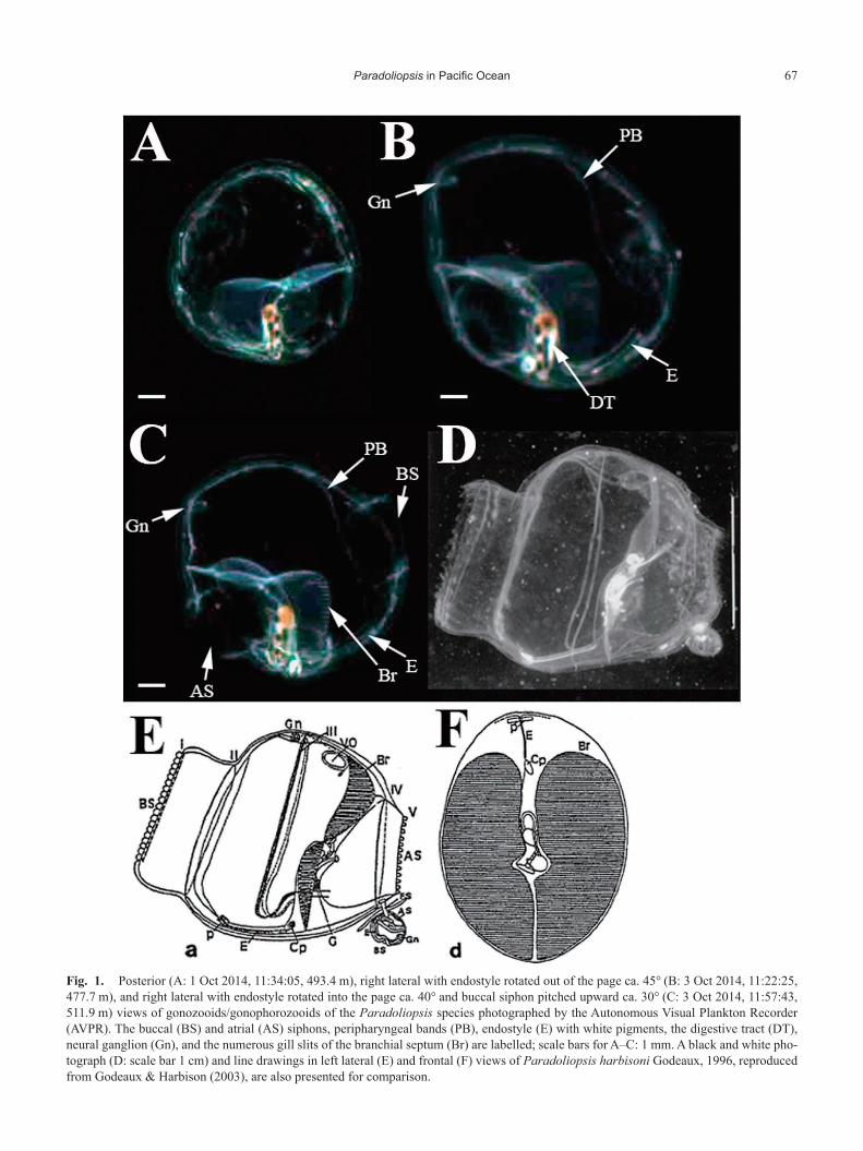
In situ photographs of three individual doliolids with a dis-tinctive anatomy were taken by the AVPR between 1–3 Oct 2014, at depths between 478–512 m (Fig. 1). Although the resolution of the images was not particularly high, it was ob-vious that these individuals belonged to the same species, each imaged from a different angle. In the individual in Fig. 1C, approximated 8–9 mm in length, the wide-open buccal and atrial siphons are visible. The body length was slightly longer than high, the atrial siphon was long, the buccal siphon wide, and the buccal vestibule was capacious. The endostyle was visible on the ventral side, along with two associated pigmented structures at either end in the individuals in Figs. 1B and C. No stolon was visible. The peripharyngeal bands, extending from the anterior end of the endostyle in the ventral mid-line of the zooid, were also evident (Fig. 1B, C). Conspic-uous red-orange and yellow-gold pigmentation existed in the U-shaped digestive canals, which also contained three to four black fecal pellets. The numerous slits of the branchial septa in the gill were visible and the gills were colorless through-
out their extent. The dorsal neural ganglion was evident and slightly pigmented (see Fig. 1B, C). The above charac-ters are congruent with these animals being gonozooids and/or gonophorozooids of the genus Paradoliopsis. They more closely resemble P. harbisoni Godeaux, 1996, than any other Doliposis-like doliolid (see Table 3 in Godeaux & Harbison, 2003), although several differences were noted. The buccal siphon was approximately 35–40% of the height of the zooid rather than 70% as reported for the single known representa-tive of the genus, P. harbisoni, and the gills were not com-pletely vertical. Because the present animals were less than 1 cm in length, while the description of P. harbisoni is based on individuals of 2.2 and 2.5 cm in length, it is possible that these morphological differences may just be growth-related and they can thus be thought referable to P. harbisoni. How-ever, because all observed individuals were of a similar size, it is possible that these animals actually belong to an as-yet undescribed species in the genus Paradoliopsis.
Two deployments of the AVPR were made during the cruise̶on the nights of the 1st and 3rd of October. The field of view was set to 43 mm by 43 mm with a pixel resolution of 48 µm pixel−1. On 1 October 2014, the VPR was deployed to a maximum depth of 720 m at a ship speed vs water of 0.5 knots and a wire reel-out speed of 0.5 m/s. On 3 October, ship speed was kept stable at 1 knot vs water and wire reel-out and reel-in speeds were 0.5 m/s with a maximum depth reached of 1000 m. Gonozooids (gonophorozooids?) of do-liopsids belonging to the genus Paradoliopsis were photo-graphed by the AVPR on 1 October at 493 m depth (Fig. 1A,
Note
* Corresponding author: Dhugal J. Lindsay; E-mail, [email protected]
Plankton & Benthos Research
© The Plankton Society of Japan
67Paradoliopsis in Pacific Ocean
Fig. 1. Posterior (A: 1 Oct 2014, 11:34:05, 493.4 m), right lateral with endostyle rotated out of the page ca. 45° (B: 3 Oct 2014, 11:22:25, 477.7 m), and right lateral with endostyle rotated into the page ca. 40° and buccal siphon pitched upward ca. 30° (C: 3 Oct 2014, 11:57:43, 511.9 m) views of gonozooids/gonophorozooids of the Paradoliopsis species photographed by the Autonomous Visual Plankton Recorder (AVPR). The buccal (BS) and atrial (AS) siphons, peripharyngeal bands (PB), endostyle (E) with white pigments, the digestive tract (DT), neural ganglion (Gn), and the numerous gill slits of the branchial septum (Br) are labelled; scale bars for A–C: 1 mm. A black and white pho-tograph (D: scale bar 1 cm) and line drawings in left lateral (E) and frontal (F) views of Paradoliopsis harbisoni Godeaux, 1996, reproduced from Godeaux & Harbison (2003), are also presented for comparison.

D. J. LinDsay et al.68
temperature 3.4°C, salinity 33.93, 38°59.62′N 142°16.45′E) and on 3 October 2014 at 478 m (Fig. 1B, temperature 3.4°C, sa-linity 33.87) and 512 m (Fig. 1C, temperature 3.6°C, salinity 34.03, 38°53.88′N 142°30.09′E) depth. Two doliolids with the same gross morphology as the present material were also cap-tured on the video record of the ROV Crambon at 372–373 m depth (temperature 3.2°C, salinity 33.79, dissolved oxygen 7.5 mL/L, 38°36.51′N 142°24.51′E) during a dive to 948 m depth on the morning of 4 October. CTD profiles from the AVPR and ROV Crambon deployments are shown in Fig. 2.
During the ROV Crambon dive, the cold-water indicator species Aglantha digitale (O.F. Müller, 1776) was observed between 204–338 m, shallower than the depth at which Para-doliopsis was observed. According to the water mass clas-sification scheme of Hanawa and Mitsudera (1987) for water masses of the Sanriku Coast, the water mass occurring below 300 m depth, in which Paradoliopsis was observed, was the cold lower-layer water system (Fig. 3). This water mass oc-curred just below the depths at which the cold, low salinity waters of the Oya shio Current prevailed (Fig. 3), and its ori-gin was supposed by Hanawa and Mitsudera (1987) to be Ku-roshio waters cooled by air-sea interaction for waters above salinity 34.0 and derived from Oyashio waters when salinity was lower than 34. The presence of the Oyashio-associated A. digitale in the shallower layers of this water mass during the ROV Crambon dive lends support to this hypothesis. Para-doliopsis was observed in waters both higher and lower than
34.0 salinity (Fig. 3).Paradoliopsis, specifically P. harbisoni, has been recorded
in the literature only off the Atlantic coast of North Ameri-ca, specifically over George’s Bank (40°02′N, 69°02′W) and in the Bahamas (24°31′N, 83°45′W) at 735–739 m depth in both locations, and at temperatures ranging between 4.8°C and 5.7°C (Godeaux & Harbison 2003). The present observa-tions are only the second of the genus, extending its known biogeographical distribution, but are both colder (3.2°C) and shallower (372 m) than previous records. It remains unclear whether the present observations are referable to a second, as-yet-undescribed, species that prefers colder and/or shallower waters, or whether these observations of smaller zooids would extend the known habitat preferences for P. harbisoni.
The size distribution of marine snow particles at the depths at which Paradoliopsis occurred were analyzed (Fig. 3) ac-cording to the methods of Lindsay et al. (2014), where the minimum measured dimension of particles was 144 µm in or-der to filter out compression artefacts and other “noise”. These data are presented as histograms of minimum Feret’s diameter (shortest dimension) vs particle number (Fig. 3). Minimum feret size was investigated because, as elongated particles generally align in flow with their long axes parallel to flow streamlines, this will determine whether the particles could be trapped within the feeding filter of the doliolid zooids. Although the pore widths of the mucous feeding filter of these zooids is as yet unknown, in most doliolids they are usually
Fig. 2. CTD profiles from the AVPR deployments on 1 October (A) and 3 October (B) 2014, and the ROV Crambon dive on 4 October (C). Depths at which Paradoliopsis was observed are indicated by open triangles.

69Paradoliopsis in Pacific Ocean
from 0.2–5 µm (Bone et al. 2003). Consequently, these zooids would therefore be able to consume almost the entire range of particles present at the depths at which they were observed (Fig. 3), including particles that were too small to be imaged with the AVPR, but with the possible exception of the larger particles (e.g. >1 mm), which may clog the feeding filter.
A VPR has been successfully used to elucidate in situ asso-ciations between doliolids, specifically Dolioletta gegenbauri (Uljanin, 1884), and sapphirinid copepods (Takahashi et al. 2015a), and to characterize a surface bloom of D. gegenbauri doliolids in the Oyashio-Kuroshio frontal region (Takahashi et al. 2015b), but the present report is the first to identify a deep sea doliolid to putative species level using VPR images and to characterize the marine snow particle field upon which it may feed, at the same depths.
Development of an imaging system able to be deployed to mesopelagic depths and taking higher resolution images than the AVPR (1024×1024 pixels) will certainly enable new discoveries of fragile zooplankton, their ecology and their en-vironment in the future. Existing net samples and net samples collected into the future from the present survey area should be (re-) examined to locate specimens for a taxonomic study
to unequivocally determine the specific identity of these Par-adoliopsis zooids.
Acknowledgements
We are grateful to the reviewers for critical and construc-tive comments on the manuscript. This study is a contribu-tion to the Census of Marine Zooplankton (CMarZ), an ocean realm field project of the Census of Marine Life, the Interna-tional Network for Scientific Investigations of Deep-Sea Eco-systems (INDEEP), and the Deep Ocean Stewardship Initia-tive (DOSI). This work was partially funded by Japan Society for the Promotion of Science (JSPS) KAKENHI, grant num-ber 24248032, JST grant CREST and research project funds for “Tohoku Ecosystem-Associated Marine Sciences”, from the Ministry of Education, Culture, Sports, Science and Tech-nology.
ReferencesBone Q, Carré C, Chang P (2003) Tunicate feeding filters. J Mar Biol
Ass UK 83: 907–919.
Fig. 3. T–S diagram based on CTD data from the two Autonomous Visual Plankton Recorder (AVPR) tows and the ROV Crambon dive, with the six water systems off the Sanriku Coast, according to Hanawa & Mitsudera (1987), overlaid: 1. the Tsugaru Warm Current water system; 2. the Oyashio water system; 3. the Kuroshio water system; 4. the cold lower-layer water system; 5. the surface layer water system; 6. the Coastal Oyashio water system. Open circles and crosses represent Paradoliopsis observations during the ROV Crambon and AVPR surveys, respectively. Marine snow particle size (minimum Feret’s diameter >144 µm) histograms, for the depths at which Paradoliopsis was observed with the AVPR, are inserted top left.
D. J. LinDsay et al.70
Godeaux JEA (1996) On the systematics of Doliolida. In: Workshop Bel-gian Oceanographic Research (8–9 January 1996). Bull Soc Roy Sci Liège 65: 83–86.
Godeaux JEA, Harbison GR (2003) On some pelagic doliolid tunicates (Thaliacea, Doliolida) collected by a submersible off the eastern North American coast. Bull Mar Sci 72(3): 589–612.
Hanawa K, Mitsudera H (1987) Variation of water system distribution in the Sanriku coastal area. J Oceanogr Soc Jpn 42: 435–446.
Lindsay DJ, Yamaguchi A, Grossmann MM, Nishikawa J, Sabates A, Fuentes V, Hall M, Sunahara K, Yamamoto H (2014) Vertical profiles of marine particulates: a step towards global scale comparisons us-
ing an Autonomous Visual Plankton Recorder. Bull Plankton Soc Jpn 61(1): 72–81.
Takahashi K, Ichikawa T, Tadokoro K (2015a) Diel colour changes in male Sapphirina nigromaculata (Cyclopoida, Copepoda). J Plankton Res 37(6): 1181–1189. doi: 10.1093/plankt/fbv088
Takahashi K, Ichikawa T, Fukugama C, Yamane M, Kakehi S, Oka-zaki Y, Kubota H, Furuya K (2015b) In situ observations of a do-liolid bloom in a warm water filament using a video plankton re-corder: Bloom development, fate, and effect on biogeochemical cycles and planktonic food webs. Limnol Oceanogr 60(5): 1763–1780. doi: 10.1002/lno.10133