histiocytic lesions of the skin - yourhosting and... · histiocytic lesions of the skin that might...
TRANSCRIPT

HARVARDMEDICAL SCHOOL
Histiocytic Lesions of the Histiocytic Lesions of the SkinSkin
Nancy Lee Harris, M. D.Nancy Lee Harris, M. D.Massachusetts General HospitalMassachusetts General Hospital
Harvard Medical SchoolHarvard Medical School

HARVARDMEDICAL SCHOOL
Histiocytic Lesions of the Skin: Histiocytic Lesions of the Skin: DisclosureDisclosure
• I am not an expert on this!
• Sophie and Jonathan: “We’re sure you’ll do a
great job!”⁻
My translation: “The experts turned us down. We’re
sure you’ll read up on it, find some cool cases, and
share with us what you’ve learned.”
• Focus: Cases that Dermatopathologists
might
show to a Hematopathologist
(what we need to know!)
⁻ Not the zillion things they sign out themselves (from
xanthelasma
to sarcoidosis)
⁻ Emphasize things we don’t typically think about and
won’t find in Hematopathology
WHO book or texts

HARVARDMEDICAL SCHOOL
Histiocytic Lesions of the Skin that Might be Shown to a Hematopathologist
•
Benign/reactive lesions⁻
Rosai‐Dorfman disease
⁻ Intravascular histiocytosis
•
Uncertain (non‐Langerhans’
cell histiocytoses)⁻
Xanthogranuloma
group
⁻ Reticulohistiocytoma
group
•
Neoplastic/malignant lesions⁻
Langerhans cell histiocytosis
⁻ Langerhans
cell sarcoma
⁻ IDC sarcoma
⁻ FDC sarcoma
⁻ Histiocytic sarcoma
⁻ Blastic plasmacytoid dendritic cell neoplasm

HARVARDMEDICAL SCHOOL
GMGM--CSFCSFILIL--44TGFTGF--ββ
LangerhansLangerhans DCDCLangerinLangerin++CD1a+CD1a+S100+S100+
GMGM--CSFCSFILIL--44
Interstitial DCInterstitial DCCD1aCD1a
--FXIIIaFXIIIa++CD68+CD68+CD168+CD168+
DCSIGN+DCSIGN+
CD14CD14--CD11c+CD11c+CD1a+CD1a+CLA+CLA+
GMGM--CSFCSFTNFTNF--aa
CD14CD14--CD11cCD11c--CD1aCD1a--BDCA2+BDCA2+CD123+CD123+
FLT3LFLT3L
PlasmacytoidPlasmacytoid
DCDC
IL3IL3CD40LCD40LInterdigitatingInterdigitatingDCDC
LangerinLangerin--CD1aCD1a
--S100+S100+
MM--CSFCSF
Follicular DCFollicular DCCD21CD21CD23CD23CD35CD35
DesmoplakinDesmoplakin
MonocyteMonocyteCD14+CD14+CD11c+CD11c+CD68+CD68+CD1aCD1a--CLACLA--
CD34+CD34+
MyeloidMyeloidstem cellstem cell
HistiocyteHistiocyteCD14+CD14+CD11c+CD11c+CD68+CD68+CD163+CD163+
MesenchymalMesenchymalstem cellstem cell
TT
iiss
ss
uu
ee
ss
In vitroIn vitro
Veiled CellVeiled Cell
LangerinLangerin--CD1a+CD1a+
S100+S100+
Fibroblastic Fibroblastic reticulum reticulum
cellcellMuscle Muscle actinactin++
KeratinsKeratins--/+/+
BB
lloo
oo
dd
pp
rr
ee
cc
uu
rr
ss
oo
rr
ss

HARVARDMEDICAL SCHOOL
Benign/reactive histiocytic
lesions•
Rosai‐Dorfman
disease
•
Intravascular histiocytosis

HARVARDMEDICAL SCHOOL
Rosai‐Dorfman disease: Cutaneous Manifestations
•
Extranodal involvement: 25‐40%⁻
Skin (9%) > upper respiratory tract > soft tissue > orbit >
bone > salivary gland > CNS > breast > pancreas*•
Primary presentation in skin = rare⁻
Median age 45yr (older than nodal cases)
⁻ Female predominance (unlike nodal cases)
⁻ Whites and Asians predominate (ditto)
•
Clinical features⁻
Solitary or multiple dermal or subcutaneous nodules or
plaques on face, trunk or extremities⁻
May resolve spontaneously or become chronic
*Rezk et al, Diagnostic Hematopathology, Ch. 51, 2010

HARVARDMEDICAL SCHOOL
64-Year-Old African American Woman: 8 cm painful nodule on abdomen

HARVARDMEDICAL SCHOOL

HARVARDMEDICAL SCHOOL

HARVARDMEDICAL SCHOOL

HARVARDMEDICAL SCHOOL
HARVARDMEDICAL SCHOOL

HARVARDMEDICAL SCHOOL

HARVARDMEDICAL SCHOOL
S100

HARVARDMEDICAL SCHOOL
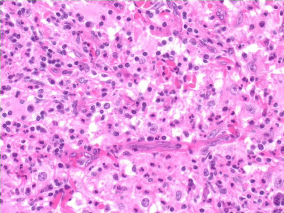
39 Year‐Old African‐American Woman: 4 cm subcutaneous plaque on flank
HARVARDMEDICAL SCHOOL
CD20 CD3

HARVARDMEDICAL SCHOOL

HARVARDMEDICAL SCHOOL
Diagnostic Area

HARVARDMEDICAL SCHOOL
Enperipolesis

HARVARDMEDICAL SCHOOL
Rosai‐Dorfman
Disease: Features•
Involvement of dermis, subcutis, either or both
•
Large S100+ histiocytes with prominent nucleoli and emperipolesis
⁻ May be present only very focally extranodal sites
⁻ May form aggregates that resemble sinuses
•
Numerous plasma cells, neutrophils, histiocytes may be present, especially at edges of lesion
•
Characteristic edematous plasma‐cell‐rich background
⁻ A clue to look for the large histiocytes

HARVARDMEDICAL SCHOOL
Intralymphatic histiocytosis•
Dilated lymphatics containing histiocytes⁻
Frequent association with rheumatoid arthritis
⁻ Erythematous plaques near affected joints
• 16 cases reported by Requena
et al
⁻ 12 on arms or legs
5 rheumatoid arthritis
2 in scar of THR
2 in scar of mastectomy⁻
1 presented as unilateral swelling of the eyelid
Clinical diagnosis: Melkerson‐Rosenthal syndrome
Requena et al. Am J Dermatopathol 2009;31:140-51

HARVARDMEDICAL SCHOOL
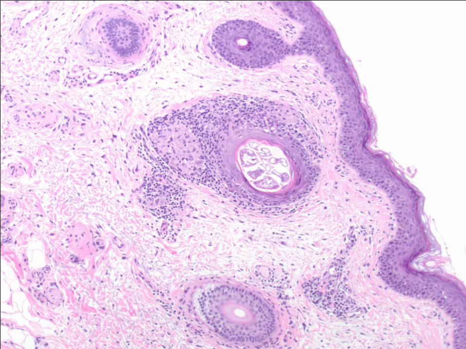
Intralymphatic Histiocytosis•
56‐year‐old man⁻
History of mild facial rosacea
⁻ MGUS
⁻ 2‐year history of unilateral periorbital edema
Difficulty opening his eye

HARVARDMEDICAL SCHOOL

HARVARDMEDICAL SCHOOL

HARVARDMEDICAL SCHOOL
HARVARDMEDICAL SCHOOL

HARVARDMEDICAL SCHOOL
HARVARDMEDICAL SCHOOL

HARVARDMEDICAL SCHOOL
Diagnosis•
Changes consistent with granulomatous
rosacea
(dilated blood vessels, superfical edema, perifollicular
and perivascular
granulomatous
inflammation)•
Demodex
follicularum
⁻ Often associated with rosacea
•
Intralymphatic
histiocytosis
with marked superficial and deep dermal edema

HARVARDMEDICAL SCHOOL
Intralymphatic histiocytosis•
Dilated lymphatics containing histiocytes⁻
Frequent association with rheumatoid arthritis
⁻ Erythematous plaques near affected joints
•
16 cases reported by Requena
et al⁻
12 on arms or legs
⁻ 5 rheumatoid arthritis
⁻ 2 in scar of THR
⁻ 2 in scar of mastectomy
⁻ 1 presented as unilateral swelling of the eyelid
Clinical diagnosis: Melkerson‐Rosenthal syndrome
Requena et al. Am J Dermatopathol 2009;31:140-51

HARVARDMEDICAL SCHOOL
Solid Facial Edema of Rosacea
•
Unilateral periorbital/facial swelling in a patient with
rosacea⁻
Rare complication, may be presenting symptom⁻
Unresponsive to treatment for rosacea
(antibiotics)•
Related entities:⁻
Solid facial edema of acne⁻
Morbihan’s
syndrome (idiopathic)⁻
Melkerson‐Rosenthal syndrome/granulomatous
cheilitis
Facial/lip swelling
Furrowed tongue
Facial nerve palsy
May be familial•
Characteristic features⁻
Edema in all layers of the dermis⁻
Perivascular
and periadnexal
inflammatory infiltrates⁻
No mention of intravascular histiocytes

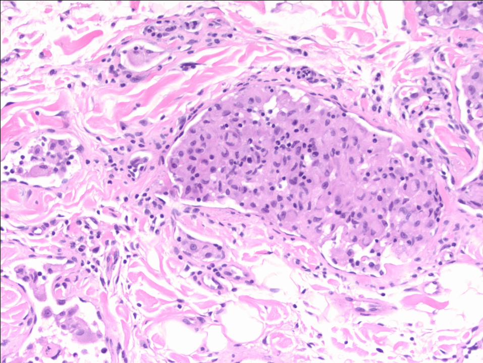
HARVARDMEDICAL SCHOOLSolid Facial Edema and Intralymphatic
Histiocytosis
•
Two cases:⁻
Case 15 of Requena
(rosacea
history not known)⁻
Current case (mild rosacea)•
Both characterized by granuloma‐like lymphohistiocytic
clusters in dermal lymphatics⁻
Distinct from bland, purely histiocytic
collections in
RA/scar‐associated cases
⁻
Blockage of lymphatics
by histiocytes
may produce edema•
Our case⁻
Methotrexate
and prednisone (given because of diagnosis
of “histiocytosis”
[a neoplasm]): no response⁻
Clinicians were contemplating radiation ⁻
Case re‐reviewed and edema of rosacea
suggested
Currently on isotretinoin
therapy
Rx of demodex
with metronidazole
HARVARDMEDICAL SCHOOL
Histiocytoses
• Xanthogranuloma
group
• Reticulohistiocytoma
group
• Langerhans
cell histiocytosis

HARVARDMEDICAL SCHOOL
Xanthogranuloma Group
•
Normal counterpart⁻
Histiocyte
or interstitial dendritic cell •
Histopathology/Immunophenotype⁻
Histiocytic infiltrate with foam cells and Touton
giant cells⁻
CD68+ CD163+ F13a+ S100‐
CD1a‐
Langerin‐•
Clinical syndromes⁻
Localized cutaneous
(Juvenile/adult xanthogranuloma)⁻
Disseminated cutaneous
(JXG, progressive nodular and generalized eruptive histocytosis)⁻
Disseminated: skin, mucosae, CNS (diabetes insipidus), bones,
soft tissue, lung, heart
(Xanthoma
disseminatum, disseminated JXG, Erdheim
Chester
Disease)
•
Genetic features⁻
Not known for cutaneous cases⁻
ECD clonal
in some (most?) cases

HARVARDMEDICAL SCHOOL
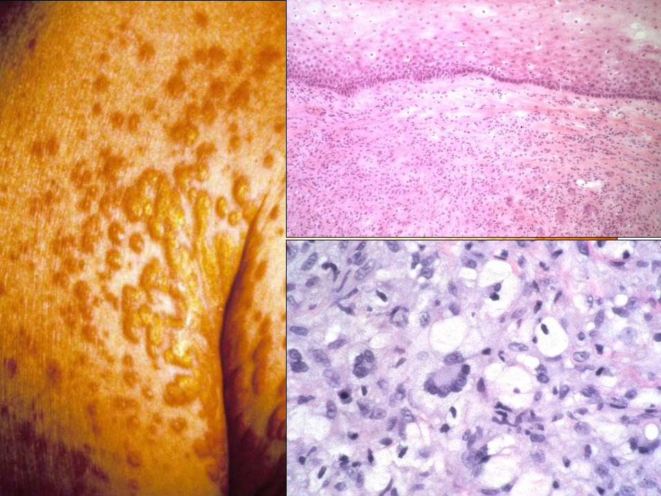
Juvenile Xanthogranuloma•
(Not likely to be shown to a hematopathologist!)•
Clinical ⁻
Child (80%), young adult (20%)⁻
Cutaneous: solitary orange‐red‐brown lesions: head‐neck >
trunk, limbs, oral cavity, eye
⁻
Disseminated: viscera, soft tissues, heart, CNS, bones•
Histology⁻
Histiocytes
with eosinophilic
or vacuolated cytoplasm,
bland nuclei
⁻
Spindle cells⁻
Multinucleated Touton
giant cells
Peripheral rim of vacuolated cytoplasm
Central ring of nuclei
Inner zone of eosinophilic
cytoplasm
(May not be identifiable in extracutaneous
sites)

HARVARDMEDICAL SCHOOL34‐Year‐Old Man
Pink Papule, Lower Abdomen

HARVARDMEDICAL SCHOOL

HARVARDMEDICAL SCHOOL
Xanthoma
Disseminatum•
A 63‐year‐old man with ⁻
Diabetes insipidus
⁻ Skin lesions on face (eyelids, nasolabial
folds)
and flexor surfaces of limbs (axillae, groin) ⁻
Submucosal
lesions of larynx with stenosis
and
stridor
Case 12‐1988.NEJM 1988; 338: 1138
HARVARDMEDICAL SCHOOL

HARVARDMEDICAL SCHOOL
Erdheim‐Chester Disease
Case Records of the Massachusetts General Hospital
Case 25‐2008 —A 43‐Year‐Old Man with Fatigue and Lesions in the Pituitary and Cerebellum
John A. Mills,.D., R. Gilberto Gonzalez, M.D., Ph.D., and Ronald Jaffe, M.D.
N Engl
J MedVolume 359(7):736‐747
August 14, 2008

HARVARDMEDICAL SCHOOLMRI Scans of the Brain
Mills J et al. N Engl J Med 2008;359:736-747

HARVARDMEDICAL SCHOOLCT Scans of the Chest and Abdomen
Mills J et al. N Engl J Med 2008;359:736-747

HARVARDMEDICAL SCHOOLPathologyPathology
•
Renal, perinephric
tissue biopsy ⁻
Mild, non‐diagnostic kidney changes
⁻ Histiocytic infiltrate in peri‐renal fat, nonspecific
•
Clinical diagnosis⁻
Neurosarcoidosis
with involvement of soft tissues;
possible aortitis⁻
Rheumatology consultant (Dr. Mills) made the
diagnosis of ECD based on the imaging studies•
Pathology re‐examined by Ron Jaffe

HARVARDMEDICAL SCHOOLPathological Features of the Perirenal Infiltrate
CD68
CD168 F13A

HARVARDMEDICAL SCHOOL
PeriPeri‐‐renal Tissuesrenal Tissues
• Bland histiocytes, lacking malignant
features•
Xanthomatous, foamy component
• Few giant cells, no Touton
giant cells
⁻ No granulomas
or epithelioid
cells
⁻ No evidence of vasculitis
• Immunophenotype(CD68+ CD163+
F13A+ CD1a‐) consistent with ECD

HARVARDMEDICAL SCHOOL
Characteristic Clinical Picture of ECD•
In this patient⁻
Hypothalamic‐pituitary axis, CNS⁻
Retroperitoneum, kidney⁻
Heart and peri‐aortic tissue⁻
Bone•
Other sites that may be involved⁻
Lung⁻
Orbit•
Tissue diagnosis⁻
Histiocytes, foam cells, +/‐
Touton
giant cells⁻
Foci of Langerhans
cells may occur⁻
May appear nonspecific; need clinical correlation⁻
Important to exclude granulomatous
diseases, vasculitis•
Mortality >50% due to lung, cardiac involvement

HARVARDMEDICAL SCHOOL
Xanthogranuloma
Group•
A group of probably related diseases with similar
histological and immunophenotypic
features but variable
clinical manifestations•
Unisystem: Cutaneous (single/multiple)⁻
Juvenile xanthogranuloma
(single or multiple)⁻
Adult xanthogranuloma
(single)⁻
Generalized eruptive histiocytosis
(multiple, relapsing)⁻
Progressive nodular histiocytosis
(multiple)•
Multisystem⁻
Xanthoma
disseminatum
Skin, CNS (diabetes insipidus, mucosae) ⁻
Disseminated JXG [child]/Erdheim‐Chester disease [adult]
Bones, CNS (diabetes insipidus), soft tissue, lung, skin (in
children; rarely in adults)

HARVARDMEDICAL SCHOOL
Reticulohistiocytoma
Group•
Clinical⁻
Nodular yellow papules, single or multiple (face, hands,
ears)
(Reticulohistiocytoma) ⁻
Systemic condition with skin lesions, destructive arthritis,
fever; occasionally with occult malignancy (25%),
rheumatic disease
(Multicentric
reticulohistioctyosis)⁻
Both: mucous membranes (nose, oral cavity)⁻
F>M, child or adult•
Morphology: ⁻
Bland nuclei, eosinophilic
ground glass cytoplasm, giant
cells (non‐Touton)
⁻
Similar infiltrate in affected joints•
Immunophenotype⁻
CD68+ S100‐
CD1a‐
lysozyme+/‐
F13a‐

HARVARDMEDICAL SCHOOLMulticentric
Reticulohistiocytosis
Adult F with generalized papular eruption, fever, uveitis.Courtesy of Jonathan Said and Larry Weiss

HARVARDMEDICAL SCHOOL
Multicentric
Reticulohistiocytosis

HARVARDMEDICAL SCHOOL
Multicentric
reticulohistiocytosis

HARVARDMEDICAL SCHOOL
Multicentric
Reticulohistiocytosis•
Immunophenotype⁻
CD68+ CD163+ S100+
⁻ Lysozyme, CD15, CD30, EMA, CD4: negative
•
Morphologic features most c/w multicentric
reticulohistiocytosis
⁻ Atypical immunophenotype
⁻ No mention of arthritis

HARVARDMEDICAL SCHOOL
• A clonal
tumor of Langerhans
cells
• Two subtypes⁻
Langerhans
cell histiocytosis
Morphologically typical Langerhans
cells⁻
Langerhans
cell sarcoma
High‐grade sarcomatous
lesion with the
immunophenotype of LC
• Morphology (LCH)⁻
Grooved nuclei, eosinophils
• Immunophenotype⁻
S100+ CD1a+ Langerin+ Birbeck
granules (EM)
⁻ CD68 variable, lysozyme‐
Langerhans
Cell Neoplasms

HARVARDMEDICAL SCHOOL
Langerhans
Cell Histiocytosis•
Unisystem
(bone/soft tissue, skin, lymph node, lung [some
cases])⁻
Unicentric
(single site)
Eosinophilic
granuloma⁻
Multicentric
(multiple cutaneous or bony sites)
Hand‐Schuller‐Christian disease (bone and adjacent soft
tissue, pituitary stalk; skin may be involved [multisystem])
•
Multisystem⁻
Acute (Letterer Siwe
disease)
Skin, bone, liver, spleen, lung, lymph nodes, bone marrow⁻
Chronic (Hand‐Schuller‐Christian disease) •
Prognosis⁻
Good for unisystem
disease⁻
May be poor for multisystem disease ~50% mortality in LS

HARVARDMEDICAL SCHOOL
84‐Year‐Old Woman Lesions on Chest and Arm

HARVARDMEDICAL SCHOOL

HARVARDMEDICAL SCHOOL
LCH CD1A

HARVARDMEDICAL SCHOOL
Histiocytoses: Common Features
•
Within each group, tumors of identical cells have a
spectrum of clinical presentation and behavior⁻
Child vs
adult⁻
Localized vs
disseminated⁻
Unisystem
vs
multisystem•
Nomenclature traditionally based on clinical presentation⁻
Leads to a confusing array of names/eponyms •
Problem largely solved for LCH group⁻
Known to be clonal, neoplastic⁻
Classify by systems involved (stage)⁻
Unclear what genetic abnormalities underly
behavior•
Need unifying nomenclature for the other 2⁻
Evidence that ECD is clonal; lacking for others⁻
Rarity of lesions hampers further study

HARVARDMEDICAL SCHOOLHistiocytoses
Revisited
•
Xanthogranuloma
group ⁻
Unisystem
(skin)
Unicentric
Multicentric⁻
Multisytem
Skin, CNS, bone, soft tissue, mucosae, heart, lungs•
Reticulohistiocytoma
group⁻
Unisystem
(skin)
Unicentric
Multicentric⁻
Multisystem
Skin, joints•
Langerhans
cell histiocytosis
(clonal)⁻
Unisystem
(skin, bone, lymph node, lung)
Unicentric
Multicentric⁻
Multisystem
Skin, bone, liver, spleen, lung, lymph nodesbone
marrow

HARVARDMEDICAL SCHOOL
Neoplastic
Diseases•
Langerhans
cell histiocytosis
•
FDC sarcoma•
IDC sarcoma
•
Histiocytic sarcoma•
Blastic
plasmacytoid
dendritic cell tumor

HARVARDMEDICAL SCHOOL
Interdigitating
Dendritic Cell Sarcoma
•
A tumor of cells resembling IDC of lymph nodes
•
Lymph nodes > skin, soft tissues•
Oval to spindled cells, variable nuclear
atypia, lymphocyte‐rich background•
S100+ CD1a‐
FDC markers–
CD68+/‐
lysozyme‐/+ ; lymphocytes T>>B•
Clinically aggressive, often fatal

HARVARDMEDICAL SCHOOL
IDC Sarcoma WHO 2008
S100
IDC Sarcoma WHO 208

HARVARDMEDICAL SCHOOL
Follicular Dendritic Cell sarcoma•
A neoplasm of oval to spindled cells with
morphology and immunophenotype of FDC•
Lymph nodes (2/3), skin, soft tissue, GI, liver,
spleen•
Spindled cells with bland nuclei, often binucleate,
forming whorls; lymphocyte‐rich background with follicles
•
Immunophenotype⁻
CD21+ CD23+ CD35+ CD68‐/+ S100‐/+ CD1a‐
⁻ Lymphocytes B>T
•
Usually indolent, may recur after excision; late metastases, death in up to 20%

HARVARDMEDICAL SCHOOLFollicular Dendritic Cell Sarcoma of Skin
67F lesion of lower leg; ipsilateral
inguinal lymphadenopathy

HARVARDMEDICAL SCHOOL
FDC Skin: Follicles

HARVARDMEDICAL SCHOOL
FDC Skin Cytologic
Features
CD21

HARVARDMEDICAL SCHOOL
Histiocytic Sarcoma•
Malignant tumor with morphologic and
immunophenotypic
features of a mature tissue histiocyte
•
Sites⁻
GI tract, skin, soft tissues, lymph nodes
⁻ Solitary or multiple cutaneous nodules
•
Large eosinophilic
cells, giant cells, vacuoles•
Immunophenotype⁻
Lysozyme, CD68, CD168 positive
⁻ Myeloperoxidase, CD1a, FDC markers negative
⁻ CD45+/‐
CD4+/‐
CD43+ S100 variable

HARVARDMEDICAL SCHOOL
Histiocytic Sarcoma 50F: bilateral tonsillar
enlargement

HARVARDMEDICAL SCHOOL
Histiocytic Sarcoma: Immunophenotype
CD1AS100
CD68 LYSOZYME

HARVARDMEDICAL SCHOOL
Histiocytic Sarcoma: DDX•
Non‐hematopoietic tumors⁻
Melanoma
⁻ Other sarcomas
•
Other hematopoietic tumors (more common!)
⁻ Myeloid or monocytic
leukemia
⁻ Blastic
plasmacytoid
dendritic cell tumor
⁻ Dendritic cell sarcomas
⁻ T‐cell or B‐cell lymphomas

HARVARDMEDICAL SCHOOLBlastic
Plasmacytoid
Dendritic Cell
Neoplasm•
A tumour
of a precursor of the plasmacytoid
dendritic cell (professional type 1 interferon producing cell or plasmacytoid
monocyte),
involving skin and bone marrow•
Sites: ⁻
Skin, bone marrow, lymph nodes
• Morphology: ⁻
Monomorphous
medium‐sized blastic
cells;
mitoses•
Immunophenotype: ⁻
CD4+ CD56+ CD123+ BCDA1+ Tcl1+ CD68+
(50%) TdT
+ (30%)⁻
CD2, CD7, CD33 variably+
⁻ CD3‐
CD5‐
CD20‐
MPO‐
lysozyme‐
CD34‐
GCP‐

HARVARDMEDICAL SCHOOL
BPDC Cutaneous Lesions

HARVARDMEDICAL SCHOOL
BPDC Immunophenotype
CD123 CD4
CD56 CD3

HARVARDMEDICAL SCHOOL
BPDC: Differential Diagnosis•
Leukemic infiltrate of AML or T‐ALL⁻
May be CD33+ CD2+ CD7+
⁻ AML or ALL may be CD56+
⁻ Monocytes
may be CD123+
⁻ Absence of MPO, CD3 helpful; need a broad panel
⁻ Treatment like AML, so distinction may not affect
management•
Expansion of mature PDC in MDS/AMML⁻
Expression of CD56, TdT
⁻ Increased proliferation fraction
HARVARDMEDICAL SCHOOL
Histiocytic Lesions of the Skin: Summary•
A heterogeneous group that range from idiopathic/reactive
to aggressive malignancies
•
True neoplasms
are relatively straightforward⁻
Rare, and thus always challenging⁻
Some cases cannot be classified precisely by normal
counterpart
⁻
Extensive immunophenotyping
often required•
Idiopathic/non‐LCH histiocytoses
more difficult⁻
Histologic
and immunophenotypic
features may not
distinguish nonspecific histiocytic
infiltrates from
specific lesions and histiocytoses
⁻
Small biopsies yield non‐diagnostic areas (RDD)⁻
Characteristic cells may be lacking in non‐lymph node or
non‐cutaneous sites
Emperipolesis
less common in skin (RDD)
Touton
giant cells less common in soft tissue (ECD)

HARVARDMEDICAL SCHOOL
Histiocytic Lesions of the Skin: Summary
•
Need clues to look for diagnostic cells⁻
Plasma cells, edema, histiocytes, neutrophils: think of RDD⁻
Foamy cells in soft tissue with no apparent cause: think of
ECD
•
Don’t “blow off”
a histiocytic
infiltrate (cutaneous or non‐
cutaneous) as non‐specific inflammation without a search
for the cause⁻
Discuss with clinicians whether there is reason to suspect a
histiocytosis
•
Avoid the term “histiocytosis”
for obviously reactive lesions ⁻
E.g. “Intralymphatic
histiocytosis”⁻
Clinicians may interpret this as indicating a neoplastic
process
•
I hope this was helpful!

HARVARDMEDICAL SCHOOL
Acknowledgments•
Judith Ferry
•
Russell Ryan•
Valentina Nardi
•
Rosalyn Nazarian•
Lyn Duncan
•
Jonathan Said•
Ronald Jaffe
•
Others….

I’ll be happy to answer any questions….
Dan Harris, ABC news, Kandahar (Oct 2001)