husam alhurani adham aldamin salma dh …...adham aldamin … mohammad salma dh 2 | p a g e motor...
TRANSCRIPT

1 | P a g e
15
Husam Alhurani
Adham Aldamin
… Mohammad
Salma Dh

2 | P a g e
Motor Tracts
-Introduction: Last lecture we talked about the stretch reflex which is very important
in studying the motor system. We also said that the innervation for Extrafusal muscle
fibers is by alpha motor neurons (bigger ones), while the Intrafusal muscle fibers are
innervated by gamma motor neurons. We studied about Renshaw cells that inhibit
alpha motor neurons because they tend to over react. Alpha fibers give collateral
branches that activate inhibitory interneurons, these interneurons when activated will
secrete glycine (inhibitory neurotransmitter) which in turn will inhibit alpha motor
neurons.
-Strychnine poisoning: inhibits the Renshaw cells and prevents them from secreting
glycine, so alpha motor neurons will over react and that will cause contractions and
convulsions “تشنجات”.
-B and C fibers are related to the autonomic nervous system:
> B fibers for preganglionic autonomic fibers “white ramus communicans” (bigger)
> C fibers for postganglionic autonomic fibers (smaller).
Now let’s start with our lecture……
*********************************************************************
-Motor system starts from the cortex down to
the spinal cord (opposite to sensory system).
-The frontal lobe of the brain that is anterior to the
central gyrus is called “Motor lobe”.
-This frontal lobe is different divided into areas such
as:
*Area 4: primary motor cortex.
*Area 6: motor association area and it includes 2 areas:
a)Premotor area: uses external cues (Lateral)
b)Supplementary motor area: uses internal cues
(Medial)

3 | P a g e
-The tracts that descend from the cortex down to the spinal cord are divided into 2
main types of tracts:
* Pyramidal Tracts. * Extrapyramidal tracts.
Type of tract Pyramidal tracts Extrapyramidal tracts
Responsibility
(Function)
Conscious control of skeletal
muscles movement (Final
execution of the movement)
Subconscious control of skeletal
muscles movements.
Area of the
cortex that is
related to it
Related Mainly to area 4 of
the cortex. (they are also
related to premotor area from
area 6 and to the sensory
cortex “area 312” )
Related to Premotor area which is a
portion from area 6 of the cortex.
-Conscious control of skeletal muscles: Upper motor neuron controls lower motor
neuron which will make the muscle contract.
-When we say subconscious control, we DON’T mean smooth muscles (they make
part of the autonomic nervous system). We mean that even skeletal muscles
(voluntary muscles) need some sort of subconscious control.
-What is the difference between pyramidal/extrapyramidal tracts:
*If a lesion happens in area 4 (primary motor area) which is related to Pyramidal
tracts, paralysis will happen and the patient will lose the ability to move.
*On the other hand, if a lesion happens in area 6 which is related to Extrapyramidal
tracts (Subconscious control/Coordination), the patient will lose the coordination
effect but NOT the whole movement (No paralysis) ( مثال رح يواجه صعوبة يدخل ابرة بخيط
بس بقدر يحرك ايدو واصابعو(
4 | P a g e
-So we conclude that fine movements that need coordination are controlled by the
subconscious level.
-We said earlier that premotor area from area 6 uses external cues, while
supplementary motor area uses internal cues, what do we mean by cues?? And what is
the difference between external/internal cues??
-External cues: (Such as vision, voice) to understand them, the doctor talked about an
experiment that we tried on a monkey. In this experiment, there were 3 lamps with 3
buttons, each lamp has a button, the monkey was trained to click on the button (a
motor task) corresponding to the lamp that turns on only.
معين. motor activityيعني باختصار كإنو حافز من البيئة الخارجية بحفزنا انو نعمل -
>> If the premotor area which uses the external cues was absent, the monkey will still
can move and see the light, but will lose the ability to use the vision information to
integrate (turn) it to a motor activity.
-Internal cues: another experiment, imagine that there were 3 buttons that have
numbers on them, when the monkey clicks on the buttons in a certain order (button 1
then 2 then 3)for example the monkey will receive a reward, now this order will be
stored in the monkey’s memory, and each time it will use the stored memory to click
on the buttons in the right order to get the reward ( the monkey used an internal cue
.(to do the motor task which is clicking on the buttons (زي حافز)
>> If the supplementary area was absent, the monkey will not be able to use the
memory information to integrate it to a motor action.
Now to the tracts….
*********************************************************************
Pyramidal Tracts
1) Lateral Corticospinal tract
-It starts from the cortex, from the primary motor cortex in the frontal lobe
which contains the cell bodies of the upper motor neuron, so:

5 | P a g e
Cell bodies of the upper motor neurons of this tract are located in the
primary motor cortex in the frontal lobe.
-Upper motor neuron starts from the
cortex, which is divided into homunculi,
each homunculus is responsible of the
motor activity of a certain area in the
body (each homunculus contains the cell
bodies of neurons that are responsible of
the movement of a certain part of the
body).
-Then the fibers descend down from the
cortex, passing and forming corona
radiata, then they will enter the internal
capsule (which is between caudate
nucleus and thalamus medially and
lentiform laterally).
-After that fibers reach the brain stem,
exactly the midbrain.
-In the center of the midbrain there is a
cavity called “Cerebral aqueduct”, a
duct between the third and the fourth
ventricles, it allows the passage of
cerebrospinal fluid.
-Behind the cerebral aqueduct there is
something called “Tectum”, which
contains the colliculi, and anterior to the
cerebral aqueduct there is something
called “Tegmentum”, the anterior part
of the tegmentum is called “crus
cerebri” or “basis pedunculi of the
midbrain”.

6 | P a g e
-While the fibers are descending down, they form a nice bundle in the crus cerebri,
exactly in the middle three-fifths (one fifth medial and one fifth lateral are excluded,
and fibers pass through middle three-fifths).
-So, when we take a cross section in the midbrain, we see corticospinal fibers in the
middle three-fifths of crus cerebri.
-Now, after the midbrain, fibers descend down to pons, to its anterior part called
“basal/basilar part” (the name is related to the basilar artery which is a union of two
vertebral arteries), this basilar part is characterized by the presence of pontine nuclei
(collection of cell bodies) these nuclei maintain connection “cross talk” between
cerebrum and cerebellum (its pathway called “cerebro-ponto-cerebellar pathway).
-When fibers reach these nuclei, they will be scattered between them ( they won’t
form a nice bundle as the one in the crus cerebri in the midbrain)
-So a cross section in pons will not show a nice bundle of corticospinal fibers.
-Down from pons to the lower level, the medulla oblongata (lowest part of the
brainstem), fibers will gather together forming a bundle (bulge) anterior and medial to
the medulla oblongata, this bulge is called “Pyramid”, and because of that this tract is
called pyramidal tract.
So, pyramid is a bulge of corticospinal fibers (white matter) located anterior and
medial to the medulla oblongata.
-Until now, the fibers are descending in an ipsilateral direction (the right pyramid is
descending from the right portion of cortex, and the left one from the left portion of
the cortex).
-At the lower part of medulla oblongata, 85% of the fibers cross the midline (fibers in
right pyramid go to the left side and vice versa), forming something called
“pyramidal decussation” or “Major motor decussation”.
- Fibers that crossed the midline (the 85%) are called “lateral corticospinal fibers”.

7 | P a g e
-The remaining 15% of fibers (minority) that didn’t cross the midline and continued
descending ipsilaterally are called “Anterior corticospinal fibers”.
-The lateral corticospinal fibers continue descending down until they reach the
anterior horn in the spinal cord, and their will synapse.
-Lateral corticospinal fibers: crossed the midline happened at the lower part of
medulla oblongata.
-Anterior corticospinal fibers: crossed the midline happened at the level of the spinal
cord.
(Anatomical difference between lateral and anterior corticospinal fibers).
Anterior
lateral -The anterior corticospinal fibers
continue descending down in ipsilateral
direction.
-When these fibers reach the spinal cord
and at the level of their corresponding
spinal segment, they will cross the
midline eventually forming the “anterior
white commissure”
-So in conclusion, all fibers will
cross the midline, in fact the right
cortex controls the left side of the
body and the left cortex controls
the right side of the body.
8 | P a g e
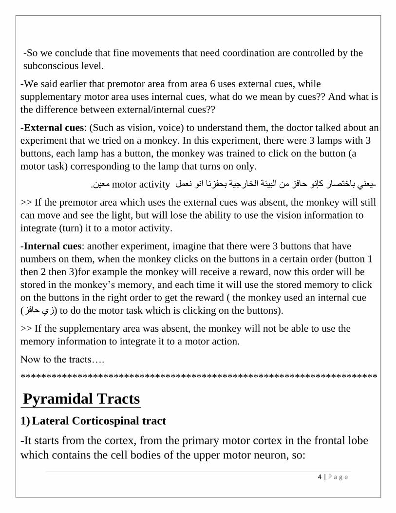
-As we said earlier (from Rexed divisions), the anterior horn took laminae 8 and 9.
-Muscles in our body are divided into:
1) Axial (medial) muscles: related to posture of the body (they don’t provide skilled
movements).
2) Peripheral (lateral) muscles (like muscles of the foot, hand): responsible for the
skilled movements.
-Now going back to our tract, lateral corticospinal fibers, they descend down
(decussation happens in pyramids) and reach the anterior horn, they will synapse their
and innervate the regions of anterior horn that are related to the lateral muscles.
-On the other hand, the anterior corticospinal fibers descend ipsilaterally and
eventually at the level of spinal cord they cross the midline through the anterior white
commissure which is related to the axial muscles (posture).
The anterior corticospinal tract acts on the proximal muscles of upper limb (shoulder
muscle) of the ipsilateral and contralateral sides (this sentence is from slide 80)
-So, we have Lateral motor system and Anterior motor system in our bodies.
(This is the difference in function between anterior and lateral corticospinal fibers,
one for the skilled movements and one for maintaining the body in a good posture).
-Inside the anterior horn there is some
kind of organization like a “map”,
each part is responsible of the muscles
of a certain area in the body.
-For example, lateral portion of the
anterior horn is responsible for the
innervation of muscles of our hands,
and the medial one for muscles of the
trunk.
-

9 | P a g e
-Lateral corticospinal tract descends the full length of the spinal cord.
-Lateral corticospinal fibers synapse with alpha and gamma nuclei of:
-55% at the cervical region (55% of the lateral corticospinal fibers end at the cervical
region (cervical segments of spinal cord) (no role in the lower limb), because these
fibers are responsible for the skilled movements, and most of them are done by the
hands (lateral muscles), and the hand is innervated by brachial plexus
(C5,C6,C7,C8,T1) so we need them mostly in the cervical segments.
-20% at the thoracic region (segments).
-25% at the lumbar and sacral regions.
-If we made a cross section at cervical region of spinal cord, we will be able to see the
representation of hand in the spinal cord (in anterior horn), but moving down to
thoracic, lumbar and sacral segments, we will not be able to see representation of hand.
-For example, at the thoracic region we can see the representation of intercostal
muscles in the anterior horn of spinal cord, so the anterior horn is narrow here.
*********************************************************
-In most cases, upper motor neuron synapses with
interneuron, then the interneuron synapses with
lower motor neuron (majority of cases).
-In 3% of the cases, upper motor neuron synapses
with the lower motor neuron directly without
interneuron (exception). (originates from the fifth
layer of area 4 (giant cells of betz) )
-The lateral corticospinal tract synapses mainly
with interneurons In laminae 4,5,6,7,8.
-Laminae 8 (and 7 somehow) are in anterior horn,
so it’s logical to synapse there, but what about
laminae 4,5,6??
-Laminae 4,5,6 are in the posterior horn (sensory
horn), so what is going here??
-

10 | P a g e
-To answer that we need to go back to the fibers that descend from the somatosensory
cortex and participate in the pyramidal tract, the fibers that originate from sensory
origins go to the sensory portions (to the posterior horn).
-There are many theories about this, one of them links what happens here with
learning new motor skills and movements (such as learning carving of teeth ☺), so we
need sensory data to learn new motor skills.
- In the 3% of cases, upper motor neurons from “Giant cells of betz” in area 4 in the
cortex descend down to lamina 9 and synapse there and control lower motor neurons
directly with no interneurons, and the goal from this direct connection between upper
and lower motor neurons is to provide very accurate movements (we need few
motor neurons to contract).
-Most of muscles of head and neck area are supplied by cranial nerves (Such as
muscles of facial expression, muscles of mastication, muscles of larynx, muscles of
pharynx..) and when talking about cranial nerves, there is no anterior horn, we have
instead motor nuclei.
1)The tract of the motor cranial nerves is called Corticonuclear tract.
2)This tract is composed of fibers originating from the precentral gyrus of the lower
quarter of the motor cortex.
3)The descending fibers terminate in the motor nuclei of cranial nerves III and IV in
the midbrain / V, VI. and VII in the pons; and IX, X, XI, and XII in the medulla.
4)The corticobulbar fibers from one side of the brain project to the motor nuclei on
both sides of the brainstem (bilateral input)
(last 4 sentences are from slide 81)
-For example, Trigeminal nerve has motor nuclei in pons (this nerve is mainly sensory
but it has a small motor portion) so upper motor neuron descends down from the
cortex to the motor nuclei, then lower motor neuron goes from the nucleus to the
muscle. (للعضلة المعنية) >> 1خاص لسكشن << وتحية ألحمد المعني
11 | P a g e
-For cranial nerves, sometimes the upper motor neuron is called also “supranuclear
neuron” (from cortex to the nuclei) and the lower motor neuron is called “infranuclear
neuron” (from nuclei to the muscle).
-
-There is an exception for the bilateral direction of cranial nerves, which is the part of
facial nerve that supplies the lower part of the face, to make it clear, when we look at
the motor nucleus of facial nerve, we will find something like a “map” in the nucleus,
and each part of this map is responsible for the innervation of certain muscles in the
face, the part of nucleus that is responsible for the innervation of the lower aspect of the
face is supplied in contralateral direction (exception for the general role).
-Another exception is the hypoglossal nerve (12th cranial nerve that supplies the
tongue), and like trigeminal, each part of the motor nucleus of hypoglossal nerve is
responsible for the innervation of certain muscles, the part that is responsible for the
innervation of genioglossus muscle is supplied in contralateral direction also.
-So to conclude, the corticoneuclear input is bilateral Except :
1- Part of 7th ( which supplies LOWER facial muscles)
2- Part of 12th (which supplies genioglossus muscle)
Now moving to the extrapyramidal tracts…
*********************************************************************
-As a conclusion in the spinal system (very important), the anterior horn on the
right side is supplied by the left cortex and vice versa because of decussation.
(for spinal nerves)
-For cranial nerves, motor nuclei are supplied bilaterally (from both sides,
contralateral+ipsilateral), that means the right motor nuclei for trigeminal nerve
for example, is supplied by the left and the right cortexes, and vice versa.

12 | P a g e
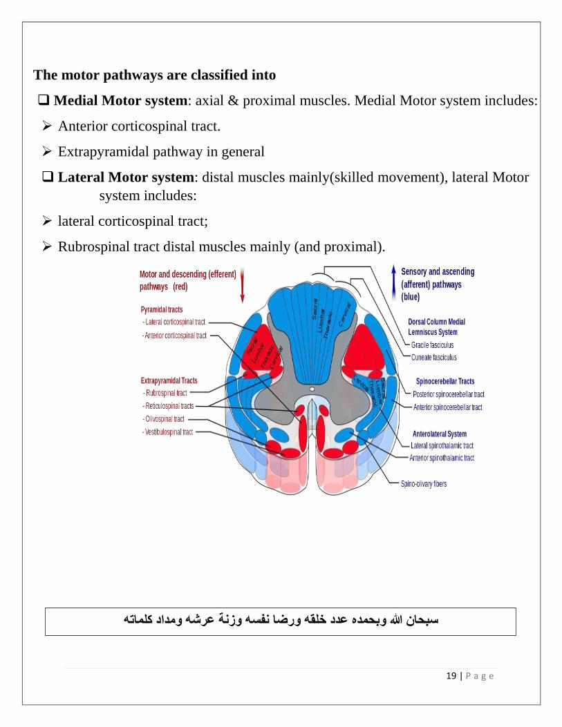
Extrapyramidal tracts
-Vesitibulospinal tracts.
-Tectospinal tracts.
-Reticulospinal tracts.
-Rubrospinal tracts.
*Their names suggesting that they originate/start from structures in the brain stem then
descend down to the spinal cord, for example:
“Rubro” refers to the red nucleus in the brain stem.
“Reticulo” refers to the reticular formation in the brain stem.
“Vestibulo” refers to the vestibular nucleus in the brain stem.
-PAY ATTENTION!! these tract’s names may be misleading because we would think
from the names that they originate from brain stem without any higher control, but in
fact they are under the control of cortex (area 6 “premotor area+supplementary
motor area) so for more accuracy their names should start with “Cortico”, for example
cortico-rubro-spinal tract.
>> “cortico” refers to cortex, we remove it from the name for simplicity, but
always remember that these tracts are under the control of CORTEX.
-Extrapyramidal tracts are related to (their functions) Coordination, muscle tone,
posture and subconscious control of skeletal muscles movement.
Now let’s start with the first tract..

13 | P a g e
Rubrospinal Tract
-This tract also receives data from cerebrum (Big boss) which contains the higher
centers (command/intention centers) (Before we do any movement, an idea about the
movement is formed)
-The word “Rubro=red” suggesting
that this tract descends down from the
red nucleus (red because its highly
vascular) in the midbrain (brain stem)
behind something called “Substantia
nigra”,
-The red nucleus has very important
role in the motor system, it receives
input from the cerebellum through a
pathway called “Globose-embolifrom-
rubral pathway”
(Globose+embolifrom are deep
cerebellur nuclei, will be discussed
later)
-Cerebellum receives afferent data
from spinocerebellar tracts from spinal
cord that gives the muscle-joint sense.
-So cerebellum is well informed about
our bodies situations in space (knows
which muscles are relaxed and which
are contracted)

14 | P a g e
-So, intention is in the cerebrum and current position of the body is in the cerebellum,
so consultation must happen (before we do any movement the cerebrum does some
consultation with structures involved in the motor system like cerebellum and basal
ganglia and take the feedback from them).
-The Rubrospinal tract is crossed, it does early crossing at the level of red nucleus
itself (when we take a section in midbrain, we see the red nucleus and the fibers
crossing the midline there).
-Then after crossing, fibers descend to the lateral white column.
-The function of Rubrospinal tract is to:
*Facilitate the activity of flexors and inhibit the activity of extensors.
-So this tract (Rubrospinal) is the coordinator for the lateral corticospinal tract
(which is responsible for skilled movements as we said earlier)
- Lateral corticospinal tract and Rubrospinal tract collectively form something
called “Lateral Motor System”. (Facilitate flexors and inhibit extensors).
-So we conclude that muscles of skilled movements (such as writing muscles) are
flexors in general.
-Extrapyramidal tracts in general are related to the proximal muscles, and because
it’s related to the coordination of Skilled movement muscles (lateral/distal muscles),
the Rubrospinal tract is an exception.
15 | P a g e
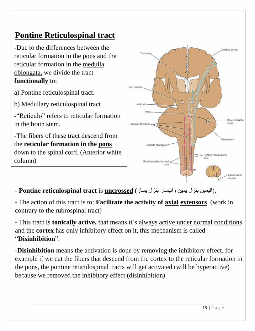
Pontine Reticulospinal tract
- Pontine reticulospinal tract is uncrossed (اليمين بنزل يمين واليسار بنزل يسار).
- The action of this tract is to: Facilitate the activity of axial extensors. (work in
contrary to the rubrospinal tract)
- This tract is tonically active, that means it’s always active under normal conditions
and the cortex has only inhibitory effect on it, this mechanism is called
“Disinhibition”.
-Disinhibition means the activation is done by removing the inhibitory effect, for
example if we cut the fibers that descend from the cortex to the reticular formation in
the pons, the pontine reticulospinal tracts will get activated (will be hyperactive)
because we removed the inhibitory effect (disinhibition)
-Due to the differences between the
reticular formation in the pons and the
reticular formation in the medulla
oblongata, we divide the tract
functionally to:
a) Pontine reticulospinal tract.
b) Medullary reticulospinal tract
-“Reticulo” refers to reticular formation
in the brain stem.
-The fibers of these tract descend from
the reticular formation in the pons
down to the spinal cord. (Anterior white
column)
.

16 | P a g e
-
Pontine reticulospinal tract and Vestibulospinal tract work together.
Medullary reticulospinal tract and Rubrospinal tract work together.
Medullary reticulospinal tracts
-Its nearly completely opposite to the pontine
reticulospinal tracts.
-Fibers descend from the reticular
formation in medulla oblongata
crossed and uncrossed down to the
spinal cord (Lateral white column)
-Not tonically active (at normal
conditions it’s not active and needs
stimulation to get activated)
-Its function is to inhibit the axial and
proximal limb extensors and to
facilitate the flexors.
-In our bodies, there are flexors and
opposite to them functionally there
are extensors (it’s impossible for
example to activate the biceps and
triceps at the same time, each time the
biceps gets activated the triceps gets
inhibited) the same concept is applied
to the tracts, when the tract that
activates the flexors is highly active,
the antagonist system that inhibits
them gets inhibited).
-Fibers descending from hypothalamus
(big boss of autonomic nervous system) to the
lateral horn cells pass with the medullary
reticulospinal tract.

17 | P a g e
Vestibulospinal tract (similar to pontine reticulospinal tract)
-Fibers descend from vestibular nucleus in
the brain stem (in Ponto-medullary junction).
-Vestibular nucleus receives data from:
1) Vestibular nerve which is a part of
vestibulocochlear nerve in inner ear (vestibule)
which contains the saccule, semicircular canals
(contain endolymph fluid) and hair cells.
-When your head moves, the fluid in
semicircular canals moves and the movement
causes firing in hair cells, so the data about the
position of your body in relation to the gravity
is transmitted to the nucleus (balance info).
2) Cerebellum
-Cerebellum is divided into 3 parts:
Cerebrocerebellum, spinocerebellum and
vestibulocerebellum >> related to vestibular
system.
-The fibers descend uncrossed from the nucleus to the spinal cord (Anterior
white column)
• -Then they synapse with neuron in the anterior gray column of the spinal cord
(from slide 88)
-The function of this tract is to facilitate the activity of extensors (to stand up in
fully extended position) and (opposite to rubro and medullary tracts, similar to
pontine tract)
-To stand up in a fully extended position, 2 tracts is involved:
Pontine and vestibulospinal tracts.
18 | P a g e
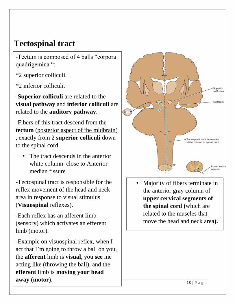
Tectospinal tract
-Tectum is composed of 4 balls “corpora
quadrigemina “:
*2 superior colliculi.
*2 inferior colliculi.
-Superior colliculi are related to the
visual pathway and inferior colliculi are
related to the auditory pathway.
-Fibers of this tract descend from the
tectum (posterior aspect of the midbrain)
, exactly from 2 superior colliculi down
to the spinal cord.
• The tract descends in the anterior
white column close to Anterior
median fissure
-Tectospinal tract is responsible for the
reflex movement of the head and neck
area in response to visual stimulus
(Visuospinal reflexes).
-Each reflex has an afferent limb
(sensory) which activates an efferent
limb (motor).
-Example on visuospinal reflex, when I
act that I’m going to throw a ball on you,
the afferent limb is visual, you see me
acting like (throwing the ball), and the
efferent limb is moving your head
away (motor).
• Majority of fibers terminate in
the anterior gray column of
upper cervical segments of
the spinal cord (which are
related to the muscles that
move the head and neck area).
•
19 | P a g e
The motor pathways are classified into
❑ Medial Motor system: axial & proximal muscles. Medial Motor system includes:
➢ Anterior corticospinal tract.
➢ Extrapyramidal pathway in general
❑ Lateral Motor system: distal muscles mainly(skilled movement), lateral Motor
system includes:
➢ lateral corticospinal tract;
➢ Rubrospinal tract distal muscles mainly (and proximal).
سبحان هللا وبحمده عدد خلقه ورضا نفسه وزنة عرشه ومداد كلماته