immunohistochemical staining for p63 is useful in the diagnosis of anal squamous cell carcinomas am...
TRANSCRIPT

Immunohistochemical Staining for p63 is
Useful in the Diagnosis of Anal Squamous
Cell Carcinomas
Am J Surg Pathol 2007;31:285–290
Int 賴雨欣

Background
Carcinomas in the anal canal account for about 1.5% of gastrointestinal cancers in the United States, and approximately 80% of these are squamous cell carcinomas (SCCs).SCCs of the anus are frequently related to chronic infection with human papilloma virus (HPV)
Background
Usually occur in the sixth to seventh decade of life, occur in younger patients when they are immunocompromisedMale:Female=2:1HIV/AIDS, and the increasing use of immunosuppressive therapy for solid organ transplantation, inflammatory bowel disease and collagen vascular diseases has meant an increasing incidence of HPV infection and anal SCC.

Background
The main differential diagnosis of carcinomas in the anus includes SCC, well-differentiated neuroendocrine carcinoma/carcinoid tumor (NEC), and poorly differentiated adenocarcinoma (ADC).
Background
Morphologic clues can be sought to aid in diagnosis, including squamous eddy/keratin pearl formation in SCC and lumen formation in ADC.Such carcinomas can look remarkably similar in small biopsies, and a panel of immunostains is often used for diagnosis.

Background
In diagnosing primary anal SCC, such panels rely on negativity for markers of ADC and neuroendocrine differentiation, along with positivity for one or more cytokeratins (CK), suggesting squamous differentiation.Combination of CK 5 and 6 (CK5/6) is used as evidence for squamous lineage.

Background
Loss of keratin expression in poorly differentiated SCC !! Accurate diagnosis is imperative for the development of an effective treatment planP63 gene !!

Background
The p63 gene resides on the long arm of chromosome 3 at 3q27-28.P63 is a member of the p53 gene family and, as such, participates in epithelial proliferation and differentiation.

Background
Antibody to p63 is frequently used in the diagnosis of prostate carcinoma, because normal prostatic glands are lined by p63-positive basal cells, whereas carcinomatous glands lack a basal layer.Be found to be associated with SCC in a number of sites, including the head and neck, lung and uterine cervix.

Background
It is also expressed in some breast carcinomas (particularly metaplastic carcinoma) and in urothelial carcinomas.Given its association with SCC in other sites, authors elected to investigate the utility of p63 staining in the diagnosis of anal SCC!!


Methods and Materials
All cases were submitted for diagnosis during the period from January 1, 2004 to December 31, 2005, and originated within the University of Michigan system.24 anal squamous carcinomas, 68 colorectal adenocarcinomas (including a tissue microarray of 49 ADC), and 32 colorectal neuroendocrine carcinomas from the archives at the University of Michigan

Methods and Materials
11 of the SCC cases were deemed ‘‘poorly differentiated’’ based on the presence of large areas consisting of monotonous, immature-appearing cells and the relative lack of areas showing squamous eddy or keratin pearl formation.
Methods and Materials
Colorectal NECs included tumors from throughout the colon, and the appendix.ADCs from throughout the colon and rectum were included.Both biopsy and resection specimens were used.

Methods and Materials
The squamous carcinoma cases included 11 resection specimens and 13 biopsies. Three resection specimens containing normal anal transition zone (ATZ) were also stained to evaluate the staining pattern in the ATZ.

Methods and Materials
Immunohistochemical stain:P63 CK5/6 chromogranin A(CGA) synaptophysin(SYN)Only nuclear staining for p63 was considered positive. A section of prostate tissue was used as a control, with p63 staining the basal cells in the normal prostate
Methods and Materials
A section of tonsil was used as a positive control for CK5/6 staining, and a section of adrenal gland for CGA and SYN staining.Staining for CK5/6, CGA, and SYN was considered positive when cytoplasmic staining was present.

Methods and Materials
Positive predictive values (PPV) and specificities were calculated for p63 and CK5/6 staining in SCC.

Diseasepositive negative
Test positive True Positive(TP) False Positive(FP)negative False Negative(FN) True Negative(TN)
Positive predictive values(PPV) =TP/TP+FP

Results-P63
All 24 (24/24; 100%) of the SCC were positive for nuclear p63 staining.None of the ADCs were positive(0/68; 0%).Two NEC, both classic appendiceal carcinoids, showed nuclear positivity with p63(2/32; 6.25%).

Results-P63
Within the SCC, staining was present in both well and poorly differentiated areas.SCC cases included 4 carcinomas with basaloid morphology, a pattern termed ‘‘cloacogenic carcinoma’’ in the past. These carcinomas also stained strongly with p63.

Invasive SCC stained with p63, showing strong nuclear staining (100X)

Classic appendiceal carcinoid tumor showing strong nuclear staining with p63 (100X)

Basaloid SCC (‘‘cloacogenic carcinoma’’) with nuclear staining for p63 (100X)

Results-P63
Although none of the ADC and only 2 NEC showed nuclear staining, there was scattered nonspecific cytoplasmic staining. However, the lack of nuclear staining was easily discerned in these cases.

Invasive ADC stained with p63, showing lack of nuclear staining. Some nonspecific cytoplasmic staining is present (100X)

Results-CK5/6
All of the SCCs (24/24; 100%) were also strongly positive for CK5/6 staining, and negative for CGA and SYN.Cytoplasmic staining with CK5/6 was so strong as to obscure most cellular and nuclear detail in the carcinomas.

Results-CK5/6
The CK5/6 immunostain was strongly positive in adnexal structures of the perianal skin where present on resection specimens, again obscuring most cytologic detail.
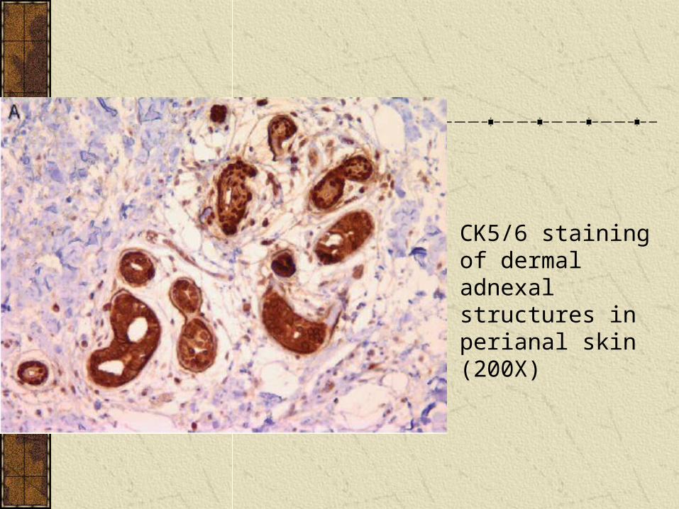
CK5/6 staining of dermal adnexal structures in perianal skin (200X)

Results-CK5/6
Five ADC (5/19; 26%) showed strong staining with CK5/6 and 6 NEC (6/32; 19%) showed a unique, punctate cytoplasmic staining with CK5/6.These results lead to the calculation of a PPV of 69% and a specificity of 78% for CK5/6 staining in SCC in our series.

CK5/6 staining in an ADC (200X)

CK5/6 staining in a NEC (200X)
punctate cytoplasmic staining

Results-P63 & CK5/6
Staining with p63 was found in the basal layers of normal squamous epithelium, and in dysplastic cells in areas of AIN(I~III).As the degree and thickness of dysplasia increased, so did the amount of p63 staining.In contrast, the normal ATZ showed staining restricted to the basal layer of cells

Results-P63 & CK5/6
With the CK5/6 antibody, all of the overlying squamous epithelium, whether normal or dysplastic, manifested full-thickness staining.

High-grade AIN III with full-thickness p63 staining (200X)

ATZ, showing basal layer staining with p63 (400X)

Full-thickness staining of squamous epithelium with CK5/6 (200X)

Discussion
Current diagnostic algorithms for anal SCC rely on negative staining with markers of ADC, including CDX-2 and CK20, and NEC, including chromogranin and SYN.Combined with putative evidence of squamous differentiation by staining with one or more CK, most often CK5 and/or CK6.

Discussion
Specificity of the CK5/6 combination for SCC is not perfect, and these keratins can stain other types of carcinoma, including up to 30% of colorectal ADC.In our series, 26% of ADC were positive for CK5/6 and 19% of NEC had a punctate, cytoplasmic positivity for CK5/6.

Discussion-p63
We found that immunohistochemical staining for p63 is a highly specific and useful tool in the diagnosis of SCCs of the anal canal.Our series reveals a specificity of 98% and a PPV of 92% for squamous differentiation in invasive carcinomas.

Discussion-p63
In addition, basaloid anal squamous carcinomas, the type formerly designated cloacogenic carcinoma, also stained with p63 antibody.Given their dark, homogeneous nuclear chromatin and their nested growth pattern, these carcinomas are easily confused with other types of carcinoma, particularly neuroendocrine tumors (different treatment protocol.)

Discussion-p63
The p63 immunostain should prove especially useful in the small, often poorly preserved biopsies from which the diagnosis of anal carcinoma is usually made.Widespread staining of dysplastic squamous epithelium in AIN precludes its use for diagnosis of SCC.(to distinguish from the normal ATZ)
Discussion-p63
The product of the p63 gene has been shown to be related to SCC in many sites, including the head and neck, the lung, and the uterine cervix.And is expressed by a handful of other carcinomas as well, including metaplastic breast carcinoma and urothelial carcinoma.

Discussion-p63
Although positive staining with p63 is very specific for anal SCC, it will not distinguish carcinomas primary in the anal canal from those directly extending or metastasis!!
Discussion-p63
The staining of 2 cases of NEC from our series, both classic appendiceal carcinoid tumors??The NEC in our series came from throughout the colon and rectum, but only the carcinoid tumors from the appendix showed nuclear positivity.

Discussion-p63
The appendix is a product of the embryonic midgut, which also includes the distal duodenum, the small intestine, the right colon, and the proximal three-quarters of the transverse colon. It is possible that p63 staining varies among NEC from different parts of the embryologic gut, and future studies could address this by staining more NEC, from throughout the gastrointestinal tract.

Discussion-CK5/6
In our series, no additional utility was gained in the diagnosis of SCC by adding the CK5/6 immunostain.On the basis of our study, staining with CK5/6 can be eliminated from any immunohistochemical panel for anal carcinomas.
Discussion-CK5/6
It provides no added utility to the diagnosis of SCC, is potentially confusing in its staining of perianal dermal adnexal structures and normal squamous epithelium, has a lower specificity and PPV than p63 for SCC, and provides no utility in highlighting squamous dysplasia in the overlying epithelium.
Conclusion
The p63 antibody should prove especially useful in small biopsies of the type commonly sent to the pathology laboratory for the diagnosis of anal carcinomas.
