in vitro antiplasmodial activity and cytotoxicity of …...in vitro antiplasmodial activity and...
TRANSCRIPT
In vitro Antiplasmodial Activity and Cytotoxicity of Extracts and Fractionsof Bidens pilosaNoumedem Anangmo Christelle Nadia1, Wabo Pone J1*, Kumar Kaushi N2, Mohana krishnan D2,Mittal Garima2, Nguemfo Tchakugni Arlette1, Mishral Shubbi2, Mpoame Mbida1 and Dinkar Sahal2
1Research Unit of Biology and Applied Ecology, University of Dschang, Cameroon2Malaria Drug Discovery Laboratory, International Centre for Genetic Engineering and Biotechnology, New Delhi,India
Abstract
Malaria is one of the most important health problems in Cameroon. The aim of this study was toevaluate the in vitro activity of the extracts and fractions of Bidens pilosa on Plasmodium falciparumstrains. Plasmodium falciparum were cultivated by the method of Trager and Jensen (1976) andmaintained in fresh O+ human erythrocytes at 4 % haematocrit in complete medium (RPMI 1640 withAlbumax II). The ring stage synchronized P. falciparum strains Pf3D7 (Chloroquine sensitive) and PfINDO (Chloroquine resistant), 1% parasitemia, 2% hematocrit} were incubated in a 96-well microplatefor 48 h with different concentrations of plant extracts and fractions of the most active extract rangingfrom 1.56 to 100 µg/ml. RPMI and 0.4% DMSO were used as negative controls; while Chloroquine (1µM) was used as positive control. The results were obtained by the microtiter plate using SYBR Green Ifluorescence assay. Cytotoxicity was determined against HEPG2 and L929 cells using MTT assay. Themost potent extract was chromatographed on reverse phase HPLC towards antiplasmodial activityguided purification of metabolites. Of the 3 plant extracts tested, the highest antiplasmodial (IC50 of 8.18±2.15 µg/ml) activity was observed with Ethyl Acetate extract of the leaves of Bidens pilosa. This extractwas then selected for antiplasmodial activity guided fractionation and isolation of active fractions.Neighbouring fractions were combined judiciously to ensure maximum purity to obtain 15 pools.Among the 15 pools evaluated, the most potent was number 12 with an IC50 of 0.73 ± 0.07 µg/ml (Pf3D7) and 3.53 ± 1.85 µg/ml (Pf INDO). Pool 12 exhibited a CC50 of 88.34 ± 1.56 µg/ml (HEPG2 cells)and 81.31 ± 2.42 µg/ml (L929 cells). Therefore, pool 12 can be considered as highly active and non-toxic.Our findings, therefore, corroborate the use of B. pilosa as antimalarial in Cameroonian folk medicine.
Keywords: Bidens pilosa; Antiplasmodial activity; Plasmodium falciparum; Cytotoxicity; Cameroon.Accepted on June 06, 2017
IntroductionMalaria is one of the most prevalent diseases in the world. Itaffects about 300–500 million people each year; mostly fromsub-Saharan Africa and causes about 2.3 million deaths everyyear [1]. Malaria represents the world’s greatest public healthproblem in terms of number of people affected, levels ofmorbidity and mortality (800,000 deaths among the 3 billionpeople at risk in 2009). About 91% of the total deaths occurredin Africa with pregnant women and children under 5 yearsbeing the most affected groups of the population [2].Cameroon is among the 18 countries bearing 90% of deathscaused by malaria in Africa, with 71% of its population livingin high-transmission areas [1]. The problems of the resistanceof the vector mosquitoes to insecticides and that of parasites tomost of the commercially available antimalarials seriouslyweaken the control approaches [3]. The success of quinine inthe treatment of malaria for many decades, and later of
artemisinin and its derivatives for treatment of cerebralmalaria, has turned attention to plants as potential sources ofantimalarial drugs [4]. Most of the people in rural areas rely ontraditional medicine for the treatment of many infectiousdiseases. They commonly treat the recurrent fever typical ofmalaria with plant extracts. It is not clear whether those plantscontain ingredients with antimalarial activity or that they exerttheir actions through other mechanisms such as immuno-modulation [5].
The emergence of multi-drug resistant strains of Plasmodiumexacerbates the situation further; posing a major obstacle tosuccessful chemoprophylaxis and chemotherapy of the disease[6]. The resistance of P. falciparum to the commonly usedantimalarial drugs including the newly introduced Artemisininshas resulted in resurgence in treatment failures [7]. This rapidspread of parasite resistance has spurred a renewed interest inthe search for new alternative antiplasmodial agents [8]. Bidens
Asian Journal of Biomedical and Pharmaceutical Sciences
ISSN 2249 – 622X
Asian J Biomed Pharmaceut Sci 2017 Volume 7 Issue 61 28

pilosa is a medicinal plant used in Cameroon bytradipractitioners for the treatment of some diseases likehelminthiases, typhoids and malaria [9]. It is in this light thatthe present study wishes to assess the efficacy of this plant invitro in order to justify its usage by traditional healers.
Materials and Methods
Plant materialFresh leaves of plant were collected from Dschang-Cameroonin March 2015 and brought to the National Herbarium ofCameroon where a specimen was kept under number 18572/SRF-CAM. After identification as B. pilosa the leaves of theplant were collected for the second time, air dried and reducedto powder, before extractions were undertaken. Three types ofextracts (methanolic, Ethyl Acetate and aqueous extracts) wereprepared and tested on both Chloroquino-Sensitive 3D7 andChloroquino-Resistant INDO strains of P. falciparum.
Preparation of extractThe methanolic and Ethyl Acetate extracts were obtained usingthe procedure described by Wabo Poné et al. [10]. Briefly, 100g of stored powder were macerated in 1.5 l of 95% methanolwhich removed the polar ingredient of the plants. The mixturewas placed on a shaker. 72 h later, the suspension was sievedand filtered using filter paper of pore size 2.5 μm. The filtratewas aligoted in a portion of 250 ml, introduced in a vial andconcentrated for about 5 minutes using a rotavapor Buchi-R-210 model heated at 65°C. The concentration of all thefiltrate took about 8 h.
For the ethyl acetate extract, the same quantity of plant powderwas macerated in 1.5 l of ethyl acetate for 72 h. After filtration,the same procedure was followed for methanolic extract.
For the aqueous extract, a similar procedure was carried out;except for the fact that maceration in distilled water took 48 h.The filtrate was evaporated in a vacuum pomp heated at 42ºCand the residues were lyophilized. The methanolic, EthylAcetate and aqueous extract obtained were kept in arefrigerator at 4ºC for further usages.
Reference drugsThe reference drugs, Chloroquine (CQ) and Artemisinin(ATR), were obtained from SIGMA and were used as positivecontrols for Pf3D7 and PfINDO respectively. Zero point four(0.4%) DMSO was used as negative controls.
Dilution of extracts and reference drugs6.5 mg of plant extracts and ATR were dissolved in 250 μl ofDMSO while the same quantity of CQ was diluted using water(Milli-Q grade) making stock solutions of 25 mg/ml. Completeculture medium (RPMI 1640 with 0.2% sodium bicarbonate,0.5% Albumax, 45 mg/l hypoxanthine and 20 mg/l gentamicin)was then added to solutions to prepare the desiredconcentrations ranging from 100 to 1.56 μg/ml.
Maintenance of culturePlasmodium falciparum was cultivated in vitro by the methodof Trager and Jensen [11] with minor modifications. Cultureswere maintained in fresh O+ human erythrocytes at 4%haematocrit in complete medium at 37ºC under reduced O2(gas mixture 5% O2, 5% CO2, and 90% N2).
Synchronization of Plasmodium falciparum parasitedevelopment using sorbitolThe cell culture was centrifuged at 1800 rpm for 5 minutes.After centrifugation, the supernatant was discarded and thepellet retained. Then, 10 pellet volume of 5% sorbitol solutionwas added. This solution was mixed and kept at 37ºC for 7minutes. The sorbitol solution containing the cells was takenout of the incubator and centrifuged at 1800 rpm for 5 minutes.The supernatant (sorbitol) was discarded and the synchronizedculture was suspended in fresh cRPMI and transferred to thePetri dishes. The parasite culture was incubated at normalcultures conditions (37ºC under reduced O2). After one hour, athin blood smear was prepared and stained slides wereexamined under a microscope at 100 X magnification for theparasites stages identification and parasitemia [12].
Antiplasmodial assay of Bidens pilosa extractsThe in vitro antiplasmodial activity of Bidens pilosa extractswas determined by fluorescence against CQ sensitive Pf3D7and resistant PfINDO strains of Plasmodium falciparum.Synchronized culture at 2% hematocrit and 1% parasitemiawas aliquoted with test drugs [plant extracts (100, 50, 25, 12.5,6.25, 3.125 and 1.56), 1Nm of CQ and ART] to 96-well flat-bottom tissue culture to a final volume of 100 µl. After 48 h ofincubation, wells tested were supplemented with equalvolumes of Lysis buffer (Tris-20 mM, EDTA-5 mM, Saponin-0.008%, Triton-X 100 – 0.08%) containing 1X SYBR Green Idye. After the addition of lysis buffer, the plates wereincubated for another one hour. After this period, the plateswere read using 96-well fluorescence plate reader (Victor,Perkin-Elmer), with excitation and emission wavelengths of497 and 520 nm respectively. The fluorescence readings wereplotted against drug concentration, and IC50 values weredetermined. In order to validate the SYBR green data, thinblood smears of treated and untreated wells were prepared andstained [13].
Evaluation of plants extracts cytotoxicityPlant extracts cytotoxicity were evaluated on animal cell linesfibroblast L929 and Hella cells using MTT assay as describedby Mosmann [14]. Briefly, cells (104 cells/200 ml/well) wereseeded into 96-well flat-bottom tissue culture plates incomplete medium (10% foetal bovine serum, 0.21% SodiumBicarbonate (Sigma, USA) and 50 mg/ml gentamicin). After24 h, plant extracts at different concentrations were added andplates incubated for 48 h in a humidified atmosphere at 37 ºCand 5% CO2, 10% DMSO (v/v) was used as a positiveinhibitor. Thereafter, 20 µl of a stock solution of MTT (5
In vitro Antiplasmodial Activity and Cytotoxicity of Extracts and Fractions of Bidens pilosa
Asian J Biomed Pharmaceut Sci 2017 Volume 7 Issue 61 29

mg/mL in 1X phosphate buffered saline) was added to eachwell, gently mixed and each plate was incubated for another 4h. After spinning the plates at 1500 rpm for 5 min, supernatantswere removed and 100 ml of 10% DMSO were added in eachwell to stop the reaction of extracts. The formation offormazon obtained after the transformation of tetrazolium wasread on a microtiter plate reader at 570 nm. The 50% cytotoxicconcentration (CC50) of plant extract was determined byanalysis of dose–response curves, according to the cytotoxicitygradient of plant extracts established by Malebo et al. (Table 1)[15]. Also, the Selectivity index (SI) was calculated using thefollowing formula:
Table 1: The IC50, the CC50 and the selectivity index (SI) of the effectof the different extract on the development of P. falciparum strains andmammalian cells line (Legend: AE: Aqueous extract, ME: Methanolicextract, EAE: Ethyl Acetate extract, SI: Selectivity index).
Strains Bidens pilosa
AE ME EAE
IC50 (µg/ml)
3D7 14.31 ± 1.37 26.66 ± 2.56 8.18 ± 2.15
INDO 22.6 ± 1.99 14.07 ± 2.09 13.37 ± 0.04
CC50 (µg/ml)
HEPG2 73.23 ± 1.22 29.36 ± 4.01 71.23 ± 2.11
L929 76.11 ± 3.21 29.02 ± 1.68 68.46 ± 3.17
SI
5.86 4.83 12.54
Bioassay guided fractionation and isolation of activefractionsThe ethyl acetate extract of B. pilosa which showed goodantiplasmodial potency was chosen for antiplasmodial activity
guided reverse phase HPLC fractionation. DMSO supernatantof 200 mg/ml was injected into C18 Deltapak (19 × 300 mm,15 m) column (Waters, USA) using water–methanol gradient(5–95%, 1%/min) at a flow rate of 9 ml/min on a Gilson prepHPLC system. Dual wavelength detections were made at 214and 254 nm. Fractions were collected, dried, weighed andevaluated for antiplasmodial and cytotoxic activities
Determination of molecules contents in the ethylacetate extract and fractions by GCMSMolecules contained in the crude Ethyl Acetate extract and inmost effective fractions were determined using ChromatogramD:\GCMS-QP2010Ultra\GCMS method.
Statistical analysisData obtained from this work were analyzed statistically usingStudent’s t-test and ANOVA (One-way) followed by a post test(Turkey–Kramer multiple comparison test) using SPSS(version 22.0). Differences between means was consideredsignificant at 5% level of significance (P<0.05). The 50%inhibitory concentrations (IC50) were determined from linearregression curve obtained between the inhibition rate expressedin probit and the decimal logarithm of the concentrations (μg/ml).
ResultsThe variation of the mean inhibition rate on the growth ofPf3D7 and PfINDO strains of P. falciparum according to thedifferent concentrations of extracts of B. pilosa is shown inFigure 1.
Figure 1: Percentage growth of Plasmodium falciparum 3d7 and INDO strains according to the different concentrations of Bidens pilosa extract.(Legend: AE: Aqueous Extract, ME: Methanolic extract, EAE: Ethyl Acetate Extract).
Figure 1 shows that RPMI 1640 and 0.4% DMSO did notaffect (0% inhibition rate) the development of P. falciparum. Inthe treated wells, the inhibition rate increases with the
increasing concentration of the tested extracts. Considering theCQ sensitive 3D7 strain, the mean inhibition rates of theaqueous extract (49.55 ± 7.46, 57.35 ± 3.30 and 63.58 ± 2.68)
Nadia/Pone/Kaushi/krishnan/Garima/Arlette/Shubbi/Mbida/Sahal
30 Asian J Biomed Pharmaceut Sci 2017 Volume 7 Issue 61

and methanolic extract (24.01 ± 9.31, 27.10 ± 8.77 and 35.00 ±4.36) at the concentrations of 12.5, 25, and 50 µg/mlrespectively were quite similar (P>0.05). Meanwhile, theseinhibition rates were less (P<0.05) than that of the EthylAcetate extract (66.54 ± 7.62, 73.92 ± 18.35 and 94.15 ± 8.68).However, at 100 µg/ml concentration, Ethyl Acetate andaqueous extracts exhibited mean inhibition rates of 98.59 ±9.94 and 98.44 ± 7.28% respectively which are higher ascompared to the mean inhibition rate of methanolic extract(41.79 ± 9.00%).
On the CQ resistant INDO strain, the aqueous extract exhibiteda poor activity with a mean inhibition rate of 3.35 ± 1.3, 29.11± 3.11, 37.51 ± 4.97, 45.70 ± 4.70 at the concentrations of12.5, 25, 50 and 100 μg/ml respectively. For concentrationsgreater than 12.5 μg/ml, the mean inhibition rates ofmethanolic extract (59.43 ± 2.44, 67.44 ± 5.20, 87.30 ± 5.30)were less (P<0.05) than the effect of Ethyl Acetate extract(84.09 ± 3.33, 91.44 ± 2.47, 91.51 ± 3.81). Below theseconcentrations, the antiplasmodial activity is similar for bothextracts.
The IC50, the CC50 and the selectivity index (SI) of thedifferent extracts are shown on Table 1. The effect of thedifferent plant extracts was more potent on CQ sensitive 3D7strain compared to the CQ sensitive INDO strain. The EthylAcetate extract exhibited the highest antiplasmodial activity(IC50 of 8.18 ± 2.15) with a (P<0.05) as compared to otherextracts except for the IC50 of the methanolic extract. Aqueousextract showed the highest cytotoxic concentration (76.11 ±3.21 μg/ml). However, the best selectively index (121.01) wasobtained in the Ethyl Acetate extract.
Bioassay guided fractionation of Ethyl Acetateextracts of Bidens pilosaAmong the extracts of B. pilosa tested, the ethyl Acetateextract was the most potent and possessed the highestselectivity index. Therefore, it was selected for activity guided
fractionation by RPHPLC using water–methanol gradient(Figure 2). Orange and blue color bars indicate fractions (1–60)collected. The zoom of 60–120 min (marked by dotted box)has been shown just above the chromatogram tracing.
Figure 2: Semiprep Reverse phase HPLC Chromatogram of ethylacetate extract of Bidens pilosa.
Table 2 shows the IC50 and the CC50 fractions of Ethyl Acetateextract of B. pilosa on the development of P. falciparum strainsand mammalian cells line.
The fractions showed a wide range of antiplasmodial potencieswith IC50 values ranging from 2.38 to 71.46 μg/ml against aCQ resistant strain (INDO) and 0.73 to 67.36 mg/ml for CQsensitive strain (3D7) of P. falciparum (Table 2).
Just like crude extracts of B. pilosa, most chromatographicfractions derived from these products were also found to bemore effective against a CQ sensitive 3D7 strain than CQresistant INDO strain. More interestingly, fractions led to asignificant increase on the inhibition of the development of P.falciparum with the lowest IC50 of 0.73 ± 0.07 against Pf3D7compared to crude extract which exhibited a lowest IC50 of8.18 ± 2.15 (P<0.05).
Table 2: The IC50, the CC50 and the selectivity index (SI) of the effect of the different fractions on the development of Plasmodium falciparumstrains and mammalian cells line (Legend: SI Selectivity index).
Fractions weight (mg) PfINDO Pf3D7 HEPG2 L929 SI
IC50 (µg/ml) CC50 (µg/ml)
1 (F1-F5) 59.26 ±1.21 58.48 ±3.29 17.42 ± 9.92 23.28 ± 4.87 0,30
2 (F6-F14) 5.99 65.38 ± 0.88 61.22 ± 1.81 4.62 ± 3.82 2.46 ± 9136 0,08
3 (F15-F20) 5.26 50.15 ± 2.2 53.18 ± 1.52 14.30 ± 1.22 12.02 ± 3.98 0,27
4 (F21-F23) 2.6 61.75 ± 1.55 59.41 ± 2.30 2.21 ± 1.81 0.87 ± 0.23 0,04
5 (F24-F27) 3.64 71.46 ± 1.48 68.73 ± 2.33 3.75 ± 1.58 1.45 ± 0.64 0,05
6 (F28) 0.65 8.29 ± 1.35 6.95 ± 1.89 17.72 ± 5.65 29.31 ± 2.42 2,55
7 (F29-31) 2.65 31.76 ± 1.73 67.36 ± 11.04 9.49 ± 1.01 11.38 ±2.87 0,14
8 (F32) 0.75 16.8 ± 8.33 14.65 ± 7.51 21.6 ± 0.9 17.46 ± 1.36 1,47
In vitro Antiplasmodial Activity and Cytotoxicity of Extracts and Fractions of Bidens pilosa
Asian J Biomed Pharmaceut Sci 2017 Volume 7 Issue 61 31

9 (F33-34) 2.17 5.33 ± 0.01 4.87 ± 0.20 36.6 ± 5.16 25.02 ± 3.98 0,08
10 (F35-36) 2.45 6.64 ± 0.87 4.39 ± 0.30 40.3 ± 7.01 36.47 ± 5.23 9,18
11 (F37-38) 3.96 13.55 ± 0.66 12.41 ± 1.24 22.2 ± 2.35 30.45 ± 6.64 1,79
12 (F39-43) 4.53 3.53 ± 1.85 0.73 ± 0.07 88.34 ± 1.56 81.31 ± 2.42 121,01
13 (F44-45) 0.69 2.38 ± 0.09 1.86 ± 0.10 62.2 ± 6.40 33.40 ± 0.62 33,44
14 (F46-51) 1.61 3.49 ± 0.04 3.11 ± 0.12 77.51 ± 616 41.93 ± 6.67 24,92
15 (F52-60) 11.12 3.91 ± 0.11 3.24 ± 1.27 73.70 ± 1.69 62.80 ± 7.02 22,75
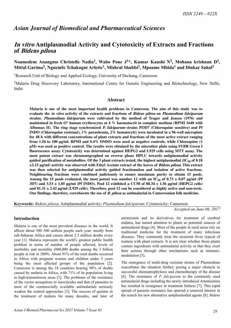
Characterization of molecules in the most effectiveextract and the most effective fractions by GCMSFigure 3 presents the number of molecules contained in a crude(A) and fraction 12 (B) of Ethyl Acetate extract of B. pilosa(F39-43).
Figure 3A has shown 68 pics of molecules contained in thecrude Ethyl Acetate extract. Each pic corresponds to a specificmolecule. The different names have been given according tothe record time of pics, the area and the percentage of area(Table 3).
Figure 3A: Chromatogram of pictures of the molecules contained inthe crude extract of ethyl acetate (A) and in the most effective fraction(12: F39-43).
Table 3: Names and percentage area of different molecules illustratein Chromatogram A: (crude extract of ethyl acetate).
Figure 3B has shown only 14 molecules in the most effectivefraction as compared to the Ethyl Acetate extract (68molecules). The different names have been given as earlierstated (Table 4).
Nadia/Pone/Kaushi/krishnan/Garima/Arlette/Shubbi/Mbida/Sahal
32 Asian J Biomed Pharmaceut Sci 2017 Volume 7 Issue 61
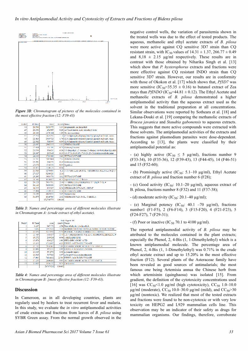
Figure 3B: Chromatogram of pictures of the molecules contained inthe most effective fraction (12: F39-43)
Table 3: Names and percentage area of different molecules illustratein Chromatogram A: (crude extract of ethyl acetate).
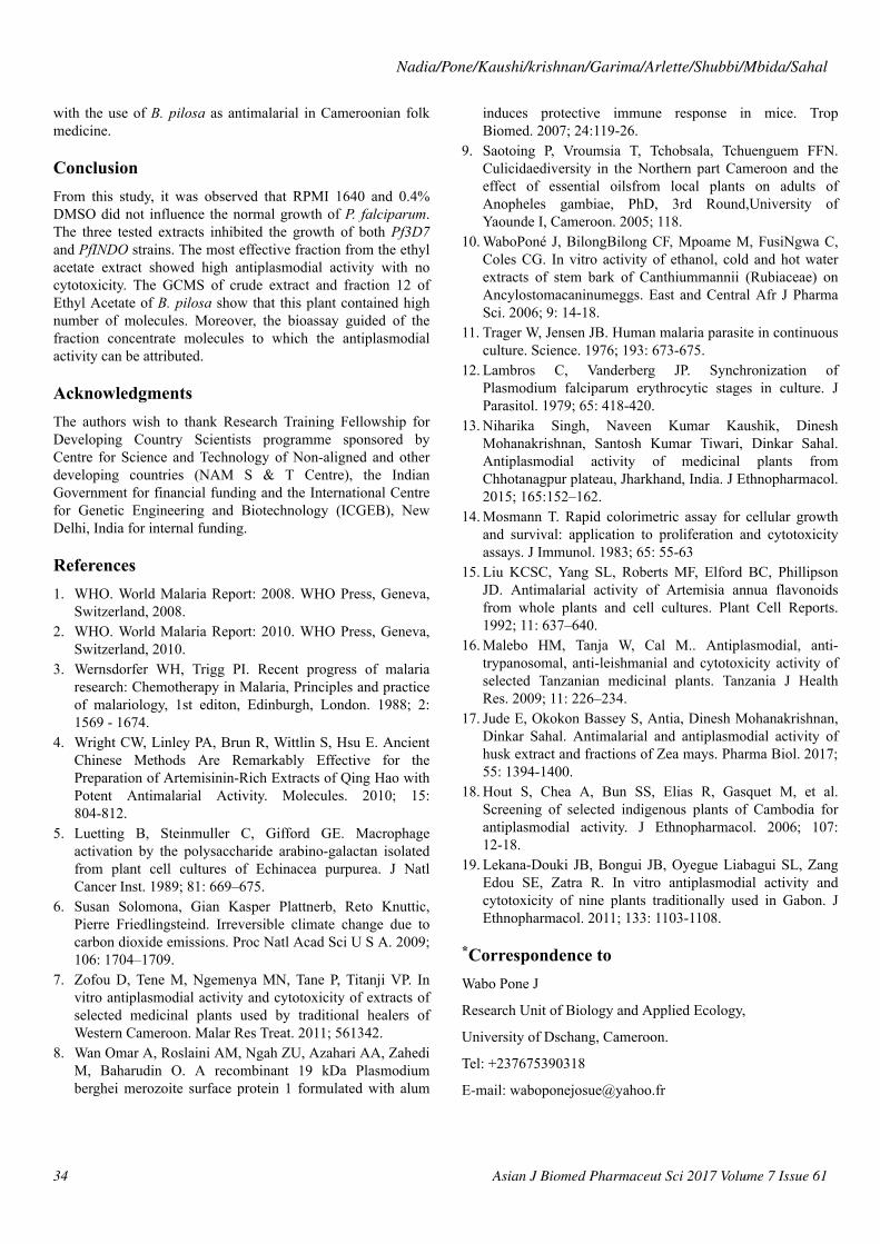
Table 4: Names and percentage area of different molecules illustratein Chromatogram B: [most effective fraction (12: F39-43).
DiscussionIn Cameroon, as in all developing countries, plants areregularly used by healers to treat recurrent fever and malaria.In this study, we evaluate the in vitro antiplasmodial activitiesof crude extracts and fractions from leaves of B. pilosa usingSYBR Green assay. From the normal growth observed in the
negative control wells, the variation of parasitemia shown inthe treated wells was due to the effect of tested products. Theaqueous, methanolic and ethyl acetate extracts of B. pilosawere more active against CQ sensitive 3D7 strain than CQresistant strain, with IC50 values of 14.31 ± 1.37, 266.77 ± 0.49and 8,18 ± 2.15 μg/ml respectively. These results are incontrast with those obtained by Niharika Singh et al. [13]which show that P. hysterophorus extracts and fractions weremore effective against CQ resistant INDO strain than CQsensitive 3D7 strain. However, our results are in conformitywith those of Okokon et al. [17] which shows that, Pf3D7 wasmore sensitive (IC50=35.35 ± 0.16) to butanol extract of Zeamays than PfINDO (IC50=44.81 ± 0.12). The Ethyl Acetate andmethanolic extracts of B. pilosa demonstrated a higherantiplasmodial activity than the aqueous extract used as thesolvent in the traditional preparation at all concentrations.Similar observations were reported by Sotheara et al. [18] andLekana-Douki et al. [19] comparing the methanolic extracts ofBrucea javanica and Staudtia gabonensis to aqueous extracts.This suggests that more active compounds were extracted withthose solvents. The antiplasmodial activities of the extracts andfractions against plasmodium parasites were dose-dependent.According to [13], the plants were classified by theirantiplasmodial potential as:
- (a) highly active (IC50 ≤ 5 µg/ml), fractions number 9(F33-34), 10 (F35-36), 12 (F39-43), 13 (F44-45), 14 (F46-51)and 15 (F52-60);
- (b) Promisingly active (IC50: 5.1–10 µg/ml), Ethyl Acetateextract of B. pilosa and fraction number 6 (F28);
- (c) Good activity (IC50: 10.1–20 µg/ml), aqueous extract ofB. pilosa, fractions number 8 (F32) and 11 (F37-38);
- (d) moderate activity (IC50: 20.1–40 µg/ml);
- (e) Marginal potency (IC50: 40.1 –70 µg/ml), fractionsnumber1 (F1-F5), 2 (F6-F14), 3 (F15-F20), 4 (F21-F23), 5(F24-F27), 7 (F29-31);
- (f) Poor or inactive (IC50 70.1 to 4100 µg/ml).
The reported antiplasmodial activity of B. pilosa may beattributed to the molecules contained in the plant extracts;especially the Phenol, 2, 4-Bis (1, 1-Dimethylethyl) which is aknown antiplasmodial molecule. The percentage area ofPhenol, 2, 4-Bis (1, 1-Dimethylethyl) was 0.71% in the crudeethyl acetate extract and up to 15.20% in the most effectivefraction (F12). Several plants of the Asteraceae family havebeen revealed as good sources of antimalarials; the mostfamous one being Artemisia annua the Chinese herb fromwhich artemisinin (quinghaosu) was isolated [15]. Fromgradient, the definition of the cytotoxicity concentrations used[16] was CC50<1.0 μg/ml (high cytotoxicity), CC50 1.0–10.0μg/ml (moderate), CC50 10.0–30.0 μg/ml (mild), and CC50>30μg/ml (nontoxic). We realized that most of the tested extractsand fractions were found to be non-cytotoxic or with very lowtoxicity on HEPG2 and L929 mammalian cells line. Thisobservation may be an indicator of their safety as drugs formammalian organisms. Our findings, therefore, corroborate
In vitro Antiplasmodial Activity and Cytotoxicity of Extracts and Fractions of Bidens pilosa
Asian J Biomed Pharmaceut Sci 2017 Volume 7 Issue 61 33
with the use of B. pilosa as antimalarial in Cameroonian folkmedicine.
ConclusionFrom this study, it was observed that RPMI 1640 and 0.4%DMSO did not influence the normal growth of P. falciparum.The three tested extracts inhibited the growth of both Pf3D7and PfINDO strains. The most effective fraction from the ethylacetate extract showed high antiplasmodial activity with nocytotoxicity. The GCMS of crude extract and fraction 12 ofEthyl Acetate of B. pilosa show that this plant contained highnumber of molecules. Moreover, the bioassay guided of thefraction concentrate molecules to which the antiplasmodialactivity can be attributed.
AcknowledgmentsThe authors wish to thank Research Training Fellowship forDeveloping Country Scientists programme sponsored byCentre for Science and Technology of Non-aligned and otherdeveloping countries (NAM S & T Centre), the IndianGovernment for financial funding and the International Centrefor Genetic Engineering and Biotechnology (ICGEB), NewDelhi, India for internal funding.
References1. WHO. World Malaria Report: 2008. WHO Press, Geneva,
Switzerland, 2008.2. WHO. World Malaria Report: 2010. WHO Press, Geneva,
Switzerland, 2010.3. Wernsdorfer WH, Trigg PI. Recent progress of malaria
research: Chemotherapy in Malaria, Principles and practiceof malariology, 1st editon, Edinburgh, London. 1988; 2:1569 - 1674.
4. Wright CW, Linley PA, Brun R, Wittlin S, Hsu E. AncientChinese Methods Are Remarkably Effective for thePreparation of Artemisinin-Rich Extracts of Qing Hao withPotent Antimalarial Activity. Molecules. 2010; 15:804-812.
5. Luetting B, Steinmuller C, Gifford GE. Macrophageactivation by the polysaccharide arabino-galactan isolatedfrom plant cell cultures of Echinacea purpurea. J NatlCancer Inst. 1989; 81: 669–675.
6. Susan Solomona, Gian Kasper Plattnerb, Reto Knuttic,Pierre Friedlingsteind. Irreversible climate change due tocarbon dioxide emissions. Proc Natl Acad Sci U S A. 2009;106: 1704–1709.
7. Zofou D, Tene M, Ngemenya MN, Tane P, Titanji VP. Invitro antiplasmodial activity and cytotoxicity of extracts ofselected medicinal plants used by traditional healers ofWestern Cameroon. Malar Res Treat. 2011; 561342.
8. Wan Omar A, Roslaini AM, Ngah ZU, Azahari AA, ZahediM, Baharudin O. A recombinant 19 kDa Plasmodiumberghei merozoite surface protein 1 formulated with alum
induces protective immune response in mice. TropBiomed. 2007; 24:119-26.
9. Saotoing P, Vroumsia T, Tchobsala, Tchuenguem FFN.Culicidaediversity in the Northern part Cameroon and theeffect of essential oilsfrom local plants on adults ofAnopheles gambiae, PhD, 3rd Round,University ofYaounde I, Cameroon. 2005; 118.
10. WaboPoné J, BilongBilong CF, Mpoame M, FusiNgwa C,Coles CG. In vitro activity of ethanol, cold and hot waterextracts of stem bark of Canthiummannii (Rubiaceae) onAncylostomacaninumeggs. East and Central Afr J PharmaSci. 2006; 9: 14-18.
11. Trager W, Jensen JB. Human malaria parasite in continuousculture. Science. 1976; 193: 673-675.
12. Lambros C, Vanderberg JP. Synchronization ofPlasmodium falciparum erythrocytic stages in culture. JParasitol. 1979; 65: 418-420.
13. Niharika Singh, Naveen Kumar Kaushik, DineshMohanakrishnan, Santosh Kumar Tiwari, Dinkar Sahal.Antiplasmodial activity of medicinal plants fromChhotanagpur plateau, Jharkhand, India. J Ethnopharmacol.2015; 165:152–162.
14. Mosmann T. Rapid colorimetric assay for cellular growthand survival: application to proliferation and cytotoxicityassays. J Immunol. 1983; 65: 55-63
15. Liu KCSC, Yang SL, Roberts MF, Elford BC, PhillipsonJD. Antimalarial activity of Artemisia annua flavonoidsfrom whole plants and cell cultures. Plant Cell Reports.1992; 11: 637–640.
16. Malebo HM, Tanja W, Cal M.. Antiplasmodial, anti-trypanosomal, anti-leishmanial and cytotoxicity activity ofselected Tanzanian medicinal plants. Tanzania J HealthRes. 2009; 11: 226–234.
17. Jude E, Okokon Bassey S, Antia, Dinesh Mohanakrishnan,Dinkar Sahal. Antimalarial and antiplasmodial activity ofhusk extract and fractions of Zea mays. Pharma Biol. 2017;55: 1394-1400.
18. Hout S, Chea A, Bun SS, Elias R, Gasquet M, et al.Screening of selected indigenous plants of Cambodia forantiplasmodial activity. J Ethnopharmacol. 2006; 107:12-18.
19. Lekana-Douki JB, Bongui JB, Oyegue Liabagui SL, ZangEdou SE, Zatra R. In vitro antiplasmodial activity andcytotoxicity of nine plants traditionally used in Gabon. JEthnopharmacol. 2011; 133: 1103-1108.
*Correspondence toWabo Pone J
Research Unit of Biology and Applied Ecology,
University of Dschang, Cameroon.
Tel: +237675390318
E-mail: [email protected]
Nadia/Pone/Kaushi/krishnan/Garima/Arlette/Shubbi/Mbida/Sahal
34 Asian J Biomed Pharmaceut Sci 2017 Volume 7 Issue 61