infra temporal fossa
TRANSCRIPT

Dr. Anitha Nancy T, Asst. professor, Department of Anatomy

Infratemporal fossa
Location
Boundaries
Contents
Maxillary artery & its branches

Boundaries:
Lateral=Ramus of Mandible
Anterior=Maxilla
Medial=Lat. Pterygoid Plate
Roof=Sphenoid
Temporal
Maxilla
Parietal
Sphenoid
Frontal
Z
Lat. Pterygoid Plate
Pterygomaxillary Fissure
Infratemporal Fossa
Temporal Fossa

Infratemporal
Fossa

Anterior:
Maxilla.
Superior:
Greater wing of sphenoid.
Medial: Lateral pterygoid plate.
Lateral: Coronoid process and ramus of mandible.

Inferior: Continuous with neck.
Posterior: TMJ and styloid process.

COMMUNICATIONS: 1. With temporal fossa: through a gap deep
to zygomatic arch 2. With cranial cavity: through foramen
ovale, foramen spinosum, foramen lacerum
3. With orbit: through inferior orbital fissure 4. With pterygopalatine fossa: through
pterygomaxillary fissure


Mandibular nerve.
Maxillary artery.
Pterygoid plexus of vein
Medial and lateral pterygoid muscles.
Lower part of temporalis muscle.
Chorda tympani nerve.
Otic ganglion.

Temporalis
Masseter
Medial Pterygoid Buccinator*
Lateral Pterygoid

Elevate mandible EXCEPT Lateral pterygoid Protrude mandible EXCEPT Temporalis Are supplied by anterior division of
mandibular nerve EXCEPT Medial pterygoid

Inferior alveolar n.
Lingual n.
Facial n..
Submandibular ganglion.
Otic ganglion. Lesser petrosal n.
Chorda tympani
Auriculotemporal n.

Ext. Cartotid
Inf. Alveolar Post. Sup. Alveolar
Maxillary
Post. Deep Temporal
Ant. Deep Temporal
Infra-orbital
Ext. Cartotid
Post. Deep Temporal Ant. Deep Temporal
Middle
Meningeal
Septal brr.
Descending palatine Greater palatine Lesser palatine
Sphenopalatine
V3

Terminal branch of external carotid artery.
Divided into 3 parts by lateral pterygoid
muscle: Artery may pass superficial or deep to
muscle.

First part: Origin to lower margin inferior to lateral
pterygoid.
Second part: Superficial or deep to inferior
lateral pterygoid.
Third part: Lies within pterygopalatine fossa.

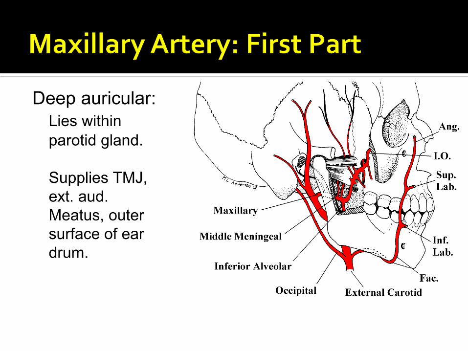
Deep auricular:
Lies within
parotid gland.
Supplies TMJ,
ext. aud.
Meatus, outer
surface of ear
drum.

Anterior
tympanic:
Arises within
parotid gland.
Supplies
tympanic cavity
and inner
surface of ear
drum.

Middle
meningeal:
Enters cranial
cavity via
foramen
spinosum.
Supplies skull
and dura mater.

Accessory
meningeal:
Enters cranial
cavity via
foramen ovale.
Supplies
semilunar
ganglion (CN V)
and dura mater.

Inferior alveolar: Enters mandibular
foramen.
Supplies lower teeth and gums.
Passes through
mental foramen and becomes mental artery.


Branches to
muscles of
mastication.
Buccal artery.

Posterior superior
alveolar:
Runs with superior
alveolar nerve (CN
V-2).
Supplies upper
molars and
premolars.

Infraorbital: Enters orbit via
inferior orbital fissure.
Runs with
infraorbital nerve. Supplies lower
eyelid, side of nose, upper lip, oral mucosa.
Gives rise to
anterior and middle superior alveolar arteries.

Descending palatine: To soft and hard
palates. Terminal branch. Artery of pterygoid canal: To pharynx and
auditory tube.

