involvement of a disintegrin and metalloproteinase 10 and 17 in shedding of tumor necrosis factor-α
TRANSCRIPT

Involvement of a disintegrin andmetalloproteinase 10 and 17 in shedding of tumornecrosis factor-a
Atsuhiko Hikita, Nobuho Tanaka, Shoji Yamane, Yasuko Ikeda, Hiroshi Furukawa,Shigeto Tohma, Ryuji Suzuki, Sakae Tanaka, Hiroyuki Mitomi, and Naoshi Fukui
Abstract: Tumor necrosis factor-a (TNF-a) is initially synthesized as a membrane-bound protein and converted into asoluble form by proteolytic cleavage. Although a disintegrin and metalloproteinase 17 (ADAM17) is considered to be theprimary sheddase for TNF-a, it is not known whether ADAM17 is solely responsible for that process in any type of cells.To identify the TNF-a sheddase(s) in varieties of cells, we performed experiments using a unique screening system andobserved that ADAM9, ADAM10, ADAM17, and ADAM19 were capable of cleaving TNF-a. We then performed RNAinterference experiments and confirmed that ADAM10 and ADAM17 were in fact involved in TNF-a shedding in 293Acells. In mouse macrophages, ADAM17 was confirmed to be the primary sheddase, but the involvement of ADAM10 wasalso demonstrated. In NIH3T3 cells, ADAM10 could be more important in the shedding than ADAM17. In mouse vascularendothelial cell line UV,2, ADAM10 and ADAM17 were equally involved in TNF-a shedding, whereas ADAM17 was amajor sheddase in human osteoarthritic chondrocytes. From these observations and others, we concluded that bothADAM10 and ADAM17 can be a TNF-a sheddase and that their significance could be determined by their expression lev-els and the abundance of tissue inhibitor of metalloproteinases.
Key words: TNF-a, ADAM10, ADAM17, TIMP, ectodomain shedding.
Resume : Le TNF-a est d’abord synthetise sous forme de proteine liee a la membrane et sa forme soluble est produite parun clivage proteolytique. Meme si la desintegrine metalloprotease ADAM17 est consideree comme la principale sheddaseagissant sur le TNF-a, on ignore si ADAM17 est la seule responsable de ce processus pour tous les types de cellules. Afind’identifier les sheddases de TNF-a dans une variete de cellules, nous avons realise des experiences a l’aide d’un systemede criblage unique et nous avons observe que ADAM9, ADAM10, ADAM17 et ADAM19 sont capables de cliver le TNF-a. Nous avons ensuite realise des experiences d’interference par ARN et nous avons confirme que ADAM10 et ADAM17sont effectivement impliquees dans la liberation du TNF-a chez les cellules 293A. Chez les macrophages de souris,ADAM17 s’est averee comme sheddase principale mais l’implication de ADAM10 a aussi ete demontree. Chez les cellulesNIH3T3, ADAM10 pourrait etre plus importante pour la liberation que ADAM17. Dans la lignee cellulaire vasculaire en-dotheliale UV,2, ADAM10 et ADAM17 sont egalement impliquees dans la liberation de TNF-a, alors que ADAM17 estla principale sheddase chez les chondrocytes osteoarthritiques humains. A partir de ces observations et d’autres, nousavons conclu que ADAM10 et ADAM17 peuvent toutes deux etre des sheddases du TNF-a et que leur importance pourraitetre determinee par leur niveau d’expression et par l’abondance des TIMP (« tissue inhibitor of metalloproteinases »).
Mots-cles : TNF-a, ADAM10, ADAM17, TIMP, liberation d’ectodomaine.
[Traduit par la Redaction]
Introduction
Ectodomain shedding is a process in which transmem-brane proteins are proteolytically released from the plasmamembrane. Ectodomain shedding modifies the biologic andpathologic functions of the substrate protein. For example,amyloid precursor protein is cleaved by a disintegrin andmetalloproteinase (ADAM) or the b-site of amyloid precur-
sor protein cleaving enzyme and converted to a nonpatho-genic and a pathogenic protein, respectively (Selkoe 1991;Buxbaum et al. 1998; Koike et al. 1999; Lammich et al.1999; Vassar et al. 1999; Li et al. 2000). On the other hand,Fas ligand is cleaved by ADAM10 and largely loses its ac-tivity (Schneider et al. 1998; Schulte et al. 2007).
Tumor necrosis factor-a (TNF-a) is a pro-inflammatorycytokine that is involved in various pathologic conditions
Received 26 January 2009. Revision received 31 March 2009. Accepted 3 April 2009. Published on the NRC Research Press Web site atbcb.nrc.ca on 4 June 2009.
A. Hikita, N. Tanaka, S. Yamane, Y. Ikeda, H. Furukawa, S. Tohma, R. Suzuki, H. Mitomi, and N. Fukui.1 Department ofPathomechanisms, Clinical Research Center, National Hospital Organization Sagamihara Hospital, Sagamihara, Kanagawa 228-8522,Japan.S. Tanaka. Department of Orthopaedic Surgery, Faculty of Medicine, University of Tokyo, Hongo 7-3-1, Tokyo, Japan.
1Corresponding author (e-mail: [email protected]).
581
Biochem. Cell Biol. 87: 581–593 (2009) doi:10.1139/O09-015 Published by NRC Research Press

such as rheumatoid arthritis, osteoarthritis, Crohn’s disease,and endotoxin shock (Tracey et al. 1987; Kriegler et al.1988; Brennan et al. 1989; Reinecker et al. 1993; Fernandeset al. 2002). TNF-a is expressed mainly by hematopoieticcells such as macrophages and T lymphocytes but is alsopresent in nonhematopoietic cells such as mast cells andvascular endothelial cells (Carswell et al. 1975; Kobayashiet al. 1986; Steffen et al. 1989; Nilsen et al. 1998). In thosecells, TNF-a is initially synthesized as a transmembrane pro-tein and then becomes a soluble form via ectodomain shed-ding (McGeehan et al. 1994; Mohler et al. 1994).
Like other proteins, the pathologic significance of TNF-amay differ between membrane-bound and soluble forms.Soluble TNF-a is responsible for endotoxin shock becausethe blockade of TNF-a release protected mice from death(Mohler et al. 1994). On the other hand, TNF-a-deficientmice reconstituted with mutated TNF-a (muTNF delta 1–12), which lost its activity when cleaved into a solubleform, were shown to develop experimental hepatitis (Kusterset al. 1997). The role of TNF-a shedding in arthritis is con-troversial. The muTNF delta 1–12 mice developed arthritisspontaneously, implying that membrane-bound TNF-a maybe important in its etiology (Alexopoulou et al. 1997). Onthe contrary, tissue inhibitor of metalloproteinase (TIMP)-3-deficient mice were more susceptible to bovine serum albu-min induced arthritis (Mahmoodi et al. 2005). Since TNF-ashedding by ADAM17 was likely to be enhanced in thosemice, that result might indicate the importance of solubleTNF-a in the development of arthritis.
Several proteinases are known to cleave TNF-a into asoluble form. At present, ADAM17 (also called TNF-a con-verting enzyme, or TACE) is thought to be the major shed-dase for TNF-a (Black et al. 1997; Moss et al. 1997;Condon et al. 2001; Zheng et al. 2004; Bell et al. 2007; Ho-riuchi et al. 2007). In T lymphocytes, deficiency or suppres-sion of ADAM17 blocked solubilization of membrane-bound TNF-a (Black et al. 1997; Condon et al. 2001). Inmouse embryonic fibroblasts, deficiency of ADAM17 re-sulted in suppression of phorbol 12-myristate 13-acetatestimulated TNF-a shedding (Zheng et al. 2004). More re-cently, two other groups reported that ADAM17 is the pri-mal sheddase for TNF-a in mouse macrophages (Bell et al.2007; Horiuchi et al. 2007). However, these results do notnecessarily mean that ADAM17 is the primal TNF-a shed-dase in all types of cells because the endogenous sheddasefor a substrate protein can differ from cell to cell. For exam-ple, the sheddase for receptor activator of NF-kb ligand(RANKL) in the mouse bone stromal cell line TM8B2 ismainly ADAM10, but a large part of soluble RANKL isproduced by membrane type-1 matrix metalloproteinase(MT1-MMP) in osteoblasts (Hikita et al. 2006). Thus, theendogenous sheddase for TNF-a should be determined inthe respective cell types.
In this study, we attempted to identify the endogenousTNF-a sheddase in several types of cells using a screeningsystem for proteinases.
Materials and methods
ReagentsDNA polymerase, KOD plus, was purchased from
TOYOBO (Osaka, Japan). Antibodies were obtained fromthe following companies: TNF-a, Cell Signaling TechnologyInc. (Beverly, Massachusetts); ADAM10, Kamiya Biochem-ical (Seattle, Washington); ADAM17, Santa Cruz Biotech-nology, Inc. (Santa Cruz, California); actin, Sigma-AldrichCo. (St. Louis, Missouri); V5 tag, Invitrogen (Carlsbad, Cal-ifornia). Lipopolysaccharide (LPS) was purchased fromWako Pure Chemical Industries, Ltd. (Osaka, Japan). Re-combinant mouse TIMP-1 and TIMP-2 were purchasedfrom Acris Antibodies GmbH (Hiddenhausen, Germany)and recombinant mouse TIMP-3 from R&D Systems (Min-neapolis, Minnesota). Recombinant human TIMP-1 andTIMP-3 were obtained from Daiichi Fine Chemical(Toyama, Japan) and R&D Systems, respectively.
Cell cultureThe human kidney cell line 293A (Invitrogen), mouse fi-
broblastic cell line NIH3T3, and mouse vascular endothelialcell line UV,2 (RCB1994) (Riken BioResource Center,Tsukuba, Japan) were cultured in Dulbecco’s modified Ea-gle medium (D-MEM) (Wako Pure Chemical Industries,Ltd.) supplemented with 10% FBS (SAFC Biosciences, Le-nexa, Kansas) and 1% penicillin–streptomycin solution(Sigma-Aldrich Co.). Macrophages were collected fromspleens of 4- to 6-week-old male ddy mice and cultured ina-MEM (Invitrogen) supplemented with 10% FBS, 1% pen-icillin–streptomycin solution, and 200 ng/mL M-CSF fromthe culture supernatant of CMG14-12 (a generous gift fromDr. Sunao Takeshita, Department of Bone and Joint Disease,Research Institute, National Center for Geriatrics and Geron-tology, Obu City, Japan). Methods for collection and cultureof human chondrocytes were previously described (Fukui etal. 2006). In brief, human articular chondrocytes were ob-tained from osteoarthritic knee cartilage at prosthetic sur-gery. The chondrocytes were isolated from the surroundingmatrix by serial enzymic digestion and cultured in mono-layer at the density of 2 � 105 cells/cm2 in D-MEM/F-12containing 10% FBS, 25 mg/mL ascorbic acid, and penicil-lin–streptomycin. Cells were incubated at 37 8C in a humidi-fied atmosphere containing 5% CO2. Expression vectorswere transfected to these cells using FuGENE 6 transfectionreagent (Roche Diagnostics, Indianapolis, Indiana) accordingto the manufacturer’s instructions.
ConstructscDNA of mouse TNF-a was cloned from cDNA of mouse
macrophages by PCR and inserted in pCR-blunt II TOPO(Invitrogen) using protocols recommended by the manufac-turer. The cDNA encoding TNF-a was subcloned into pSG5vector (Stratagene, La Jolla, California) together with cDNAfor myc or V5 and His tag subcloned from pCMV-Tag5A(Stratagene) or pcDNA3.1-V5HisA (Invitrogen), respec-tively. The tTNF-SEAP expression vector was constructedby inserting the cDNA fragment of the mouse TNF-a (corre-sponding to amino acids 1–90 including the cytoplasmic re-gion, the transmembrane domain, and the stalk region) andthe cDNA for SEAP subcloned from pSEAP2-Control(Clontech, Palo Alto, California) into pcDNA3.1-V5HisA.The construction of the expression vector for ADAM10 waspreviously described (Hikita et al. 2006). Expression vectorsfor ADAM9, ADAM17, ADAM19, and MMP7 were con-
582 Biochem. Cell Biol. Vol. 87, 2009
Published by NRC Research Press

structed by inserting the cDNA subcloned from correspond-ing pcDNA3.1-V5HisA constructs (Hikita et al. 2006) alongwith V5 and His tag into pSG5 vector. Expression vectorsfor MT1-MMP, MT2-MMP, MT3-MMP, MT4-MMP, MT5-MMP, and MT6-MMP were generous gifts from Dr. Moto-haru Seiki (Center for Experimental Medicine, Institute ofMedical Science, The University of Tokyo). Expression vec-tors for TIMP-1, TIMP-2, and TIMP-3 were constructed byinserting cDNA coding in these TIMPs to the EcoRI andXbaI sites of pcDNA3.1-V5HisA. Short hairpin RNA(shRNA) plasmids for GFP, mouse or human ADAM10 andADAM17, and mouse TIMP-1 were constructed using pi-GENE mU6 vector or piGENE hU6 vector (iGENE Thera-peutics Inc., Tsukuba, Japan) according to themanufacturer’s protocol. Target sites are listed in Table 1.Retroviral shRNA vectors were constructed as follows:cDNA fragments for shRNA including the mouse U6 pro-moter were subcloned from piGENE constructs by PCR us-ing M13F and M13R primers and ligated into pCR-blunt IITOPO (Invitrogen), digested by EcoRI, and inserted in theEcoRI site of pMX-IRES-bsr (a generous gift from Dr. Su-nao Takeshita).
Screening of TNF-a sheddasesAlkaline phosphatase activity in culture media was as-
sayed by a previously described method except for the useof tTNF-SEAP instead of tRANKL-SEAP (Hikita et al.2005). The method for Western blot analysis was also de-scribed previously (Yamamoto et al. 2002). Soluble TNF-ain culture media was precipitated at 85% saturation of am-monium sulfate (Mueller et al. 1999), dissolved in TNE buf-fer [10 mmol/L Tris-HCl (pH 7.5), 150 mmol/L NaCl,1 mmol/L EDTA, 1% NP-40], and subjected to SDS–PAGE. Soluble TNF-a was collected using Ni-NTA beads(Qiagen, Hilden, Germany) when it contained 6x His tag.
Determination of cleavage sites of TNF-apSG5-mTNFa-V5His was transfected to 293A cells. Sev-
enty-two hours after transfection, 200 mL of culture mediumwas collected and soluble TNF-a was recovered using Ni-NTA beads. Samples were subjected to SDS–PAGE, trans-ferred to polyvinylidene membrane, and stained with Coo-massie Brilliant Blue. The band was excised and the N-terminal amino acid sequence was determined by HokkaidoSystem Science Co., Ltd. (Hokkaido, Japan).
ELISAThe concentration of soluble TNF-a in culture media was
determined using a Murine TNF-a ELISA development kit(Peprotech, Rocky Hill, New Jersey) for mouse macro-phages and NIH3T3 and UV,2 cells. The amount of TNF-areleased from primary cultured chondrocytes was deter-mined as follows. Forty-eight hours after plating, culturemedia were replaced with those containing 3 mg/mL LPS.For some cells, recombinant human TIMP-1 or TIMP-3 wasadded to the media at a concentration of 100 nmol/L.Twenty-four hours later, the media were collected and theamount of TNF-a was determined by ELISA (QuantiGloChemiluminescent Sandwich ELISA kit, R&D Systems).
Real-time RT-PCRThe reaction mixture for real-time RT-PCR was prepared
using SYBR Premix Ex Taq (TAKARA Biochemicals,Shiga, Japan) and analyzed using LightCycler (Roche Ap-plied Science, Indianapolis, Indiana) according to the manu-facturer’s protocol. The sets of primers used are listed inTable 2.
Retrovirus infectionThe procedure for the preparation of the retrovirus was
described previously (Kitamura 1998). Spleen macrophageswere incubated with culture media containing retrovirus sup-plemented with 4 mg/mL polybrane (Sigma-Aldrich Co.) and200 ng/mL M-CSF for 6 h. Eighteen hours later, infectedcells were selected by culturing in the media containing1 mg/mL blasticidin S (Kaken Pharmaceutical Co., Tokyo,Japan) for 48 h.
ImmunostainingA skin biopsy specimen was obtained from a 57-year-old
female patient with Henoch-Schonlein purpura, and a controlspecimen was acquired from a 73-year-old female who diedof an unrelated cause. Immunostaining of the skin tissueswas performed as follows. Four-micrometre-thick sectionswere prepared, deparaffinized in xylene, and rehydrated ingraded concentrations of ethanol. After blocking endogenousperoxidase activity, antigen retrieval was performed by anto-claving sections for 30 min in Target Retrieval Solution(pH 6.0) (Dako Japan, Tokyo, Japan). Primary antibodiesfor ADAM10 and TNF-a were both purchased from Abcam(Cambridge, Masschusetts) and used at a concentration of 1/100. Color detection of those antibodies was performed by acommercially available kit (EnVision HRP kit, Dako Japan).Immunostaining of cartilage tissue was described before(Fukui et al. 2006). In brief, 6-mm-thick cryosections wereprepared from osteoarthritic or control cartilage, fixed withice-cold acetone, and digested with sheep testis hyaluroni-dase (type IV, Sigma-Aldrich Co.) for antigen retrieval. Os-teoarthric cartilage was obtained at prosthetic surgery, andcontrol cartilage was obtained from a nonarthritic knee jointof a donor who died of an unrelated disease. The anti-ADAM10 and anti-TNF-a antibodies were used at a concen-tration of 1:100 and visualized with a commercially avail-able kit (ABC Staining System, Santa Cruz Biotechnology)
Table 1. Target sequences for RNAi.
Gene SequenceGFP 5’-GCTACGTCCAGGAGCGCACCA-3’Mouse Adam10 5’-GACATTATGAAGGATTATCTT-3’
5’-GGGTCTGTCATTGATGGAAGA-3’Mouse Adam17 5’-GCGACACACTTAGAAACATTA-3’
5’-GGAACTCTTGGATTAGCTTAC-3’Human ADAM10 5’-GACATTATGAAGGATTATCTT-3’
5’-GGTCTCATGTACCTCCCAAAG-3’Human ADAM17 5’-GCTCTCAGACTACGATATTCT-3’
5’-GCTAGAGCAATTTAGCTTTGA-3’Mouse Timp1 5’-GCAACTCGGACCTGGTCATAA-3’
5’-GGAACGGAAATTTGCACATCA-3’5’-GCACAGTGTTTCCCTGTTTAT-3’
Note: GFP was used as a control.
Hikita et al. 583
Published by NRC Research Press

coupled with 3-amino-9-ethylcarbazole (AEC Liquid Sub-strate Chromogen, Dako Japan).
Results
Screening of MMPs and ADAMs as TNF-a sheddasesWe constructed an expression vector encoding a fusion
protein of secreted placental alkaline phosphatase (SEAP)with the C-terminally truncated form of TNF-a, which con-tained the stalk region, the transmembrane domain, and theintracellular domain of TNF-a (tTNF-SEAP) (Fig. 1A). Ex-pression vectors of various MMPs or ADAMs were trans-fected to 293A cells together with tTNF-SEAP plasmidsand the alkaline phosphatase activity of culture media wasmeasured. To achieve sufficient levels of proteinase expres-sion, proteinase expression vectors were transfected in 80-fold excess of the tTNF-SEAP expression vector. In this as-say system, increased alkaline phosphatase activity in themedium indicates an increase in TNF-a shedding. ADAM9,ADAM10, ADAM17, ADAM19, MMP7, MT1-MMP, MT3-MMP, and MT5-MMP exhibited tTNF-SEAP shedding ac-tivities (Fig. 1B). In contrast, MMP1, MMP2, MMP3,MMP9, MMP11, MMP13, MMP23, MMP28, MT2-MMP,MT4-MMP, and MT6-MMP failed to cleave tTNF-SEAP(data not shown).
ADAM10 and ADAM17 are major TNF-a sheddases in293A cells
To confirm the capacity of these proteinases to cleavefull-length TNF-a, they were expressed in 293A cells to-gether with full-length TNF-a, which was C-terminallytagged with myc. In this experiment, expression vectors forproteinases and those for TNF-a were transfected at anequal molarity. Soluble TNF-a was recovered from culturemedia by ammonium sulfate precipitation and subjected toWestern blotting analysis. When TNF-a was overexpressedin 293A cells, soluble TNF-a cleaved by endogenous protei-
nase in the cells appeared in culture media (Fig. 2A). TheN-terminal sequence of soluble TNF-a was determined(Fig. 2B), which agreed with the cleavage site reported pre-viously (Cseh and Beutler 1989). Among proteinases that
Table 2. Primer sequences for mouse and human genes used for real-time PCR.
Gene Forward Reverse
MouseAdam9 5’-GGATATGGAGGAAGCGTGGA-3’ 5’-GCAACAAGGGGGACGATTAG-3’Adam10 5’-AGCAACATCTGGGGACAAAC-3’ 5’-TGGCCAGATTCAACAAAACA-3’Adam17 5’-GTACGTCGATGCAGAGCAAA-3’ 5’-GAAATCCCAAAATCGCTCAA-3’Adam19 5’-GGTTCTGCTTGCTGGCTCTC-3’ 5’-CCTTCTTGGCTTCCTCTTGTG-3’Mmp7 5’-AGGCGGAGATGCTCACTTTG-3’ 5’-GGTGGCAGCAAACAGGAAG-3’Mmp14 (MT1-MMP) 5’-CCCAAGGCAGCAACTTCA-3’ 5’-CAATGGCAGCTGAGAGTGAC-3’Mmp16 (MT3-MMP) 5’-ATCATGGCCCCATTTTATCA-3’ 5’-GCATTGGGTATCCATCCATC-3’Mmp24 (MT5-MMP) 5’-TTGAGCAGGAGGAGGAGAAA-3’ 5’-GAGTCACCTTCTGCCACACA-3’Timp1 5’-ATCTGGCATCCTCTTGTTGC-3’ 5’-CGTTGATTTCTGGGGAACC-3’Timp2 5’-CACCCAGAAGAAGAGCCTGA-3’ 5’-GTGACCCAGTCCATCCAGAG-3’Timp3 5’-GCGTGTATGAAGGCAAGATGTA-3’ 5’-GAGGTCACAAAACAAGGCAAGTA-3’Actb (actin, beta) 5’-AGATGTGGATCAGCAAGCAG-3’ 5’-GCGCAAGTTAGGTTTTGTCA-3’
HumanADAM10 5’-TTTGAAGGATTCATCCAGACTC-3’ 5’-ACACCAGTCATCTGGTATTTCC-3’ADAM17 5’-AAGCTTGATTCTTTGCTCTCAG-3’ 5’-TACTCGCTTTCGTTTTTACCAT-3’TIMP1 5’-AGCGTTATGAGATCAAGATGACCA-3’ 5’-GTTTTCCAGCAATGAGAAACTCCT-3’TIMP2 5’-ATGATAGGTGAACCTGAGTTGCAG-3’ 5’-CTATCCTAACCCCCATATCACTGG-3’TIMP3 5’-AACTCCGACATCGTGATCCG-3’ 5’-CGTAGTGTTTGGACTGGTAGC-3’GAPDH 5’-AAAACCTGCCAAATATGATGAC-3’ 5’-CAGGAAATGAGCTTGACAAAGT-3’
Fig. 1. Screening of potential TNF-a sheddases, MMPs, andADAMs. (A) Schematic representations of TNF-a and the tTNF-SEAP fusion protein. tTNF-SEAP is a fusion protein of SEAP withC-terminally truncated TNF-a, which contains the stalk region butlacks the TNF domain. The protein has a V5 and 6x His tag at theC terminus. Cleavage of tTNF-SEAP can be evaluated by alkalinephosphatase activity in culture supernatants. (B) Alkaline phospha-tase activity of culture media of 293A cells transfected with tTNF-SEAP and expression vectors for ADAMs or MMPs. **P < 0.01.
584 Biochem. Cell Biol. Vol. 87, 2009
Published by NRC Research Press

had cleaved tTNF-SEAP, ADAM9, ADAM10, ADAM17,and ADAM19 showed shedding activities for full-lengthTNF-a (Fig. 2A). The cleavage of TNF-a by MMP7, MT1-MMP, MT3-MMP, and MT5-MMP was relatively low com-pared with the result of the screening using tTNF-SEAP(Fig. 1B). TIMPs are biologic inhibitors for metalloprotei-nases and four subtypes have been reported (Gomez et al.1997). Spectra of inhibition are different among the sub-types, so TIMPs have been widely used for identification ofendogenous proteinase(s). To identify the endogenous shed-dase(s) in 293A cells, expression vectors for TIMP-1, TIMP-2, and TIMP-3 were cotransfected with an expression vectorfor full-length TNF-a to 293A cells. Overexpression ofTIMP-1 and TIMP-3 decreased soluble TNF-a in culturemedia, while TIMP-2 had no apparent effect (Fig. 2C). Thisresult ruled out the involvement of ADAM9 or ADAM19 inTNF-a shedding because the activities of these proteinasesare not inhibited by any of those three TIMPs (Amour et al.2002; Chesneau et al. 2003). Meanwhile, the reduction ofsoluble TNF-a by TIMP-1 and TIMP-3 suggested the in-volvement of ADAM10 in the shedding, since the activityof ADAM10 is inhibited by these TIMPs (Amour et al.2000). The involvement of ADAM17 was also possible be-cause ADAM17 is inhibited by TIMP-3 (Amour et al.1998).
To clarify the involvement of these two ADAMs, shRNAvectors for ADAM10 and ADAM17 were constructed. Trans-fection of these constructs specifically reduced the expres-sion of ADAM10 and ADAM17, respectively (Fig. 2D).Suppression of ADAM10 or ADAM17 resulted in decreaseof soluble TNF-a in the culture media, and a further reduc-tion was observed by simultaneous suppression of these twoADAMs (Fig. 2E). These results indicated that ADAM10 andADAM17 are two major TNF-a sheddases in 293A cells.
Determination of endogenous sheddase(s) for TNF-a inmacrophages
Macrophages are known to express several ADAMs (Ver-rier et al. 2004). To confirm the expression of proteinases
that had shedding activities for tTNF-SEAP, RNA frommacrophages with or without LPS treatment was analyzedby real-time RT-PCR. Considerable amounts of ADAM9,ADAM10, ADAM17, and MT1-MMP were expressed inmacrophages with or without LPS stimuli, but the expres-sion of ADAM19, which showed a shedding activity forfull-length TNF-a, was considerably lower than that of theother four proteinases (Fig. 3A). Because ADAM10, one ofthe major TNF-a sheddases in 293A cells, is expressed inmacrophages at a level comparable with ADAM17, we at-tempted to specify the endogenous sheddase(s) for TNF-ain macrophages. LPS was able to stimulate macrophages torelease soluble TNF-a to culture media (Carswell et al.1975). To identify the endogenous TNF-a sheddase(s), re-combinant TIMP-1, TIMP-2, and TIMP-3 were added to
Fig. 2. Shedding of TNF-a in 293A cells. (A) Shedding of full-length TNF-a. 293A cells were transfected with pSG5-mTNFa-myctogether with plasmids expressing MMPs or ADAMs. Seventy-twohours later, culture media were collected and proteins were precipi-tated with ammonium sulfate and subjected to Western blottinganalysis using anti-TNF-a antibody. Soluble TNF-a was detected asa band of approximately 20 kDa. (B) Cleavage site of TNF-a in293A cells. (C) Inhibition of TNF-a shedding by endogenous pro-teinases with TIMPs. 293A cells were transfected with pSG5-mTNFa-myc and pcDNA3.1-TIMP1-V5HisA, pcDNA3.1-TIMP2-V5HisA, or pcDNA3.1-TIMP3-V5HisA. Shedding of TNF-a wasdetermined as described for Fig. 2A. (D) Suppression of ADAM10and ADAM17 by RNAi. shRNA plasmids for ADAM10 orADAM17 were transfected to 293A cells, and 48 h later, cell ly-sates were obtained and subjected to Western blotting analysis toevaluate suppression of endogenous ADAM10 or ADAM17. (E)Effects of RNAi for ADAM10 and ADAM17 on the shedding ofTNF-a. 293A cells were transfected with pSG5-mTNFa-myc andshRNA plasmids for ADAM10 or ADAM17. Shedding of TNF-awas evaluated in the same manner.
Hikita et al. 585
Published by NRC Research Press

culture media together with LPS and the concentration ofsoluble TNF-a in culture media was determined by ELISA.TIMP-3 suppressed shedding of TNF-a in a dose-dependent
manner, while TIMP-1 and TIMP-2 failed to suppress it(Fig. 3B). This result suggested that ADAM17 might be theendogenous sheddase in macrophages, while ADAM10
Fig. 3. Shedding of TNF-a in mouse spleen macrophages. (A) Expression of MMPs and ADAMs in mouse spleen macrophages. After cul-turing for 16 h in the presence or absence of LPS (1 mg/mL), RNA was obtained and the expression of respective genes was analyzed byreal-time RT-PCR together with that of b-actin. Results are shown by expression ratios against b-actin. (B) Inhibition of TNF-a shedding byTIMPs. Macrophages were cultured with or without 1 mg/mL LPS for 16 h together with recombinant TIMPs at the indicated concentrationsand the concentration of soluble TNF-a in culture media was measured by ELISA. *P < 0.05; **P < 0.01. (C) RNAi for ADAM10 andADAM17. Macrophages were infected with retroviruses containing shRNA sequence and infected cells were selected with 1 mg/mL blastci-din S for 48 h. RNA was extracted from cells and gene expression was analyzed by real-time RT-PCR. **P < 0.01. (D) Effects of RNAi forADAM10 and ADAM17 on the shedding of TNF-a. Retroviruses containing shRNA sequence for ADAM10 or ADAM17 were infected tomacrophages and infected cells were selected with 1 mg/mL blastcidin S for 48 h. Some cells were then treated with 1 mg/mL LPS. Sixteenhours later, soluble TNF-a in culture media was measured by ELISA. **P < 0.01.
586 Biochem. Cell Biol. Vol. 87, 2009
Published by NRC Research Press

could have little contribution to the shedding. To confirmthis, we constructed retroviral vectors carrying shRNA forthese proteinases. Suppression of ADAM10 or ADAM17 ex-pression by those retrovirus vectors was confirmed by real-time RT-PCR (Fig. 3C). Using these vectors, the involve-ment of these two proteinases in TNF-a shedding was inves-tigated in macrophages. RNAi for ADAM17 dramaticallysuppressed TNF-a shedding with LPS stimulation (Fig. 3D).On the other hand, suppression of ADAM10 showed a par-tial effect on the concentration of soluble TNF-a with LPSstimuli. These results confirmed the results of previous stud-ies that ADAM17 is the major sheddase in macrophages(Bell et al. 2007; Horiuchi et al. 2007).
TIMP-1 inhibits ADAM10 activity in macrophagesAlthough ADAM10 played a significant role in TNF-a
shedding in 293A cells, it was not a major TNF-a sheddasein macrophages in spite of its substantial expression. In anattempt to understand this seeming contradiction, we eval-uated expression levels of ADAM10, ADAM17, TIMP-1,TIMP-2, and TIMP-3 in macrophages. In macrophages,ADAM10 was expressed at a level similar to that ofADAM17 (Fig. 3A). Meanwhile, TIMP-1 may be expressedmore abundantly than TIMP-3, and that trend was aug-mented when the cells were stimulated with LPS (Fig. 4A).Considering the difference in inhibitory effects betweenthese TIMPs, such a difference in their expression may ac-count for the major role of ADAM17 in TNF-a shedding inmacrophages. To confirm this speculation, we made retrovi-ruses carrying shRNAs for TIMP-1 and evaluated their ef-fects on the release of TNF-a (Fig. 4B). Suppression ofTIMP-1 in macrophages by these constructs in fact in-creased shedding of TNF-a (Fig. 4C). Thus, the activity ofADAM10 was considered to be suppressed in macrophagesby the abundance of endogenous TIMP-1.
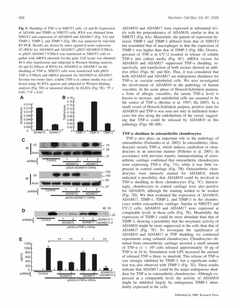
ADAM10 is the major TNF-a sheddase in NIH3T3 cellsThe endogenous sheddase(s) for a substrate protein may
differ from cell to cell. As shown above, the endogenoussheddase(s) could be determined by the expression of theproteinase(s) and its endogenous inhibitor(s). Thus, we nextexamined expression of ADAM10, ADAM17, TIMP-1,TIMP-2, and TIMP-3 in NIH3T3. Compared with macro-phages, NIH3T3 cells expressed ADAM10 more abundantlythan ADAM17 (Fig. 5A). In NIH3T3, the difference in ex-pression between TIMP-1 and TIMP-3 was not so obviousas in macrophages (Fig. 5B). These data suggested thatADAM10 could have a greater contribution to TNF-a shed-ding in NIH3T3 than in macrophages. shRNA vectors formouse ADAM10 and ADAM17 showed specific inhibitionof respective proteinases in this cell line (Fig. 5C). Theseconstructs were transfected with an expression vector forfull-length TNF-a, and soluble TNF-a was collected fromculture media using Ni-NTA beads. Although suppressionof ADAM10 or ADAM17 resulted in reduction of solubleTNF-a, the reduction was more obvious with the suppres-sion of ADAM10 than with that of ADAM17 (Figs. 5D and5E). This result indicated that unlike in macrophages,ADAM10 is the major TNF-a sheddase in NIH3T3 cells.
Involvement of ADAM10 and ADAM17 in TNF-ashedding in vascular endothelial cell line
Vascular endothelial cells were known to release TNF-awith several stimulations such as high-mobility group pro-tein-1, cell walls of Streptococcus pneumoniae, and LPS.Such release of TNF-a by endothelial cells is profoundly in-volved in several pathologies such as sepsis or bacterialmeningitis (Freyer et al. 1999; Fiuza et al. 2003). To iden-tify the endogenous sheddase(s) for TNF-a in vascular endo-thelial cells, we conducted experiments using a mousevascular endothelial cell line, UV,2. In this cell, both
Fig. 4. Suppression of TNF-a shedding by TIMP-1 in mouse spleenmacrophages. (A) Expression of TIMPs in mouse spleen macro-phages. Expression of TIMP-1, TIMP-2, and TIMP-3 in macro-phages was determined by real-time RT-PCR as described forFig. 3A. Results are shown by expression ratios against b-actin. (B)RNAi for TIMP-1. Effect of retroviruses carrying shRNA con-structs for TIMP-1 was confirmed in the manner described forFig. 3D. Expression of TIMP-1 is shown by the ratios against thatof b-actin. (C) Effects of RNAi for TIMP-1 on the shedding ofTNF-a. TIMP-1 expression was suppressed and the change of TNF-a release into the media was evaluated as described for Fig. 3D.
Hikita et al. 587
Published by NRC Research Press
ADAM10 and ADAM17 were expressed at substantial lev-els with the preponderance of ADAM10, similar to that inNIH3T3 (Fig. 6A). Meanwhile, the pattern of expression be-tween TIMP-1 and TIMP-3 differed from that of NIH3T3but resembled that of macrophages in that the expression ofTIMP-1 was higher than that of TIMP-3 (Fig. 6B). Overex-pression of TNF-a in UV,2 resulted in release of solubleTNF-a into culture media (Fig. 6C). shRNA vectors forADAM10 and ADAM17 suppressed TNF-a shedding, re-spectively, and transfection of both vectors showed an addi-tive effect (Figs. 6C and 6D). Thus, it was considered thatboth ADAM10 and ADAM17 are endogenous sheddases forTNF-a in vascular endothelial cells. We next investigatedthe involvement of ADAM10 in the pathology of humanvasculitis. In the acute phase of Henoch-Schonlein purpura,a form of allergic vasculitis, the serum TNF-a level isknown to increase, and endothelial cells are assumed to bethe source of TNF-a (Besbas et al. 1997; Ha 2005). In asmall vessel of Henoch-Schonlein purpura, positive stain forADAM10 and TNF-a was seen not only in infiltrated leuko-cytes but also along the endothelium of the vessel, suggest-ing that TNF-a could be released by ADAM10 in thispathology (Figs. 6E–6H).
TNF-a sheddase in osteoarthritic chondrocytesTNF-a also plays an important role in the pathology of
osteoarthritis (Fernandes et al. 2002). In osteoarthritis, chon-drocytes secrete TNF-a, which induces catabolism in chon-drocytes in an autocrine manner (Pelletier et al. 2001). Inaccordance with previous reports, immunostaining of osteo-arthritic cartilage confirmed that osteoarthritic chondrocyteswere expressing TNF-a (Fig. 7A), while it was little ex-pressed in control cartilage (Fig. 7B). Osteoarthritic chon-drocytes were intensely stained for ADAM10, whichindicated a possibility that ADAM10 could be involved inTNF-a shedding in those chondrocytes (Fig. 7C). Interest-ingly, chondrocytes in control cartilage were also positivefor ADAM10, although the staining tended to be weaker(Fig. 7D). We then evaluated the expression of ADAM10,ADAM17, TIMP-1, TIMP-2, and TIMP-3 in the chondro-cytes within osteoarthritic cartilage. Similar to NIH3T3 andUV,2 cells, ADAM10 and ADAM17 were expressed atcomparable levels in those cells (Fig. 7E). Meanwhile, theexpression of TIMP-1 could be more abundant than that ofTIMP-3, showing a possibility that the enzymatic activity ofADAM10 might be more suppressed in the cells than that ofADAM17 (Fig. 7F). To investigate the significance ofADAM10 and ADAM17 in TNF shedding, we conductedexperiments using cultured chondrocytes. Chondrocytes ob-tained from osteoarthritic cartilage secreted a small amountof TNF-a (1 � 106 cells released approximately 10 pg ofTNF-a in 24 h). Stimulation with LPS increased the amountof released TNF-a three- to ninefold. This release of TNF-awas strongly inhibited by TIMP-3, but a significant reduc-tion was also observed with TIMP-1 (Fig. 7G). These resultsindicate that ADAM17 could be the major endogenous shed-dase for TNF-a in osteoarthritic chondrocytes. Although ex-pressed at a comparable level, the activity of ADAM10might be inhibited largely by endogenous TIMP-1 abun-dantly expressed in the cells.
Fig. 5. Shedding of TNF-a in NIH3T3 cells. (A and B) Expressionof ADAMs and TIMPs in NIH3T3 cells. RNA was obtained fromNIH3T3 and expression of ADAM10 and ADAM17 (Fig. 5A) andTIMP-1, TIMP-2, and TIMP-3 (Fig. 5B) was analyzed by real-timeRT-PCR. Results are shown by ratios against b-actin expression.(C) RNAi for ADAM10 and ADAM17. pSG5-ADAM10-V5HisAor pSG5-ADAM17-V5HisA was transfected to NIH3T3 cells to-gether with shRNA plasmids for the gene. Cell lysate was obtained48 h after transfection and subjected to Western blotting analysis.(D and E) Effects of RNAi for ADAM10 or ADAM17 on theshedding of TNF-a. NIH3T3 cells were transfected with pSG5-TNF-a-V5HisA and shRNA plasmids for ADAM10 or ADAM17.Seventy-two hours later, soluble TNF-a in culture media was col-lected using Ni-NTA agarose and subjected to Western blottinganalysis (Fig. 5D) or measured directly by ELISA (Fig. 5E). *P <0.05; **P < 0.01.
588 Biochem. Cell Biol. Vol. 87, 2009
Published by NRC Research Press

Discussion
In this study, we employed a unique screening system tofind possible sheddases for TNF-a. Although the system isnot the one well established, we have already used a similarsystem to identify RANKL sheddases and found it to be areliable method (Hikita et al. 2006). In this system, the re-sult could be affected by expression levels of the protei-nases. If proteinases are expressed at low levels, theircapacity to cleave the substrate may be underestimated. Toavoid this, in the present study, we transfected proteinaseexpression vectors in excessive amounts. Although therewas some discrepancy between the result of the initialscreening (Fig. 1B) and that of the second experiment withfull-length TNF-a (Fig. 2A), it could not be related to thepoor reliability of the screening but more likely to the differ-ence in substrate proteins.
Currently, ADAM17 is considered to be the major shed-dase for TNF-a (Black et al. 1997; Moss et al. 1997; Con-don et al. 2001; Zheng et al. 2004; Bell et al. 2007;Horiuchi et al. 2007). However, besides ADAM17, severalproteinases are known to cleave membrane-bound TNF-a.Haro et al. (2000) showed that MMP7 null macrophagesfailed to release soluble TNF-a, suggesting that MMP7could be responsible for TNF-a shedding in macrophages.Purified proteinase 3 from activated neutrophils was alsoshown to have a shedding activity for TNF-a (Coeshott etal. 1999). ADAM19 could work as a sheddase for TNF-awhen overexpressed in COS-7 and CHO cells (Chesneau etal. 2003). ADAM10 is also reported to have a shedding ac-tivity for TNF-a. Purified bovine ADAM10 and ADAM10overexpressed in 293A EBNA cells processed TNF-a in vi-tro (Lunn et al. 1997). In contrast, when expressed in COS-7cells or CHO cells, ADAM10 failed to release TNF-a(Zheng et al. 2004). Again, the lack of ADAM10 expressiondid not change TNF-a shedding in mouse embryonic fibro-blasts (Zheng et al. 2004). In this study, we showed that notonly ADAM17 but also ADAM9, ADAM10, and ADAM19are capable of cleaving TNF-a in a cell-based assay. Wealso showed, for the first time, that endogenous ADAM10
is a major TNF-a sheddase in 293A cells, NIH3T3 cells,and UV,2 cells. Our current result differed from those inprevious reports in that endogenous ADAM10 can cleaveTNF-a in certain types of cells. Since proteinases involvedin protein shedding often differ among cell types (Schlon-dorff et al. 2001; Hikita et al. 2006), the discrepancy be-tween our current result and those reported previously couldbe ascribed to the difference in cell types used in the assay.Also, the difference in used constructs or substrate proteinscould account for the discrepancy. In the report of Zheng etal. (2004), the expression vector for a fusion protein of alka-line phosphatase and TNF-a was used, while myc-tagged
Fig. 6. Shedding of TNF-a in UV,2 cells. (A and B) Expression ofADAMs and TIMPs in UV,2 cells. RNA was obtained from UV,2cells and expression of ADAM10 and ADAM17 (Fig. 6A) andTIMP-1, TIMP-2, and TIMP-3 (Fig. 6B) was analyzed by real-timeRT-PCR. Results are shown by ratios against b-actin expression. (Cand D) Effects of RNAi for ADAM10 or ADAM17 on the sheddingof TNF-a. UV,2 cells were transfected with pSG5-mTNFa-V5Hisand shRNA plasmids for ADAM10 or ADAM17. Forty-eight hourslater, soluble TNF-a in culture media was collected using Ni-NTAagarose and subjected to Western blotting analysis (Fig. 6C) ormeasured directly by ELISA (Fig. 6D). **P < 0.01. (E–H) Expres-sion of ADAM10 and TNF-a in vasculitic endothelium. Skinbiopsy specimens obtained from a patient with Henoch-Schonleinpurpura (Figs. 6E and 6F) and a control donor (Figs. 6G and 6H)were immunostained for ADAM10 (Figs. 6E and 6G) and TNF-a(Figs. 6F and 6H), respectively. In Fig. 6F, a higher magnificationimage of the boxed area is shown in the inset in which staining forTNF-a is indicated by arrowheads. Cross-sectional images of ablood vessel are shown. Bar = 50 mm.
Hikita et al. 589
Published by NRC Research Press

TNF-a was used in our assay. We often failed to detect al-kaline phosphatase activity in the culture media of 293 cellswhen fusion proteins of the alkaline phosphatase and sub-strate proteins were overexpressed, possibly due to the inter-action between the enzyme and the substrate proteins.
In this study, we confirmed that the major sheddase for
TNF-a in macrophages is ADAM17. This result is consistentwith those of two recent reports that dealt with ADAM17conditional knockout mice and radiation chimeric mice re-constituted with leukocytes lacking functional ADAM17(Bell et al. 2007; Horiuchi et al. 2007). ADAM10, whichturned out to be the major TNF-a sheddase in the above-
Fig. 7. Shedding of TNF-a in articular chondrocytes. (A–D) Expression of TNF-a and ADAM10 in osteoarthritic and control cartilage.Histological sections of osteoarthritic and control cartilage were immunostained for TNF-a (Figs. 7A and 7B, respectively) and ADAM10(Figs. 7C and 7D, respectively). Bar = 100 mm. (E and F) Expression of ADAMs and TIMPs in chondrocytes. RNA was obtained fromhuman osteoarthritic chondrocytes and expression of ADAM10 and ADAM17 (Fig. 7E) and TIMP-1, TIMP-2 and TIMP-3 (Fig. 7F) wasanalyzed by real-time RT-PCR. Results are shown by ratios against GAPDH expression. (G) Effects of TIMP-1 and TIMP-3 on the secre-tion of TNF-a from primary cultured chondrocytes stimulated with LPS. *P < 0.05; **P < 0.01.
590 Biochem. Cell Biol. Vol. 87, 2009
Published by NRC Research Press

mentioned three types of cells, was known to play only alimited role in the release of TNF-a in macrophages. Thisdiscrepancy could be explained by the difference in expres-sion levels between ADAM10 and ADAM17. Comparedwith macrophages, the expression of ADAM10 was moreabundant than that of ADAM17 in NIH3T3 cells. Likewise,in UV,2 cells in which both ADAM10 and ADAM17 playsignificant roles in TNF-a shedding, the expression ofADAM10 was relatively greater than that of ADAM17.Presence of TIMPs may be another factor that determinesthe significance of the proteinases in the shedding. Whilethe enzymic activity of ADAM10 was inhibited by TIMP-1and TIMP-3, the activity of ADAM17 was suppressed byTIMP-3 but not by TIMP-1. In macrophages, the expressionof TIMP-1 could be much greater than that of TIMP-3.Therefore, it is possible that in those cells, the significanceof ADAM10 in TNF-a shedding could be reduced by theabundance of TIMP-1. In an analogy of macrophages, theexpression of TIMP-1 could be more abundant than that ofTIMP-3 in articular chondrocytes, which might explain thepredominance of ADAM17 over ADAM10 in TNF-a shed-ding in the cells.
Vascular endothelial cells have been shown to secreteTNF-a in vitro and in vivo. In spinal cord injury in rats, vas-cular endothelial cells as well as neurons and glial cells ex-pressed TNF-a around the injury site (Yan et al. 2001).Since the inhibition of TNF-a led to the suppression of in-flammation and tissue injury in spinal cord injury in mice(Genovese et al. 2008), TNF-a could play an important rolein this pathology. Considering the result with UV,2 cells, itseems possible in that condition that TNF-a is expressed bythe endothelial cells and processed by both ADAM10 andADAM17. The involvement of ADAM10 in the actual hu-man vasculitis has been suggested by the result of immunos-taining performed on the skin tissues from a patient withHenoch-Schonlein purpura. In the acute phase of this dis-ease, TNF-a increases in serum and endothelial cells arelikely the source of this cytokine (Besbas et al. 1997; Ha2005). Therefore, the observation that endothelial cells ex-press TNF-a and ADAM10 together convincingly indicatesthat ADAM10 could play a significant role in the release ofTNF-a in the disease. Furthermore, the fact that human en-dothelial cells were intensely stained for ADAM10 (Fig. 6E)would suggest that those cells have a capacity to express theproteinase at significant levels. Considering this, ADAM10could indeed be a major TNF-a sheddase in certain patholo-gies in which endothelial cells express TNF-a.
Although ADAM9 could process TNF-a and is abun-dantly expressed in macrophages, we could not find any evi-dence for its involvement in TNF-a shedding. The reasonfor this was not clarified in this study. Since ADAM9 is notsuppressed by TIMPs (Amour et al. 2002), other endogenousinhibitor(s) for ADAM9 might have been expressed and pre-vented it from shedding.
The result of this study suggested a possibility thatADAM10 can be a major TNF-a sheddase in certain celltypes or certain biological situations. As shown in macro-phages, ADAM17 is the principal sheddase for TNF-a incertain cell types when ADAM10 activity is inhibited byTIMP-1. In fact, even in macrophages, ADAM10 can be amajor sheddase when TIMP-1 expression is reduced for
some reason. Thus, we have shown some evidence that bothADAM10 and ADAM17 can work as a sheddase for TNF-a.Their significance in the shedding may be determined byvarious factors, such as cell types, their expression levels,and the abundance of TIMPs. Since TNF-a is profoundly in-volved in various biological events, the present observationcould be of some help in understanding the kinetics of mo-bilization of the cytokine.
AcknowledgementsThe authors thank Dr. Sunao Takeshita for providing
CMG14-12 and pMX-IRES-bsr and Dr. Motoharu Seiki forthe expression vectors for MT1-MMP, MT2-MMP, MT3-MMP, MT4-MMP, MT5-MMP, and MT6-MMP. This workwas supported in part by a Grant-in-Aid for Young Scien-tists (B) from the Japan Society for the Promotion of Sci-ence to A.H.
ReferencesAlexopoulou, L., Pasparakis, M., and Kollias, G. 1997. A murine
transmembrane tumor necrosis factor (TNF) transgene inducesarthritis by cooperative p55/p75 TNF receptor signaling. Eur. J.Immunol. 27(10): 2588–2592. doi:10.1002/eji.1830271018.PMID:9368614.
Amour, A., Slocombe, P.M., Webster, A., Butler, M., Knight, C.G.,Smith, B.J., et al. 1998. TNF-alpha converting enzyme (TACE)is inhibited by TIMP-3. FEBS Lett. 435(1): 39–44. doi:10.1016/S0014-5793(98)01031-X. PMID:9755855.
Amour, A., Knight, C.G., Webster, A., Slocombe, P.M., Stephens,P.E., Knauper, V., et al. 2000. The in vitro activity of ADAM-10is inhibited by TIMP-1 and TIMP-3. FEBS Lett. 473(3): 275–279. doi:10.1016/S0014-5793(00)01528-3. PMID:10818225.
Amour, A., Knight, C.G., English, W.R., Webster, A., Slocombe,P.M., Knauper, V., et al. 2002. The enzymatic activity ofADAM8 and ADAM9 is not regulated by TIMPs. FEBS Lett.524(1-3): 154–158. doi:10.1016/S0014-5793(02)03047-8. PMID:12135759.
Bell, J.H., Herrera, A.H., Li, Y., and Walcheck, B. 2007. Role ofADAM17 in the ectodomain shedding of TNF-a and its recep-tors by neutrophils and macrophages. J. Leukoc. Biol. 82(1):173–176. doi:10.1189/jlb.0307193. PMID:17510296.
Besbas, N., Saatci, U., Ruacan, S., Ozen, S., Sungur, A., Bakkalo-glu, A., and Elnahas, A.M. 1997. The role of cytokines in He-noch Schonlein purpura. Scand. J. Rheumatol. 26(6): 456–460.PMID:9433407.
Black, R.A., Rauch, C.T., Kozlosky, C.J., Peschon, J.J., Slack, J.L.,Wolfson, M.F., et al. 1997. A metalloproteinase disintegrin thatreleases tumour-necrosis factor-alpha from cells. Nature,385(6618): 729–733. doi:10.1038/385729a0. PMID:9034190.
Brennan, F.M., Chantry, D., Jackson, A., Maini, R., and Feldmann,M. 1989. Inhibitory effect of TNF alpha antibodies on synovialcell interleukin-1 production in rheumatoid arthritis. Lancet,2(8657): 244–247. doi:10.1016/S0140-6736(89)90430-3. PMID:2569055.
Buxbaum, J.D., Liu, K.N., Luo, Y., Slack, J.L., Stocking, K.L.,Peschon, J.J., et al. 1998. Evidence that tumor necrosis factor al-pha converting enzyme is involved in regulated alpha-secretasecleavage of the Alzheimer amyloid protein precursor. J. Biol.Chem. 273(43): 27765–27767. doi:10.1074/jbc.273.43.27765.PMID:9774383.
Carswell, E.A., Old, L.J., Kassel, R.L., Green, S., Fiore, N., andWilliamson, B. 1975. An endotoxin-induced serum factor that
Hikita et al. 591
Published by NRC Research Press

causes necrosis of tumors. Proc. Natl. Acad. Sci. U.S.A. 72(9):3666–3670. doi:10.1073/pnas.72.9.3666. PMID:1103152.
Chesneau, V., Becherer, J.D., Zheng, Y., Erdjument-Bromage, H.,Tempst, P., and Blobel, C.P. 2003. Catalytic properties ofADAM19. J. Biol. Chem. 278(25): 22331–22340. doi:10.1074/jbc.M302781200. PMID:12682046.
Coeshott, C., Ohnemus, C., Pilyavskaya, A., Ross, S., Wieczorek,M., Kroona, H., et al. 1999. Converting enzyme-independent re-lease of tumor necrosis factor alpha and IL-1beta from a stimu-lated human monocytic cell line in the presence of activatedneutrophils or purified proteinase 3. Proc. Natl. Acad. Sci.U.S.A. 96(11): 6261–6266. doi:10.1073/pnas.96.11.6261. PMID:10339575.
Condon, T.P., Flournoy, S., Sawyer, G.J., Baker, B.F., Kishimoto,T.K., and Bennett, C.F. 2001. ADAM17 but not ADAM10 med-iates tumor necrosis factor-alpha and L-selectin shedding fromleukocyte membranes. Antisense Nucleic Acid Drug Dev. 11(2):107–116. doi:10.1089/108729001750171353. PMID:11334139.
Cseh, K., and Beutler, B. 1989. Alternative cleavage of the cachec-tin/tumor necrosis factor propeptide results in a larger, inactiveform of secreted protein. J. Biol. Chem. 264(27): 16256–16260.PMID:2777790.
Fernandes, J.C., Martel-Pelletier, J., and Pelletier, J.P. 2002. Therole of cytokines in osteoarthritis pathophysiology. Biorheology,39(1–2): 237–246. PMID:12082286.
Fiuza, C., Bustin, M., Talwar, S., Tropea, M., Gerstenberger, E.,Shelhamer, J.H., and Suffredini, A.F. 2003. Inflammation-pro-moting activity of HMGB1 on human microvascular endothelialcells. Blood, 101(7): 2652–2660. doi:10.1182/blood-2002-05-1300. PMID:12456506.
Freyer, D., Manz, R., Ziegenhorn, A., Weih, M., Angstwurm, K.,Docke, W.D., et al. 1999. Cerebral endothelial cells releaseTNF-alpha after stimulation with cell walls of Streptococcuspneumoniae and regulate inducible nitric oxide synthase andICAM-1 expression via autocrine loops. J. Immunol. 163(8):4308–4314. PMID:10510370.
Fukui, N., Ikeda, Y., Ohnuki, T., Hikita, A., Tanaka, S., Yamane,S., et al. 2006. Pro-inflammatory cytokine tumor necrosis fac-tor-a induces bone morphogenetic protein-2 in chondrocytes viamRNA stabilization and transcriptional up-regulation. J. Biol.Chem. 281(37): 27229–27241. doi:10.1074/jbc.M603385200.PMID:16835229.
Genovese, T., Mazzon, E., Crisafulli, C., Di Paola, R., Muia, C.,Esposito, E., et al. 2008. TNF-alpha blockage in a mouse modelof SCI: evidence for improved outcome. Shock, 29(1): 32–41.PMID:17621255.
Gomez, D.E., Alonso, D.F., Yoshiji, H., and Thorgeirsson, U.P.1997. Tissue inhibitors of metalloproteinases: structure, regula-tion and biological functions. Eur. J. Cell Biol. 74(2): 111–122.PMID:9352216.
Ha, T.S. 2005. The role of tumor necrosis factor-alpha in Henoch-Schonlein purpura. Pediatr. Nephrol. 20(2): 149–153. doi:10.1007/s00467-004-1726-3. PMID:15627167.
Haro, H., Crawford, H.C., Fingleton, B., Shinomiya, K., Spengler,D.M., and Matrisian, L.M. 2000. Matrix metalloproteinase-7-de-pendent release of tumor necrosis factor-alpha in a model ofherniated disc resorption. J. Clin. Invest. 105(2): 143–150.doi:10.1172/JCI7091. PMID:10642592.
Hikita, A., Kadono, Y., Chikuda, H., Fukuda, A., Wakeyama, H.,Yasuda, H., et al. 2005. Identification of an alternatively splicedvariant of Ca2+-promoted Ras inactivator as a possible regulatorof RANKL shedding. J. Biol. Chem. 280(50): 41700–41706.doi:10.1074/jbc.M507000200. PMID:16234249.
Hikita, A., Yana, I., Wakeyama, H., Nakamura, M., Kadono, Y.,
Oshima, Y., et al. 2006. Negative regulation of osteoclastogen-esis by ectodomain shedding of receptor activator of NF-kappaBligand. J. Biol. Chem. 281(48): 36846–36855. doi:10.1074/jbc.M606656200. PMID:17018528.
Horiuchi, K., Kimura, T., Miyamoto, T., Takaishi, H., Okada, Y.,Toyama, Y., and Blobel, C.P. 2007. Cutting edge: TNF-a-con-verting enzyme (TACE/ADAM17) inactivation in mouse mye-loid cells prevents lethality from endotoxin shock. J. Immunol.179(5): 2686–2689. PMID:17709479.
Kitamura, T. 1998. New experimental approaches in retrovirus-mediated expression screening. Int. J. Hematol. 67(4): 351–359.doi:10.1016/S0925-5710(98)00025-5. PMID:9695408.
Kobayashi, M., Plunkett, J.M., Masunaka, I.K., Yamamoto, R.S.,and Granger, G.A. 1986. The human LT system. XII. Purifica-tion and functional studies of LT and ‘‘TNF-like’’ LT formsfrom a continuous human T cell line. J. Immunol. 137(6):1885–1892. PMID:2427583.
Koike, H., Tomioka, S., Sorimachi, H., Saido, T.C., Maruyama, K.,Okuyama, A., et al. 1999. Membrane-anchored metalloproteaseMDC9 has an alpha-secretase activity responsible for processingthe amyloid precursor protein. Biochem. J. 343(Pt. 2): 371–375.doi:10.1042/0264-6021:3430371. PMID:10510302.
Kriegler, M., Perez, C., DeFay, K., Albert, I., and Lu, S.D. 1988. Anovel form of TNF/cachectin is a cell surface cytotoxic trans-membrane protein: ramifications for the complex physiology ofTNF. Cell, 53(1): 45–53. doi:10.1016/0092-8674(88)90486-2.PMID:3349526.
Kusters, S., Tiegs, G., Alexopoulou, L., Pasparakis, M., Douni, E.,Kunstle, G., et al. 1997. In vivo evidence for a functional role ofboth tumor necrosis factor (TNF) receptors and transmembraneTNF in experimental hepatitis. Eur. J. Immunol. 27(11): 2870–2875. doi:10.1002/eji.1830271119. PMID:9394812.
Lammich, S., Kojro, E., Postina, R., Gilbert, S., Pfeiffer, R., Jasio-nowski, M., et al. 1999. Constitutive and regulated alpha-secre-tase cleavage of Alzheimer’s amyloid precursor protein by adisintegrin metalloprotease. Proc. Natl. Acad. Sci. U.S.A. 96(7):3922–3927. doi:10.1073/pnas.96.7.3922. PMID:10097139.
Li, Y.M., Lai, M.T., Xu, M., Huang, Q., DiMuzio-Mower, J., Sar-dana, M.K., et al. 2000. Presenilin 1 is linked with gamma-se-cretase activity in the detergent solubilized state. Proc. Natl.Acad. Sci. U.S.A. 97(11): 6138–6143. doi:10.1073/pnas.110126897. PMID:10801983.
Lunn, C.A., Fan, X., Dalie, B., Miller, K., Zavodny, P.J., Narula,S.K., and Lundell, D. 1997. Purification of ADAM 10 from bo-vine spleen as a TNFalpha convertase. FEBS Lett. 400(3): 333–335. doi:10.1016/S0014-5793(96)01410-X. PMID:9009225.
Mahmoodi, M., Sahebjam, S., Smookler, D., Khokha, R., and Mort,J.S. 2005. Lack of tissue inhibitor of metalloproteinases-3 resultsin an enhanced inflammatory response in antigen-induced arthri-tis. Am. J. Pathol. 166(6): 1733–1740. PMID:15920158.
McGeehan, G.M., Becherer, J.D., Bast, R.C., Jr., Boyer, C.M.,Champion, B., Connolly, K.M., et al. 1994. Regulation of tu-mour necrosis factor-alpha processing by a metalloproteinase in-hibitor. Nature, 370(6490): 558–561. doi:10.1038/370558a0.PMID:8052311.
Mohler, K.M., Sleath, P.R., Fitzner, J.N., Cerretti, D.P., Alderson,M., Kerwar, S.S., et al. 1994. Protection against a lethal dose ofendotoxin by an inhibitor of tumour necrosis factor processing.Nature, 370(6486): 218–220. doi:10.1038/370218a0. PMID:8028669.
Moss, M.L., Jin, S.L., Milla, M.E., Bickett, D.M., Burkhart, W., Car-ter, H.L., et al. 1997. Cloning of a disintegrin metalloproteinasethat processes precursor tumour-necrosis factor-alpha. Nature,385(6618): 733–736. doi:10.1038/385733a0. PMID:9034191.
592 Biochem. Cell Biol. Vol. 87, 2009
Published by NRC Research Press

Mueller, C., Corazza, N., Trachsel-Løseth, S., Eugster, H.P., Buh-ler-Jungo, M., Brunner, T., and Imboden, M.A. 1999. Nonclea-vable transmembrane mouse tumor necrosis factor-a (TNF-a)mediates effects distinct from those of wild-type TNF-a in vitroand in vivo. J. Biol. Chem. 274(53): 38112–38118. doi:10.1074/jbc.274.53.38112. PMID:10608881.
Nilsen, E.M., Johansen, F.E., Jahnsen, F.L., Lundin, K.E., Scholz,T., Brandtzaeg, P., and Haraldsen, G. 1998. Cytokine profilesof cultured microvascular endothelial cells from the human in-testine. Gut, 42(5): 635–642. PMID:9659156.
Pelletier, J.P., Martel-Pelletier, J., and Abramson, S.B. 2001. Os-teoarthritis, an inflammatory disease: potential implication forthe selection of new therapeutic targets. Arthritis Rheum. 44(6):1237–1247. doi:10.1002/1529-0131(200106)44:6<1237::AID-ART214>3.0.CO;2-F. PMID:11407681.
Reinecker, H.C., Steffen, M., Witthoeft, T., Pflueger, I., Schreiber,S., MacDermott, R.P., and Raedler, A. 1993. Enhanced secretionof tumour necrosis factor-alpha, IL-6, and IL-1 beta by isolatedlamina propria mononuclear cells from patients with ulcerativecolitis and Crohn’s disease. Clin. Exp. Immunol. 94(1): 174–181. PMID:8403503.
Schlondorff, J., Lum, L., and Blobel, C.P. 2001. Biochemical andpharmacological criteria define two shedding activities forTRANCE/OPGL that are distinct from the tumor necrosis factoralpha convertase. J. Biol. Chem. 276(18): 14665–14674. doi:10.1074/jbc.M010741200. PMID:11278735.
Schneider, P., Holler, N., Bodmer, J.L., Hahne, M., Frei, K., Fon-tana, A., and Tschopp, J. 1998. Conversion of membrane-boundFas(CD95) ligand to its soluble form is associated with downre-gulation of its proapoptotic activity and loss of liver toxicity. J.Exp. Med. 187(8): 1205–1213. doi:10.1084/jem.187.8.1205.PMID:9547332.
Schulte, M., Reiss, K., Lettau, M., Maretzky, T., Ludwig, A., Hart-mann, D., et al. 2007. ADAM10 regulates FasL cell surface ex-pression and modulates FasL-induced cytotoxicity andactivation-induced cell death. Cell Death Differ. 14(5): 1040–1049. PMID:17290285.
Selkoe, D.J. 1991. The molecular pathology of Alzheimer’s dis-ease. Neuron, 6(4): 487–498. doi:10.1016/0896-6273(91)90052-2. PMID:1673054.
Steffen, M., Abboud, M., Potter, G.K., Yung, Y.P., and Moore,M.A. 1989. Presence of tumour necrosis factor or a related fac-tor in human basophil/mast cells. Immunology, 66(3): 445–450.PMID:2703257.
Tracey, K.J., Fong, Y., Hesse, D.G., Manogue, K.R., Lee, A.T.,Kuo, G.C., et al. 1987. Anti-cachectin/TNF monoclonal antibo-dies prevent septic shock during lethal bacteraemia. Nature,330(6149): 662–664. doi:10.1038/330662a0. PMID:3317066.
Vassar, R., Bennett, B.D., Babu-Khan, S., Kahn, S., Mendiaz, E.A.,Denis, P., et al. 1999. Beta-secretase cleavage of Alzheimer’samyloid precursor protein by the transmembrane aspartic pro-tease BACE. Science, 286(5440): 735–741. doi:10.1126/science.286.5440.735. PMID:10531052.
Verrier, S., Hogan, A., McKie, N., and Horton, M. 2004. ADAMgene expression and regulation during human osteoclast forma-tion. Bone, 35(1): 34–46. doi:10.1016/j.bone.2003.12.029.PMID:15207739.
Yamamoto, A., Miyazaki, T., Kadono, Y., Takayanagi, H., Miura,T., Nishina, H., et al. 2002. Possible involvement of IkB kinase2 and MKK7 in osteoclastogenesis induced by receptor activatorof nuclear factor kB ligand. J. Bone Miner. Res. 17(4): 612–621.doi:10.1359/jbmr.2002.17.4.612. PMID:11918218.
Yan, P., Li, Q., Kim, G.M., Xu, J., Hsu, C.Y., and Xu, X.M. 2001.Cellular localization of tumor necrosis factor-alpha followingacute spinal cord injury in adult rats. J. Neurotrauma, 18(5):563–568. doi:10.1089/089771501300227369. PMID:11393259.
Zheng, Y., Saftig, P., Hartmann, D., and Blobel, C. 2004. Evalua-tion of the contribution of different ADAMs to tumor necrosisfactor alpha (TNF-alpha) shedding and of the function of theTNF-alpha ectodomain in ensuring selective stimulated sheddingby the TNF-alpha convertase (TACE/ADAM17). J. Biol. Chem.279(41): 42898–42906. doi:10.1074/jbc.M403193200. PMID:15292243.
Hikita et al. 593
Published by NRC Research Press