ipsc-derived microglia as a model of immune …
TRANSCRIPT

iPSC-DERIVED MICROGLIA AS A MODEL OF IMMUNE DYSFUNCTION IN
NEURODEGENERATIVE DISEASES
A thesis submitted for the degree of
Doctor of Philosophy (PhD)
by
Aurelien Bunga
2018

ii
ACKNOWLEDGEMENTS
“Ndenge nini na zongisela Mokonzi Nzambe wa nga malamu manso asali na
ngai, Nazali na nyongo ya bolingo bwa ye, Bolingo bwa ye ezali bolingo bwa
seko.”
I would like to sincerely thank my supervisors Nick and Julie for their invaluable guidance, and
support throughout my time at Cardiff. I am lucky to have worked with them and learned a
huge amount under their mentorship and I am very grateful for the opportunities that I have
had over the last few years. I’d also like to thank the Wellcome Trust for funding my PhD
studentship.
I really appreciate all the help and encouragement along the way from many colleagues and
friends – a conversation here, a piece of advice there. Thank you. Thanks also to Robert
Andrews and Katherine Tansey for their vital help and support with RNA-sequencing analysis.
Thank you to everyone in the lab for putting up with me and my funny ways: Beth, Elisa and
Jincy; Kimmy J and Jasmine; Emma and her funny Welsh accent for teaching me the ropes.
Thanks to Oly, Lee, Kira, Ellen, Rae, Rachael and Jon – this experience would have been a lot
less enjoyable without you!
A special thank you to my sisters Youssillya, Aurore and Jenny, and my parents Julien and
Jeanette, who are my biggest inspiration and without whom this would not have been
possible. I’ll be eternally grateful for their faith in me and their many sacrifices and for
everything that they do for us.
Nzambe azwa lokumu mpe nkembo.

iii
ABSTRACT
It is now increasingly evident that neurodegenerative diseases such as Alzheimer’s disease
(AD) and Huntington’s disease (HD) trigger immune activation within the CNS and activate
innate immune responses, which are primarily driven by microglia.
HD is caused by a CAG repeat expansion in the huntingtin gene (HTT), leading to translation
of an aberrant and pathogenic mutant HTT. Studies in mouse models have shown that HD
microglia acquire a hyper-reactive inflammatory phenotype, mediated by a gain of toxicity of
mutant HTT. Using a HD induced pluripotent stem cell (iPSC) line containing 109 CAG repeats
in the HTT gene, microglia-like cells were generated with the aim of characterising their
phenotype. RNA sequencing was used to explore the microglial-specific transcriptional
changes associated with mutant HTT and pathway analysis carried out to predict any
downstream processes affected in the HD109 microglia. Several immune-related functions
including chemotaxis and phagocytosis were subsequently identified as dysregulated or
impaired. However, upon functional assay validation, no phenotypic abnormalities were
manifest in the HD109 microglia, with comparisons made against WT Kolf2 and isogenic
HD109 corrected iPSC-derived microglia. Similarly, the in vitro microglia did not exhibit the
inflammatory phenotype characteristic of diseased HD microglia and other immune cells.
Loss-of-function variants in the microglia-enriched gene encoding ATP-binding cassette
transporter A7 (ABCA7) increase Alzheimer’s disease risk. Although it has been suggested to
play a role in amyloid beta phagocytosis, the precise role of ABCA7 in AD pathogenesis
remains unknown. To that end, ABCA7 knockout iPSCs were generated using CRISPR/Cas9
and in preliminary results, loss-of-function mutations in ABCA7 were found to impair
phagocytosis of E. coli bioparticles and regulate in vitro microglial inflammatory responses.
This thesis demonstrates that iPSC-derived microglia can serve as a platform for exploring the
inflammatory pathways mediating microglial involvement in AD and HD pathogenesis, thus
enabling in-depth mechanistic studies that bridge the gap between clinical and animal
models.

iv
ABBREVIATIONS
Aß Amyloid ß
ABCA7 ATP-binding cassette transporter A7
ABI3 ABI family member 3
AßO Amyloid ß oligomers
AD Alzheimer’s disease
ADF Advanced DMEM F12
ADGC Alzheimer’s Disease Genetic Consortium
ADORA3 Adenosine A3 receptor
ADP Adenosine diphosphate
AGM Aorta-gonad mesonephros
ANOVA Analysis of variance
APC Antigen presenting cell
ApoE Apolipoprotein E
APP Amyloid precursor protein
ATP Adenosine triphosphate
BBB Blood brain barrier
BIN1 Bridging Integrator-1
BMP4 Bone morphogenetic protein 4
C1QA Complement C1q subcomponent subunit A
CABLES1 Cdk5 and ABL1 enzyme substrate 1
CAMP Cathelin-related antimicrobial peptide
CCL Chemokine (C-C motif) ligand
CD Cluster of differentiation
CD2AP CD1-associated protein
CLU Clusterin
CNS Central nervous system
CORO1A Coronin-1A
COX Cyclooxygenase
CR1 Complement receptor 1

v
CRISPR Clustered Regularly Interspaced Short Palindromic Repeats
CSF Cerebrospinal fluid
CSF1R Colony stimulating factor 1 receptor
CXCL Chemokine (C-X-C motif) ligand
CX3CL1 Chemokine (C-X3-C motif) ligand 1 (or fractalkine ligand)
CX3CR1 CX3C chemokine receptor 1 (or fractalkine receptor)
CYBB Cytochrome b-245 heavy chain (or NADPH oxidase 2)
DAMP Damage-associated molecular pattern
DAVID Database for Annotation, Visualization and Integrated Discovery
DE Differentially expressed
DMEM Dulbecco’s Modified Eagle Media
EAAT Excitatory amino acid transporter
EB Embryoid body
ECM Extracellular matrix
EMP Erythro-myeloid progenitors
EPHA-1 Ephrin type-A receptor 1
ERK Extracellular signal-regulated kinase
FACS Fluorescence-activated cell sorting
FABP4 Fatty acid binding protein 4
FASN Fatty acid synthase
FCCP Carbonyl cyanide-4-(trifluoromethoxy)phenylhydrazone
FCGR Fc gamma receptor
FDR False discovery rate
FITC Fluorescein Isothiocyanate
FLK1 Fetal Liver Kinase 1 (also VEGF Receptor)
GABA Gamma-aminobutyric acid
GAS6 Growth arrest-specific 6
GERAD Genetic and Environmental Risk in Alzheimer’s Disease Consortium
GFP Green fluorescent protein
GM-CSF Granulocyte Macrophage Colony Stimulating Factor
GO Gene Ontology
GPR34 G protein-coupled receptor 34

vi
GSEA Gene set enrichment analysis
GWAS Genome-wide association studies
HD Huntington’s disease
HEXB Beta-hexosaminidase subunit beta
HMG-coA β-Hydroxy β-methylglutaryl-CoA
HSC Haematopoietic stem cell
HTT Huntingtin
IBA1 Ionised Calcium-Binding Adapter Molecule 1
ICAM1 Intracellular Adhesion Molecule 1
IFITM Interferon-induced transmembrane protein
IFN Interferon
IL Interleukin
INPP5D Inositol polyphosphate-5-phosphatase D
iPSC Induced Pluripotent Stem Cell
IRF Interferon regulatory factor
KEGG Kyoto Encyclopaedia of Genes and Genomes
KO Knockout
LD Linkage disequilibrium
LDLR Low-density lipoprotein receptor
LGMN Legumain
LOAD Late-onset Alzheimer’s disease
LPS Lipopolysaccharide
LRP1 Low density lipoprotein receptor-related protein 1
MAPK Mitogen-activated protein kinase
MAO-B Monoamine oxidase B
MARCO Macrophage receptor with collagenous structure
M-CSF Macrophage Colony Stimulating Factor
Macpre Macrophage precursor
MCI Mild Cognitive Impairment
MCP-1 Monocyte chemoattractant protein 1 (or CCL2)
MEF2C Monocyte-specific enhancer factor 2C
MERTK MER proto-oncogene Tyrosine Kinase

vii
MG Microglia
MHC Major Histocompatibility complex
mHTT Mutant HTT
MMP Matrix metalloproteinase
mRNA Messenger RNA
MS4A Membrane-spanning 4-domain family, subfamily A
MSN Medium spiny neuron
NADPH Nicotinamide adenine dinucleotide phosphate
NBD Nucleotide binding domain
NFB Nuclear factor kappa-light-chain-enhancer of activated B cells
NLRC5 NLR family CARD domain containing 5
NLRP3 NOD-Like Receptor family Pyrin domain 3
NSAID Non-steroidal anti-inflammatory drug
OCR Oxygen consumption rate
OLFML3 Olfactomedin-3 like protein 3
PAMP Pathogen-associated molecular pattern
PCA Principal component analysis
PE Phycoerythrin
PET Positron emission tomography
PICALM Phosphatidylinositol binding clathrin assembly protein
PLCG2 Phospholipase C Gamma 2
PPAR Peroxisome proliferator-activated receptor gamma
PROS1 Protein S
PRR Pattern recognition receptor
PC Phosphatidylcholine
PS Phosphatidylserine
PSEN Presenilin
P2RX Purinergic receptor P2X
P2RY Purinergic receptor P2Y
ROS Reactive oxygen species
RUNX1 Runt Related Transcription Factor 1
SERPINB2 Serpin family B member 2

viii
SCF Stem Cell Factor
SIGLEC Sialic acid-binding immunoglobulin-like lectin
SLCO2B1 solute carrier organic anion transporter family member 2B1
SNP Single nucleotide polymorphism
SORL1 Sortilin-related receptor 1
SPI1 Transcription factor PU.1
SREBF2 Sterol regulatory element-binding protein 2
STAT Signal Transducer and Activator of Transcription
SYK Spleen tyrosine kinase
TCA Tricarboxylic acid
TGF-ß Transforming Growth Factor ß
TGFBR1 Transforming Growth Factor ß receptor 1
TLR Toll Like Receptor
TMD Transmembrane domain
TMEM119 Transmembrane protein 119
TNF Tumour necrosis factor
TREM2 Triggering Receptor Expressed on Myeloid Cells 2
tSNE t-distributed stochastic neighbour embedding
TSPO Translocator Protein
TYROBP TYRO protein tyrosine Kinase-binding protein (or DAP12)
VEGF Vascular Endothelial Growth Factor
WT Wild-type
YS Yolk sac

ix
TABLE OF CONTENTS
1. INTRODUCTION .................................................................................................................................... 12
1.1 NEUROINFLAMMATION IN ALZHEIMER’S AND HUNTINGTON’S DISEASE .................................................................... 12 1.1.1 Neuroinflammation in Alzheimer’s and Huntington’s disease ........................................................... 12 1.1.2 Immune system and microglial dysfunction in Huntington’s disease ................................................ 18 1.1.3 Genetics of Alzheimer’s disease implicate microglia dysfunction in Alzheimer’s disease .................. 20
1.2 MICROGLIA .................................................................................................................................................... 26 1.2.1 Origins of microglia ............................................................................................................................ 26 1.2.2 Microglia arise from precursors formed during embryonic haematopoiesis ..................................... 27 1.2.3 Microglia physiology and functional diversity .................................................................................... 30 1.2.4 Microglia phenotypes and activity states ........................................................................................... 35
1.3 ASSOCIATION OF ABCA7 WITH ALZHEIMER’S DISEASE .......................................................................................... 38 1.3.1 Expression profile and structure of ABCA7 ......................................................................................... 40 1.3.2 Function and physiological properties of ABCA7 ................................................................................ 42 1.3.3 ABCA7 function in Alzheimer’s disease ............................................................................................... 44
1.4 PROJECT AIMS ................................................................................................................................................ 47
2. MATERIAL AND METHODS .................................................................................................................... 12
2.1 MATERIALS .................................................................................................................................................... 12 2.1.1 Cell culture reagents ........................................................................................................................... 12 2.1.4 Buffers, consumables and kits ............................................................................................................ 13
2.2 MAINTENANCE AND CULTURE OF HUMAN INDUCED PLURIPOTENT STEM CELLS ........................................................... 14 2.2.1 Maintenance of human iPSC lines ...................................................................................................... 14 2.2.2 Maintenance of THP-1 monocytes ..................................................................................................... 14
2.3 DIRECTED DIFFERENTIATION OF IPSCS INTO MICROGLIA-LIKE CELLS .......................................................................... 15 2.3.1 Differentiation of human iPSCs into embryoid bodies (EBs) ............................................................... 15 2.3.2 Differentiation of EBs into monocyte-like macrophage precursor cells ............................................. 15 2.3.3 Induction of microglia-like phenotype ................................................................................................ 15
2.4 FUNCTIONAL ASSAYS........................................................................................................................................ 16 2.4.1 Stimulation with amyloid beta oligomers and reverse amyloid beta ................................................ 16 2.4.2 Phagocytic uptake of pHrodo red E. coli bioparticles ......................................................................... 17 2.4.3 IncuCyte S3 chemotaxis assay ............................................................................................................ 18 2.4.4 Seahorse XFe96 assays for mitochondrial bioenergetics and glycolytic flux ..................................... 18 2.4.5 Cholesterol staining ............................................................................................................................ 20 2.4.6 Sphingomyelin staining....................................................................................................................... 20 2.4.7 Phospholipid staining.......................................................................................................................... 20
2.5 FLOW CYTOMETRY .......................................................................................................................................... 21 2.6 IMMUNOCYTOCHEMISTRY ................................................................................................................................ 21
2.6.1 Immunocytochemistry ........................................................................................................................ 21 2.6.2 Cytocentrifugation .............................................................................................................................. 22
2.7 RNA EXTRACTION AND QRT-PCR ..................................................................................................................... 23 2.7.1 PowerUp SYBR Green qRT-PCR ........................................................................................................... 23 2.7.2 Fluidigm High Throughput qRT-PCR ................................................................................................... 24 2.7.3 qRT-PCR primers ................................................................................................................................. 27
2.8 CRISPR/CAS9-MEDIATED MUTATION OF ABCA7 GENE ....................................................................................... 28 2.8.1 CRISPR guide RNAs design and preparation ....................................................................................... 28 2.8.2 Cell culture, transfection and selection of targeted clones ................................................................ 29
2.9 RNA SEQUENCING AND ANALYSES .................................................................................................................... 31 2.9.1 RNA Sequencing library construction and sequencing ....................................................................... 31 2.9.2 RNA-seq read mapping, gene counts estimation and differential gene expression analysis............. 31 2.9.3 Comparative Gene Set Enrichment Analysis of iPSC-derived microglia ............................................. 32 2.9.4 iPathwayGuide Impact Analysis ......................................................................................................... 32 2.9.5 Upstream regulator analysis .............................................................................................................. 33
x
2.10 STATISTICAL ANALYSIS ..................................................................................................................................... 33
3. TRANSCRIPTOMIC AND FUNCTIONAL VALIDATION OF HUMAN IPSC-DERIVED MICROGLIA ...................... 34
3.1 INTRODUCTION ............................................................................................................................................... 34 3.2 AIMS ............................................................................................................................................................. 35 3.3 RESULTS......................................................................................................................................................... 36
3.2.1 Human iPSC-derived microglia cells express characteristic microglial markers................................. 36 3.2.2 Transcriptomic profiling of human iPSC-derived microglia reveals a microglial signature................ 42 3.2.3 Functional analysis and validation of Kolf2 iPSC-derived microglia ................................................... 53
3.3 SUMMARY AND DISCUSSION .............................................................................................................................. 58
4. CHARACTERISATION OF THE CELL-AUTONOMOUS EFFECTS OF THE HTT GENE CAG EXPANSION ON IPSC-DERIVED MICROGLIA ................................................................................................................................ 63
4.1 INTRODUCTION ............................................................................................................................................... 63 4.2 AIMS ............................................................................................................................................................. 65 4.3 RESULTS......................................................................................................................................................... 66
4.3.1 Transcriptomic analysis of HD109 iPSC-derived microglia ................................................................. 66 4.3.2 Functional validation of HD109 of iPSC-derived microglia ................................................................. 81
4.4 SUMMARY AND DISCUSSION .............................................................................................................................. 88
5. EXPLORING THE EFFECTS OF CORRECTING THE HD MUTATION ON IMMUNE DYSFUNCTION IN IPSC-DERIVED HD MICROGLIA .......................................................................................................................... 92
5.1 INTRODUCTION ............................................................................................................................................... 92 5.2 AIMS ............................................................................................................................................................. 94 5.3 RESULTS......................................................................................................................................................... 95
5.3.1 Does correcting the HTT CAG expansion reverse the gene expression changes observed in HD109 iPSC-derived microglia? ............................................................................................................................... 95 5.3.2 Effects of correcting the HD mutation on the microglial inflammatory response under stimulatory conditions .................................................................................................................................................... 99 5.3.3 HD109 microglia do not manifest transcriptome-associated functional deficits in phagocytosis and chemotaxis ................................................................................................................................................. 103 5.3.4 HD109 microglia do not exhibit deficits in mitochondrial respiration associated with other HD cell types .......................................................................................................................................................... 108
5.4 SUMMARY AND DISCUSSION ............................................................................................................................ 114
6. INVESTIGATION OF THE EFFECTS OF ABCA7 LOSS-OF-FUNCTION IN HUMAN IPSC-DERIVED MICROGLIA . 119
6.1 INTRODUCTION ............................................................................................................................................. 119 6.2 AIMS ........................................................................................................................................................... 121 6.3 RESULTS....................................................................................................................................................... 122
6.3.1 Generation of ABCA7 KO iPSC lines by CRIPSR-Cas9 ........................................................................ 122 6.3.2 Validation of ABCA7-edited iPSC-derived microglial cells ................................................................ 127 6.3.3 ABCA7 loss of function impairs phagocytosis in iPSC-derived microglia .......................................... 132 6.3.4 Is ABCA7 involved in intracellular lipid trafficking? .......................................................................... 136
6.3 SUMMARY AND DISCUSSION ............................................................................................................................ 137
7. GENERAL DISCUSSION ........................................................................................................................ 141
7.1 GENERAL DISCUSSION AND FUTURE PERSPECTIVES ................................................................................................ 141 7.1.1 Characterisation of the phenotype of HD109 iPSC-derived microglia .............................................. 141 7.1.2 The effects of correcting the HD CAG mutation on the microglial phenotype ................................. 143 7.1.3 Modelling neuroinflammation with iPSC-derived microglia ............................................................ 145
7.2 CONCLUDING REMARKS .................................................................................................................................. 147
8. REFERENCES ....................................................................................................................................... 148
9. APPENDICES ....................................................................................................................................... 173
Appendix 1. Top 50 differentially expressed genes in HD109 iPSC-derived microglia. ............................. 173

xi
Appendix 2. IPA output of upstream transcriptional regulators inhibited in HD109 iPSC-derived microglia. ................................................................................................................................................................... 174 Appendix 3. IPA output of upstream transcriptional regulators activated in HD109 iPSC-derived microglia. ................................................................................................................................................................... 175 Appendix 4. IPA output of inhibited upstream transcriptional regulators specific to HD109 iPSC-derived macrophage precursors. ............................................................................................................................ 175 Appendix 5. Microglia response to cholesterol depletion ......................................................................... 176 Appendix 6. List of qRT-PCR primers used in this project (continued) ....................................................... 176

2. MATERIAL AND METHODS 2.1 Materials
2.1.1 Cell culture reagents
Cell culture reagent Supplier Catalogue number
RPMI 1640 Gibco 1185093
Fetal Bovine Serum Gibco 10439024
Glutamax Thermo Fisher 35050-038
Penicillin/Streptomycin (5000U/5000µg)
Gibco 15070063
BMP-4 PeproTech 120-05ET
SCF Miltenyi Biotec 130-093-991
VEGF PeproTech 100-20
E8 Flex Medium Life Technologies A2858501
X-VIVO15 Scientific Laboratory Supplies LZBE02-060Q
M-CSF BioLegend 574806
IL-3 BioLegend 578006
ß-mercaptoethanol Life Technologies 31350
IL-34 BioLegend 577906
GM-CSF BioLegend 572902
Fibronectin Merck Millipore FC010-10MG
PBS pH 7.4 Gibco 10010015
Vitronectin Gibco A31804
ReLeSR Stem Cell Technologies 05872
Y-27632 dihydrochloride (ROCK inhibitor)
Tocris 1254
40 µm cell strainers Scientific Laboratory Supplies 431750
D-PBS Life Technologies 14190250
Trypan Blue solution Sigma Aldrich T8154
DMEM-F12, no phenol red Thermo Fisher 11039021
Table 2.1 List of reagents used for cell culture and differentiation.
Cell culture media Composition
THP-1 maintenance media RPMI 1640 + 10% FBS + 2 mM Glutamax + Penicillin/Streptomycin 50U/50µg
E8-3G (EB differentiation medium)
E8 + 50 ng/ml BMP4 + 50 ng/ml VEGF + 20 ng/ml SCF

X-VIVO factory medium X-VIVO15 + 2 mM Glutamax + 50U/50µg Penicillin/Streptomycin + 50 µM ß-mercaptoethanol + 50 ng/ml M-CSF + 25 ng/ml IL-3
Microglia differentiation medium
X-VIVO15 + 100 ng/ml IL-34 + 10 ng/ml GM-CSF
Table 2.2 List of media used for cell culture and microglial differentiation
2.1.4 Buffers, consumables and kits
Composition
DNA lysis buffer 1 M Tris pH 8.0 (2.5 ml), 0.5 M EDTA (1 ml) , 10% SDS (155 µl), 5 M NaCl (200 µl), Deionised water (46 ml)
ICC Blocking buffer (external markers)
3% Bovine Serum Albumin (BSA), 2% Normal Serum (Goat/Chicken), PBS pH 7.4 (up to 50 ml)
ICC Blocking buffer (internal markers)
3% BSA, 2% Normal Serum (Goat/Chicken), 0.1% Triton X-100, PBS pH 7.4 (up to 50 ml)
FACS buffer 0.1% BSA, PBS pH 7.4
Table 2.3 List of buffers.
Consumable Supplier Catalogue number
Superscript II Reverse Transcriptase kit Invitrogen 18064014
RNEasy Mini kit Qiagen 74104
PowerUp SYBR Green Master Mix Applied Biosystems A25778
P3 Primary Cell 4D-Nucleofector Kit Lonza V4XP-3032
Bovine Serum Albumin (BSA) Sigma Aldrich A7906
Normal Goat/Chicken Serum Dako X0907/X0903
Triton X-100 Sigma Aldrich T8787
Formalin (4% FPA) Sigma Aldrich HT5011
Poly-D-Lysine Poly-D-Lysine Poly-D-Lysine
Sigma Aldrich Sigma Aldrich Sigma Aldrich
Cytospin funnels Fisher Scientific 11972345
Table 2.4 List of consumables and kits used.

2.2 Maintenance and culture of human induced pluripotent stem cells
2.2.1 Maintenance of human iPSC lines
Studies were performed using two feeder-free human iPSC lines: Kolf2-C1 and HD109. Kolf2-
C1 iPSC lines were generated from fibroblasts collected from a healthy 55-59-year-old male
and reprogramming carried out by non-integrating Sendai virus transduction of Oct4, Sox2,
Klf4 and cMyc. HD109 iPSC lines were derived from fibroblasts collected from a 9-year-old
female with juvenile onset HD from the age of 4, with severe bradykinesia, rigidity and
dystonia at time of biopsy (HD iPSC Consortium 2012). Reprogramming was conducted by
lentiviral transduction of Oct4, Sox2, Klf4 and cMyc. For both cell lines, normal karyotypes
and expression of pluripotent stem cell markers were confirmed. All cell cultures were
maintained in a humidified incubator (37˚C, 5% CO2).
iPSCs were cultured in feeder-free conditions on 6-well plates (Corning) coated in vitronectin
(0.5 µg/cm2; Gibco, Life Technologies) and fed with E8 Flex medium. Medium was changed
daily, and cells were passaged using ReLeSR every 3-5 days (or at 80% confluence). For
passaging, cells were washed once with PBS pH 7.4 and the ReLeSR added before incubation
at 37˚C for 5 minutes. The dissociation reagent was removed by aspiration and fresh E8 Flex
medium added to gently detach the colonies by homogenisation with a pipette. The mixture
was transferred to a 15 ml Falcon tube and centrifuged for 3 minutes at 1000 rpm. The
supernatant was aspirated, and cell pellet re-suspended in warm E8 Flex for plating.
2.2.2 Maintenance of THP-1 monocytes
THP-1 monocytes were a kind gift from Dr Shane Wainwright (Cardiff University). The cells
were cultured with monocyte maintenance medium (see section 2.1.1) in non-adherent T75
flasks. The cells were seeded at a density of 2x10⁵ cells per ml and re-suspended in 19 ml of
media. Once concentration exceeded 1x10⁶ cells per ml, they were collected by centrifugation
(1000 rpm for 3 minutes) and split in a 1:5 ratio into T75 flasks.

2.3 Directed differentiation of iPSCs into microglia-like cells
2.3.1 Differentiation of human iPSCs into embryoid bodies (EBs)
Day 0: iPSC colonies grown to confluence on 6-well plates were washed with 1x PBS pH 7.4 at
room temperature, ReLeSR was added (1 ml per well) and the cells placed in incubator. After
2-3 minutes, the dissociation reagent was aspirated, and the side of the plate was lightly
tapped for 1-2 minutes to allow colonies to dislodge. Following this step, 2ml of warm E8
medium was gently added to each well to allow the aggregated cells to form floating EBs and
the plate returned to the incubator for one day.
Day 1: EBs were carefully transferred to a 15-ml tube and allowed to settle by gravity. The
supernatant medium was removed and replaced with E8 + 50 ng/ml BMP-4, 50 ng/ml VEGF
and 20 ng/ml SCF. EBs were cultured in E8-3G media for 7 days, with a 50% media change
every two days.
2.3.2 Differentiation of EBs into monocyte-like macrophage precursor cells
E8-3G- cultured EBs were collected and further differentiated to make ‘factories’ by seeding
15 EBs into one well of a 6-well plate in X-VIVO15 media supplemented with 50 ng/ml M-CSF
and 25 ng/ml IL-3. The culture media from the factories was changed weekly. After
approximately 2 weeks, non-adherent (monocyte-like) myeloid cells would become visible in
the supernatant media of the factories, were harvested weekly and the culture media
replenished. For harvesting, the supernatant was passed through a 40 µm cell strainer,
centrifuged for 3 minutes at 1000 rpm and the resulting pellet gently re-suspended for use in
assays described below or plated onto glass coverslips or tissue-culture plates.
2.3.3 Induction of microglia-like phenotype
Non-adherent myeloid cells harvested from the factories were plated at a density of 2-3 x 10⁵
cells per well and 10,000 onto PDL-treated (100 µg/ml in borate buffer) tissue culture 6-well
plates and glass coverslips respectively, pre- coated with 5 µg/cm2 fibronectin. The cells were
cultured in XVIVO15 medium supplemented with 10ng/ml GM-CSF and 100 ng/ml IL-34. The
medium was changed after 7 days and cells incubated in standard culture conditions for 14
days, prior to being used for experiments.

2.4 Functional assays
Name Supplier Catalogue number
Aß1-42 peptide California Peptide Research 641-15
Reverse Aß42-1 Tocris 3391
HFIP Sigma Aldrich H-8508
Bafilomycin A1 Sigma Aldrich B1793-10UG
Cytochalasin D Sigma Aldrich C8273-1MG
pHrodo red E. coli bioparticles Life Technologies P35361
Live Cell Imaging Solution Life Technologies A14291DJ
96-well black-walled µ-clear microplates
Greiner Bio-One 655097
ClearView 96-well Migration plates
Essen BioScience 4582
ClearView 96-well reservoir plates
Essen BioScience 4600
Seahorse XF Cell Mito Stress Test
Agilent Technologies 103015-100
Seahorse XF96 V3 PS cell culture microplates
Agilent Technologies 101085-004
Seahorse XF calibrant Agilent Technologies 100840
Seahorse XF 1.0 M Glucose solution
Agilent Technologies 103577-100
Seahorse XF RPMI Medium pH 7.4
Agilent Technologies 103576-100
Seahorse XF96 sensor cartridge
Agilent Technologies 102601-100
Filipin Sigma Aldrich F9765
Ibidi 8-well µ-slides Thistle Scientific 80826
Lysenin Antiserum Peptide International NLY-14802-v
LipidTOX Red Phospholipidosis Detection Reagent
Invitrogen H34351
Complement C1q Merck Millipore 204876-1MG
Lipopolysaccharide Sigma Aldrich L2654-1MG
IL-4 PeproTech 200-04
ADP Sigma Aldrich A2754-500MG
CX3CL1 BioLegend 583404
Table 2.5 List of material, media and consumables used for functional assays.
2.4.1 Stimulation with amyloid beta oligomers and reverse amyloid beta
Synthetic Aß1-42 peptides were resuspended in ice-cold HFIP (or 1,1,1,3,3,3,-
hexafluoropropan-2-ol), by adding 222 µl to 1mg and aliquoted into microcentrifuge tubes

(100 µl = 0.45 mg). Each aliquot was dried overnight under a stream of N2 gas to allow HFIP
evaporation (and stored long term at -80˚C). The resulting peptide films were dissolved to 5
mM Aß stock by adding 20 µl DMSO to 0.45 mg peptide. The peptide stock was further diluted
to 100 µM in phenol red-free F-12 medium and the suspension incubated at 4˚C for 24 hours
to allow oligomerisation. Following incubation, the Aß mixture was centrifuged in the cold for
10 min at 14,000 rpm to pellet out any fibrils. The supernatant was collected as Aß oligomeric
preparation, diluted in phenol red-free medium before use.
2.4.2 Phagocytic uptake of pHrodo red E. coli bioparticles
This assay was used to measure the formation of acidified phagosomes: as the pHrodo red E.
coli bioparticles reach the acidic environment of the phagosome (pH 4.5-5.5), the intensity of
the fluorescence is greatly enhanced, enabling direct quantification of phagocytosis in real-
time. First, pHrodo red E. coli bioparticles were resuspended to 1mg/ml in Live Cell Imaging
Solution. The mixture was homogenised, transferred to glass vial and sonicated for 5 min prior
to each experiment.
Precursor cells collected from differentiation factories were plated onto 96-well PDL-treated,
black walled µ-clear flat bottom microplates at a density of 20,000 cells/well in a 100 µl
suspension and differentiated to microglia for 14 days. Before the assay, the growth medium
was removed and replaced with 75 µl of live cell imaging solution. After 10 µg per well of
pHrodo E. coli was added, the microplates were transferred into the IncuCyte S3 live-cell
imaging system (Essen BioScience), housed inside a 37˚C/5% CO2 incubator. The microplates
were imaged in the IncuCyte for 4-6 hours with a 20x objective. At least four images per well
were taken every 15 or 20 min and analysis carried out using the IncuCyte S3 Base software.
Red channel acquisition was 400 ms and phase contrast masking and cell segmentation was
applied to exclude cells from background, with an area filter of 50 µm2 to exclude objects.
Background red channel noise was subtracted using Top-Hat correction with a radius of 20
µm and a threshold of 2 red corrected units (Kapellos et al. 2016). As a positive control for
inhibited phagocytosis, cells were pre-treated for 1 hour with 10 µM of the actin
polymerisation inhibitor cytochalasin D or bafilomycin A1, an inhibitor of the phagolysosome
V-ATPase.

2.4.3 IncuCyte S3 chemotaxis assay
The chemotaxis assays were conducted with IncuCyte ClearView 96-well Migration plates,
featuring a low pore density membrane that separates an upper and lower chamber and
requires cells to migrate across the membrane surface to reach a pore. Prior to cell plating,
both sides of the IncuCyte ClearView plate membrane were coated with fibronectin: 150 µl
per well was added to an IncuCyte ClearView 96-well reservoir plate and 20 µl was added to
the insert wells, before the insert wells were gently placed onto the reservoir plate. The
migration plate was then incubated at 37˚C for 30 min before being allowed to cool down to
room temperature for another 30 min. The fibronectin was then aspirated from both the
reservoir plate and insert and replaced with 200 µl of D-PBS to the reservoir plate. Microglia
precursor cells were then seeded into each well of the insert plate in 60 µl at 3000 cells per
well and allowed to settle on the membrane at room temperature for 15-30 min. The cells
were differentiated to microglia within the plate for 14 days, with a media change after 7
days.
On the day of the assay, chemoattractants were made to desired concentrations in XVIVO
medium, with 200 µl added to the appropriate wells of a reservoir plate and the insert plate
carefully placed onto the chemoattractant-containing reservoir plate. The migration plate
was transferred to the IncuCyte S3 system for scanning. 10x objective, phase channel scans
of both the top and bottom sides of the insert were scheduled every 30 min for 24 hours.
Migration was quantified by the IncuCyte analysis software as the appearance of in focus cells
on the bottom of the well and the surface area they occupied (total phase object area in µm2
/well).
2.4.4 Seahorse XFe96 assays for mitochondrial bioenergetics and glycolytic flux
The Seahorse XFe96 Analyser measures oxygen consumption rate (OCR) and extracellular
acidification rate (ECAR) over a specified time period and at particular time intervals as a way
of assessing mitochondrial respiration (via cellular oxygen consumption) and glycolytic proton
efflux. Using the manufacturer’s standardised protocol for the Seahorse XF Cell Mito Stress
Test, cell respiration can be measured under basal conditions and subsequent exposures to
the ATP coupler oligomycin, the uncoupler carbonyl cyanide-p-
trifluoromethoxyphenylhydrazone (FCCP), and the complex I inhibitor Rotenone. Oligomycin,

which acts by inhibiting the proton channel of the ATP synthase (complex V), is used in this
protocol to calculate the percentage of oxygen consumption allocated to ATP synthesis.
Treatment with FCCP, which disturbs the transport of hydrogen ions across the mitochondrial
membrane, affects the mitochondrial membrane potential and results in a rapid increase in
OCR. Lastly, rotenone injection leads to a shutdown of mitochondrial respiration. Therefore,
each administered compound allows the assessment of different aspects of mitochondrial
respiration, including basal respiration, maximal respiration, spare respiratory capacity,
proton leak and ATP production.
The optimal seeding density and test compound concentrations were determined during
preliminary experiments to identify the optimal number of cells required to detect a sufficient
shift in OCR and ECAR. The Seahorse XF Cell Mito Stress test, which assays mitochondrial
respiration, generates a bioenergetics profile, whereby each injected drug allows different
properties of mitochondrial respiration to be measured.
20,000 microglia precursor cells per well were seeded onto PDL-coated Seahorse XF96 V3 PS
cell culture microplates and differentiated for 14 days in XVIVO with IL-34 and GM-CSF. Prior
to plating, the plates were coated with fibronectin. Cells were seeded in a 80 µl suspension
of Seahorse XF RPMI media pH 7.4 supplemented with 10 mM glucose, 1 mM sodium
pyruvate and 2 mM L-glutamine and allowed to adhere to the plate for 1 hour at room
temperature to allow for even distribution across the well before being moved to a humidified
5% CO2 incubator at 37˚C. Four wells were kept empty to serve as background control.
The day prior to the experiment, the Seahorse XFe96 Analyser was turned on to allow the
temperature to stabilise. 200 µl of Seahorse XF calibrant was added to the Seahorse XF96
sensor cartridge and kept in a non-CO2 incubator overnight. The day of the experiment, the
hydrated cartridge was loaded with the test compounds oligomycin (2µM, 25 µl), FCCP
(2.5µM, 25 µl) and antimycin A/rotenone (0.5µM, 25 µl) diluted in pre-warmed Seahorse XF
RPMI media at a 10x concentration to dose delivered to cells. The cartridge was then inserted
into the Seahorse XFe96 machine and the Seahorse XF Cell Mito Stress test protocol set up
for calibration according to the manufacturer’s instructions. While the machine was
calibrating, the cell culture microplate was removed from the 37˚C CO2 incubator, the XVIVO
growth medium removed, the cells washed once with Seahorse XF calibrant and incubated
with 175 µl per well of Seahorse XF RPMI assay medium for 45-60 min at 37˚C in a non-CO2

incubator. Following calibration and assay medium incubation, the cell culture microplate was
inserted into the analyser and the Seahorse XF Cell Mito Stress test protocol was run: 3
successive readings (every 5 min) of basal respiration, followed by a drug injection sequence
of oligomycin, FCCP and rotenone/antimycin A with 3 sequential readings every 5 min under
condition.
2.4.5 Cholesterol staining
Cholesterol staining was performed using filipin, a cytochemical cholesterol-specific probe
(Te Vruchte et al. 2004). 14-day differentiated microglia on PDL-treated and fibronectin-
coated 8-well µ-slides were washed once with PBS and fixed in 4% PFA for 15 min followed
by three PBS washes. Cells were incubated with 125 µg/ml filipin (diluted in XVIVO medium)
for 30 min in the dark at room temperature, after which the filipin-containing media was
removed, and cells washed in PBS. Imaging was performed using a Zeiss Axio Imager A1
microscope in conjunction with an Axiocam High-Resolution Camera and Axiovision software
v4.8.
2.4.6 Sphingomyelin staining
The sphingomyelin-specific stain used for these experiments was lysenin (0.1 µg/ml; Peptide
International). Microglia cells plated on Ibidi chamber slides were washed with PBS once,
fixed with 4% PFA and then stained with the sphingomyelin stain lysenin overnight (diluted in
PBS + 1% BSA + 0.1% saponin). Following overnight incubation, cells were washed with PBS
three times, incubated with lysenin anti-serum (1:500 dilution in PBS) at room temperature
for 1 hour before incubation with a fluorescent secondary antibody (1:500 dilution in PBS) at
room temperature for 30 min. Incubation medium was removed, Hoechst nuclear staining
was applied followed by two PBS washes. Cells were imaged with a Zeiss Axio Imager A1
microscope and High-Resolution Camera and Axiovision software v4.8.
2.4.7 Phospholipid staining
Phospholipidosis was analysed using LipidTOX Red Phospholipidosis Detection Reagent (Nioi
et al. 2007). Microglia differentiated on Ibidi chamber slides were washed once with PBS and
incubated with LipidTOX (1:1000 dilution in X-VIVO medium) for 4h to detect

phospholipidosis. Subsequently, the media was removed, and nuclei were stained with
Hoechst before images were obtained with Zeiss Axio Imager A1 microscope and High-
Resolution Camera and Axiovision software v4.8. LipidTOX was excited at 543 nm and its
emission was detected at 594 nm.
2.5 Flow Cytometry
Fluorescence-activated cell sorting (FACS) was carried out for the analysis of cell surface
molecules on the non-adherent cells collected from supernatant of differentiated factories as
described above. Cells were washed in 0.1% BSA in PBS, centrifuged at 1000 rom for 3 minutes
and the resulting pellet was re-suspended in 200 µl of 0.1% BSA in PBS. For single-colour
staining, 5 µl of the conjugated antibody or isotype-matching control (with the same
fluorophore, from the same manufacturer) was added to the suspension and the mixture
incubated at room temperature in the dark for one hour. For two-colour staining, two
antibodies or two isotype controls (attached to different fluorophores) were added together.
Following primary staining, the cells were washed twice with 0.1% BSA in PBS and centrifuged,
before being transferred to 5 ml round bottom tubes (BD Falcon) in 200 µl of 0.1% BSA in PBS.
Fluorescence was measured using a BD LSR Fortessa and data analysed using FlowJo software.
The following antibodies and isotype controls were used.
Antibody Clone Supplier Catalogue number
CD14-APC 61D3 eBioscience 17-0149-41
CD11b-APC ICRF44 eBioscience 17-0118-42
CD45-FITC 2D1 eBioscience 11-9459-42
CD34-PECy7 4H11 eBioscience 25-0349-42
IgG1κ-APC APC isotype control eBioscience 17-4714-81
IgG1κ-FITC FITC isotype control eBioscience 11-4714-81
IgG1κ-PECy7 PE-Cy7 isotype control eBioscience 25-4714-80
Table 2.6 List of conjugated antibodies used for flow cytometry analysis experiments.
2.6 Immunocytochemistry
2.6.1 Immunocytochemistry
Non-adherent myeloid cells were collected from the differentiated factories and plated onto
glass coverslips. Cultured cells were washed in 1xPBS pH7.4, fixed with 4% paraformaldehyde
(PFA) for 15 min at room temperature and washed 3 times with PBS. Fixed cells were

incubated with blocking buffer (3% goat/chicken/donkey serum, 0.1% Triton-X in PBS) for 1
hour at room temperature before overnight incubation at 4˚C with primary antibodies (see
list below) diluted in blocking buffer. Following overnight incubation, cells were subjected to
3 washes of 5 min, followed by a 1-hour incubation at room temperature, protected from
light, with fluorescent secondary antibodies diluted in blocking solution. Coverslips were
subsequently incubated with Hoechst at 1:5000 in blocking buffer and mounted on
microscope slides. Images were taken using an Olympus BX61 fluorescent microscope.
The ICC primary and secondary antibodies used were as follows:
1˚ antibody Host Dilution Supplier
CD34 Mouse anti-human 1:100 Abcam AB6330
CD45 Mouse anti-human 1:100 R&D Systems MAB1430
P2RY12 Rabbit anti-human 1:100 Abcam AB188968
IBA-1 Goat anti-human 1:100 Abcam AB5076
TGFBR1 Rabbit anti-human 1:50 Abcam AB31013
TMEM119 Rabbit anti-human 1:100 Abcam AB185333
CX3CR1 Rabbit anti-human 1:100 Bio-Rad AHP1589
Table 2.7 List of primary antibodies used for immunocytochemistry.
2˚ antibody Dilution Supplier
Alexa Fluor goat anti-mouse IgG 488 1:400 Invitrogen A11001
Alexa Fluor goat anti-mouse IgG 594 1:400 Invitrogen A11032
Alexa Fluor goat anti-rabbit IgG 594 1:400 Invitrogen A11037
Alexa Fluor goat anti-rabbit IgG 488 1:400 Invitrogen A11034
Alexa Fluor chicken anti-goat IgG 594 1:400 Invitrogen A21468
Alexa Fluor chicken anti-rabbit IgG 488 1:400 Invitrogen A21441
Alexa Fluor chicken anti-mouse IgG 594 1:400 Invitrogen A21201
Table 2.8 List of secondary antibodies used for immunocytochemistry.
2.6.2 Cytocentrifugation
Cells were washed and fixed as described. To make cytospins, 1x10⁵ cells were spun at 800
rpm for 1 min through pre-wet filters onto glass slides using a Centurion CYT04 centrifuge
before staining as described in section 2.6.1.

2.7 RNA extraction and qRT-PCR
For RNA extraction, samples were harvested and lysed in RLT buffer (RNEasy Mini kit, Qiagen)
containing 10 µl/ml ß-mercaptoethanol. Total RNA was extracted according to the
manufacturer’s protocols. The concentration and purity of the extracted RNA samples were
measured by spectrophotometry (Nanodrop).
2.7.1 PowerUp SYBR Green qRT-PCR
Reverse transcription was carried out using Superscript II kit, with 1 µg template RNA in a 20
µl reaction volume. The resulting cDNA samples were diluted 1:5 by adding 80 µl of deionised
water and readied for qRT-PCR. Each qRT-PCR reaction, comprising 1 µl of the diluted cDNA
product in a 20 µl volume, was placed in a thermocycler set up with the cycling parameters:
25˚C for 10 min, 42˚C for 50 min and 70˚C for 15 min.
RT-qPCR was carried out on a CFX Connect Real Time System (Bio-Rad) using PowerUp SYBR
Green Master Mix (Applied Biosystems). Quantification of target gene transcripts was carried
out using primers designed and validated with established efficiencies between 90 and 105%.
Primers were specifically designed following these parameters: Tm close to 60˚C and qPCR
product between 80 and 120 bp.
The qPCR reaction mix was added to a 96-well plate, which was centrifuged for 1 minute at
1500 rpm and put through the cycling program described below.
qPCR component Volume
10 µM Primer mix 1 µl
PowerUp SYBR Green Master Mix 10 µl
RNAse-free deionised water 8 µl
cDNA 1 µl
Step Temperature Time Cycle
UDG activation 50˚C 2 min Hold
Polymerase activation 95˚C 2 min Hold
Denaturation 95˚C 15 sec
40 Annealing/extension 60˚C 1 min
Stage Step Ramp rate Temp Time
Step 1 1.6˚C/sec 95˚C 15 sec

For analysis, the delta-delta CT method was used to determine relative gene expression
values, with 3 biological replicates. Each biological replicate was analysed in triplicate.
2.7.2 Fluidigm High Throughput qRT-PCR
Reagent Catalogue number Supplier
Random hexamers C1181 Promega
Fluidigm Reverse
Transcription Master Mix kit 100-6300 Fluidigm
Preamp Master Mix kit 100-5580 Fluidigm
Exonuclease I M0293S New England BioLabs
TE buffer 12090015 Invitrogen
Fluidigm 96.96 Dynamic Array
IFC chip BMK-M-96.96 Fluidigm
96.96 Dynamic Array DNA
Binding Dye and Assay
loading reagent kits
100-3400 Fluidigm
SsoFast EvaGreen Supermix
with low ROX 172-5211 BioRad
20X DNA Binding Dye Sample
Loading Reagent 100-3400 Fluidigm
Control line fluid 89000021 Fluidigm
Table 2.8 List of consumables used for Fluidigm qRT-PCR.
Samples were harvested and RNA extraction carried out as described above. cDNA synthesis
was performed using 100 ng of total RNA and random hexamers with the Fluidigm Reverse
Transcription Master Mix kit. According to the manufacturer’s instructions, samples had to
undergo a round of preamplification prior to use with the high throughput array. 1.25 µl of
each cDNA sample was pre-amplified using the Preamp Master Mix along with 0.5 µl of the
pooled gene primers (500 nM per primer). The preamplification reactions (see table 2.8) was
carried out using a 2 min 95˚C denaturation step and 10 cycles of 15s at 95˚C and 4 min at
Melt curve stage Step 2 1.6˚C/sec 60˚C 1 min
Step 3 (dissociation) 0.15˚C/sec 95˚C 15 sec

60˚C. The pre-amplified samples then underwent a clean-up step with Exonuclease I, where
2 µl of diluted Exo I at 4U/µl was added to each 5-µl preamplified reaction. The mixtures were
put on a cycling program of digestion at 37˚C for 30 min and inactivation at 80˚C for 15 min.
Following this treatment, the pre-amplified reactions were diluted 5 times using TE buffer.
Component Vol per reaction (µl) Vol for 96 reactions + 20% overage* (µl)
PREAMPLIFICATION PRE-MIX
Preamp Master Mix 1.00 115.2
Pooled Gene primers 0.50 57.6
DNase-free H2O 2.25 259.2
cDNA 1.25 -
Total 5.00 -
Table 2.8 Preamplification reaction using Fluidigm Preamp Master Mix. *20% overage for ease of pipetting.
For the high throughput qPCR, a Fluidigm 96.96 Dynamic Array IFC chip, which is formatted
to run reactions for 96 samples and 96 genes, was used (Spurgeon et al. 2008). The primer
reaction or assay mixes (see table 2.9) were prepared on a 96-well PCR plate using the 96.96
Dynamic Array DNA Binding Dye and Assay loading reagent kits. The forward and reverse
primers were first combined in microcentrifuge tubes before being added to loading reagent
and TE DNA suspension buffer in individual wells of a 96-well plate. The plate was later
vortexed for 30 sec and centrifuged at 1000 g for 1 min before pipetting into the IFC inlets.
Component Vol per inlet (µl) Vol for 50 µl stock
2x Assay Loading Reagent 2.5 25
1X TE buffer 2.25 22.5
100 µM Forward + Reverse primers 0.25 2.5
Total 5 50
Table 2.9 Fluidigm 96.96 IFC primer reaction mix preparation. Final concentration of each primer is 5 µM in the inlet and 500 nM in the final reaction. For ease of pipetting, mixtures were made up as 50 µl stocks.
The sample reaction mixtures containing preamplified and Exo I-treated cDNA were prepared
as shown in table 10 using SsoFast EvaGreen Supermix with low ROX and 20X DNA Binding
Dye Sample Loading Reagent. The sample pre-mix was made up in a microcentrifuge tube
being aliquoted as 3.3 µl per well of a 96-well plate, to which each preamplified cDNA sample
was added. The cDNA sample mixture plate was vortexed and centrifuged as above before
loading onto the IFC inlets.

Component Vol per inlet + 20% overage
(µl)
Vol for 96.96 IFC for 60 samples
(µl)
SAMPLE PRE-MIX
2X Sso fast EvaGreen 3.0 360
20X DNA Binding Dye 0.3 36
Preamplified & Exo I-treated cDNA 2.7 -
Total 6.00 -
Table 2.10 Fluidigm 96.96 IFC sample reaction mixture preparation.
Having prepared the sample and assay mixes, the chip was first primed after control line fluid
was injected into each accumulator on the IFC. Following the priming script, 5 µl of primer
assay mix and 5 µl of sample were loaded into their respective inlets of the 96.96 IFC chip and
the chip placed in the HX controller for automatic loading and mixing. After loading, the chip
was moved to the BioMark HD Real-Time PCR System (Fluidigm). The cycling program was GE
Fast 96x96 PCR+Melt v2.
Segment Type Temp (˚C) Duration (sec) BioMark HD Ramp rate (˚C/s)
1 Thermal mix 70 2400 5.5
60 30 5.5
2 Hot Start 95 60 5.5
3 PCR (30 cycles) 96 5 5.5
60 20 5.5
4 Melting curve 60
3 1
60-95 1˚C/3s
Table 2.11 Cycling parameters for BioMark HD Real-Time PCR platform for use with 96.96 IFC chip.
Post chip run qPCR data analysis was initially done using the BioMark Gene Expression Data
Analysis software, which analyses individual data points and generates Ct values using “pass”
and “fail” scores. Fail data points, which feature melting curves with aberrant Tm or from
non-specific amplification, were removed after this initial analysis. Raw Ct values were then
downloaded from the software as a data frame, and subsequently processed using Singular
Analysis Toolset (Fluidigm), which runs the R script ‘fluidigmSC”. This script performs a series
of differential gene expression analysis such as ANOVA, PCA, tSNE and hierarchical clustering
to identify differentially expressed genes of interest. Gene expression values are generated
as log2 expression counts, converted from Ct values above the recommended background
default of 24 (Livak et al. 2013).

2.7.3 qRT-PCR primers
Gene Forward Reverse
Nestin GTTCCAGCTGGCTGTGGAGGC GCTGCTGCCGACCTTCCAGG
Brachyury TCAGCAAAGTCAAGCTCACCA CCCCAACTCTCACTATGTGGATT
Flk1 GGCCCAATAATCAGAGTGGCA CCAGTGTCATTTCCGATCACTTT
RUNX1 CTGCCCATCGCTTTCAAGGT GCCGAGTAGTTTTCATCATTGCC
CD34 TGGACCGCGGCTTTGCT CCCTGGGTAGGTAACTCTGGG
CD45 CATTTGGCTTTGCCTTTCTG TTCTCTTTCAAAGGTGCTTGC
IBA1 GCTGAGCTATGAGCCAAACC TCATCCAGCCTCTCTTCCTG
Kir2.1 GGTTTGCTTTGGCTCAGTCG GAACATGTCCTGTTGCTGGC
NaV1.6 GGCAATGTTTCAGCTCTACGC ATTGTCTTCAGGCCTGGGATT
TREM2 CTGGAGATCTCTGGTTCCCC AGAAGGATGGAAGTGGGTGG
CSF1R TATGTCAAAGACCCTGCCCG GTGAGCAGACAGGGCAGTAG
CD11b GAAAGGCAAGGAAGCCGGAG TGGATCTGTCCTTCTCTTAGCCG
TMEM119 CTTCCTGGATGGGATAGTGGAC GCACAGACGATGAACATCAGC
ABCA7 GCTGAGGAACTTGACCAAGGTAT CCTGCTCCATTCACACCCAG
PLCG2 AACTGCAGACTTCGAGGAGC AGGGGCTTTCACGACGTTAT
INPP5D CTGCGTGCTGTATCGGAATTG AAGAACCTCATGGAGACGCC
SREBF2 AGGCAGGCTTTGAAGACGAA AGCTTCCCTGTGATGTGCAG
IL-1ß TTCGAGGCACAAGGCACAA TGGCTGCTTCAGACACTTGAG
TNF GCCCATGTTGTAGCAAACCC TATCTCTCAGCTCCACGACA
IL-6 ACCCCCAATAAATATAGGACTGGA TTCTCTTTCGTTCCCGGTGG
TGFß1 GGCTGTATTTAAGGACACCCGT GACACAGAGATCCGCAGTCC
IL-10 AAGACCCAGACATCAAGGCG AATCGATGACAGCGCCGTAG
P2RY12 GGAGCTGCAGAACAGAACACT AGTTGCCAAACCTCTTTGTGATA
GAS6 ACC TGACCGTGGGAGGTATT GTGTCTTCTCCGTTCAGCCA
ABCA1 ACTGGTTTGGCGAGGAAAGT CAGCTTCAAGTGGGTGGGTT
LRP1 CTCGGATGAGCCCAAGGAAG TCACCGCAATCGTTGTCGTA
FASN GTGCCCTGAGCTGGACTACT AAGCCGTAGTTGCTCTGTCC
HMGCS1 GACTGTCCTTTCGTGGCTCA GAAAGAGCTGTGTGAAGGATAGA
HMGCR TCGGTGGCCTCTAGTGAGAT ACAAAGAGGCCATGCATTCG
PPARG ACAAGGCCATTTTCTCAAACGAG CAAAGTTGGTGGGCCAGAATG
LDLR ACCACAGAGGATGAGGTCCA GTCATCCTCCAGACTGACCATC
CX3CR1 TGGGGCCTTCACCATGGAT GCCAATGGCAAAGATGACGGAG
Table 2.12 List of primers used for Fluidigm qRT-PCR. For rest of the list, see appendix 6.

2.8 CRISPR/Cas9-mediated mutation of ABCA7 gene
2.8.1 CRISPR guide RNAs design and preparation
Single guide RNAs (gRNA) targeting the ABCA7 gene were designed using the Wellcome
Sanger Institute and Deskgen CRISPR design tools, with focus on reducing off target effects
such as modification of unintended genomic sites. The Alt-R CRISPR-Cas9 system (Integrated
DNA Technologies or IDT) was used because of its high editing efficiency compared to vector
constructs (Kim et al. 2014; Zuris et al. 2015). This method forms a ribonucleoprotein (RNP)
complex by combining a S.p. Cas9 Nuclease (1081058 – IDT) with a target crRNA and a
fluorescently labelled ATTO 550 tracrRNA (1075928 – IDT). Delivery of the activated RNP
complex causes double-stranded DNA cleavage of the target DNA by Cas9, activating the non-
homologous end joining system (NHEJ). The gRNAs chosen for the study were mapped onto
coding exons 2 (Ex2P1) and 8 (Ex8P1) of the ABCA7 gene and were predicted to have minimal
off-target effects in the genome.
gRNA name Target region Sequence
Ex2P1 Exon 2 GGAAGAATTTCATGTATCGCCGG
Ex8P1 Exon 8 CACTGCTGCAGAGACCCCGAGGG
To generate the RNP complex, each crRNA oligo and tracrRNA was resuspended to 200 µM in
nuclease-free duplex buffer (IDT). Then, a cRNA:tracrRNA duplex was formed by combining
each oligo with a tracrRNA in equimolar concentration: 1.7 µl crRNA + 1.7 µl tracrRNA + 1.7
µl of nuclease-free duplex buffer. The reaction mixture was heated at 95˚C for 5 min and
allowed to cool at room temperature. To assemble the RNP complex for each oligo, the
reaction mixture below was set up and incubated for 20 min at room temperature prior to
transfection. The Cas9 nuclease was made up by diluting 3.1 µl of 10 µg/µl Cas9 stock with
1.9 µl Cas9 storage buffer (10 mM tris-HCl pH 7.4, 300 mM NaCl, 0.1 mM EDTA, 1mM DTT).
Component Volume (µl)
cRNA:tracrRNA complex (1µM) 2.5
Cas9 nuclease (6.2 µg/µl) 2.5
Total volume 5

2.8.2 Cell culture, transfection and selection of targeted clones
Kolf2 iPS cells were grown to 70-80% confluence and maintained in E8 medium, as described
before. On the day of transfection, media was removed and replaced with E8 media
containing 10 µM ROCK inhibitor for 1-2 hours before transfection. Cells were washed once
with PBS pH 7.4 and dissociated with pre-warmed accutase for approximately 5 min (Life
Technologies). E8 media containing ROCK inhibitor was added to inactivate the accutase, the
mixture homogenised, centrifuged at 1500 rpm for 3 min and counted with 1 million cells
being used. Cells were then resuspended in 100 µl of nucleofection buffer, made up of 78 µl
P3 buffer and 22 µl supplement from the P3 Primary Cell 4D-Nucleofector Kit. 5 µl of each
assembled RNP was added to the suspension, which was then transferred to an Amaxa
cuvette. The cuvette was gently tapped onto a flat surface to eliminate bubbles. Cells were
transfected with RNP complexes by electroporation using the Amaxa 4D Nucleofector system
(program CA137). Following nucleofection, the cells were allowed to recover for ~ 10 min at
37˚C, before addition of pre-warmed E8 + ROCK inhibitor media and plating onto a culture
dish pre-coated with vitronectin. 24 hours post nucleofection, the transfected cells were FACS
sorted to select for the top 10% of the most fluorescent cells, for increased editing efficiency.
Briefly, the transfected cells were washed in PBS and dissociated as single cells with accutase,
as above. The cells were then centrifuged and resuspended in 500 µl in E8 media + ROCK
inhibitor into FACS tubes for sorting in a FACS ARIA Fusion (BD Biosciences) using the tracrRNA
ATTO550. The sorted cells were collected in a FACS tube containing 1 ml of E8 + ROCK
inhibitor, replated onto a 10 cm tissue culture dish pre-coated with vitronectin in order to
obtain single cell clones. The following day, the culture media was changed to E8 medium and
thereafter the cells were fed every other day, being monitored daily for the formation of
colonies. Around 7 days after transfection, single colonies were cut in half and picked onto
two separate 96-well plates by using a P20 pipette set at 10 µl – cells grown one plate were
used for genomic DNA extraction and PCR screening to detect successful excision of the target
region (see table 2.13 below). The corresponding 96-well plate was used for clonal expansion.
PCR primer Target region Sequence
Ex2F Exon 2 ATTGGTTTACTCCACCCCTGGG
Ex2R Exon 2 GAGTCGTTGAAGTTGCTCAGGC
Ex8F Exon 8 ctttgcctctcagagcctcagt
Ex8R Exon 8 GGAAACCGAGGTCAGTCACTCA

PCR component Volume (µl)
5x colourless GoTaq Flexi buffer 10
25 mM MgCl2 solution 5
10 µM dNTPs 1
Ex2F2 Primer 1
Ex8R6 Primer 1
Extracted DNA with DNA lysis buffer 1
GoTaq DNA polymerase 0.25
Nuclease-free water to 50
Table 2.13 ABCA7 KO PCR screening reaction.
For genomic DNA extraction, cells in 96-well plates were washed once with PBS before
addition of DNA lysis buffer (see section 2.1.4) supplemented with 2% proteinase K. 50 µl of
DNA lysis buffer was added per well and the mixture incubated at 55˚C for 4 hours or
overnight. This was followed by inactivation at 95˚C for 30 minutes, where each extracted
DNA sample was diluted 1 in 10 in 10 mM Tris pH 8.0. The diluted and inactivated DNA sample
was then used for PCR assay outlined below. The PCR cycling parameters were set up as
follows:
Step Temperature Time Cycle
Initial denaturation 95˚C 2 min 1
Denaturation 95˚C 15 secs
30 Annealing 60˚C 45 secs
Extension 72˚C 1 min
Final extension 72˚C 5 min 1
96-well plate clones found to carry the successful mutation after PCR screening were
subjected to sub-cloning. To do this, the cells were washed with PBS and incubated with
accutase for 5 min to ensure a single cell suspension. Pre-warmed E8 media was added to the
cell suspension to inactive the accutase and the mixture centrifuged at 1000rpm for 3 min.
The cells were then re-suspended in E8 + ROCK inhibitor media before plating at very low
density in a vitronectin-coated 10 cm culture dish. This allowed the cells to expand from single
cells, before individual colonies from the dish were picked, grown and expanded in 24-well
plates before another round of PCR screening. All expanded sub-clones were also kept as

frozen pellets. The PCR-amplified DNA were separated by agarose gel electrophoresis using
2% gels.
2.9 RNA Sequencing and Analyses
2.9.1 RNA Sequencing library construction and sequencing
Samples used for RNA sequencing (RNA-seq) were harvested using RLT buffer from RNeasy
Mini Kit before total RNA was extracted from lysates following the protocols of the
manufacturer. RNA-seq was performed at the Genomics Research Hub of Cardiff University
School of Biosciences by Angela Marchbank. RNA integrity was measured for all samples with
the Agilent Bioanalyser 2100 (Agilent Technologies) and samples with RNA integrity number
above 8 were retained for library construction. The Illumina TruSeq mRNA stranded protocol
was used to perform cDNA synthesis from all samples and generate poly-A mRNA libraries. 1
µg of RNA was used to construct RNA-seq libraries. Libraries were then assessed for their
quality with the Agilent 4200 TapeStation (Agilent Technologies) and sequenced as single-end
75 bp reads on the Illumina NextSeq500 sequencer, generating 40 million reads per sample.
2.9.2 RNA-seq read mapping, gene counts estimation and differential gene expression
analysis
RNA-seq reads (fastq files) were first trimmed using Trimmomatic (version 0.35) (Bolger et al.
2014) and then mapped to the GRCh38.89 reference human genome using STAR (version
2.5.1b) (Dobin et al. 2013). Transcripts were assembled before raw read counts per gene were
generated using the software FeatureCounts (Liao et al. 2014). Transcripts with at least 1
count per million in at least one sample were considered to represent expressed genes and
retained for further downstream analysis, otherwise removed. Differential gene expression
analysis was carried out with the Bioconductor package DESeq2 (release 3.6) to generate gene
expression values as FPKM (Fragments per Kilobase of transcript per Million mapped reads)
(Love et al. 2014). The Benjamini-Hochberg (BH) multiple testing correction procedure, which
decreases false discovery rate, was employed to compute adjusted p-values. Principal
component analysis of the normalised expression counts was performed using the R function
“prcomp” and plotted with “ggplot”. Hierarchical clustering heatmap based on normalised
gene expression counts was conducted using the R function “pheatmap”.

2.9.3 Comparative Gene Set Enrichment Analysis of iPSC-derived microglia
Single-ended RNA-seq data for human cortical neurons, astrocytes and myeloid cells (Zhang
et al., 2016) was retrieved from Gene Expression Omnibus (accession GSE73721). The
downloaded raw fastq files were processed along the same pipeline as described above to
produce raw gene counts. These were combined with values from our data to estimate
normalised gene expression counts with corresponding adjusted p-values. Genes with p-
adjusted < 0.05 were chosen to carry out a comprehensive Gene Set Enrichment Analysis
(GSEA) of the stem cell-derived microglia datasets. GSEA analyses whether enrichment of
differential expression among gene sets relates to biological hypotheses and pathways
(Subramanian et al. 2005). GSEA orders all genes according to their differential expression
statistic, and tests whether genes in a specific gene set have a higher overall rank than would
be expected by chance. A pathway set was chosen consisting of Gene Ontology (GO) (Harris
et al. 2004), Kyoto Encyclopaedia of Genes and Genomes (KEGG) (Kanehisa et al. 2012),
Reactome (Croft et al. 2014), BioCarta and NCI pathway interaction database (Schaefer et al.
2009). Upregulated and downregulated genes in iPSC-derived microglia were assessed
separately.
2.9.4 iPathwayGuide Impact Analysis
Pathway Impact Analysis was run using AdvaitaBio iPathwayGuide
(https://www.advaitabio.com/ipathwayguide.html) to identify significantly impacted
pathways. This method utilises a systems biology approach and incorporates the classical
probabilistic aspects of analysis (e.g. GSEA) with important biological factors: “the magnitude
of the expression changes of each gene, the position of the differentially expressed genes on
the given pathways, the topology of the pathway that describes how these genes interact,
and the type of signalling interactions between them” (Draghici et al. 2007; Donato et al.
2013).
This method analyses DE gene datasets in the context of pathways from the KEGG database
(Kanehisa et al., 2012), gene ontologies from Gene Ontology consortium database (Ashburner
et al. 2000) and miRNAs from the miRBase (Release 21). The pathways are then scored
according to two criteria: (i) the over-representation of DE genes in a given pathway and (ii)
the perturbation of that pathway computed by propagating the measured expression

changes across the pathway topology. These criteria are taken account to calculate two
probability values: pORA and pAcc. pORA expresses the probability of observing the number
of DE genes in a given pathway that is greater than or equal to the probability by random
chance. The second probability pAcc is calculated based on the amount of total accumulation
measured in each pathway and the sum of all of all absolute accumulations of the genes in a
given pathway. The probability values are then combined into one final pathway score by
calculating a p value using Fisher’s method, corrected for multiple comparisons by False
discovery rate (FDR).
2.9.5 Upstream regulator analysis
Upstream regulator analysis was performed using the Ingenuity Pathway Analysis (Ingenuity
Systems, Qiagen). This program predicts potential upstream regulators and mechanistic
networks that explain the gene expression changes observed in a dataset, by calculating P-
values of overlap and activation z-score statistics. The P-value of overlap establishes whether
there is a statistically significant overlap between gene expression changes in a dataset and
the genes affected by an upstream regulator (regulators with p value of < 0.01 qualified). The
activation z-sore considers the direction of gene expression changes and is used to predict
whether regulators are activated or inhibited in the dataset. Generally, a z-score of > 2 or < -
2 is regarded as significant. A cut-off of 0.01 was used to decide which genes were included
in this analysis.
2.10 Statistical analysis
Statistical analysis of Fluidigm qRT-PCR data was carried out using GraphPad Prism 7.0
software. Data were represented as average log2 expression values generated by Fluidigm
analysis R package (see section 2.7.1). When comparing the log2 expression from multiple
conditions against a control group, one-way ANOVA with Dunnett’s post hoc comparison was
used. For assessing data from experiments with independent variables and one continuous
dependent variable, a two-way ANOVA with Tukey’s post hoc multiple comparisons test was
carried out. Asterisks representing significance are defined as follows: ns – not significant; P
> 0.05; ** P < 0.01; *** P < 0.001; **** P < 0.0001.

34
3. TRANSCRIPTOMIC AND
FUNCTIONAL VALIDATION OF HUMAN
IPSC-DERIVED MICROGLIA
3.1 Introduction
Animal models have been invaluable in advancing our understanding of
neurodegenerative diseases such as Alzheimer’s and Huntington’s disease (Götz &
Ittner 2008; Laferla & Green 2012). Much has been gained from animal studies,
including the now recognised importance of research focusing on the role of
neuroinflammation, and specifically microglia in those disorders (see section 1.1).
Nonetheless, the emphasis needs to shift towards elucidating the phenotypic
characteristics of diseased microglia, as well as how they can be harnessed to treat
disease. Indeed, it can be argued that the failure of several clinical trials and paucity
of new therapies (Cummings et al. 2014) highlight a necessity for improved disease
models that bridge the gap between the clinic and animal models.
In that respect, advances in stem cell technology since the advent of induced
pluripotent stem cells (iPSCs) (Takahashi & Yamanaka 2006; Okita et al. 2007;
Takahashi et al. 2007) have revolutionised in vitro disease modelling, leading to the
development of several reliable protocols for differentiation of iPSCs into neurons and
astrocytes (Krencik et al. 2011; Serio et al. 2013; Shaltouki et al. 2013). Prior to the
beginning of this study, there were no published protocols for in vitro differentiation
of microglia. As established through lineage tracing studies, microglia arise from
primitive yolk-sac derived haematopoietic precursors during embryonic development
that colonise the developing brain (Ginhoux et al. 2010). Therefore, any strategy for
microglial differentiation would require mimicking the steps leading to yolk sac
haematopoiesis (Ginhoux et al. 2013). Based on this rationale, a number of microglia
differentiation protocols have since been reported (Muffat et al. 2016; Abud et al.
2017; Haenseler et al. 2017; Pandya et al. 2017). The protocol used in this study (see

35
section 2.3) was adapted from work by (Karlsson et al. 2008; van Wilgenburg et al.
2013), who showed that monocyte-like primitive macrophage precursors can be
derived from human stem cells. The differentiation method involves culturing
embryoid bodies (EBs) in serum-free and feeder-free medium containing the
cytokines IL-3 and macrophage colony-stimulating factor (M-CSF) to promote the
formation of the non-adherent myeloid cells in the culture supernatants. These
macrophage precursors are then harvested from the culture medium and
differentiated to a microglial phenotype by the addition of two growth factors: the
astrocyte-derived granulocyte M-CSF (GM-CSF) and IL-34, which have been shown to
induce a ramified microglia phenotype from human monocytes (Ohigdani et al. 2014).
In the CNS, IL-34 is produced by neurons and is required to maintain microglial survival
(Wang et al. 2012; Greter et al. 2012). This protocol compares favourably to the others
mentioned in its simplicity and straightforwardness, while it involves little
manipulation other than a simple weekly harvest of the macrophage precursors,
compared to a trituration-based method (Muffat et al. 2016) and rounds of FACS-
sorting (Pandya et al. 2017).
3.2 Aims
This chapter set out to characterise microglia differentiated from iPSCs using the
protocol described in section 2.3. Two cell lines of different genetic backgrounds Kolf2
and HD109 were used, enabling an appraisal of the efficacy and reproducibility of the
differentiation protocol. Using RNA sequencing, the transcriptome profile of the
differentiated microglia was investigated, with emphasis on the differences between
macrophage precursors and microglia highlighted here.

36
3.3 Results
3.2.1 Human iPSC-derived microglia cells express characteristic microglial
markers
Figure 3.1 displays a schematic of the differentiation process, including the time
course and changes in culture conditions through the early parts of the protocol. Split
into three parts, the early stages of the protocol involve the formation of EBs and
culture in E8-3G medium to induce mesoderm layer differentiation. Preliminary
experiments indicated that E8-3G-treated EBs were committed to the mesoderm
lineage, increasing expression of Brachyury, a transcription factor required for
mesoderm layer formation and haematopoietic stem cell differentiation. Similarly,
this treatment was found to increase expression of the haematopoietic stem cell
marker CD34 and of RUNX1, a transcription factor that regulates haematopoietic stem
cell differentiation (data not shown). Taken together, these results indicated a definite
switch upon EB formation and culture with VEGF, BMP4 and SCF.
Figure 3.1 Schematic representation of the protocol for the differentiation of human iPSCs into macrophage precursor cells. Feeder-free iPSCs were enzymatically dissociated using ReLeSR to form embryoid bodies (A), which were subsequently cultured in E8 medium with 50 ng/ml BMP-4, 50 ng/ml VEGF and 20 ng/ml SCF (or E8-3G) for 7-10 days to allow the EBs to form cystic structures. EBs were further differentiated by seeding 6-8 EBs into a well of 6-well plate cultured in X-VIVO15 media supplemented with M-CSF and IL-3. Under these conditions, EBs adhere to the culture surface and begin to spread, forming factories (B). As the factories continue to differentiate, there is formation of phase bright cells, which increase in number and are shed from their factory colonies, allowing them to be harvested from the supernatant weekly (C). Scale bars represent 50 µm.
In the second stage of differentiation, non-adherent cells produced in the supernatant
of differentiation factories were harvested and assayed for expression of various
myeloid-specific markers by immunocytochemistry (Figure 3.2). Cytocentrifugation
and immunostaining of the cells revealed a highly homogeneous population of cells
where typical myeloid surface antigens CD45 and CD11b were expressed.
Interestingly, the supernatant cells were also positive for CD34, suggesting that they

37
were still undergoing maturation. This trend was similar in both the HD109 and Kolf2
differentiated cells (Figure 3.2).
Figure 3.2 Schematic representation of the protocol for the differentiation of human iPSCs into macrophage precursor cells. Representative images of non-adherent myeloid cells formed during M-CSF and IL-3 directed differentiation of HD109 and Kolf2 iPS cell lines. The cells were shed into the culture supernatant of the differentiated factories and harvested before cytospin and staining for the expression of haematopoietic markers CD34 (green), CD11b (red) and CD45 (green). Scale bar represents 50 µm.
Next, the non-adherent myeloid cells harvested at different time points from the
differentiation factories (weeks 3 and 5) were analysed for the expression of CD34,
CD45, CD14 and CD11b by flow cytometry (Figure 3.3). The majority of the HD109
harvested cells were positive cells at week 3 (82%), whereas CD34 expression was
negligible (Figure 3.3A). Cells harvested two weeks later displayed a similar FACS
profile, with an equally high proportion of CD45+ CD14+ double positive cells (80%),
while CD11b expression was measured at 15% (Figure 3.3B). An immortalised human
monocytic cell line THP-1 (Tsuchiya et al. 1980), featuring high CD45 and CD11b
expression, was then used to confirm the monocyte-like antigen profile of the
supernatant cells (Figure 3.3C).
Flow cytometry analysis was extended to Kolf2 supernatant cells harvested at weeks
3 and 5 from the differentiation factories, with similar results. CD45 and CD14
expression at both time points was measured at over 90%, with CD34 equally low at
1% (Figure 3.3 D-E). CD11b was also expressed at around 30% for the two harvests,
showing that the phenotype of the monocyte-like supernatant cells does not vary
greatly over the course of a differentiation.

38
A
B
CD14
CD
45
CD
45
CD
14
CD
34
CD
45
CD11b
CD
34
CD
45
CD
14
CD14
CD
45

39
D
E
C
CD11b
CD11b+ 27%
CD14
CD14+ 94.9%
CD45
CD45+ 92.8%
CD34
CD34+ 1.5%
CD11b+ 36.7%
CD14+ 94.5%
CD45+ 93.3%
CD34+ 1.51%
CD11b CD14 CD45 CD34

40
Figure 3.3 Non-adherent supernatant cells harvested from microglia differentiation factories are positive for haematopoietic markers. (A) A high proportion of HD109 supernatant cells harvested from M-CSF & IL-3 differentiation factories at week 3 expressed the myeloid markers CD45 and CD14 (95% and 82%). CD34 expression in the cells harvested from the same differentiation factories was less than 1%. Co-expression of CD45 and CD14 showed a large population of 82%. Gates were defined by using the appropriate isotype control antibodies. (B) Staining for HD109 supernatant cells harvested from the same differentiation factories at week 5 showed a similarly high proportion of CD45+ (over 85%), CD14+ cells (78%) produced in the culture supernatant. Double staining for CD45 and CD11b showed that 15% of the harvested cells were double positive. (C) Expression of CD45 and CD11b in the HD109 and Kolf2 supernatant cells was compared to the myeloid control cell line THP-1 monocytes, which express high levels of CD45 (over 90%) and CD11b (57%). (D) Differentiation factories set up from Kol2 iPSCs produced a population of monocyte-like myeloid cells at week 3 expressing high levels of CD45 and CD14 (both, at over 90%), with CD11b expression measured at 27% and CD34 at 1%. Red peaks denote antigens and blue peaks denote isotype controls. (E) Kolf2 supernatant cells harvested from the same differentiation factories at week 5 displayed a similar FACS profile to those from week 3. Representative of three independent experiments.

41
As previously mentioned, the neuron-derived cytokine IL-34 is vital for the
development and maintenance of microglia in the brain, where it binds to the colony-
stimulating factor-1 receptor (CSF1R), whose ablation results in loss of microglia
(Erblich et al. 2011). Moreover, a recent study demonstrated that stimulation with IL-
34 and GM-CSF can induce a ramified phenotype in blood monocytes (Ohgidani et al.
2014). Therefore, to induce an in vitro microglial phenotype, Kolf2 and HD109
macrophage precursor cells (Macpre) were cultured in XVIVO15 media supplemented
with 10 ng/ml GM-CSF and 100 ng/ml IL-34 for 14 days, before being stained for the
canonical macrophage/microglia markers TMEM119 and IBA1 (Figure 3.4).
Figure 3.4 Differentiation of macrophage precursor cells with GM-CSF and IL-34 induces a microglial-like phenotype. Confocal images of HD109 (A) and Kolf2 (B) microglia-like cells, which were plated on PDL-treated fibronectin-coated coverslips and cultured in XVIVO + GM-CSF + IL-34 for 14 days. MG cells were fixed and stained with TMEM119, a cell marker of unknown function highly specific to human and mouse microglia, and the macrophage/microglia marker IBA1. Scale bars represent 20 µm.
A
B

42
3.2.2 Transcriptomic profiling of human iPSC-derived microglia reveals a
microglial signature
Following confirmation that the iPSC-derived IL-34 and GM-CSF-treated microglia cells
(MG) express microglial markers such as IBA1 and TMEM119, RNA sequencing was
employed to profile the transcriptome of the in vitro differentiated by comparing it to
the macrophage precursor cells. Samples used for this experiment were drawn from
three independent differentiations. Correlation analysis showed clear separation
between the macrophage precursor cells and the IL-34 differentiated microglial cells
for both genetic backgrounds (Figure 3.5A). Crucially, a disease effect on top of the
cell type segregation was apparent between the Kolf2 and HD109 samples, as shown
by the principal component analysis plot (see chapter 4). When compared to their
respective macrophage precursor cells, differential gene expression analysis (fold
change of 2 and p-value 0.05) demonstrated a total of 935 and 605 differentially
expressed (DE) genes in the Kolf2 (Figure 3.5B) and HD109 MG cells (Figure 3.5C)
respectively. In total, IL-34 and GM-CSF treatment of the respective precursor cells
increased expression of 374 and 240 genes in Kolf2 MG and HD109 MG cells
respectively. In contrast, 561 DE genes were downregulated in the Kolf2 WT cells,
compared to 365 in the HD109 iPSC-derived microglia (Figure 3.5 B-C).

43
Kolf2
Mac
pre
Kolf2
MG
HD10
9 M
acpre
HD10
9 M
G
0
5
10
15
ABI3
Log
2 (
FP
KM
+1)
**** ****
Kolf2
Mac
pre
Kolf2
MG
HD10
9 M
acpre
HD10
9 M
G
0
1
2
3
4
5
Log
2 (
FP
KM
+1)
CD48
***
**
Kolf2
Mac
pre
Kolf2
MG
HD10
9 M
acpre
HD10
9 M
G
0
5
10
15Log
2 (
FP
KM
+1)
FOS
***
*
Kolf2
Mac
pre
Kolf2
MG
HD10
9 M
acpre
HD10
9 M
G
0.0
0.5
1.0
1.5
2.0
2.5
Log
2 (
FP
KM
+1)
IL1A
ns
*
C A B
Kolf2
Mac
pre
Kolf2
MG
HD10
9 M
acpre
HD10
9 M
G
0
5
10
15
STAB1
** *
Lo
g2 (F
PK
M +
1)
Kolf2
Mac
pre
Kolf2
MG
HD10
9 M
acpre
HD10
9 M
G
0
1
2
3
4
SLC24A3
*
**
Lo
g2 (
FP
KM
+1
)
D

44
Figure 3.5 Transcriptomic profiling of Kolf2 and HD109 iPSC-derived microglia demonstrates a microglial signature in the in vitro cells. Differential gene expression was carried out using DESeq2 to generate normalised expression counts. Biological replicates were used for comparative analysis to compute significant differentially expressed genes at fold change of 2 and Benjamini-Hochberg p value-adjusted < 0.05. (A) Principal component analysis of Macpre and MG cells shows a cell-type specific segregation between Macpre samples and MG samples. PCA was performed using “prcomp” and plotted with “ggplot”. (B) Differentiation of Kolf2 Macpre cells into MG cells with IL-34 and GM-CSF resulted in 935 DE genes, with 374 upregulated (log2 fold change > 1) and 561 downregulated genes (log2 fold change < -1). (C) Treatment of HD109 Macpre cells with GM-CSF and IL-34 resulted in a total of 605 DE genes in HD109 MG cells, with 240 upregulated and 365 downregulated genes. The majority of microglia sensome genes were found to be comparably expressed between the Macpre and MG cells, except the genes highlighted as green dots on the volcano plots. (D) Differentially expressed microglia sensome genes between Macpre and MG samples. A pseudo-count, log2 transformed measure of normalised gene expression counts (FPKM+1) was used to compare expression levels between samples (Abud et al. 2017). (E) Canonical microglia genes were highly expressed in Kolf2 and HD109 Macpre and MG cells. (F) Levels of the purinergic receptor P2RY12 and surface protein CX3CR1 were surprisingly low in all the Kolf2 and HD109 samples. Data analysed by DESeq2 and Benjamini-Hochberg multiple correction procedure used to compute adjusted p-values. * p < 0.05, ** p < 0.01, *** p < 0.001. ns – not significant. n=3 independent differentiations.
PROS1
MERTK
0
2
4
6
Log
2 (
FP
KM
+1)
TREM
2
SLC
O2B
1
0
5
10
15
Log
2 (
FP
KM
+1)
F
Kolf2
Mac
pre
Kolf2
MG
HD10
9 M
acpre
HD10
9 M
G
0.0
0.5
1.0
1.5
2.0Log
2 (
FP
KM
+1)
P2RY12
ns
ns
Kolf2
Mac
pre
Kolf2
MG
HD10
9 M
acpre
HD10
9 M
G
0
1
2
3
4
5
Log
2 (
FP
KM
+1)
***
ns
CX3CR1
C1Q
A
GAS6
GPR34
0
5
10
15
Log
2 (
FP
KM
+1)
Kolf2 Macpre
Kolf2 MG
HD109 Macpre
HD109 MG
ns
E

45
To assess whether a microglial signature could be detected in our Kolf2 and HD109
microglia cells, a number of complementary analyses were undertaken. Firstly, the
expression of microglia sensome genes was compared between Macpre and MG
samples. For this purpose, a list of ~190 ‘microglia sensome’ genes, identified in recent
publications to be associated with microglia (Hickman et al. 2013; Butovsky et al. 2014;
Grabert et al. 2016; Galatro et al. 2017), was compiled and included PRRs,
chemoattractant and chemokine receptors, purinergic and cytokine receptors, cell-to-
cell interaction and ECM-protein receptors as well as various endogenous transporters
(see Table 1.1 in section 1.2.3 for examples of genes included in the list). The majority
of sensome genes showed similar expression in Macpre and MG cells, with only a
handful showing differential expression between the respective MG and Macpre cells,
highlighted as green dots on the volcano plots (Figure 3.5 B-C). Of those genes, the AD
risk gene ABI3, transmembrane receptor STAB1 and sodium/calcium transporter
SLC24A3 were significantly downregulated in HD109 and Kolf2 MG compared to
Macpre samples, along with the inflammatory-associated transcription factor FOS.
Conversely, mRNA levels of IL1A, the cytokine released in response to injury were
higher in the MG cells (Figure 3.5D). Key canonical microglial genes such as C1QA,
GAS6, GPR34, PROS1, MERTK, TREM2 and SLCO2B1 were strongly expressed, and at
comparable levels in both cell types (Figure 3.5E). Surprisingly, the microglial surface
proteins P2RY12 and CX3CR1 were found to be expressed at low levels (Figure 3.5F),
with CX3CR1 significantly downregulated in the Kolf2 MG vs Macpre (log2FC = -5.547
& p-value of 1.59 x 10⁻⁴) but not in HD109 MG vs Macpre cells (log2FC = 2.479 & p-
value of 0.113).
Secondly, the transcriptomics data was evaluated for expression of markers of
leukocytes and tissue macrophages and comparisons made with expression of
sensome genes. Markers for T cells (CD3, CD32), B cells (CD19) and dendritic cells
(CD123) were not expressed at detectable levels. Other lowly expressed genes
included CD68 (the phagocytic-promoting lysosomal protein highly expressed in
monocytes and macrophages), the carrier protein FABP4 and the serine protease
SERPINB2 (Figure 3.6A). Many recently recognised microglia-specific genes, rather
than belonging to a particular biological pathway, are mostly implicated in host

46
defence and regulation of immune response. Correspondingly, the iPSC-derived MG
cells expressed high levels of LGMN (a cysteine protease involved in the MHC II
pathway of bacterial peptide processing), SQLE (ER-localised enzyme that catalyses
squalene oxidation, a rate-limiting step in sterol biosynthesis), the lysosomal protease
cathepsin D and the purinergic receptor P2RX4, highly expressed in the nervous
system where it has been implicated in inflammation activation and synaptic
strengthening (Baxter et al. 2011; de Rivero Vaccari et al. 2012) (Figure 3.6B). The
presence of immune-related transcripts highly expressed in macrophages was
nevertheless notable in the Macpre and MG cells, including scavenger receptors
(MSR1 and MARCO) and proteins such as TLR8 (endosomal PRR for viral ssRNA -
(Cervantes et al. 2012)), IFITM (IFN-induced antiviral proteins that inhibit viral entry
into the cytoplasm - (Bailey et al. 2014)) and the chemokine receptor 1 (CCR1), which
enables recruitment of effector immune cells to an inflammation site and has been
recently identified as enriched in a subpopulation of embryonic microglia (Hammond
et al. 2018) (Figure 3.6C). Also expressed by the in vitro MG cells were a host of
immune alertness genes, including inhibitory (CD33, CD47, TREM2) and classically
activating molecules such as CD163 and TLR2, suggesting that the cells are in an
immune vigilant and competent state, capable of reacting to pathogenic stimuli
(Figure 3.6C). Several transcription factors and regulators have been identified as part
of the core human microglial transcriptome and these were evaluated in the HD109
and Kolf2 stem cell-derived cells (Figure 3.6D). While the cells expressed low mRNA
levels of the dendritic cell-associated transcription factor FLT3 (Figure 3.6A), microglia-
specific TFs SPI1, IRF8 and MEF2C were found in relatively high levels, along with the
inflammatory associated regulators IRF5 and TRIM22 (interferon-induced) (Figure
3.6F). Similarly, the cells showed robust expression of the ubiquitous MHC class I
regulator NLRC5 and the MHC class II regulator CIITA (Meissner et al. 2010), stressing
the importance of interactions between microglia and other immune cells.

47
Figure 3.6 iPSC-derived microglia display low expression levels of leukocyte markers, but high levels of immune alertness genes. Expression of markers for T cells (CD3, CD32), B cells (CD19) and dendritic cells (CD123) was undetected in HD109 and Kolf2 samples. (A) The macrophage genes CD68, SERPINB2 and FABP4 were expressed at low levels in Macpre and MG cells, along with the dendritic cell TF FLT3. (B) Newly identified MG-enriched genes P2RX4, LGMN, SQLE and CTSD were expressed at comparable levels in the MG and Macpre samples from both Kolf2 and HD109 cells. (C -D) In vitro macrophage precursors and microglia express high levels of macrophage-associated scavenger receptors (MSR1, MARCO), TLR proteins (TLR2, TLR8), viral entry IFITM proteins and immune alertness proteins (CCR1, CD33, CD47, CD163). (E) iPSC-derived macrophage precursors and microglia cells express several microglia-specific transcription factors such as SPI1, MEF2C, IRF8 and TRIM22. MG cells express higher levels of MHC II transcription regulator CIITA. Expression levels are represented as log2 transformed FPKM counts. Data analysed by DESeq2 and Benjamini-Hochberg multiple correction procedure used to compute adjusted p-values. Asterisks denote statistical significance: * < 0.05, ** < 0.01, *** < 0.001, ns – not significant. n=3 independent differentiations.
CD68
SERPIN
B2
FABP4
FLT3
0
2
4
6
8
Log
2 (
FP
KM
+1)
*
ns
ns
*
****
ns
A
P2R
X4
LGM
N
SQ
LE
CTS
D
0
5
10
15
20
Log
2 (
FP
KM
+1)
Kolf2 Macpre
Kolf2 MG
HD109 Macpre
HD109 MG
B
MSR1
MARCO
TLR8
CCR1
IFIT
M1
0
5
10
15
20
Log
2 (
FP
KM
+1)
**ns
C
CD33
CD47
CD16
3
TLR2
0
5
10
15
Log
2 (
FP
KM
+1)
D
SPI1
MEF2C
IRF8
CIIT
A
TRIM
22IR
F5
0
5
10
15
Log
2 (
FP
KM
+1)
**
*****
HD109 MG
Kolf2 Macpre
Kolf2 MG
HD109 Macpre
E

48
In the final step of analysis, the microglia sensome list described above was employed
to run a microglial signature enrichment analysis via Gene Set Enrichment Analysis
(GSEA), in order to find out how the Kolf2 and HD109 iPSC-derived microglia compared
to other brain cell types, namely brain myeloid cells, neurons and astrocytes. With the
sensome gene list as the gene set of interest, GSEA was carried out using Fisher’s exact
test, to determine whether the sensome gene set was significantly enriched in the MG
samples compared to primary brain cell type samples. Due to lack of availability and
failure to acquire good quality primary tissues, publicly available datasets were used.
Direct transcriptomic comparisons to primary samples and microglia derived from
recently published differentiation protocols (Muffat et al. 2016; Abud et al. 2017) were
intended but not undertaken given differences in cell culture conditions and
sequencing platforms – any potential gene expression changes may have reflected all
those factors, as much as variations according to cell types.
Instead, the enrichment analysis described here below was carried out by identifying
commonly DE genes in the iPSC-derived microglia cells when compared to human
cortical neurons, astrocytes and myeloid cells from the (Zhang et al. 2016) dataset,
chosen as the sequencing platform used by the authors closely matched our own.
Enrichment analysis for the microglia sensome gene set by Fisher’s exact test was
calculated, with the results shown in Table 3.1. In sum, both HD109 and Kolf2 iPSC-
derived microglia were significantly enriched for microglia-specific genes, relative to
neurons, astrocytes and cortex myeloid cells.
Kolf2 iPSC-microglia HD109 iPSC-derived microglia
Comparison Odds ratio p-value Odds ratio p-value
MG vs cortex neurons 2.79 1.57 x 10⁻¹⁰ 2.84 5.4 x 10⁻¹¹
MG vs cortex astrocytes 5.47 2.66 x 10⁻²³ 5.54 3.8 x 10⁻²⁴
MG vs cortex myeloid cells 3.51 2.18 x 10⁻¹⁵ 4.04 3.08 x 10⁻¹⁸
Table 3.1 Enrichment analysis of microglia sensome genes in iPSC-derived microglia. Dataset used for this analysis was the gene list generated by comparative analysis of iPSC-derived microglia against primary neurons, astrocytes and myeloid cells.

49
The expression of several MG markers was further validated by Fluidigm qRT-PCR,
with differential gene expression analysis carried out using the R package “Singular
Analysis Toolset” (see section 2.7.1). Gene expression values are represented as log2
expression normalised to the reference genes GAPDH, ß-actin, SDHA, HPRT1, UBC and
HMBS. Compared to THP-1 monocytes, ANOVA comparisons showed that the iPSC-
derived microglia significantly expressed higher levels of CABLES1 (p < 0.01) and
TMEM119 (p< 0.05), as well as GPR34 and C1QA (both at p< 0.01). Transcript levels
were comparable between macrophage precursor and microglia samples, confirming
RNA-seq data (Figure 3.7 A-B). Equally, the expression of these genes did not vary
significantly across the two genetic backgrounds of Kolf2 and HD109. However,
CX3CR1 was significantly downregulated in the MG samples compared to THP-1 (p <
0.05) and Macpre samples. Moreover, P2RY12 and MERTK expression was lower in
THP-1 (p< 0.01) but higher in precursor macrophages and microglia cells (Figure 3.7C).
The family of sialic acid-binding immunoglobulin-like lectins or SIGLECs is made up of
cell surface receptors that have important homeostatic functions in microglia, in
particular immunosuppression and neuroprotection. They have been shown mediate
a number of processes including activation, phagocytosis and inflammasome
formation through cytoplasmic Immunoreceptor Tyrosine-based Inhibitory or
Activation Motif or ITIM/ITAM domains (MacAuley et al. 2014). Examples include
CD33, the neuroprotective SIGLEC11 and SIGLEC12, as well as the AD risk gene TREM2,
which is associated with the ITAM TYROBP, which recruits and phosphorylates the
tyrosine kinase SYK to activate cellular responses such as phagocytosis (Takahashi et
al. 2005). As shown in Figure 3.7D, macrophage precursor and microglia samples from
both cell lines expressed similarly high levels of TREM2, SYK and TYROBP, while mRNA
levels of the neuroprotective SIGLEC12 were lower in comparison.
RNA-seq analysis for the enrichment of Alzheimer’s disease-associated risk genes
showed that the majority of these genes were comparably expressed in the Kolf2
microglia and their precursor cells (Figure 3.8A). This observation was confirmed using
qPCR, with results showing that the Macpre and MG express risk genes such as PLCG2,
CR1, CD33, INPP5D and CD2AP at levels comparable with THP-1 monocytes, with only
APOE being significantly upregulated in the in vitro derived cells at p< 0.001 (Figure

50
3.8 B-C). This confirms that iPSC-derived microglia can be used to study the influence
of these genes on microglia function and AD risk in vitro.

51
Figure 3.7 Validation of expression of canonical microglia genes in iPSC-derived microglia by Fluidigm qRT-PCR. Kolf2 and HD109 MG were differentiated for 14 days in IL-34 and GM-CSF and all samples assayed by Fluidigm qRT-PCR. Ct values generated by the Fluidigm BioMark qPCR software were analysed using the R package “fluidigmSC” to gene expression values, represented here as mean Log2 Expression. Log2 Expression was normalised, during analysis, to reference genes GAPDH, ß-actin, SDHA, HPRT1, UBS and HMBS. (A) TMEM119 and CABLES1 are upregulated in Macpre and MG samples compared to THP1, while levels are comparable between HD109 and Kolf2 Macpre and MG samples. (B) iPSC-derived Macpre and MG cells express significantly higher levels of C1QA and GPR34 than THP1 monocytes. CX3CR1 is downregulated in the MG samples (Kolf2 & HD109) compared to THP1 and Macpre cells. (C) Expression P2RY12 and MERTK is significantly upregulated in macrophage precursors and microglia compared THP1. Levels between HD109 and Kolf2 cells did not vary significantly. (D) Macpre and MG cells express members of the SIGLEC family such as SIGLEC12, CD33 and TREM2 and their associated receptors. Statistical analysis used two-way ANOVA with Tukey’s multiple comparisons test. Asterisks denote statistical significance: * < 0.05, ns – not significant. n=3 biological replicates for all samples, except THP1 (n=2).
TMEM
119
CABLES1
0
2
4
6
8
10
Log
2 E
xpre
ssio
n
Kolf2 Macpre
HD109 Macpre
Kolf2 MG
HD109 MG
THP1
ns
nsns
ns
ns
A
P2RY12
MER
TK
0
2
4
6
8
10
Log
2 E
xpre
ssio
n
Kolf2 Macpre
HD109 Macpre
Kolf2 MG
HD109 MG
THP1
ns
ns
ns
ns
C
SIGLEC
12SYK
TYRO
BP
TREM
2
0
5
10
15
Log
2 E
xpre
ssio
n
Kolf2 Macpre
HD109 Macpre
Kolf2 MG
HD109 MG
THP1
ns
D
C1Q
A
GPR
34
CX3C
R1
0
5
10
15
Log
2 E
xpre
ssio
n
Kolf2 Macpre
HD109 Macpre
Kolf2 MG
HD109 MG
THP1
*
ns
ns
ns
ns
*
B

52
Figure 3.8 iPSC-derived microglia express Alzheimer’s disease risk genes. (A) Heatmap of 29 genes with variants associated with late onset Alzheimer’s disease showed that the Kolf2 Macpre and MG express several immune genes at similar levels. Heatmap and hierarchical clustering based on normalised gene expression counts from RNA-seq data. Data plotted using R function “pheatmap”. (B) Validation of APOE, CLU, ABI3 and PLCG2 expression in HD109 and Kolf2 Macpre and MG cells by Fluidigm qRT-PCR. Changes in expression did not show a significant difference between Macpre and MG cells. APOE is upregulated in Macpre and MG cells compared to THP1 (p< 0.001). (C) Levels of CR1, CD33, INPP5D and CD2AP in microglia are comparable to expression in THP1 monocytes. Log2 Expression normalised to reference genes. Statistical analysis used two-way ANOVA with Tukey’s multiple comparisons test. Asterisks denote statistical significance: * < 0.05, ** < 0.01, *** < 0.001, ns – not significant. n=3 biological replicates for all samples, except THP1 (n=2).
A
APO
ECLU
ABI3
PLCG
2
0
5
10
15
Log
2 E
xpre
ssio
n
Kolf2 Macpre
HD109 Macpre
Kolf2 MG
HD109 MG
THP1
ns
ns
ns
ns
ns
ns
ns
ns
B
CR1
CD33
INPP5D
CD2A
P
0
5
10
15
Log
2 E
xpre
ssio
n
Kolf2 Macpre
HD109 Macpre
Kolf2 MG
HD109 MG
THP1
ns
ns
ns
ns
ns
ns
C

53
3.2.3 Functional analysis and validation of Kolf2 iPSC-derived microglia
Microglial activation is mediated by a complex interaction of cellular signals,
metabolism and their surrounding microenvironment. They can therefore assume
diverse phenotypes. Stimulation by LPS results in an M1 phenotype and is associated
with expression of pro-inflammatory cytokines. Conversely, microglia can be
stimulated by IL-4 to an M2 phenotype, for resolution of inflammation and tissue
repair (Biswas & Mantovani 2012). Though this dichotomous nomenclature doesn’t
faithfully represent the spectrum of microglial polarisation states and phenotypes, it
remains a useful tool for in vitro assays (see section 1.2.5). Thus, in order to
functionally validate the capacity of iPSC-derived microglia to show both M1 and M2
polarisation states, the inflammatory response of Kolf2 cells under stimulation with
LPS and IL-4 was investigated. 14-day-differentiated cells were incubated with 1 µg/ml
LPS or 50ng/ml IL-4 for 24 hours.
LPS-activated gene expression is mediated through TLR4 signalling and involves the
activation of several pathways including NFB and STAT1, which induce the release of
pro-inflammatory cytokines and chemokines such as IL-1ß, TNF-, IL-6 and CCL2. qPCR
results revealed that LPS increased expression of IL-1ß (though this didn’t reach
significance at p = 0.058), while reducing mRNA levels of the neuroprotective
molecules CD22 and SIGLEC12 (Figure 3.9A). The nuclear receptor PPAR plays a
critical immune role by inhibiting pro-inflammatory gene expression and promoting
anti-inflammatory processes (Clark 2002). Interestingly, PPAR expression was, after
CD22, the most significantly downregulated gene post LPS incubation (Figure 3.9A). In
addition, LPS treatment did not appreciably alter transcript levels of pro-inflammatory
markers such as IL-6, TNF-, CCL2 and IL-8 (Figure 3.9B), which suggests a diminishing
immune response after 24 hours of exposure to LPS.

54
Figure 3.9 Functional analysis of iPSC-derived microglial response to immune stimuli. (A-B) Kolf2 MG cells were incubated with 1 µg/ml LPS for 24 hours. LPS incubation led to decreased
expression of the anti-inflammatory PPAR and neuroprotective genes CD22 and SIGLEC12. M1 pro-
inflammatory markers such as IL-1ß, TNF- and ICAM1 showed a trend towards upregulation. (C-D) Kolf2 MG were stimulated with 50 ng/ml IL-4 for 24 hours to induce a M2 polarisation. This resulted in
a significant increase of anti-inflammatory genes (CCL17, CCL22, PPAR) while expression levels of M2 polarisation markers (MRC1, IL-10, CD163) remained constant. (E-F) Incubation of Kolf2 MG cells with
PPA
Rγ
CD22
IL-1ß
SIG
LEC12
NLR
P3
0
5
10
15Log
2 E
xpre
ssio
n
Kolf2 MG
Kolf2 MG + 24h LPS
**
**
ns
*
ns
A
TNF-α
IL-6
IL-8
CCL2
ICAM
1
0
5
10
15
Log
2 E
xpre
ssio
n
Kolf2 MG
Kolf2 MG + 24h LPS
ns
ns
ns
ns
ns
B
CCL17
PPARγ
CCL22
0
5
10
15
Log
2 E
xpre
ssio
n
Kolf2 MG + 24h IL-4
Kolf2 MG
***
* ***
IL-4 response
C
MRC1
CD16
3
IL-1
0
HLA
-DR
0
5
10
15
20
Log
2 E
xpre
ssio
n
Kolf2 MG + 24h IL-4
Kolf2 MG
ns
ns ns
IL-4 response
ns
D
ICAM
1
NLR
P3
STAT1
CD40
PPARγ
0
5
10
15
Log
2 E
xpre
ssio
n
Kolf2 MG
Kolf2 MG + 24h AßO
*
*
** ns**
E
IL-1ß
TGF-ß 1
IL-8
IL-6
TNF-α
0
5
10
15
Log
2 E
xpre
ssio
n
Kolf2 MG
Kolf2 MG + 24h AßO
*
ns
**
*
ns
F
PPA
Rγ
CD22
IL-8
HLA
-DR
0
5
10
15
Log
2 E
xpre
ssio
n
Kolf2 MG
Kolf2 MG + 24 rAß
**
*
** ns
G

55
1 µM oligomeric Aß1-42 resulted in significant downregulation of both pro- (ICAM1, NLRP3, STAT1, IL-
1ß, IL-8) and anti-inflammatory genes (TGF-ß1, PPAR). (G) Incubation with 1 µM rAß42-1 for 24 hours
resulted in lower expression of PPAR, CD22 and IL-8. No differences were found in the expression of other inflammatory genes. Log2 Expression normalised to reference genes. Statistical analysis used one-way ANOVA with Dunnett’s post hoc comparison. Asterisks denote statistical significance: * < 0.05, ** < 0.01, *** < 0.001, ns – not significant. n=3 biological replicates for all samples, except for Kolf2 MG 24h LPS (n=2).
Binding of IL-4 to the IL-4 receptor initiates a signalling cascade through the
transcription factor STAT6 that polarises cells towards an anti-inflammatory
phenotype typified by increased expression of classical M2 markers such as MRC1, IL-
10 and PPAR (Martinez & Gordon 2014). Stimulation of Kolf2 cells with 50 ng/ml IL-4
resulted in upregulation of CCL17, CCL22 and PPAR, while mRNA levels of the M2
markers CD163, IL-10, MRC1 and HLA-DR were not significantly induced (Figure 3.9 C-
D).
Microglia are also activated upon exposure to abnormal protein aggregates, releasing
cytokines and other inflammatory mediators to induce a cascade of pathways and
mechanisms thought to contribute to neuronal death (Block et al. 2007). In order to
measure the microglial inflammatory response to Aß, Kolf2 iPSC-derived microglia
were stimulated with 1 µM of Aß oligomers (AßO) for 24 hours. This resulted in gene
expression changes characterised by downregulation of activation molecules such as
ICAM1, the NLRP3 inflammasome, the interferon regulator STAT1, CD40 (expressed
on professional antigen presenting cells) and PPAR (Figure 3.9E). The pro-
inflammatory cytokines IL-1ß and IL-8 were also downregulated, while other M1
markers such as CCL2, IL-6 and TNF- were unchanged (Figure 3.9F). In contrast, the
anti-inflammatory TGF-ß1 was significantly reduced (p < 0.0042). As a negative control,
the cells were stimulated with 1µM rAß42-1 for 24 hours, which resulted in PPAR, CD22
and IL-8 downregulation (Figure 3.9G). Otherwise, the rAß response closely matched
the unstimulated samples.

56
Figure 3.10 The in vitro microglial response to oligomeric Aß is distinct from M1 and M2 polarisation states. (A) 24-hour incubation of Kolf2 iPSC-derived MG cells with 1 µM Aß1-42 oligomers resulted in significantly decreased expression of lipid synthesis genes. (B) Expression of
cholesterol metabolism regulator ABCA1, which stimulates anti-inflammatory signalling through the PPAR-LXR pathway (NR1H2 and NR1H3), was significantly downregulated, along with the Aß receptor LRP1. (C-D) In AßO-treated MG cells, levels of AD CD33, CD2AP, ABI3, APOE, CLU, INPP5D and ABCA7 were significantly decreased.
LDLR
SREBF2
FASN
HM
GCR
HM
GCS1
0
5
10
15
Log
2 E
xpre
ssio
n
Kolf2 MG
Kolf2 MG + 24h AßO
Kolf2 MG + 24h rAß
Kolf2 MG + 24h IL-4
***
***
***ns
ns
***
***
***
**
ns
ns
***
*
**
***
***
***ns
**ns
ns
ns
ns
ns
ns
nsns
ns
A
LRP1
ABCA1
PPARγ
NR1H
2
NR1H
3
0
5
10
15
Log
2 E
xpre
ssio
n
Kolf2 MG
Kolf2 MG + 24h AßO
Kolf2 MG + 24h rAß
Kolf2 MG + 24h IL-4
*****
***ns
ns
** **
***
*
*
ns
ns
ns
ns
**
*
ns
ns
***nsns*
**
***
ns**
***
B
TREM
2
CD33
CD2A
P
PLCG
2
0
5
10
15
Log
2 E
xpre
ssio
n
Kolf2 MG
Kolf2 MG + 24h AßO
ns**
ns
C
ABI3
INPP5D
ABCA7
APO
ECLU
0
5
10
15
Log
2 E
xpre
ssio
n
Kolf2 MG
Kolf2 MG + 24h AßO
***
**
**
***
**D

57
(E) PCA analysis revealed that AßO-treated MG cells clustered away from LPS-treated cells. Additionally, they segregated with IL-4 treated Kolf2 MG cells, despite significant differences in expression of lipid and cholesterol metabolism genes (F-G) Treatment of MG cells with Aß oligomers did not induce statistically significant differences in expression of canonical microglial genes, except for the downregulation of the phagocytosis-associated C1QA. Log2 Expression normalised to reference genes. Statistical analysis used two-way ANOVA with Tukey’s multiple comparisons test. Asterisks denote statistical significance: * < 0.05, ** < 0.01, *** < 0.001, ns – not significant. n=3 biological replicates for all samples, except for Kolf2 MG 24h LPS (n=2).
E
Done
Save
Clear
Circle
Point
Kolf2
Kolf2 24h LPS
Kolf2 24h Aß
Kolf2 24h rAß
Kolf2 24h IL-4
-20 -15 -10 -5 0 5 10 15
-10
-50
5
PCA Score Plot
PC1
PC
2
Kolf2 MG 24h rAb 2
Kolf2 MG 24h rAb 3
Kolf2 MG 24h rAb 1
Kolf2 MG 24h Ab 3
Kolf2 MG 24h Ab 2
Kolf2 MG 24h Ab 1Kolf2 MG 24h IL4 (2)
Kolf2 MG 24h IL4 (1)
Kolf2 MG 24h IL4 (3)
Kolf2 MG 24h LPS 1
Kolf2 MG2
Kolf2 MG3
Kolf2 MG1
P2RY12
C1Q
A
SIGLEC
12
ADO
RA3
CX3C
R1
0
5
10
15
Log
2 E
xpre
ssio
n
Kolf2 MG
Kolf2 MG + 24h AßO
Kolf2 MG + 24h rAß
Kolf2 MG + 24h IL-4
**
**
ns
ns
**ns
ns
ns
*
***
*
ns
ns
ns
ns
ns
nsns
ns
ns
*
ns
**
ns
nsns
ns
ns
ns
ns
F
TYRO
BP
GAS6
CABLES1
GPR
34
IBA-1
0
5
10
15
Log
2 E
xpre
ssio
n
Kolf2 MG
Kolf2 MG + 24h AßO
Kolf2 MG + 24h rAß
Kolf2 MG + 24h IL-4
**
ns
ns
*ns
ns
ns
*
**
ns
ns
ns
ns
ns
ns
ns
ns
ns
ns
**
ns
nsns
ns
ns
ns
ns
ns
ns
ns
G

58
Notably, the ANOVA results highlighted significant decreases in the expression of several
genes involved in lipid and cholesterol metabolism, including fatty acid synthase (FASN),
HMG-CoA reductase, HMG-CoA synthase 1 and the sterol response element binding factor 2
(SREBF2); these genes not only help regulate intracellular cholesterol and are also associated
with phagocytosis (Iwamoto et al. 2006). Correspondingly, levels of the multifunctional
receptor LRP1, which acts as a phagocytic receptor for Aß oligomers, were similarly reduced
following AßO stimulation (Figure 3.10 A-B). The toxic effects of oligomeric Aß also influenced
the expression of several AD risk genes, by downregulating CD2AP (p= 0.013), CD33 (p= 0.039)
and more prominently ABI3, INPP5D, ABCA7, APOE, CLU and PICALM (Figure 3.10 C-D).
Differences in PICALM, TREM2 and PLCG2 expression levels did not reach significance. This
pattern of gene expression was found to diverge from the pro-inflammatory LPS Kolf2
microglial response, as shown by the clear segregation of the LPS-treated samples on the PCA
plot, generated by using a dataset comprising inflammatory, cholesterol metabolism,
sensome and AD risk genes (Figure 3.10E). Moreover, oligomeric Aß treatment impacted on
expression of the MG-specific C1QA, though expression differences in CX3CR1, P2RY12 and
SIGLEC12 did not reach statistical significance (Figure 3.10F). Other sensome genes including
the purinergic receptor ADORA3, GPR34, TYROBP and IBA1 were not significantly altered
following Aß treatment (Figure 3.10G). However, when compared with mRNA levels of IL-4
treated Kolf2 microglia cells, expression of several sensome genes were found to be
significantly altered. Overall, the 24h AßO-treated samples seemed to segregate away from
the IL-4 and LPS-stimulated samples, which suggests that the microglial oligomeric Aß
response differs significantly from the established M1 and M2 in vitro paradigm.
3.3 Summary and Discussion
The work highlighted in this chapter set out to investigate the efficacy and reproducibility of
the microglia differentiation protocol described in section 2.3. The resulting iPSC-derived
microglia cells displayed robust expression of microglial markers by immunocytochemistry
and were enriched for several microglia-exclusive transcripts such as GAS6, C1QA, GPR34 and
the recently identified immune alertness gene CCR1. RNA sequencing was then used to
analyse the transcriptome of the in vitro derived cells to establish a microglial signature.

59
This protocol for in vitro microglial differentiation mimics development in the embryo, giving
rise to a population of monocyte-like primitive macrophage precursor cells (van Wilgenburg
et al. 2013). The myeloid phenotype of these cells was validated by FACS and immunostaining
to confirm an expression profile of myeloid markers comparable across two cell lines of
different genetic background – a wild-type Kolf2 and a Huntington’s disease iPSC line with a
109 CAG repeat expansion in the HTT gene (HD iPSC Consortium 2012). Recent work has
elucidated the developmental origin of these in vitro microglia precursor cells, demonstrating
that they are derived from a RUNX1- and SPI1-dependent lineage distinct from MYB-
dependent monocytes and macrophages (Buchrieser et al. 2017). This makes them closer
developmentally to yolk-sac-derived precursors of microglia (Ginhoux et al. 2010) and is in
accordance with the latest findings from fate mapping mouse studies (Mass et al. 2016).
Transcriptomic analysis by RNA sequencing confirmed this close similarity between
macrophage precursors and IL-34 and GM-CSF-treated microglia cells. Several genes
identified as exclusive to microglia were expressed at comparable levels in the macrophage
precursor cells (Figure 3.5). Furthermore, gene set enrichment analysis was used to highlight
a microglial signature enrichment when the MG cells were compared to other brain cell types
(see Table 3.1). Nonetheless, this signature reflected the monoculture nature of the in vitro
cells and featured low expression of CX3CR1, P2RY12 and other genes involved in the
homeostatic and surveillance functions of microglia (Figure 3.5). In vitro work by Abud et al.
2017 built on in vivo mouse studies data by Butovsky et al., 2014 to show that TGF-ß removal
from the microglia differentiating medium strongly influences the microglial transcriptome,
reducing expression of key markers such as P2RY12, CX3CR1 and TGFßR1. Interestingly, lack
of TGF-ß also downregulated expression of several key AD-associated genes such as APOE,
CD33 and BIN1. (Bennett et al. 2018) recently demonstrated that microglial identity is induced
by brain signalling, emphasising that microglia are highly sensitive to their environment and
require brain signals to sustain homeostatic gene expression. The authors used a
transplantation system to assess the survival of various macrophage populations in the
microglia-deficient mouse brain and crucially found that, though tissue macrophages were
able to survive and express some characteristic microglial genes, only yolk-sac derived cells
were able to fully attain microglial identity – emphasising the crucial role of ontogeny on
correct microglial identity. Similarly, it has been shown that though peripheral macrophages

60
can engraft into the brain, they maintain a unique transcriptional identity divergent to that of
microglia (Cronk et al. 2018).
No significant differences in the microglial signature of Kolf2 and HD109 microglia cells were
detected using gene set enrichment analysis, with transcript levels of canonical genes
expressed comparably between the two cell lines, with a few exceptions (Figures 3.3 & 3.4).
Fluidigm qPCR validation likewise found a similar pattern of sensome gene expression in the
IL-34 induced microglia from both cell lines (Figure 3.6), which strongly indicates that the
expanded CAG track in the HTT gene has no discernible effect on the intrinsic microglia
sensome. Nonetheless, the variability in the genetic background of the cell lines was reflected
in the number of differentially expressed (DE) genes in the iPSC-derived microglia samples,
when compared to their respective macrophage precursors samples. Treatment of the
precursor cells resulted in the upregulation of 374 DE genes in the Kolf2 microglia, compared
to 240 genes in the HD109 cells. Similarly, a greater number of genes were significantly
downregulated in the Kolf2 (561) than in the HD109 microglia (365). This could be attributed
to a Huntington’s disease effect, whereby the HD109 microglia are developmentally delayed
in a fashion mirroring the developmental delay in HD iPSC-derived neural stem cells (Mattis
& Svendsen 2017). However, given the presence of an intact microglial signature in the HD109
cells, this discrepancy is most likely an artefact of the in vitro differentiation process.
Microglia are capable of adopting various phenotypes. To test the functionality of the in vitro
derived microglia-like cells, Kolf2 cells were activated with LPS and IL-4, which induce
polarisation to an M1 and M2 phenotype respectively. LPS challenge for 24 hours was able to
induce an M1 phenotype, though the characteristically increased expression of several pro-
inflammatory markers did not reach statistical significance. This result suggests that the
microglial response to LPS diminishes over time and hints at a different M1 polarisation
phenotype in microglia compared to macrophages, underlining the distinct nature of the
microglial phenotype. Polarisation to an M2 phenotype using IL-4 was also attempted and
results showed no alterations in the expression of M2 markers, including the mannose
receptor MRC1, CD163 as well as HLA-DR and IL-10. Intriguingly, the anti-inflammatory
nuclear receptor PPAR was upregulated, along with the chemokines CCL17 and CCL22. Again,
this pattern of gene expression contrasts with expected changes after IL-4 induction in

61
macrophages, though these results compare favourably with reports of decreased sensitivity
to M2 polarisation in human microglia (Durafourt et al. 2012).
Previous studies carried out within our lab have shown that stimulation with monomeric Aß
gives rise to a pro-inflammatory response (Emma Cope thesis, 2014). Post treatment with
oligomeric Aß, Kolf2 microglia segregated away from LPS and IL-4-treated cells, proving that
the AßO-stimulated phenotype differs from M1 and M2 polarised activation phenotype of
microglia. Characterised by reduced expression of both pro- and anti-inflammatory genes,
this inflammatory phenotype also featured the marked downregulation of several AD risk
genes. These results particularly provide strong evidence linking the microglial response to
amyloid ß to lipid and cholesterol metabolism, highlighting the PPAR-LXR axis as an
important player in the regulation of Aß-mediated inflammation. Induction of the anti-
inflammatory PPAR and LXR pathways in mouse macrophages is promoted by LRP1
signalling, which also inhibits pro-inflammation (Zurhove et al. 2008; May et al. 2013). In
microglia, LRP1 downregulation results in increased production of pro-inflammatory
cytokines through the JNK and NFB pathways (Yang et al. 2016). Conversely, activation of
LRP1 signalling in response to inflammation induces transcription of ABCA1, which regulates
cellular cholesterol export and the formation of lipid rafts (Xian et al. 2017). The findings that
stimulation of in vitro microglia with oligomeric Aß aggregates affect LRP1 levels and
cholesterol metabolism reinforce the notion that cellular cholesterol homeostasis plays a
crucial role in the inflammatory responses of microglia. One caveat to this approach is the
fact that this is an acute model of oligomeric Aß toxicity, with 1µM AßO concentration used
in the experiments. This method has been shown to lead to increased neuronal death in a
recent study comparing the toxicity of nanomolar and micromolar Aß concentrations to iPSC-
derived neurons (Berry et al. 2018). Dahlgren and colleagues also previously showed that
oligomeric Aß42 inhibit neural viability significantly more than unaggregated peptides and
fibrils at nanomolar concentrations (Dahlgren et al. 2002). Therefore, future experiments
should focus on investigating the effects of more physiological concentrations on the
microglial phenotype, with particular attention to expression of AD risk genes, immune
response and cholesterol metabolism genes.

62
Not many studies of stem cell microglia have focused on elucidating the gene expression
changes associated with in vitro Aß challenge. There is a rationale for carrying out such
experiments, as a way to study the impact of Aß on specific pathogenic pathways. Abud et al
2017 demonstrated this when they stimulated their iPSC microglia with fibrillar Aß to show
that AD risk genes such as ABCA7, APOE, CLU, TREM2 and TYROBP were upregulated by the
treatment. Equally, the focus of many ongoing studies will be to reveal how iPSC-derived
microglia interact with AD pathology in vivo, by using humanised tau and amyloid mouse
models. This exciting avenue of research is set to greatly advance the AD neuroinflammation
field, and answer pertinent questions, for example regarding the role of microglia in tau
propagation in AD (Asai et al. 2015; Maphis et al. 2015). Moreover, transplantation of stem
cell microglia into AD mouse models will allow to investigate the influence of microglia-
enriched risk genes and specific disease-associated mutations on pathology in vivo.

63
4. CHARACTERISATION OF THE CELL-
AUTONOMOUS EFFECTS OF THE HTT
GENE CAG EXPANSION ON IPSC-
DERIVED MICROGLIA
4.1 Introduction
Immune activation in the central nervous system, primarily driven by microglia, is a
major feature of Huntington’s disease. Inflammation caused by a gain of toxicity of
mutant HTT represents an important mechanism in HD pathogenesis (Ellrichmann et
al. 2013), with current evidence suggesting that efficacy of immune system function
may act as a disease modifier (Crotti & Glass 2015).
Though generally associated with neuroprotection, microglia in HD acquire an
inflammatory phenotype, resulting in elevated levels of pro-inflammatory cytokines
in the brains of HD patients and in several mouse models (Dalrymple et al. 2007;
Silvestroni et al. 2009). Moreover, when stimulated with lipopolysaccharide (a
component of the bacterial cell wall), HD immune cells cultured ex vivo exhibit an
exaggerated immune response, secreting higher levels of pro-inflammatory cytokines
(Björkqvist et al. 2008; Trager et al. 2015). Expression of mutant HTT seems key to this
pathogenic phenotype. Indeed, Crotti et al. 2014 showed that mHTT-expressing
microglia in R6/2 and Q175 knock-in mice (Menalled et al. 2012) are primed for an
enhanced immune response through increased basal expression of pro-inflammatory
and neurotoxic genes that potentiate neuronal death (Crotti et al. 2014). In particular,
mHTT expression increased expression and transcriptional activity of the transcription
factor PU.1, leading to higher expression of PU.1-C/EBP target pro-inflammatory
genes such as Il6, Irf1, Tnf and Tlr2. The same study also found corresponding higher

64
expression of SPI1 (PU.1), TLR2, IL6 and IRF1 in the striatum of post-mortem HD brains
in comparisons with matched controls. Elsewhere, mutant HTT has also been detected
in monocytes and macrophages using time-resolved FRET, where levels tracked
disease burden (Weiss et al. 2012).
The hyper-reactivity of HD immune cells is also partially mediated through altered
intracellular signalling pathways centred around NFB, a major regulator of
inflammatory mediator activation in immune cells. Mutant HTT has been shown to
interact with the IKK complex, leading to the phosphorylation and degradation of
IB. This releases NFB from cytoplasmic sequestration, allows its nuclear
translocation and enhances its activity. Trager and colleagues recently demonstrated
the reversibility of this cellular dysfunction by using glucan-encapsulated small
interfering RNA particles to lower mHTT levels in primary monocytes and
macrophages isolated from patients with HD (Träger et al. 2014). This intervention
lowered cytokine production in macrophages and led to transcriptional changes in
monocytes. Interestingly, cytokine production was reduced in siRNA-treated WT cells,
suggesting that wild-type HTT plays a role in cytokine production.
Wild-type HTT also plays a role in actin remodelling and microtubule dynamics
(Cattaneo et al. 2005), processes required for a variety of immune functions including
cytokine trafficking and release (Lacy & Stow 2011), and cell migration. Fittingly, HD
microglia isolated from YAC128 (yeast artificial chromosome model of human HD gene
with 128 CAG repeats) and BACHD (bacterial artificial chromosome of human HD gene
with 97 CAG repeats) mice (Slow et al. 2003; Gray et al. 2008) were shown to exhibit
impaired migration, while aged BACHD microglia also showed a delayed response to
laser-induced injury (Wanda Kwan et al. 2012). This deficit was attributed to mHTT-
mediated decreased membrane ruffling and lower levels of the actin-binding protein
cofilin, which plays an essential role in the regulation of actin polymerisation and is
required for cell migration (Dawe et al. 2003). The same study also reported that
monocytes and macrophages isolated from HD patients showed severe impairments
in migration, agreeing with findings by Munsie and colleagues that revealed deficits in
actin remodelling in patient-derived HD lymphocytes (Munsie et al. 2011). It is also

65
worth noting that impaired migration has been suggested as an explanation for the
absence of infiltration of immune cells into the brain in mouse models and HD patients
(Crotti & Glass 2015).
A recent RNA-sequencing study of blood-derived monocytes from HD patients found
a significant enrichment in pro-inflammatory pathway activation in the transcriptome
of HD monocytes at the basal level (Miller et al. 2016). Functional gene sets associated
with innate immunity, cytokine production and inflammatory response, especially
genes related to the NFB pathway, were enriched, adding support to the notion that
mHTT primes HD myeloid cells.
4.2 Aims
Given the overwhelming amount of evidence for pathogenic abnormalities in HD
immune cells from patients as well as mouse models, experiments in this chapter were
aimed at characterising the cell-autonomous phenotype of human microglia derived
from HD iPS cell lines containing 109 CAG repeats. Having established correct
expression of canonical microglial markers in chapter 3, RNA-sequencing was used
here to closely examine the transcriptome of the HD109 iPSC-derived microglia and
compare it to Kolf2 WT microglia. Finally, differentiated HD109 microglia were
assayed for their inflammatory response to stimulation with LPS to investigate
whether the HD cells exhibited the exaggerated immune response characteristic of
HD immune cells.

66
4.3 Results
4.3.1 Transcriptomic analysis of HD109 iPSC-derived microglia
Gene expression analyses uncovering the transcriptional changes in human HD
immune cells have been predominantly carried out in peripheral blood cells (Runne et
al. 2007; Mastrokolias et al. 2015). Indeed, while one study has looked at the
transcriptome-wide effects associated with mHTT expression in HD patient-derived
monocytes (Miller et al. 2016), to date no study has conducted a similar analysis of
human microglia in HD. Therefore, in order to dissect any microglial-specific
transcriptional changes, microglial cells and their precursors were generated from
control Kolf2 and HD109 iPS cells for transcriptomic analysis by RNA sequencing.
Using principal component analysis (PCA), a correlation analysis showing the variance
between data, the samples were shown to segregate by cell type, macrophage
precursor samples from both cell lines showing orthogonal separation from microglia
samples from both cell lines (Figure 4.1A). Analysis on the basis of disease indicated a
distinct disease effect between the respective Kolf2 control and HD109 precursor and
microglia samples. After processing the RNA sequencing data (see section 2.9),
differential gene expression analysis was used to calculate the number of differentially
expressed (DE) genes across the different samples. To investigate the possibility of a
disease signature common to both HD cell types in comparison with the respective
Kolf2 samples, a HD-specific meta-analysis was carried out, identifying 421 genes with
significantly altered expression in both HD cell types vs Kolf2 control (Figure 4.1B).
These were obtained using a threshold of 0.05 for statistical significance and a log fold
change of expression with absolute value of at least 1, corresponding to a 2-fold
change in mRNA expression. Of that total, 196 genes were upregulated compared to
223 downregulated genes. The gene lists were then inputted into the functional
annotation tool DAVID (Huang et al. 2008) to uncover any enriched biological themes
and functionally related gene groups. Among the upregulated genes, gene sets
relating to ubiquitin conjugation, mitosis, cell cycle, DNA damage, DNA repair and cell
division were found to be enriched. On the other hand, significantly downregulated

67
gene sets included those associated with cellular organelles such as lysosome,
endoplasmic reticulum and Golgi membrane (see Table 4.1 below).
GO term Fold enrichment Benjamini corrected p value
Ubiquitin conjugation 2.398 6.55 x 10⁻⁴
Mitosis 5.035 6.91 x 10⁻³
Cell cycle 3.044 1.1 x 10⁻²
DNA damage 3.747 3.55 x 10⁻²
DNA repair 4.052 3.84 x 10⁻²
Cell division 3.400 4.96 x 10⁻²
Lysosome 4.768 1.02 x 10⁻²
Endoplasmic reticulum 2.484 1.12 x 10⁻²
Golgi membrane 2.652 3.29 x 10⁻²
Table 4.1 GO terms upregulated (top) and downregulated (bottom) in HD macrophage precursor and microglia cells.

68
Figure 4.1 Transcriptomic analysis of HD109 microglia by differential gene expression reveals immune pathways likely to be impacted. (A) Correlation analysis of Kolf2 and HD109 Macpre and MG cells segregation of the samples by cell type and by genotype, indicating a disease effect. Data presented here is same as in Figure 3.5A. (B) Differential gene expression analysis (> 2-fold, p< 0.05) identified a common disease signature in the HD Macpre and MG cells compared to Kolf2 samples. 421 genes were significantly altered in both HD Macpre and MG cells vs their respective Kolf2 WT cells - 196 upregulated and 223 downregulated genes. (C) Volcano plot highlighting differential expression of 1114 genes (red dots) in HD109 compared to Kolf2 microglia. X axis shows log2 fold change and BH corrected p-values shown on Y axis as negative log (base 10) values. n=3 independent differentiations. (D) Analysis of KEGG pathways impacted by differential expression in HD109 MG cells was performed by iPathwayGuide Impact analysis package (AdvaitaBio). Top 13 Immune pathways predicted to be affected in HD109 MG include cell adhesion, cytokine-cytokine receptor interaction, antigen processing, chemokine signalling and phagosome.
A C
0 1 2 3 4
Cell adhesion molecules (CAMs)
Cytokine-cytokine receptor interaction
Neuroactive ligand-receptor interaction
Antigen processing and presentation
ECM-receptor interaction
Hematopoietic cell lineage
Autoimmune thyroid disease
Chemokine signaling pathway
Allograft rejection
Staphylococcus aureus infection
Phagosome
Complement and coagulation cascades
-log10 FDR-adjusted p-value
Impacted KEGG Pathways in HD109 microgliaD
B

69
The next analysis focused on the microglial-specific differences between the
transcriptomes of the HD109 and Kolf2 cells. Out of a total of 17039 genes with
measured expression, DE analysis of the data identified 1114 DE genes (233
upregulated and 881 downregulated) in HD109 cells compared to Kolf2 control cells
(Figure 4.1C). The top 20 genes ranked by p-value correction contained several
immune-related transcripts, including BDKRB1, CALR, CD151 (cell adhesion molecule),
CTSB, CX3CR1, CXCL1, HLA-A/-B/-H and IL15RA (see appendix 1).
To contextualise the observed transcriptional changes in the HD109 microglia into
biological relevance, the iPathwayGuide Impact Analysis package (AdvaitaBio) was
utilised, as it incorporates analyses of pathways, biological processes and miRNAs (see
section 2.9.4). Following analysis of impacted biological processes, a number of
immune system themes and processes were represented, including immune
response, response to stimulus, cell migration, chemotaxis and extracellular matrix
organisation. Enrichment analysis showed that developmental and cell proliferation
processes were also heavily represented outside the top 10 enriched GO terms (see
Table 4.2). Table 4.3 shows the corresponding GO molecular functions. Of the
enriched molecular functions, membrane receptor binding and activities including
cytokine, chemokine and other signalling molecules featured prominently. It is worth
noting that redundancy is a feature of these analyses, due to overlapping gene sets.
GO Biological process DE genes (#/ALL) FDR-adjusted p value
Immune response 151/1200 7.783 x 10⁻¹¹
Response to stimulus 531/6428 1.503 x 10⁻⁸
Defence response 136/1142 1.503 x 10⁻⁸
Cell migration 128/1055 1.592 x 10⁻⁸
Chemotaxis 71/458 1.592 x 10⁻⁸
Response to external stimulus 173/1602 3.942 x 10⁻⁸
Cell communication 417/4835 5.188 x 10⁻⁸
Inflammatory response 76/538 1.752 x 10⁻⁷
Localisation of the cell 131/1141 1.752 x 10⁻⁷
Regulation of cell migration 82/609 2.83 x 10⁻⁷
Table 4.2 Representation of enriched gene sets in differentially expressed genes in HD109 microglia. GO terms list was generated by inputting DESeq2 data frame into iPathwayGuide Impact analysis package.

70
GO Molecular function DE genes (#/ALL) FDR-adjusted p value
Receptor activity 124/910 2.979 x 10⁻¹²
Molecular transducer activity 124/910 2.979 x 10⁻¹²
Transmembrane receptor activity 95/646 4.062 x 10⁻¹¹
Transmembrane signalling receptor activity 91/607 4.062 x 10⁻¹¹
Signalling receptor activity 97/694 3.520 x 10⁻¹⁰
Cytokine activity 32/134 3.385 x 10⁻⁸
Chemokine receptor binding 17/41 5.609 x 10⁻⁸
Chemokine activity 15/32 6.77 x 10⁻⁸
Receptor binding 126/1134 6.018 x 10⁻⁷
Collagen binding 18/60 5.01 x 10⁻⁶
Table 4.3 Molecular function gene sets represented in the DE HD109 microglia cells.
Pathway analysis identified 12 KEGG pathways significantly altered in the HD
microglia, following FDR correction (Figure 4.1D). A number of immune pathways
were predicted to be impacted, including cytokine-cytokine receptor interaction
(KEGG:04060 – FDR = 2.19 x 10⁻⁴), antigen processing and presentation (KEGG: 04612
– FDR = 0.001), hematopoietic cell lineage (KEGG: 04640 – FDR = 0.001), autoimmune
thyroid disease (KEGG: 05320 – FDR = 0.008), chemokine signalling (KEGG: 04062 –
FDR = 0.029), allograft rejection (KEGG: 05330 – FDR = 0.029), Staphylococcus aureus
infection (KEGG: 05150 – FDR = 0.035), phagosome (KEGG: 04145 – FDR = 0.035) and
complement and coagulation cascades (KEGG: 04610 – FDR = 0.047). The most
affected pathway in terms of number of DE genes was cytokine-cytokine receptor
interaction (36 DE genes), followed by neuroactive ligand-receptor interaction (29 DE
genes), cell adhesion molecules (24 DE genes) and chemokine signalling pathway (20
DE genes).
When considering the cytokine-cytokine receptor interaction and chemokine
signalling pathways together, a pattern of dysregulation becomes evident, whereby
several chemokines including CCL7, CCL17 and CCL18 and members of the CXC
subfamily were found to be downregulated compared to just one, CCL24 being
upregulated, log2FC = 3.284 & p-value of 0.021 (Figure 4.2B). In addition, the
chemokines (CXCL1, CXCL2, CXCL3, CXCL5, CXCL6) bind the common chemokine
receptor, IL8RB or CXCR2. On the other hand, only two cytokines were shown to be

71
upregulated in the HD109 stem cell-derived microglia, TNFSF18 (log2FC of 2.657 and
p-value of 0.026) and TNFSF11 (log2FC of 3.98 & p-value 0.041) (Figure 4.2A). Both are
members of the TNF ligand family, thought to be important in the regulation of T
lymphocyte survival in peripheral tissues. The anti-inflammatory IL-10 was found in
lower levels (log2FC of -1.687 & p-value 0.010) in the HD109 microglia, along with
CLCF1 or BSF3 (log2FC of -1.503 & p-value 0.036), a member of the IL-6 family which
induces tyrosine phosphorylation of the IL-6 receptor subunit gp130, LIF receptor beta
and the transcription factor STAT3. Though most chemokine receptors levels
remained unchanged, levels of CCR2 (log2FC of 5.688 and p-value 0.010) and CX3CR1
(log2FC of 9.412 and p-value 1E-06) were significantly upregulated (Figure 4.2B). The
important adaptor proteins [ADCY9 (log2FC of -1.194 and p-value 0.017), STAT1
(log2FC of -1.416 and p-value 7.139E-04) and GNG11 (log2FC of -2.17 and p-value
0.034)] were also downregulated, leading to actin regulation, cytokine production,
migration and chemotaxis listed among the predicted perturbed processes (Figure
4.2C). Crucially, levels of CSF1R, the receptor which mediates microglial proliferation
and survival, were shown to be significantly higher in the HD microglia (log2FC of 1.411
and p-value 0.020) compared to Kolf2 cells.

72
Figure 4.2 Several immune pathways are predicted to be altered in HD109 microglia. (A) The TNF family ligands TNFSF11 and TNFSF18 were upregulated in HD109 MG, whereas the majority of cytokines including IL-10 were downregulated compared to Kolf2 MG cells. (B) With the exception of CCL24, the expression of several chemokines was downregulated in the HD109 MG cells. The chemokine receptors CX3CR1 and CCR2 were significantly upregulated in the HD MG cells. (C) Dysregulation in chemokine signalling was predicted to perturb several processes, including cell migration and chemotaxis, actin regulation and cytokine production. Chemokine signalling pathway diagram (KEGG: 04062) overlaid with computed iPathway Guide perturbation analysis (bold red lines and arrows). Perturbation analysis accounts for measured fold change for the gene and for the accumulated perturbation propagated from any upstream genes. The highest negative perturbation is shown in dark blue, and the highest positive perturbation in dark red.
A
B C

73
(D) 24 cell adhesion molecules, belonging to the KEGG pathway (04514) were reported as DE in the HD109 MG cells. (E) Differential expression of extracellular matrix-receptor interaction molecules (KEGG: 04512) in HD109 microglia in comparison to Kolf2 WT cells. (F) The neuroactive ligand-receptor interaction KEGG pathway contained 29 DE genes in the HD109 microglia. Bar plots of top-30 genes out of total number of DE genes in a pathway. Upregulated genes displayed in red, downregulated in blue. Box and whisker plots outline the distribution of DE genes annotated to the KEGG pathway. Box represents the first quartile, the median and third quartile; outliers are shown as circles.
D
E
F

74
Two closely related pathways were also predicted as dysregulated: cell adhesion
molecules (KEGG: 04514 – FDR = 2.19 x 10⁻⁴) and extracellular matrix-receptor
interaction (KEGG: 04512 – FDR = 0.001). A number of cell adhesion genes that
regulate pre- and post-synaptic neuronal interactions as well as interactions between
neurons and other nervous system cells were altered (Figure 4.2D). Those include the
neuronal adhesion genes CDH2 (log2FC of -1.866 and p-value 0.006), L1CAM (log2FC
of -2.626 and p-value 0.003), NLG4Y (log2FC of 1-9.807 and p-value 1 x 10⁻⁶) and
CADM3 (log2FC of -4.139 and p-value 0.001), all significantly downregulated whereas
CNTN2 was the only neuronal adhesion gene markedly upregulated (log2FC of 8.082
and p-value 0.044). Also upregulated was the epithelial and tight junction protein
CLDN10 (log2FC of 6.563 and p-value 0.009) while the claudin family members CLDN1
(log2FC of -2.798 and p-value 0.018) and CLDN28 (log2FC of -1.133 and p-value 8.118
x 10-4) and the endothelium adhesion proteins VCAM1 (log2FC of -2.315 and p-value
0.008) and JAM2 (log2FC of -2.419 and p-value 0.032) were significantly reduced in the
HD microglia. Notably, several immune-specific cell adhesion molecules were altered,
with predicted perturbating effects on antigen processing and presentation via the
MHC I and MHC II pathways (below). Examples include ALCAM, an activated adhesion
molecule that binds the T-cell CD6 antigen (log2FC of 1.506 and p-value 0.003) and
CD80, a receptor that binds to T-cells when activated and induces their proliferation
(log2FC of -1.042 and p-value 0.005).
The HD109 microglia also displayed significantly lower levels of transcripts coding for
integrins, receptors that facilitate cell adhesion to the extracellular matrix: ITGA1,
ITGA2, ITGA3 and ITGA5. Indeed, only ITGB7 levels were higher in HD109 vs Kolf2
microglial cells (Figure 4.2E).
The KEGG pathway neuroactive ligand-receptor interaction (KEGG: 04080) was
another predicted to be dysregulated by HD109 microglia. The most perturbed gene
here was the bradykinin receptor or B1BDKRB1 (log2FC of -5.42 and p-value 1 x 10⁻⁶),
which mediates binding to bradykinin, a peptide released in inflammatory conditions.
Other downregulated neuroligand receptors were the acetylcholine receptor subunit
genes CHRNA1 and CHRNA3, the NDMA receptor subunit GRIN3B and the muscarinic
cholinergic receptor CHRM2 (Figure 4.2F). Additional notable receptors belonging to
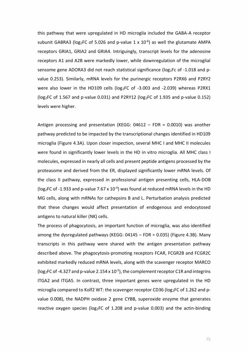
75
this pathway that were upregulated in HD microglia included the GABA-A receptor
subunit GABRA3 (log2FC of 5.026 and p-value 1 x 10⁻⁶) as well the glutamate AMPA
receptors GRIA1, GRIA2 and GRIA4. Intriguingly, transcript levels for the adenosine
receptors A1 and A2B were markedly lower, while downregulation of the microglial
sensome gene ADORA3 did not reach statistical significance (log2Fc of -1.018 and p-
value 0.253). Similarly, mRNA levels for the purinergic receptors P2RX6 and P2RY2
were also lower in the HD109 cells (log2FC of -3.003 and -2.039) whereas P2RX1
(log2FC of 1.567 and p-value 0.031) and P2RY12 (log2FC of 1.935 and p-value 0.152)
levels were higher.
Antigen processing and presentation (KEGG: 04612 – FDR = 0.0010) was another
pathway predicted to be impacted by the transcriptional changes identified in HD109
microglia (Figure 4.3A). Upon closer inspection, several MHC I and MHC II molecules
were found in significantly lower levels in the HD in vitro microglia. All MHC class I
molecules, expressed in nearly all cells and present peptide antigens processed by the
proteasome and derived from the ER, displayed significantly lower mRNA levels. Of
the class II pathway, expressed in professional antigen presenting cells, HLA-DOB
(log2FC of -1.933 and p-value 7.67 x 10⁻⁴) was found at reduced mRNA levels in the HD
MG cells, along with mRNAs for cathepsins B and L. Perturbation analysis predicted
that these changes would affect presentation of endogenous and endocytosed
antigens to natural killer (NK) cells.
The process of phagocytosis, an important function of microglia, was also identified
among the dysregulated pathways (KEGG: 04145 – FDR = 0.035) (Figure 4.3B). Many
transcripts in this pathway were shared with the antigen presentation pathway
described above. The phagocytosis-promoting receptors FCAR, FCGR2B and FCGR2C
exhibited markedly reduced mRNA levels, along with the scavenger receptor MARCO
(log2FC of -4.327 and p-value 2.154 x 10⁻⁵), the complement receptor C1R and integrins
ITGA2 and ITGA5. In contrast, three important genes were upregulated in the HD
microglia compared to Kolf2 WT: the scavenger receptor CD36 (log2FC of 1.262 and p-
value 0.008), the NADPH oxidase 2 gene CYBB, superoxide enzyme that generates
reactive oxygen species (log2FC of 1.208 and p-value 0.003) and the actin-binding

76
protein coronin 1A, which initiates fusion of the phagosome to the lysosome (log2FC
of 1.182 and p-value 0.008); perhaps hinting at a compensatory mechanism.
According to the iPathwayGuide Impact analysis, the final pathway predicted to be
significantly affected was the complement and coagulation pathway (KEGG: 04610 –
FDR = 0.047), which had 13 DE genes, of which C5 and C7 were upregulated (Figure
4.3C). CR2, CLU, MASP1, C1S, C1R and VTN were all found to be downregulated in
HD109 MG cells.

77
A

78
Figure 4.3 Phagocytosis and antigen processing and presentation are among the top pathways predicted to be dysregulated in HD109 iPSC-derived microglia. (A) Perturbation analysis by iPathway Guide identified antigen processing and presentation as likely affected in HD109 MG. Highest negative perturbation shown in dark blue. Changes predicted to affect antigen presentation to natural killer cells. (B) Phagosome KEGG pathway diagram overlaid with measured fold changes for genes including CYBB, CD36 and coronin (upregulated) and downregulated receptors such as FCGR2B and MARCO. (C) Complement pathway was enriched with 13 DE genes in HD109 MG cells. Bar plots of top-30 genes out of total number of DE genes in a pathway. Upregulated genes displayed in red, downregulated in blue. Box and whisker plots outline the distribution of DE genes annotated to the KEGG pathway. Box represents the first quartile, the median and third quartile; outliers are shown as circles.
B C

79
To gain further insight into the underlying mechanisms driving the differential gene
expression observed in the HD109 stem cell-derived microglia, upstream regulator
analysis was performed using Ingenuity Pathway Analysis software (IPA, Qiagen). This
software compares gene expression in a dataset with known gene lists regulated by
specific upstream signalling molecules and networks (p-value of overlap) and predicts
whether a specific molecule is likely to be activated or inhibited (z score). Using a z
score of > 2 or < -2 and p value of < 0.01 as cut-offs for ascribing significance, the
extensive list of predicted upstream regulators was narrowed down to 41, with the
majority (28 out of 41) found to be significantly inhibited (see Table 4.4) compared to
13 predicted to be activated (see appendix 3).
Upstream regulator Activation z-score P value of overlap Function
Lipopolysaccharide -4.272 6.42E-14 Bacterial cell wall component that elicits
strong immune response
NFB complex -4.222 0.0022 Family of TFs that regulate inflammatory
response
TP53 -4.17 1.7E-05 Tumour suppressor involved in regulation
of apoptosis
IFN-2 -4.114 6.16E-05 Type I IFN cytokine produced by virus-
infected cells
poly rI:rC-RNA -3.806 2.05E-05 Synthetic drug that activates IFN-
inducible genes
IFN- -3.668 1.04E-10 Immune response-mediating cytokine
IL1A -3.609 2.15E-03 Pro-inflammatory cytokine
TNF -3.545 1.53E-15 Pro-inflammatory cytokine
F2 -3.502 1.94E-03 Serine protease involved in clotting
process
IFN-ß -3.399 7.75E-04 Type I IFN cytokine involved in innate
immune response
Table 4.4 IPA of upstream transcriptional regulators inhibited in HD109 iPSC-derived microglia. IPA was used to predict activation of specific transcription regulators that overlap with the transcriptome changes observed in HD109 microglia. The upstream regulators were ranked by activation z-score, with a significant p value cut-off of < 0.01. The 10 most significant regulators and networks were included in the table above (for the remaining inhibited regulators – see appendix 2).
The top activated regulator in terms of z score was the p38 MAPK kinase inhibitor
SB203580 (z score of 3.885 and p-value overlap 1.33 x 10⁻⁸), followed by the nuclear
receptor retinoic acid receptor alpha or RARA (z score of 3.414 and p-value of overlap

80
5.41 x 10⁻⁷). The cytokine IL1RN, whose expression was upregulated (log2FC of 2.254
& p-value 0.034), was identified as one of the activated upstream regulators (z score
of 2.279 and p-value 1.64 x 10⁻³), along with the growth factor DKK1 (z score of 2.005
and p-value 1.94 x 10⁻⁴). Predictive activation of the transcription factor STAT3,
previously identified as an activated regulator in HD monocytes (Miller et al. 2016),
failed to reach significance (z score of 1.12 and p-value 7.29 x 10⁻⁴) whereas NKX2-3 (z
score of 2.103 and p-value 6.63 x 10⁻⁶) and MYCN (z score of 2.671 and p-value 7.85 x
10-4) were predicted as significantly activated.
In contrast, a large number of cytokines, transcription factors and pathways were
predicted to be inhibited in the HD109 microglial cells. Top of this list were the LPS
pathway (z score of -4.272 and p-value overlap 6.42 x 10⁻¹⁴), NFB complex (z score of
-4.222 and p-value overlap 2.2 x 10⁻³), TP53 (z score of -4.17 and p-value overlap 1.7 x
10-5), IFN2 (z score of -4.114 and p-value overlap 6.16 x 10⁻⁵), IFN- (z score of -3.668
and p-value overlap 1.04 x 10⁻¹⁰) and TNF (z score of -3.545 and p-value overlap 1.53
x 10-15). Several other regulators including IL6, IL1ß, IFN, IL4, ERK1/2, p38 MAPK,
IRF3, IRF1 and STAT1 were similarly predicted to be significantly inhibited, though
TLR4 didn’t fit the z score criterion (-1.58 and p-value overlap 6.65 x 10⁻⁴). Overall,
these data firmly suggest that the transcriptional changes observed in the HD109 stem
cell-derived microglia are associated with inhibition by several upstream regulators.

81
4.3.2 Functional validation of HD109 of iPSC-derived microglia
Upstream regulator analysis carried out using IPA (see section 4.3.1) projected several
inflammatory pathways to be inhibited in HD109 MG, including the NFB (Figure 4.4A)
and LPS pathways (Figure 4.5A). To test this hypothesis, the expression of NFB
pathway genes in the RNA-seq data was compared to WT Kolf2 cells. Out of a total of
50 genes, only three were differentially expressed: IL1R1 (log2FC of -1.411 and p-value
0.018), IL10 (log2FC of -1.687 and p-value 0.01) and STAT1 (log2FC of -1.416 and p-
value 7.14 x 10⁻⁴) were downregulated (Figure 4.4B). All other genes in this pathway,
including important adaptor proteins such as MYD88 were expressed at comparable
levels in both cell lines, though differences in IL-1ß did not reach statistical significance
(log2FC 1.115 and p-value 0.241) (Figure 4.4C). The expression of key NFB pathway
genes was further validated by Fluidigm qRT-PCR and results confirmed RNA-seq data
observations that mRNA levels of NFB genes did not vary significantly between
HD109 and Kolf2 microglia samples (Figure 4.4 D-E).

82
Figure 4.4 The expression of NFB pathway genes is unaltered in HD109 microglia compared to WT Kolf2 microglia. (A) IPA’s Upstream regulator analysis of the DE genes in the HD109 microglia RNA-seq dataset predicted the inhibition of several inflammatory pathways including the NFB complex. Regulators with significantly inferred activation (IL-10, SMAD7 and STAT3) are shown in orange, inhibited regulators (TNF, IL-6, IL-1ß, TGFß1, STAT1 and IRF1) are
shown in blue. (B) Expression of various components of the NFB pathway was assessed by RNA-seq. No significant differences were found in levels of NFB subunits RELA and RELB, as well as the inflammatory activation markers TNF, ICAM1, CCL2 and IRF1. (C) Out of 50 genes in the pathway, three were differentially expressed (downregulated) in HD109 microglia: IL1R1, IL-10 and STAT1. RNA-seq expression levels are represented as log2 transformed FPKM pseudo-counts. Data analysed using one-way ANOVA with Tukey’s multiple comparison test. Asterisks denote statistical significance: * < 0.05, ** < 0.01, *** < 0.001, ns – not significant. n=3 independent differentiations.
A
RELA
RELB
TNF
ICAM
1
TLR2
CCL2
IRF1
0
5
10
15
20
Log
2 (
FP
KM
+1)
Kolf2 MG
HD109 MG
B
IL1R
1IL
6IL
10
STA
T1IL
1ß
0
5
10
15
Log
2 (
FP
KM
+1)
Kolf2 MG
HD109 MG
***
ns
**
ns
*
C

83
(D-E) Fluidigm qPCR validation of NFB pathway genes expression in the HD109 microglia. No statistically significant differences were found in the expression levels of several genes between Kolf2 and HD109 iPSC-derived microglia, despite a notable trend towards upregulation. Fluidigm qRT-PCR data are presented as log2 expression normalised to reference genes. Statistical analysis used one-way ANOVA with Dunnett’s post hoc comparison. ns – not significant. n=3 biological replicates, except THP-1 (n=2).
IL-1ß
IL-6
CCL2
CD40
ICAM
1
0
5
10
15
20
Log
2 E
xpre
ssio
n ns
ns
ns
ns ns
D
IL-1
0
TNF-α
IRF1
STA
T1
TLR4
0
5
10
15
Log
2 E
xpre
ssio
n
Kolf2 MG
HD109 MG
THP-1
ns nsns
ns
ns
E

84
Mouse model studies have shown how HD immune cells cultured ex vivo exhibit a
hyper-reactive profile upon LPS stimulation, secreting higher levels of pro-
inflammatory cytokines compared to controls (Bjorkqvist et al. 2008; Trager et al.
2015). Thus, the hypothesis that LPS-stimulation of HD109 microglia may result in a
hyper-reactive was investigated, with expression of classical M1 markers and LPS
pathway genes assessed by qPCR. HD109 and Kolf2 microglia were differentiated for
14 days in IL-34 and GM-CSF, after which the cultures were incubated with 1 µg/ml
LPS for 4 and 24 hours, as gene expression changes following LPS stimulation peak at
around 4 hours (Yamamoto et al. 2004; Aung et al. 2006). No notable differences were
evident in the magnitude of change in expression of activation molecules after 4 hours
between HD and Kolf2 microglia, which suggests that both cell lines exhibited the
expected M1 activation or polarisation state. Genes such as IRF1, CCR7, ICAM1, CD40
and NLRP3 were upregulated to similar levels in both LPS-treated cell samples (Figure
4.5B). Though these differences were not statistically significant, there was a trend
towards higher mRNA levels of M1 pro-inflammatory cytokines in unstimulated
HD109 microglia, including TNF-, IL-8, IL-6, IL-1ß and CCL2. Subsequently, these
genes were upregulated in response to LPS incubation at 4 hours, with no differences
in mRNA levels post incubation between the cell lines (Figure 4.5C). However, the
magnitude of the mRNA increases between the LPS-treated microglia samples differed
for two genes, TNF- and IL-1ß. For TNF-, mRNA levels increased by a greater
magnitude in the Kolf2 WT cells (3-fold increase at p = 0.0024) compared to HD109
cells (2-fold increase at p= 0.0217) (Figure 4.5C). A similar pattern was observed for IL-
1ß, with a 3-fold increase in gene expression for Kolf2 cells (p= 0.0073) versus 2.5-fold
increase in the HD109 cells (p= 0.0256). The 4h LPS challenge also resulted in higher
CCL2 expression in HD109 microglia compared to Kolf2, whereas CCL22 expression
was higher in 4h LPS-treated HD109 compared to 4h LPS-treated WT Kolf2 cells. The
chemokine CCL18, a cytokine with both immune activation and suppressive
properties, was equally upregulated by LPS induction in both genotypes (Figure 4.5D).
Taken together, these observations hint at a tighter regulation of pro-inflammatory
cytokine production in the HD109 microglial cells.

85
Figure 4.5 The inflammatory response to LPS shows that HD109 microglia are not hyper-reactive. (A) The LPS pathway was predicted as inhibited in HD109 MG cells by IPA upstream regulator analysis. Inflammatory regulators such as IL-1ß, TNF, IFN and IFN (shown in blue) were among those predictively inhibited whereas IL-10 and STAT3 (shown in orange) were identified as upstream activators. (B) HD109 and Kolf2 MG cells were incubated with 1 µg/ml LPS for 4h. No significant differences between HD and WT cells were found in expression levels of M1 polarisation markers following LPS stimulation.
(C) In response to LPS, and in comparison with Kolf2 WT microglia, HD109 microglia produced similar levels of the pro-inflammatory cytokines TNF-, IL-1ß, IL-6 and IL-8. (D) LPS treatment resulted in higher CCL2 expression in HD109 MG cells whereas CCL22 expression was significantly higher in the LPS-treated Kolf2 MG cells than the corresponding HD109 cells.
A
B
IRF1
CCR7
ICAM
1
CD40
NLR
P3
0
5
10
15
Log
2 E
xpre
ssio
n
Kolf2 MG
Kolf2 MG + 4h LPS
HD109 MG + 4h LPS
HD109 MG
**
ns
**
*
ns
***
**
ns
** *
ns
*ns
ns
ns
B
TNF-α
IL-8
IL-1ß
IL-6
0
5
10
15
20
Log
2 E
xpre
ssio
n
*
ns
**
ns
****
**
ns
*
*
ns
ns
C
CCL2
CCL22
CCL18
IL-1
0
0
5
10
15
20
25
Log
2 E
xpre
ssio
n
ns*
ns
ns
**
**
***
ns
**
ns
ns
ns
HD109 MG + 4h LPS
Kolf2 MG
Kolf2 MG + 4h LPS
HD109 MG
D

86
Done
Save
Clear
Circle
Point
Kolf2 MG
Kolf2 MG 24h LPS
HD109 MG 24h LPS
HD109 MG
HD109 MG 4h LPS
Kolf2 MG 4h LPS
-15 -10 -5 0 5 10 15
-10
-50
5
PCA Score Plot
PC1
PC
2
Kolf2 MG 24h LPS 1Kolf2 MG 24h LPS 2
Kolf2 MG 4h LPS 2Kolf2 MG 4h LPS 3
Kolf2 MG 4h LPS 1
109n5 MG 4h LPS
109n4 MG 4h LPS109n1 MG 4h LPS
109n5 MG 24h LPS
109n4 MG 24h LPS109n1 MG 24h LPS
Kolf2 MG1
109n4 MG109n1 MG
Kolf2 MG3
Kolf2 MG2
E
CCL22
CCL18
CCL17
CCL2
0
5
10
15
20
25
Log
2 E
xpre
ssio
n
HD109 MG
HD109 MG + 4h LPS
HD109 MG + 24h LPS
ns
**
ns
ns
ns
ns
ns
ns
nsns
ns
ns
F
CCL22
CCL18
CCL17
CCL2
0
5
10
15
20
Log
2 E
xpre
ssio
n
Kolf2 MG
Kolf2 MG + 4h LPS
Kolf2 MG + 24h LPS
***
**
ns
***
ns*
nsns
*
ns
ns
ns
G
CX3C
R1
CD22
SIG
LEC12
PPA
Rγ
0
2
4
6
8
10
Log
2 E
xpre
ssio
n
HD109 MG
HD109 MG + 4h LPS
HD109 MG + 24h LPS
ns
ns
ns
*
*
nsns
ns
ns
ns
ns
ns
H
CX3C
R1
CD22
SIGLEC
12
PPARγ
0
2
4
6
8
10
Log
2 E
xpre
ssio
n
Kolf2 MG
Kolf2 MG + 4h LPS
Kolf2 MG + 24h LPS
**
*ns
**
ns
*
ns
ns
ns
**
**
ns
I
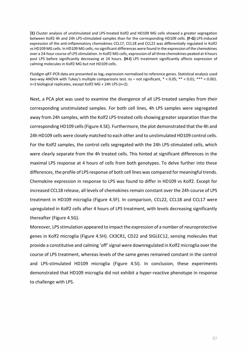
87
(E) Cluster analysis of unstimulated and LPS-treated Kolf2 and HD109 MG cells showed a greater segregation between Kolf2 4h and 24h LPS-stimulated samples than for the corresponding HD109 cells. (F-G) LPS-induced expression of the anti-inflammatory chemokines CCL17, CCL18 and CCL22 was differentially regulated in Kolf2 vs HD109 MG cells. In HD109 MG cells, no significant differences were found in the expression of the chemokines over a 24-hour course of LPS stimulation. In Kolf2 MG cells, expression of all three chemokines peaked at 4 hours post LPS before significantly decreasing at 24 hours. (H-I) LPS treatment significantly affects expression of calming molecules in Kolf2 MG but not HD109 cells. Fluidigm qRT-PCR data are presented as log2 expression normalised to reference genes. Statistical analysis used two-way ANOVA with Tukey’s multiple comparisons test. ns – not significant, * < 0.05; ** < 0.01; *** < 0.001. n=3 biological replicates, except Kolf2 MG + 24h LPS (n=2).
Next, a PCA plot was used to examine the divergence of all LPS-treated samples from their
corresponding unstimulated samples. For both cell lines, 4h LPS samples were segregated
away from 24h samples, with the Kolf2 LPS-treated cells showing greater separation than the
corresponding HD109 cells (Figure 4.5E). Furthermore, the plot demonstrated that the 4h and
24h HD109 cells were closely matched to each other and to unstimulated HD109 control cells.
For the Kolf2 samples, the control cells segregated with the 24h LPS-stimulated cells, which
were clearly separate from the 4h treated cells. This hinted at significant differences in the
maximal LPS response at 4 hours of cells from both genotypes. To delve further into these
differences, the profile of LPS response of both cell lines was compared for meaningful trends.
Chemokine expression in response to LPS was found to differ in HD109 vs Kolf2. Except for
increased CCL18 release, all levels of chemokines remain constant over the 24h course of LPS
treatment in HD109 microglia (Figure 4.5F). In comparison, CCL22, CCL18 and CCL17 were
upregulated in Kolf2 cells after 4 hours of LPS treatment, with levels decreasing significantly
thereafter (Figure 4.5G).
Moreover, LPS stimulation appeared to impact the expression of a number of neuroprotective
genes in Kolf2 microglia (Figure 4.5H). CX3CR1, CD22 and SIGLEC12, sensing molecules that
provide a constitutive and calming ‘off’ signal were downregulated in Kolf2 microglia over the
course of LPS treatment, whereas levels of the same genes remained constant in the control
and LPS-stimulated HD109 microglia (Figure 4.5I). In conclusion, these experiments
demonstrated that HD109 microglia did not exhibit a hyper-reactive phenotype in response
to challenge with LPS.

88
4.4 Summary and Discussion
In this chapter, a HD iPSC line containing 109 CAG repeats and a WT Kolf2 iPSC line were
differentiated into microglia and the phenotype of the in vitro derived microglial cells
characterised using RNA sequencing in order to analyse the microglia-specific transcriptome
effects of the HD CAG expansion. Gene Ontology analysis revealed that developmental
processes, including development of the embryo, mesenchyme, vasculature and epithelium
were among those significantly enriched in the HD109 microglia gene sets. The fact that these
processes aren’t specific to the nervous or immune system suggests that these transcriptional
changes reflect development as a whole being altered in HD. Recent studies have shown that
CAG repeat expansion results in the dysregulation of genes involved in neuronal development
and dorsal striatum formation (Ring et al. 2015). Similarly, Molero et al. 2009 found that CAG
expansion-associated developmental abnormalities in neurogenesis in the striatum
subsequently made medium spiny neurons more susceptible to degeneration in later life
(Molero et al. 2009). These findings seem to support the notion of HD as a
neurodevelopmental disorder (Kerschbamer & Biagioli 2016). Genes related to skeletal and
limb morphogenesis were also represented among the gene sets altered in the HD109
microglia, likely as a result of non-specific transcriptional changes due to aberrant mutant HTT
transcription factors interactions. These findings agree with recent RNA-seq data from
Labadorf et al. 2015, who analysed gene expression in the prefrontal cortex of HD post-
mortem samples and found evidence of a developmental signal, although it was unclear
which brain cell type was responsible for this signal (Labadorf et al. 2015). The same study
also uncovered evidence of an enrichment in inflammatory and immune response gene sets
in HD brains.
According to pathway impact analysis of the in vitro HD microglia transcriptome data, several
immune-related pathways were predicted to be dysregulated. Transcripts coding for cell
adhesion molecules that mediate antigen presentation and immune cell regulation were
downregulated in HD109 vs Kolf2 cells. Given that microglial cell adhesion molecules play a
critical role in many processes, including neural tissue development, it was unsurprising to
note a predicted dysregulation in the KEGG pathway 04080, enriched with genes coding for
neuroactive ligand-receptor interactions. This may hint at an impairment of HD microglia in
their capacity to aid the construction and development of neuronal networks. Another

89
pathway impact analysis prediction was the likely perturbation of interactions between HD
microglia and the extracellular matrix, which are mainly mediated by integrins and cell
adhesion molecules whose expression was found to be dysregulated. Given that the ECM
plays a key role in the morphogenesis and maintenance of cell and tissue structure and
function, these data appear to tally well with reports of neurovasculature disruptions in HD
patients, R6/2 mouse models and iPSC models of blood-brain barrier (Drouin-ouellet et al.
2015; Lim et al. 2017; Vatine et al. 2017).
Chemokine signalling was among the biological pathways revealed to be significantly affected
by transcriptional changes in HD109 microglia. Chemokines involved in the recruitment of
blood myeloid cells (CCL7, CCL8, CCL17) were significantly downregulated. mRNA levels of
CCL24, strongly chemotactic to resting T lymphocytes, was upregulated while levels of T cell
recruiting chemokines such as CCL1, CCL2 and CCL22 were unchanged. This pattern of
expression seems to contradict data from previous findings, which have shown that levels of
chemokines, complement proteins and the anti-inflammatory cytokine IL-10 increase with HD
burden (Dalrymple et al. 2007; Bjorkqvist et al. 2008; Wild et al. 2011). Equally, these
observations could be reflective of a dysregulation in chemokine signalling and thus compare
favourably with reports of a lack of infiltration of blood myeloid cells into the HD brain (Lucin
& Wyss-Coray 2009; Rocha et al. 2016); a phenomenon previously thought to be a
consequence of impaired immune cell migration in HD (Wanda Kwan et al. 2012). Finally,
altered chemokine signalling is likely to affect crosstalk between the HD microglia in the CNS
and cells of the adaptive immune system, and potentially have an impact on the involvement
of other immune cells in the striatum. Certainly, this view is supported by predicted deficits
in the antigen processing and presentation machinery of the HD109 microglia, perhaps an
adaptation to keep out infiltrating blood immune cells such as cytotoxic T-cells, NK cells and
monocytes.
Expression of three important cytokines and chemokine receptors (CCR2, CX3CR1 and CSF1R)
was increased in HD microglia, perhaps an indication of a proliferative and neuroprotective
adaptation of the cells to the HD neuronal environment. Interestingly, a recent study showed
in a rodent brain slice model that mutant HTT aggregation triggered by neuronal stressors led
to increased IL-34 production and corresponding microglial proliferation, exacerbating
degeneration of medium spiny neurons (Khoshnan et al. 2017). In addition, deletion of CCR2
has been shown to hamper microglial accumulation in a transgenic mouse model of

90
Alzheimer’s disease, resulting in higher amyloid burden and increased mortality (El Khoury et
al. 2007), implying that CCR2-dependent microglial accumulation can be neuroprotective. Of
course, deletion of the fractalkine receptor CX3CR1 has been shown to regulate microglial
activation in a mouse model of tauopathy (Bhaskar et al. 2010), while its overexpression
suppresses activation and diminishes tau pathology (Nash et al. 2013).
The inflammatory response of the HD109 cells was subsequently validated, to investigate the
hypothesis that HD microglia are primed for an exaggerated response to exogenous stimuli.
Following stimulation with LPS, both HD109 and Kolf2 cells exhibited the expected M1
polarisation state featuring a similar upregulation in pro-inflammatory markers including
CD40, ICAM1 and IRF1. In contrast, the same challenge appeared to induce a maintenance of
the core microglial phenotype in the HD109 microglia, with neuroprotective and calming
molecules such as SIGLEC12, CD22 and CX3CR1 being expressed at similar levels during and
following LPS treatment, whereas the same genes were downregulated in the Kolf2 cells
(Figure 4.5). Another particular point of difference in the HD109 inflammatory response
profile was that the increase in TNF- and IL-1ß expression post LPS of a higher magnitude in
Kolf2 compared to HD109 microglia. All things considered, it appears that the immune
response of HD109 microglia is more tightly regulated to prevent excessive release of
deleterious pro-inflammatory cytokines and loss of a neuroprotective signature. Cytokines
such as TNF and IL-6 are contained in carrier vesicles that are trafficked to the cell surface for
release (Stanley & Lacy 2010). Upon TLR-mediated LPS stimulation, carrier vesicles
accumulate in the Golgi prior to release. Several molecules are involved in the vesicular
transport of TNF-containing vesicles, including the lysosomal cysteine protease cathepsin B
(Ha et al. 2008), which is downregulated in HD109 microglia. Given the importance of the
Golgi network in cytokine trafficking, and given the fact that several Golgi and lysosome-
associated proteins were found to be decreased in the HD109 cells, it is plausible that altered
vesicle transport and trafficking pathways form part of the inhibitory mechanisms that
regulate cytokine release in HD stem cell derived microglia.
The hypothesis that the HD CAG expansion results in a more neuroprotective microglial
phenotype is further supported by findings from the upstream regulator analysis, which
predicted inhibition of several inflammatory pathways and signalling cascades in the HD109

91
microglia (see Table 4.4). In particular, the TNF and LPS pathways, previously shown to be
dysregulated in HD patient-derived monocytes (Miller et al. 2016), were predicted to be
inhibited, in comparison with the Kolf2 microglia cells. The NFß pathway, another
dysregulated pathway in HD immune cells (Trager et al. 2014), was similarly predictively
inhibited. These results imply a protective anti-inflammatory phenotype in the HD109 cells,
in stark contrast to the cell-autonomous pro-inflammatory state of HD mouse microglia cited
in the literature (Crotti & Glass, 2015). This discrepancy intriguingly invites questions about
the origin of this immune dampening effect – is this effect an accurate reflection of the HD
microglial phenotype or an artefact of in vitro differentiation? If this is an HD microglia-
specific effect, what are the mechanisms mediating it? The first question is the subject of the
investigation in chapter 5. To answer the second question, one has to consider the crucial role
of the brain microenvironment on the microglial phenotype.
Recent studies have intimated that neuronal co-culture highly modulates the transcriptome
of iPSC-derived microglial cells, suppressing pro-inflammatory signalling pathways relative to
monocultures (Abud et al. 2017; Haenseler et al. 2017). Despite the lack of neuronal co-
culture in this differentiation protocol, the predicted inhibitory effects observed in our HD
microglia dataset are evident when compared to the HD macrophage precursors upstream
regulator analysis dataset (see appendix 4), demonstrating that the neuronal IL-34 is partly
responsible for this particular inhibitory signal. In addition, it could be argued that in vitro
microglia are protected from this mutant HTT-mediated priming effect due to their distinct
ontogeny to other immune cell types. Indeed, the primitive macrophages used as precursors
for differentiation in this protocol have been shown to arise from a MYB-independent, RUNX1
and SPI1 lineage (Buchrieser et al. 2017) that is separate from the MYB-dependent wave of
haematopoiesis that gives rise to monocytes and macrophages (Orkin & Zon 2008). Therefore,
it is plausible that the distinct ontogeny of microglia combined with the immune-privileged
nature of the CNS and the fragility of the HD brain environment mediate the immune
dampening and inhibitory effects observed on the transcriptome of HD iPSC-derived
microglia.

92
5. EXPLORING THE EFFECTS OF
CORRECTING THE HD MUTATION ON
IMMUNE DYSFUNCTION IN IPSC-
DERIVED HD MICROGLIA
5.1 Introduction
Huntington’s disease is a fatal neurodegenerative disorder caused by expansion of a
polymorphic CAG repeat tract in exon 1 of the huntingtin (HTT) gene (MacDonald et
al. 1993). Though neurodegeneration in the HD brain primarily affects medium spiny
neurons in the striatum, mutant HTT is broadly expressed, including in immune cells
where its levels track with disease progression (Weiss et al. 2012). Thus, mutant HTT
expression in cells of the immune system has been suggested to contribute to HD
neuropathology. Activation of microglia, the brain’s resident immune cells, as
evidenced in HD post-mortem brains (Sapp et al. 2001), is not only widespread in
symptomatic patients, but it also occurs years before disease onset in HD gene carriers
(Pavese et al. 2006; Tai et al. 2007). Indeed, these pathological microglial changes
mirror those reported in dysfunctional peripheral immune cells (Dalrymple et al. 2007;
Bjorkqvist et al. 2008). Several studies have put forward convincing evidence for the
peripheral and central immune systems acting as modifiers of HD pathogenesis. In
2012, Kwan and colleagues showed that transplantation of WT bone marrow cells
attenuates motor deficits, enhances synaptogenesis and lowers plasma cytokine
levels in HD mice (W. Kwan et al. 2012). An earlier study determined that inhibiting
the enzyme kynurenine 3-monooxygenase (KMO), a key metabolite in the kynurenine
pathway, is neuroprotective, resulting in reduced microglial activation, attenuated
synaptic loss and a prolonged life span in HD mice, despite the inhibitor’s inability to
cross the BBB (Zwilling et al. 2011). Furthermore, signalling through the cannabinoid

93
receptor 2, expressed in microglia and peripheral immune cells and whose levels
increase in post-mortem HD brains and mice, was recently shown to modulate
immune activation (Palazuelos et al. 2009). Administration of a CB2 agonist dampened
CNS inflammation and inhibited motor defects and synaptic loss in R6/2 HD mice
(Bouchard et al. 2012).
The development of protocols for the differentiation of human iPSCs into several cell
types of interest has unlocked new prospects in disease modelling. Nevertheless, one
challenge of iPSC-based disease modelling that remains is the inconsistency in
differentiation quality due to variations in genetic background (Kajiwara et al. 2012),
which may affect the interpretation of in vitro diseased phenotypes. Moreover, the
impact of genetic background on observed phenotypes in iPSC-based models can be
substantial, even for a monogenic disease like HD where numerous disease-modifying
loci alter age of neurological onset (GeM-HD Consortium 2015). This should therefore
encourage the use of genetically identical control iPSC lines, to increase confidence
and authenticate observed HTT-specific mechanisms and phenotypes. For this reason,
work carried out within our lab by Jasmine Donaldson was successful in generating
isogenic control iPSC lines from the HD109n1 iPSC line, which is heterozygous for the
HD mutation with one WT allele of 21 CAG repeats and an expanded allele of 109 CAG
repeats. Using a homologous gene-targeting approach, CRISPR gRNAs were used to
target the 5’ UTR and exon 1 sequences of the expanded HTT allele. The donor DNA
featured a piggyBac transposon to allow a footprint-free removal of the selection
cassette from the targeted locus. Targeted clones were identified by PCR amplification
of exon 1 of the HTT allele, and following excision of the selection cassette by piggyBac
transposase, correct footprint-free editing of the HD iPSCs was confirmed by Sanger
sequencing and SNP array genotyping. In all, three isogenic HD109 corrected clones
were generated (5H9, 3H2 and 2H1), along with a non-corrected HD iPSC line (11B11)
that did not undergo piggyBac selection cassette to be used as an additional control
HD iPSC line. All lines were validated for pluripotency and shown to retain a normal
karyotype.

94
Successful correction of the HD mutation in iPSCs has been previously reported (An et
al. 2014; Shin et al. 2016). Efforts to characterise the effects have focused on
phenotypic abnormalities in iPSC-derived neural stem cells, namely impaired neural
rosette formation, increased susceptibility to growth factor removal and
mitochondrial respiration deficits (An et al. 2012; Mattis et al. 2015; Xu et al. 2017). In
an attempt to assess the effects of HD correction on immune cell function, a study by
the Tabrizi group used anti-HTT small interfering RNA to demonstrate that immune
dysfunction and hyperactivity caused by NFB dysregulation can be reversed by
reducing mutant HTT expression in patient-derived monocytes and macrophages
(Trager et al. 2014).
5.2 Aims
In chapter 4, RNA-seq was used to reveal any microglia-specific transcriptome changes
associated with mutant HTT expression in the HD109 iPSC-derived cells. Pathway
analysis identified several immune-related functions and pathways as being
potentially impaired or dysregulated in these cells. Following on from this earlier work,
this chapter had two principal objectives. Firstly, isogenic control HD corrected iPSC
lines were differentiated into microglia in order to study the effects of correcting the
HD mutation on the gene expression changes reported in the HD109 iPSC-microglia.
Secondly, a series of experiments aimed to investigate the predicted impaired
functions of HD microglia, characterise any potential impairments and ascertain the
extent to which any phenotypic abnormalities were rescued by correcting the
Huntington disease CAG expansion.

95
5.3 Results
5.3.1 Does correcting the HTT CAG expansion reverse the gene expression
changes observed in HD109 iPSC-derived microglia?
The presence of the expanded CAG track in the HTT gene resulted in widespread
changes in the transcriptome of the HD109 stem cell-derived microglia, as described
in chapter 4. In order to disentangle whether these transcriptional changes were
mutant HTT-specific or due to differences in genetic background between the HD109
and Kolf2 cells, isogenic control HD corrected iPSC lines were differentiated into
microglia. As well as the three HD corrected clones (5H9, 3H2 and 2H1), one non-
corrected HD iPSC line (11B11) was also used as control.
Initially, HD corrected microglia were assayed for expression of canonical markers by
immunocytochemistry. As highlighted in chapter 3, treatment of macrophage
precursors with IL-34 and GM-CSF results in robust expression of several microglial
markers. Confocal imaging analysis of microglia derived from isogenic corrected iPSCs
showed clearly that the IL-34 induced cells were positive for the microglial markers
IBA1, P2RY12 as well as the leucocyte-specific CD45 (Figure 5.1). Also expressed was
the microglia-enriched TGFBR1, a protein kinase receptor that binds the astrocyte-
derived cytokine TGF-ß (Schilling et al. 2001) shown to be essential for microglial
development and maintenance (Abutbul et al. 2012; Butovsky et al. 2014).

96
Figure 5.1 HD109 Corrected and non-corrected iPSC-derived microglia express microglial markers and display the characteristic ramified morphology of microglia. (A-B) Confocal images of microglia-like cells derived from HD109 corrected lines 3H2 (A) and 5H9 (B) shows robust expression of MG markers IBA1, TGFBR1, P2RY12 and CD45. (C) Non-corrected HD iPSC-derived MG cells were cultured for 14 days in GM-CSF and IL-34, fixed and stained with P2RY12 and CD45. (D) HD109 MG cells express the MG-enriched protein kinase TGF-ß receptor 1. Scale bars represent 20 µm.
A
B
C
D

97
Next, the expression of microglia-specific markers was analysed by Fluidigm qRT-PCR
and results showed that genes such as C1QA, GPR34, MERTK, P2RY12 and CABLES1
were expressed at similar levels in the HD corrected microglia compared to Kolf2 and
isogenic HD109 cells. Although its expression was decreased compared to THP1
monocytes, CX3CR1 followed a similar trend to other microglia sensome genes, with
transcript levels consistent across cells of different genotypes (Figure 5.2 A-B). Also,
two other sensome genes P2RY12 and ADORA3 found to be differentially expressed
in the HD109 microglia after RNA-seq data analysis were expressed at comparable
levels between the different genotypes.
The expression of AD risk genes in the HD corrected iPSC-derived microglia was
assessed, with CLU and CR2 being identified as differentially expressed in the HD109
microglia compared to Kolf2. qPCR results showed that CLU expression did not vary
markedly between the Kolf2, HD109 and HD109 corrected microglia samples. CR2
expression was not detected in THP1 monocytes and low in the microglia samples,
with the highest expression in the HD109 microglia and transcripts levels in the
corrected HD109 cells matching the low levels in the Kolf2 cells. However, these
differences did not reach statistical significance (Figure 5.2C).
Findings presented in chapter 4 established that expression levels of NFB genes were
similar in Kolf2 and HD109 microglia (see section 4.3.2). Expression of the core
microglial transcription factor PU.1 (SPI1) and its target genes, which is increased in
HD mouse immune cells, was equally similar. When the same genes were evaluated
in the corrected HD109 microglia by qPCR, results established that expression of both
sets of genes were comparable to the WT Kolf2 and HD109 microglia (Figure 5.2 D-E).
Overall, these results are consistent with the lack of altered NFB signalling observed
in HD109 microglia and suggest that reversal of the CAG mutation in HD microglia may
not affect basal activation of pro-inflammatory signalling through NFB.

98
Figure 5.2 The expression of canonical microglial and NFB pathway genes is unaffected by reversal of HD CAG repeat expansion. Kolf2, HD109 and HD109 corrected MG cells were differentiated for 14 days in IL-34 and GM-CSF and all samples assayed by Fluidigm qRT-PCR. (A-B) No significant differences were found in the expression of microglia-enriched markers between HD109 corrected MG cells, Kolf2 and HD109 MG samples. (C) ABI3, CLU and CR2 were identified as DE
in HD109 MG compared to Kolf2 by RNA-seq. Validation by qPCR shows comparable expression levels between HD109, Kolf2 and HD109 corrected samples. (D) NFB pathway genes are comparably expressed in HD109 and isogenic corrected MG cells. (E) Expression of the TF SPI1 and of its target genes is unaltered in HD109 and following correction of the CAG mutation. Log2 Expression normalised to reference genes. Statistical analysis by two-way ANOVA with Tukey’s multiple comparisons test. Asterisks denote significance: * < 0.05, ** < 0.01, *** < 0.001, ns – not significant. n=3 biological replicates for all samples, except for THP1 (n=2).
C1QA
GPR34
CX3CR1
P2RY12
ADORA3
0
5
10
15
Log 2
Exp
ress
ion
THP1
Kolf2 MG
HD109 MG
HD109 Corr MG
ns
ns
ns
nsns
ns
nsns
nsns
ns
ns
ns
ns
ns
A
MERTK
CLEC
7A
TMEM
119
CABLES
1
0
5
10
15
Log
2 E
xpre
ssio
n
THP1
Kolf2 MG
HD109 MG
HD109 Corr MG
nsns
ns
nsns
ns
nsns
ns
nsns
ns
B
APOE
CLUABI3
INPP5D
CR20
5
10
15
Log 2
Exp
ress
ion
THP1
Kolf2 MG
HD109 MG
HD109 Corr MG
ns
ns
ns
ns
ns
ns
ns
ns
nsns
ns
ns
nsns
ns
C
STA
T1
TLR4
IL-1ß
CCL2
ICAM
1
IL-1
0
0
5
10
15
20
Log
2 E
xpre
ssi
on
THP1
Kolf2 MG
HD109 MG
HD109 Corr MG
ns
ns
ns
ns
ns
ns
ns
ns
ns
ns
ns
ns
ns
ns
ns
ns
ns
nsD
SPI1
IL-6
IRF1
TNF-α
0
5
10
15
Log
2 E
xpre
ssio
n
THP1
Kolf2 MG
HD109 MG
HD109 Corr MG
nsns
ns
ns
ns
nsns
ns
ns
nsns
ns
E

99
5.3.2 Effects of correcting the HD mutation on the microglial inflammatory
response under stimulatory conditions
Having previously established that HD109 microglia exhibited an inflammatory
activation profile distinct from Kolf2 WT cells, the next set of experiments aimed at
investigating the immune response of the HD corrected iPSC-derived microglia. 14-
day-differentiated microglia derived from HD109 corrected iPSC were stimulated with
1 µg/ml LPS for 4 hours, and the expression of pro-inflammatory markers compared
to 4-hour LPS-treated Kolf2 and HD109 microglia. The activation profile of the
corrected HD109 microglia was found to be consistent with that of Kolf2 and HD109
cells, with expected increases in the M1 markers IRF1, CCR7 and ICAM1 that were
comparable in magnitude between the corresponding unstimulated and 4h LPS
samples; the only notable difference being that CCR7 mRNA levels post LPS were
significantly higher in Kolf2 vs corrected HD109 cells (Figure 5.3A). The two main pro-
inflammatory cytokines TNF- and IL-1ß were upregulated after LPS stimulation in all
three cell lines, with comparable mRNA levels post incubation (Figure 5.3B).
Intriguingly, differences in the magnitude of these changes were again apparent, with
a 3-fold upregulation in both genes for the WT Kolf2 and corrected HD109 microglia
compared to a 2.5-fold increase in TNF- and IL-1ß for the HD109 cells. Inflammatory
regulation of these two genes may therefore be an indication of the effects of
correcting the CAG mutation on HD109 microglia. The other pro-inflammatory
cytokines IL-8 and IL-6 were also upregulated after LPS incubation, with non-
significant variations between the genotypes (Figure 5.3C).
The genetic background of the cells also seemed to dictate their LPS response.
Whereas levels of the anti-inflammatory IL-10 and the pro-inflammatory CCL2 were
unchanged in the LPS-treated Kolf2 cells, these two genes were correspondingly
upregulated in the HD109 and isogenic corrected microglia (Figure 5.3D). Expression
of the main anti-inflammatory cytokine IL-10 hinted at a balanced secretion of pro-
and anti-inflammatory molecules. Fittingly, the upregulation of the chemokines CCL22
(in Kolf2 microglia only) and CCL18 (in all three samples) after LPS exposure seemed
to support this idea (Figure 5.3E). To illustrate the segregation between the
unstimulated and LPS-treated samples, a principal component analysis was carried

100
out, with the results plotted using a t-distributed Stochastic Neighbour Embedding
(tSNE) (Van Der Maaten & Hinton 2008). The resulting plot showed close proximity
between HD corrected microglia, HD109n1 and two Kolf2 microglia samples, hinting
at a close similarity between the unstimulated microglia (Figure 5.3F). The LPS-treated
samples from all three cell lines formed separate clusters that segregated according
to genotype, which suggested that LPS treatment resulted in a HD corrected
phenotype distinct from that observed in the WT Kolf2 and HD109 microglia. This
therefore hinted at differences between the HD109 and the corrected microglia and
demonstrated that correction of the CAG mutation on the HD gene had an effect on
the phenotype of microglia.

101
Figure 5.3 The LPS inflammatory response of HD corrected microglia is similar to that of HD109 microglia. (A) 4h LPS stimulation of HD109 corrected MG cells leads to upregulation of IRF1, CCR7 and ICAM1 comparable with Kolf2 and HD109 cells. (B) Despite a trend towards lower
expression of TNF- and IL-1ß in corrected compared to HD109 MG, no significant differences were found in expression of both genes in the corresponding LPS-treated MG samples. (C) Expression of the pro-inflammatory cytokines IL-8 and IL-6 did not vary significantly between the HD genotypes following LPS incubation. (D) LPS-induced changes in IL-10 and CCL2 expression are dictated by the genetic background of the MG cells; expression of both genes is upregulated in both HD genotypes and unchanged in Kolf2 cells. (E) Expression of the anti-inflammatory chemokine CCL18 is significantly increased in LPS-treated corrected MG cells and HD109 MG cells. Kolf2 MG cells upregulate CCL18 and CCL22 expression in response to LPS incubation. Fluidigm qRT-PCR analysis used to generate log2 Expression values normalised to reference genes. Statistical analysis by two-way ANOVA with Tukey’s multiple comparisons test. Asterisks denote significance: * < 0.05, ** < 0.01, *** < 0.001, ns – not significant. n=3 biological replicates for all samples.
IRF1
CCR7
ICAM
1
0
5
10
15
Log
2 E
xpre
ssio
n
Kolf2 MG
Kolf2 MG + 4h LPS
HD109 MG + 4h LPS
HD109 MG
***
ns
***
***
ns
***
***
***
***
ns
***
HD Corr MG
HD Corr MG + 4h LPS
ns
***
ns
ns
ns
**
ns
ns
nsA
IL-8
IL-6
0
5
10
15
20
Log
2 E
xpre
ssio
n
Kolf2 MG
Kolf2 MG + 4h LPS
HD109 MG + 4h LPS
HD109 MG
***
HD Corr MG
HD Corr MG + 4h LPS
*
ns
ns
******
ns
ns
****
ns
ns
ns
ns
C
TNF-α
IL-1ß
0
5
10
15
20
Log
2 E
xpre
ssio
n ***
ns
ns
ns
******
ns
ns
***
ns
ns
ns
***
***
B
IL-1
0
CCL2
0
5
10
15
20
25
Log
2 E
xpre
ssio
n
Kolf2 MG
Kolf2 MG + 4h LPS
HD109 MG + 4h LPS
HD109 MG
ns
HD Corr MG
HD Corr MG + 4h LPS
*
*
ns
****
ns
ns
****
ns
**
**ns
D
CCL22
CCL180
5
10
15
20
Log 2
Exp
ress
ion
Kolf2 MG
Kolf2 MG + 4h LPS
HD109 MG + 4h LPS
HD109 MG
*
HD Corr MG
HD Corr MG + 4h LPS
***
ns***
ns
ns
******
ns
***
ns
ns
***
E

102
(F) 4-hour LPS stimulation of Kolf2, HD109 and HD109 corrected microglia resulting in clustering effect, with MG samples segregating according to genotype. tSNE plot generated using “Fluidigm SC” package analysis of raw Ct data. Each data point represents one MG sample.
F
Done
Save
Clear
Circle
Point
Kolf2 MG
HD109 MG
HDCorr MG
HDCorr MG 4h LPS
HD109 MG 4h LPS
Kolf2 MG 4h LPS
-1500 -1000 -500 0 500 1000
-200
0-1
000
01
000
200
0
tSNE Plot
X1
X2
109n5 MG 4h LPS
109n1 MG 4h LPS
109n4 MG 4h LPS
HDCorr 2H1 MG 4h LPSHDCorr 5H9 MG 4h LPS
HDCorr 3H2 MG 4h LPS
Kolf2 MG 4h LPS 1
Kolf2 MG 4h LPS 3
Kolf2 MG 4h LPS 2
Kolf2 MG1
109n4 MG
109n1 MG
Kolf2 MG2
Kolf2 MG3
HDCorr 5H9 MG
HDCorr 2H1 MG

103
5.3.3 HD109 microglia do not manifest transcriptome-associated functional
deficits in phagocytosis and chemotaxis
As described in chapter 4, several KEGG pathways were predicted through impact
analysis to be impaired or dysregulated in HD109 microglial cells. These included
cytokine-cytokine interaction, chemokine signalling, cell adhesion and ECM-receptor
interaction as well as phagosome. Two pathways were chosen for functional
validation: phagocytosis (FDR= 0.035) and chemotaxis (0.029). Levels of chemokines
such as CCL17 and CCL18 were also downregulated in HD109 microglia, with qPCR
results confirming that only one gene followed the same trend, with negligible CCL17
levels in the HD109 and corrected microglia compared to Kolf2. Elsewhere, transcript
levels of CCL22 and CCL18 in HD109 and corrected cells matched those in Kolf2 cells
(Figure 5.5A).
Phagocytosis is a key function of microglia, enabling clearance of pathogens, protein
aggregates and apoptotic cells in the CNS. Differential gene expression identified
downregulation of several phagocytosis-promoting receptors (FCAR, FCGR2B and
FCGR2C, C1R, ITGA2, ITGA5, MARCO) in HD109 microglia compared to Kolf2 WT
microglia. Interestingly, genes coding for the multi-ligand scavenger receptor CD36
and the enzyme NADPH oxidase or CYBB were upregulated. Validation of CD36 and
CYBB expression by qPCR indicated that both genes were comparably expressed in the
Kolf2 and HD microglia from both genotypes (Figure 5.4A). Also downregulated was
CORO1A, a protein that initiates fusion of the phagosome to the lysosome during
pathogen killing. These findings seemed to indicate a compensatory mechanism. To
confirm this observation, phagocytosis of pHrodo red E. coli particles was monitored
in real time using the IncuCyte S3 live imaging platform, which quantifies the increase
in red fluorescence triggered by the uptake of pHrodo cells and internalisation into
the acidic phagosome by professional phagocytes (Kapellos et al. 2016). Microglia
from Kolf2, HD109 and HD109 corrected iPSCs were seeded on 96-well plates at
35,000 cells per well, differentiated for 14 days and then incubated with 10 µg pHrodo
red particles for 6 hours while confocal images were taken every 10 min. All HD
derived microglial cells (HD109, non-corrected and corrected) displayed a higher rate

104
of uptake compared to Kolf2 microglia, indicating that they retained efficient ability
to phagocytose bioparticles (Figure 5.4B). Interestingly, Kofl2 WT cells pre-treated
with the phagolysosome v-ATPase inhibitor bafilomycin A1 were capable of ingesting
the fluorescent particles, though the final count of phagocytosed particles was
significantly reduced compared to Kolf2 and all HD genetic backgrounds (two-way
ANOVA with Tukey’s post hoc test, p<0.0001). This was hypothesised to be due to the
high cell density in each well (Figure 5.4C). Comparisons between the HD109n1 and
the HD109 corrected clones showed that correction of the HD CAG mutation had no
effect on the cells’ ability to phagocytose bioparticles, despite clonal variations in
pHrodo E. coli bioparticle uptake. This experiment was repeated with a lower number
of 20 000 plated cells per well (Figure 5.5D). On this occasion, pre-treatment of the
cells with bafilomycin A1 and cytochalasin D resulted in a larger reduction in
fluorescence signal, demonstrating that the rate of bioparticle uptake is dependent
on cell numbers (Figure 5.4E). At the peak of around 6 hours of incubation, MG cells
from two of the HD109 corrected clones (5H9 and 2H1) ingested more particles than
cells derived from the isogenic HD109 line. The HD109 corrected clone 3H2
phagocytosed less particles than cells generated from the parental HD109n1
(p<0.0001), although this rate of phagocytosis was found to be as efficient as that of
the HD109 non-corrected iPSC-derived microglial cells (p = 0.5897). To conclude, while
clonal differences in the kinetics of pHrodo phagocytosis were evident, HD microglia
have shown to be competent phagocytes of bioparticles.

105
Figure 5.4 Phagocytosis of E. coli bioparticles and chemotaxis are not impaired in HD109 iPSC-derived microglia. (A) qPCR validation of phagocytosis and chemokine transcripts shows expression of CCL17 and CCL22 is higher in Kolf2 MG than HD109 MG. CYBB and CD36 expression levels are comparable in HD109 and Kolf2 MG. (B) MG cells from HD109 and HD109 corrected iPSCs exhibit a higher rate of uptake of bioparticles than Kolf2 MG cells. MG cells were plated on a 96-well plate at 35,000 per well and incubated with 10 µg pHrodo E. coli for 6 hours. For negative control, Kolf2 MG cells were pre-treated with 10 µM bafilomycin A1 before 1hr before E. coli incubation. (C) End point fluorescence values from (B) were plotted. Data presented as mean ± SEM from each condition with 8 technical replicates. n=1 independent experiment.
CYBB
CD36
CCL22
CCL17
CCL18
0
5
10
15Log
2 E
xpre
ssio
n
THP1
Kolf2 MG
HD109 MG
HD109 Corr MG
ns
ns
ns
ns
ns
ns
*
ns
ns
ns
ns
ns
A
B
HD10
9n1
HD10
9 non C
orr
HD10
9 Corr
3H2
HD10
9 Corr
5H9
HD10
9 Corr
2H1
Kolf2
Baf
ilom
ycin
A1
0
100000
200000
300000
400000
Tota
l red o
bje
ct a
rea (
µm
2/Im
age)
****
ns
ns
ns
ns
ns****
***ns
C

106
(D) HD109 corrected microglia phagocytose E. coli bioparticles as efficiently as microglia derived from the HD109 parental clonal line. MG cells were plated at 20,000 cells per well. HD109 MG cells were pre-incubated with 10 µM cytochalasin D for 1hr before E. coli treatment. End point fluorescence values were plotted in (E). Data presented as mean ± SEM from each condition or genotype with 8 technical replicates. n=2 independent experiments.
D
HD10
9n1
HD10
9 non C
orr
HD10
9 Corr
3H2
HD10
9 Corr
5H9
HD10
9 Corr
2H1
Cyt
ochal
asin
D
Baf
ilom
ycin
A1
0
50000
100000
150000
200000
250000
Tota
l red o
bje
ct a
rea (
µm
2/Im
age)
ns
ns****
****
****
****
ns
nsE

107
(F) HD109 and HD109 corrected MG cells display efficient cell migration in response to stimulation with CX3CL1. (G) End point (12h) values for top membrane index show that compared to cytochalasin D-treated MG cells, HD109 and HD109 corrected microglia retain proper chemotactic functions. (H) Chemotactic response of MG cells to ADP is unaffected in HD1009 iPSC-derived microglia. For pHrodo E. coli phagocytosis assays, red channel fluorescence (400ms) was measured every 10 mins in an IncuCyte S3 live imaging platform. Data was analysed using the IncuCyte S3 Base software (see section 2.5.3 for analysis parameters). For chemotaxis assays, MG cells were plated and differentiated for 14 days on the upper chamber of 96-well ClearView chemotaxis plates. Cells were stimulated with 200 ng/ml CX3CL1 or 8 µM ADP to induce migration to the lower chamber of the chemotaxis plate. Phase channel images were taken using IncuCyte S3 every 30 mins for 12-24 hours and quantified by IncuCyte analysis software. n=1 independent experiment. Statistical significance was tested using two-way ANOVA with Tukey’s multiple comparisons test. * < 0.05, ** < 0.01, *** < 0.001, **** < 0.0001, ns – not significant. n=1 independent experiment.
F
HD10
9 Corr
3H2
HD10
9 Corr
2H1
HD10
9n1
HD10
9n5
HD10
9 Corr
5H9
Cyt
ochal
asin
D
0.0
0.5
1.0
1.5
Top m
em
bra
ne o
bje
ct count
CX3CL1
ns
ns
ns
ns
**
G
H

108
Kwan et al. 2012 revealed using myeloid cells isolated from premanifest HD patients
and early postnatal mouse microglia overexpressing mutant HTT that immune cells
display mHTT-dependent deficits in migration to chemotactic stimuli. In order to
characterise any potential impairments in chemotaxis in HD109 iPSC-derived
microglia, cell migration assays were carried out using the IncuCyte S3 platform (see
section 2.5.3). Microglial cells from Kolf2, HD corrected and HD109 iPSCs were
differentiated for 14 days on a 96-well ClearView Chemotaxis, where each well is
made up of a membrane that contains a lower and upper chamber. Cells are required
to migrate across the membrane, through 8µm pores to reach the lower chamber, a
process visualised and quantified in real-time by the IncuCyte analysis software.
Migration in response to two chemoattractants, CX3CL1 and ADP was assayed. Cells
stimulated with 200 ng/ml CX3CL1 were shown to be responsive to the chemotactic
stimulus (Figure 5.4F), as shown by the reduced top membrane index (Figure 5.4G).
When compared to cytochalasin D-treated MG HD 109 cells, all cell lines were found
to be efficient at cell migration through the membrane. Interestingly, no significant
variations in response were found between the HD109 and the corrected iPSC-derived
microglia. Chemotactic response to ATP was then investigated. Along with CX3CL1,
sphingosine-1-phosphate (S1P) and lysophosphatidylcholine (lysoPC), ATP and other
nucleotides are so called “find-me signals” released by cells undergoing apoptosis that
attract phagocytes towards them (Lauber et al. 2004). Chemotactic response of the
HD MG cells to 8 µM ADP was quantified and results showed no statistically significant
differences in the migration of HD109 and HD109 corrected microglia compared to
Kolf2 WT microglia (Figure 5.4H). Overall, these results suggest that migration and
regulation of actin remodelling of HD iPSC-derived microglia is not impaired, with no
rescue effect of the CAG expansion excision on those properties.
5.3.4 HD109 microglia do not exhibit deficits in mitochondrial respiration
associated with other HD cell types
Mitochondrial dysfunction has been strongly implicated in HD pathogenesis, with
impairments in ATP production, mitochondrial basal respiration and glycolysis among
early reported disturbances in the HD brain (Damiano et al. 2010; Costa & Scorrano

109
2012). Studies in mouse models and post-mortem human brains have described
mitochondrial impairments in several HD cell types, e.g. primary lymphoblasts,
fibroblasts and striatal cells (Polyzos & Mcmurray 2017) and stem cell-derived neural
stem cells (An et al., 2012; Xu et al. 2017). To date, impairments in HD microglia have
not been extensively reported or characterised. Given the metabolic demands that
homeostatic and immune functions place on microglia (Cherry et al. 2014), an
investigation of the bioenergetics profile of HD109 microglia was performed to initially
compare it to that of WT Kolf2 microglia (Figure 5.5A). For this series of preliminary
experiments, microglial cells from Kolf2, HD109 and corrected HD109 iPSC lines were
differentiated in Seahorse XF96 plates and responsiveness to the mitochondrial
stressors oligomycin, FCCP and rotenone measured to calculate the oxygen
consumption rate (OCR). The Seahorse XF Cell Mito Stress Test protocol was used (see
section 2.5.4), which allows the assessment of different aspects of mitochondrial
respiration, including basal respiration, maximal respiration, spare respiratory
capacity and ATP production.
Though earlier results had hinted at the HD109 MG cells exhibiting a lower basal
respiration rate, the preliminary examinations presented here showed that basal
respiration was similar between the WT and HD109 microglial cells (Figure 5.5B).
Microglia derived from the HD109n1 clonal line seemed to exhibit a greater
mitochondrial spare respiratory capacity compared to cells derived from Kolf2,
HD109n4 and HD109n5 lines, with a similar pattern emerging in the maximal
respiration OCR rates between the lines (Figure 5.5C). ATP production rates were also
comparable between the HD109 and Kolf2 WT MG cells (Figure 5.5D).
Similarly, the profile of HD corrected iPSC-derived microglia was compared to that of
HD109n1, the parent clonal line (Figure 5.5E), with preliminary data showing small
increases in maximal respiratory capacity for two out of three clones (Figure 5.5F), but
no changes in ATP production or basal respiration (Figure 5.5 G-H). Taken together,
these preliminary results into the bioenergetics profile of iPSC-derived microglia
suggest that mitochondrial function may not be impaired in the HD109 microglia.

110
Figure 5.5 Preliminary results show that mitochondrial oxidative phosphorylation is unimpaired in HD109 iPSC-derived microglia. MG cells (20,000 per well) were plated and differentiated on Seahorse XF 96-well plates. Oxygen consumption rate (OCR) was measured with sequential injections of 2 µM oligomycin, 2.5 µM FCCP and 0.5 µM antimycin A & rotenone. (A-D) Measurements of basal respiration and ATP production show no differences between HD109 and Kolf2 MG cells. MG cells derived from HD109n1 iPSCs seemed to show greater maximal respiration rate than WT Kolf2 MG cells. Data presented as mean ± SEM from each genotype with at least 8 technical replicates; OCR measures normalised to absorbance at 560 nm; n=1 independent experiment.
0 20 40 60 800
200
400
600
800
1000
Time (minutes)
OC
R (
pm
ol/m
in/A
560) Kolf2
HD109n5
HD109n1
Oligomycin
HD109n4
FCCP
Antimycin A & RotenoneA B
HD10
9n4
Kolf2
HD10
9n1
HD10
9n5
0
50
100
150
200
250
Basal respiration
OC
R (
pm
ol/m
in/A
560)
C
HD10
9n4
Kolf2
HD10
9n1
HD10
9n5
0
200
400
600
800
1000
Maximal respiration
OC
R (
pm
ol/m
in/A
560)
D
HD10
9n4
Kolf2
HD10
9n1
HD10
9n5
0
50
100
150
200
ATP production
OC
R (
pm
ol/m
in/A
560)

111
(E-H) Mitochondrial basal respiration, maximal respiration and ATP production rates in HD109 corrected microglia are comparable to measurements in the microglia cells derived from the parental clonal line HD109n1. n=1 independent experiment.
20 40 60 80
-100
0
100
200
300
400
500
Time (minutes)
OC
R (
pm
ol/m
in/A
560) HD109 Corr 5H9
HD109 Corr 3H2
HD109 Non Corr
HD109 Corr 2H1
HD109n1
Oligomycin
FCCP
Antimycin A & RotenoneE F
HD10
9n1
HD10
9 non C
orr
HD10
9 Corr
2H1
HD10
9 Corr
3H2
HD10
9 Corr
5H9
0
100
200
300
400
500
OC
R (
pm
ol/m
in/A
560)
Maximal respiration
G
HD10
9n1
HD10
9 non C
orr
HD10
9 Corr
2H1
HD10
9 Corr
3H2
HD10
9 Corr
5H9
0
20
40
60
80
100
OC
R (
pm
ol/m
in/A
560)
ATP production H
HD10
9n1
HD10
9 non C
orr
HD10
9 Corr
2H1
HD10
9 Corr
3H2
HD10
9 Corr
5H9
0
20
40
60
80
100
OC
R (
pm
ol/m
in/A
560)
Basal respiration

112
A complex interplay exists between the immune response and the major metabolic
pathways of glycolysis, the TCA cycle, fatty acid synthesis and oxidation, amino acid
metabolism and the pentose phosphate pathway. In particular, changes in the
metabolites of these pathways have been shown to regulate the innate inflammatory
response (Galván-peña & O’Neill 2014; O’Neill et al. 2016). For example, Tannahill et
al. 2013 showed that LPS stimulation of mouse macrophages causes an increase in
intracellular levels of the TCA cycle intermediate succinate, which subsequently drives
production of IL-1ß (Tannahill et al. 2013). Stimulation by LPS, which results in an M1
phenotype and is associated with expression of pro-inflammatory cytokines, is
accompanied by a shift from oxidative phosphorylation to glycolysis for energy
production, called the Warburg effect (López-Lázaro 2008). A second aspect of this
shift during inflammation is mitochondrial dysfunction, whereby mitochondria are
reprogrammed by undergoing fission, which induces formation of ROS and other
inflammatory mediators such as NFB (Park et al. 2013; Park et al. 2015). To
investigate mitochondrial contribution to the energetic demands concurrent with
inflammation, preliminary experiments were undertaken to study the effects of LPS
stimulation on the bioenergetics of microglia. Cells were plated and differentiated on
Seahorse XF96 plates for 14 days. After differentiation, the microglia were stimulated
in situ with 1 µg/ml LPS for 4 hours before measurements of mitochondrial respiration
(Figure 5.6A). LPS challenge was found not to cause any changes in ATP production or
basal respiration in the respective HD109 and Kolf2 microglial cells (Figure 5.6 B-C). A
similar pattern was observed with microglia derived from the HD corrected iPSC lines
(Figure 5.6 D-F). When comparing challenged and unstimulated cells, LPS stimulation
only resulted in subtle differences in the maximal respiration rates (Figure 5.6E). Taken
together, these preliminary experiments appear to indicate that LPS activation of in
vitro microglia does not result in deficits in mitochondrial respiration.

113
Figure 5.6 LPS stimulation of iPSC-derived microglia does not seem to impair mitochondrial respiration. MG cells (20,000 per well) differentiated on Seahorse XF 96-well plates were stimulated with 1 µg/ml LPS for 4 hours before OCR measurements. (A-C) Measurements of basal respiration and ATP production in HD109 and Kolf2 MG cells show that LPS stimulation does not affect mitochondrial function in iPSC-derived microglia. (D-F) Preliminary comparisons of mitochondrial respiration measures between HD109 and HD109 corrected microglia suggest that mitochondrial function is unchanged following LPS stimulation. Data presented as mean ± SEM from each genotype with at least 8 technical replicates; OCR measures normalised to absorbance at 560 nm; n=1 independent experiment.
0 20 40 60 800
200
400
600
800
1000
Time (minutes)
OC
R (
pm
ol/m
in/A
560)
Kolf2
Kolf2 4h LPS
HD109n5
HD109n5 4h LPS
HD109n1
HD109n1 4h LPS
HD109n4
HD109n4 4h LPS
Oligomycin
FCCP
Antimycin A & RotenoneA
HD10
9n4
Kolf2
HD10
9n1
HD10
9n5
0
50
100
150
200
250
Basal respiration
OC
R (
pm
ol/m
in/A
560)
Unstimulated
4h LPS
B C
HD10
9n4
Kolf2
HD10
9n1
HD10
9n5
0
50
100
150
200
ATP production
OC
R (
pm
ol/m
in/A
560)
Unstimulated
4h LPS
FCCP
0 20 40 60 800
200
400
600
800
1000
Time (minutes)
OC
R (
pm
ol/m
in/N
orm
. U
nit)
Kolf2
Kolf2 4h LPS
HD109n1
HD109n1 4h LPS
HD Corr 5H9
HD Corr 5H9 4h LPS
HD corr 2H1
HD Corr 2H1 4h LPS
HD Corr 3H2
HD Corr 3H2 4h LPS
Oligomycin
Antimycin A & RotenoneD E
Kolf2
HD10
9n1
HD C
orr 5
H9
HD C
orr 2
H1
HD C
orr 3
H2
0
200
400
600
800
1000
Maximal respiration
OC
R (
pm
ol/m
in/A
560)
Unstimulated
4h LPS
F
Kolf2
HD10
9n1
HD C
orr 5
H9
HD C
orr 2
H1
HD C
orr 3
H2
0
50
100
150
200
250
Basal respiration
OC
R (
pm
ol/m
in/A
560)
Unstimulated
4h LPS

114
5.4 Summary and Discussion
As described in chapter 4, pathway impact analysis of changes in the transcriptome
HD109 microglia predicted potential impairments in crucial immune functions such as
chemotaxis, antigen processing and presentation, phagocytosis. The work undertaken
in this chapter was thus based on these transcriptomic findings and experiments were
carried out to characterise any phenotypic and functional abnormalities in the HD109
microglia. In parallel, isogenic control HD corrected iPSCs, generated by CRISPR-Cas9
and piggyBac transposon editing, were used to derived microglia, with the aim of
establishing the extent to which any functional abnormalities observed in the HD109
microglia could be rescued by correction of the HD CAG expansion mutation.
Differential gene expression of RNA-seq data highlighted decreased expression levels
of several phagocytosis-associated transcripts in HD109 microglia, prompting
questions about whether these expression changes could translate to an observed
phenotype. This was assayed with pHrodo E. coli particles, where no functional
abnormalities in the HD109 microglia were apparent. The lack of phenotypic
manifestations of these transcriptional changes was first suggested to be due to the
likely compensatory upregulation of CD36, CORO1A and CYBB, key genes in the
phagocytosis pathway. Indeed, the scavenger receptor CD36 is crucial to engagement
of apoptotic particles and the subsequent actin cytoskeleton signalling that facilitates
phagocytosis (Stuart et al. 2007). In addition, it has been shown to mediate
phagocytosis through interactions with TLRs (Erdman et al. 2009). However, qPCR
validation of CD36 and CYBB revealed that mRNA levels of these two genes were
similar in the Kolf2 and HD109 microglia. Alternatively, the absence of observed
phagocytosis deficits may reflect the relatively young age of the microglial cells in the
monocultures. Since iPSC microglial differentiation mimics embryonic
haematopoiesis, it is conceivable that these phenotypic abnormalities would not
manifest at the embryonic stage of microglial development, but rather later as the
cells age and mature. Another point to consider is that phagocytosis has been shown
to differ according to macrophage polarisation, whereby a slower rate of phagosome
acidification and maturation is thought to facilitate antigen presentation (Canton

115
2014). Thus, it could be hypothesised that any phagocytic deficits in the HD109
microglia would become more apparent upon stimulation with LPS for example.
Likewise, these deficits could be more evident in the inflamed environment of the HD
brain, using viable humanised transgenic mouse models.
Similarly, cell migration in response to a chemotactic gradient was not altered in the
HD109 microglia. Given that these microglial functions were intact in the HD109
microglia, it was unsurprising to discover that cells derived from corrected HD iPS cell
lines did not exhibit any impairments when chemotaxis was assayed in response to
CCL5, ADP and CX3CL1. Despite transcriptomic evidence pointing to the contrary,
these findings indicate that the CAG track expansion in the HTT gene is unlikely to
impact the phagocytosis and chemotaxis-associated homeostatic functions of HD
microglia. However, these results should also be analysed in the context of a recent
study by Galatro et al. 2018, which analysed the transcriptome of human microglia
isolated from aged patients and identified several age-related transcriptional changes
in genes associated with actin cytoskeleton dynamics and cell motility. This could
mean that ultimately the HD cells have either adapted to their transcriptome or, as
with the prediction of phagocytic deficits, that the embryonic nature of the HD109
iPSC-derived microglia is masking manifestations of any potential deficits. Ultimately,
these findings demonstrate that HD iPSC-derived microglia-like cells are competent
immune cells capable of phagocytosis and chemotaxis.
There is currently little published data on the direct contribution of metabolic
pathways to in vivo microglial function, an important area of research, given recent
mouse model findings that variations in metabolic and bioenergetic pathways
contribute to regional diversity in microglial population (Grabert et al. 2016).
Furthermore, mitochondrial dysfunction has been strongly implicated in HD
pathogenesis, with impairments described in several HD cell types but not microglia
(Polyzos & McMurray 2017). The hypothesis that HD microglia would exhibit potential
mitochondrial deficits was investigated in preliminary experiments, with the assay
results suggesting that in vitro HD microglia did not present deficiencies in
mitochondrial respiration. This implies that impairments in mitochondrial function

116
may be due to cell type specific effects of mutant HTT. These findings are in contrast
to reports of impairments in HD iPSCs and iPSC-derived neural precursor cells (Xu et
al. 2017). However, these results seem to align with our transcriptomic data where we
found no differences in the expression of genes required for glycolysis and oxidative
phosphorylation were found in the HD109 microglia dataset compared to Kolf2. They
also confirm the intact metabolic capacity of HD109 microglia and are in agreement
with other functional data proving the competency of HD microglia in phagocytosis,
chemotaxis and inflammatory response. Exploratory mito stress results showed no
changes in OCR levels in response to LPS activation, indicating that microglia maintain
mitochondrial function under inflammatory stimulatory conditions in vitro. This
suggests that the bioenergetic profile of human iPSC-derived microglia is distinct from
that of mouse microglia (Orihuela et al. 2016; Park et al. 2013; Park et al. 2015) and
macrophages (Everts et al. 2012; Everts et al. 2014). In those cells, inflammatory
stimulation is characterised by a metabolic shift towards glycolysis for energy
production, whereas preliminary results from proton efflux rate experiments hinted
that microglia undergo a less pronounced glycolytic shift under stimulatory conditions
(data not shown).
Aberrant NFB signalling, which is mediated by mutant HTT transcriptional activity, is
a widely reported feature of HD immune cells. In particular, mHTT expression leads to
increased expression and transcriptional activity of the transcription factor PU.1 (SPI1)
and results in higher expression of SPI1 target pro-inflammatory genes such as Il6, Irf1,
Tnf and Tlr2 in microglia isolated from new-born R6/2 mice, with similar findings
reported in primary microglia derived from Q175/Q175 mutant mice (Crotti et al.
2014). Fluidigm qRT-PCR was therefore used to validate expression of genes belonging
to this pathway, with comparisons made between Kolf2, HD109 and HD corrected
microglial cells. It was found that expression of NFB pathway and SPI1-target pro-
inflammatory genes in the HD109 corrected were expressed at comparable levels to
those in WT Kolf2 and HD109 microglia. Notably, these results were consistent with
findings of a lack of pro-inflammatory NFB-mediated signature in the HD109 iPSC-
derived microglia, as described in chapter 4.

117
Mutant HTT expression in mouse models has been shown to have several other cell-
autonomous effects on immune cells. These include a priming effect, such that the
cells exhibit an exaggerated inflammatory response to exogenous stimuli (Crotti &
Glass 2015). The same priming effect has been recapitulated in HD patient-derived
monocytes (Miller et al. 2016). However, qPCR data from the LPS stimulation
experiments revealed an activation profile similar to that of microglia derived from
the parental clonal line HD109 iPSCs (Figure 5.3). Yet, principal component analysis of
the LPS-treated samples showed a separation between HD corrected and HD109 LPS-
treated samples, hinting at slight differences in the immune response of the cells.
Given that the unstimulated HD corrected microglia clustered together with the
HD109 microglia, these overall findings suggest that correction of the HTT gene CAG
mutation did impact on the phenotype of the HD microglia, with these effects evident
only after inflammatory stimulation.
Moreover, the LPS experiments indicated that aspects of the immune response of the
in vitro microglia varied according to the genetic background of the cells. For example,
whilst unaffected in the HD genotypes, LPS stimulation in the Kolf2 microglia led to
increased expression of the CCR4 agonist CCL22, an inflammatory chemokine
associated with M2 polarisation expressed by various cells (Mantovani et al. 2004).
Likewise, this was noted for the M2a-associated chemokine CCL17 (data not shown),
which also binds to CCR4 and is induced by production of IL-10 to inhibit M1 activation
(Katakura et al. 2004). These observations may reflect a genotype-specific divergent
regulation of immune response in microglia. Still, despite these genetic background-
related differences, there were parallels in the LPS response of the three cell lines,
centring around the upregulation of activation markers such as ICAM1, IRF1 and CCR7,
and the pro-inflammatory molecules IL-8 and IL-6. In this regard, the response from
all three cell lines exhibited a consistent M1 activation profile. Logically, this variability
in microglial immune response across different iPSC lines highlights the need for
careful thought and planning of experiments, especially those involving the use of
non-isogenic control-matched iPSC models.
LPS exposure was also found to elicit expression of CCL18, induced by anti-
inflammatory cytokines such as IL-4, IL-13 and IL-10 (Politz et al. 2000). The presence
of this anti-inflammatory signature in an overall pro-inflammatory environment

118
suggests that the gene expression changes evident in the results do not take into
account and/or may mask the immune response and behaviour at the single cell level.
It is likely, that individual cells or a subpopulation of cells are driving the expression of
specific anti-inflammatory mediators. In addition, based on the prediction of potential
inhibition of specific inflammatory pathways in HD microglia, it may transpire that the
transcription and inhibitory nuances sought after are more apparent at the level of
the single cell, but may be masked overall. Therefore, future experiments should
endeavour to investigate the transcriptome of LPS-treated HD109 and HD corrected
iPSC-derived microglia at the single cell level, in combination with comparisons of bulk
samples of HD corrected and HD109 iPSC-derived microglia. In addition, a proteomics
approach using mass spectrometry analysis of culture supernatants could be
integrated to these experiments, by assessing whether any gene expression changes
observed at the transcriptomic levels match protein and cytokine expression at the
level of the secretome.
Finally, it should be noted that while the results described herein have considered the
downstream effects of their immune response, the transcriptomic data hinted at
complex changes upstream of HD microglia’s immune response genes. These could
include transcription factor interactions and regulatory gene networks, which could
be the focus of future transcriptomic studies of HD109 and corrected iPSC-derived
microglia. Several analysis tools are available to help with these imputations, for
example weighted gene co-expression network analysis or WGCNA, which can be used
for finding clusters of highly correlated genes and identifying patterns of expression
(Langfelder & Horvath 2008). Another example is the Bioconductor package
CoRegNet, which analyses large-scale gene expression datasets and imputes the
transcriptional co-regulator networks responsible. It integrates data from ChIP and TF
binding sites (Nicolle et al. 2015). These packages could be used in conjunction with
the upstream regulator analysis from IPA to build a more complete map of the
transcriptional changes specific to the HD microglia.

119
6. INVESTIGATION OF THE EFFECTS OF
ABCA7 LOSS-OF-FUNCTION IN
HUMAN IPSC-DERIVED MICROGLIA
6.1 Introduction
Phagocytosis is an important and highly complex process central to several biological
processes, including development, tissue homeostasis and innate immunity.
Mechanistically, it involves the recognition and ingestion of particles larger than 0.5
µm into membrane-bound vacuolar structures known as phagosomes. It is primarily
performed by professional phagocytes, a subset of cells that comprises monocytes,
macrophages, dendritic cells, neutrophils, eosinophils and osteoblasts (Rabinovitch
1995). Phagocytes express a spectrum of distinct receptors that mirrors the diversity
of particles they encounter, ranging from foreign pathogens such as bacteria and yeast
to apoptotic cells.
As the resident phagocytes of the CNS, microglia express a range of PRRs,
PAMPs and DAMPs (see section 1.2.4) that facilitate particle recognition, engagement
and initiation of appropriate downstream signalling. Their phagocytic ability is thus
crucial in modulating their response to apoptotic or damaged neurons (Stolzing &
Grune 2004; Fraser et al. 2010), harmful protein aggregates such as amyloid beta (Ard
et al. 1996), as well as the capacity to shape neural circuits (Paolicelli et al. 2011;
Schafer et al. 2012). Following on from the notion of microglial phagocytosis being
essential to normal brain health, variants in phagocytosis-related genes such as
TREM2 and CD33 have been identified as risk factors for Alzheimer’s disease
(Kleinberger et al. 2014; Griciuc et al. 2013).
First identified as a risk factor for AD by two genome-wide association studies in 2011
(Naj et al. 2011; Hollingworth et al. 2011), and later by Lambert et al. 2013, ABCA7 is
one of four risk loci significantly associated with plaque burden in Alzheimer’s disease,

120
alongside CR1, APOE and CD2AP (Shulman et al. 2013). The common ABCA7 GWAS
risk allele rs3764650G, located in intron 13, is associated with reduced expression in
the brain and increased plaque burden (Vasquez et al. 2013), suggesting that lower
levels of ABCA7 protein may underline increased disease risk. Fittingly, rare loss-of-
function coding variants were found in a Belgian cohort by Cuyvers et al. 2015,
demonstrating that several ABCA7 coding mutations segregate with disease in an
autosomal dominant manner. Some of these findings were replicated in an Icelandic
cohort (Steinberg et al. 2015), while a recent study by the European Early-Onset
Dementia consortium identified ten novel deleterious ABCA7 mutations (Figure 6.1)
(Roeck et al. 2017). Le Guennec et al. 2016 performed a meta-analysis of recently
published data from France and Belgium cohorts to find that ABCA7 rare loss-of-
function variants of ABCA7 account for around 3% of AD cases (Le Guennec et al.
2016). These and more recent early onset AD findings from Bellenguez et al. 2017
provide strong genetic evidence that ABCA7 mutations significantly contribute to AD
risk (Bellenguez et al. 2017).
Figure 6.1 Schematic representation of rare loss-of-function variants in ABCA7. Loss-of-function variants identified by Steinberg et al. 2015, Cuyvers et al. 2015 and Roeck et al. 2017. fs denotes frameshift mutations; * denotes nonsense mutations. Figure adapted from Cuyvers et al. 2015; Aikawa et al. 2018.

121
ABCA7 encodes ATP-binding cassette transporter A7, a member of the ABC
transporter superfamily involved in the transport across cell membranes of a variety
of substrates ranging from small molecules such as amino acids and sugars to larger
compounds including peptides, oligonucleotides and lipids (Dean & Annilo 2005). Its
expression can be detected in several tissues including thymus, spleen and the brain,
where it is most abundant in microglia (Kim et al. 2006). Structurally, ABC transporters
are organised into four functional units: two nucleotide-binding domains (NBDs) and
two transmembrane domains (TMDs). The NBDs or ATP-binding cassettes are highly
conserved in sequence and structure across all ABC transporters, with each composed
of seven motifs that include the Walker A, Walker B and ABC signature LSGGQ motif
(Beek et al. 2014). The -helices of the two TMDs are arranged such that they form a
transmembrane pore accessible either from the cytoplasm (inward-facing
conformation) or the outside of the cell (outward-facing conformation) (Rees et al.
2009).
ABCA7 has previously been shown to regulate phagocytosis of apoptotic cells. Jehle et
al. 2006 demonstrated that Abca7+/- mice-derived macrophages exhibit a deficiency in
the phagocytosis of apoptotic Jurkat T cells and neutrophils. Similarly, Iwamoto and
colleagues have shown that siRNA-knockdown of ABCA7 in BALB/3T3 fibroblasts
significantly reduces phagocytic activity (Iwamoto et al. 2006). Studies assessing
ABCA7 function in AD mouse models have shown that ABCA7 deletion increases
amyloid plaque load (Kim et al. 2013; Li et al. 2015; Sakae et al. 2016). Importantly,
ABCA7 deficiency has been shown to impair Aß phagocytosis in mouse macrophages
and microglia, suggesting that ABCA7 may play a role in phagocytic clearance of
amyloid from the brain. However, given the lack of direct mechanistic evidence for
the link between ABCA7 and microglial phagocytosis, more work is required to further
our understanding of the role of ABCA7 in microglia and of its contribution to the
pathogenesis of Alzheimer’s disease.
6.2 Aims
The objective of this chapter was to investigate the function of ABCA7 in human
microglia derived from iPSCs. ABCA7 single and double allele KO iPSC lines were
122
generated by CRIPSR-Cas9 genome editing and validated. Microglia were
subsequently differentiated from the successfully generated ABCA7-edited clones and
the cells used to analyse the effects of these ABCA7 mutations on phagocytosis,
immune response and lipid metabolism.
6.3 Results
6.3.1 Generation of ABCA7 KO iPSC lines by CRIPSR-Cas9
The full length (type I) ABCA7 encodes a protein of 2146 amino acids (Abe-Dohmae et
al. 2006). Ikeda et al. 2003 detected a splicing variant of the protein (designated type
II), with a translation initiation codon between exon 5 and 6 of the full-length isoform.
This novel N-terminus in type II ABCA7 is thought to affect both the size, subcellular
localisation and lipid release function of the protein. To generate a loss-of-function
allele of ABCA7 in iPSCs, two CRISPR guide RNAs were designed to target the coding
exons 2 (Ex2P1) and 8 (Ex8P1) in the N-terminus of the protein. The guide RNAs were
chosen as they were predicted to have no potential off-target sites – this was
confirmed by using the Cas-OFFinder algorithm (Bae et al. 2014)
(http://www.rgenome.net/cas-offinder/). In addition, this combination of gRNAs
would result in a successful excision of a 2.4kb region at the 5’ end of the gene and a
subsequent frameshift mutation (Figure 6.2A).
Following transfection and the first round of single colonies picking and screening (see
section 2.8.2), 9 clones were initially identified as potential ABCA7 positive KO by PCR
screening, from which four (2G, 7G, 11H and 12H) were chosen for sub-cloning onto
10 cm culture dishes (Figure 6.2B). A total of ~ 200 single sub-clones were picked from
this pool (48 colonies per clones) and PCR screened for successful ABCA7 excision. A
number of potential KO positive sub-colonies were identified by PCR screen, from
which 4 mutant clones were expanded and re-screened to confirm excision of the
intervening DNA between exons 2 and 8 (Figure 6.2C). The PCR fragments from these
screens were sequenced to reveal the Cas9 cleavage sites on ABCA7 exon 2 and exon
8 loci. Sequencing results revealed a great degree of variability in the Cas9 cleavage
sites between clones. Cleavage resulted in identical sequences for two clones 11H-7C
and 11H-11B, with the Cas9 cleaving the target sequence 5 base pairs upstream of the
PAM sequence and the insertion of two base pairs (Figure 6.2D). In both cases, the

123
mutation would result in a premature stop codon. Sequencing for the third single KO
clone 11H-9G showed the Cas9 cut site to be 3 base pairs upstream of the PAM
sequences in both exons 2 and 8 (Figure 6.2E), resulting in a frameshift mutation, as
verified using the CRISPR-mediated indel detection tool CRISPR-ID (Dehairs et al.
2016).
ABCA7 double KO alleles were generated using the same strategy, by submitting the
ABCA7 heterozygous clone 11H-11B to a round of transfection, picking and screening,
followed by sub-cloning and expansion of potential double KO positive clones. PCR
screening showed the presence of single 500 bp bands indicative of successful excision
of the target region between the exons 2 and 8 of ABCA7 gene (Figure 6.2F). The
double KO clones identified in this way were subsequently sequenced: clones T2-9G
and T2-11B returned similar sequences, as shown in Figure 6.2G. On the other hand,
sequencing for the T2-1F and T2-1G clones revealed the Cas9 cut site for the exon 8
Ex8P1 guide RNA to be located three base pairs upstream of the PAM site, with the
exon 2 cut site five base pairs upstream of the CGG sequence (Figure 6.2H). To further
differentiate between heterozygous and homozygous ABCA7 mutants, an additional
PCR screen was carried using the Ex2F forward primer and Ex2R reverse primer, which
binds in the guide RNA-targeted region (Figure 6.2I). PCR products of 800 bp could be
detected for WT Kolf2 and ABCA7+/- cells, with no bands detected for the homozygous
clones T2-1F, T2-9G and T2-11B.

124
Figure 6.2 CRISPR/Cas9-edited generation of ABCA7 knock out mutants in Kolf2 iPS cells. (A) Guide RNAs (Ex2P1 and Ex8P1) were designed to target exons 2 and 8 at the N-terminal end of ABCA7 in Kolf2 iPSCs. Successful Cas9 cleavage would result in deletion of a 2.4kb region that could identified by PCR screen using Ex2F and Ex8R primers flanking the target DNA sequence to give a knock out PCR fragment of 500 bp and WT fragment of 2400 bp. (B) Following transfection and picking of single colonies in a 96-well plate, 9 clones were identified as potential KO clones by the initial PCR screening – targeted efficiency of 9%. From those potential clones, four (2G, 7G, 11H and 12H) were selected for sub-cloning on 10 cm TC dishes and single colonies picked for further screening. (C) 200 sub-clones were picked and screened, with four 11H sub-clones identified as potential ABCA7+/- mutants. (D) Sequencing analysis showed Cas9 cleavage sites for 11H-7C and 11H-11B ABCA7+/- mutants were 5 bp upstream of PAM sites, with 2 bp insertion. The mutated DNA sequence would result in the insertion of a premature stop codon in the translated protein.
A
B
C
D
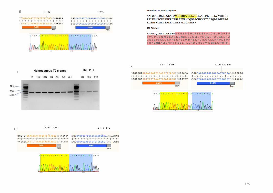
125
E
F
G
H

126
(E) Sequencing for the ABCA7+/- mutant 11H-9G revealed the Cas9 cleavage sites to be 3 bp upstream of the PAM sites, leading to a frameshift mutation. (F) ABCA7-/- mutants were generated by transfecting the heterozygous mutant 11H-11B, followed by colony picking and sub-cloning. PCR screening identified seven T2 homozygous clones, by the presence of 500 bp bands of stronger intensity than the heterozygous 11H ABCA7+/- mutants. (G-H) Sequencing analysis of the homozygous T2 clones identified variability in the Cas9 cleavage sites between the different clones. (I) PCR screens revealed differences between ABCA7 heterozygous and homozygous clones, using the Ex2R reverse primer that binds in the guide RNA-targeted region. For heterozygous clones, the reaction would yield a PCR product of 800bp while no band would be produced for homozygous clones.
I
127
6.3.2 Validation of ABCA7-edited iPSC-derived microglial cells
Having generated the ABCA7 mutant iPSC lines, microglia were derived from the
edited iPS cells before the expression of canonical MG markers was confirmed by
immunostaining (Figure 6.3 A-B). Expression of MG-specific transcripts CABLES1,
C1QA and GPR34 was also measured by qPCR, with comparisons showing significant
upregulation in the microglial samples compared to THP1 monocytes and comparable
expression across the ABCA7 genotypes (Figure 6.3C). Validation of protein expression
in the ABCA7-edited MG cells by immunocytochemistry and western blotting was
attempted over the course of the study, but no suitable antibodies could be found. As
a result, Fluidigm qRT-PCR analysis was used to assess reduction of ABCA7 mRNA
expression in the ABCA7 mutant microglia generated from the heterozygous KO
clones 11H-7C and 11H-11B as well as the homozygous KO clones T2-1G, T2-9G and
T1-11B. Principal component analysis showed a clear separation between samples
from the double KO microglia and Kolf2 WT microglia, while the ABCA7+/- mutants
segregated with the Kolf2 WT samples (Figure 6.3D). Gene expression results revealed
no significant differences in the ABCA7 mRNA levels between the samples, although
there was a trend towards lower expression in the homozygous KO samples (Figure
6.3E). ABCA7 shares sequence homology with ABCA1, the ATP cassette transporter
essential for the regulation of intracellular cholesterol levels (Kaminski et al. 2000).
The potential for a compensatory upregulation of ABCA1 after ABCA7 deletion was
also investigated, with results showing that ABCA1 expression levels did not differ
between WT and ABCA7-edited MG cells (Figure 6.3E).

128
Figure 6.3 Microglia derived from ABCA7+/- and ABCA7-/- iPSCs express canonical microglia markers. (A) Confocal images of ABCA7+/- microglia-like cells positive for P2RY12 and CD45. (B) Differentiation of ABCA7-/- clones generated TMEM119+ IBA1+ iPSC-derived microglia. Scale bar represents 20 µm. (C) Expression of microglia-enriched genes C1QA, GPR34 and CABLES1 in microglia differentiated from ABCA7-edited Kolf2 iPSCs was validated by Fluidigm qPCR. (D) Cluster analysis revealed that ABCA7-/- samples segregated away from WT and ABCA7+/- samples.
Done
Save
Clear
Circle
Point
Kolf2 MG
Kolf2 ABCA7+/-
Kolf2 ABCA7-/-
-4 -2 0 2 4
-1.5
-1.0
-0.5
0.0
0.5
1.0
1.5
2.0
PCA Score Plot
PC1
PC
2
Kolf2 MG2
Kolf2 MG3
11H-11B MG
11H-7C MG
Kolf2 MG1
T2-9G MG
T2-1G MG
T2-11B MGD
ABCA7
ABCA1
0
5
10
15
Log
2 E
xpre
ssio
n
THP-1
Kolf2 MG
Kolf2 ABCA7+/- MG
Kolf2 ABCA7-/- MG
ns
ns
ns
ns
nsns
nsns
ns
ns
ns
ns
E
WT ABCA7 MG
ABCA7+/-
MG
ABCA7-/-
MG
C1QA
GPR34
CABLES10
5
10
15
Log 2
Exp
ress
ion
THP-1
Kolf2 MG
Kolf2 ABCA7+/- MG
Kolf2 ABCA7-/- MG
ns
nsns
ns
ns
ns
ns
ns
ns
C

129
(E) ANOVA comparisons revealed no significant differences in ABCA7 mRNA expression between ABCA7+/-, WT and ABCA7-/- Kolf2 microglia. Levels of the related ATP transporter ABCA1 were similarly unchanged in the ABCA7-edited MG cells. Log2 Expression normalised to reference genes. Statistical analysis used two-way ANOVA with Tukey’s multiple comparison test. Asterisks denote statistical significance: * < 0.05, ** < 0.01, *** < 0.001, ns – not significant. n=3 biological replicates for all samples, except for THP-1 (n=2), Kolf2 ABCA7+/- MG (n=2).
Next, the impact of the ABCA7+/- mutations generated on the microglial immune
response was investigated. ABCA7+/- cells were differentiated on 6-well plates for 14
days and stimulated with 1 µg/ml LPS for 4 hours or 50 ng/ml IL-4 for 24 hours. Under
control unstimulated conditions, both ABCA7+/- and WT MG cells expressed
comparable mRNA levels of M1 activation markers. Following LPS treatment, only
three markers CCR7, TNF- and IL-1ß were significantly upregulated in the Kolf2
ABCA7+/- cells vs control ABCA7+/- cells (p < 0.01) (Figure 6.4A). In contrast, transcript
levels of several other activation markers were significantly upregulated in the LPS-
treated WT Kolf2 cells (p < 0.001), including the chemokines IL-8, CCL18 and CCL22 (p
< 0.01) and the inflammation-induced adhesion molecule ICAM1 (Figure 6.4B). This
suggests that though the LPS-treated cells displayed the classical M1 phenotype, the
ABCA7 mutations restricted the magnitude of the induced pro-inflammatory
response.
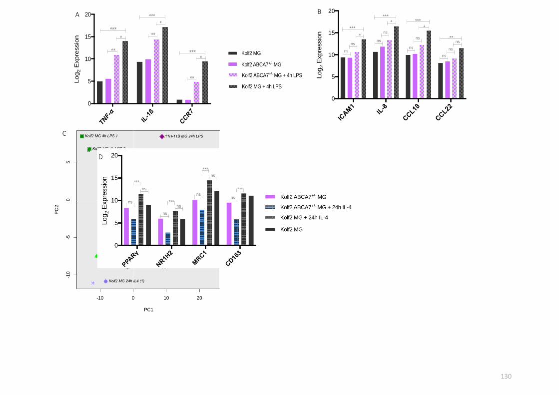
130
TNF-α
IL-1ß
CCR7
0
5
10
15
20
Log
2 E
xpre
ssio
n
Kolf2 MG
Kolf2 ABCA7+/- MG
Kolf2 ABCA7+/- MG + 4h LPS
Kolf2 MG + 4h LPS
*
**
****
**
***
*
**
***
A
ICAM
1IL
-8
CCL18
CCL22
0
5
10
15
20
Log
2 E
xpre
ssio
n
Kolf2 MG
Kolf2 ABCA7+/- MG
Kolf2 ABCA7+/- MG + 4h LPS
Kolf2 MG + 4h LPS
*
ns
****
***
ns*
***
nsns
**
nsns
ns
ns
ns
B
Done
Save
Clear
Circle
Point
Kolf2 MG ABCA7+/-
Kolf2 MG
Kolf2 ABCA7+/- 24h IL-4
Kolf2 ABCA7+/- 4h LPS
Kolf2 ABCA7+/- 24h LPS
Kolf2 24h LPS
Kolf2 4h LPS
Kolf2 24h IL-4
-10 0 10 20
-10
-50
5
PCA Score Plot
PC1
PC
2
11H-11B MG 24h IL-4
11H-7C MG 24h IL-4
11H-11B MG
11H-7C MG
Kolf2 MG2
Kolf2 MG3
Kolf2 MG1
Kolf2 MG 24h IL4 (2)
Kolf2 MG 24h IL4 (1)
11H-7C MG 4h LPS
11H-11B MG 24h LPS
11H-7C MG 24h LPS
Kolf2 MG 24h LPS 2
Kolf2 MG 24h LPS 1
Kolf2 MG 4h LPS 2
Kolf2 MG 4h LPS 1C
PPA
Rγ
NR1H
2
MRC1
CD16
3
0
5
10
15
20
Log
2 E
xpre
ssio
n
Kolf2 ABCA7+/- MG
Kolf2 ABCA7+/- MG + 24h IL-4
Kolf2 MG + 24h IL-4
Kolf2 MG
ns
***
nsns
***
ns
ns
***
nsns
***
D

131
Figure 6.4 The inflammatory response of iPSC-derived microglia to LPS and IL-4 stimulation is influenced by ABCA7 deleterious mutations. (A) Stimulation of Kolf2 ABCA7+/- MG cells with 1 µg/ml LPS for 4 hours resulted in increasing expression of three M1 markers, TNF-, IL-1ß and CCR7. Compared to WT 4h LPS-treated MG cells, this pro-inflammatory response was markedly restrained, as demonstrated by higher expression levels of those three genes in the corresponding Kolf2 WT LPS samples. (B) Expression of ICAM1, IL-8, CCL18 and CCL22 was unchanged in the ABCA7-edited MG cells after the same LPS challenge, whereas the same genes were significantly upregulated in 4h LPS-stimulated WT MG cells. (C) Kolf2 WT and ABCA7+/- MG cells were stimulated with 50 ng/ml IL-4 for 24 hours. Cluster analysis showed that the M2 phenotype of ABCA7-edited IL-4 treated MG cells was markedly different from that of IL-4 treated WT Kolf2 MG cells. (D) IL-4 stimulation resulted in
lower expression of the anti-inflammatory genes (PPAR and NR1H2) and M2-specific markers (MRC1 and CD163) in the ABCA7+/- MG cells compared to WT MG cells. (E) The pro-inflammatory genes CCL2, ICAM1 and NLRP3 were expressed at lower levels in compared to WT MG cells. (F) Differences in the expression of ABCA7 and cholesterol-associated genes were statistically significant between WT and ABCA7+/- MG cells after treatment with IL-4. Log2 Expression normalised to reference genes. Statistical analysis used two-way ANOVA with Tukey’s multiple comparison test. Asterisks denote statistical significance: * < 0.05, ** < 0.01, *** < 0.001, ns – not significant. n=3 biological replicates for all samples, except for Kolf2 ABCA7+/- MG (n=2); Kolf2 ABCA7+/- MG + 4h LPS (n=2); Kolf2 ABCA7+/- MG + 24h IL-4 (n=2).
CCL2
ICAM
1
NLR
P3
IL-1ß
0
5
10
15
Log
2 E
xpre
ssio
n
Kolf2 ABCA7+/- MG
Kolf2 ABCA7+/- MG + 24h IL-4
Kolf2 MG + 24h IL-4
Kolf2 MG
ns*
nsns
**
nsns
*
ns
*
ns
ns
E
ABCA7
SREB
F2
LRP1
LDLR
0
5
10
15
Log
2 E
xpre
ssio
n
Kolf2 ABCA7+/- MG
Kolf2 ABCA7+/- MG + 24h IL-4
Kolf2 MG + 24h IL-4
Kolf2 MG
ns*
ns
ns*
ns
ns
*ns
ns
ns
ns
F

132
This immune response-altering effect was more apparent with stimulation with IL-4,
which induces an M2 phenotype. As shown by the PCA plot in Figure 6.4C, the
inflammatory phenotype of the ABCA7+/- cells was markedly different from that of WT
microglia after IL-4 treatment, with a distinct segregation based on expression of
several immune response genes. This difference was particularly illustrated by the fact
that the M2-specific markers PPAR- and CD163, and the nuclear factor NR1H2 (or
LXR-ß), which showed a trend towards upregulation in the WT IL-4 stimulated cells,
trended towards downregulation in the corresponding ABCA7-edited MG cells (Figure
6.4D). In addition, the pro-inflammatory markers ICAM1, NLRP3 and CCL2 showed a
trend towards reduction in the IL-4-treated ABCA7 mutant microglia, while IL-1ß
reduction was found to be statistically significant. Crucially, the differences in
expression levels of these genes were significant when comparing WT and ABCA7+/-
IL-4 treated cells (Figure 6.4E). It is also worth noting that, following IL-4 stimulation,
levels of ABCA7 and the cholesterol related transcripts SREBF2 and LRP1 were lower
in ABCA7+/- cells vs WT cells (Figure 6.4F). Taken together, these results showed that,
along with pro-inflammation, M2 anti-inflammatory responses were likely to be
affected by ABCA7 loss-of-function mutations.
6.3.3 ABCA7 loss of function impairs phagocytosis in iPSC-derived microglia
ABCA7+/- and ABCA7-/- microglia cells were functionally validated for their capacity to
phagocytose pHrodo red E. coli particles. 96-well plate differentiated microglial cells
(20,000 per well) were incubated with 10 µg pHrodo particles and red channel
fluorescence measurements were taken every 15 minutes for 4 hours in the IncuCyte
S3 live imaging platform (see section 2.5). pHrodo red fluorescence was found to be
markedly reduced in ABCA7+/- and ABCA7-/- cells compared to Kolf2 WT microglial cells
(Figure 6.5A). Quantification of the endpoint fluorescence indicated that ABCA7
homozygous and heterozygous mutant microglia cells ingested significantly fewer
particles than WT Kolf2 cells (p <0.0001) (Figure 6.5B). Pre-treatment of the WT cells
with 1 µM bafilomycin A1 resulted in ablation of the fluorescent signal, confirming
that phagolysosome acidification is a prerequisite for this process. Tellingly, ABCA7
mutant cells showed a higher rate of particle uptake than the inhibitor-treated cells,

133
except for one ABCA7-/- clone T2-1F where the difference was non-significant.
Differences in rate of particle uptake were also uncovered between the three different
ABCA7-/- clones, which individually ingested significantly fewer particles (p< 0.0001)
than the single ABCA7+/- clone studied in this experiment. Therefore, these preliminary
results suggest that microglial phagocytosis of red E. coli bioparticles is considerably
impaired by genetic modification of ABCA7, implying that ABCA7 is essential for
phagocytosis of pathogenic particles.

134
Figure 6.5 Phagocytosis of pHrodo E. coli bioparticles seems to be impaired by ABCA7 heterozygous and homozygous KO mutations in preliminary examinations. iPSC-derived MG cells plated at 20,000 per well were incubated with 10 µg pHrodo E. coli for 4 hours. (A) For negative control, WT MG cells were pre-treated with 10 µM of bafilomycin A1 for 1 hour before E. coli treatment. (B) End point fluorescence values from the experiment in (A) were plotted. Data are presented as mean ± SEM from each genotype or condition with 8-16 technical replicates. n=1 independent experiment.
A B
0
100000
200000
300000
Tota
l red o
bje
ct are
a (
µm
2/Im
age)
Bafilomycin A1
Kolf2
HD109n1
ABCA7 +/- 11H-9G
ABCA7 -/- T2-1F
ABCA7 -/- T2-11B
ABCA7 -/- T2-9G

135
Figure 6.6 Stimulation with the apoptotic molecule C1q induces intracellular accumulation of phospholipids and cholesterol in iPSC-derived microglia. Kolf2 WT and ABCA7+/- MG cells were cultured on PDL-treated and fibronectin-coated Ibidi 8-well chamber slides before stimulation with 10 µg/ml C1q for 1 hour. (A) MG cells were incubated with the phospholipidosis detection reagent LipidTOX for 4 hours. Fluorescence images were taken with a high-resolution microscope – LipidTOX was excited at 543 nm and its emission detected at 594 nm. (B) Following C1q stimulation, MG cells were incubated for 30 mins with 125 µg/ml of the cholesterol-specific probe filipin. Fluorescence images were taken at 300nm. Scale bars represent 20 µm.
Phospholipids Phospholipids Cholesterol

136
6.3.4 Is ABCA7 involved in intracellular lipid trafficking?
ABCA7 has been reported to exert its phagocytosis-mediating activity in mouse macrophages
through recruitment to the cell surface of the low-density lipoprotein receptor-related
protein 1 or LRP1, with both proteins colocalised to phagocytic cups when the cells were
stimulated with C1q (Jehle et al. 2006). C1q is expressed on apoptotic cells, where it binds to
LRP1 on phagocytes, initiating uptake of apoptotic cells (Ogden et al. 2001).
Particle engulfment by phagocytes is a tightly orchestrated process that requires several
proteins operating in concert to generate approximately 100 lipids in order to replenish the
cell membrane expended during initial engulfment (Aderem 2002). This rapid rate of lipid
synthesis is accompanied by increased transcription of many proteins, including the LDL
receptor, fatty acid synthase and the cholesterol synthesis enzymes 3-hydroxy-3-
methylglutaryl CoA synthase and 3-hydroxy-3-methylglutaryl CoA reductase (Castoreno et al.
2005). In order to explore the dynamics of cell membrane biogenesis and lipid metabolism
during microglial phagocytosis, lipid trafficking assays focusing on two species (cholesterol
and phospholipids) were carried out. Microglia differentiated from WT and ABCA7+/- cell lines
were cultured on Ibidi 8-well chamber slides and incubated with 10 µg/ml C1q for 1 hour
before staining for cholesterol and phospholipids with filipin and LipidTOX respectively (see
section 2.5). High resolution images showed that unstimulated microglial cells exhibited low
levels of phospholipids, with the LipidTOX staining enclosed in intracellular puncta (Figure
6.6A). Upon stimulation with C1q, cells exhibited a noticeable intracellular accumulation of
phospholipids, indicative of an increased rate of lipid synthesis. Crucially, no differences in
staining pattern were observed between WT and single KO microglial cells, suggesting that
ABCA7 mutations do not affect phagocytosis-mediated synthesis of phospholipids in microglia
cells (Figure 6.6A). Cholesterol was similarly observed in intracellular punctate structures, as
shown by staining with filipin (Figure 6.6B). ABCA7+/- and WT microglia treated with C1q for 1
h showed elevated levels of filipin staining, the accumulation of cholesterol being
concentrated in intracellular compartments (Figure 6.6B). Though not quantified, cholesterol
staining in the ABCA7+/- cells was less pronounced compared to WT cells, hinting at an altered
capacity to regulate cholesterol levels in response to a phagocytic challenge.

137
6.3 Summary and Discussion
Transcriptomic and gene expression studies described in chapter 3 demonstrated that the
human iPSC-derived microglia generated using the protocol described in this study express
several disease-associated risk genes for AD. To validate the suitability of these in vitro
differentiated cells for modelling the function of microglial-enriched AD risk genes, ABCA7
was chosen as a candidate gene with the intention of knocking out the gene and studying the
subsequent effects of these mutations on microglial function. Using CRISPR/Cas9 genome
editing, ABCA7 mutant clones were generated by excision of a 2.5 kb region at the N-terminal
end of the protein. Potential heterozygous and homozygous KO clones were identified by PCR
screening and sequencing.
Validation by Fluidigm qRT-PCR showed that ABCA7 mRNA expression was found to be
unaltered following genome editing. The mRNA primers used were mapped to the coding
exons 40 and 41 of the full-length protein, with the forward mRNA primer flanking the exon-
exon boundary for increased specificity. No reduction in ABCA7 mRNA levels in either the
heterozygous or homozygous mutants suggests that the mutated mRNA transcripts may have
escaped the nonsense mediated decay machinery. One important limitation of this
investigation was the failure to validate ABCA7 protein expression and therefore the KO lines
generated by Western blotting. A number of antibodies were trialled, namely the rabbit
polyclonal antibody 25339-1-AP from ProteinTech Europe and another rabbit polyclonal
antibody ARP-43690_P050 from Aviva Systems Biology (USA), which targets the middle region
of the protein. Neither antibody worked convincingly by producing any bands when Western
blots experiments were carried out. Recently, a study published the first paper using an
antibody specific to human ABCA7 (Allen et al. 2017), designed to bind the epitope aa 2096-
2146 in the C-terminus of the full-length protein (LS-C291064, LifeSpan BioSciences - USA).
This antibody looks like a good candidate for validation of the KO lines generated in this study.
Nevertheless, preliminary functional assays carried out with pHrodo red E. coli bioparticles
demonstrated a deficit in phagocytosis in ABCA7-edited microglia, showing that ABCA7 is
required for efficient phagocytosis of bioparticles. In addition, the findings underlined the
importance of actin remodelling and phagolysosome fusion in microglial phagocytosis, as the

138
use of the respective inhibitors cytochalasin D and bafilomycin A1 resulted in loss of pHrodo
fluorescence. These observations are in agreement with recent studies of iPSC-derived
macrophage and microglia phagocytosis performed using similar live cell imaging platforms
(Kapellos et al. 2016; Brownjohn et al. 2018). Though this study recapitulates previous mouse
model findings that ABCA7 mediated phagocytosis, another limitation is that only one aspect
of bioparticle phagocytosis, through engagement of the TLR4 was investigated herein. Future
experiments would need to establish whether mechanisms of phagocytosis impairment
through ABCA7 loss-of-function are receptor- and particle-dependent, by challenging cells
with larger bioparticles such as zymosan, a ligand found on the surface of fungi that stimulates
TLR2-mediated uptake. Another essential question that has to be answered is whether these
ABCA7 mutations would affect binding and internalisation of oligomeric Aß, as ABCA7
deletion in AD mouse models enhances plaque load (Kim et al. 2013; Li et al. 2015; Sakae et
al. 2016). Given that iPSC-derived microglia were shown to express several Aß receptors
including TREM2, CD36 and CR1, impairments in Aß phagocytosis would likely point towards
a general phagocytic mechanism that requires ABCA7.
Studies in mouse macrophages have shown that ABCA7 staining is mostly intracellular
(Liensel-Nitschke et al. 2005), whereas the protein co-localises with LRP1 following
stimulation with C1q (Jehle et al. 2006). The latter work also proved that ABCA7 is required
for signalling through LRP1. The ubiquitously expressed receptor LRP1 regulates a diverse
range of physiological processes including endocytosis, lipid metabolism and signal
transduction (May et al. 2005). In macrophages, it promotes anti-inflammatory signalling via
inhibition of pro-inflammatory gene expression (Zurhove et al. 2008; May et al. 2013). This in
turn affects polarisation by favouring an M2 anti-inflammatory phenotype. Correspondingly,
LRP1 downregulation in mouse microglia leads to increased pro-inflammation via JNK and
NFB activation (Yang et al. 2016). LPS and IL-4 stimulation experiments showed that the
inflammatory response of microglia was affected by ABCA7 mutations, suggesting that the
ATP transporter may play a role in microglial anti-inflammatory signalling. ABCA7+/- microglia
exhibited a more restrained inflammatory response to LPS incubation compared to WT Kolf2
cells, as indicated by fewer and statistically smaller increases in pro-inflammatory and M1
activation molecules (Figure 6.4). Similarly, a distinct immune profile was obtained following
stimulation with the anti-inflammatory cytokine IL-4. These data therefore provide

139
preliminary evidence for the involvement of ABCA7 in both the pro- and anti-inflammatory
response of microglial cells, perhaps in modulating the appropriate response to pathogens
and inflammatory stimuli. Given the evidence for LRP1 in this context and that ABCA7 is
required for LRP1 signalling, it is plausible that ABCA7 exerts its inflammation- and
phagocytosis-mediating effects through interactions with LRP1 in lipid rafts. Consequently,
more work is needed to certify these potential links between LRP1 and ABCA7, including their
intracellular localisation in unstimulated microglia and during phagocytosis. This could be
done using confocal microscopy, as well as using co-immunoprecipitation assays that could
enable the identification of proteins and complexes that interact with ABCA7 during
phagocytosis.
C1q treatment of iPSC-derived microglia resulted in increased intracellular cholesterol and
phospholipid staining, indicative of a higher rate of lipid and cholesterol synthesis in response
to or during phagocytosis. Future experiments should aim to quantify these increases and
assess whether ABCA7 mutations inhibit or enhance these processes. Additionally, ABCA7
expression is regulated by intracellular cholesterol levels through the SREBP2 pathway,
whereby its expression is upregulated by cholesterol depletion and downregulated by higher
cellular cholesterol levels (Iwamoto et al. 2006). A series of experiments focusing on this
pathway in the ABCA7 KO iPSC lines were planned, with cholesterol depletion to be mimicked
using the cholesterol-lowering drug mevastatin, an inhibitor of the HMG-CoA reductase
enzyme (Bamji-Mirza et al. 2016). Preliminary results with Kolf2 WT microglia stimulated with
10 µM mevastatin (ML-236B, Sigma Aldrich) for 4 hours showed that mevastatin treatment
significantly increased expression of SREBF2 and the cholesterol-synthesis enzymes fatty acid
synthase (FASN), low density lipoprotein receptor (LDLR) and HMG-CoA synthase 1 (HMGCS1)
(see appendix 5). Likewise, experiments assessing the effects of C1q stimulation on gene
expression of cholesterol pathway genes should be carried out, and comparisons made
between WT and ABCA7-deficient microglia.
Phagocytosis and clearance of debris is a quintessential and defining function of microglia
(Gordon & Martinez 2010). Indeed, a growing research area within the field revolves around
their involvement in synaptic pruning, whereby microglia engulf and eliminate synapses
during development (Paolicelli et al. 2011; Schafer et al. 2012). Though there is very little

140
evidence for microglial-mediated synaptic pruning after development, proper microglia
function is critical for the correcting wiring of embryonic brain circuits (Squarzoni et al. 2014).
Furthermore, microglia have been shown to mediate synaptic loss in AD mouse models
through the CR3 pathway, a process that precedes plaque deposition and is dependent on
C1q expression (Hong et al. 2016). Fluorescence imaging of synaptic membrane dynamics has
been reported using the pH-sensitive pHluorin (Miesenböck et al. 1998; Royle et al. 2008) and
thus can be adapted for in vitro live imaging assays, by co-culturing microglia with synapto-
pHluorin-expressing iPSC-derived neurons. This would allow to investigate synaptic pruning
in vitro as well as the effects of ABCA7 mutations on this microglial activity.

141
7. GENERAL DISCUSSION
7.1 General discussion and future perspectives
7.1.1 Characterisation of the phenotype of HD109 iPSC-derived microglia
The accumulation of reactive microglia is a key feature of the HD brain (Sapp et al. 2001), with
imaging studies going on to demonstrate that this extensive microglial activation is apparent
long before age of onset in HD gene carriers (Tai et al. 2007) and that it correlates with
neuronal loss in symptomatic patients (Pavese et al. 2006). Moreover, studies in mouse
models and post-mortem human brains have shown that HD microglia acquire an
inflammatory phenotype, resulting in elevated levels of proinflammatory cytokines
(Dalrymple et al. 2007; Silvestroni et al. 2009). Though microglial activation in the HD brain
has been relatively well reported, it remains unclear whether this phenomenon is a secondary
response to neuronal degeneration or mainly down to the cell-autonomous effects of mutant
HTT expression in immune cells.
The first aim of this thesis was to investigate the phenotype of microglia derived from a
Huntington’s disease human iPS cell line containing 109 CAG repeats in the HTT gene. The
transcriptome of HD109 iPSC-derived microglia was compared to that of WT cells derived
from a different genetic background by RNA sequencing. The transcriptional hallmarks of
mutant HTT-associated immune dysfunction, which include NFB dysregulation and higher
expression of pro-inflammatory SPI1-target genes, were not detected in the HD109 microglia.
In fact, the findings presented in chapter 4 point towards the notion that the pathogenic
hallmarks of mutant HTT-expressing microglia, as described in the literature, may not be
innate to their basal transcriptome.
The study’s focus then turned to assessing whether the pro-inflammatory properties of HD
microglia would manifest following exogenous stimulation. Surprisingly, results showed that
the in vitro HD microglia did not exhibit a hyper-reactive activation profile when challenged
with LPS. When paying close attention to genes belonging to the two HD affected pathways
of NFB and LPS, no evidence of dysregulation in gene expression were found in those
pathways, contrary to findings reported elsewhere (Crotti et al. 2014).

142
These results therefore indicate that the transcriptome of human HD microglia may not be
primed; instead, evidence points towards a distinct neuroprotective signature with several
pathways highlighted as activated in patient-isolated monocytes predicted to be inhibited. As
already mentioned, one attractive explanation is that this is down to the impact of the
neuronal environment, particularly the neuron-derived cytokine IL-34 on the microglial
phenotype, which illustrates the transcriptional alterations that microglia undergo as they
populate the embryonic brain. This could be viewed as an adaptation of the HD microglial
transcriptome to its surrounding neuronal environment, shown to be susceptible to
degeneration early in neurodevelopment (Molero et al. 2009; Ring et al. 2015). Indeed, the
fragile HD brain may initially potentiate the phenotype of microglia in HD, potentially
dampening the priming effects of mutant HTT expression.
Another possibility is that this inhibitory transcriptomic signal represents a human microglial-
specific effect, given that microglia from HD mouse models exhibit activated inflammatory
pathways. A crucial point of difference between the findings outlined in chapter 4 and those
of patient-derived monocytes resides in the age of the patient-derived cells from Miller et al.
2016, where the mean age of participants at the early or moderate stage of the disease was
48 years old. In contrast, a feature of differentiation protocols is that they seldom give rise to
adult differentiated cells. Indeed, this remains a great challenge for the iPSC modelling field.
Given this caveat, it is plausible that differences in transcriptome-wide pathway activation
from the respective studies reflect different stages of the choreography of inflammation in
Huntington’s disease. Furthermore, these findings imply that the basal transcriptome of
microglia in HD may be non-pathogenic and/or neuroprotective, with gradual changes in
phenotype towards those reported in mouse models and HD patients with characteristics
such as increased pro-inflammation, as a likely consequence of exposure to the HD brain
environment and degeneration in the striatum. One way to test this hypothesis would be to
use human iPSC-derived microglia from HD and isogenic control lines in combination with
xenotransplantation compatible WT mice (e.g. MITRG) (Rongvaux et al. 2014) and
corresponding HD mouse models, along with single cell transcriptomic approaches to
investigate the effects of both ageing and exposure to the HD brain microenvironment on the
immune phenotype of human microglia. Such an experiment would answer pertinent

143
questions about the effect of ageing on the phenotype of HD microglia. In addition, this would
shed a light on the contribution of diseased and non-diseased microglia on inflammatory
processes in the HD brain, and subsequently on neural degeneration in the striatum.
7.1.2 The effects of correcting the HD CAG mutation on the microglial phenotype
Comparing iPSC-derived neural stem cells, Xu et al. 2017 recently found that transcriptome
differences between HD and non-isogenic lines were not detected when comparing HD with
isogenic corrected line samples, suggesting that those differences resulted from non-HD-
specific effects, a consequence of genetic background variations. The second aim of this thesis
was therefore to determine the effects of correcting the HD CAG expansion mutation on the
phenotype of HD109 iPSC-derived microglia. Given the predictions of potential impairments
in several immune pathways in the HD109 microglia (when compared to WT Kolf2 iPSC-
derived microglia), experiments were carried out to ascertain the HD-specific nature of these
HD109 transcriptome changes, with comparisons against microglia differentiated from HD
isogenic corrected iPSC lines. When assayed, the HD109 cells displayed no functional deficits
in phagocytosis or chemotaxis compared to HD corrected and Kolf2 cells. Similarly, an
investigation into the immune response of the corrected HD microglia failed to pinpoint
mutant HTT-specific phenotypic differences in the LPS response of HD109 microglia. Taken
together, the data point towards genetic background, rather than HD-specific, differences
between the transcriptome of HD109 and Kolf2 microglia. This is best illustrated by the fact
that while RNA-seq data showed lower levels of chemokines expressed in HD109 cells
implying a dysregulation in chemokine signalling, validation by Fluidigm qRT-PCR confirmed
that these differences were most likely due to genetic background, as the LPS chemokine
profile of the HD109 microglia post LPS was found to match that of the LPS-stimulated HD
corrected cells.
The absence of any predicted transcriptome-associated functional dysfunctions in the HD109
microglia, allied with the lack of manifestation of phenotypic differences with the HD
corrected cells, could be explained by an absence of mutant HTT expression in the in vitro
derived HD109 microglia. One way to confirm this hypothesis would be to measure mutant

144
HTT expression by Western blotting, which, due to time constraints, was not undertaken and
remains a significant limitation of this study.
Mutant HTT detection has been reported in new-born mice microglia (Crotti et al. 2014), but
not human microglia, although it was found to be expressed in other immune cells isolated
from symptomatic HD patients such monocytes, T cells and B cells (Weiss et al. 2012).
However, it should be pointed out that the characteristics and reactivity of microglia in rodent
models of Huntington’s disease vary greatly across species. In a recent study, Harrison et al.
2018 used quinolinic acid lesion models of HD to evaluate differences in survival of engrafted
tissues in mice and rats and demonstrated that mice models exhibited a lower engraftment
survival than equivalent rat grafts, and substantially higher microglia activation and
recruitment to the site of transplantation (Harrison et al. 2018). The outcome of higher
mouse microglia reactivity has many implications that are important to consider, not just for
mouse transplantation studies (since mouse models comprise the majority of transgenic HD
models) but also for the fields of microglial research in general and in neurodegenerative
diseases such HD and AD. Additionally, it emphasises a particular disparity in the immune
response between rodent models that is under-reported in the neuroinflammation field and
is highly likely to translate into differences with human cells. In turn, it could be argued that
this could be reflected in the human iPSC models used in this work.
No matter whether mutant HTT expression can be detected in HD iPSC-derived microglia in
future experiments, the absence of a HD phenotype in these embryonic-like cells
demonstrates that the cell autonomous effects of mutant HTT on the microglial phenotype
must be triggered by either intrinsic cellular events (e.g. mutant HTT aggregation or ageing)
or by extrinsic factors, such as interactions between microglia and the HD brain
microenvironment. This entails that the presence of the CAG expansion track in the HTT gene
on its own is insufficient to trigger the manifestation of the pathogenic effects characteristic
of HD microglia, an observation compatible with reports of increased microglial activation
with age in HD patients (Pavese et al. 2006; Tai et al. 2007). It also suggests that, rather than
a causal disease mechanism, microglial activation in HD should be considered as a disease
biomarker. Nonetheless, this does not preclude from microglia contributing to striatal
degeneration.

145
This is the first study to evaluate the effects of reversing the HD mutation on immune
dysfunction in human microglia. Given the extensive evidence for the involvement of
microglia and other immune cells in HD neuropathology, it is imperative to investigate the
effects of mutant HTT lowering on HD human immune cells. In addition, this area of research
is of greater importance in the light of current encouraging developments in human clinical
trials of the mutant HTT-lowering antisense oligonucleotide-based IONIS-HTTRx (RG6042)
(Østergaard et al. 2013; Skotte et al. 2014). Preliminary phase 1/2 trials have assessed safety
of this treatment and confirmed a dose-dependent reduction of mutant HTT in the CSF of HD
patients, using a single-molecule counting mHTT immunoassay (Wild et al. 2015). Studies in
HD mouse models had earlier demonstrated that human mutant HTT-targeting antisense
oligonucleotides produced long-lasting reductions in HTT brain mRNA and protein levels,
which correlated with improved motor function and survival (Kordasiewicz et al. 2012).
7.1.3 Modelling neuroinflammation with iPSC-derived microglia
As well as helping to elucidate the pathogenic mechanisms underlying microglial involvement
in HD, iPSC-based models hold great potential for Alzheimer’s disease. Firstly, they provide a
good alternative to animal models for drug screening. In vitro microglia can certainly be used
to develop assays that probe the effects of immunomodulatory therapies on immune
activation. One aspect of microglial function that can certainly be exploited in this manner,
using Seahorse assays, is the link between metabolism and inflammatory processes, which
work in this thesis attempted to investigate. This is especially relevant to AD, with evidence
for the influence of metabolic processes on microglial function coming from mouse model
studies by Ulland et al. 2017, where the authors demonstrated that TREM2 deletion disrupts
microglial function by impairing other metabolic pathways including glycolysis, the TCA cycle
and the pentose phosphate pathway (Ulland et al. 2017). Indeed, TREM2-deficient mouse
microglia exhibit mitochondrial impairments including reduced mitochondrial mass and
increased autophagy.
Secondly, iPSC-derived microglia can and are being used to understand the role of microglia-
enriched disease-associated risk genes in microglial biology, enabling researchers to look at
both rare and common disease variants. While the former offer an insight into pathological
mechanisms, the latter allows us to model predisposition to disease, increasing the likelihood

146
of discovering preventative therapies. The advent of CRISPR/Cas9 genome editing, along with
the progress made in generating patient-specific iPSCs, have opened the door for in vitro
functional studies in these two contexts.
One important challenge of this microglial differentiation protocol remains that there are
doubts surrounding the suitability of monocultured in vitro microglia. Reasons for this include
the lack of elaborate ramifications in the in vitro microglia and the relatively high expression
of inflammatory genes in the IL-34 and GM-CSF treated iPSC-derived microglia. The first
observation is partly reflective of the neuronal-free culture conditions, while the second is a
reported feature of ex vivo microglial culture (Bohlen et al. 2017). Another possibility,
currently being investigated in our lab, is that the absence of a dampened immune signature
in the in vitro monocultured microglia cells could represent an artefact of in vitro
differentiation with GM-CSF, which has been shown to regulate cell proliferation and enhance
several inflammatory functions (Shiomi & Usui 2015).
Given our rapidly evolving understanding of how the transcriptome, phenotype and functions
of microglia is modulated by the CNS microenvironment, the findings described in this thesis
underline the importance of using the appropriate culture conditions that more faithfully
recapitulate microglia origins, maturation and homeostasis. Therefore, the use of more
complex model systems such as co-cultures that better mimic the natural microglial habitat
should be greatly encouraged, where possible. These systems would ideally integrate
neuronal and astrocytic cell types in order to more faithfully study the effects of the functional
interactions between the relevant cell types on the phenotypes and functionality of the
individual cell types.

147
7.2 Concluding remarks
Immune activation within the CNS is now accepted as a key feature of Alzheimer’s disease,
Huntington’s disease and many other neurodegenerative diseases. In AD, recent GWAS
findings highlighting several microglia-enriched genes as disease-associated risk loci have
focused current research on the role of microglia-mediated inflammation. Using iPSC-derived
microglia, loss-of-function mutations in ABCA7 have been found to impair phagocytosis and
regulate microglial inflammatory responses in preliminary studies. Although limited, the
ABCA7 work in this thesis provides the starting point for more in-depth mechanistic studies
to explore the role of ABCA7 in microglial biology.
Data from human imaging studies in Huntington’s disease point towards the detrimental
influence of microglia-mediated neuroinflammatory processes to disease progression. In
addition, studies in HD mouse models have established that mutant HTT induces pathogenic
cell-autonomous effects on the phenotype of microglia. Using microglia derived from HD
iPSCs, this study demonstrates that the pathogenic and inflammatory phenotype of diseased
HD microglia is not innate to their transcriptome, suggesting that it may therefore be a
consequence of intrinsic factors or extrinsic interactions with the degenerative HD brain
microenvironment. In particular, the lack of manifestation of the phenotypic abnormalities
characteristic of diseased HD microglia in the embryonic-like in vitro microglia suggests that
the HD microglial phenotype may not be accurately reflected outside of the diseased HD brain
environment. These findings provide an insight into the limitations of in vitro models of
microglia, whilst emphasising the need for iPSC-based models that better recapitulate disease
pathology and subsequently better enable dissecting the contribution of microglia to
pathogenesis.

148
8. REFERENCES Abe-dohmae, S. et al., 2004. Human ABCA7 Supports Apolipoprotein-mediated Release of
Cellular Cholesterol and Phospholipid to Generate High Density Lipoprotein. Journal of Biological Chemistry, 279(1), pp.604–611.
Abe-Dohmae, S., Ueda, K. & Yokoyama, S., 2006. ABCA7, a molecule with unknown function. FEBS Letters, 580(4), pp.1178–1182.
Abud, E.M. et al., 2017. iPSC-Derived Human Microglia-like Cells to Study Neurological Disease. Neuron, 94(2), pp.278–293. Available at: http://dx.doi.org/10.1016/j.neuron.2017.03.042.
Abutbul, S. et al., 2012. TGF-β signaling through SMAD2/3 induces the quiescent microglial phenotype within the CNS environment. Glia, 60(7), pp.1160–1171.
Aderem, A., 2002. How to eat something bigger than your head. Cell, 110(1), pp.5–8. Aguzzi, A. et al., 2013. Microglia: Scapegoat, Saboteur, or Something Else ? Science,
156(January), pp.156–162. Aikawa, T., Holm, M. & Kanekiyo, T., 2018. ABCA7 and Pathogenic Pathways of Alzheimer’s
Disease. Brain Sciences, 1, pp.1–13. Akiyama, H. & McGeer, P.L., 1990. Brain microglia constitutively express beta-2 integrins.
Journal of neuroimmunology, 30(1), pp.81–93. Allen, M. et al., 2017. ABCA7 loss-of-function variants, expression, and neurologic disease
risk. Neurology Genetics, 0, pp.1–7. An, M.C. et al., 2012. Genetic Correction of Huntington’s Disease Phenotypes in Induced
Pluripotent Stem Cells. Cell Stem Cell, 11, pp.253–263. An, M.C. et al., 2014. Polyglutamine Disease Modeling: Epitope Based Screen for
Homologous Recombination using CRISPR/Cas9 System. PLoS currents, 6. Andrew, S.E. et al., 1993. The relationship between trinucleotide (CAG) repeat length and
clinical features of Huntington’s disease. Nature genetics, 4(4), pp.398–403. Angelopoulos, P. et al., 2008. Cytokines in Alzheimer’s disease and vascular dementia. The
International journal of neuroscience, 118(12), pp.1659–1672. Ard, M.D. et al., 1996. Scavenging of Alzheimer’s amyloid beta-protein by microglia in
culture. Journal of neuroscience research, 43(2), pp.190–202. Armulik, A. et al., 2010. Pericytes regulate the blood-brain barrier. Nature, 468(7323),
pp.557–561. Asai, H. et al., 2015. Depletion of microglia and inhibition of exosome synthesis halt tau
propagation. Nature Neuroscience, 18(11), pp.1584–1593. Available at: http://www.nature.com/doifinder/10.1038/nn.4132.
Ashburner, M. et al., 2000. Gene ontology: tool for the unification of biology. The Gene Ontology Consortium. Nature genetics, 25(1), pp.25–29.
Askew, K. et al., 2017. Coupled Proliferation and Apoptosis Maintain the Rapid Turnover of Microglia in the Adult Brain. Cell Reports, 18(2), pp.391–405.
Aung, H.T. et al., 2006. LPS regulates proinflammatory gene expression in macrophages by altering histone deacetylase expression. The FASEB journal, 20(9), pp.1315–1327.
Avramopoulos, D., 2009. Genetics of Alzheimer’s disease: recent advances. Genome Medicine, 1(3), pp.1–7.
Bae, S., Park, J. & Kim, J.S., 2014. Cas-OFFinder: A fast and versatile algorithm that searches

149
for potential off-target sites of Cas9 RNA-guided endonucleases. Bioinformatics, 30(10), pp.1473–1475.
Bailey, C.C. et al., 2014. IFITM-Family Proteins: The Cell’s First Line of Antiviral Defense. Annual review of virology, 1, pp.261–283. Available at: http://www.ncbi.nlm.nih.gov/pmc/articles/PMC4295558/.
Bamji-Mirza, M. et al., 2016. Genetic Variations in ABCA7 Can Increase Secreted Levels of Amyloid-40 and Amyloid-42 Peptides and ABCA7 Transcription in Cell Culture Models. Journal of Alzheimer’s disease : JAD, 53, pp.875–892.
Banati, R.B., 2002. Visualising microglial activation in vivo. Glia, 40(2), pp.206–217. Baxter, A.W. et al., 2011. Role of P2X4 receptors in synaptic strengthening in mouse CA1
hippocampal neurons. European Journal of Neuroscience, 34(2), pp.213–220. Beach, T.G., Walker, R. & McGeer, E.G., 1989. Patterns of gliosis in Alzheimer’s disease and
aging cerebrum. Glia, 2(6), pp.420–436. Beek, J., Guskov, A. & Slotboom, D.J., 2014. Structural diversity of ABC transporters. The
Journal of General Physiology, pp.419–435. Bellenguez, C. et al., 2017. Contribution to Alzheimer’s disease risk of rare variants in
TREM2, SORL1, and ABCA7 in 1779 cases and 1273 controls. Neurobiology of Aging, 59, p.220.e1-220.e9.
Bennett, F.C. et al., 2018. A Combination of Ontogeny and CNS Environment Establishes Microglial Identity. Neuron, 98(6), p.1170–1183.e8. Available at: https://doi.org/10.1016/j.neuron.2018.05.014.
Berry, B.J. et al., 2018. Physiological Aβ Concentrations Produce a More Biomimetic Representation of the Alzheimer’s Disease Phenotype in iPSC Derived Human Neurons. ACS Chemical Neuroscience, pp.1–9.
Bertrand, J.Y. et al., 2005. Three pathways to mature macrophages in the early mouse yolk sac Three pathways to mature macrophages in the early mouse yolk sac. Blood, 106(9), pp.3004–3011.
Bhaskar, K. et al., 2010. Regulation of tau pathology by the microglial fractalkine receptor. Neuron, 68(1), pp.19–31. Available at: http://dx.doi.org/10.1016/j.neuron.2010.08.023.
Biber, K. et al., 2007. Neuronal ‘On’ and ‘Off’ signals control microglia. Trends in Neurosciences, 30(11), pp.596–602.
Biswas, S.K. & Mantovani, A., 2010. Macrophage plasticity and interaction with lymphocyte subsets: Cancer as a paradigm. Nature Immunology, 11(10), pp.889–896. Available at: http://dx.doi.org/10.1038/ni.1937.
Biswas, S.K. & Mantovani, A., 2012. Orchestration of metabolism by macrophages. Cell Metabolism, 15(4), pp.432–437. Available at: http://dx.doi.org/10.1016/j.cmet.2011.11.013.
Björkqvist, M. et al., 2008. A novel pathogenic pathway of immune activation detectable before clinical onset in Huntington’s disease. The Journal of experimental medicine, 205(8), pp.1869–77. Available at: http://jem.rupress.org/content/205/8/1869.
Block, M.L., Zecca, L. & Hong, J.-S., 2007. Microglia-mediated neurotoxicity: uncovering the molecular mechanisms. Nature reviews. Neuroscience, 8(1), pp.57–69. Available at: http://www.nature.com/nrn/journal/v8/n1/pdf/nrn2038.pdf.
Bodzioch, M. et al., 1999. The gene encoding ATP-binding cassette transporter 1 is mutated in Tangier disease. Nature genetics, 22(4), pp.347–351.
Bohlen, C.J. et al., 2017. Diverse Requirements for Microglial Survival, Specification, and Function Revealed by Defined-Medium Cultures. Neuron, 94(4), p.759–773.e8.

150
Available at: http://dx.doi.org/10.1016/j.neuron.2017.04.043. Bolger, A.M., Lohse, M. & Usadel, B., 2014. Trimmomatic: a flexible trimmer for Illumina
sequence data. Bioinformatics, 30(15), pp.2114–2120. Bouchard, J. et al., 2012. Cannabinoid Receptor 2 Signaling in Peripheral Immune Cells
Modulates Disease Onset and Severity in Mouse Models of Huntington’s Disease. Journal of Neuroscience, 32(50), pp.18259–18268. Available at: http://www.jneurosci.org/cgi/doi/10.1523/JNEUROSCI.4008-12.2012.
Bradford, J. et al., 2009. Expression of mutant huntingtin in mouse brain astrocytes causes age-dependent neurological symptoms. Proceedings of the National Academy of Sciences, 106(52), pp.22480–22485. Available at: http://www.pnas.org/lookup/doi/10.1073/pnas.0911503106.
Breteler, M.M.B., 2000. Vascular risk factors for Alzheimer’s disease: An epidemiologic perspective. Neurobiology of Aging, 21(2), pp.153–160. Available at: http://www.sciencedirect.com/science/article/pii/S0197458099001104?via%3Dihub [Accessed October 9, 2017].
Broccardo, C. et al., 2001. Comparative analysis of the promoter structure and genomic organization of the human and mouse ABCA7 gene encoding a novel ABCA transporter. Cytogenetics and cell genetics, 92(3–4), pp.264–270.
Brooke, G. et al., 2004. Human lymphocytes interact directly with CD47 through a novel member of the signal regulatory protein (SIRP) family. Journal of immunology, 173(4), pp.2562–2570.
Brown, G.C. & Neher, J.J., 2014. Microglial phagocytosis of live neurons. Nature Reviews Neuroscience, 15(4), pp.209–216. Available at: http://dx.doi.org/10.1038/nrn3710%5Cnpapers2://publication/doi/10.1038/nrn3710.
Brownjohn, P.W. et al., 2018. Functional Studies of Missense TREM2 Mutations in Human Stem Cell-Derived Microglia. Stem Cell Reports, 10, pp.1–14. Available at: https://doi.org/10.1016/j.stemcr.2018.03.003.
Buchrieser, J., James, W. & Moore, M.D., 2017. Human Induced Pluripotent Stem Cell-Derived Macrophages Share Ontogeny with MYB-Independent Tissue-Resident Macrophages. Stem Cell Reports, 8(2), pp.334–345. Available at: http://dx.doi.org/10.1016/j.stemcr.2016.12.020.
Budka, H. et al., 1995. Neuropathological diagnostic criteria for Creutzfeldt-Jakob disease (CJD) and other human spongiform encephalopathies (prion diseases). Brain pathology, 5(4), pp.459–466.
Butovsky, O. et al., 2014. Identification of a unique TGF-β-dependent molecular and functional signature in microglia. Nature neuroscience, 17(1), pp.131–43. Available at: http://www.pubmedcentral.nih.gov/articlerender.fcgi?artid=4066672&tool=pmcentrez&rendertype=abstract.
Buttgereit, A. et al., 2016. Sall1 is a transcriptional regulator defining microglia identity and function. Nature Immunology, 17(12), pp.1397–1406. Available at: http://www.nature.com/doifinder/10.1038/ni.3585.
Cagnin, A. et al., 2001. In-vivo measurement of activated microglia in dementia. Lancet, 358(9280), pp.461–467.
Cajal, 1913 (Contribución al conocimiento de la neuroglia del cerebro humano. Trab. Lab. Invest. Biol. 11, 255–345
Canton, J., 2014. Phagosome maturation in polarized macrophages. Journal of Leukocyte Biology, 96(5), pp.729–738. Available at:

151
http://www.jleukbio.org/cgi/doi/10.1189/jlb.1MR0114-021R. Cardona, A.E. et al., 2006. Control of microglial neurotoxicity by the fractalkine receptor.
Nature Neuroscience, 9(7), pp.917–924. Carter, S.F. et al., 2012. Evidence for Astrocytosis in Prodromal Alzheimer Disease Provided
by 11C-Deuterium-L-Deprenyl: A Multitracer PET Paradigm Combining 11C-Pittsburgh Compound B and 18F-FDG. Journal of Nuclear Medicine, 53(1), pp.37–46. Available at: http://jnm.snmjournals.org/cgi/doi/10.2967/jnumed.110.087031.
Castellano, J.M. et al., 2011. Human apoE Isoforms Differentially Regulate Brain Amyloid-ß Peptide Clearance. Science Translational Medicine, 3(89).
Castoreno, A.B. et al., 2005. Transcriptional regulation of phagocytosis-induced membrane biogenesis by sterol regulatory element binding proteins. Proceedings of the National Academy of Sciences of the United States of America, 102(37), pp.13129–13134.
Cattaneo, E., Zuccato, C. & Tartari, M., 2005. Normal huntingtin function: An alternative approach to Huntington’s disease. Nature Reviews Neuroscience, 6(12), pp.919–930.
Cervantes, J.L. et al., 2012. TLR8: the forgotten relative revindicated. Cellular And Molecular Immunology, 9, p.434. Available at: http://dx.doi.org/10.1038/cmi.2012.38.
Chakrabarty, P. et al., 2015. IL-10 Alters Immunoproteostasis in APP Mice, Increasing Plaque Burden and Worsening Cognitive Behavior. Neuron, 85(3), pp.519–533. Available at: http://dx.doi.org/10.1016/j.neuron.2014.11.020.
Chan, S.L. et al., 2008. ATP-binding cassette transporter A7 regulates processing of amyloid precursor protein in vitro. Journal of Neurochemistry, 106(2), pp.793–804.
Chen, H. et al., 2003. Nonsteroidal anti-inflammatory drugs and the risk of Parkinson disease. Archives of neurology, 60(8), pp.1059–1064.
Cherry, J.D., Olschowka, J.A. & O’Banion, M.K., 2014. Are ‘resting’ microglia more ‘M2’? Frontiers in Immunology, 5(NOV), pp.2–5.
Chiu, I.M. et al., 2013. A neurodegeneration-specific gene-expression signature of acutely isolated microglia from an amyotrophic lateral sclerosis mouse model. Cell Reports, 4(2), pp.385–401. Available at: http://dx.doi.org/10.1016/j.celrep.2013.06.018.
Choi, S.H. et al., 2012. Inhibition of NADPH oxidase promotes alternative and anti-inflammatory microglial activation during neuroinflammation. Journal of Neurochemistry, 120(2), pp.292–301.
Choo, I.L.H. et al., 2014. Astrocytosis measured by (1)(1)C-deprenyl PET correlates with decrease in gray matter density in the parahippocampus of prodromal Alzheimer’s patients. European journal of nuclear medicine and molecular imaging, 41(11), pp.2120–2126.
Clark, R.B., 2002. The role of PPARs in inflammation and immunity. Journal of leukocyte biology, 71(3), pp.388–400.
Colantuoni, C. et al., 2011. Temporal dynamics and genetic control of transcription in the human prefrontal cortex. Nature, 478(7370), pp.519–523.
Colonna, M., 2003. Trems in the immune system and beyond. Nature Reviews Immunology, 3(6), pp.445–453.
Corder, E.H. et al., 1993. Gene Dose of Apolipoprotein E Type 4 Allele and the Risk of Alzheimer’s Disease in Late Onset Families. Science, 261(14), pp.921–923.
Costa, V. & Scorrano, L., 2012. Shaping the role of mitochondria in the pathogenesis of Huntington’s disease. EMBO Journal, 31(8), pp.1853–1864. Available at: http://dx.doi.org/10.1038/emboj.2012.65.
Croft, D. et al., 2014. The Reactome pathway knowledgebase. Nucleic acids research,

152
42(Database issue), pp.D472-7. Cronk, J.C. et al., 2018. Peripherally derived macrophages can engraft the brain independent
of irradiation and maintain an identity distinct from microglia. The Journal of Experimental Medicine, 215(6), pp.1627–1647. Available at: http://www.jem.org/lookup/doi/10.1084/jem.20180247.
Crotti, A. et al., 2014. Mutant Huntingtin promotes autonomous microglia activation via myeloid lineage-determining factors. Nature Neuroscience, 17(4), pp.513–521.
Crotti, A. & Glass, C.K., 2015. The choreography of neuroinflammation in Huntington’s disease. Trends in Immunology, 36(6), pp.364–373. Available at: http://dx.doi.org/10.1016/j.it.2015.04.007.
Cummings, J.L., Morstorf, T. & Zhong, K., 2014. Alzheimer’s disease drug-development pipeline : few candidates , frequent failures. Alzheimer’s research & therapy, pp.1–7.
Cuyvers, E. et al., 2015. Mutations in ABCA7 in a Belgian cohort of Alzheimer’s disease patients: A targeted resequencing study. The Lancet Neurology, 14(8), pp.814–822. Available at: http://dx.doi.org/10.1016/S1474-4422(15)00133-7.
D’Andrea, M.R., Cole, G.M. & Ard, M.D., 2004. The microglial phagocytic role with specific plaque types in the Alzheimer disease brain. Neurobiology of Aging, 25, pp.675–683.
Dagher, N.N. et al., 2015. Colony-stimulating factor 1 receptor inhibition prevents microglial plaque association and improves cognition in 3xTg-AD mice. Journal of Neuroinflammation, 12(1), pp.1–14. Available at: http://dx.doi.org/10.1186/s12974-015-0366-9.
Dahlgren, K.N. et al., 2002. Oligomeric and fibrillar species of amyloid-β peptides differentially affect neuronal viability. Journal of Biological Chemistry, 277(35), pp.32046–32053.
Dalrymple, A. et al., 2007. Proteomic profiling of plasma in Huntington’s disease reveals neuroinflammatory activation and biomarker candidates. Journal of Proteome Research, 6(7), pp.2833–2840.
Damiano, M. et al., 2010. Mitochondria in Huntington’s disease. Biochimica et Biophysica Acta - Molecular Basis of Disease, 1802(1), pp.52–61. Available at: http://dx.doi.org/10.1016/j.bbadis.2009.07.012.
Danbolt, N.C., 2001. Glutamate uptake. Progress in neurobiology, 65(1), pp.1–105. Davalos, D. et al., 2005. ATP mediates rapid microglial response to local brain injury in vivo.
Nature Neuroscience, 8(6), pp.752–758. Davis, E.J., Foster, T.D. & Thomas, W.E., 1994. Cellular forms and functions of brain
microglia. Brain research bulletin, 34(1), pp.73–78. Dawe, H.R. et al., 2003. ADF/cofilin controls cell polarity during fibroblast migration. Current
Biology, 13(3), pp.252–257. Dean, M. & Annilo, T., 2005. Evolution of the ATP-binding cassette (ABC) transporter
superfamily in vertebrates. Annual review of genomics and human genetics, 6, pp.123–142.
Dehairs, J. et al., 2016. CRISP-ID: Decoding CRISPR mediated indels by Sanger sequencing. Scientific Reports, 6(July), pp.1–5. Available at: http://dx.doi.org/10.1038/srep28973.
Dobin, A. et al., 2013. STAR: ultrafast universal RNA-seq aligner. Bioinformatics, 29(1), pp.15–21.
Donato, M. et al., 2013. Analysis and correction of crosstalk effects in pathway analysis. Genome Research, 23, pp.1885–1893.
Draghici, S. et al., 2007. A systems biology approach for pathway level analysis. Genome

153
Research, 17, pp.1537–1545. Drouin-ouellet, J. et al., 2015. Cerebrovascular and Blood – Brain Barrier Impairments in
Huntington’s Disease : Potential Implications for Its Pathophysiology. Annals of Neurology, 78, pp.160–177.
Durafourt, B.A. et al., 2012. Comparison of Polarization Properties of Human Adult Microglia and Blood-Derived Macrophages. Glia, 727(August 2011), pp.717–727.
Dursun, E. et al., 2015. The interleukin 1 alpha, interleukin 1 beta, interleukin 6 and alpha-2-macroglobulin serum levels in patients with early or late onset Alzheimer’s disease, mild cognitive impairment or Parkinson’s disease. Journal of neuroimmunology, 283, pp.50–57.
Edison, P. et al., 2008. Microglia, amyloid, and cognition in Alzheimer’s disease: An [11C](R)PK11195-PET and [11C]PIB-PET study. Neurobiology of disease, 32(3), pp.412–419.
Ellrichmann, G. et al., 2013. The Role of the Immune System in Huntington’s Disease. Clinical and Developmental Immunology, 2013(541259), pp.1–11. Available at: http://dx.doi.org/10.1155/2013/541259%0AReview.
Erblich, B. et al., 2011. Absence of colony stimulation factor-1 receptor results in loss of microglia, disrupted brain development and olfactory deficits. PLoS ONE, 6(10), p.e26317.
Erdman, L.K. et al., 2009. CD36 and TLR interactions in inflammation and phagocytosis: implications for malaria. Journal of immunology (Baltimore, Md. : 1950), 183(10), pp.6452–6459.
Esiri, M.M., Al Izzi, M.S. & Reading, M.C., 1991. Macrophages, microglial cells, and HLA-DR antigens in fetal and infant brain. Journal of Clinical Pathology, 44, pp.102–106.
Estrada-Sánchez, A.M. & Rebec, G. V., 2012. Corticostriatal dysfunction and glutamate transporter 1 (GLT1) in Huntington’s disease: Interactions between neurons and astrocytes. Basal Ganglia, 2(2), pp.57–66.
Etminan, M., Gill, S. & Samii, A., 2003. Effect of non-steroidal anti-inflammatory drugs on risk of Alzheimer’s disease: systematic review and meta-analysis of observational studies. BMJ, 327, pp.1–5.
Everts, B. et al., 2012. Commitment to glycolysis sustains survival of NO-producing inflammatory dendritic cells. Blood, 120(7), pp.1422–1431.
Everts, B. et al., 2014. TLR-driven early glycolytic reprogramming via the kinases TBK1-IKKvarepsilon supports the anabolic demands of dendritic cell activation. Nature immunology, 15(4), pp.323–332.
Fabelo, N. et al., 2014. Altered lipid composition in cortical lipid rafts occurs at early stages of sporadic Alzheimer’s disease and facilitates APP/BACE1 interactions. Neurobiology of Aging, 35(8), pp.1801–1812. Available at: http://dx.doi.org/10.1016/j.neurobiolaging.2014.02.005.
Fan, R. et al., 2007. Minocycline Reduces Microglial Activation and Improves Behavioral Deficits in a Transgenic Model of Cerebral Microvascular Amyloid. Journal of Neuroscience, 27(12), pp.3057–3063. Available at: http://www.jneurosci.org/cgi/doi/10.1523/JNEUROSCI.4371-06.2007.
Farkas, E. & Luiten, P.G.M., 2001. Cerebral microvascular pathology in aging and Alzheimer’s disease,
Fedoroff, S., Zhai, R. & Novak, J.P., 1997. Microglia and Astroglia Have a Common Progenitor Cell. Journal of Neuroscience Research, 50, pp.477–486.

154
Forlenza, O.V. et al., 2009. Increased serum IL-1beta level in Alzheimer’s disease and mild cognitive impairment. Dementia and geriatric cognitive disorders, 28(6), pp.507–512.
Fowler, J.S. et al., 2005. Translational neuroimaging: positron emission tomography studies of monoamine oxidase. Molecular imaging and biology : MIB : the official publication of the Academy of Molecular Imaging, 7(6), pp.377–387.
Franciosi, S. et al., 2012. Age-dependent neurovascular abnormalities and altered microglial morphology in the YAC128 mouse model of Huntington disease. Neurobiology of Disease, 45(1), pp.438–449. Available at: http://dx.doi.org/10.1016/j.nbd.2011.09.003.
Francis, G.A., Knopp, R.H. & Oram, J.F., 1995. Defective removal of cellular cholesterol and phospholipids by apolipoprotein A-I in Tangier Disease. The Journal of clinical investigation, 96(1), pp.78–87.
Fraser, D.A., Pisalyaput, K. & Tenner, A.J., 2010. C1q enhances microglial clearance of apoptotic neurons and neuronal blebs, and modulates subsequent inflammatory cytokine production. Journal of Neurochemistry, 112(3), pp.733–743.
Fu, Y. et al., 2016. ABCA7 Mediates Phagocytic Clearance of Amyloid-ß in the Brain. Journal of Alzheimer’s disease : JAD, 54, pp.569–584.
Galatro, T.F. et al., 2017. Transcriptomic analysis of purified human cortical microglia reveals age-associated changes. Nature Neuroscience, 20(8), pp.1162–1174.
Galea, I., Bechmann, I. & Perry, V.H., 2007. What is immune privilege (not)? Trends in Immunology, 28(1), pp.12–18.
Galimberti, D. et al., 2006. Serum MCP-1 levels are increased in mild cognitive impairment and mild Alzheimer’s disease. Neurobiology of aging, 27(12), pp.1763–1768.
Galván-peña, S. & O’Neill, L.A.J., 2014. Metabolic reprograming in macrophage polarization. Frontiers in Immunology, 5(September), pp.1–6.
Gatz, M. et al., 2006. Role of Genes and Environments for Explaining Alzheimer Disease. Archives in Genetic Psychiatry, 63(2), pp.168–174.
GeM-HD Consortium, 2015. Identification of Genetic Factors that Modify Clinical Onset of Huntington’s Disease. Cell, 162, pp.516–526.
Ginhoux, F. et al., 2010. Hematopoietic Waves of Microglial Recruitment and Differentiation Occur in the Central Nervous. Science, 330(2009).
Ginhoux, F. et al., 2013. Origin and differentiation of microglia. Frontiers in cellular neuroscience, 7(April), p.45. Available at: http://www.pubmedcentral.nih.gov/articlerender.fcgi?artid=3627983&tool=pmcentrez&rendertype=abstract.
Goate, A.M. et al., 1991. Segregation of a missense mutation in the amyloid precursor protein gene with familial Alzheimer’s disease. Nature, 349.
Gomez-Nicola, D. & Perry, V.H., 2015. Microglial Dynamics and Role in the Healthy and Diseased Brain: A Paradigm of Functional Plasticity. The Neuroscientist, 21(1), pp.169–184.
Gomez Perdiguero, E. et al., 2014. Tissue-resident macrophages originate from yolk-sac-derived erythro-myeloid progenitors. Nature, 518(7540), pp.547–551. Available at: http://www.nature.com/doifinder/10.1038/nature13989.
Gordon, S. & Martinez, F.O., 2010. Alternative activation of macrophages: Mechanism and functions. Immunity, 32(5), pp.593–604. Available at: http://dx.doi.org/10.1016/j.immuni.2010.05.007.
Gosselin, D. et al., 2014. Environment drives selection and function of enhancers controlling tissue-specific macrophage identities. Cell, 159(6), pp.1327–1340. Available at:

155
http://dx.doi.org/10.1016/j.cell.2014.11.023. Gotz, J. & Ittner, L.M., 2008. Animal models of Alzheimer’s disease and frontotemporal
dementia. Nature Neuroscience, 9(july). Grabert, K. et al., 2016. Microglial brain region−dependent diversity and selective regional
sensitivities to aging. Nature Neuroscience, (January). Available at: http://www.nature.com/doifinder/10.1038/nn.4222.
Graeber, M.B., Li, W. & Rodriguez, M.L., 2011. Role of microglia in CNS inflammation. FEBS Letters, 585(23), pp.3798–3805. Available at: http://dx.doi.org/10.1016/j.febslet.2011.08.033.
Gray, M. et al., 2008. Full-Length Human Mutant Huntingtin with a Stable Polyglutamine Repeat Can Elicit Progressive and Selective Neuropathogenesis in BACHD Mice. Journal of Neuroscience, 28(24), pp.6182–6195. Available at: http://www.jneurosci.org/cgi/doi/10.1523/JNEUROSCI.0857-08.2008.
Greter, M. et al., 2012. Stroma-Derived Interleukin-34 Controls the Development and Maintenance of Langerhans Cells and the Maintenance of Microglia. Immunity, 37, pp.1050–1060.
Griciuc, A. et al., 2013. Alzheimer’s Disease Risk Gene CD33 Inhibits Microglial Uptake of Amyloid Beta. Neuron, 78(4), pp.631–643. Available at: http://dx.doi.org/10.1016/j.neuron.2013.04.014.
Le Guennec, K. et al., 2016. ABCA7 rare variants and Alzheimer disease risk. Neurology, 86(23), pp.2134–2137.
Guerreiro, R. et al., 2013. TREM2 Variants in Alzheimer’s Disease. New England Journal of Medicine, 368(2), pp.117–127.
Gulyas, B. et al., 2011. Activated MAO-B in the brain of Alzheimer patients, demonstrated by [11C]-L-deprenyl using whole hemisphere autoradiography. Neurochemistry international, 58(1), pp.60–68.
Ha, S.-D. et al., 2008. Cathepsin B Is Involved in the Trafficking of TNF- -Containing Vesicles to the Plasma Membrane in Macrophages. The Journal of Immunology, 181(1), pp.690–697. Available at: http://www.jimmunol.org/cgi/doi/10.4049/jimmunol.181.1.690.
van de Haar, H.J. et al., 2016. Blood-Brain Barrier Leakage in Patients with Early Alzheimer Disease. Radiology, 281(2), pp.527–535. Available at: https://doi.org/10.1148/radiol.2016152244.
van de Haar, H.J. et al., 2016. Neurovascular unit impairment in early Alzheimer’s disease measured with magnetic resonance imaging. Neurobiology of Aging, 45, pp.190–196. Available at: http://dx.doi.org/10.1016/j.neurobiolaging.2016.06.006.
de Haas, A.H., Boddeke, H.W.G.M. & Biber, K., 2008. Region-specific expression of immunoregulatory proteins on microglia in the healthy CNS. Glia, 56(8), pp.888–894.
Haenseler, W. et al., 2017. Excess α-synuclein compromises phagocytosis in iPSC-derived macrophages. Scientific Reports, (July), pp.1–11. Available at: http://dx.doi.org/10.1038/s41598-017-09362-3.
Halliday, G.M. et al., 1998. Regional specificity of brain atrophy in Huntington’s disease. Experimental neurology, 154(2), pp.663–672.
Hamelin, L. et al., 2016. Early and protective microglial activation in Alzheimer’s disease: a prospective study using 18 F-DPA-714 PET imaging. Brain, 139(4), pp.1252–1264. Available at: https://academic.oup.com/brain/article-lookup/doi/10.1093/brain/aww017.
Hammond, T.R. et al., 2018. Complex cell-state changes revealed by single cell RNA

156
sequencing of 76 , 149 microglia throughout the mouse lifespan and in the injured brain. Biorxiv, pp.1–22.
Hanisch, U.-K.K. & Kettenmann, H., 2007. Microglia: active sensor and versatile effector cells in the normal and pathologic brain. Nature Neuroscience, 10(11), pp.1387–1394. Available at: isi:000250508400013%5Cnhttp://www.ncbi.nlm.nih.gov/pubmed/17965659.
Hanisch, U.K., Johnson, T. V. & Kipnis, J., 2008. Toll-like receptors: roles in neuroprotection? Trends in Neurosciences, 31(4), pp.176–182.
Hao, C., Richardson, A. & Fedoroff, S., 1991. Macrophage-like cells originate from neuroepithelium in culture: characterization and properties of the macrophage-like cells. International journal of developmental neuroscience, 9(1), pp.1–14.
Hardy, J. & Selkoe, D.J., 2002. The amyloid hypothesis of Alzheimer’s disease: progress and problems on the road to therapeutics. Science (New York, N.Y.), 297(5580), pp.353–356. Available at: http://www.ncbi.nlm.nih.gov/pubmed/12130773.
Hardy, J.A. & Higgins, G.A., 1992. Alzheimer’s disease: the amyloid cascade hypothesis. Science (New York, N.Y.), 256(5054), pp.184–185.
Harold, D. et al., 2009. Genome-wide association study identifies variants at CLU and PICALM associated with Alzheimer ’ s disease. Nature genetics, 41(10).
Harris, M.A. et al., 2004. The Gene Ontology (GO) database and informatics resource. Nucleic acids research, 32(Database issue), pp.D258-61.
Harrison, D.J. et al., 2018. The Effect of Tissue Preparation and Donor Age on Striatal Graft Morphology in the Mouse. Cell Transplantation, 27(2), pp.230–244.
Haynes, S.E. et al., 2006. The P2Y12 receptor regulates microglial activation by extracellular nucleotides. Nature neuroscience, 9(12), pp.1512–1519.
HD iPSC Consortium, 2012. Induced Pluripotent Stem Cells from Patients with Huntington’s Disease Show CAG-Repeat-Extension Associated Phenotypes. Cell Stem Cell, 11, pp.264–278.
Heneka, M.T. et al., 2013. NLRP3 is activated in Alzheimer’s disease and contributes to pathology in APP/PS1 mice. Nature, 493(7434), pp.674–678. Available at: http://www.ncbi.nlm.nih.gov/pubmed/23254930%5Cnhttp://www.nature.com/nature/journal/v493/n7434/pdf/nature11729.pdf.
Herrup, K., 2015. The case for rejecting the amyloid cascade hypothesis. Nature Neuroscience, 18(6), pp.794–799. Available at: http://www.nature.com/doifinder/10.1038/nn.4017%5Cnhttp://www.ncbi.nlm.nih.gov/pubmed/26007212%5Cnhttp://www.nature.com/neuro/journal/v18/n6/pdf/nn.4017.pdf.
Hickman, S.E. et al., 2013. The microglial sensome revealed by direct RNA sequencing. Nature neuroscience, 16(12), pp.1896–905. Available at: http://www.pubmedcentral.nih.gov/articlerender.fcgi?artid=3840123&tool=pmcentrez&rendertype=abstract.
Higgins, C.F., 1992. ABC transporters: from microorganisms to man. Annual review of cell biology, 8, pp.67–113.
Higgins, C.F. & Linton, K.J., 2004. The ATP switch model for ABC transporters. Nature reviews. Structural & molecular biology, 11(10), pp.918–926.
Hirschhorn, J.N. & Daly, M.J., 2005. genome-wide association studies for common diseases and complex traits. Nature reviews. Genetics, 6(February), pp.95–108.
Hoek, R.M. et al., 2000. Down-regulation of the macrophage lineage through interaction

157
with OX2 (CD200). Science (New York, N.Y.), 290(5497), pp.1768–1771. Hollingworth, P. et al., 2011. Common variants at ABCA7, MS4A6A/MS4A4E, EPHA1, CD33
and CD2AP are associated with Alzheimer’s disease. Nature genetics, 43(5), pp.429–436.
Holtzman, D.M., Morris, J.C. & Goate, A.M., 2011. Alzheimer’s Disease : The Challenge of the Second Century. Science Translational Medicine, 3(77), pp.1–18.
Hong, S. et al., 2016. Complement and microglia mediate early synapse loss in Alzheimer mouse models. Science, 8373, pp.1–9. Available at: http://www.sciencemag.org/cgi/doi/10.1126/science.aad8373.
Hoozemans, J.J.M. et al., 2011. Soothing the inflamed brain: effect of non-steroidal anti-inflammatory drugs on Alzheimer’s disease pathology. CNS & neurological disorders drug targets, 10(1), pp.57–67.
Hsiao, H.-Y. et al., 2014. Inhibition of soluble tumor necrosis factor is therapeutic in Huntington’s disease. Human Molecular Genetics, 23(16), pp.4328–4344. Available at: https://academic.oup.com/hmg/article-lookup/doi/10.1093/hmg/ddu151.
Huang, D.W., Sherman, B.T. & Lempicki, R.A., 2008. Systematic and integrative analysis of large gene lists using DAVID bioinformatics resources. Nature Protocols, 4, p.44. Available at: http://dx.doi.org/10.1038/nprot.2008.211.
Huyhn, a et al., 1995. Characterization of hematopoietic progenitors from human yolk sacs and embryos. Blood, 86(12), pp.4474–85. Available at: http://www.ncbi.nlm.nih.gov/pubmed/8541536.
Ikeda, Y. et al., 2003. Posttranscriptional regulation of human ABCA7 and its function for the apoA-I-dependent lipid release. Biochemical and Biophysical Research Communications, 311(2), pp.313–318.
Imai, F. et al., 2007. Neuroprotective effect of exogenous microglia in global brain ischemia. Journal of cerebral blood flow and metabolism, 27(3), pp.488–500.
Ishii, K. et al., 1997. Increased Aß42(43)-plaque deposition in early-onset familial Alzheimer’s disease brains with the deletion of exon 9 and the missense point mutation ( H163R ) in the PS-1 gene. Neuroscience Letters, 228, pp.17–20.
Itagaki, S. et al., 1989. Relationship of microglia and astrocytes to amyloid deposits of Alzheimer disease. Journal of neuroimmunology, 24(3), pp.173–182.
Iwamoto, N. et al., 2006. ABCA7 expression is regulated by cellular cholesterol through the SREBP2 pathway and associated with phagocytosis. Journal of lipid research, 47(9), pp.1915–1927.
Jankowsky, J.L. et al., 2004. Mutant presenilins specifically elevate the levels of the 42 residue beta-amyloid peptide in vivo: evidence for augmentation of a 42-specific gamma secretase. Human molecular genetics, 13(2), pp.159–170.
Jehle, A.W. et al., 2006. ATP-binding cassette transporter A7 enhances phagocytosis of apoptotic cells and associated ERK signaling in macrophages. The Journal of cell biology, 174(4), pp.547–556.
Jones, L. et al., 2015. Convergent genetic and expression data implicate immunity in Alzheimer’s disease. Alzheimer’s and Dementia, 11(6), pp.658–671.
Jonsson, T. et al., 2013. Variant of TREM2 Associated with the Risk of Alzheimer’s Disease. New England Journal of Medicine, 368(2), pp.107–116.
Jossan, S.S. et al., 1991. Monoamine oxidase B in brains from patients with Alzheimer’s disease: a biochemical and autoradiographical study. Neuroscience, 45(1), pp.1–12.
Kajiwara, M. et al., 2012. Donor-dependent variations in hepatic differentiation from

158
human-induced pluripotent stem cells. Proceedings of the National Academy of Sciences, 109(36), pp.14716–14716. Available at: http://www.pnas.org/cgi/doi/10.1073/pnas.1212710109.
Kalonia, H. et al., 2010. Protective effect of rofecoxib and nimesulide against intra-striatal quinolinic acid-induced behavioral, oxidative stress and mitochondrial dysfunctions in rats. Neurotoxicology, 31(2), pp.195–203.
Kalonia, H. & Kumar, A., 2011. Suppressing inflammatory cascade by cyclo-oxygenase inhibitors attenuates quinolinic acid induced Huntington’s disease-like alterations in rats. Life sciences, 88(17–18), pp.784–791.
Kaminski, W.E. et al., 2000. Identification of a novel human sterol-sensitive ATP-binding cassette transporter (ABCA7). Biochemical and biophysical research communications, 273, pp.532–538.
Kanehisa, M. et al., 2012. KEGG for integration and interpretation of large-scale molecular data sets. Nucleic acids research, 40(Database issue), pp.D109-14.
Kapellos, T.S. et al., 2016. A novel real time imaging platform to quantify macrophage phagocytosis. Biochemical Pharmacology, 116, pp.107–119. Available at: http://dx.doi.org/10.1016/j.bcp.2016.07.011.
Karch, C.M. et al., 2012. Expression of Novel Alzheimer’s Disease Risk Genes in Control and Alzheimer’s Disease Brains. PLoS ONE, 7(11), p.e50976.
Karch, C.M. & Goate, A.M., 2015. Alzheimer’s Disease Risk Genes and Mechanisms of Disease Pathogenesis. Biological Psychiatry, 77(1), pp.43–51. Available at: http://linkinghub.elsevier.com/retrieve/pii/S0006322314003394.
Karlsson, K.R. et al., 2008. Homogeneous monocytes and macrophages from human embryonic stem cells following coculture-free differentiation in M-CSF and IL-3. Experimental Hematology, 36(9), pp.1167–1175.
Kashon, M.L. et al., 2004. Associations of cortical astrogliosis with cognitive performance and dementia status. Journal of Alzheimer’s disease : JAD, 6(6), pp.581–595.
Katakura, T. et al., 2004. CCL17 and IL-10 as effectors that enable alternatively activated macrophages to inhibit the generation of classically activated macrophages. Journal of immunology (Baltimore, Md. : 1950), 172(3), pp.1407–1413.
Kerschbamer, E. & Biagioli, M., 2016. Huntington’s disease as neurodevelopmental disorder: Altered chromatin regulation, coding, and non-coding RNA transcription. Frontiers in Neuroscience, 9(JAN), pp.1–5.
Kettenmann, H. et al., 2011. Physiology of microglia. Physiological reviews, 91(2), pp.461–553. Available at: http://physrev.physiology.org/content/91/2/461.abstract.
Khoshnan, A. et al., 2017. IKKß and mutant huntingtin interactions regulate the expression of IL-34: implications for microglial-mediated neurodegeneration in HD. Human Molecular Genetics, 0(0), pp.1–11.
El Khoury, J. et al., 2007. Ccr2 deficiency impairs microglial accumulation and accelerates progression of Alzheimer-like disease. Nature Medicine, 13(4), pp.432–438.
Kierdorf, K. et al., 2013. Microglia emerge from erythromyeloid precursors via Pu.1- and Irf8-dependent pathways. Nature Neuroscience, 16(3), pp.273–280. Available at: http://dx.doi.org/10.1038/nn.3318.
Kim, S. et al., 2014. Highly efficient RNA-guided genome editing in human cells via delivery of purified Cas9 ribonucleoproteins. Genome Research, 24, pp.1012–1019.
Kim, T.-S. et al., 2008. Changes in the levels of plasma soluble fractalkine in patients with mild cognitive impairment and Alzheimer’s disease. Neuroscience letters, 436(2),

159
pp.196–200. Kim, W.S. et al., 2005. Abca7 null mice retain normal macrophage phosphatidylcholine and
cholesterol efflux activity despite alterations in adipose mass and serum cholesterol levels. Journal of Biological Chemistry, 280(5), pp.3989–3995.
Kim, W.S. et al., 2013. Deletion of Abca7 increases cerebral amyloid-β accumulation in the J20 mouse model of Alzheimer’s disease. Journal of Neuroscience, 33(10), pp.4387–94. Available at: http://www.ncbi.nlm.nih.gov/pubmed/23467355.
Kim, W.S. et al., 2006. Quantitation of ATP-binding cassette subfamily-A transporter gene expression in primary human brain cells. Neuroreport, 17(9), pp.891–6. Available at: http://www.ncbi.nlm.nih.gov/pubmed/16738483.
Kinchen, J.M. et al., 2005. Two pathways converge at CED-10 to mediate actin rearrangement and corpse removal in C . elegans. Nature, 434(649), pp.93–100.
Kitamura, T., Miyake, T. & Fujita, S., 1984. Genesis of Resting Microglia in the Gray Matter of Mouse Hippocampus. Journal of Comparative Neurology, 226, pp.421–433.
Kiyota, T. et al., 2009. CCL2 accelerates microglia-mediated Abeta oligomer formation and progression of neurocognitive dysfunction. PloS one, 4(7), p.e6197.
Kleinberger, G. et al., 2014. TREM2 mutations implicated in neurodegeneration impair cell surface transport and phagocytosis. Science Translational Medicine, 6(243).
Koldamova, R., Staufenbiel, M. & Lefterov, I., 2005. Lack of ABCA1 Considerably Decreases Brain ApoE Level and Increases Amyloid Deposition in APP23 Mice. Journal of Biological Chemistry, 280(52), pp.43224–43235.
Kordasiewicz, H.B. et al., 2012. Sustained Therapeutic Reversal of Huntington’s Disease by Transient Repression of Huntingtin Synthesis. Neuron, 74(6), pp.1031–1044. Available at: http://dx.doi.org/10.1016/j.neuron.2012.05.009.
Krabbe, G. et al., 2013. Functional Impairment of Microglia Coincides with Beta-Amyloid Deposition in Mice with Alzheimer-Like Pathology. PLoS ONE, 8(4).
Krasemann, S. et al., 2017. The TREM2-APOE Pathway Drives the Transcriptional Phenotype of Dysfunctional Microglia in Neurodegenerative Diseases. Immunity, 47(3), pp.566–581. Available at: http://dx.doi.org/10.1016/j.immuni.2017.08.008.
Kreisl, W.C. et al., 2013. In vivo radioligand binding to translocator protein correlates with severity of Alzheimer’s disease. Brain, 136(7), pp.2228–2238.
Krencik, R. et al., 2011. Specification of transplantable astroglial subtypes from human pluripotent stem cells. Nature Biotechnology, 29(6), pp.528–534. Available at: http://www.nature.com/doifinder/10.1038/nbt.1877.
Kreutzberg, G.W., 1996. Microglia: a sensor for pathological events in the CNS. Trends in neurosciences, 19(8), pp.312–318.
Kumaravelu, P. et al., 2002. Quantitative developmental anatomy of definitive haematopoietic stem cells/long-term repopulating units (HSC/RUs): role of the aorta-gonad-mesonephros (AGM) region and the yolk sac in colonisation of the mouse embryonic liver. Development (Cambridge, England), 129(21), pp.4891–4899.
Kunkle, B.W. et al., 2017. Targeted sequencing of ABCA7 identifies splicing, stop-gain and intronic risk variants for Alzheimer disease. Neuroscience Letters, 649, pp.124–129. Available at: http://dx.doi.org/10.1016/j.neulet.2017.04.014.
Kwan, W. et al., 2012. Bone Marrow Transplantation Confers Modest Benefits in Mouse Models of Huntington’s Disease. Journal of Neuroscience, 32(1), pp.133–142. Available at: http://www.jneurosci.org/cgi/doi/10.1523/JNEUROSCI.4846-11.2012.
Kwan, W. et al., 2012. Mutant huntingtin impairs immune cell migration in Huntington

160
disease. J Clin Invest., 122(12), pp.4737–4747. Labadorf, A. et al., 2015. RNA Sequence Analysis of Human Huntington Disease Brain
Reveals an Extensive Increase in Inflammatory and Developmental Gene Expression. Plos One, 10(12), p.e0143563. Available at: http://dx.plos.org/10.1371/journal.pone.0143563.
Lacy, P. & Stow, J.L., 2011. Cytokine release from innate immune cells : association with diverse membrane trafficking pathways. Blood, 118(1), pp.9–19.
Laferla, F.M. & Green, K.N., 2012. Animal Models of Alzheimer Disease. Cold Spring Harbour Perspectives in Medicine, pp.1–14.
Lambert, J. et al., 2009. Genome-wide association study identifies variants at CLU and CR1 associated with Alzheimer’s disease. Nature Genetics, 41(10), pp.1094–1099. Available at: http://dx.doi.org/10.1038/ng.439.
Lambert, J.C. et al., 2013. Meta-analysis of 74,046 individuals identifies 11 new susceptibility loci for Alzheimer’s disease. Nature genetics, 45(12), pp.1452–8. Available at: http://www.pubmedcentral.nih.gov/articlerender.fcgi?artid=3896259&tool=pmcentrez&rendertype=abstract.
Langfelder, P. & Horvath, S., 2008. WGCNA: An R package for weighted correlation network analysis. BMC Bioinformatics, 9.
Langmann, T. et al., 2003. Real-Time Reverse Transcription-PCR Expression Profiling of the Complete Human ATP-Binding Cassette Transporter Superfamily in Various Tissues. Clinical Chemistry, 238, pp.230–238.
Lapchak, P.A. et al., 2001. Neuroprotection by the selective cyclooxygenase-2 inhibitor SC-236 results in improvements in behavioral deficits induced by reversible spinal cord ischemia. Stroke, 32(5), pp.1220–1225.
Lauber, K. et al., 2004. Clearance of Apoptotic Cells : Getting Rid of the Corpses. Molecular Cell, 14, pp.277–287.
Lavin, Y. et al., 2014. Tissue-Resident Macrophage Enhancer Landscapes Are Shaped by the Local Microenvironment. Cell, 159(6), pp.1312–1326. Available at: http://dx.doi.org/10.1016/j.cell.2014.11.018.
Lawson, L.J. et al., 1990. Heterogeneity in the distribution and morphology of microglia in the normal adult mouse brain. Neuroscience, 39(I), pp.151–170.
Lawson, L.J., Perry, V.H. & Gordon, S., 1992. Turnover of resident microglia in the normal adult mouse brain. Neuroscience, 48(2), pp.405–415.
Li, H., Karl, T. & Garner, B., 2015. Understanding the function of ABCA7 in Alzheimer’s disease. Biochemical Society Transactions, 43(5), pp.920–923. Available at: http://biochemsoctrans.org/cgi/doi/10.1042/BST20150105.
Li, S. et al., 1997. Glutamate transporter alterations in Alzheimer disease are possibly associated with abnormal APP expression. Journal of neuropathology and experimental neurology, 56(8), pp.901–911.
Liao, Y., Smyth, G.K. & Shi, W., 2014. featureCounts: an efficient general purpose program for assigning sequence reads to genomic features. Bioinformatics, 30(7), pp.923–930.
Liévens, J.C. et al., 2001. Impaired glutamate uptake in the R6 Huntington’s disease transgenic mice. Neurobiology of Disease, 8(5), pp.807–821.
Lim, R.G. et al., 2017. Huntington’s Disease iPSC-Derived Brain Microvascular Endothelial Cells Reveal WNT-Mediated Angiogenic and Blood-Brain Barrier Deficits. Cell Reports, 19(7), pp.1365–1377. Available at: http://dx.doi.org/10.1016/j.celrep.2017.04.021.
Linsel-nitschke, P. et al., 2005. Potential role of ABCA7 in cellular lipid efflux to apoA-I.

161
Journal of lipid research, 46, pp.86–93. Liu, D. & Niu, Z.-X., 2009. The structure, genetic polymorphisms, expression and biological
functions of complement receptor type 1 (CR1/CD35). Immunopharmacology and Immunotoxicology, 31(4), pp.524–535. Available at: http://www.tandfonline.com/doi/full/10.3109/08923970902845768.
Livak, K.J. et al., 2013. Methods for qPCR gene expression profiling applied to 1440 lymphoblastoid single cells. Methods (San Diego, Calif.), 59(1), pp.71–79.
López-Lázaro, M., 2008. The warburg effect: why and how do cancer cells activate glycolysis in the presence of oxygen? Anti-cancer agents in medicinal chemistry, 8(3), pp.305–312.
Lotze, M.T. et al., 2007. The grateful dead: Damage-associated molecular pattern molecules and reduction/oxidation regulate immunity. Immunological Reviews, 220(1), pp.60–81.
Love, M.I., Huber, W. & Anders, S., 2014. Moderated estimation of fold change and dispersion for RNA-seq data with DESeq2. Genome Biology, 15(12), p.550. Available at: https://doi.org/10.1186/s13059-014-0550-8.
Lucin, K.M. & Wyss-Coray, T., 2009. Immune Activation in Brain Aging and Neurodegeneration: Too Much or Too Little? Neuron, 64(1), pp.110–122. Available at: http://dx.doi.org/10.1016/j.neuron.2009.08.039.
Van Der Maaten, L.J.P. & Hinton, G.E., 2008. Visualizing high-dimensional data using t-sne. Journal of Machine Learning Research, 9, pp.2579–2605. Available at: https://lvdmaaten.github.io/publications/papers/JMLR_2008.pdf%0Ahttp://www.ncbi.nlm.nih.gov/entrez/query.fcgi?db=pubmed&cmd=Retrieve&dopt=AbstractPlus&list_uids=7911431479148734548related:VOiAgwMNy20J.
MacAuley, M.S., Crocker, P.R. & Paulson, J.C., 2014. Siglec-mediated regulation of immune cell function in disease. Nature Reviews Immunology, 14(10), pp.653–666.
MacDonald, M.E. et al., 1993. A novel gene containing a trinucleotide repeat that is expanded and unstable on Huntington’s disease chromosomes. Cell, 72(6), pp.971–983.
Magno, L. et al., 2018. Alzheimer’s disease Phospholipase C-gamma-2 (PLCG2) protective variant is a functional hypermorph. Biorxiv, 2, pp.1–21.
Mahley, R.W. & Rall, S.C., 2000. APOLIPOPROTEIN E : Far More Than a Lipid Transport Protein. Annual Review of Genomics and Human Genetics, 1(59), pp.507–537.
Mann, D.M.A. & Pickering-brown, S.M., 2001. Amyloid Angiopathy and Variability in Amyloid ß Deposition Is Determined by Mutation Position in Presenilin-1-Linked Alzheimer’s Disease. The American Journal of Pathology, 158(6), pp.2165–2175. Available at: http://dx.doi.org/10.1016/S0002-9440(10)64688-3.
Mantovani, A. et al., 2004. The chemokine system in diverse forms of macrophage activation and polarization. Trends in Immunology, 25(12), pp.677–686.
Maphis, N. et al., 2015. Reactive microglia drive tau pathology and contribute to the spreading of pathological tau in the brain. Brain, 138(6), pp.1738–1755.
Maragakis, N.J. & Rothstein, J.D., 2006. Mechanisms of Disease: astrocytes in neurodegenerative disease. Nature clinical practice. Neurology, 2(12), pp.679–689.
Martinez, F.O. & Gordon, S., 2014. The M1 and M2 paradigm of macrophage activation: time for reassessment. F1000prime reports, 6(March), p.13. Available at: http://www.pubmedcentral.nih.gov/articlerender.fcgi?artid=3944738&tool=pmcentrez&rendertype=abstract.
Masliah, E. et al., 1996. Deficient glutamate transport is associated with neurodegeneration

162
in Alzheimer’s disease. Annals of neurology, 40(5), pp.759–766. Mass, E. et al., 2016. Specification of tissue-resident macrophages during organogenesis.
Science, 4238(August). Mastrokolias, A. et al., 2015. Huntington’s disease biomarker progression profile identified
by transcriptome sequencing in peripheral blood. European Journal of Human Genetics, 23(10), pp.1349–1356.
Mattis, V.B. et al., 2015. HD iPSC-derived neural progenitors accumulate in culture and are susceptible to BDNF withdrawal due to glutamate toxicity. Human molecular genetics, 24(11), pp.3257–3271.
Mattis, V.B. & Svendsen, C.N., 2017. Modeling Huntington׳s disease with patient-derived neurons. Brain Research, 1656, pp.76–87. Available at: http://dx.doi.org/10.1016/j.brainres.2015.10.001.
May, P., Bock, H.H. & Nofer, J., 2013. Low density receptor-related protein 1 (LRP1) promotes anti-inflammatory phenotype in murine macrophages. Cell Tissue Research, 354, pp.887–889.
May, P., Herz, J. & Bock, H.H., 2005. Molecular mechanisms of lipoprotein receptor signalling. Cellular and Molecular Life Science, 62, pp.2325–2338.
McGeer, P.L. et al., 1988. Reactive microglia are positive for HLA-DR in the substantia nigra of Parkinson’s and Alzheimer’s disease brains. Neurology, 38(8), pp.1285–1291.
McGrath, K.E. et al., 2003. Circulation is established in a stepwise pattern in the mammalian embryo. Blood, 101(5), pp.1669–1676.
McKercher, S.R. et al., 1996. Targeted disruption of the PU.1 gene results in multiple hematopoietic abnormalities. The EMBO journal, 15(20), pp.5647–5658.
McMahon, H.T., Wigge, P. & Smith, C., 1997. Clathrin interacts specifically with amphiphysin and is displaced by dynamin. FEBS letters, 413(2), pp.319–322.
Meissner, T.B. et al., 2010. NLR family member NLRC5 is a transcriptional regulator of MHC class I genes. Proceedings of the National Academy of Sciences of the United States of America, 107(31), pp.13794–13799.
Menalled, L.B. et al., 2012. Comprehensive Behavioral and Molecular Characterization of a New Knock-In Mouse Model of Huntington’s Disease: ZQ175. PLoS ONE, 7(12).
Meyerholz, A. et al., 2005. Effect of Clathrin Assembly Lymphoid Myeloid Leukemia Protein Depletion on Clathrin Coat Formation. Traffic, 6(13), pp.1225–1234.
Miesenböck, G., De Angelis, D.A. & Rothman, J.E., 1998. Visualizing secretion and synaptic transmission with pH-sensitive green fluorescent proteins. Nature, 394(6689), pp.192–195.
Migliaccio, G. et al., 1986. Human Embryonic Hemopoiesis. J Clin Invest, 78, pp.51–60. Miller, J.R. et al., 2016. RNA-Seq of Huntington’s Disease Patient Myeloid Cells Reveals
Innate Transcriptional Dysregulation Associated With Proinflammatory Pathway Activation. Human Molecular Genetics, 0(0), p.ddw142. Available at: http://www.hmg.oxfordjournals.org/lookup/doi/10.1093/hmg/ddw142.
Miller, J.R.C. et al., 2015. Mutant Huntingtin Does Not Affect the Intrinsic Phenotype of Human Huntington’s Disease T Lymphocytes. Plos One, 10(11), pp.1–16. Available at: http://dx.plos.org/10.1371/journal.pone.0141793.
Mittelbronn, M. et al., 2001. Local distribution of microglia in the normal adult human central nervous system differs by up to one order of magnitude. Acta neuropathologica, 101(3), pp.249–255.
Molero, A.E. et al., 2009. Impairment of developmental stem cell-mediated striatal

163
neurogenesis and pluripotency genes in a knock-in model of Huntington’s disease. Proceedings of the National Academy of Sciences of the United States of America, 106(51), pp.21900–5. Available at: http://www.pubmedcentral.nih.gov/articlerender.fcgi?artid=2799796&tool=pmcentrez&rendertype=abstract.
Monier, A. et al., 2007. Entry and distribution of microglial cells in human embryonic and fetal cerebral cortex. Journal of Neuropathology and Experimental Neurology, 66(5), pp.372–382. Available at: http://www.embase.com/search/results?subaction=viewrecord&from=export&id=L46701319%5Cnhttp://dx.doi.org/10.1097/nen.0b013e3180517b46.
Montagne, A., Zhao, Z. & Zlokovic, B. V, 2017. Alzheimer’s disease : A matter of blood – brain barrier dysfunction ? The Journal of Experimental Medicine. Journal of Experimental Medicine, 214(11), pp.3151–3169.
Moore, M.A. & Metcalf, D., 1970. Ontogeny of the haemopoietic system: yolk sac origin of in vivo and in vitro colony forming cells in the developing mouse embryo. British journal of haematology, 18(3), pp.279–296. Available at: http://www.ncbi.nlm.nih.gov/pubmed/5491581.
Morris, J.C. et al., 2010. APOE Predicts Amyloid-Beta but Not Tau Alzheimer Pathology in Cognitively Normal Aging. Annals of Neurology, 67, pp.122–131.
Mosher, K.I. & Wyss-Coray, T., 2014. Microglial dysfunction in brain aging and Alzheimer’s disease. Biochemical Pharmacology, 88(4), pp.594–604. Available at: http://dx.doi.org/10.1016/j.bcp.2014.01.008.
Mucke, L. et al., 2000. High-level neuronal expression of abeta 1-42 in wild-type human amyloid protein precursor transgenic mice: synaptotoxicity without plaque formation. Journal of Neuroscience, 20(11), pp.4050–4058.
Muffat, J. et al., 2016. Derivation of microglia-like cells from human pluripotent stem cells. Nature Medicine, (September). Available at: http://dx.doi.org/10.1038/nm.4189.
Munsie, L. et al., 2011. Mutant huntingtin causes defective actin remodeling during stress: Defining a new role for transglutaminase 2 in neurodegenerative disease. Human Molecular Genetics, 20(10), pp.1937–1951.
Murabe, Y. & Sano, Y., 1982. Morphological studies on neuroglia. VI. Postnatal development of microglial cells. Cell and tissue research, 225(3), pp.469–485.
Murabe, Y. & Sano, Y., 1983. Morphological studies on neuroglia. VII. Distribution of ‘brain macrophages’ in brains of neonatal and adult rats, as determined by means of immunohistochemistry. Cell and tissue research, 229(1), pp.85–95.
Myers, R.H. et al., 1991. Decreased neuronal and increased oligodendroglial densities in Huntington’s disease caudate nucleus. Journal of neuropathology and experimental neurology, 50(6), pp.729–742.
Nagele, R.G. et al., 2003. Astrocytes accumulate A beta 42 and give rise to astrocytic amyloid plaques in Alzheimer disease brains. Brain research, 971, pp.197–209.
Naj, A.C. et al., 2011. Common variants at MS4A4/MS4A6E, CD2AP, CD33 and EPHA1 are associated with late-onset Alzheimer’s disease. Nature genetics, 43(5).
Nakamura, Y., 2000. Regulating factors for microglial activation. Biological and Pharmaceutical Bulletin, 72(6), pp.465–468.
Nash, K.R. et al., 2013. Fractalkine overexpression suppresses tau pathology in a mouse model of tauopathy. Neurobiology of Aging, 34(6), pp.1540–1548. Available at: http://dx.doi.org/10.1016/j.neurobiolaging.2012.12.011.

164
Nicolle, R., Radvanyi, F. & Elati, M., 2015. CoRegNet: Reconstruction and integrated analysis of co-regulatory networks. Bioinformatics, 31(18), pp.3066–3068.
Nimmerjahn, A., Kirchhoff, F. & Helmchen, F., 2005. Resting Microglial Cells Are Highly Dynamic Surveillants of Brain Parenchyma in Vivo. Science, 308(May), pp.1314–1319. Available at: http://www.sciencemag.org/content/suppl/2005/05/26/1110647.DC1.
Nioi, P. et al., 2007. In vitro detection of drug-induced phospholipidosis using gene expression and fluorescent phospholipid-based methodologies. Toxicological Sciences, 99(1), pp.162–173.
Norflus, F. et al., 2004. Anti-inflammatory treatment with acetylsalicylate or rofecoxib is not neuroprotective in Huntington’s disease transgenic mice. Neurobiology of disease, 17(2), pp.319–325.
O’Neill, L.A.J., Kishton, R.J. & Rathmell, J., 2016. A guide to immunometabolism for immunologists. Nature reviews. Immunology, 16(9), pp.553–65. Available at: http://www.ncbi.nlm.nih.gov/pubmed/27396447%5Cnhttp://www.pubmedcentral.nih.gov/articlerender.fcgi?artid=PMC5001910.
Ogden, C.A. et al., 2001. C1q and Mannose Binding Lectin Engagement of Cell Surface Calreticulin and CD91 Initiates Macropinocytosis and Uptake of Apoptotic Cells. Journal of Experimental Medicine, 194(6).
Ohgidani, M. et al., 2014. Direct induction of ramified microglia-like cells from human monocytes: Dynamic microglial dysfunction in Nasu-Hakola disease. Scientific Reports, 4(4957), pp.1–7. Available at: http://www.nature.com/doifinder/10.1038/srep04957.
Okello, A. et al., 2009. Microglial activation and amyloid deposition in mild cognitive impairment: a PET study. Neurology, 72(1), pp.56–62.
Okita, K., Ichisaka, T. & Yamanaka, S., 2007. Generation of germline-competent induced pluripotent stem cells. Nature, 448(July), pp.313–318.
Olmos-Alonso, A. et al., 2016. Pharmacological targeting of CSF1R inhibits microglial proliferation and prevents the progression of Alzheimer’s-like pathology. Brain, 139(3), pp.891–907.
Olson, J.K. & Miller, S.D., 2004. Microglia Initiate Central Nervous System Innate and Adaptive Immune Responses through Multiple TLRs. The Journal of Immunology, 173(6), pp.3916–3924. Available at: http://www.jimmunol.org/cgi/doi/10.4049/jimmunol.173.6.3916.
Orihuela, R., Mcpherson, C.A. & Harry, G.J., 2016. Microglial M1/M2 polarization and metabolic states. British Journal of Pharmacology, 173, pp.649–665.
Orkin, S.H. & Zon, L.I., 2008. Hematopoiesis: An Evolving Paradigm for Stem Cell Biology. Cell, 132(4), pp.631–644.
Orre, M. et al., 2014. Isolation of glia from Alzheimer’s mice reveals inflammation and dysfunction. Neurobiology of Aging, 35(12), pp.2746–2760.
Østergaard, M.E. et al., 2013. Rational design of antisense oligonucleotides targeting single nucleotide polymorphisms for potent and allele selective suppression of mutant Huntingtin in the CNS. Nucleic Acids Research, 41(21), pp.9634–9650.
Palazuelos, J. et al., 2009. Microglial CB2 cannabinoid receptors are neuroprotective in Huntington’s disease excitotoxicity. Brain, 132(11), pp.3152–3164.
Palis, J. et al., 1999. Development of erythroid and myeloid progenitors in the yolk sac and embryo proper of the mouse. Development (Cambridge, England), 126(22), pp.5073–5084. Available at: http://www.ncbi.nlm.nih.gov/pubmed/10529424.
Pandya, H. et al., 2017. Differentiation of human and murine induced pluripotent stem cells

165
to microglia-like cells. Nature Neuroscience, (March), pp.2–11. Available at: http://www.nature.com/neuro/journal/vaop/ncurrent/full/nn.4534.html.
Paolicelli, R.C. et al., 2011. Synaptic Pruning by Microglia Is Necessary for Normal Brain Development. Science, 333(September), pp.1456–1459.
Park, J. et al., 2013. Mitochondrial dynamics modulate the expression of pro-inflammatory mediators in microglial cells. Journal of Neurochemistry, 127(2), pp.221–232. Available at: http://doi.wiley.com/10.1111/jnc.12361.
Park, J. et al., 2015. Mitochondrial ROS govern the LPS-induced pro-inflammatory response in microglia cells by regulating MAPK and NF-κB pathways. Neuroscience letters, 584, pp.191–6. Available at: http://www.ncbi.nlm.nih.gov/pubmed/25459294.
Pavese, N. et al., 2006. Microglial activation correlates with severity in Huntington disease: a clinical and PET study. Neurology, 66(11), pp.1638–1643.
Perry, V.H., 1998. A revised view of the central nervous system microenvironment and major histocompatibility complex class II antigen presentation. Journal of neuroimmunology, 90(2), pp.113–121.
Perry, V.H., Hume, D.A. & Gordon, S., 1985. Immunohistochemical localization of macrophages and microglia in the adult and developing mouse brain. Neuroscience, 15(2), pp.313–326.
Pike, L.J., Han, X. & Gross, R.W., 2005. Epidermal growth factor receptors are localized to lipid rafts that contain a balance of inner and outer leaflet lipids: a shotgun lipidomics study. The Journal of biological chemistry, 280(29), pp.26796–26804.
Pocock, J.M. & Kettenmann, H., 2007. Neurotransmitter receptors on microglia. Trends in Neurosciences, 30(10), pp.527–535.
Politis, M. et al., 2015. Increased central microglial activation associated with peripheral cytokine levels in premanifest Huntington’s disease gene carriers. Neurobiology of Disease, 83, pp.115–121. Available at: http://dx.doi.org/10.1016/j.nbd.2015.08.011.
Politz, O. et al., 2000. Pseudoexons and regulatory elements in the genomic sequence of the beta-chemokine, alternative macrophage activation-associated CC-chemokine (AMAC)-1. Cytokine, 12(2), pp.120–126.
Polyzos, A.A. & Mcmurray, C.T., 2017. The chicken or the egg: mitochondrial dysfunction as a cause or consequence of toxicity in Huntington’s disease. Mechanisms of Ageing and Development, 161, pp.181–197. Available at: http://dx.doi.org/10.1016/j.mad.2016.09.003.
Prinz, M. et al., 2011. Heterogeneity of CNS myeloid cells and their roles in neurodegeneration. Nature neuroscience, 14(10), pp.1227–1235.
Prokop, S., Miller, K.R. & Heppner, F.L., 2013. Microglia actions in Alzheimer’s disease. Acta neuropathologica, 126(4), pp.461–477.
Quazi, F. & Molday, R.S., 2013. Differential phospholipid substrates and directional transport by ATP-binding cassette proteins ABCA1, ABCA7, and ABCA4 and disease-causing mutants. Journal of Biological Chemistry, 288(48), pp.34414–34426.
Querfurth, H.W. & Laferla, F.M., 2010. Alzheimer’s Disease. The New England Journal of Medicine, 362(4), pp.329–344.
Rabinovitch, M., 1995. Professional and non-professional phagocytes: an introduction. Trends in cell biology, 5(3), pp.85–87.
Rajagopalan P, Hibar DP, T.P., 2013. TREM2 and Neurodegenerative Disease. New England Journal of Medicine, 369(16), pp.1565–1567. Available at: http://www.nejm.org/doi/10.1056/NEJMc1306509.

166
Ramjaun, A.R. & Mcpherson, P.S., 1998. Multiple Amphiphysin II Splice Variants Display Differential Clathrin Binding : Identification of Two Distinct. Journal of Neurochemistry, 70(6), pp.2369–2376.
Ransohoff, R.M., 2016. How neuroinflammation contributes to neurodegeneration. Science, 353(6301), pp.168–175.
Ransohoff, R.M. & Perry, V.H., 2009. Microglial physiology: unique stimuli, specialized responses. Annual review of immunology, 27, pp.119–45. Available at: http://www.ncbi.nlm.nih.gov/pubmed/19302036.
Rebeck, C.W. et al., 1993. Apolipoprotein E in Sporadic Alzheimer’s Disease : Allelic Variation and Receptor Interactions. Neuron, 11, pp.575–580.
Rees, D.C., Johnson, E. & Lewinson, O., 2009. ABC transporters: the power to change. Nature reviews. Molecular cell biology, 10(3), pp.218–27. Available at: http://dx.doi.org/10.1038/nrm2646.
Reich, D.E. et al., 2001. Linkage disequilibrium in the human genome. Nature, 411, pp.199–204.
Reitz, C. et al., 2013. Variants in the ATP-Binding Cassette and the Risk of Late-Onset Alzheimer Disease. JAMA, 309(14), pp.1483–1493.
Remaley, A.T. et al., 1997. Decreased reverse cholesterol transport from Tangier disease fibroblasts. Acceptor specificity and effect of brefeldin on lipid efflux. Arteriosclerosis, thrombosis, and vascular biology, 17(9), pp.1813–1821.
Ren, G. et al., 2006. The BAR Domain Proteins : Molding Membranes in Fission, Fusion, and Phagy. Microbiology and Molecular Biology Reviews, 70(1), pp.37–120.
Rezaie, P. & Male, D., 1999. Colonisation of the developing human brain and spinal cord by microglia: a review. Microscopy research and technique, 45(6), pp.359–382.
Ring, K.L. et al., 2015. Genomic Analysis Reveals Disruption of Striatal Neuronal Development and Therapeutic Targets in Human Huntington’s Disease Neural Stem Cells. Stem Cell Reports, 5(6), pp.1023–1038. Available at: http://dx.doi.org/10.1016/j.stemcr.2015.11.005.
del Rio-Hortega, P. (1919) El tercer elemento de los centros nerviosos. Biol. Soc. Esp. Biol. 9, 69–120. del Rio-Hortega, D. (1932). “Microglia,” in Cytology and Cellular Pathology of the Nervous System, Vol. 2, ed W. Penfield, (New York, NY: P. B. Hoeber, Inc.), 482–534. de Rivero Vaccari, J.P. et al., 2012. P2X4 Receptors Influence Inflammasome Activation after
Spinal Cord Injury. Journal of Neuroscience, 32(9), pp.3058–3066. Available at: http://www.jneurosci.org/cgi/doi/10.1523/JNEUROSCI.4930-11.2012.
Robinson, M., Lee, B.Y. & Hane, F.T., 2017. Recent Progress in Alzheimer’s Disease Research, Part 2: Genetics and Epidemiology. Journal of Alzheimer’s disease : JAD, 57(2), pp.317–330.
Rocha, N.P. et al., 2016. Neuroimmunology of Huntington’s Disease : Revisiting Evidence from Human Studies. Mediators of Inflammation, 2016, pp.1–10. Available at: http://dx.doi.org/10.1155/2016/8653132.
Rodriguez-Vieitez, E. et al., 2016. Diverging longitudinal changes in astrocytosis and amyloid PET in autosomal dominant Alzheimer’s disease. Brain, 139(Pt 3), pp.922–936.
Roeck, A. De et al., 2017. Deleterious ABCA7 mutations and transcript rescue mechanisms in early onset Alzheimer’s disease. Acta Neuropathologica.
Rogers, J. et al., 1996. Inflammation and Alzheimer’s disease pathogenesis. Neurobiology of aging, 17(5), pp.681–686.

167
Rogers, J. et al., 2002. Microglia and Inflammatory Mechanisms in the Clearance of Amyloid-ß Peptide. Glia, 269(July), pp.260–269.
Rogers, J. et al., 2006. Peripheral clearance of amyloid β peptide by complement C3-dependent adherence to erythrocytes. Neurobiology of Aging, 27(12), pp.1733–1739.
Rongvaux, A. et al., 2014. Development and function of human innate immune cells in a humanized mouse model. Nature Biotechnology, 32(4), pp.364–372.
Ross, C.A. et al., 2014. Huntington disease: Natural history, biomarkers and prospects for therapeutics. Nature Reviews Neurology, 10(4), pp.204–216. Available at: http://dx.doi.org/10.1038/nrneurol.2014.24.
Ross, C.A. & Tabrizi, S.J., 2011. Huntington’s disease: From molecular pathogenesis to clinical treatment. The Lancet Neurology, 10(1), pp.83–98. Available at: http://dx.doi.org/10.1016/S1474-4422(10)70245-3.
Royle, S.J. et al., 2008. Imaging pHluorin-based probes at hippocampal synapses. Methods in Molecular Biology, 457(8), pp.293–303.
Runne, H. et al., 2007. Analysis of potential transcriptomic biomarkers for Huntington’s disease in peripheral blood. Proceedings of the National Academy of Sciences of the United States of America, 104(36), pp.14424–14429.
Rupprecht, R. et al., 2010. Translocator protein (18 kDa) (TSPO) as a therapeutic target for neurological and psychiatric disorders. Nature Reviews Drug Discovery, 9(12), pp.971–988. Available at: http://dx.doi.org/10.1038/nrd3295.
Sagare, A.P. et al., 2013. Pericyte loss influences Alzheimer-like neurodegeneration in mice. Nature Communications, 4, pp.1–14. Available at: http://dx.doi.org/10.1038/ncomms3932.
Sakae, N. et al., 2016. ABCA7 Deficiency Accelerates Amyloid-beta Generation and Alzheimer’s Neuronal Pathology. Journal of Neuroscience, 36(13), pp.3848–3859.
Santillo, A.F. et al., 2011. In vivo imaging of astrocytosis in Alzheimer’s disease: an (1)(1)C-L-deuteriodeprenyl and PIB PET study. European journal of nuclear medicine and molecular imaging, 38(12), pp.2202–2208.
Sapp, E. et al., 2001. Early and progressive accumulation of reactive microglia in the Huntington disease brain. Journal of neuropathology and experimental neurology, 60(2), pp.161–172.
Satoh, K. et al., 2015. ATP-binding cassette transporter A7 (ABCA7) loss of function alters Alzheimer amyloid processing. Journal of Biological Chemistry, 290(40), pp.24152–24165.
Saura, J. et al., 1994. Increased monoamine oxidase B activity in plaque-associated astrocytes of Alzheimer brains revealed by quantitative enzyme radioautography. Neuroscience, 62(1), pp.15–30.
Saura, J. et al., 1996. Molecular neuroanatomy of human monoamine oxidases A and B revealed by quantitative enzyme radioautography and in situ hybridization histochemistry. Neuroscience, 70(3), pp.755–774.
Schaefer, C.F. et al., 2009. PID: the Pathway Interaction Database. Nucleic acids research, 37(Database issue), pp.D674-9.
Schafer, D.P. et al., 2012. Microglia Sculpt Postnatal Neural Circuits in an Activity and Complement-Dependent Manner. Neuron, 74(4), pp.691–705. Available at: http://dx.doi.org/10.1016/j.neuron.2012.03.026.
Schellenberg, G.D. & Montine, T.J., 2012. The genetics and neuropathology of Alzheimer’s disease. Acta Neuropathologica, 124, pp.305–323.

168
Schilling, T. et al., 2001. Astrocyte-released cytokines induce ramification and outward K+channel expression in microglia via distinct signalling pathways. European Journal of Neuroscience, 14(3), pp.463–473.
Schmid, C.D. et al., 2002. Heterogeneous expression of the triggering receptor expressed on myeloid cells-2 on adult murine microglia. Journal of neurochemistry, 83(6), pp.1309–1320.
Schulz, C. et al., 2012. A Lineage of Myeloid Cells Independent of Myb and Hematopoietic Stem Cells. Science, 336(86), pp.2–7.
Serio, A. et al., 2013. Astrocyte pathology and the absence of non-cell autonomy in an induced pluripotent stem cell model of TDP-43 proteinopathy. Proceedings of the National Academy of Sciences of the United States of America, 110(12), pp.4697–702. Available at: http://www.pubmedcentral.nih.gov/articlerender.fcgi?artid=3607024&tool=pmcentrez&rendertype=abstract.
Shaltouki, A. et al., 2013. Efficient generation of astrocytes from human pluripotent stem cells in defined conditions. Stem Cells, 31(5), pp.941–952.
Sheng, J.G., Mrak, R.E. & Griffin, W.S.T., 1997. Neuritic plaque evolution in Alzheimer’s disease is accompanied by transition of activated microglia from primed to enlarged to phagocytic forms. Acta Neuropathologica, 94(1), pp.1–5.
Shi, Q. et al., 2017. Complement C3 deficiency protects against neurodegeneration in aged plaque-rich APP/PS1 mice. Science Translational Medicine, 6295(May).
Shin, J.-Y. et al., 2005. Expression of mutant huntingtin in glial cells contributes to neuronal excitotoxicity. The Journal of Cell Biology, 171(6), pp.1001–1012. Available at: http://www.jcb.org/lookup/doi/10.1083/jcb.200508072.
Shin, J.W. et al., 2016. Permanent inactivation of Huntington’s disease mutation by personalized allele-specific CRISPR/Cas9. Human molecular genetics, 25(20), pp.4566–4576. Available at: http://www.ncbi.nlm.nih.gov/pubmed/27634650.
Shiomi, A. & Usui, T., 2015. Pivotal roles of GM-CSF in autoimmunity and inflammation. Mediators of Inflammation, 2015.
Shulman, J. et al., 2013. Genetic Susceptibility for Alzheimer Disease Neuritic Plaque Pathology. JAMA Neurology, 70(9), pp.1150–1157.
Sierra, A. et al., 2013. Janus-faced microglia: beneficial and detrimental consequences of microglial phagocytosis. Frontiers in Cellular Neuroscience, 7(January), pp.1–22. Available at: http://journal.frontiersin.org/article/10.3389/fncel.2013.00006/abstract.
Silvestroni, A. et al., 2009. Distinct neuroinflammatory profile in post-mortem human Huntington’s disease. NeuroReport, 20(12), pp.1098–1103.
Simard, A.R. et al., 2006. Bone marrow-derived microglia play a critical role in restricting senile plaque formation in Alzheimer’s disease. Neuron, 49(4), pp.489–502.
Simmons, D.A. et al., 2007. Ferritin accumulation in dystrophic microglia is an early event in the development of Huntington’s disease. Glia, 55(10), pp.1074–1084.
Simpson, J.E. et al., 2010. Astrocyte phenotype in relation to Alzheimer-type pathology in the ageing brain. Neurobiology of Aging, 31(4), pp.578–590. Available at: http://dx.doi.org/10.1016/j.neurobiolaging.2008.05.015.
Sims, R. et al., 2017. Rare coding variants in PLCG2, ABI3, and TREM2 implicate microglial-mediated innate immunity in Alzheimer’s disease. Nature genetics, (June), pp.1–15.
Singhrao, S.K. et al., 1999. Increased complement biosynthesis by microglia and complement activation on neurons in Huntington’s disease. Experimental Neurology,

169
159(2), pp.362–376. Skotte, N.H. et al., 2014. Allele-specific suppression of mutant huntingtin using antisense
oligonucleotides: Providing a therapeutic option for all Huntington disease patients. PLoS ONE, 9(9).
Slow, E.J. et al., 2003. Selective striatal neuronal loss in a YAC128 mouse model of Huntington disease. Human molecular genetics, 12(13), pp.1555–1567.
Smith, J.D. et al., 2004. ABCA1 mediates concurrent cholesterol and phospholipid efflux to apolipoprotein A-I. Journal of lipid research, 45(4), pp.635–644.
Soreq, L. et al., 2017. Major Shifts in Glial Regional Identity Are a Transcriptional Hallmark of Human Brain Aging. Cell Reports, 18(2), pp.557–570. Available at: http://dx.doi.org/10.1016/j.celrep.2016.12.011.
Spangenberg, E.E. et al., 2016. Eliminating microglia in Alzheimer’s mice prevents neuronal loss without modulating amyloid-ß pathology. Brain, 139(4), pp.1265–1281.
Spurgeon, S.L., Jones, R.C. & Ramakrishnan, R., 2008. High throughput gene expression measurement with real time PCR in a microfluidic dynamic array. PLoS ONE, 3(2).
Squarzoni, P. et al., 2014. Microglia Modulate Wiring of the Embryonic Forebrain. Cell Reports, 8(5), pp.1271–1279. Available at: http://dx.doi.org/10.1016/j.celrep.2014.07.042.
Stanley, A.C. & Lacy, P., 2010. Pathways for Cytokine Secretion. Physiology, 25(4), pp.218–229. Available at: http://physiologyonline.physiology.org/cgi/doi/10.1152/physiol.00017.2010.
Steinberg, S. et al., 2015. Loss-of-function variants in ABCA7 confer risk of Alzheimer’s disease. Nature Genetics, 47(5), pp.445–447. Available at: http://www.nature.com/doifinder/10.1038/ng.3246.
Stolzing, A. & Grune, T., 2004. Neuronal apoptotic bodies: phagocytosis and degradation by primary microglial cells. FASEB Journal, 18(6).
Strittmatter, W.J. et al., 1993. Apolipoprotein E : High-avidity binding to , B-amyloid and increased frequency of type 4 allele in late-onset familial Alzheimer disease 4 ° C ,. Proceedings of the National Academy of Sciences of the United States of America, 90(March), pp.1977–1981.
Stuart, L.M. et al., 2007. CD36 signals to the actin cytoskeleton and regulates microglial migration via a p130Cas complex. Journal of Biological Chemistry, 282(37), pp.27392–27401.
Subramanian, A. et al., 2005. Gene set enrichment analysis: a knowledge-based approach for interpreting genome-wide expression profiles. Proceedings of the National Academy of Sciences of the United States of America, 102(43), pp.15545–15550.
Tai, F. et al., 2007. Microglial activation in presymptomatic Huntington’s disease gene carriers. Brain, 130, pp.1759–1766.
Takahashi, K. et al., 2007. Induction of Pluripotent Stem Cells from Adult Human Fibroblasts by Defined Factors. Cell, pp.861–872.
Takahashi, K., Rochford, C.D.P. & Neumann, H., 2005. Clearance of apoptotic neurons without inflammation by microglial triggering receptor expressed on myeloid cells-2. The Journal of Experimental Medicine, 201(4), pp.647–657. Available at: http://www.jem.org/lookup/doi/10.1084/jem.20041611.
Takahashi, K. & Yamanaka, S., 2006. Induction of Pluripotent Stem Cells from Mouse Embryonic and Adult Fibroblast Cultures by Defined Factors. Cell, 2, pp.663–676.
Tanaka, N. et al., 2011. HMG-CoA reductase inhibitors enhance phagocytosis by

170
upregulating ATP-binding cassette transporter A7. Atherosclerosis, 217(2), pp.407–414. Available at: http://dx.doi.org/10.1016/j.atherosclerosis.2011.06.031.
Tannahill, G.M. et al., 2013. Succinate is an inflammatory signal that induces IL-1β through HIF-1α. Nature, 496(7444), pp.238–242. Available at: http://dx.doi.org/10.1038/nature11986.
Tavian, M. & Péault, B., 2005. Embryonic development of the human hematopoietic system. International Journal of Developmental Biology, 49, pp.243–250.
Tay, T.L. et al., 2017. A new fate mapping system reveals context-dependent random or clonal expansion of microglia. Nature Neuroscience, 20(6), pp.793–805.
Thinakaran, G. & Koo, E.H., 2008. Amyloid precursor protein trafficking, processing, and function. Journal of Biological Chemistry, 283(44), pp.29615–29619.
Tian, Y. et al., 2013. Adaptor complex AP2/PICALM, through interaction with LC3, targets Alzheimer’s APP-CTF for terminal degradation via autophagy. Proceedings of the National Academy of Sciences of the United States of America, 110(42), pp.17071–17076.
Tomioka, M. et al., 2017. Lysophosphatidylcholine export by human ABCA7. BBA - Molecular and Cell Biology of Lipids, 1862(7), pp.658–665. Available at: http://dx.doi.org/10.1016/j.bbalip.2017.03.012.
Trager, U. et al., 2015. Characterisation of immune cell function in fragment and full-length Huntington’s disease mouse models. Neurobiology of Disease, 73, pp.388–398.
Träger, U. et al., 2014. HTT-lowering reverses Huntington’s disease immune dysfunction caused by NFκB pathway dysregulation. Brain, 137(3), pp.819–833.
Trapp, B.D. et al., 2007. Evidence for synaptic stripping by cortical microglia. Glia, 55(4), pp.360–368.
Tsuchiya, S. et al., 1980. Establishment and characterization of a human acute monocytic leukemia cell line (THP-1). International journal of cancer, 26(2), pp.171–176.
Ulland, T.K. et al., 2017. TREM2 Maintains Microglial Metabolic Fitness in Alzheimer’s Disease. Cell, 170(4), p.649–656.e13. Available at: http://dx.doi.org/10.1016/j.cell.2017.07.023.
Vasquez, J.B., Fardo, D.W. & Estus, S., 2013. ABCA7 expression is associated with Alzheimer’s disease polymorphism and disease status. Neuroscience Letters, 556, pp.58–62. Available at: http://dx.doi.org/10.1016/j.neulet.2013.09.058.
Vatine, G.D. et al., 2017. Modeling Psychomotor Retardation using iPSCs from MCT8-Deficient Patients Indicates a Prominent Role for the Blood-Brain Barrier. Cell Stem Cell, 20(6), p.831–843.e5. Available at: http://dx.doi.org/10.1016/j.stem.2017.04.002.
Velazquez, P. et al., 1997. Aspartate residue 7 in amyloid beta-protein is critical for classical complement pathway activation: implications for Alzheimer’s disease pathogenesis. Nature medicine, 3, pp.77–79.
Verghese, P.B. et al., 2013. ApoE influences amyloid-β (Aβ) clearance despite minimal apoE/Aβ association in physiological conditions. Proceedings of the National Academy of Sciences of the United States of America, 110(19), pp.E1807–E1816.
Verney, C. et al., 2010. Early microglial colonization of the human forebrain and possible involvement in periventricular white-matter injury of preterm infants. Journal of Anatomy, 217(4), pp.436–448.
Vetrivel, K.S. & Thinakaran, G., 2010. Membrane rafts in Alzheimer’s disease beta-amyloid production. Biochimica et Biophysica Acta, 1801(8), pp.860–867. Available at: http://dx.doi.org/10.1016/j.bbalip.2010.03.007.

171
Vonsattel, J.P. et al., 1985. Neuropathological classification of Huntington’s disease. Journal of neuropathology and experimental neurology, 44(6), pp.559–577.
Te Vruchte, D. et al., 2004. Accumulation of glycosphingolipids in Niemann-Pick C disease disrupts endosomal transport. Journal of Biological Chemistry, 279(25), pp.26167–26175.
van der Wal, E.A., Gomez-Pinilla, F. & Cotman, C.W., 1993. Transforming growth factor-beta 1 is in plaques in Alzheimer and Down pathologies. Neuroreport, 4(1), pp.69–72.
Walker, D.G. et al., 2009. Decreased expression of CD200 and CD200 receptor in Alzheimer’s disease: A potential mechanism leading to chronic inflammation. Experimental Neurology, 215(1), pp.5–19. Available at: http://dx.doi.org/10.1016/j.expneurol.2008.09.003.
Wang, C.C., Wen, C.Y. & Medical, T., 1996. Immunohistochemical study of amoeboid microglial cells. Journal of Anatomy, 189, pp.567–574.
Wang, N. et al., 2003. ATP-binding Cassette Transporter A7 (ABCA7) Binds Apolipoprotein A-I and Mediates Cellular Phospholipid but Not Cholesterol Efflux. Journal of Biological Chemistry, 278(44), pp.42906–42912.
Wang, N. et al., 2000. Specific Binding of ApoA-I , Enhanced Cholesterol Efflux , and Altered Plasma Membrane Morphology in Cells Expressing ABC1. Journal of Biological Chemistry, 275(42), pp.33053–33058.
Wang, Y. et al., 2012. IL-34 is a tissue-restricted ligand of CSF1R required for the development of Langerhans cells and microglia. Nature immunology, 13(8), pp.753–60. Available at: http://dx.doi.org/10.1038/ni.2360.
Weiss, A. et al., 2012. Mutant huntingtin fragmentation in immune cells tracks Huntington’s disease progression. Journal of Clinical Investigation, 122(10), pp.3731–3736.
Westin, K. et al., 2012. CCL2 is associated with a faster rate of cognitive decline during early stages of Alzheimer’s disease. PloS one, 7(1), p.e30525.
Wild, E. et al., 2011. Abnormal peripheral chemokine profile in Huntington’s disease. PLoS currents, 3, p.RRN1231.
Wild, E.J. et al., 2015. Quantification of mutant huntingtin protein in cerebrospinal fluid from Huntington’s disease patients. The Journal of clinical investigation, 125(5), pp.1–8.
van Wilgenburg, B. et al., 2013. Efficient, long term production of monocyte-derived macrophages from human pluripotent stem cells under partly-defined and fully-defined conditions. PloS one, 8(8), p.e71098. Available at: http://www.pubmedcentral.nih.gov/articlerender.fcgi?artid=3741356&tool=pmcentrez&rendertype=abstract.
Wright, G.J. et al., 2000. Lymphoid/neuronal cell surface OX2 glycoprotein recognizes a novel receptor on macrophages implicated in the control of their function. Immunity, 13(2), pp.233–242.
Wright, G.J. et al., 2001. The unusual distribution of the neuronal/lymphoid cell surface CD200 (OX2) glycoprotein is conserved in humans. Immunology, 102(2), pp.173–179.
Wynn, T.A., Chawla, A. & Pollard, J.W., 2013. Macrophage biology in development, homeostasis and disease. Nature, 496(7446), pp.445–455.
Wyss-Coray, T. et al., 2003. Adult mouse astrocytes degrade amyloid-beta in vitro and in situ. Nature medicine, 9(4), pp.453–457.
Wyss-Coray, T. et al., 2001. TGF-beta1 promotes microglial amyloid-beta clearance and reduces plaque burden in transgenic mice. Nature medicine, 7(5), pp.612–618.
Xian, X. et al., 2017. LRP1 integrates murine macrophage cholesterol homeostasis and

172
inflammatory responses in atherosclerosis. eLife, 6(e29292), pp.1–31. Xiao, Q. et al., 2012. Role of Phosphatidylinositol Clathrin Assembly Lymphoid-Myeloid
Leukemia ( PICALM ) in Intracellular Amyloid Precursor Protein ( APP ) Processing and Amyloid Plaque Pathogenesis *. Journal of Biological Chemistry, 287(25), pp.21279–21289.
Xu, J. et al., 2016. Microglia Colonization of Developing Zebrafish Midbrain Is Promoted by Apoptotic Neuron and Lysophosphatidylcholine. Developmental Cell, 38, pp.214–222.
Xu, X. et al., 2017. Reversal of Phenotypic Abnormalities by CRISPR/Cas9-Mediated Gene Correction in Huntington Disease Patient-Derived Induced Pluripotent Stem Cells. Stem Cell Reports, 8(3), pp.619–633.
Yamamoto, M. et al., 2005. Overexpression of monocyte chemotactic protein-1/CCL2 in beta-amyloid precursor protein transgenic mice show accelerated diffuse beta-amyloid deposition. The American journal of pathology, 166(5), pp.1475–1485.
Yamamoto, M. et al., 2004. Regulation of Toll/IL-1-receptor-mediated gene expression by the inducible nuclear protein IκBζ. Nature, 430, p.218. Available at: http://dx.doi.org/10.1038/nature02738.
Yang, L. et al., 2016. LRP1 modulates the microglial immune response via regulation of JNK and NF-κB signaling pathways. Journal of Neuroinflammation, 13(304), pp.1–13. Available at: http://dx.doi.org/10.1186/s12974-016-0772-7.
Yusa, K., 2013. Seamless genome editing in human pluripotent stem cells using custom endonuclease-based gene targeting and the piggyBac transposon. Nature Protocols, 8(10), pp.2061–2078.
Zetterberg, H., Andreasen, N. & Blennow, K., 2004. Increased cerebrospinal fluid levels of transforming growth factor-beta1 in Alzheimer’s disease. Neuroscience letters, 367(2), pp.194–196.
Zhang, B. et al., 2013. Integrated Systems Approach Identifies Genetic Nodes and Networks in Late-Onset Alzheimer’s Disease. Cell, 153(3), pp.707–720. Available at: http://linkinghub.elsevier.com/retrieve/pii/S0092867413003875.
Zhang, Y. et al., 2016. Purification and Characterization of Progenitor and Mature Human Astrocytes Reveals Transcriptional and Functional Differences with Mouse. Neuron, 89(1), pp.37–53. Available at: http://dx.doi.org/10.1016/j.neuron.2015.11.013.
Zhou, Q. et al., 2012. A hypermorphic missense mutation in PLCG2, encoding phospholipase Cγ2, causes a dominantly inherited autoinflammatory disease with immunodeficiency. American Journal of Human Genetics, 91(4), pp.713–720.
Zipser, B.D. et al., 2007. Microvascular injury and blood-brain barrier leakage in Alzheimer’s disease. Neurobiology of aging, 28(7), pp.977–986.
Zlokovic, B. V., 2011. Neurovascular pathways to neurodegeneration in Alzheimer’s disease and other disorders. Nature Reviews Neuroscience, 12(12), pp.723–738. Available at: http://dx.doi.org/10.1038/nrn3114.
Zurhove, K. et al., 2008. γ -Secretase Limits the Inflammatory Response Through the Processing of LRP1. Science Signalling, 1(47), pp.1–13.
Zuris, J.A. et al., 2015. Cationic lipid-mediated delivery of proteins enables efficient protein-based genome editing in vitro and in vivo. Nature Biotechnology, 33(1), pp.73–80.
Zwilling, D. et al., 2011. Kynurenine 3-monooxygenase inhibition in blood ameliorates neurodegeneration. Cell, 145(6), pp.863–874.

173
9. APPENDICES Appendix 1. Top 50 differentially expressed genes in HD109 iPSC-derived microglia. Genes were ranked by p-value correction with
Symbol LogFC p-value
LRRC61 -4.101 1.00E-06
BDKRB1 -5.42 1.00E-06
CALR -1.127 1.00E-06
CD151 -1.339 1.00E-06
CTSB -1.108 1.00E-06
CX3CR1 9.412 1.00E-06
DGKG -2.242 1.00E-06
EVI2A 1.026 1.00E-06
F3 -2.066 1.00E-06
F10 -3.127 1.00E-06
GABRA3 5.026 1.00E-06
GALNT2 -1.083 1.00E-06
CXCL1 -2.356 1.00E-06
GSTM1 8.009 1.00E-06
HLA-A -1.878 1.00E-06
HLA-B -1.728 1.00E-06
HLA-H -2.147 1.00E-06
ID2 1.159 1.00E-06
IL15RA -2.306 1.00E-06
ITGA2 -4.622 1.00E-06
JARID2 1.027 1.00E-06
CD82 -1.989 1.00E-06
MFGE8 -2.753 1.00E-06
MELTF -3.198 1.00E-06
MYO10 -2.137 1.00E-06
P4HB -1.314 1.00E-06
PRKY -10 1.00E-06
MASP1 -5.793 1.00E-06
RPA1 1.079 1.00E-06
RPS4Y1 -10 1.00E-06
RRAD -4.167 1.00E-06

174
CCL7 -4.399 1.00E-06
SLC22A3 -4.6 1.00E-06
SMPD1 -1.023 1.00E-06
TAP1 -1.182 1.00E-06
TCN2 -2.424 1.00E-06
MEG9 -3.74 1.00E-06
TIMP1 -3.057 1.00E-06
FAM155A -2.434 1.00E-06
UTY -10 1.00E-06
VTN -4.171 1.00E-06
ZFX 1.171 1.00E-06
MIR4458HG -9.571 1.00E-06
ZFY -10 1.00E-06
ZNF232 -1.438 1.00E-06
TFPI2 -6.756 1.00E-06
KDM5D -10 1.00E-06
USP9Y -10 1.00E-06
DDX3Y -10 1.00E-06
PLOD3 -1.182 1.00E-06
Appendix 2. IPA output of upstream transcriptional regulators inhibited in HD109 iPSC-derived microglia. This table contains the rest of the transcriptional regulators identified as inhibited in HD109 via IPA.
Upstream regulator Activation z-score P value of overlap
IL6 -3.348 0.000811
IFNL1 -3.286 0.000048
ERK1/2 -3.236 0.000124 SMARCA4 -3.168 0.0000153
IRF1 -3.16 0.00513
P38 MAPK -2.728 0.00112
Interferon alpha -2.66 0.00000881 IRF3 -2.655 0.00357
IL4 -2.591 0.00000563
IL1B -2.576 1.1E-09 JAK -2.433 0.000599
STAT1 -2.374 0.00928
SREBF1 -2.301 0.00284
PRL -2.184 7.86E-08
CCL11 -2.084 0.00000379

175
forskolin -2.082 0.0000173
VEGFA -2.012 0.0000252
Appendix 3. IPA output of upstream transcriptional regulators activated in HD109 iPSC-derived microglia.
Upstream regulator Activation z-score P value of overlap
SB203580 3.885 1.33E-08 calphostin C 2.42 2.16E-07
RARA 3.414 5.49E-07
NKX2-3 2.103 6.63E-06 Go 6976 2.97 1.30E-05
miR-199a-5p 2.72 2.48E-05
tyrphostin AG 1478 2.397 1.32E-04
PD98059 2.746 1.86E-04
DKK1 2.005 1.94E-04
UDP-D-glucose 2.449 1.61E-09
MYCN 2.671 7.85E-04
2-aminopurine 2.165 9.46E-04
IL1RN 2.279 1.64E-03
Appendix 4. IPA output of inhibited upstream transcriptional regulators specific to HD109 iPSC-derived macrophage precursors.
Upstream regulator Activation z-score P value of overlap
3-deazaneplanocin -2.561 2.48E-05
OSM -2.709 1.07E-04
lipopolysaccharide -3.881 4.60E-04
enterotoxin B -2.414 1.27E-03
pyrrolidine dithiocarbamate 2.584 1.35E-03
TGFB1 -2.206 1.37E-03
IRF7 -2.18 5.04E-03 TP53 -2.251 5.38E-03

176
Appendix 5. Microglia response to cholesterol depletion Kolf2 WT microglia were stimulated with 10 µM mevastatin for 4 hours.
Appendix 6. List of qRT-PCR primers used in this project (continued)
Gene Forward Reverse
MERTK TCC ATC CGT CCG GAG AGA AA CCC TTG CCT CAG TGA TAG CTC
C1QA TGG AGT TGA CAA CAG GAG GC ATA TGG CCA GCA CAC AGA GC
PROS1 ATGACCCGGAAACGGATTATT AGCATTAGTTGACTGACGTGC
GPR34 CCCAGCTGACACAACCAAGA TGCATTAAAGTTCAGGTTCGCC
CR1 ATAGCAGGGTCCTTCCTTGAC TTTAGCACGAGGCAGAAGGG
CR2 AATCGGATCACCAATGGAACCC CTGCAGCAATACCACAAAGGACAG
SDHA TGG GAA CAA GAG GGC ATC TG CCA CCA CTG CAT CAA ATT CAT G
HPRT1 TGA CAC TGG CAA AAC AAT GCA GGT CCT TTT CAC CAG CAA GCT
UBC CGG TGA ACG CCG ATG ATT AT ATC TGC ATT GTC AAG TGA CGA
HMBS TGC AAC GGC GGA AGA AAA ACG AGG CTT TCA ATG TTG CC
GAPDH TGC ACC ACC AAC TGC TTA GC GGC ATG GAC TGT GGT CAT GAG
CD33 TCAACGTCACCTATGTTCCAC CACTCCTGCTCTGGTCTCTTG
TYROBP CTGCGGAGGCAGCGA GGCCTCTGTGTGTTGAGGTC
SYK GGGAAAGAAGTTCGACACGC ACATTTCCCTGTGTGCCGATT
APOE CAGGTCACCCAGGAACTGAG CGGGGTCAGTTGTTCCTCC
ß-actin CCC AGC ACA ATG AAG ATC AA ACA TCT GCT GGA AGG TGG AC PICALM TGGAGTCAACCAGGTGAAAAG GGGTGCGTATTGTGGAAAATG
CD36 GCCAGGTATTGCAGTTCTTTTC TGTCTGGGTTTTCAACTGGAG
IL8 ATACTCCAAACCTTTCCACCC TCTGCACCCAGTTTTCCTTG
IRF1 CAACAGATGAGGATGAGGAAGG GGTTCATTGAGTAGGTACCCC
ADORA3 CTTATCTTTACCCACGCCTCC CAGGAATGACACCAGCCA
CLEC7A TGTCACTAAATTCCTGGGATGG ATCAGGTTGGGAAGACACTTG
SIGLEC12 TCCCTGCAAAACGAGTACAC GGACCTCACTACAACGAAGATG
CD22 TGTAATAGTTGGTGCTCGTGG AGAAGTCACATTGGAGGCTG
ABCA7
SREBF2
FASN
HM
GCS1
HM
GCR
LDLR
0
5
10
15
Log
2 E
xpre
ssio
n
Kolf2 MG
Kolf2 MG + 4h Mev
ns
ns ****
** *

177
CLU ATTCAAAATGCTGTCAACGGG CTTTGTCTCTGATTCCCTGGTC
ICAM1 CAATGTGCTATTCAAACTGCCC CAGCGTAGGGTAAGGTTCTTG
FCER1G TCGACTGAAGATCCAAGTGC ACCGCATCTATTCTAAAGCTACTG
NR1H2 GATGTCCCAGGCACTGATG ACAGACACGGCAAAGCTC
TLR4 TGCGTGAGACCAGAAAGC TTAAAGCTCAGGTCCAGGTTC
CD40 AAGCTGTGAGACCAAAGACC ATAAAGACCAGCACCAAGAGG
CABLES1 TCATTGGTCTGGAAGGTGTG GTTACGGAACTGGGAGAAAGAG
STAT1 TGAACTTACCCAGAATGCCC CAGACTCTCCGCAACTATAGTG
MRC1 CACCTTAACAACTGGATTTGCCA TGGTGGATTGTCTTGAGGAGC
CCL18 CTTGTCCTCGTCTGCACCAT TTGTGGAATCTGCCAGGAGG
CCL17 CTTCAAGGGAGCCATTCCCC CCTGCCCTGCACAGTTACA
CCL2 AATCAATGCCCCAGTCACCT CTTCTTTGGGACACTTGCTGC
CD163 TATTTCTGGAATGGAAAAGGAGGC ATCTTAAAGGCTGAACTCACTGGG
CCR2 GGTTCAGTTGCTGAGAAGCC GTACTGGGGAAATGCGTCCT
CYBB GGAATGCCCAATCCCTCAGT AAAACCGCACCAACCTCTCA
NLRP3 GCGATCAACAGGAGAGACCT TTCAATGCTGTCTTCCTGGCA
NR1H3 TTCTGGACAGGAAACTGCACC TGGTCATTACCAAGGCACTGT
SOCS3 GGAGACTTCGATTCGGGACC GGAGCCAGCGTGGATCTG
CCR7 CTTCCTGTGTGGTTTTACCGC CACAGGCATACCTGGAAAATGA
CCL22 TACTGGACCTCAGACTCCTGC GAATCATCTTCACCCAGGGCA
CD68 ATCCCCACCTGCTTCTCTCA CCGAGAATGTCCACTGTGCT
SPI1 GATCTGACCGACTCGGAGC GTCTTCTGATGGCTGAGGGG
HLA-DR TTCAGGAATCAGAAAGGACACTC TCTGCATTTCAGCTCAGGAA