jojournalurnal spatial and temporal role of the...
TRANSCRIPT
Spatial and temporal role of the apelin/APJsystem in the caliber size regulation of bloodvessels during angiogenesis
Hiroyasu Kidoya1, Masaya Ueno1,Yoshihiro Yamada1, Naoki Mochizuki2,Mitsugu Nakata3, Takashi Yano3,Ryo Fujii4 and Nobuyuki Takakura1,*1Department of Signal Transduction, Research Institute for MicrobialDiseases, Osaka University, Suita, Osaka, Japan, 2Department ofStructural Analysis, National Cardiovascular Center Research Institute,Suita, Osaka, Japan, 3Pharmaceutical Research Laboratories 1,Pharmaceutical Research Division, Takeda Pharmaceutical CompanyLimited, Yodogawa, Osaka, Japan and 4Frontier Research Laboratories,Pharmaceutical Research Division, Takeda Pharmaceutical CompanyLimited, Tsukuba-shi, Ibaraki, Japan
Blood vessels change their caliber to adapt to the demands
of tissues or organs for oxygen and nutrients. This event is
mainly organized at the capillary level and requires a size-
sensing mechanism. However, the molecular regulatory
mechanism involved in caliber size modification in blood
vessels is not clear. Here we show that apelin, a protein
secreted from endothelial cells under the activation of Tie2
receptor tyrosine kinase on endothelial cells, plays a role
in the regulation of caliber size of blood vessel through its
cognate receptor APJ, which is expressed on endothelial
cells. During early embryogenesis, APJ is expressed on
endothelial cells of the new blood vessels sprouted from
the dorsal aorta, but not on pre-existing endothelial cells
of the dorsal aorta. Apelin-deficient mice showed narrow
blood vessels in intersomitic vessels during embryo-
genesis. Apelin enhanced endothelial cell proliferation in
the presence of vascular endothelial growth factor and
promoted cell-to-cell aggregation. These results indicated
that the apelin/APJ system is involved in the regulation of
blood vessel diameter during angiogenesis.
The EMBO Journal (2008) 27, 522–534. doi:10.1038/
sj.emboj.7601982; Published online 17 January 2008
Subject Categories: development
Keywords: angiogenesis; angiopoietin-1; apelin; APJ; lumen
size
Introduction
The vascular system of vertebrates has a highly organized
and hierarchical structure, ranging from large blood vessels
down to finely sized capillaries. The intraluminal cavity of
blood vessels is lined almost exclusively with endothelial
cells (ECs). The formation of blood vessels is initiated by
the assembly and tube formation of ECs, or EC progenitors.
This process is termed vasculogenesis and is followed by
angiogenesis, which results in the emergence of new vessels
through sprouting and elongation from, or the remodelling
of, pre-existing vessels (Risau, 1997).
Many genes involved in these processes have been iso-
lated and their roles in the specification of vascular lineage
from mesodermal cells and vascular morphogenesis have
been analysed (Wang et al, 1998; Adams et al, 1999; Gale
and Yancopoulos, 1999; Oettgen, 2001; Zhong et al, 2001;
Carmeliet, 2003; Gerhardt and Betsholtz, 2003; Simon, 2004).
Among many molecules, vascular endothelial growth factors
(VEGFs) and their cognate receptors (VEGFRs) play central
roles in the differentiation (arterial), proliferation, migration
and survival of ECs in physiological and pathological condi-
tions (Ferrara et al, 2003). Based on the diverse functions of
VEGFs in blood vessel formation, the VEGF/VEGFR system
has proved effective in the clinical management of cancer
patients by negatively regulating angiogenesis (Ferrara and
Alitalo, 1999; Jain, 2005). Therefore, these results indicate
the importance of developmental studies for understanding
blood vessel formation.
In the maturation process involved in blood vessel forma-
tion, the ECs, which form the tube, recruit supporting mural
cells (MCs) such as periendothelial cells (pericytes) or vascular
smooth muscle cells, by releasing platelet-derived growth factor
(PDGF)-BB (Lindahl et al, 1997). MCs subsequently adhere to
ECs resulting in the formation of a structurally stable blood
vessel. It has been proposed that this cell adhesion between
ECs and MCs is induced when angiopoietin 1 (Ang1), produced
from MCs, stimulates Tie2, a receptor tyrosine kinase on ECs
(Dumont et al, 1994; Sato et al, 1995; Suri et al, 1996).
During angiogenesis, blood vessels need to be able to
adjust their caliber, in order to allow them to respond
adequately to the changes in demand for oxygen and nutri-
ents made by the organs and tissues. This caliber adjustment
is involved in the maturation process during angiogenesis;
however, the molecular mechanism involved in the determi-
nation of blood vessel size has not been elucidated. A potent
regulator of the enlargement of blood vessel caliber is the
Ang1/Tie2 system, because transgenic overexpression of
Ang1 in the keratinocyte-induced enlarged blood vessels in
the dermis (Suri et al, 1998) and administration of a potent
Ang1 variant were also reported to induce enlargement of
blood vessels (Cho et al, 2005; Thurston et al, 2005). There-
fore, the analysis of the precise molecular mechanism of how
the Ang1/Tie2 system induces enlargement of blood vessels
would allow us to understand the process of determination of
blood vessel size during angiogenesis.
In this report, by the analysis of downstream signalling of
Ang1/Tie2 in ECs, we found that apelin, a recently isolated
bioactive peptide from bovine gastric extract working as aReceived: 19 April 2007; accepted: 18 December 2007; publishedonline: 17 January 2008
*Corresponding author. Department of Signal Transduction, ResearchInstitute for Microbial Diseases, Osaka University, 3-1 Yamadaoka,Suita, Osaka 565-0871, Japan. Tel.: þ 81 6 6879 8316;Fax: þ 81 6 6879 8314; E-mail: [email protected]
The EMBO Journal (2008) 27, 522–534 | & 2008 European Molecular Biology Organization |All Rights Reserved 0261-4189/08
www.embojournal.org
The EMBO Journal VOL 27 | NO 3 | 2008 &2008 European Molecular Biology Organization
EMBO
THE
EMBOJOURNAL
THE
EMBOJOURNAL
522

ligand for APJ, is upregulated by Ang1 stimulation of human
umbilical vein endothelial cells (HUVECs). A sequence of
apelin cDNA encodes a protein of 77 amino acids, which can
generate two active polypeptides: the long (42–77) and the
short (65–77) forms of apelin (Tatemoto et al, 1998; Kawamata
et al, 2001; Masri et al, 2005). Both forms activate APJ.
APJ is a G protein-coupled receptor, which has been reported
to be expressed in the cardiovascular and central nervous
systems (O’Dowd et al, 1993; Devic et al, 1999). In brain tissues,
APJ expression is observed in neurons (Edinger et al, 1998) as
well as in oligodendrocytes and astrocytes (Croitoru-Lamoury
et al, 2003). In the brain, the apelin/APJ system plays a role in
maintaining body fluid homeostasis and regulating the release of
vasopressin from the hypothalamus (De Mota et al, 2004). In the
cardiovascular system, APJ is expressed in an endothelial lineage
in various species such as amphibian, mouse and human (Devic
et al, 1996, 1999; Katugampola et al, 2001). In the mouse and
human, the expression of the receptor has also been detected by
immunocytochemistry in vascular smooth muscle cells and
cardiomyocytes (Kleinz and Davenport, 2004). Apelin/APJ func-
tion in cardiomyocytes is thought to be associated with a very
strong inotropic activity (Szokodi et al, 2002; Ashley et al, 2005).
The function of apelin/APJ in EC lineage is reported to be
associated with the hypotensive activity of apelin (Ishida et al,
2004), as the activation of APJ leads to nitric oxide (NO)
production by the ECs (Tatemoto et al, 2001), and this possibly
plays a role in the relaxation of smooth muscle cells.
Using the morpholino antisense oligonucleotide, requisite
roles of the apelin/APJ system have been reported in the
cardiovascular system of Xenopus laevis (Cox et al, 2006; Inui
et al, 2006) and zebrafish (Scott et al, 2007). Xenopus apelin
(Xapelin) was detected in the region around the presumptive
blood vessels during early embryogenesis and overlapped
with the expression of Xmsr, the Xenopus homolog of APJ.
Overexpression of Xapelin disorganized the expression of the
endothelial precursor cell marker XlFli at the neurula stage.
Knockdown of Xapelin or Xmsr induced abnormal heart
morphology and attenuated the expression of Tie2, resulting
in the disruption of blood vessel formation in the posterior
cardinal vein, intersomitic vessels (ISVs) and vitelline ves-
sels. By contrast, apelin protein has been shown to induce
angiogenesis in the chicken chorioallantoic membrane assay
(Cox et al, 2006). Although the involvement of apelin/APJ in
angiogenesis and the regulation of proliferation of ECs has
been suggested, the precise function of apelin/APJ in the
morphology of blood vessels in mammals is not clear.
Here, using various in vitro and in vivo assays, we show that
apelin induces enlargement of blood vessels. Moreover, the
physiological function of apelin has been studied through the
generation of apelin-mutant mice and the relationship of Ang1/
Tie2 signalling to apelin has been studied by mating apelin-
mutant mice with Ang1 transgenic mice. Finally, using the
para-aortic splanchnopleural mesoderm (P-Sp) organ culture
system that mimics in vivo vasculogenesis and angiogenesis
and various in vitro HUVEC culture systems, we have studied
how apelin regulates the enlargement of blood vessels.
Results
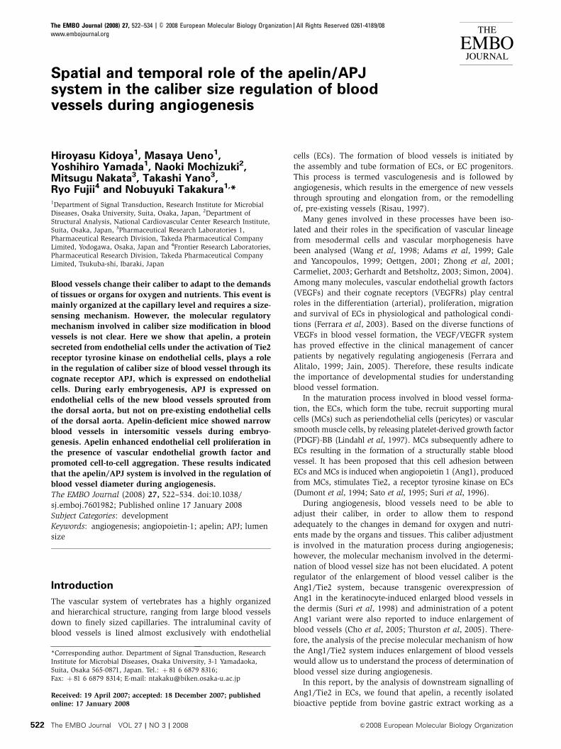
Ang1 induces apelin expression on ECs
To elucidate the molecular mechanism by which Tie2 regu-
lates the caliber change of blood vessels from small to larger
ones, and in order to isolate the genes encoding proteins that
are involved in caliber change and specifically expressed in
HUVECs stimulated by Ang1, we constructed a subtraction
library from HUVECs with Tie2 stimulated by Ang1 as a
tester, and HUVECs with no stimulation of Tie2 as the driver
(Figure 1A). One of these isolated cDNA clones was the
human gene encoding apelin (Figure 1B–D). Real-time poly-
merase chain reaction (PCR) analysis revealed that apelin
mRNA was potently increased in HUVECs after stimulation
by Ang1 in a time-dependent manner (Figure 1B) and we
confirmed that the expression of apelin protein was markedly
upregulated on HUVECs stimulated by Ang1 (Figure 1C).
Moreover, the dose-dependent effect of Ang1 on apelin pro-
duction from HUVECs was confirmed by enzyme immunoas-
say, using the culture supernatant of HUVECs (Figure 1D).
In order to confirm further the upregulation of apelin in
ECs in vivo, we analysed apelin expression in the dermis
of Ang1 transgenic (Ang1Tg) mice, in which Ang1 was over-
expressed in the keratinocytes under the transcriptional
control of K14 promoter (Suri et al, 1998). As shown in
Figure 1E, apelin expression on ECs in the dermis at postnatal
day 7 was increased in Ang1Tg mice compared to that in
wild-type (WT) mice. We confirmed the overexpression of
apelin mRNA in Ang1Tg mice by quantitative real-time
RT–PCR using ECs fractionated from the dermis by a cell
sorter (Figure 1F). As it is well known that Ang1 is involved
in angiogenesis, next we observed the effect of other
proangiogenic molecules on the expression of apelin on
ECs. bFGF induced apelin expression on HUVECs; however,
VEGF, PDGF-BB or EGF did not affect apelin expression
(Supplementary Figure 1A and B).
Apelin with VEGF induces proliferation of ECs
With respect to the enlargement of blood vessels, it seems
likely that apelin causes the proliferation of ECs. To test this
ability, firstly we studied the proliferation of ECs using
HUVECs. As shown in Figure 2A, apelin was not effective
in inducing proliferation of HUVECs. However, upon stimula-
tion with VEGF, the expression level of APJ was upregulated
in HUVECs, at both the mRNA and protein levels (Figure 2B–
D and Supplementary Figure 2). Cell surface expression of
APJ on HUVECs was confirmed by both cell surface bio-
tinylation experiment (Figure 2D) and confocal laser scan-
ning analysis (Supplementary Figure 2). Consistent with this
result, VEGF-induced proliferation of HUVECs was enhanced
by the addition of apelin in a dose-dependent manner
(Figure 2A). Among proangiogenic cytokines, such as Ang1,
EGF, bFGF, PDGF-BB and VEGF, only VEGF induced APJ
expression on HUVECs (Figure 2B–D and Supplementary
Figure 1C and D).
These results suggested that APJ is expressed and affects
ECs during angiogenesis in which VEGF levels are upregu-
lated. Next we observed the proliferation of primary ECs from
the culture of the AGM region (aorta–gonad–mesonephros
region followed by P-Sp region at embryonic day (E) 10.5 to
E11.5) in which angiogenesis was actively taking place. APJ
was highly expressed in the AGM region compared to other
tissues, such as E10.5 yolk sac, head region and heart, and
adult heart (Figure 3A). Furthermore, APJ was expressed
strongly in CD45�CD31þ ECs from the AGM region com-
pared to those from E10.5 heart and adult heart. Although
ECs in E10.5 yolk sac and head region expressed APJ, the
Apelin regulates lumen size of blood vesselsH Kidoya et al
&2008 European Molecular Biology Organization The EMBO Journal VOL 27 | NO 3 | 2008 523
expression level was weaker than that in the AGM region.
When cells from the AGM region were cultured on apelin-
expressing OP9 cells (Figure 3B), proliferation of CD45�CD31þ
was increased compared with that on control OP9 cells and this
proliferation by apelin was abrogated by the addition of anti-
apelin blocking antibody (Figure 3C and D), suggesting that
this action of proliferation by apelin is specific to the apelin/
APJ system. Moreover, as APJ expression was weaker in ECs
from adult heart (Figure 3A) or adult liver (data not shown)
than in those from the AGM region, apelin did not induce
proliferation of ECs in such adult tissues compared to those in
the AGM region (Supplementary Figure 3).
Apelin induces the assembly of ECs
Although the proliferation of ECs is one of the factors
involved in the construction of larger vessels, it is not the
only one. The assembly or aggregation of ECs or endothelial
progenitors, resulting in abundant cell-to-cell contact, is also
necessary for the induction of a caliber change of blood
vessels into larger ones. Therefore, next we tested the ability
of apelin to regulate cell-to-cell contact. When cells from the
AGM region were put on OP9 feeder cells, the control OP9
cells induced a cord-like structure of ECs, in contrast to the
OP9 cells expressing apelin, which induced a sheet-like layer
of ECs in abundance (Figure 4A and C). When cell-to-cell
contact was observed using anti-VE-cadherin or -claudin5
antibodies, we confirmed that the sheet-like structure
was composed of ECs connected with the junctional proteins,
VE-cadherin (Figure 4B) or claudin5 (Supplementary
Figure 4). Moreover, the addition of anti-apelin monoclonal
antibody (mAb) inhibited the sheet-like layer formation of
ECs induced by apelin (Figure 4B and Supplementary
Figure 4). This sheet-like structure was already observed in
the early stage of this culture (Figure 4C), suggesting that cell
aggregation was initiated when ECs were seeded on OP9 cells
expressing apelin.
Among many adhesion molecules tested, we found that
the expression of the junctional protein, claudin5, was
significantly induced by apelin on HUVECs, while that of
VE-cadherin was only slightly induced, at both the mRNA
Figure 1 Ang1 stimulation induces apelin expression in ECs in vitro and in vivo. (A) Tie2 phosphorylation on HUVECs by Ang1 in our system.HUVECs, serum-starved for 2 h, were either treated or not treated with 500 ng/ml Ang1 for 10min. Phosphorylation was studied byimmunoblotting using phosphospecific antibody (p-Tie2). (B) Quantitative real-time RT–PCR analysis of apelin mRNA in HUVECs. TotalRNAwas extracted from HUVECs that had been stimulated with Ang1 for 0–22h. Results are shown as fold increase in comparison with basallevels of HUVECs (0 h). (C) Immunocytochemical analysis of apelin expression in HUVECs, non-stimulated (a) and stimulated (b) by Ang1(500ng/ml) for 20h. Cells were stained with anti-apelin mAb (green). The inset in (b) shows HUVECs stained with secondary antibody as anegative control. Nuclei were stained with propidium iodide (PI; red). Scale bar indicates 50mm. (D) Quantitative enzyme immunoassay ofapelin production from HUVECs stimulated by various doses of Ang1. *Po0.001 (n¼ 3). (E) Immunochemical detection of apelin peptide inthe dermis. Sections of skin from WT and Ang1Tg neonatal mice were stained with anti-CD31 (green) and anti-apelin (red) mAb. Arrowsindicate CD31þ blood vessels. Scale bar indicates 30mm. (F) Quantitative real-time RT–PCR analysis of apelin mRNA in ECs and hematopoieticcells (HCs) of Ang1Tg mice. RNAwas prepared from sorted CD31þCD45� ECs or CD31�CD45þ HCs from the dermis of WTor Ang1Tg neonatalmouse skin. *Po0.01 (n¼ 3).
Apelin regulates lumen size of blood vesselsH Kidoya et al
The EMBO Journal VOL 27 | NO 3 | 2008 &2008 European Molecular Biology Organization524

and protein levels (Supplementary Figure 5). In vitro sheet-
like formation of ECs and upregulation of cell-to-cell adhe-
sion molecules by apelin indicated the involvement of the
apelin/APJ system in the assembly of ECs. Next, we per-
formed the cord formation assay of HUVECs on Matrigel in
the presence or absence of apelin (Figure 5A). After 20h of
culture of HUVECs on Matrigel, they formed a cord-like
structure in the absence of apelin (Figure 5Aa). However, in
the presence of apelin, the HUVECs formed an enlarged cord-
like structure (Figure 5Abd). In this assay, by using confocal
laser scanning analysis, we confirmed that enlargement of this
cord-like structure was induced by cell aggregation, but not by
cell spreading (Supplementary Figure 6). This enlargement
was completely blocked by anti-VE-cadherin blocking anti-
body (Figure 5Acd), suggesting that the enlarged cord-like
formation induced by apelin was initiated by cell-to-cell con-
tact. Moreover, in the spheroid assay (Korff and Augustin,
1998), HUVECs formed large spheroids in the presence of
apelin (Figure 5B) and this action was abrogated by anti-apelin
antibody. Therefore, these results strongly support the notion
that the apelin/APJ system induces EC-to-EC assembly.
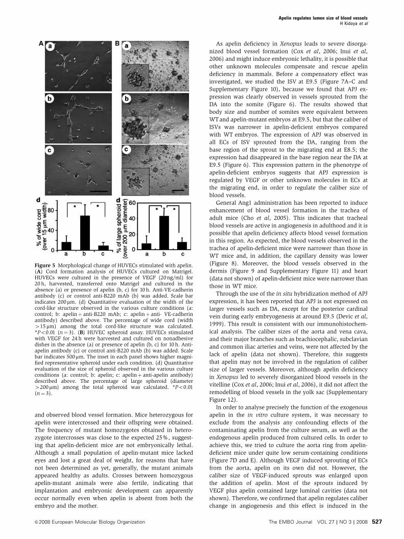
APJ expression in ECs during early embryogenesis
As APJ expression was observed in ECs during early embry-
ogenesis (Figure 3), we studied which vessels expressed
APJ in mouse embryos. At E8.5, cells committed into the
endothelial lineage formed the dorsal aorta (DA), from which
ECs started to sprout. As observed in Figure 6A, APJ expres-
sion was observed in those ECs that had sprouted from the
DA but not in those that were forming the DA. At E9.5, APJ
expression was observed in the migrating end region of ISVs
sprouting from the DA (Figure 6B). Besides the expression of
APJ in the ISVs, weak APJ expression was observed in the
somites. These expression profiles were not very different
from the results obtained by in situ hybridization analysis, as
reported previously (Devic et al, 1999). In another area of the
E9.5 embryo, we found that the anterior cardinal vein (ACV)
expressed APJ. However, when compared to CD31 expression
in ECs, APJ-positive ECs were observed in the migrating
end of the ACV, but not in the base (Supplementary
Figure 7). These expression profiles suggest that the apelin/
APJ system may be associated with angiogenesis but not
with vasculogenesis. Moreover, when apelin expression was
observed in the somite region at E9.5, we found that apelin
protein was detected in ISV (Supplementary Figure 8),
suggesting that the apelin/APJ system may be associated
with the formation of ISV.
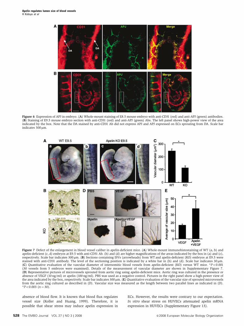
Narrow blood vessels are induced in
apelin-mutant mice
In order to understand the physiological function of apelin,
we generated apelin-mutant mice (Supplementary Figure 9)
Figure 2 Apelin induces proliferation of HUVECs in a VEGF-dependent manner. (A) Proliferation of HUVECs by apelin. HUVECs (5�103) werecultured with apelin (0–1000ng/ml) in the presence or absence of VEGF (20 ng/ml) for 48 h and the number of cells was counted. *Po0.001(n¼ 3). (B) Quantitative real-time RT–PCR analysis of the induction of APJ expression by VEGF in HUVECs. HUVECs were stimulated withVEGF (10 ng/ml) for 0–18 h. Results are shown as fold increase in expression in comparison with levels in stimulated HUVECs at 0 h. *Po0.001(n¼ 3). (C) APJ expression on HUVECs. HUVECs were cultured in the absence (a) or presence (b, c) of VEGF (20ng/ml) for 24 h and stainedwith anti-APJ antibody (green) (a, b). (c) Cells stained with a secondary antibody alone as a negative control. Nuclei were stained withpropidium iodide (red). Scale bar indicates 50 mm. (D) Western blot analysis of cell surface APJ expression on HUVECs that had beenstimulated with VEGF (20 ng/ml) for 24 h. The purity of cell membrane protein was confirmed by the lack of intracellular protein GAPDHexpression. Claudin5 expression was analysed for the experimental control of another cell surface protein. Note that the expression of a 60 kDaAPJ protein was increased in HUVECs in the presence of VEGF.
Apelin regulates lumen size of blood vesselsH Kidoya et al
&2008 European Molecular Biology Organization The EMBO Journal VOL 27 | NO 3 | 2008 525

Figure 3 ECs from the AGM region express APJ and are induced to proliferate by apelin. (A) Quantitative real-time RT–PCR analysis of APJexpression in various tissues, as indicated. RNA from whole tissue, or CD45�CD31þ ECs sorted from various tissues, was evaluated for theexpression of APJ. (B) Western blot analysis of apelin expression on OP9 cells induced by mock vector (OP9/vector) or apelin expression vector(OP9/apelin). An 8 kDa apelin protein was detected in OP9/apelin. GAPDH was used for the internal control. (C) Apelin-induced proliferationof ECs from E10.5 AGM region. Cells from the AGM region were cultured for 7 days, on an OP9/vector or OP9/apelin, in the presence orabsence of anti-apelin or control B220 mAb. AGM cells harvested from cultures were stained with anti-CD31 and -CD45 mAbs and analysed byFACS. (D) Quantitative evaluation of the percentage of CD31þCD45� vascular ECs cultured as described in (C). *Po0.001 (n¼ 5).
Figure 4 Endothelial sheet formation by apelin. (A) Cells from E11.5 AGM region were cocultured with an OP9/vector (a) or OP9/apelin (b) for2–6 days, and CD31 immunostaining was performed. The arrow indicates the aggregated EC sheet. Scale bar indicates 100mm. (B) Cells fromE11.5 AGM region were cocultured for 6 days with an OP9/vector (a–c), OP9/apelin in the presence of B220 control antibody (d–f) or OP9/apelin in the presence of anti-apelin blocking antibody (g–i). ECs on OP9 cells were stained with anti-CD31 (a, d, g) and anti-VE-cadherin(b, e, h) antibodies. (c, f, i) Merged images of (a) and (b), (d) and (e), or (g) and (h), respectively. Scale bar indicates 50 mm. (C) The proportionof sheet-like or cord-like structures of ECs on OP9/apelin or OP9/vector stromal cells (n¼ 3).
Apelin regulates lumen size of blood vesselsH Kidoya et al
The EMBO Journal VOL 27 | NO 3 | 2008 &2008 European Molecular Biology Organization526
and observed blood vessel formation. Mice heterozygous for
apelin were intercrossed and their offspring were obtained.
The frequency of mutant homozygotes obtained in hetero-
zygote intercrosses was close to the expected 25%, suggest-
ing that apelin-deficient mice are not embryonically lethal.
Although a small population of apelin-mutant mice lacked
eyes and lost a great deal of weight, for reasons that have
not been determined as yet, generally, the mutant animals
appeared healthy as adults. Crosses between homozygous
apelin-mutant animals were also fertile, indicating that
implantation and embryonic development can apparently
occur normally even when apelin is absent from both the
embryo and the mother.
As apelin deficiency in Xenopus leads to severe disorga-
nized blood vessel formation (Cox et al, 2006; Inui et al,
2006) and might induce embryonic lethality, it is possible that
other unknown molecules compensate and rescue apelin
deficiency in mammals. Before a compensatory effect was
investigated, we studied the ISV at E9.5 (Figure 7A–C and
Supplementary Figure 10), because we found that APJ ex-
pression was clearly observed in vessels sprouted from the
DA into the somite (Figure 6). The results showed that
body size and number of somites were equivalent between
WTand apelin-mutant embryos at E9.5, but that the caliber of
ISVs was narrower in apelin-deficient embryos compared
with WT embryos. The expression of APJ was observed in
all ECs of ISV sprouted from the DA, ranging from the
base region of the sprout to the migrating end at E8.5; the
expression had disappeared in the base region near the DA at
E9.5 (Figure 6). This expression pattern in the phenotype of
apelin-deficient embryos suggests that APJ expression is
regulated by VEGF or other unknown molecules in ECs at
the migrating end, in order to regulate the caliber size of
blood vessels.
General Ang1 administration has been reported to induce
enhancement of blood vessel formation in the trachea of
adult mice (Cho et al, 2005). This indicates that tracheal
blood vessels are active in angiogenesis in adulthood and it is
possible that apelin deficiency affects blood vessel formation
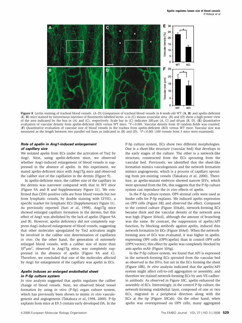
in this region. As expected, the blood vessels observed in the
trachea of apelin-deficient mice were narrower than those in
WT mice and, in addition, the capillary density was lower
(Figure 8). Moreover, the blood vessels observed in the
dermis (Figure 9 and Supplementary Figure 11) and heart
(data not shown) of apelin-deficient mice were narrower than
those in WT mice.
Through the use of the in situ hybridization method of APJ
expression, it has been reported that APJ is not expressed on
larger vessels such as DA, except for the posterior cardinal
vein during early embryogenesis at around E9.5 (Devic et al,
1999). This result is consistent with our immunohistochem-
ical analysis. The caliber sizes of the aorta and vena cava,
and their major branches such as brachiocephalic, subclavian
and common iliac arteries and veins, were not affected by the
lack of apelin (data not shown). Therefore, this suggests
that apelin may not be involved in the regulation of caliber
size of larger vessels. Moreover, although apelin deficiency
in Xenopus led to severely disorganized blood vessels in the
vitelline (Cox et al, 2006; Inui et al, 2006), it did not affect the
remodelling of blood vessels in the yolk sac (Supplementary
Figure 12).
In order to analyse precisely the function of the exogenous
apelin in the in vitro culture system, it was necessary to
exclude from the analysis any confounding effects of the
contaminating apelin from the culture serum, as well as the
endogenous apelin produced from cultured cells. In order to
achieve this, we tried to culture the aorta ring from apelin-
deficient mice under quite low serum-containing conditions
(Figure 7D and E). Although VEGF induced sprouting of ECs
from the aorta, apelin on its own did not. However, the
caliber size of VEGF-induced sprouts was enlarged upon
the addition of apelin. Most of the sprouts induced by
VEGF plus apelin contained large luminal cavities (data not
shown). Therefore, we confirmed that apelin regulates caliber
change in angiogenesis and this effect is induced in the
Figure 5 Morphological change of HUVECs stimulated with apelin.(A) Cord formation analysis of HUVECs cultured on Matrigel.HUVECs were cultured in the presence of VEGF (20ng/ml) for20 h, harvested, transferred onto Matrigel and cultured in theabsence (a) or presence of apelin (b, c) for 10 h. Anti-VE-cadherinantibody (c) or control anti-B220 mAb (b) was added. Scale barindicates 200mm. (d) Quantitative evaluation of the width of thecord-like structure observed in the various culture conditions (a:control; b: apelinþ anti-B220 mAb; c: apelinþ anti- VE-cadherinantibody) described above. The percentage of wide cord (width415mm) among the total cord-like structure was calculated.*Po0.01 (n¼ 3). (B) HUVEC spheroid assay. HUVECs stimulatedwith VEGF for 24 h were harvested and cultured on nonadhesivedishes in the absence (a) or presence of apelin (b, c) for 10 h. Anti-apelin antibody (c) or control anti-B220 mAb (b) was added. Scalebar indicates 500mm. The inset in each panel shows higher magni-fied representative spheroid under each condition. (d) Quantitativeevaluation of the size of spheroid observed in the various cultureconditions (a: control; b: apelin; c: apelinþ anti-apelin antibody)described above. The percentage of large spheroid (diameter4200mm) among the total spheroid was calculated. *Po0.01(n¼ 3).
Apelin regulates lumen size of blood vesselsH Kidoya et al
&2008 European Molecular Biology Organization The EMBO Journal VOL 27 | NO 3 | 2008 527
absence of blood flow. It is known that blood flux regulates
vessel size (Koller and Huang, 1999). Therefore, it is
possible that shear stress may induce apelin expression in
ECs. However, the results were contrary to our expectation.
In vitro shear stress on HUVECs attenuated apelin mRNA
expression in HUVECs (Supplementary Figure 13).
Figure 6 Expression of APJ in embryo. (A) Whole-mount staining of E8.5 mouse embryo with anti-CD31 (red) and anti-APJ (green) antibodies.(B) Staining of E9.5 mouse embryo section with anti-CD31 (red) and anti-APJ (green) Abs. The left panel shows high-power view of the areaindicated by the box. Note that the DA stained by anti-CD31 Ab did not express APJ and APJ expressed on ECs sprouting from DA. Scale barindicates 500mm.
Figure 7 Defect of the enlargement in blood vessel caliber in apelin-deficient mice. (A) Whole-mount immunohistostaining of WT (a, b) andapelin-deficient (c, d) embryos at E9.5 with anti-CD31 Ab. (b) and (d) are higher magnifications of the areas indicated by the box in (a) and (c),respectively. Scale bar indicates 300 mm. (B) Sections containing ISVs (arrowheads) from WTand apelin-deficient (KO) embryos at E9.5 werestained with anti-CD31 antibody. The level of the sectioning position is indicated by a white bar in (b) and (d). Scale bar indicates 30mm.(C) Quantitative evaluation of the vascular diameter of intersomitic blood vessels from apelin-deficient (KO) versus WT mice. *Po0.001(30 vessels from 5 embryos were examined). Details of the measurement of vascular diameter are shown in Supplementary Figure 7.(D) Representative pictures of microvessels sprouted from aortic ring using apelin-deficient mice. Aortic ring was cultured in the presence orabsence of VEGF (10 ng/ml) or apelin (100 ng/ml). PBS was used as a negative control. Pictures in the right panel show a high-power view ofthe area indicated by the box, respectively. Scale bar indicates 300mm. (E) Quantitative evaluation of the vascular size of sprouted microvesselsfrom the aortic ring cultured as described in (D). Vascular size was measured as the length between two parallel lines as indicated in (D).*Po0.003 (n¼ 30).
Apelin regulates lumen size of blood vesselsH Kidoya et al
The EMBO Journal VOL 27 | NO 3 | 2008 &2008 European Molecular Biology Organization528
Role of apelin in Ang1-induced enlargement
of capillary size
We isolated apelin from ECs under the activation of Tie2 by
Ang1. Next, using apelin-deficient mice, we observed
whether Ang1-induced enlargement of blood vessels is sup-
pressed in the absence of apelin. In this experiment, we
mated apelin-deficient mice with Ang1Tg mice and observed
the caliber size of the capillaries in the dermis (Figure 9).
In apelin-deficient mice, the caliber size of the capillary in
the dermis was narrower compared with that in WT mice
(Figure 9A and B and Supplementary Figure 11). We con-
firmed that CD31-positive cells are from blood vessels but not
from lymphatic vessels, by double staining with LYVE1, a
specific marker for lymphatic ECs (Supplementary Figure 11).
As previously reported (Suri et al, 1998), Ang1Tg mice
showed enlarged capillary formation in the dermis, but this
effect of Ang1 was abolished by the lack of apelin (Figure 9A
and B). However, apelin deficiency did not completely sup-
press Ang1-induced enlargement of blood vessels, suggesting
that other molecules upregulated by Tie2 activation might
be involved in the caliber size determination of capillaries
in vivo. On the other hand, the generation of extremely
enlarged blood vessels, with a caliber size of more than
104 mm2, observed in Ang1Tg mice, was completely sup-
pressed in the absence of apelin (Figure 9A and C).
Therefore, we concluded that one of the molecules affected
by Ang1 for enlargement of the capillary was apelin in ECs.
Apelin induces an enlarged endothelial sheet
in P-Sp culture system
In vivo analysis suggested that apelin regulates the caliber
change of blood vessels. Next, we observed blood vessel
formation by using in vitro (P-Sp) organ culture system,
which has previously been shown to mimic in vivo vasculo-
genesis and angiogenesis (Takakura et al, 1998, 2000). P-Sp
explants from mice at E9.5 contain early developed DA. In the
P-Sp culture system, ECs show two different morphologies.
One is a sheet-like structure (vascular bed) that develops in
the early stages of the culture. The other is a network-like
structure, constructed from the ECs sprouting from the
vascular bed. Previously, we identified that the sheet-like
formation mimics vasculogenesis and the network formation
mimics angiogenesis, which is a process of capillary sprout-
ing from pre-existing vessels (Takakura et al, 2000). There-
fore, as apelin-mutant embryos showed narrow ISVs, which
were sprouted from the DA, this suggests that the P-Sp culture
system can reproduce the in vivo effects of apelin.
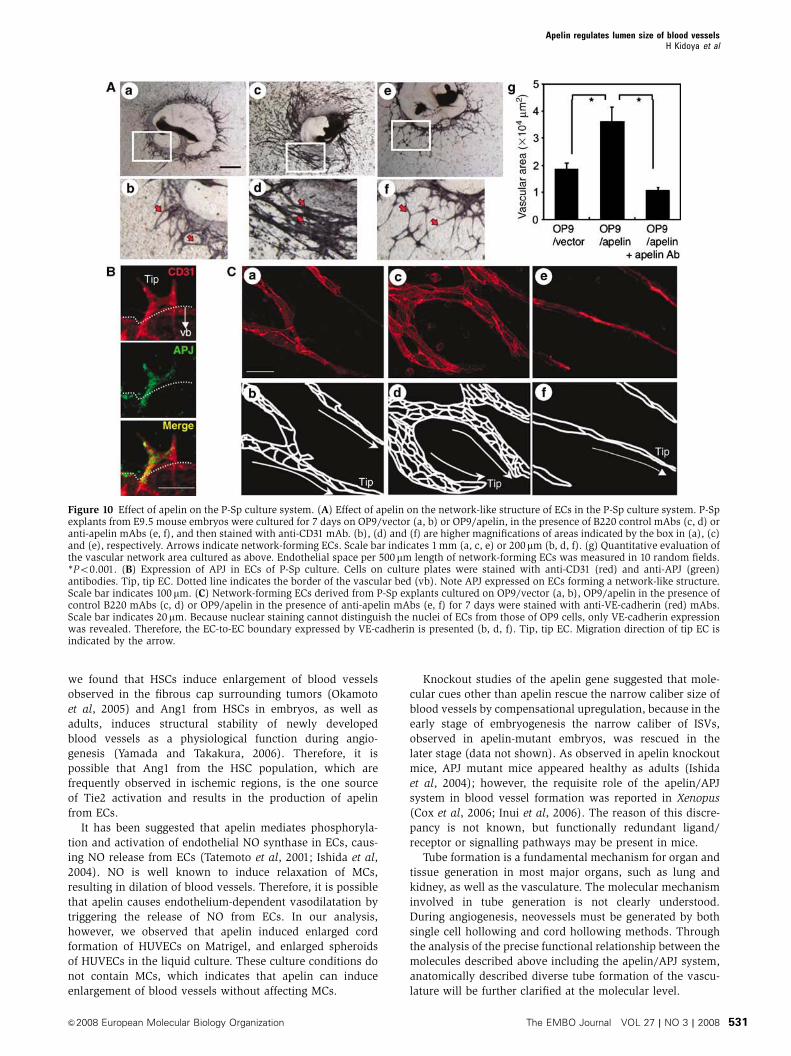
In the P-Sp culture system, OP9 stromal cells were used as
feeder cells for P-Sp explants. We induced apelin expression
on OP9 cells (Figure 3B) and observed the effect. Compared
to the control culture (Figure 10Aab), network-forming ECs
became thick and the vascular density of the network area
was high (Figure 10Acd), although the amount of branching
was the same. By contrast, the suppression of apelin/APJ
function, by blocking antibody against apelin, induced thin
network formation by ECs (Figure 10Aef). When the network-
forming area of ECs was evaluated, it was higher in apelin-
expressing OP9 cells (OP9/apelin) than in control OP9 cells
(OP9/vector); this effect by apelin was completely blocked by
anti-apelin mAb (Figure 10Ag).
In the P-Sp culture system, we found that APJ is expressed
in the network-forming ECs sprouted from the vascular bed
as observed in the ISVs, but not in the ECs forming the sheet
(Figure 10B). In vitro analysis indicated that the apelin/APJ
system might affect cell-to-cell aggregation or assembly, and
therefore we stained network-forming ECs by anti-VE-cadher-
in antibody. As observed in Figure 10C, apelin enhanced the
assembly of ECs. Interestingly, in the control P-Sp culture, the
network-forming endothelial layer, composed of one or two
ECs, migrated in a peripheral direction along with the
ECs at the tip (Figure 10Cab). On the other hand, when
apelin was overexpressed on OP9 cells, many aggregated
Figure 8 Lectin staining of tracheal blood vessels. (A–D) Comparison of tracheal blood vessels in 8-week-old WT (A, B) and apelin-deficient(C, D) mice stained by intravenous injection of fluorescein-labelled lectin. a in (C) means avascular area. (B) and (D) show a high-power viewof the area indicated by the box in (A) and (C), respectively. Scale bar in (C) indicates 200 mm (A, C) and 60mm (B, D). (E) Quantitativeevaluation of vascular density from apelin-deficient (KO) versus WT mice. *Po0.001. Vascular density from 10 random fields was counted.(F) Quantitative evaluation of vascular size of blood vessels in the trachea from apelin-deficient (KO) versus WT mice. Vascular size wasmeasured as the length between two parallel red lines as indicated in (B) and (D). *Po0.001 (100 vessels from 3 mice were examined).
Apelin regulates lumen size of blood vesselsH Kidoya et al
&2008 European Molecular Biology Organization The EMBO Journal VOL 27 | NO 3 | 2008 529

ECs migrated along with the ECs at the tip (Figure 10Ccd),
and this effect was completely suppressed by anti-apelin mAb
(Figure 10Cef). These results indicated that apelin induces an
enlarged endothelial sheet when angiogenesis is taking place.
Discussion
The knowledge of how vascular cells commit from their
progenitor cells and generate a closed cardiovascular circu-
latory system has accumulated in recent years, mostly by the
isolation and functional analysis of molecules associated with
blood vessel formation. However, little is known regarding
the molecular events that regulate EC morphogenesis, espe-
cially the caliber size determination of blood vessels. Data
documented here, from both in vitro and in vivo analysis,
showed that apelin regulates the enlargement of newly
developed blood vessels during angiogenesis.
In angiogenesis, how blood vessels ‘decide’ their appro-
priate size is very important to the organization of the
adjustment of tissue and organ demand for oxygen and
nutrients. Our analysis clearly showed that APJ expression
was induced by VEGF, which, in turn, is well known to be
induced by tissue hypoxia (Liu et al, 1995). This indicates
that, under tissue hypoxia, blood vessels have an opportunity
to enlarge their size and the reduction of APJ expression
finalizes the enlargement of blood vessel caliber under tissue
normoxia. Indeed, in the retina, APJ was observed temporally
in the radial vessels and the associated capillaries of retina
from day 3 to day 12 after birth, but APJ expression on ECs
was attenuated in the later stage (Saint-Geniez et al, 2002).
As reported in the retina, we also found that APJ expression
was observed in ECs sprouted from the DA and ECs on blood
vessels in the neonatal dermis of mice (data not shown), but
that it gradually disappeared with maturity. These expression
patterns strongly suggested that APJ plays a spatio-temporal
role in the maturation of blood vessels by transient expres-
sion on ECs of blood vessels where angiogenesis is taking
place. Therefore, we concluded that one of the molecules
associated with the regulation of blood vessel diameter was
apelin in the ECs.
Based on our results presented here, it appears that VEGF,
Ang1 and apelin regulate caliber size in a concerted fashion,
as follows. Upon stimulation by VEGF, ECs sprouted from
pre-existing vessels may express APJ. Subsequently, Ang1
stimulates these sprouted ECs to induce apelin expression. In
the presence of both VEGF and apelin, the ECs start to
proliferate, adhere and form contacts with each other through
junctional proteins, and construct enlarged blood vessels.
Apelin has been reported to induce angiogenesis in the
Matrigel plug assay (Kasai et al, 2004) and also chemotaxis
(Cox et al, 2006). In our experiments using the Matrigel plug
assay, we found that apelin induced migration, rather than
proliferation, of ECs (Supplementary Figure 14). Moreover,
we confirmed that like VEGF, apelin modified the cytos-
keleton structure (Supplementary Figure 15). Therefore, ape-
lin may induce mobilization of ECs in the process of EC-to-EC
assembly.
As we found, apelin deficiency suppressed the enlarge-
ment of ISVs during early embryogenesis. Furthermore, it has
been reported elsewhere that Ang1 and VEGF are expressed
in intersomitic or somitic tissues (Davis et al, 1996; Lawson
et al, 2002) and that apelin is coexpressed with APJ-positive
ECs in ISVs. Indeed both Tie2 and Ang1 mutant embryos
showed impaired ISV formation (Dumont et al, 1994; Sato
et al, 1995). Therefore, it appears that these three compo-
nents may be involved in the regulation of caliber size change
of the ISVs.
Transgenic overexpression of Ang1 in the keratinocyte
induced enlarged blood vessels in the dermis (Suri et al,
1998) and administration of a potent Ang1 variant was also
reported to induce enlargement of blood vessels (Cho et al,
2005; Thurston et al, 2005). Therefore, Ang1 expression may
be a key determinant of caliber size during angiogenesis.
Ang1 is usually produced from MCs in cells composing blood
vessels (Davis et al, 1996), However, we previously reported
that hematopoietic stem cells (HSCs) producing Ang1 migrate
into avascular areas before the ECs start to migrate, and that
this Ang1 from HSCs induces angiogenesis by promoting the
chemotaxis of ECs (Takakura et al, 2000). Moreover, recently,
Figure 9 Apelin/APJ system is involved in Ang1-induced vascularenlargement. (A) Sections of ear skin stained with anti-CD31 mAb.Ear skin was prepared from 8-week-old WT, apelin-deficient (apelinKO), Ang1Tg mice, or apelin-deficient mice mated with Ang1Tgmice (apelin KO/Ang1 Tg). Scale bar indicates 30 mm. (B, C)Quantitative evaluation of the number of enlarged blood vesselscomposed of a luminal cavity of more than 5000 mm2 (B) or morethan 104mm2 (C) in the skin from mice as described in (A). Thirtyrandom fields were observed from sections of three independentmice as described in (A). *Po0.01.
Apelin regulates lumen size of blood vesselsH Kidoya et al
The EMBO Journal VOL 27 | NO 3 | 2008 &2008 European Molecular Biology Organization530
we found that HSCs induce enlargement of blood vessels
observed in the fibrous cap surrounding tumors (Okamoto
et al, 2005) and Ang1 from HSCs in embryos, as well as
adults, induces structural stability of newly developed
blood vessels as a physiological function during angio-
genesis (Yamada and Takakura, 2006). Therefore, it is
possible that Ang1 from the HSC population, which are
frequently observed in ischemic regions, is the one source
of Tie2 activation and results in the production of apelin
from ECs.
It has been suggested that apelin mediates phosphoryla-
tion and activation of endothelial NO synthase in ECs, caus-
ing NO release from ECs (Tatemoto et al, 2001; Ishida et al,
2004). NO is well known to induce relaxation of MCs,
resulting in dilation of blood vessels. Therefore, it is possible
that apelin causes endothelium-dependent vasodilatation by
triggering the release of NO from ECs. In our analysis,
however, we observed that apelin induced enlarged cord
formation of HUVECs on Matrigel, and enlarged spheroids
of HUVECs in the liquid culture. These culture conditions do
not contain MCs, which indicates that apelin can induce
enlargement of blood vessels without affecting MCs.
Knockout studies of the apelin gene suggested that mole-
cular cues other than apelin rescue the narrow caliber size of
blood vessels by compensational upregulation, because in the
early stage of embryogenesis the narrow caliber of ISVs,
observed in apelin-mutant embryos, was rescued in the
later stage (data not shown). As observed in apelin knockout
mice, APJ mutant mice appeared healthy as adults (Ishida
et al, 2004); however, the requisite role of the apelin/APJ
system in blood vessel formation was reported in Xenopus
(Cox et al, 2006; Inui et al, 2006). The reason of this discre-
pancy is not known, but functionally redundant ligand/
receptor or signalling pathways may be present in mice.
Tube formation is a fundamental mechanism for organ and
tissue generation in most major organs, such as lung and
kidney, as well as the vasculature. The molecular mechanism
involved in tube generation is not clearly understood.
During angiogenesis, neovessels must be generated by both
single cell hollowing and cord hollowing methods. Through
the analysis of the precise functional relationship between the
molecules described above including the apelin/APJ system,
anatomically described diverse tube formation of the vascu-
lature will be further clarified at the molecular level.
Figure 10 Effect of apelin on the P-Sp culture system. (A) Effect of apelin on the network-like structure of ECs in the P-Sp culture system. P-Spexplants from E9.5 mouse embryos were cultured for 7 days on OP9/vector (a, b) or OP9/apelin, in the presence of B220 control mAbs (c, d) oranti-apelin mAbs (e, f), and then stained with anti-CD31 mAb. (b), (d) and (f) are higher magnifications of areas indicated by the box in (a), (c)and (e), respectively. Arrows indicate network-forming ECs. Scale bar indicates 1mm (a, c, e) or 200mm (b, d, f). (g) Quantitative evaluation ofthe vascular network area cultured as above. Endothelial space per 500 mm length of network-forming ECs was measured in 10 random fields.*Po0.001. (B) Expression of APJ in ECs of P-Sp culture. Cells on culture plates were stained with anti-CD31 (red) and anti-APJ (green)antibodies. Tip, tip EC. Dotted line indicates the border of the vascular bed (vb). Note APJ expressed on ECs forming a network-like structure.Scale bar indicates 100 mm. (C) Network-forming ECs derived from P-Sp explants cultured on OP9/vector (a, b), OP9/apelin in the presence ofcontrol B220 mAbs (c, d) or OP9/apelin in the presence of anti-apelin mAbs (e, f) for 7 days were stained with anti-VE-cadherin (red) mAbs.Scale bar indicates 20mm. Because nuclear staining cannot distinguish the nuclei of ECs from those of OP9 cells, only VE-cadherin expressionwas revealed. Therefore, the EC-to-EC boundary expressed by VE-cadherin is presented (b, d, f). Tip, tip EC. Migration direction of tip EC isindicated by the arrow.
Apelin regulates lumen size of blood vesselsH Kidoya et al
&2008 European Molecular Biology Organization The EMBO Journal VOL 27 | NO 3 | 2008 531

Materials and methods
AnimalsC57BL/6 mice and ICR mice were purchased from Japan SLC(Shizuoka, Japan) at 8 weeks of age and used between 8 and 12weeks of age. Ang1Tg mice (Suri et al, 1998) with a C57BL/6 back-ground were provided by Dr GD Yanchopoulos (RegeneronPharmaceuticals Inc., Tarrytown, NY). Animal care in our labo-ratory was in accordance with the guidelines of Kanazawa andOsaka University for animal and recombinant DNA experiments.
Plasmids and transfectionThe mouse Apelin gene was cloned into the pCAGSIH expressionvector. Lipofectamine Plus reagent (Invitrogen Life Technologies,Carlsbad, CA) was used to transfect cells with this plasmid andclones of cells exhibiting stable transfection were obtained by anti-biotic resistance selection using G418 (Gibco, Grand Island, NY).Primer pairs for PCR to detect transfected gene are listed inSupplementary Table S1.
Tissue preparation, immunohistochemistry and flowcytometryTissue fixation, preparation of tissue sections and staining ofsections or cultured cells with antibodies were performed asdescribed previously (Takakura et al, 2000). An biotin-conjugatedanti-CD31 mAb (Pharmingen, San Diego, CA), anti-apelin Ab (4G5;Kawamata et al, 2001) and anti-APJ polyclonal Ab were used in thestaining of tissue sections or cultured cells. To obtain a specificantibody against mouse APJ, a rabbit was immunized with asynthetic peptide (CHEKSIPYSQETLVD) derived from the C-terminal region of APJ. Antisera were affinity purified with thesame peptide. Preimmunized rabbit immunoglobulins were used asa negative control to confirm specific staining. Sections werecounterstained with hematoxylin or propidium iodide. The sectionswere observed using an Olympus IX-70 microscope (Olympus,Tokyo, Japan) and images were acquired with a CoolSnap digitalcamera (Roper Scientific, Trenton, NJ). Whole-mount immunohis-tochemistry using anti-CD31 mAb or anti-APJ was performed aspreviously described (Takakura et al, 1998). Stained embryos wereobserved under a Leica MZ16FA stereomicroscope (Leica, Solms,Germany) and photographed with a DC120 digital camera (Pixera,Los Gatos, CA). In all assays, we used an isotype-matched control Igas a negative control and confirmed that the positive signals werenot derived from nonspecific background. Investigation of thedensity and morphology of microvessels in lectin-stained wholemount of tracheal blood vessels was performed as described(Yamada and Takakura, 2006). In brief, after anaesthesia withsodium pentobarbital, mice were injected into the tail vein withfluorescein-labelled Lycopersicon esculentum lectin (Vector Labora-tories, Burlingame, CA), which binds uniformly to the luminalsurface of ECs and adherent leukocytes. Lectin-stained tracheaswere removed from apelin-deficient and WT mice and analysedunder a fluorescence microscope. Images were processed usingAdobe Photoshop 6.0 software (Adobe Systems, San Jose, CA).Flow cytometric analysis was performed as previously described(Yamada and Takakura, 2006). FITC-conjugated anti-CD45 mAb andPE-conjugated anti-CD31 mAb (Pharmingen) were used. Thestained cells were analysed by FACS Calibur (Becton Dickinson,Franklin Lakes, NJ) and sorted by EPICS flow cytometer (ALTRA;Beckman Coulter, Fullerton, CA).
Cell culture4�106 HUVECs were cultured in six-well plates for 12 h in HumediaEG2 (Kurabo, Osaka, Japan). Cells were then incubated in M199medium supplemented with 1% fetal bovine serum (FBS). After 3 h
of serum deprivation, cells were incubated with basal mediumcontaining 500ng/ml of Ang1 (R&D Systems, Minneapolis, MN),100 ng/ml of apelin (Bachem, Bubendorf, Switzerland) or 20 ng/mlof VEGF-A165 (PeproTech, Rocky Hill, NJ).
The culture of P-Sp explant was performed as previouslyreported (Takakura et al, 2000). OP9 cells stably transfected witha pCAGSIH expression vector carrying the cDNA for mouse apelinor mock vector were used as feeder cells (OP9/apelin or OP9/vector, respectively) in the presence or absence of anti-apelinmonoclonal blocking antibody (4G5; see Supplementary Figure 16for functional analysis of this antibody in the inhibition of theapelin/APJ system) or control anti-B220 mAb. After 7 days ofculture, the cultured cells on OP9 cells were fixed and stained withmAbs. For the culture of dissociated ECs from the AGM region, theregions were excised from E11.5 ICR embryos and dissociated bydispase II (Boehringer Mannheim, Mannheim, Germany) as pre-viously described (Yamada and Takakura, 2006). Cells weresuspended in DMEM supplemented with 15% fetal calf serumand cultured in OP9/vector or OP9/apelin seeded on 24-well platesin the presence of murine SCF (100 ng/ml; PeproTech), bFGF (1 ng/ml; R&D Systems) and OSM (10ng/ml; R&D Systems). To thisculture, we added 10 mg/ml anti-apelin or anti-B220 mAb. After 6days of culture, the cells were fixed and double stained with anti-CD31 mAb and anti-VE-cadherin mAb (BD Pharmingen, San Jose,CA), or anti-CD31 mAb and claudin5 mAb (Abcam Inc., Cambridge,MA). Stained samples were analysed by confocal laser scanningmicroscopy (LSM510, Carl Zeiss, Germany). For the evaluation ofapelin in promoting proliferation of HUVECs, HUVECs werecultured for 24 h in medium plus growth supplements and thenfor an additional 48 h in medium with or without apelin (10–100ng/ml), VEGF (10 ng/ml) or apelin plus VEGF. Cell proliferation wasthen evaluated by directly counting the cell number.
Aortic ring culture for angiogenesis assayDescending thoracic aortas were isolated from apelin-deficientmice. Under a stereomicroscope, multiple 1-mm-thick aortic ringswere prepared. Rings were then placed between two layers of type Icollagen gel (Cellmatrix Type IA, Nitta Zeratin, Osaka, Japan),supplemented with Medium 199, 20% FBS in the presence orabsence of VEGF (20 ng/ml) or apelin (200 ng/ml). The cultureswere kept at 371C in a humidified environment for a week andexamined every second day under an Olympus microscope (IX70).
Statistical analysisAll data are presented as mean7standard deviation (s.d.). Forstatistical analysis, the statcel2 software package (OMS) was usedwith analysis of variance performed on all data followed by Tukey–Kramer multiple comparison test. When only two groups werecompared, two-sided Student’s t-test was used.
Supplementary dataSupplementary data are available at The EMBO Journal Online(http://www.embojournal.org).
Acknowledgements
We thank Dr GD Yanchopoulos (Regeneron Pharmaceuticals Inc.)and Dr Y Oike (Keio University, Tokyo, Japan) for providing us withAng1Tg mice and with anti-LYVE-1 antibody, respectively.Moreover, we thank N Fujimoto for preparation of plasmid DNAand K Fukuhara for administrative assistance. This work was partlysupported by a Grant-in-Aid from The Ministry of Education,Culture, Sports, Science and Technology of Japan to NT.
References
Adams RH, Wilkinson GA, Weiss C, Diella F, Gale NW, Deutsch U,Risau W, Klein R (1999) Roles of ephrinB ligands and EphBreceptors in cardiovascular development: demarcation of arter-ial/venous domains, vascular morphogenesis, and sproutingangiogenesis. Genes Dev 13: 295–306
Ashley EA, Powers J, Chen M, Kundu R, Finsterbach T, Caffarelli A,Deng A, Eichhorn J, Mahajan R, Agrawal R, Greve J, Robbins R,
Patterson AJ, Bernstein D, Quertermous T (2005) The endo-genous peptide apelin potently improves cardiac contractilityand reduces cardiac loading in vivo. Cardiovasc Res 65: 73–82
Carmeliet P (2003) Angiogenesis in health and disease. Nat Med 9:653–660
Cho CH, Kim KE, Byun J, Jang HS, Kim DK, Baluk P, Baffert F, LeeGM, Mochizuki N, Kim J, Jeon BH, McDonald DM, Koh GY
Apelin regulates lumen size of blood vesselsH Kidoya et al
The EMBO Journal VOL 27 | NO 3 | 2008 &2008 European Molecular Biology Organization532
(2005) Long-term and sustained COMP-Ang1 induces long-lasting vascular enlargement and enhanced blood flow. Circ Res97: 86–94
Cox CM, D’Agostino SL, Miller MK, Heimark RL, Krieg PA (2006)Apelin, the ligand for the endothelial G-protein-coupledreceptor, APJ, is a potent angiogenic factor required fornormal vascular development of the frog embryo. Dev Biol 296:177–189
Croitoru-Lamoury J, Guillemin GJ, Boussin FD, Mognetti B,Gigout LI, Cheret A, Vaslin B, Le Grand R, Brew BJ, Dormont D(2003) Expression of chemokines and their receptors in humanand simian astrocytes: evidence for a central role of TNFalpha and IFN gamma in CXCR4 and CCR5 modulation. Glia41: 354–370
Davis S, Aldrich TH, Jones PF, Acheson A, Compton DL, Jain V,Ryan TE, Bruno J, Radziejewski C, Maisonpierre PC,Yancopoulos GD (1996) Isolation of angiopoietin-1, a ligand forthe TIE2 receptor, by secretion-trap expression cloning. Cell 87:1161–1169
De Mota N, Reaux-Le Goazigo A, El Messari S, Chartrel N, RoeschD, Dujardin C, Kordon C, Vaudry H, Moos F, Llorens-Cortes C(2004) Apelin, a potent diuretic neuropeptide counteractingvasopressin actions through inhibition of vasopressin neuronactivity and vasopressin release. Proc Natl Acad Sci USA 101:10464–10469
Devic E, Paquereau L, Vernier P, Knibiehler B, Audigier Y (1996)Expression of a new G protein-coupled receptor X-msr is asso-ciated with an endothelial lineage in Xenopus laevis. Mech Dev59: 129–140
Devic E, Rizzoti K, Bodin S, Knibiehler B, Audigier Y (1999)Amino acid sequence and embryonic expression of msr/apj, themouse homolog of Xenopus X-msr and human APJ. Mech Dev 84:199–203
Dumont DJ, Gradwohl G, Fong GH, Puri MC, Gerstenstein M,Auerbach A, Breitman ML (1994) Dominant-negative and tar-geted null mutations in the endothelial receptor tyrosine kinase,tek, reveal a critical role in vasculogenesis of the embryo. GenesDev 8: 1897–1909
Edinger AL, Hoffman TL, Sharron M, Lee B, Yi Y, Choe W, KolsonDL, Mitrovic B, Zhou Y, Faulds D, Collman RG, Hesselgesser J,Horuk R, Doms RW (1998) An orphan seven-transmembranedomain receptor expressed widely in the brain functions as acoreceptor for human immunodeficiency virus type 1 and simianimmunodeficiency virus. J Virol 72: 7934–7940
Ferrara N, Alitalo K (1999) Clinical applications of angiogenicgrowth factors and their inhibitors. Nat Med 5: 1359–1364
Ferrara N, Gerber HP, LeCouter J (2003) The biology of VEGFand itsreceptors. Nat Med 9: 669–676
Gale NW, Yancopoulos GD (1999) Growth factors acting via en-dothelial cell-specific receptor tyrosine kinases: VEGFs, angio-poietins, and ephrins in vascular development. Genes Dev 13:1055–1066
Gerhardt H, Betsholtz C (2003) Endothelial-pericyte interactions inangiogenesis. Cell Tissue Res 314: 15–23
Inui M, Fukui A, Ito Y, Asashima M (2006) Xapelin and Xmsr arerequired for cardiovascular development in Xenopus laevis. DevBiol 298: 188–200
Ishida J, Hashimoto T, Hashimoto Y, Nishiwaki S, Iguchi T, HaradaS, Sugaya T, Matsuzaki H, Yamamoto R, Shiota N, Okunishi H,Kihara M, Umemura S, Sugiyama F, Yagami K, Kasuya Y,Mochizuki N, Fukamizu A (2004) Regulatory roles for APJ, aseven-transmembrane receptor related to angiotensin-type 1 re-ceptor in blood pressure in vivo. J Biol Chem 279: 26274–26279
Jain RK (2005) Normalization of tumor vasculature: an emergingconcept in antiangiogenic therapy. Science 307: 58–62
Kasai A, Shintani N, Oda M, Kakuda M, Hashimoto H, Matsuda T,Hinuma S, Baba A (2004) Apelin is a novel angiogenic factorin retinal endothelial cells. Biochem Biophys Res Commun 325:395–400
Katugampola SD, Maguire JJ, Matthewson SR, Davenport AP (2001)[(125)I]-(Pyr(1))Apelin-13 is a novel radioligand for localizingthe APJ orphan receptor in human and rat tissues with evidencefor a vasoconstrictor role in man. Br J Pharmacol 132: 1255–1260
Kawamata Y, Habata Y, Fukusumi S, Hosoya M, Fujii R, Hinuma S,Nishizawa N, Kitada C, Onda H, Nishimura O, Fujino M (2001)Molecular properties of apelin: tissue distribution and receptorbinding. Biochim Biophys Acta 1538: 162–171
Kleinz MJ, Davenport AP (2004) Immunocytochemical localizationof the endogenous vasoactive peptide apelin to human vascularand endocardial endothelial cells. Regul Pept 118: 119–125
Koller A, Huang A (1999) Development of nitric oxide and pros-taglandin mediation of shear stress-induced arteriolar dilationwith aging and hypertension. Hypertension 34: 1073–1079
Korff T, Augustin HG (1998) Integration of endothelial cells inmulticellular spheroids prevents apoptosis and induces differen-tiation. J Cell Biol 143: 1341–1352
Lawson ND, Vogel AM, Weinstein BM (2002) sonic hedgehog andvascular endothelial growth factor act upstream of the Notchpathway during arterial endothelial differentiation. Dev Cell 3:127–136
Lindahl P, Johansson BR, Leveen P, Betsholtz C (1997) Pericyte lossand microaneurysm formation in PDGF-B-deficient mice. Science277: 242–245
Liu Y, Cox SR, Morita T, Kourembanas S (1995) Hypoxia regulatesvascular endothelial growth factor gene expression in endothelialcells. Identification of a 50 enhancer. Circ Res 77: 638–643
Masri B, Knibiehler B, Audigier Y (2005) Apelin signalling: apromising pathway from cloning to pharmacology. Cell Signal17: 415–426
O’Dowd BF, Heiber M, Chan A, Heng HH, Tsui LC, Kennedy JL, ShiX, Petronis A, George SR, Nguyen T (1993) A human gene thatshows identity with the gene encoding the angiotensin receptor islocated on chromosome 11. Gene 136: 355–360
Oettgen P (2001) Transcriptional regulation of vascular develop-ment. Circ Res 89: 380–388
Okamoto R, Ueno M, Yamada Y, Takahashi N, Sano H, Suda T,Takakura N (2005) Hematopoietic cells regulate the angiogenicswitch during tumorigenesis. Blood 105: 2757–2763
Risau W (1997) Mechanisms of angiogenesis. Nature 386:671–674
Saint-Geniez M, Masri B, Malecaze F, Knibiehler B, Audigier Y(2002) Expression of the murine msr/apj receptor and its ligandapelin is upregulated during formation of the retinal vessels.Mech Dev 110: 183–186
Sato TN, Tozawa Y, Deutsch U, Wolburg-Buchholz K, Fujiwara Y,Gendron-Maguire M, Gridley T, Wolburg H, Risau W, Qin Y(1995) Distinct roles of the receptor tyrosine kinases Tie-1 andTie-2 in blood vessel formation. Nature 376: 70–74
Scott IC, Masri B, D’Amico LA, Jin SW, Jungblut B, Wehman AM,Baier H, Audigier Y, Stainier DY (2007) The G protein-coupledreceptor agtrl1b regulates early development of myocardial pro-genitors. Dev Cell 12: 403–413
Simon MC (2004) Vascular morphogenesis and the formation ofvascular networks. Dev Cell 6: 479–482
Suri C, Jones PF, Patan S, Bartunkova S, Maisonpierre PC, Davis S,Sato TN, Yancopoulos GD (1996) Requisite role of angiopoietin-1,a ligand for the Tie-2 receptor, during embryonic angiogenesis.Cell 87: 1171–1180
Suri C, McClain J, Thurston G, McDonald DM, Zhou H, OldmixonEH, Sato TN, Yancopoulos GD (1998) Increased vascularization inmice overexpressing angiopoietin-1. Science 282: 468–471
Szokodi I, Tavi P, Foldes G, Voutilainen-Myllyla S, Ilves M, TokolaH, Pikkarainen S, Piuhola J, Rysa J, Toth M, Ruskoaho H (2002)Apelin, the novel endogenous ligand of the orphan receptor APJ,regulates cardiac contractility. Circ Res 91: 434–440
Takakura N, Huang XL, Naruse T, Hamaguchi I, Dumont DJ,Yancopoulos GD, Suda T (1998) Critical role of the TIE2 endothe-lial cell receptor in the development of definitive hematopoiesis.Immunity 9: 677–686
Takakura N, Watanabe T, Suenobu S, Yamada Y, Noda T, Ito Y,Satake M, Suda T (2000) A role for hematopoietic stem cells inpromoting angiogenesis. Cell 102: 199–209
Tatemoto K, Hosoya M, Habata Y, Fujii R, Kakegawa T, Zou MX,Kawamata Y, Fukusumi S, Hinuma S, Kitada C, Kurokawa T,Onda H, Fujino M (1998) Isolation and characterization of a novelendogenous peptide ligand for the human APJ receptor. BiochemBiophys Res Commun 251: 471–476
Tatemoto K, Takayama K, Zou MX, Kumaki I, Zhang W, Kumano K,Fujimiya M (2001) The novel peptide apelin lowers bloodpressure via a nitric oxide-dependent mechanism. Regul Pept99: 87–92
Thurston G, Wang Q, Baffert F, Rudge J, Papadopoulos N, Jean-Guillaume D, Wiegand S, Yancopoulos GD, McDonald DM (2005)Angiopoietin 1 causes vessel enlargement, without angiogenic
Apelin regulates lumen size of blood vesselsH Kidoya et al
&2008 European Molecular Biology Organization The EMBO Journal VOL 27 | NO 3 | 2008 533

sprouting, during a critical developmental period. Development132: 3317–3326
Wang HU, Chen ZF, Anderson DJ (1998) Molecular distinction andangiogenic interaction between embryonic arteries and veinsrevealed by ephrin-B2 and its receptor Eph-B4. Cell 93: 741–753
Yamada Y, Takakura N (2006) Physiological pathway of differentia-tion of hematopoietic stem cell population into mural cells. J ExpMed 203: 1055–1065
Zhong TP, Childs S, Leu JP, Fishman MC (2001) Gridlock signallingpathway fashions the first embryonic artery. Nature 414: 216–220
Apelin regulates lumen size of blood vesselsH Kidoya et al
The EMBO Journal VOL 27 | NO 3 | 2008 &2008 European Molecular Biology Organization534