journal of magnetism and magnetic materials · acid comb-like copolymer (pega-aa) is synthesized by...
TRANSCRIPT

Journal of Magnetism and Magnetic Materials 380 (2015) 132–139
Contents lists available at ScienceDirect
Journal of Magnetism and Magnetic Materials
http://d0304-88
n CorrE-m
tombac
journal homepage: www.elsevier.com/locate/jmmm
Novel carboxylated PEG-coating on magnetite nanoparticles designedfor biomedical applications
Erzsébet Illés a,n, Etelka Tombácz a,n, Márta Szekeres a, Ildikó Y. Tóth a, Ákos Szabó b,Béla Iván b
a Department of Physical Chemistry and Materials Science, University of Szeged, Aradi Vt. 1, H-6720 Szeged, Hungaryb Department of Polymer Chemistry, Research Centre for Natural Sciences, HAS, P.O. Box 286, H-1519 Budapest, Hungary
a r t i c l e i n f o
Article history:Received 30 June 2014Received in revised form18 October 2014Accepted 19 October 2014Available online 31 October 2014
Keywords:Magnetite nanoparticlePEGylated nanomagnetsSuperhydrophilic coatingColloid stabilityBiomedical application
x.doi.org/10.1016/j.jmmm.2014.10.14653/& 2014 Elsevier B.V. All rights reserved.
esponding authors.ail addresses: [email protected] (E. Illés),[email protected] (E. Tombácz).
a b s t r a c t
Fabrication of PEG coating on magnetite nanoparticles (MNPs) is one of the most favoured ways to ensurebiocompatibility. Surface modification of magnetite by an own-prepared comb-like PEG-copolymer(PEGA-AA) was compared with two commercially available ones (carboxy-PEG (PEG-C) and phosphate-PEG (PEG-P)). ATR FT-IR data revealed that all polymers form complexes on the surface of MNPs. Elec-trophoresis and dynamic light scattering (DLS) experiments showed that both the type and quantity ofthe polymers' anchoring groups influence the aggregation of coated nanomagnets. PEG-C shell does notprovide excess negative charges, so magnetite particles became aggregated. However PEG-P and PEGA-AA gradually modify the surface: neutralizing the originally positively charged MNPs below loading0.5 mmol/g, while above it a polyanionic layer forms on nanomagnets dispersing them in salty media atpH �6.5. The PEGA-AA comb-like copolymer is more efficient for MNPs PEGylation due to the uniformdistribution of carboxylates and PEG chains along the carbon skeleton.
& 2014 Elsevier B.V. All rights reserved.
1. Introduction
Designing hydrophilic magnetic nanoparticles (MNPs) for di-agnostic and/or therapeutic applications, such as MRI contrastenhancement, magnetic hypertermia, drug-delivery, etc. has beenin the focus of scientific interest for the last decades [1–7]. Aqu-eous magnetic fluids (MF) designed for biomedical applicationsshould be non-toxic, biocompatible, chemically stable, and remainuniformly sized even under physiological conditions [8,9]. BareMNPs (nanomagnets) inherently aggregate in biorelevant media(neutral pH, high salt and protein content) so they cannot be ap-plied in living systems. A protective layer on the particle's surfaceis necessary to prevent the aggregation, to stabilize the dispersionand to hinder their chemical and biological degradation.
Wide varieties of coating compounds have been published inthe literature, among them are frequently used small molecules(e.g. citrate [9–12], surfactants [13–15]) and larger ones (e.g. dex-trane [16,17], polyelectrolytes [9,10,18], PEO–PPO–PEO triblockcopolymers [2,19]) as well. Beside the chemical resistance of theshell, very hydrophilic surfaces are required to avoid the non-specific protein adsorption (opsonisation) in the blood. Recently
the PEGylation, i.e. creating protective polyethylene glycol (PEG)(also called as polyethylene oxide (PEO)) layer on MNPs is themost widely applied method to ensure the biocompatibility ofnanomagnets [2,20]. The superhydrophilic PEG coating can pre-vent opsonisation in blood [2,19] and hence it presumably in-creases the life-time of the PEGylated MNPs in the circulationsystem. The prolonged circulation of PEG coated MNPs in livingsystems raises their theranostic potential.
Numerous methods related to the binding of PEG to MNP sur-face have been described elsewhere [1,2,20–27]. Among severalapproaches including in situ and post-synthesis coatings, one ofthe popular ways is to prepare PEG coating on surfacted (mainlyoleate double layer covered) nanoparticles [28–31]. Althoughthese PEGylated MNPs exhibit remarkable relaxivity (r2) valuespromising for MRI contrast enhancement, the colloidal stability isnot satisfactory due the weakly bound top shell of the triple layercoated MNPs [32]. Currently published results proved that theprotective layer can be fastened properly on the surface of nano-magnets via functional (e.g. carboxyl, phosphate) groups of or-ganic molecules [9,24,25,27,31,33]. Therefore polymers harnessingthe joint combination of high hydrophilicity of the PEG chains andanchoring sites that form chemical bonds through carboxylic/phosphate groups open opportunities to construct a new genera-tion of biocompatible core–shell MNPs.
The colloid stability is essential for biomedical applications,since the aggregation of MNPs can lead to embolism or thrombosis

Fig. 1. Chemical structure of the studied PEG-polymers.
Table 1Some characteristics of compounds used for coating MNPs.
Coatingmaterials
Mn
(g/mol)Specific amount of func-tional groupsa (mmol/g)
Type ofcompound
E. Illés et al. / Journal of Magnetism and Magnetic Materials 380 (2015) 132–139 133
in the blood vessels. An optimization procedure of carbox-ylate@MNP core–shell nanoparticle preparation was established inour group [9], which guides us through a sequence of the physico-chemical tests (e.g. adsorption, electrophoresis and dynamic lightscattering measurements) to select the potential candidates forbiomedical applications. Briefly only the samples stable under thephysiological conditions (pH �7, 150 mM NaCl) can be chosen forin vitro tests. The steps of this optimization procedure can be usedas a guideline to compare the efficiency of PEG-polymers in MNPstabilization.
The main goal of this paper is to prepare water-based, bio-compatible magnetic fluids consisting of core–shell PEGylatedMNPs stable under physiological conditions. We compared theadsorption and colloidal stabilizing efficiency of three differentPEG-polymers with various structures and anchoring (carboxyland phosphate) groups on magnetite in order to choose the mostefficient PEG-coating.
PEG-C 3000 0.3 Linear polymerPEG-P 5000 0.2 Linear polymerPEGA-AA 4500 5.0 Comb-like
polymer
a Characteristic functional group is COOH for PEG-C and PEGA-AA, and phos-phate for PEG-P expressed in millimoles per mass of polymer.
2. Materials and methods
2.1. Materials
Magnetite nanoparticles (MNPs) were synthesized by co-pre-cipitation method starting from analytical grade FeCl2 and FeCl3salts (Molar, Hungary) as described in detail in our previous andrecent papers [8,9,18,32]. The size of naked nanomagnets is�10 nm determined by TEM (picture is not shown here) [9], thematerial is of black colour and exhibits strong magnetism. Thecrystalline structure was identified by X-Ray diffraction using thesix characteristic peaks of magnetite according to the JCPDS da-tabase [34].
Two commercially available linear PEG-polymers with differentend groups and an own-synthesized one were chosen for coatingthe MNPs. The α-hydroxy-ω-carboxy PEG (Iris Biotech, Germany)is abbreviated as PEG-C, and the α-methoxy-ω-phosphate PEG(Chemicell, Germany) is marked as PEG-P. The PEG-acrylate-acrylicacid comb-like copolymer (PEGA-AA) is synthesized by quasilivingatomic transfer radical polymerisation (ATRP) using a complex ofCu(I)-chloride and hexamethyl-triethylene-tetramine as catalyst.PEGA-AA was purified by passing it through a neutral Al2O3 col-umn; PEG-C and PEG-P were used as received. The chemicalstructures of the studied polymers are shown in Fig. 1 and theirmain features are summarized in Table 1.
All experiments were performed at room temperature(2571 °C) and at atmospheric pressure. Considering future bio-medical application the coated nanomagnets were studied at nearphysiological pH. The pH was adjusted to 6.570.2 (denoted as�6.5) to avoid the remarkable carbon dioxide absorption at higherpHs and the use of various buffers during experiments, which caninfluence the adsorption equilibrium. This pH approaches theconditions in cancer cells (pH¼6.3–6.8) [35].
2.2. Preparation of PEG-coated core–shell nanomagnets
The nanomagnets were coated by the adsorption method, equili-brating a given mass of MNP with various amounts (0–1.5 mmol/gMNP) of PEG-polymers (PEG-C, PEG-P and PEGA-AA respectively) inthe volume of 10 mls. The adsorbed amount of PEG polymers is ex-pressed in mmol/g, where the molar amount of their functionalgroups is related to the unit mass of magnetite. The pH was set to pH�6.5 with a 0.1 M NaOH solution. The samples containing MNPs andpolymer were left to stand for an hour (adsorption time). The MFsconsisting of core–shell nanoparticles designated as PEG-C@MNP,PEG-P@MNP and PEGA-AA@MNP, were further characterized withoutany additional purification process and stored in fridge prior to use.
2.3. Infrared spectroscopy (ATR FT-IR)
Bio-Rad Digilab Division FTS-65A/896 spectrometer (with DTGSdetector) equipped with a Harrick's Meridian Split Pea DiamondATR accessory was applied to record the ATR FT-IR spectra. Theabsorbance of the samples was measured in single reflection modeover the 400–4000 cm�1 range (with resolution of 2 cm�1), ac-cumulating 256 scans. One drop of the nanomagnets' dispersions(MNP, PEG-C@MNP, PEG-P@MNP and PEGA-AA@MNP) or of thepure polymer solutions (PEG-C, PEG-P and PEGA-AA) was driedonto the surface of the diamond crystal. The polymer loading was1 mmol/g MNP, the pH was set to �6.5 and the NaCl concentrationto 10 mM. The background spectra were measured on a clean anddry diamond crystal.
2.4. Electrophoresis measurements
Zeta potentials of the bare and coated MNPs (MNP, PEG-C@MNP, PEG-P@MNP and PEGA-AA@MNP) were determined usingNanoZS (Malvern, UK) apparatus with a 4 mW He–Ne laser source(λ¼633 nm). Electrophoretic mobilities of dilute dispersions (0.1 gMNP/L) were measured in DTS 1061 cells at 2570.1 °C and theSmoluchowski equation was used to convert them to zeta poten-tials. The surface modification effect of the PEG-polymers wasstudied at various loadings (0–1.5 mmol/g MNP) at pH �6.5 and10 mM NaCl. The pH-dependent surface charging of the nano-magnets was examined between pH �3 and �10 set by using0.1 M HCl and NaOH solutions.
2.5. Particle size determination
The mean diameter of the bare and coated nanomagnets (MNP,PEG-C@MNP, PEG-P@MNP and PEGA-AA@MNP) was measured byNanoZS (Malvern, UK) dynamic light scattering (DLS) instrumentoperating at 173° (backscattering mode) at 2570.1 °C. Same so-lution conditions were ensured as for electrophoresis experiments(0.1 g MNP/L, pH¼3–10, 10 mM NaCl), the amount of the addedPEG-polymer was varied between 0 and 1.5 mmol/g MNP. Ag-gregation state of the core-shell MNPs was characterized by thechange of the average hydrodynamic diameter (Z-Ave). We used
E. Illés et al. / Journal of Magnetism and Magnetic Materials 380 (2015) 132–139134
the second- or third-order cumulant fit of the autocorrelationfunctions.
3. Results and discussion
3.1. Adsorption of PEG-polymers on magnetite
The adsorption of the three different PEG-polymers (PEG-C,PEG-P and PEGA-AA) on magnetite nanoparticles was studied byadding increasing amounts of the compounds. The effect of theincreasing PEG-C and PEG-P polymer concentration on particleaggregation can be clearly seen in the following photos (Figs. 2 and3, respectively). The addition of PEG-C (Fig. 2) did not lead tostable samples even at higher loadings (1.5 mmol/g MNP), the
Fig. 2. Adsorption of PEG-C on magnetite at pH �6.5, in the presence of 10 mMNaCl; the amount of polymer increases from 0 up to 1.5 mmol/g MNP (from left toright).
Fig. 3. Adsorption of PEG-P on magnetite at pH �6.5, in the presence of 10 mMNaCl; the amount of polymer increases from 0 up to 1.5 mmol/g MNP (from left toright).
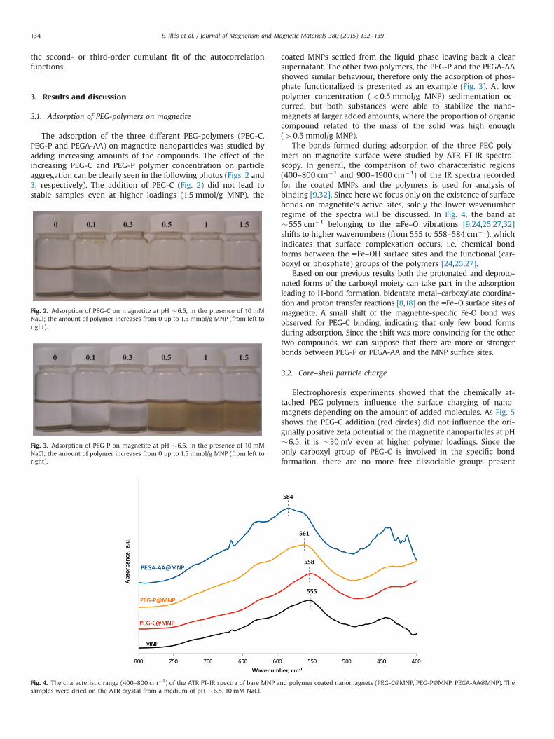
Fig. 4. The characteristic range (400–800 cm�1) of the ATR FT-IR spectra of bare MNP asamples were dried on the ATR crystal from a medium of pH �6.5, 10 mM NaCl.
coated MNPs settled from the liquid phase leaving back a clearsupernatant. The other two polymers, the PEG-P and the PEGA-AAshowed similar behaviour, therefore only the adsorption of phos-phate functionalized is presented as an example (Fig. 3). At lowpolymer concentration (o0.5 mmol/g MNP) sedimentation oc-curred, but both substances were able to stabilize the nano-magnets at larger added amounts, where the proportion of organiccompound related to the mass of the solid was high enough(40.5 mmol/g MNP).
The bonds formed during adsorption of the three PEG-poly-mers on magnetite surface were studied by ATR FT-IR spectro-scopy. In general, the comparison of two characteristic regions(400–800 cm�1 and 900–1900 cm�1) of the IR spectra recordedfor the coated MNPs and the polymers is used for analysis ofbinding [9,32]. Since here we focus only on the existence of surfacebonds on magnetite's active sites, solely the lower wavenumberregime of the spectra will be discussed. In Fig. 4, the band at�555 cm�1 belonging to the ≡Fe–O vibrations [9,24,25,27,32]shifts to higher wavenumbers (from 555 to 558–584 cm�1), whichindicates that surface complexation occurs, i.e. chemical bondforms between the ≡Fe–OH surface sites and the functional (car-boxyl or phosphate) groups of the polymers [24,25,27].
Based on our previous results both the protonated and deproto-nated forms of the carboxyl moiety can take part in the adsorptionleading to H-bond formation, bidentate metal–carboxylate coordina-tion and proton transfer reactions [8,18] on the ≡Fe–O surface sites ofmagnetite. A small shift of the magnetite-specific Fe-O bond wasobserved for PEG-C binding, indicating that only few bond formsduring adsorption. Since the shift was more convincing for the othertwo compounds, we can suppose that there are more or strongerbonds between PEG-P or PEGA-AA and the MNP surface sites.
3.2. Core–shell particle charge
Electrophoresis experiments showed that the chemically at-tached PEG-polymers influence the surface charging of nano-magnets depending on the amount of added molecules. As Fig. 5shows the PEG-C addition (red circles) did not influence the ori-ginally positive zeta potential of the magnetite nanoparticles at pH�6.5, it is �30 mV even at higher polymer loadings. Since theonly carboxyl group of PEG-C is involved in the specific bondformation, there are no more free dissociable groups present
nd polymer coated nanomagnets (PEG-C@MNP, PEG-P@MNP, PEGA-AA@MNP). The

Fig. 5. The zeta potentials of PEG-C@MNPs (red circles), PEG-P@MNPs (orange squares) and PEGA-AA@MNPs (blue diamonds) at various polymer loadings and constant pH�6.5 and 10 mM NaCl. (Inset: upper photo – PEG-C@MNPs and the lower one – PEG-P@MNPs). (For interpretation of the references to color in this figure legend, the readeris referred to the web version of this article.)
Fig. 6. The pH-dependent zeta potential of naked (black diamonds) and PEG-C coated MNPs at 0.2 (red squares) and 1 (blue circles) mmol/g polymer loadings in thepresence of 10 mM NaCl. (For interpretation of the references to color in this figure legend, the reader is referred to the web version of this article.)
E. Illés et al. / Journal of Magnetism and Magnetic Materials 380 (2015) 132–139 135
providing extra charges for electrostatic stabilization of MNPs.Similar feature was observed for salicylic acid adsorption on alu-mina [36], where the electrostatic stability also could not bereached as a consequence of the aromatic carboxylic acid baringonly one COOH group.
On the other hand, an increase in the PEG-P and PEGA-AAadsorption leads to significant lowering of the MNPs' zeta poten-tial (orange squares and blue diamonds in Fig. 5). Increasing thepolymer addition, first the charge neutralization of MNP's positivesurface charge at pH�6.5 takes place. The isoelectric point (IEP) ofthe MNPs was reached at �0.15 mmol/g loading in harmony withour previous experiences [10,18]. Complete charge reversal can be
achieved (zeta potential became ��35 mV), if the amount ofunbound functional groups is large enough at higher polymerloadings (40.5 mmol/g), as it was found by others as well [26,27].Similarly to PEG-C, PEG-P has also only one anchoring group at-tached at the end of the PEO chain with similar functional groupdensity (�0.2 and �0.3 mmol/g for PEG-C and PEG-P polymers,respectively, as seen in Table 1), but it is still able to stabilize theMNPs due to the multivalence of the phosphate group. Supposingmono- or bidentate complex formation between Fe–O surface sitesand phosphate groups take place, free negative charges still re-main in the polymer shell providing electrostatic stabilization fornanomagnets. Since PEGA-AA is a polycarboxylate decorated with

Fig. 7. The pH-dependent zeta potential of naked (black diamonds) and PEG-P coated MNPs at 0.2 (red squares), 0.5 (orange triangles) and 1 (blue circles) mmol/g polymerloadings in the presence of 10 mM NaCl. (For interpretation of the references to color in this figure legend, the reader is referred to the web version of this article.)
E. Illés et al. / Journal of Magnetism and Magnetic Materials 380 (2015) 132–139136
PEO side chains, its polyanionic character at pH�6.5 provides anelectrostatic repulsion between the core–shell particles [26,27].
Studying the pH-dependent charge state of the coated MNPs, itcan be seen that zeta potential of the PEG-C coated MNPs did notshow characteristic difference at various polymer loadings (Fig. 6)compared to the uncoated nanomagnets (pHIEP �8). The apparentincrease in zeta potential values may be attributed to a shift of theslipping plane apart from the particle surface due to the presenceof an adsorption layer; however, the accuracy of zeta potentialmeasurement (75 mV) also has to be kept in mind.
PEG-P and PEGA-AA coated MNPs have similar pH-dependentsurface charging behaviour, so only the data set of PEG-P@MNPsare presented in Fig. 7. At small added amount of PEG-P(0.2 mmol/g MNP, red squares) zeta potential slightly decreasedand the pHIEP shifted to a lower pH (from �8 to �6). An increasein the added amount (0.5 mmol/g MNP, orange triangles in Fig. 7)of both polymers led to gradual shifting of the pHIEP to lowerpH values (from �8 to �4). The further addition of polymer(1 mmol/g MNP, blue circles) leads to more negative zeta poten-tials due to the excess of negative charges originating from thenon-bonded acidic groups of the polymers, the minimum value is��40 mV. In addition, the IEP almost disappeared in the studiedpH range (pH¼3–10) as experienced for other polyelectrolytecoatings too [9,10,26,27].
3.3. Colloid stability
In biological applications such as intravenous administration,the aggregation of MNPs must be prevented. Testing the colloidalstability under biorelevant conditions is perhaps the most im-portant task. Hydrodynamic diameters of bare (MNP) and coatednanoparticles (PEG-C@MNP, PEG-P@MNP and PEGA-AA@MNP) atvarious polymer loadings were measured by DLS (Fig. 8).
The recorded size values characterize the aggregation state ofthe nanomagnets in harmony with the measured zeta potentials(Fig. 5). PEG-C coated nanoparticles exhibit large mean values(4800 nm) independently of the added polymer amounts, in-dicating that aggregation takes place between the non-stabilizedMNPs. The samples containing PEG-P (orange squares) and PEGA-AA (blue diamonds in Fig. 8) show different colloidal stability
states depending on the polymer loading, since large sizes wererecorded at low polymer additions, while small particles weredetected at higher degree of coating (40.5 mmol/g MNP).
In Figs. 9 and 10, the hydrodynamic sizes measured betweenpH �3 and �10 for PEG-C@MNP and PEG-P@MNP, respectively,are presented. The naked MNPs show pH-dependent aggregationstate in harmony with the change of the zeta potential values (seeblack diamonds in Figs. 6 and 7). In the absence of electrostaticstabilization aggregation takes place around the pH of the IEP(�8) exhibiting a maximum in the measured size values(41000 nm in Fig. 9). Further from the pHIEP, however, themagnetite sols are stable, since the particles become positively(pHo8) or negatively (pH48) charged. With increasing amountof PEG-C added to the nanoparticles, no change in pH-dependentaggregation behaviour could be detected even at higher loadingscompared to the size values of bare MNPs (Fig. 9). This finding,together with the electrophoresis measurements, supports ouridea, that PEG-C is not appropriate for stabilization of nano-particles at pHs close to the physiological condition (�7).
In contrary, PEG-P and PEGA-AA adsorption significantly in-fluences the pH-dependent aggregation state of MNPs. Since thesecompounds showed similar behaviour regarding the colloid sta-bility, only the data measured for PEG-P are presented in Fig. 10.Addition of a trace amount of PEG-P resulted in the widening ofthe aggregation zone with particle sizes larger than 1000 nm (redsquares) due to the partial charge neutralization. An increase inthe added amount of both polymers (orange triangles) led to theshifting of the maxima of the aggregation zone to lower pH values(from �8 to �4) in harmony with the change of pHIEP (Figs. 6 and7). If the polymers were added at a larger amount (1 mmol/g MNP,blue circles) stable samples could be obtained, containing particleswith an average diameter of �95 nm. Since significant change(from 80 nm to �95 nm) could be measured for coated (PEG-P@MNP and PEGA-AA@MNP) particles' size compared to that ofthe bare nanomagnets, it means, that enough thick layer of poly-mers formed, and so the steric contribution to the stabilization ofMNPs can be assumed. This combined effect, the so-called electro-steric stabilization of nanoparticles was also experienced for otherpolyelectrolyte coatings [9,10,26,27].

Fig. 9. The pH-dependent hydrodynamic diameter (Z-Ave values) of naked (black diamonds) and PEG-C coated MNPs at 0.2 (red squares) and 1 (blue circles) mmol/g MNPloadings in the presence of 10 mM NaCl. (For interpretation of the references to color in this figure legend, the reader is referred to the web version of this article.)
Fig. 8. Hydrodynamic diameter of PEG-C@MNPs (red circles), PEG-P@MNPs (orange squares) and PEGA-AA@MNPs (blue diamonds) at various loadings, measured at pH�6.5 and 10 mM NaCl. (For interpretation of the references to color in this figure legend, the reader is referred to the web version of this article.)
E. Illés et al. / Journal of Magnetism and Magnetic Materials 380 (2015) 132–139 137
As it was mentioned before, the colloidal stability is essentialfor MNPs designed for biomedical application. The preliminaryresults of coagulation kinetic experiments performed by a methodpublished before [9,32] showed that the salt tolerance of thecoated MNPs stable at pH �6.5 exceed the physiological saltcontent (�150 mM). As outlined above both the type and thenumber of PEG-polymers' anchoring groups determine the ag-gregation of covered MNPs. It was revealed that multiple chargesin the protecting layer can provide appropriate stabilization ofnanomagnets. The uniform distribution of carboxylates and PEGchains along the carbon skeleton makes the PEGA-AA comb-likecopolymer to be a more sophisticated variant for the PEGylation ofMNPs.
The promising physico-chemical results open a new perspec-tive for the biomedical application of these functionalized PEGcoated nanomagnets, e.g., MRI contrast enhancement, magnetichyperthermia and perhaps targeted drug delivery, but to test theirreal theranostic potential further experiments (e.g. hemocompat-ibility assay, cytotoxicity tests, MRI contrast enhancement andmagnetic hyperthermia measurements) [9,32] are necessary.
4. Conclusions
The naked magnetite nanoparticles are not suitable for bio-medical applications due to the aggregation near the physiologicalpH (�7) in the absence of (electrostatic or steric) stabilization.

Fig. 10. The pH-dependent hydrodynamic diameter (Z-Ave values) of the naked (black diamonds) and the PEG-P@MNPs at 0.2 (red squares), 0.5 (orange triangles) and 1(blue circles) mmol/g MNP loadings in the presence of 10 mM NaCl. (For interpretation of the references to color in this figure legend, the reader is referred to the webversion of this article.)
E. Illés et al. / Journal of Magnetism and Magnetic Materials 380 (2015) 132–139138
Huge efforts are made to find a coating for MNPs, which fulfills theserious criteria of biocompatibility, and the chemical and thecolloidal stability as well. The latter remain untouched; re-searchers do not pay enough attention to them. Although nu-merous papers are published in literature related to the MNPcoatings they are based on empirical considerations in general.Recently, PEGylation is one of the most widely applied methods toensure the biocompatibility of nanomagnets. In this paper, ourpurpose was to provide a qualitative comparison of the colloidalstability of core-shell MNPs bearing various functionalized PEG-polymer shells.
Three functionalized PEG-polymers (PEG-C, PEG-P and PEGA-AA) were used to coat MNPs. Surface modification efficiency ofcommercially available linear PEGs with one functional (carboxylor phosphate) end-group and an own-synthesized comb-like car-boxylated PEG compound was compared. The influence of che-mical difference and the number of functional groups present inthe PEG-polymer on the colloid stability of MNPs was the funda-mental question of this work.
ATR FT-IR measurements proved that these PEG-polymers ad-sorb by forming chemical bonds on magnetite. It is worthy of at-tention, that PEG-C, the linear mono-functionalized (carboxylated)PEO polymer was not able to stabilize the magnetic nanoparticles.The only COOH of PEG-C binds to the surface of MNP, so no excesscharges are present in the polymer shell providing electrostaticstabilization for nanoparticles.
Other important outcome of the experiments, if these polymerscoat the MNPs completely, is that both the addition of PEG-P andPEGA-AA led to colloidally stable samples under near-biorelevantconditions due to the multiple charges linked to each polymerchain. As a conclusion we can state, that beside appropriateloading of polymers, multiple charges are necessary for electro-static stabilization, originating from either one (phosphate) ormore functional groups distributed along the polymer chain(polycarboxylate type structure). Based on these and previousexperiences, the continuous distribution of charges in the own-prepared carboxylated comb-like polymer (PEGA-AA) makes itmore suitable for stabilization of MNPs. In addition, it opens the
possibility for fine tuning of the PEG-polymer shell structure bychanging the number and type of functional groups.
Acknowledgement
This research was supported by the OTKA (NK 84014) grant aswell as by European Union and the State of Hungary, co-financedin the framework of TÁMOP-4.2.4.A/2-11/1-2012-0001 ‘NationalExcellence Program’.
References
[1] Q.A. Pankhurst, N.K.T. Than, S.K. Jones, J. Dobson, Progress in applications ofmagnetic nanoparticles in biomedicine, J. Phys. D: Appl. Phys 42 (2009)224001 (15 pp.).
[2] J.K. Oh, J.M. Park, Iron oxide-based superparamagnetic polymeric nanoma-terials: design, preparation, and biomedical application, Prog. Polym. Sci 36(2011) 168–189.
[3] S. Mura, P. Couvreur, Nanotheranostics for personalized medicine, Adv. DrugDeliv. Rev. 64 (2012) 1394–1416.
[4] C. Corot, D. Warlin, Superparamagnetic iron oxide nanoparticles for MRI:contrast media pharmaceutical company R&D perspective, Wiley Interdiscip.Rev. Nanomed. Nanobiotechnol. 5 (2013) 411–422.
[5] A.S. Wadajkar, J.U. Menon, T. Kadapure, R.T. Tran, J. Yang, K.T. Nguyen, Designand application of magnetic-based theranostic nanoparticle systems, RecentPat. Biomed. Eng. 6 (2013) 47–57.
[6] S. Dürr, C. Janko, S. Lyer, P. Tripal, M. Schwarz, J. Zaloga, R. Tietze, C. Alexiou,Magnetic nanoparticles for cancer therapy, Nanotechnol. Rev. 2 (4) (2013)395–409.
[7] M.L. Etheridge, S.A. Campbell, A.G. Erdman, C.L. Haynes, S.M. Wolf,J. McCullough, Journal of Nanomedicine: Nanotechnology, Biol. Med. 9 (2013)1–14.
[8] I.Y. Tóth, E. Illés, R.A. Bauer, D. Nesztor, M. Szekeres, I. Zupkó, E. Tombácz,Designed polyelectrolyte shell on magnetite nanocore for dilution-resistantbiocompatible magnetic fluids, Langmuir 28 (2012) 16638–16646.
[9] M. Szekeres, I.Y. Tóth, E. Illés, A. Hajdú, I. Zupkó, K. Farkas, G. Oszlánczi,L. Tiszlavicz, E. Tombácz, Chemical and colloidal stability of carboxylated core–shell magnetite nanoparticles designed for biomedical applications, Int. J. Mol.Sci. 14 (2013) 14550–14574.
[10] E. Tombácz, I.Y. Tóth, D. Nesztor, E. Illés, A. Hajdú, M. Szekeres, L. Vékás, Ad-sorption of organic acids on magnetite nanoparticles, pH-dependent colloidalstability and salt tolerance, Colloids Surf. A 435 (2013) 91–96.

E. Illés et al. / Journal of Magnetism and Magnetic Materials 380 (2015) 132–139 139
[11] J. Liu, Z. Sun, Y. Deng, Y. Zou, C. Li, X. Guo, L. Xiong, Y. Gao, F. Li, D. Zhao, Highlywater-dispersible biocompatible magnetite particles with low cytotoxicitystabilized by citrate groups, Angew. Chem. Int. Ed. 48 (2009) 5875–5879.
[12] R.G. RuizMoreno, A.I. Martinez, R. Castro-Rodriguez, P. Bartolo, Synthesis andcharacterization of citrate coated magnetite nanoparticles, J. Supercond. Nov.Magn. 26 (2013) 709–712.
[13] D. Bica, L. Veka s, M.V. Avdeev, O. Marinica, V. Socoliuc, M. Balasoiu, V.M. Garamus, Sterically stabilized water based magnetic fluids: synthesis,structure and properties, J. Magn. Magn. Mater. 311 (2007) 17–21.
[14] E. Tombácz, D. Bica, A. Hajdú, E. Illés, A. Majzik, L. Vékás, Surfactant doublelayer stabilized magnetic nanofluids for biomedical application, J. Phys. Cond.Matter 20 (2008) 20410 (6 pp.).
[15] R.P. Araújo-Neto, E.L. Silva-Freitas, J.F. Carvalho, T.R.F. Pontes, K.L. Silva, I.H.M. Damasceno, E.S.T. Egito, A.L. Dantas, M.A. Morales, A.S. Carriço, Mono-disperse sodium oleate coated magnetite high susceptibility nanoparticles forhyperthermia applications, J. Magn. Magn. Mater. 364 (2014) 72–79.
[16] S. Dutz, M. Kettering, I. Hilger, R. Müller, M. Zeisberger, Magnetic multicorenanoparticles for hyperthermia-influence of particle immobilization in tu-mour tissue on magnetic properties, Nanotechnology 22 (2011) 265102 (7pp.).
[17] Y. Wang, H.-Z. Jia, K. Han, R.-X. Zhuo, X.-Z. Zhang, Theranostic magnetic na-noparticles for efficient capture and in situ chemotherapy of circulating tumorcells, J. Mater. Chem. B 1 (2013) 3344–3352.
[18] A. Hajdú, M. Szekeres, I.Y. Tóth, R.A. Bauer, J. Mihály, I. Zupkó, E. Tombácz,Enhanced stability of polyacrylate-coated magnetite nanoparticles in bior-elevant media, Colloids Surf. B: Biointerfaces 94 (2012) 242–249.
[19] V.I. Shubayev, T.R. Pisanic, S. Jin, Magnetic nanoparticles for theragnostics, Adv.Drug Deliv. Rev 61 (2009) 467–477.
[20] B. Mishra, B.B. Patel, S. Tiwari, Colloidal nanocarriers: a review on formulationtechnology, types and applications toward targeted drug delivery, Nanomed.Nanotechnol. 6 (2010) 9–24.
[21] B. Thong-On, B. Rutnakornpituk, U. Wichai, M. Rutnakornpituk, Magnetitenanoparticle coated with amphiphilic bilayer surfactant of polysiloxane andpoly(poly(ethylene glycol) methacrylate), J. Nanopart. Res. 14 (2012) 953.
[22] X. Cao, B. Zhang, F. Zhao, L. Feng, Synthesis and properties of MPEG-coatedsuperparamagnetic magnetite nanoparticles, J. Nanomater. 2012 (2012)607296 (6 pp.).
[23] O. Veiseh, J.W. Gunn, M. Zhang, Design and fabrication of magnetic nano-particles for targeted drug delivery and imaging, Adv. Drug. Deliv. Rev 62(2010) 284–304.
[24] D. Dorniani, A.U. Kura, S.H. Hussein-Al-Ali, M.Z. Bin Hussein, S. Fakurazi, A.H. Shaari, Z. Ahmad, In vitro sustained release study of gallic acid coated with
magnetite-PEG and Magnetite-PVA for drug delivery system, Sci. World J. 2014(2014) 416354 (11 pp.).
[25] S.R. Kumar, L. Marianna, S. Gianni, A.J. Nathanael, S.I. Hong, T.H. Oh,D. Mangalaraj, C. Viswanathan, N. Ponpandian, Hydrophilic polymer coatedmonodispersed Fe3O4 nanostructures and their cytotoxicity, Mater. Res. Ex-press 1 (2014) 015015.
[26] C. Barrera, A. Herrera, Y. Zayas, C. Rinaldi, Surface modification of magnetitenanoparticles for biomedical applications, J. Magn. Magn. Mater. 321 (2009)1397–1399.
[27] A. Masoudi, H.R.M. Hosseini, M.A. Shokrgozar, R. Ahmadi, M.A. Oghabian, Theeffect of poly(ethylene glycol) coating on colloidal stability of super-paramagnetic iron oxide nanoparticles as potential MRI contrast agent, Int. J.Pharm. 433 (2012) 129–141.
[28] A. Jozefczak, A. Skumiel, Ultrasonic investigation of magnetic nanoparticlessuspension with PEG biocompatible coating, J. Magn. Magn. Mater. 323 (2011)1509–1516.
[29] L. Sun, C. Huang, T. Gong, S. Zhou, A biocompatible approach to surfacemodification: Biodegradable polymer functionalized super-paramagnetic ironoxide nanoparticles, Mater. Sci. Eng. C 30 (2010) 583–589.
[30] V. Zavisova, M. Koneracka, M. Muckova, J. Lazova, A. Jurıkova, G. Lancz,N. Tomasovicova, M. Timko, J. Kovac, I. Vavra, M. Fabian, A.V. Feoktystov, V.M. Garamus, M.V. Avdeev, P. Kopcansky, Magnetic fluid poly(ethylene glycol)with moderate anticancer activity, J. Magn. Magn. Mater. 323 (2011)1408–1412.
[31] M. Mahdavi, M.B. Ahmad, M.J. Haron, F. Namvar, B. Nadi, M.Z.A. Rahman,J. Amin, Synthesis, surface modification and characterisation of biocompatiblemagnetic iron oxide nanoparticles for biomedical applications, Molecules 18(2013) 7533–7548.
[32] E. Illés, M. Szekeres, E. Kupcsik, I.Y. Tóth, K. Farkas, A. Jedlovszky-Hajdú,E. Tombácz, PEGylation of surfacted magnetite core-shell nanoparticles forbiomedical application, Colloids Surf. A 460 (2014) 429–440.
[33] U.I. Tromsdorf, O.T. Bruns, S.C. Salmen, U. Beisiegel, H. Weller, A highly ef-fective, nontoxic T1 MR contrast agent based on ultrasmall pegylated ironoxide nanoparticles, Nanoletters 9 (2009) 4434–4440.
[34] R.M. Cornell, U. Schwertmann, The Iron Oxides, VCH, Weimheim, 1996.[35] K. An, P. Zhao, C. Lin, H. Liu, A pH and redox dual responsive 4-arm poly
(ethylene glycol)-block-poly(disulfide histamine) copolymer for non-viralgene transfection in vitro and in vivo, Int. J. Mol. Sci. 15 (2014) 9067–9081.
[36] M. Szekeres, E. Tombácz, K. Ferencz, I. Dékány, Adsorption of salicylate onalumina surfaces, Colloids Surf. A 141 (1998) 319–325.