jpet #2 19 49 3 polythiol-containing, recombinant
TRANSCRIPT

JPET #219493
1
Polythiol-containing, recombinant mannosylated-albumin is a superior CD68+/CD206+ Kupffer
cell-targeted nano-antioxidant for the treatment of two acute hepatitis models
Hitoshi Maeda, Kenshiro Hirata, Hiroshi Watanabe, Yu Ishima, Victor Tuan Giam Chuang, Kazuaki
Taguchi, Akihito Inatsu, Manabu Kinoshita, Motohiko Tanaka, Yutaka Sasaki, Masaki Otagiri, and Toru
Maruyama
Department of Biopharmaceutics, Graduate School of Pharmaceutical Sciences, Kumamoto University,
5-1 Oe-Honmachi, Chuo-ku, Kumamoto 862-0973, Japan. (H.M., K.H., H.W., Y.I., V.C., T.M.)
Center for Clinical Pharmaceutical Sciences, School of Pharmacy, Kumamoto University, 5-1
Oe-Honmachi, Chuo-ku, Kumamoto 862-0973, Japan. (H.W., Y.I., T.M.)
School of Pharmacy, Faculty of Health Sciences, Curtin Health Innovation Research Institute, Curtin
University, GPO Box U1987, Perth 6845, Western Australia. (V.C., T.M.)
Faculty of Pharmaceutical Sciences, Sojo University, 1-22-4 Ikeda, Nishi-ku, Kumamoto 860-0082,
Japan. (K.T., M.O.)
DDS Research Institute, Sojo University, 1-22-4 Ikeda, Nishi-ku, Kumamoto 860-0082, Japan. (M.O.)
Department of Immunology and Microbiology, National Defense Medical College, 3-2 Namiki,
Tokorozawa, Saitama 359-8513, Japan. (A. I., M.K.)
Department of Gastroenterology and Hepatology, Graduate School of Medical Sciences, Kumamoto
University, Kumamoto 860-8556, Japan. (M.T., Y.S.)
This article has not been copyedited and formatted. The final version may differ from this version.JPET Fast Forward. Published on November 14, 2014 as DOI: 10.1124/jpet.114.219493
at ASPE
T Journals on N
ovember 4, 2021
jpet.aspetjournals.orgD
ownloaded from

JPET #219493
2
Running title: SH-Man-HSA attenuates acute liver injury
Corresponding author: Toru Maruyama, Ph.D., Professor,
Department of Biopharmaceutics, Graduate School of Pharmaceutical Sciences, Kumamoto University,
5-1 Oe-honmachi, Kumamoto 862-0973, Japan; Phone: +81-96-370-4150; Fax: +81-96-362-7690;
E-mail address: [email protected]
The number of text pages: 38
The number of tables: 0
The number of Figures: 8
The number of references: 35
The number of words in the Abstract: 250
The number of words in the Introduction: 552
The number of words in the Discussion: 2113
Abbreviations: ROS, reactive oxygen species; KC, Kupffer cells; TNF, tumor necrosis factor; CD,
cluster of differentiation; Con-A, concanavalin-A; GdCl3, gadolinium chloride; APAP, acetaminophen;
CD206, mannose receptor; HSA, Human serum albumin; Man-HSA, mannosylated-HSA; SH, thiol;
GSH, glutathione; NAC, N-acetyl cysteine; SH-Man-HSA, polythiolated-Man-HSA; DTPA,
diethylenetriamine-pentaacetic acid; DTNB, 5,5'-dithiobis-(2-nitrobenzoic acid); DMPO, 5,
5-dimethyl-1-pyrroline N-oxide; POBN, α-(4-pyridyl-1-oxide)-N-tert-butylnitrone; rHSAs, recombinant
HSAs; KPB, potassium phosphate buffer; PBS, phosphate buffered saline; EPR, electron paramagnetic
resonance; ALT, alanine aminotransferase; AST, aspartate aminotransferase; HE, hematoxylin and eosin;
8-OHdG, 8-hydroxy-2'-deoxyguanosine; NO2-Tyr, nitrotyrosine; GSH/GSSG, reduced/oxidized
This article has not been copyedited and formatted. The final version may differ from this version.JPET Fast Forward. Published on November 14, 2014 as DOI: 10.1124/jpet.114.219493
at ASPE
T Journals on N
ovember 4, 2021
jpet.aspetjournals.orgD
ownloaded from
JPET #219493
3
glutathione ratio; LOO˙, lipid peroxide radical; iNOS, inducible NO synthase; MNCs, mononuclear
cells; IFN-γ, interferon-γ; NAPQI, n-acetyl-p-benzoquinone imine; HIF-1α, hypoxia-inducible factor-1α
A recommended section assignment: Drug Discovery and Translational Medicine
This article has not been copyedited and formatted. The final version may differ from this version.JPET Fast Forward. Published on November 14, 2014 as DOI: 10.1124/jpet.114.219493
at ASPE
T Journals on N
ovember 4, 2021
jpet.aspetjournals.orgD
ownloaded from
JPET #219493
4
ABSTRACT
Since reactive oxygen species (ROS) derived from Kupffer cells (KC), especially CD68+ KC,
play a key role in the induction of hepatic oxidative stress and injuries, we newly developed a
polythiolated- and mannosylated-human serum albumin (SH-Man-HSA), which functions as a novel
nano-antioxidant, for delivering thiol to CD68+ KC. In vitro electron paramagnetic resonance (EPR)
coupled with pharmacokinetics and immunohistochemical studies showed that SH-Man-HSA possessed
powerful radical scavenging activity and rapidly and selectively delivered thiols to the liver via mannose
receptor (CD206) on CD68+ cells. SH-Man-HSA significantly improved the survival rate of
concanavalin-A (Con-A) treated mice. Moreover, SH-Man-HSA exhibited excellent hepato-protective
functions, not by decreasing TNF or IFN-γ production that is closely associated with Con-A induced
hepatitis, but by suppressing ROS production. Interestingly, the protective effect of SH-Man-HSA was
superior to N-acetylcysteine (NAC). This could be due to the difference in the inhibition of hepatic
oxidative stress between the two antioxidants depending upon their potential for thiol delivery to the
liver. Similar results were also observed for acetaminophen (APAP)-induced hepatopathy models. Flow
cytometric data further confirmed that an increase in F4/80+/ROS+ cells was dramatically decreased by
SH-Man-HSA. The administration of SH-Man-HSA at 4 hours following a Con-A or APAP injection
also exhibited a profound hepato-protective action against these hepatitis models, while this was not
observed for NAC. It therefore can be concluded that SH-Man-HSA has great potential for use as a
rescue therapy for hepatopathy as a nano-antioxidant because of its ability to efficiently and rapidly
deliver thiols to CD68+/CD206+ KC.
This article has not been copyedited and formatted. The final version may differ from this version.JPET Fast Forward. Published on November 14, 2014 as DOI: 10.1124/jpet.114.219493
at ASPE
T Journals on N
ovember 4, 2021
jpet.aspetjournals.orgD
ownloaded from
JPET #219493
5
Introduction
Oxidative stress plays a key role in the onset and progression of various liver diseases, including
viral hepatitis (Korenaga et al., 2005), autoimmune hepatitis (Moreno-Otero, 2013), alcoholic
hepatopathy (Tsukamoto and Lu, 2001), and drug-induced hepatic impairment (Laskin and Laskin, 2001).
Reactive oxygen species (ROS) produced upon the activation of Kupffer cells (KC), and the subsequent
production of inflammatory cytokines such as tumor necrosis factor (TNF)-cytokines triggered by the
presence of ROS have been shown to be involved in the progression of a number of liver pathological
conditions (Roberts et al., 2007; Jaeschke, 2011). Kinoshita et al. reported on the subclassification of KC
based on their surface markers and functions, namely, cluster differentiation (CD) 68+, residental, and
CD11b+ KC, monocyte-derived. CD68+ KC have a potent capacity to produce ROS, whereas CD11b+
cells did not. Conversely, CD11b+ KC have a strong capacity for the production of TNF, which was much
less prominent in CD68+ cells (Kinoshita et al., 2010). Moreover, ROS produced by CD68+ KC have
been identified as an aggravating factor for the onset and progression of hepatitis in a concanavalin-A
(Con-A)-induced hepatitis mouse model (Nakashima et al., 2008). In addition, mice that were pretreated
with gadolinium chloride (GdCl3), a KC inhibitor, especially CD68+ cells, showed a decreased
acetaminophen (APAP)-induced hepato-toxicity, possibly via suppressing the induction of oxidative
stress (Laskin et al., 1995; Michael et al., 1999). This was particularly evident in the case of CD68+ KC.
It therefore appears that KC, especially CD68+, play a major role in the progression of liver pathological
conditions. In fact, it had been shown that the intensity of KC activation, based on CD68 expression, is
directly correlated with the degree of disease severity in patients with alcoholic liver disease (Chedid et
al., 2004). Interestingly, mannose receptors (CD206) are present on the KC surface, including CD68+
(Hu et al., 2012). An effective ROS-scavenging agent that can be delivered to CD68+ KC in the liver
through CD206 would therefore be predicted to result in promising therapeutic outcomes. However, as
far as we know, no carrier that targets CD68+/CD206+ KC is currently available.
This article has not been copyedited and formatted. The final version may differ from this version.JPET Fast Forward. Published on November 14, 2014 as DOI: 10.1124/jpet.114.219493
at ASPE
T Journals on N
ovember 4, 2021
jpet.aspetjournals.orgD
ownloaded from
JPET #219493
6
Human serum albumin (HSA) is a simple protein with no oligosaccharide chain structures.
However, the insertion of a consensus sequence for an oligosaccharide chain into the albumin gene
results in the production of a protein that contains an oligosaccharide chain, as in some reported genetic
variants (Minchiotti et al., 2001). Using three of these HSA variants, Albumin Dalakarlia, Casebrook and
Redhill (Kragh-Hansen et al., 2001), as a template for designing a recombinant glycosylated-HSA, we
recently succeeded in producing a recombinant mannosylated-HSA (Man-HSA) containing a
biosynthetic oligosaccharide chain with a high content of 12 mannose residues per HSA molecule using a
yeast expression system (Supplemental Figure 1) (Hirata et al., 2010). Therefore, Man-HSA would be
anticipated to be efficiently distributed to CD68+ KC via CD206.
Antioxidants containing a thiol (SH) group, such as glutathione (GSH) and N-acetylcysteine
(NAC), possess excellent ROS-scavenging activities (Saito et al., 2010). In fact, NAC is currently the
only remedy available for the treatment of APAP-induced hepato-toxicity (Lee et al., 2008). Hence,
polythiolated-Man-HSA (SH-Man-HSA), produced by introducing SH groups into Man-HSA, has the
potential to function as a novel nano-antioxidant that targets ROS derived from KC, especially
CD68+/CD206+ and thereby, as a candidate of the rescue therapy for both acute and chronic hepatopathy.
This article has not been copyedited and formatted. The final version may differ from this version.JPET Fast Forward. Published on November 14, 2014 as DOI: 10.1124/jpet.114.219493
at ASPE
T Journals on N
ovember 4, 2021
jpet.aspetjournals.orgD
ownloaded from

JPET #219493
7
Materials and Methods
Animals
Male C57BL/6 mice (8 weeks old) were purchased from Japan SLC (Shizuoka, Japan). All
animal experiments were conducted in accordance with the guidelines of Kumamoto University for the
care and use of laboratory animals.
Reagents
Diethylenetriamine-pentaacetic acid (DTPA) and 5, 5'-dithiobis (2-nitrobenzoic acid) (DTNB)
were obtained from Dojindo Laboratories (Kumamoto, Japan). 5-dimethyl-1-pyrroline N-oxide (DMPO)
and α-(4-pyridyl-1-oxide)-N-tert-butylnitrone (POBN) were purchased from Alexis Biochemicals
(Lausen, Switzerland). All other chemicals were of the highest analytical grade available.
Production and purification of recombinant HSAs
The protocol used to express the recombinant HSAs (rHSAs; Man-HSA and HSA) by Pichia
pastoris was a modification of a previously published procedure (Hirata et al., 2010). The eluted rHSAs
were deionized and defatted by charcoal treatment, freeze-dried, and then stored at −80 °C until used.
Sample purity was estimated by a density analysis of the coomassie brilliant blue-stained protein bands
on a 10% SDS-PAGE gels. The rHSAs were estimated to be more than 97% pure. A MALDI-TOF-MS
analysis revealed that approximately 12 mannose (N-acetyl glucosamine=2) units are present in the
glycan chain structure of the Man-HSA (Supplemental Figure 1).
Synthesis of SH-rHSAs and determination of thiolation efficiency
Terminal SH groups were added to the rHSAs molecule by incubating 0.15 mM rHSAs with 8
This article has not been copyedited and formatted. The final version may differ from this version.JPET Fast Forward. Published on November 14, 2014 as DOI: 10.1124/jpet.114.219493
at ASPE
T Journals on N
ovember 4, 2021
jpet.aspetjournals.orgD
ownloaded from

JPET #219493
8
mM 2-iminothiolane in 100 mM potassium phosphate buffer (KPB) containing 0.5 mM DTPA, pH 7.8,
for 1 hour at room temperature (Katayama et al., 2008). The amount of SH groups in the SH-rHSAs was
quantified using the DTNB method. First, 20 μL aliquots of SH-rHSAs solution and reduced glutathione
(standard) were incubated in a 96-well plate with 0.2 mL of 100 mM KPB, pH 7.0, containing 1 mM
DTPA and 0.5 mM DTNB, for 15 minutes at room temperature. The absorbance was then measured at
405 nm. The number of SH groups attached to Man-HSA or HSA was estimated to be 7.52 ± 0.01 mol
SH/mol Man-HSA or 7.82 ± 0.02 mol SH/mol HSA, respectively (Supplemental Figure 2B).
Scavenging activity of SH-Man-HSA against ·OH generated by H2O2/UV system
The reaction solution contained 100 μM DTPA, 9 mM DMPO and 500 μM H2O2 in the absence
or presence of 75 μM HSA, Man-HSA or SH-Man-HSA in phosphate buffered saline (PBS) (pH 7.4).
After the sample was irradiated with UV (254 nm) for 1 minute in the electron paramagnetic resonance
(EPR) flat cell, EPR spectra were measured immediately.
Pharmacokinetic analysis of SH-Man-HSA
SH-Man-HSA was radiolabeled with 111In using the bifunctional chelating reagent DTPA
anhydride according to the method of Hnatowich et al. (Hnatowich et al., 1982). Mice received tail vein
injections of 111In-labeled SH-Man-HSA in saline, at a dose of 10.0 nmol SH/kg. In the early period after
injection, the efflux of 111In radioactivity from organs is assumed to be negligible, because the
degradation products of 111In-labeled ligands using DTPA anhydride cannot easily pass through
biological membranes. This assumption was confirmed by the finding that 111In was not detectable in the
urine throughout the 120 minutes period. At appropriate intervals after the injection, blood was collected
from the vena cava under ether anesthesia and plasma was obtained by centrifugation (3000 xg, 10
minutes). The liver, kidney, spleen and lung were excised, rinsed with saline and weighed. The
This article has not been copyedited and formatted. The final version may differ from this version.JPET Fast Forward. Published on November 14, 2014 as DOI: 10.1124/jpet.114.219493
at ASPE
T Journals on N
ovember 4, 2021
jpet.aspetjournals.orgD
ownloaded from

JPET #219493
9
radioactivity of each sample was measured in a well-type NaI scintillation counter (ARC-500, Aloka,
Tokyo).
Preparation of bromobimane-labeled SH-Man-HSA
A solution of 75 μM bromobimane in 0.1 M KPB was added to a solution of 100 μM
SH-Man-HSA in 0.1 M KPB, followed by incubation for 2 hours at 4 °C. The resulting solution was
purified using a Pharmacia Bio-Gel PD-10 column (GE Healthcare UK Ltd) and concentrated by
Vivaspin (Sartorius Stedim Biotech S.A., France).
Evaluation of CD68+/CD206+ KC distribution of SH-Man-HSA
The prepared bromobimane-labeled SH-Man-HSA (20.0 μmol SH/kg) was injected into the tail
vein of mice. Mannan (6.0 mg/mouse) was injected intravenously 30 minutes before the
bromobimane-labeled SH-Man-HSA administration. At 1 and 12 hours after the administration of
bromobimane-labeled SH-Man-HSA, the liver was removed, covered with OCT compound and frozen at
−80 °C. Fresh frozen sections (4 μm thickness) of the liver were cut on a cryostat (Leica, CM3000II,
Germany), collected on slides, and immediately dried. The sections were fixed with phosphate-buffered
4% paraformaldehyde and washed. After incubation with 1% Block Ace (DS Pharma Biomedical,
Osaka) for 10 minutes, the slides were incubated overnight with anti-HSA (BETHYL), CD68 (FA-11,
BioLegend), CD206 (C068C2, BioLegend) antibody diluted 100, 100 or 200 times, respectively. After
the reaction, the slide was observed using a Microscope (Keyence, BZ-8000, Osaka, Japan).
Effect of SH-Man-HSA on the survival rate of lethal Con-A treated mice
A lethal Con-A treated mouse model was produced using C57BL/6 mice that were intravenously
injected with Con-A (1.0 mg/mouse). The same SH content (20.0 μmol SH/kg) of SH-Man-HSA and
This article has not been copyedited and formatted. The final version may differ from this version.JPET Fast Forward. Published on November 14, 2014 as DOI: 10.1124/jpet.114.219493
at ASPE
T Journals on N
ovember 4, 2021
jpet.aspetjournals.orgD
ownloaded from

JPET #219493
10
NAC was administered intravenously just prior to the Con-A injection.
Hepato-protective effect of SH-Man-HSA on Con-A or APAP treated mice
Con-A- and APAP-induced liver injury mouse model was produced as previously reported
(Ayoub et al., 2004; Nakashima et al., 2008; Saito et al., 2010). Animal experiment protocol was shown
in Supplemental figure 4 and 5. Alanine aminotransferase (ALT) and aspartate aminotransferase (AST)
activities levels were determined using a transaminase C2 test kit from Wako Chemicals (Saitama, Japan).
In case of Con-A, the mice received 0.25 mg of Con-A intravenously (Sigma Chemical Co., St. Louis,
MO). 5.0, 10.0, 20.0 μmol SH/kg of SH-Man-HSA and 20.0 μmol SH/kg of NAC were administered
intravenously to the mice just prior to Con-A injection. Similarly, SH-HSA (20.0 μmol SH/kg) or
Man-HSA (2.66 μmol SH/kg) was administered intravenously. In case of APAP, the mice received 300
mg/kg of APAP intraperitoneally (Sigma Chemical Co., St. Louis, MO). The same SH content (20.0
μmol SH/kg) of SH-Man-HSA and NAC were administered intravenously to the mice just prior to APAP
injection. Furthermore, we administered SH-Man-HSA or NAC (i.v.) at 2 and 4 hours after the injection
of Con-A or APAP. To inhibit the KC, especially CD68+ cells, 200 μg of GdCl3 was administered (Sigma
Chemical Co., St. Louis, MO) intravenously via the caudal vein 24 hours before Con-A or APAP
injection. All mice were sacrificed 12 hours after Con-A or APAP injection.
Histological examination of liver tissues
12 hours after Con-A or APAP injection, the whole liver was removed. For the histological
analyses, the liver tissues were fixed in 10% neutral-buffered formalin (Wako, Japan), embedded in
paraffin (Sakura Finetek Japan, Japan) and sectioned at a 4 µm thickness. The liver sections were
subjected to hematoxyline and eosin (HE) staining for morphologic analysis, TUNEL staining for cell
apoptosis and immunohistochemistry for 8-hydroxy-2'-deoxyguanosine (8-OHdG) [15A3] (Santa Cruz
This article has not been copyedited and formatted. The final version may differ from this version.JPET Fast Forward. Published on November 14, 2014 as DOI: 10.1124/jpet.114.219493
at ASPE
T Journals on N
ovember 4, 2021
jpet.aspetjournals.orgD
ownloaded from

JPET #219493
11
Biotechnology Inc, cat#: sc-66036, Santa Cruz, USA) and nitrotyrosine (NO2-Tyr) (Millipore, cat#:
AB5411, Billerica, USA). All assays were performed in triplicate. For TUNEL staining, sections were
stained using an in situ cell death detection kit, Fluorescein (Roche, Basel, Switzerland), according to the
manufacturer’s protocol for paraffin-embedded sections. For the immunohistochemistry of 8-OHdG and
NO2-Tyr, first, antigen retrieval was performed by means of an immunosaver (Nisshin EM Corporation,
Tokyo, Japan). A solution containing 50 mM Tris-HCl (pH= 7.4) and 0.1% Tween-20 (T-TB) was then
used to solubilize the liver slices, followed by blocking with Block Ace (Dainippon Pharmaceutics,
Osaka, Japan) at room temperature for 10 minutes. Next, reaction with the primary antibody was carried
out overnight at a temperature below 4 °C. In addition, the primary antibody against 8-OHdG or NO2-Tyr
was diluted with 0.5% BSA in PBS 50 times before use. The liver slices were then washed with T-TB,
followed by reaction with the secondary antibody at room temperature for 1.5 hours. For 8-OHdG
immunostaining, Alexa Fluor 488 goat anti-mouse IgG (H+L) (Invitrogen, Tokyo, Japan) and for
NO2-Tyr immunostaining, Alexa Fluor 546 goat anti-mouse IgG (H+L) (Invitrogen, Tokyo, Japan) were
diluted with 0.5% BSA in PBS 200 times before use. After the reaction, the slide was observed under a
microscope (Keyence, BZ-8000, Osaka, Japan).
Quantification of liver SH and GSH levels
Tissue homogenates were prepared by homogenizing the tissues with 5% (w/v) sulfosalicylic
acid. The homogenates were then centrifuged at 10,000 xg for 10 minutes at 4 °C. SH levels were
determined according to the DTNB method. Reduced and oxidized glutathione levels in the livers were
determined using a reduced/oxidized GSH ratio (GSH/GSSG) quantification kit (Dojinkagaku,
Kumamoto, Japan).
Determination of ROS in liver using EPR
This article has not been copyedited and formatted. The final version may differ from this version.JPET Fast Forward. Published on November 14, 2014 as DOI: 10.1124/jpet.114.219493
at ASPE
T Journals on N
ovember 4, 2021
jpet.aspetjournals.orgD
ownloaded from

JPET #219493
12
The in vivo EPR analysis was performed as described previously with some modifications (Sato
et al., 2002). The EPR spectra of POBN spin adducts of lipid peroxide radical (LOO˙) were determined at
4 hours after Con-A or APAP injection. POBN (1.0 g/kg) was injected intraperitoneally at 30 minutes
before the mice were sacrificed. The livers were homogenized in 1 mL of saline on an ice bath. The
homogenates were added in 2 mL of 2:1 chloroform/methanol, shaken then centrifuged at 2800 rpm for
10 minutes. The chloroform layer was isolated. After evaporating the sample by bubbling with N2 in a
vial, the sample was resuspended in organic solvent (200 µL of chloroform/methanol). EPR spectra were
immediately recorded at room temperature in a JES-TE200 spectrometer (JEOL, Tokyo, Japan) under the
following conditions: modulation frequency, 100 kHz; microwave frequency, 9.43 GHz; microwave
power, 40 mW; scanning field, 333.5 mT; sweep time, 2 minutes; field modulation width, 0.25 mT;
receiver gain, 630; and time count, 0.3 seconds. Every buffer and solutions of the reaction mixture used
for EPR measurement were treated with chelex 100 resin (Bio-Rad Laboratories) before use to remove
metals.
Isolation of hepatic mononuclear cells (MNCs) and flow cytometry
MNCs were prepared as described previously (Dobashi et al., 1999). Liver specimens were
minced with scissors. After adding a 0.25% collagenase solution, the specimens were shaken for 20
minutes in a 37 °C constant-temperature bath. The liver specimen was then filtered through a nylon mesh
(108 μm). After mixing in 33% Percoll solution containing heparin (1%), the sample was centrifuged for
20 minutes at 500 xg at room temperature. After removing the supernatant, the pellet was resuspended in
a red blood cell lysis solution and then was washed twice in 10% fetal bovine serum Roswell Park
Memorial Institute 1640. For identification of KC, MNCs were stained by phycoerythrin Cy5-
conjugated anti-F4/80 antibody (eBioscience, San Diego, CA). For the detection of intracellular ROS,
CM-H2DCFDA (Invitrogen, Carlsbad, CA, USA), a ROS-sensitive fluorescent dye used as an ROS
This article has not been copyedited and formatted. The final version may differ from this version.JPET Fast Forward. Published on November 14, 2014 as DOI: 10.1124/jpet.114.219493
at ASPE
T Journals on N
ovember 4, 2021
jpet.aspetjournals.orgD
ownloaded from
JPET #219493
13
probe. At also, for the detection of macrophage polarization status, anti-inducible NO synthase (iNOS)
(Santa Cruz, California, USA) and CD206 (C068C2, BioLegend) antibody were used. F4/80+ KC were
isolated by positive selection according to the manufacturer’s protocol (MACS isolation system,
Militenyi Biotec, Auburn, CA). Flow cytometry was performed using an FACS Calibur (BD
Biosciences).
Quantification of TNF and interferon-γ (IFN-γ) levels
TNF and IFN-γ levels in plasma at 1 and 12 hours, respectively, after Con-A (0.25 mg/mouse)
injection were measured by ELISA, following the manufacturer’s recommended protocol.
Statistical analysis
All data are expressed as mean ± SE. The means for groups were compared by analysis of
variance followed by Turkey’s multiple comparison and Student’s t test. The survival rates were
compared using Kaplan-Meier survival curves and the log-rank test. A probability value of P < 0.05 was
considered to be significant.
This article has not been copyedited and formatted. The final version may differ from this version.JPET Fast Forward. Published on November 14, 2014 as DOI: 10.1124/jpet.114.219493
at ASPE
T Journals on N
ovember 4, 2021
jpet.aspetjournals.orgD
ownloaded from

JPET #219493
14
Results
Radical-scavenging activity of the SH-Man-HSA
We developed SH-Man-HSA (7.5 mol SH/mol of Man-HSA) using 2-iminothiolane
(Supplemental Figure 2A) and its radical-scavenging activity against ·OH radicals was evaluated using
an EPR method. Figure 1A shows the EPR signal derived from ·OH radicals in the absence (control) and
presence of HSA, Man-HSA and SH-Man-HSA. In the presence of HSA and Man-HSA, the signal
intensity decreased to 40% and 50% of the control, respectively, while SH-Man-HSA inhibited the
intensity by nearly 80%, indicating that SH-Man-HSA has more potent ROS-scavenging activity than
HSA and Man-HSA (Figure 1B).
Pharmacokinetic analysis of the SH-Man-HSA
A pharmacokinetic analysis of the SH-Man-HSA showed that 50% of the administered dose was
distributed to the liver within about 20 minutes, similar to previously reported findings for Man-HSA
(Figure 2A) (Hirata et al., 2010). Furthermore, the hepatic distribution of SH-Man-HSA was investigated
by means of a fluorescence imaging technique. In this procedure, the SH of SH-Man-HSA were
completely modified by the treatment with bromobimane. Liver sections were observed at 1 hour after
the administration of bromobimane-labeled SH-Man-HSA (20.0 μmol SH/kg), and modified SH groups
were detected with a fluorescence probe in the form of a blue emission. As shown in figure 2B,
substantial fluorescence was observed in the liver sections, especially near the sinusoidal regions. Such a
fluorescence was largely inhibited by the pre-administration of a 2 times higher dose of mannan, a typical
substrate for CD206, suggesting the importance of this receptor for distributing SH-Man-HSA to the
liver.
At the region near the sinusoid, there are several cells that express CD206, such as KC,
endothelial cells, dendritic cells (Kogelberg et al., 2007). Our previous study revealed that Man-HSA
This article has not been copyedited and formatted. The final version may differ from this version.JPET Fast Forward. Published on November 14, 2014 as DOI: 10.1124/jpet.114.219493
at ASPE
T Journals on N
ovember 4, 2021
jpet.aspetjournals.orgD
ownloaded from

JPET #219493
15
was preferentially distributed to KC, but not endothelial cells (Hirata et al., 2010). It has been
demonstrated that the C-type lectin like domain binds complex carbohydrates in which the chains are
terminated with mannose, such as Man(n)GlcNAc2 (n=numbers of mannose residues). For instance, the
carbohydrate-recognition domain of Dectin-2 is a C-type lectin with a very high affinity for
high-mannose structures, especially structures that contain more than 7 mannose residues and its
specificity is enhanced with an increase in the number of mannose residues (McGreal et al., 2006).
Interestingly, Dectin-2 expression was predominantly restricted to CD206 positive macrophages (Sun et
al., 2013). Since SH-Man-HSA possesses an oligosaccharide chain with Man(12)GlcNAc2, it would be
expected to be preferentially recognized by Dectin-2 on CD206 expressed KC, but not endothelial cells.
Evaluation of CD68+/CD206+ KC distribution of SH-Man-HSA
To further characterize the distribution of SH-Man-HSA to KC subtype, liver sections were
subjected to immunostaining with anti-HSA, CD68 and CD206 antibodies. The anti-HSA antibody used
in this study did not cross react with the mouse species. At 1 hour after the administration of
bromobimane-labeled SH-Man-HSA, the fluorescence derived from bromobimane (blue) was merged
with both the fluorescence of the anti-HSA antibody (red) and the anti-CD68 antibody (green) as shown
by white spots in the inset image (Figure 3A). Similar white spots that had merged with the fluorescence
of bromobimane, the anti-HSA antibody and the anti-CD206 antibody (green) were also observed
(Figure 3B). These data indicate that SH-Man-HSA was efficiently distributed to CD68+/CD206+ KC
within 1 hour after the administration of bromobimane-labeled SH-Man-HSA depending upon its unique
oligosaccharide moiety. However, the fluorescence of bromobimane did not become merged with both
the fluorescences of the anti-HSA antibody and the anti-CD68 antibody at 12 hours after the
administration of bromobimane-labeled SH-Man-HSA (Figure 3C right panel), suggesting that
SH-Man-HSA is susceptible to lysosomal degradation in CD68+/CD206+ KC, and that the majority of the
This article has not been copyedited and formatted. The final version may differ from this version.JPET Fast Forward. Published on November 14, 2014 as DOI: 10.1124/jpet.114.219493
at ASPE
T Journals on N
ovember 4, 2021
jpet.aspetjournals.orgD
ownloaded from

JPET #219493
16
SH moieties librated from SH-Man-HSA and moved to the outside of CD68+/CD206+ KC.
Hepato-protective effect of SH-Man-HSA on Con-A treated mice
To examine the effect of SH-Man-HSA on liver injury, we used a Con-A induced hepatitis model,
because it has been widely used as a model for liver injury, such as auto-immune and viral types. As
shown in figure 4A, all of the saline treated mice died within 28 hours after a lethal dose (1.0 mg/mouse;
i.v.) of Con-A, while 20% of the NAC (20.0 μmol SH/kg) treated group mice survived. Interestingly, the
administration of SH-Man-HSA (20.0 μmol SH/kg) resulted in a significant increase in survival rate,
with 70% of the mice being alive at 60 hours after the Con-A injection.
The hepato-protective effects of SH-Man-HSA were evaluated to monitor ALT and AST levels
at 12 hours after the injection of a low dose (0.25 mg/mouse; i.v.) of Con-A because the plasma ALT and
AST levels were at a maximum at 12 hours post Con-A injection (Supplemental Figure 6). SH-Man-HSA
suppressed the elevation of plasma ALT and AST levels that had been induced by Con-A in a
dose-dependent manner. In particular, 20.0 µmol SH/kg of SH-Man-HSA inhibited the elevation of ALT
and AST plasma levels by 98% and 99% for Con-A treated mice (Figure 4B). Therefore, 20.0 µmol
SH/kg of SH-Man-HSA was adopted in the subsequent experiments (Supplemental Figure 3).
20.0 µmol SH/kg of NAC also suppressed the elevation in plasma ALT and AST levels in Con-A
treated mice but the effect was much less than SH-Man-HSA. On the other hand, pre-treatment with
GdCl3 at 24 hours before the Con-A injection resulted in a hepato-protective effect that was equivalent to
20.0 µmol SH/kg of SH-Man-HSA (Figure 4B).
Histological examinations of liver tissue in Con-A-induced hepatitis were also performed with
HE and TUNEL staining (Figure 4C). HE staining of liver sections showed evidence of massive necrosis
within the liver lobules in the saline treatment group of the Con-A challenged mice. Similar results were
obtained for TUNEL staining. In the saline treatment groups, a number of TUNEL-positive hepatocytes
This article has not been copyedited and formatted. The final version may differ from this version.JPET Fast Forward. Published on November 14, 2014 as DOI: 10.1124/jpet.114.219493
at ASPE
T Journals on N
ovember 4, 2021
jpet.aspetjournals.orgD
ownloaded from

JPET #219493
17
were observed for the Con-A-induced model, while no evidence for such positive cells was found in the
case of the SH-Man-HSA and GdCl3 treatment groups. NAC also reduced TUNEL-positive cells, but to a
lesser extent as compared to SH-Man-HSA.
The contribution of each functional component was examined using two HSA derivatives,
namely, Man-HSA, which contains mannose residues but no additional SH groups, and SH-HSA, which
contains additional SH moieties, but no mannose residues, in Con-A-induced model. As shown in figure
4D, Man-HSA (2.66 µmol SH/kg) and SH-HSA (20.0 µmol SH/kg) significantly attenuated the
Con-A-induced elevation in plasma ALT and AST levels. The extent of attenuation was in the following
order: Man-HSA < SH-HSA < SH-Man-HSA.
Effect of SH-Man-HSA on hepatic SH level and oxidative stress induced by Con-A
SH levels in liver homogenates were determined 12 hours after the injection of Con-A (Figure
5A) to evaluate the efficacy of SH delivery to the liver by SH-Man-HSA and NAC, both of which have
the same SH content (20.0 µmol SH/kg). The SH level of the saline treated groups decreased by Con-A
injection, while it was recovered in the following treatment order: NAC < GdCl3 < SH-Man-HSA.
Furthermore, as shown in figure 5B, SH-Man-HSA suppressed the decrease in GSH/GSSG, indicating
that SH-Man-HSA is superior to NAC in terms of delivering SH to the liver.
The effects of SH-Man-HSA or NAC on Con-A induced free radical formation in the liver were
directly monitored in vivo using an EPR technique (Figure 5C), in which LOO˙ levels were determined in
a liver homogenate. Radical production increased in a time-dependent manner, reaching a maximum at 4
or 5 hours after the Con-A injection (Supplemental Figure 7). As shown in figure 5D, more than 80% of
the EPR signal induced by the Con-A challenges was inhibited by both SH-Man-HSA and GdCl3, while
less than 40% of the signal intensity was decreased by NAC at 4 hours after the Con-A injection. Similar
antioxidative effects of SH-Man-HSA were also confirmed by immunostaining of 8-OHdG and NO2-Tyr,
This article has not been copyedited and formatted. The final version may differ from this version.JPET Fast Forward. Published on November 14, 2014 as DOI: 10.1124/jpet.114.219493
at ASPE
T Journals on N
ovember 4, 2021
jpet.aspetjournals.orgD
ownloaded from

JPET #219493
18
markers for oxidative and nitrative stress, respectively, in the liver (Figure 5E). In addition, Con-A
markedly increased the fluorescence intensity of ROS derived from F4/80+/ROS+ detecting probe (168 ±
27), while it was significantly inhibited by both SH-Man-HSA (57 ± 6) and GdCl3 (70 ± 5) (Figure 5F).
Such similar inhibitions of ROS production in F4/80+/ROS+ cell between SH-Man-HSA and GdCl3 could
reflect the comparable action against Con-A induced hepatic damage.
On the other hand, the possibility that the ROS-scavenging activity of SH-Man-HSA might be
mediated via the inhibition of TNF or IFN-γ that is closely associated with Con-A induced hepatic
damage, cannot be completely excluded. However, SH-Man-HSA did not decrease to the levels of TNF
or IFN-γ levels (Figure 5G).
Hepato-protective effect of SH-Man-HSA on APAP treated mice
The hepato-protective effect of SH-Man-HSA was also examined against an APAP-induced
hepatic injury model that was prepared by an intraperitoneal injection of mice with 300 mg/kg of APAP
(Figure 6). As in the case of Con-A induced hepatitis, equal SH contents (20.0 µmol SH/kg) of
SH-Man-HSA or NAC were administered (i.v.) just before APAP injection, and the plasma ALT and
AST levels were measured at 12 hours after APAP injection (Supplemental Figure 8). As a result,
SH-Man-HSA largely inhibited APAP-induced elevation of plasma ALT and AST levels by 96% and
94%, respectively. NAC also significantly suppressed the plasma ALT and AST levels by 61% and 57%,
respectively, but this effect was much less than that of SH-Man-HSA. Pre-treatment with GdCl3 showed
similar hepato-protective effects as SH-Man-HSA administration (Figure 6A). This is consistent with the
previous finding that KC play an important role in the development of APAP-induced hepatic injury.
HE staining of liver sections indicated that the APAP induced massive necrosis within the
centralobular region of the livers, as evidenced by the loss of basophilic staining, vacuolization, cell
swelling, and karyolysis around the centrilobular veins, as previously reported (Figure 6B upper panel).
This article has not been copyedited and formatted. The final version may differ from this version.JPET Fast Forward. Published on November 14, 2014 as DOI: 10.1124/jpet.114.219493
at ASPE
T Journals on N
ovember 4, 2021
jpet.aspetjournals.orgD
ownloaded from
JPET #219493
19
However, liver sections from SH-Man-HSA administration showed little damage and a nearly normal
morphology was preserved. Similar results were also obtained for a GdCl3 pre-treatment. HE staining of
tissue from the NAC administration group showed some improvement in the integrity of liver sections
against APAP-induced hepato-toxicity, but its efficacy was less than that of SH-Man-HSA and GdCl3.
Similar results were obtained for TUNEL staining (Figure 6B lower panel). In the saline treatment group,
a number of TUNEL-positive hepatocytes were observed, while such positive cells could not be found
after SH-Man-HSA and GdCl3 administration groups. NAC also reduced TUNEL-positive cells, but to a
lesser extent as compared to SH-Man-HSA.
Effect of SH-Man-HSA on hepatic SH level and oxidative stress induced by APAP
Figure 7A shows SH levels in liver homogenates 12 hours after the injection of mice with
APAP. The SH levels in liver were significantly decreased as the result of the APAP injection, but the
level was completely recovered by the subsequent SH-Man-HSA administration or a GaCl3
pre-treatment. In contrast, the administration of NAC was ineffective in recovering APAP-induced SH
depletion in the liver. Furthermore, as shown in figure 7B, SH-Man-HSA suppressed the decrease in
GSH/GSSG, indicating that SH-Man-HSA is superior to NAC in terms of delivery SH to the liver.
Figure 7C and 7D showed the effect of SH-Man-HSA on the lipid peroxide radical in liver
homogenate after APAP injection to mice. As observed in the Con-A-induced hepatopathy model, a
remarkable EPR signal was detected at 4 hours after APAP injection (Supplemental Figure 9). This EPR
signal was significantly suppressed by the SH-Man-HSA or GdCl3 pre-treatment by approximately 70%,
but NAC treatment only decreased the signal intensity by approximately 40%.
Previous findings revealed that oxidative and nitrative toxicity in liver induced by APAP
included lipid peroxidation and protein nitration (Michael et al., 1999). As shown in figure 7E, our
immunostaining data against 8-OHdG and NO2-Tyr demonstrated that SH-Man-HSA administration
This article has not been copyedited and formatted. The final version may differ from this version.JPET Fast Forward. Published on November 14, 2014 as DOI: 10.1124/jpet.114.219493
at ASPE
T Journals on N
ovember 4, 2021
jpet.aspetjournals.orgD
ownloaded from
JPET #219493
20
effectively inhibited the accumulation of those oxidized products in the liver induced by APAP, while
NAC was less effective in inhibiting the accumulation of those oxidative stress markers. Furthermore, to
clarify the anti-oxidative activity of SH-Man-HSA in KC of APAP-induced hepatic injury mice, the
F4/80+/ROS+ KC population was estimated at 4 hours after APAP injection (300 mg/kg, i.p.) by flow
cytometry (Figure 7F). F4/80+/ROS+ KC increased following the APAP injection, while it was decreased
by SH-Man-HSA administration. Pre-treatment with GdCl3 also decreased the content of F4/80+/ROS+
KC, to the lesser extent.
Effect of post-administration of SH-Man-HSA on Con-A or APAP treated mice
To investigate the potential of SH-Man-HSA as a rescue therapy after Con-A or APAP
challenges, equal SH contents (20.0 µmol SH/kg) of SH-Man-HSA or NAC were administered 2 and 4
hours following an injection of Con-A (i.v.) or APAP (i.p.).
As shown in figure 8, the administration of SH-Man-HSA 2 hours after Con-A or APAP
injection largely inhibited the elevation in ALT and AST levels. In addition, even 4 hours after the Con-A
or APAP injection, SH-Man-HSA significantly reduced the elevation of ALT and AST. In contrast, the
administration of NAC 2 or 4 hours later did not exhibit sufficient hepato-protective action in either of
the models.
A previous study reported that the administration of a high dose (650 µmol SH/kg) of NAC (i.v.)
at 1.5 hours after a APAP injection (300 mg/kg; i.p.) exhibited a significant hepato-protective action
(Saito et al., 2010). Thus, the effect of 650 µmol SH/kg of NAC on APAP-induced hepatic injury at 2 or
4 hours after an APAP injection (300 mg/kg; i.p.) was also examined. At 2 hours following the APAP
injection, a high dose of NAC showed a greater suppressive effect on ALT (84%) and AST (80%)
elevation than a low dose (20.0 µmol SH/kg), as shown in figure 8B. However, a high dose of NAC
showed only a small inhibitory effect against ALT and AST elevations at 4 hours after the APAP injection.
This article has not been copyedited and formatted. The final version may differ from this version.JPET Fast Forward. Published on November 14, 2014 as DOI: 10.1124/jpet.114.219493
at ASPE
T Journals on N
ovember 4, 2021
jpet.aspetjournals.orgD
ownloaded from

JPET #219493
21
These data indicate that SH-Man-HSA was a more effective agent for treating these experimental acute
liver injuries compared with NAC.
To confirm whether the hepato-protective action derived from the post-administration of
SH-Man-HSA was due to the inhibition of hepatic oxidative stress as well as the pre-treatment, we
carried out histopathological and oxidative stress analyses after a post-treatment with SH-Man-HSA to
Con-A- and APAP-induced hepatopathy models. The liver sections of HE and the 8-OHdG staining of
the SH-Man-HSA treatment at 2 hours after the Con-A and APAP injections clearly showed that
SH-Man-HSA effectively inhibited liver damage and hepatic oxidative stress (Supplemental Figure 10),
confirming that even though a post-treatment, SH-Man-HSA showed hepato-protective action via the
inhibition of hepatic oxidative stress similar to that observed for the pre-treatment.
This article has not been copyedited and formatted. The final version may differ from this version.JPET Fast Forward. Published on November 14, 2014 as DOI: 10.1124/jpet.114.219493
at ASPE
T Journals on N
ovember 4, 2021
jpet.aspetjournals.orgD
ownloaded from
JPET #219493
22
Discussion
The main goal of the present study was to design a system for delivering a nano-antioxidant
containing SH groups to KC, especially CD68+/CD206+, using mannose as a recognition element for use
as a rescue therapeutic agent in treating various acute liver injuries. HSA is a simple protein and contains
no oligosaccharide chain structures. However, the insertion of a consensus sequence for an
oligosaccharide chain into the albumin gene (D63N, A320T, D494N) results in a protein that contains an
oligosaccharide chain, as in some reported genetic variants (Minchiotti et al., 2001). The
pharmacokinetic properties and biological activities of these glycosylated-mutants of HSA are similar to
the wild type molecule. Yeast expression systems were used to produce the Man-HSA because generally
glycoproteins that are expressed in yeast possess high mannose type glycan chains, which are different
from those expressed by mammalian cells. We previously showed that Man-HSA is suitable for use as a
drug delivery system carrier in targeting KC owing to its pharmacokinetic properties, which includes its
rapid and efficient distribution to the liver and the fact that it is selectively distributed to KC by CD206,
unlike other chemically modified mannosylated-carriers (Hirata et al., 2010). The subsequent addition of
an average of 7.5 mol SH groups to each Man-HSA molecule did not alter its unique hepatic distribution
properties, and furthermore, the immunohistochemical evaluations reported here clearly showed that,
among the KC subtypes, SH-Man-HSA is efficiently distributed to CD68+/CD206+ KC (Figure 3). As far
as we know, this is the first study to report on the development of a carrier for targeting CD68+/CD206+
KC. Thus, SH-Man-HSA meets the requirement for use as a CD68+/CD206+ KC directed
nano-antioxidant because it confers more potent ROS-scavenging activity than HSA (Figure 1).
Con-A- (Nakashima et al., 2008) and APAP-induced hepatitis (Ayoub et al., 2004; Saito et al.,
2010) mouse models were used to evaluate the efficacy of SH-Man-HSA as a CD68+ KC directed
anti-oxidant in the present study. The most frequent single cause of acute liver failure is an APAP
overdose, accounting for 46% of the reported cases in the United States (Lee et al., 2008). In addition, the
This article has not been copyedited and formatted. The final version may differ from this version.JPET Fast Forward. Published on November 14, 2014 as DOI: 10.1124/jpet.114.219493
at ASPE
T Journals on N
ovember 4, 2021
jpet.aspetjournals.orgD
ownloaded from
JPET #219493
23
characteristics of an APAP-induced hepatitis mouse model of liver injury are similar to those for human
patients based on clinical observations and biochemical and histopathological data, and has been used in
the past to identify potential therapeutic interventions. In fact, the present study demonstrated that the
efficient delivery of SH to CD68+/CD206+ KC exhibited excellent hepato-protective action against two
models of acute hepatopathy. As mentioned in the introduction section, it is generally thought that ROS
generated by CD68+ KC play an important role in the initiation of or the progression of liver pathological
conditions. Our results provide more insights into involvement of CD68+/CD206+ KC in the
development of acute liver injury. In addition, the present study also demonstrated that at an equal dose
of SH (20.0 µmol SH/kg), SH-Man-HSA was superior to NAC in terms of improving the survival rate of
Con-A treated mice (Figure 4A), and of alleviating liver damage in Con-A- and APAP-induced hepatitis
models as evidenced by the suppression of ALT and AST (Figure 4B, 6A) and HE staining (Figure 4C,
6B). Because the hepato-protective effect of Man-HSA and SH-HSA were not comparable to that of
SH-Man-HSA (Figure 4D), this suggests that both mannose residues and SH are essential for the
therapeutic effect of SH-Man-HSA against a hepatic injury. The hepato-protective effect observed for
SH-Man-HSA was similar to that for a GdCl3 pre-treatment (Figure 4B, 6A), indicating that
SH-Man-HSA, when delivered to CD68+ KC, could efficiently scavenge ROS generated in response to
Con-A or APAP. In addition, excellent ROS-scavenging activity was evident for SH-Man-HSA, as
evidenced by in vivo EPR, flow cytometry and oxidative or nitrative stress markers (8-OHdG, NO2-Tyr)
(Figure 5C-F, 7C-F).
On the other hand, it is well known that Con-A activates T cells to produce IFN-γ via binding to
T-cell receptors. IFN-γ, in turn, activates CD68+ KC to produce TNF that interacts with the TNF receptor
on the CD68+ KC to produce ROS (Nakashima et al., 2008; Kinoshita et al., 2010). The possibility that
the ROS-scavenging activity of SH-Man-HSA might be mediated via the inhibition of TNF or IFN-γ
cannot be completely excluded. Interestingly, SH-Man-HSA levels did not decrease to the levels of these
This article has not been copyedited and formatted. The final version may differ from this version.JPET Fast Forward. Published on November 14, 2014 as DOI: 10.1124/jpet.114.219493
at ASPE
T Journals on N
ovember 4, 2021
jpet.aspetjournals.orgD
ownloaded from

JPET #219493
24
inflammatory factors. This result indicates that SH-Man-HSA attenuates Con-A induced hepatopathy
regardless of the levels of these inflammatory factors (Figure 5G). Therefore, SH-Man-HSA exhibited its
hepato-protective effect, not by influencing TNF or IFN-γ production, but by suppressing ROS
production produced by CD68+/CD206+ KC, which occurs downstream from the production of these
cytokines. Similar results were reported for other anti-oxidants, such as edarabone and lecithinized
superoxide dismutase, on Con-A-induced hepatitis (Nakashima et al., 2008).
There is a possibility that mannose residues or HSA molecules of SH-Man-HSA directly
interact with the injected Con-A molecules, thus leading to suppressing the toxicity of Con-A. If this is
correct, TNF and IFN-γ, which are involved in the development of Con-A-induced hepatopathy should
not be increased after the co-administration of Con-A and SH-Man-HSA. However, we found similar
elevations of these inflammatory factors between Con-A injection and co-administration of
SH-Man-HSA and Con-A, indicating that Con-A still preserved the potency that causes the induction of
hepatic damage when SH-Man-HSA and Con-A were co-administrated. This clearly excludes the
possibility that mannose residues or HSA of SH-Man-HSA directly inactivate the hepatic toxicity of
Con-A. In fact, the post-administration of SH-Man-HSA also ameliorated the Con-A-induced liver
injury via the mechanism similar to that of the pre-treatment (Supplemental Figure 10).
The role of ROS in APAP toxicity has been a subject of debate for decades and is not without
its controversies (James et al., 2003). However, the pathological role of ROS, has not been fully
understood because its effects can vary, depending on experimental conditions. For example, in their
comprehensive review that drug toxicity, Jaeschke H et al. mentioned that ROS can induce an
inflammatory response in immune cells including KC, and these extracellularly generated oxidants can
diffuse into hepatocytes and trigger various types of mitochondrial dysfunction and oxidant stress, which
subsequently induce cell death (Jaeschke et al., 2012). In addition, Botta et al. commented on the
importance of glutathione synthesis by glutamate-cysteine ligase against APAP-induced liver injury
This article has not been copyedited and formatted. The final version may differ from this version.JPET Fast Forward. Published on November 14, 2014 as DOI: 10.1124/jpet.114.219493
at ASPE
T Journals on N
ovember 4, 2021
jpet.aspetjournals.orgD
ownloaded from

JPET #219493
25
using glutamate-cysteine ligase transgenic mice (Botta et al., 2006). On the other hand, Lewerenz V et al.
reported that antioxidants protect primary rat hepatocyte cultures against APAP-induced DNA strand
breaks but not against APAP-induced cytotoxicity (Lewerenz et al., 2003). Moreover, the knockout of Cu,
Zn-superoxide dismutase, a major intracellular antioxidant enzyme, was reported to be resistant to
APAP-induced hepatotoxicity (Lei et al., 2006). In this study, we demonstrated that both the pre- and
post-administration of SH-Man-HSA that targets ROS derived from CD68+/CD206+ KC significantly
improve the Con-A- and APAP-induced hepatopathy mouse model via the inhibition of hepatic oxidative
stress. This implies that ROS derived from CD68+/CD206+ KC could be responsible for the development
of liver damage in both diseased models. In addition, we found that the SH-Man-HSA treatment did not
significantly influence the early metabolic pathway of APAP-induced liver injury (data not shown). A
similar result was also reported by Micheael et al. that a pre-treatment of GdCl3 prevented
APAP-induced hepatopathy without a decrease in the APAP-protein adducts (Michael et al., 1999).
These findings indicate that ROS derived from CD68+/CD206+ KC is not likely to be involved in the
early event of APAP toxicity and that SH-Man-HSA functions at the stage after the formation of
n-acetyl-p-benzoquinone imine (NAPQI). Moreover, hypoxia-inducible factor-1α (HIF-1α) is a critical
transcription factor in response to oxidative stress (James et al., 2006). It has been revealed that ROS are
important contributors to the early HIF-1α stabilization that enhances APAP toxicity (Sparkenbaugh et
al., 2011). Since NAC prevented HIF-1α accumulation via the inactivation of the NAPQI formation, it
would be interesting to know whether SH-Man-HSA also inhibits the stabilization of HIF-1α or ROS
derived from CD68+/CD206+ KC is involved in the HIF-1α accumulation. As mentioned above,
SH-Man-HSA does not significantly influence the early metabolic pathway of APAP toxicity,
SH-Man-HSA or ROS derived from KC may not be directly involved in the HIF-1α accumulation. These
findings led us to conclude that, even though the early events of the onset of acute liver injury are
different, depending on the disease, ROS derived from CD68+/CD206+ KC might be a common
This article has not been copyedited and formatted. The final version may differ from this version.JPET Fast Forward. Published on November 14, 2014 as DOI: 10.1124/jpet.114.219493
at ASPE
T Journals on N
ovember 4, 2021
jpet.aspetjournals.orgD
ownloaded from

JPET #219493
26
contributor for the development of acute liver injury. Future investigations will be necessary to clarify
these interesting issues in terms of understanding the pathological role of ROS derived from
CD68+/CD206+ KC. In addition, CD206 has been frequently used as a marker of M2 macrophages that
are a type of anti-inflammatory immune cells (Choi et al., 2010). Thus, it is possible that SH-Man-HSA
inhibits hepatic injury via an increasing the population of M2 macrophage. However, SH-Man-HSA did
not significantly influence to the balance of M1/M2 macrophage in both disease models (Supplemental
Figure 11). This indicates that the hepato-protective effect of SH-Man-HSA is not due to the changes in
the polarization of macrophage in liver.
Clinically, NAC is only effective if given to patients the first few hours post ingestion of an
overdose of APAP. In the case of the APAP hepato-toxicity mouse model, NAC was reported to be the
least beneficial if administered 3 or 4 hours after APAP injection (Saito et al., 2010). Consistent with
previous reports, we also found that, even at a high dose (650 µmol SH/kg), NAC was no longer effective
at 4 hours after APAP injection (Figure 8B). Surprisingly, 20.0 µmol SH/kg of SH-Man-HSA, a dose that
was approximately 33 times less than the NAC dose (650 µmol SH/kg) exhibited a significant
hepato-protective effect, even though when it was administered 4 hours later in both pathologic models.
In vivo EPR data clearly showed that ROS were mainly generated in the liver at 4 to 5 hours after the
injection of Con-A or APAP and was completely inhibited by SH-Man-HSA or GdCl3. Taking into
account the rapid and efficient delivery of SH into CD68+/CD206+ KC by SH-Man-HSA (Figure 3) and
the elevation in the hepatic SH content (Figure 5A, 7A), these findings suggest that the difference in the
therapeutic effect between SH-Man-HSA and NAC given after an injection of Con-A or APAP could be
due to the difference in the rate and site of distribution to the liver, especially CD68+/CD206+ KC
between these two drugs.
The issue of whether the recovery of the hepatic SH content to control levels by SH-Man-HSA
can be attributed to the SH moieties in SH-Man-HSA, or suppressing the consumption of GSH, an
This article has not been copyedited and formatted. The final version may differ from this version.JPET Fast Forward. Published on November 14, 2014 as DOI: 10.1124/jpet.114.219493
at ASPE
T Journals on N
ovember 4, 2021
jpet.aspetjournals.orgD
ownloaded from
JPET #219493
27
intracellular substance with SH, or both, remains unknown. In contrast to the hepatic SH content, the
hepatic GSH/GSSG ratio was not recovered to the control level as the result of the SH-Man-HSA
treatment (Figure 5B, 7B). Such an inconsistency might explain why, initially, GSH is consumed as the
result of a Con-A or APAP challenge, and then, SH derived from SH-Man-HSA makes up for the deficit
in hepatic SH levels. In fact, immunohistochemical analyses (Figure 2, 3) and the SH levels in liver
homogenates (Figure 5A, 7A) are supportive of the hypothesis that SH librated from SH-Man-HSA may
still be retained within the liver, but not CD68+ KC at 12 hours after SH-Man-HSA administration
(Figure 3C). SH-Man-HSA would be hydrolyzed in lysosomes of CD68+/CD206+ KC after endocytosis
by CD206, and the SH groups would then be liberated from SH-Man-HSA because 2-iminothiolane, the
SH labeling agent for HSA used in this study, is also hydrolyzed at a low pH, such as pH 5 (Singh et al.,
1996). These SH groups then move to the outside of the CD68+/CD206+ KC and would contribute to the
elevation of the hepatic SH levels in diseased models. Therefore, SH-Man-HSA is not only able to
scavenge ROS in the liver more efficiently than NAC, but also ROS-scavenging activity is retained in the
liver longer than NAC. It might be possible to treat a liver impairment with SH-Man-HSA at a lower dose
and a lesser dosing frequency than is needed for NAC.
In conclusion, SH-Man-HSA is an unique and powerful nano-antioxidant that targets ROS
derived from CD68+/CD206+ KC because of its efficient and rapid delivery of SH to CD68+/CD206+ KC.
SH-Man-HSA exhibited an excellent hepato-protective action against two experimental acute hepatitis
models that was superior to NAC due to the sufficient suppression of oxidative stress. Therefore, it has
great potential for use as a rescue therapy for acute hepatopathy. Moreover, the therapeutic impact of
SH-Man-HSA in chronic hepatic diseases in which CD68+ KC contributes to the onset and progression of
diseases such as nonalcoholic steatohepatitis (Chedid et al., 2004) and alcoholic hepatitis (Gadd et al.,
2013) is deserving of further examination.
Authorship Contributions
This article has not been copyedited and formatted. The final version may differ from this version.JPET Fast Forward. Published on November 14, 2014 as DOI: 10.1124/jpet.114.219493
at ASPE
T Journals on N
ovember 4, 2021
jpet.aspetjournals.orgD
ownloaded from
JPET #219493
28
Participated in research design: Maeda, Hirata, Watanabe, Ishima, Inatsu, Kinoshita, Otagiri, Maruyama
Conducted experiments: Maeda, Hirata, Tanaka, Sasaki
Contributed new reagents or analytic tool: Maeda, Watanabe, Ishima, Inatsu, Kinoshita, Tanaka, Sasaki,
Maruyama
Performed data analysis: Maeda, Watanabe, Ishima, Inatsu, Kinoshita, Tanaka, Sasaki, Maruyama
Wrote or contributed to writing of the manuscript: Maeda, Watanabe, Ishima, Chuang, Taguchi,
Kinoshita, Tanaka, Sasaki, Otagiri, Maruyama
This article has not been copyedited and formatted. The final version may differ from this version.JPET Fast Forward. Published on November 14, 2014 as DOI: 10.1124/jpet.114.219493
at ASPE
T Journals on N
ovember 4, 2021
jpet.aspetjournals.orgD
ownloaded from

JPET #219493
29
References
Ayoub SS, Botting RM, Goorha S, Colville-Nash PR, Willoughby DA and Ballou LR (2004)
Acetaminophen-induced hypothermia in mice is mediated by a prostaglandin endoperoxide
synthase 1 gene-derived protein. Proc Natl Acad Sci U S A 101:11165-11169.
Botta D, Shi S, White CC, Dabrowski MJ, Keener CL, Srinouanprachanh SL, Farin FM, Ware CB,
Ladiges WC, Pierce RH, Fausto N and Kavanagh TJ (2006) Acetaminophen-induced liver injury
is attenuated in male glutamate-cysteine ligase transgenic mice. J Biol Chem 281:28865-28875.
Chedid A, Arain S, Snyder A, Mathurin P, Capron F and Naveau S (2004) The immunology of
fibrogenesis in alcoholic liver disease. Arch Pathol Lab Med 128:1230-1238.
Choi KM, Kashyap PC, Dutta N, Stoltz GJ, Ordog T, Shea Donohue T, Bauer AJ, Linden DR,
Szurszewski JH, Gibbons SJ and Farrugia G (2010) CD206-positive M2 macrophages that
express heme oxygenase-1 protect against diabetic gastroparesis in mice. Gastroenterology
138:2399-2409, 2409.e2391.
Dobashi H, Seki S, Habu Y, Ohkawa T, Takeshita S, Hiraide H and Sekine I (1999) Activation of mouse
liver natural killer cells and NK1.1(+) T cells by bacterial superantigen-primed Kupffer cells.
Hepatology 30:430-436.
Gadd VL, Skoien R, Powell EE, Fagan KJ, Winterford C, Horsfall L, Irvine K and Clouston AD (2013)
The portal inflammatory infiltrate and ductular reaction in human non-alcoholic fatty liver
disease. Hepatology.
Hirata K, Maruyama T, Watanabe H, Maeda H, Nakajou K, Iwao Y, Ishima Y, Katsumi H, Hashida M and
Otagiri M (2010) Genetically engineered mannosylated-human serum albumin as a versatile
carrier for liver-selective therapeutics. J Control Release 145:9-16.
Hnatowich DJ, Layne WW and Childs RL (1982) The preparation and labeling of DTPA-coupled
albumin. Int J Appl Radiat Isot 33:327-332.
This article has not been copyedited and formatted. The final version may differ from this version.JPET Fast Forward. Published on November 14, 2014 as DOI: 10.1124/jpet.114.219493
at ASPE
T Journals on N
ovember 4, 2021
jpet.aspetjournals.orgD
ownloaded from

JPET #219493
30
Hu W, Jiang Z, Zhang Y, Liu Q, Fan J, Luo N, Dong X and Yu X (2012) Characterization of infiltrating
macrophages in high glucose-induced peritoneal fibrosis in rats. Mol Med Rep 6:93-99.
Jaeschke H (2011) Reactive oxygen and mechanisms of inflammatory liver injury: Present concepts. J
Gastroenterol Hepatol 26 Suppl 1:173-179.
Jaeschke H, McGill MR and Ramachandran A (2012) Oxidant stress, mitochondria, and cell death
mechanisms in drug-induced liver injury: lessons learned from acetaminophen hepatotoxicity.
Drug Metab Rev 44:88-106.
James LP, Donahower B, Burke AS, McCullough S and Hinson JA (2006) Induction of the nuclear factor
HIF-1alpha in acetaminophen toxicity: evidence for oxidative stress. Biochem Biophys Res
Commun 343:171-176.
James LP, Mayeux PR and Hinson JA (2003) Acetaminophen-induced hepatotoxicity. Drug Metab
Dispos 31:1499-1506.
Katayama N, Nakajou K, Komori H, Uchida K, Yokoe J, Yasui N, Yamamoto H, Kai T, Sato M,
Nakagawa T, Takeya M, Maruyama T and Otagiri M (2008) Design and evaluation of
S-nitrosylated human serum albumin as a novel anticancer drug. J Pharmacol Exp Ther
325:69-76.
Kinoshita M, Uchida T, Sato A, Nakashima M, Nakashima H, Shono S, Habu Y, Miyazaki H, Hiroi S and
Seki S (2010) Characterization of two F4/80-positive Kupffer cell subsets by their function and
phenotype in mice. J Hepatol 53:903-910.
Kogelberg H, Tolner B, Sharma SK, Lowdell MW, Qureshi U, Robson M, Hillyer T, Pedley RB,
Vervecken W, Contreras R, Begent RH and Chester KA (2007) Clearance mechanism of a
mannosylated antibody-enzyme fusion protein used in experimental cancer therapy.
Glycobiology 17:36-45.
Korenaga M, Wang T, Li Y, Showalter LA, Chan T, Sun J and Weinman SA (2005) Hepatitis C virus core
This article has not been copyedited and formatted. The final version may differ from this version.JPET Fast Forward. Published on November 14, 2014 as DOI: 10.1124/jpet.114.219493
at ASPE
T Journals on N
ovember 4, 2021
jpet.aspetjournals.orgD
ownloaded from
JPET #219493
31
protein inhibits mitochondrial electron transport and increases reactive oxygen species (ROS)
production. J Biol Chem 280:37481-37488.
Kragh-Hansen U, Donaldson D and Jensen PH (2001) The glycan structure of albumin Redhill, a
glycosylated variant of human serum albumin. Biochim Biophys Acta 1550:20-26.
Laskin DL, Gardner CR, Price VF and Jollow DJ (1995) Modulation of macrophage functioning
abrogates the acute hepatotoxicity of acetaminophen. Hepatology 21:1045-1050.
Laskin DL and Laskin JD (2001) Role of macrophages and inflammatory mediators in chemically
induced toxicity. Toxicology 160:111-118.
Lee WM, Squires RH, Nyberg SL, Doo E and Hoofnagle JH (2008) Acute liver failure: Summary of a
workshop. Hepatology 47:1401-1415.
Lei XG, Zhu JH, McClung JP, Aregullin M and Roneker CA (2006) Mice deficient in Cu,Zn-superoxide
dismutase are resistant to acetaminophen toxicity. Biochem J 399:455-461.
Lewerenz V, Hanelt S, Nastevska C, El-Bahay C, Röhrdanz E and Kahl R (2003) Antioxidants protect
primary rat hepatocyte cultures against acetaminophen-induced DNA strand breaks but not
against acetaminophen-induced cytotoxicity. Toxicology 191:179-187.
McGreal EP, Rosas M, Brown GD, Zamze S, Wong SY, Gordon S, Martinez-Pomares L and Taylor PR
(2006) The carbohydrate-recognition domain of Dectin-2 is a C-type lectin with specificity for
high mannose. Glycobiology 16:422-430.
Michael SL, Pumford NR, Mayeux PR, Niesman MR and Hinson JA (1999) Pretreatment of mice with
macrophage inactivators decreases acetaminophen hepatotoxicity and the formation of reactive
oxygen and nitrogen species. Hepatology 30:186-195.
Minchiotti L, Campagnoli M, Rossi A, Cosulich ME, Monti M, Pucci P, Kragh-Hansen U, Granel B,
Disdier P, Weiller PJ and Galliano M (2001) A nucleotide insertion and frameshift cause albumin
Kénitra, an extended and O-glycosylated mutant of human serum albumin with two additional
This article has not been copyedited and formatted. The final version may differ from this version.JPET Fast Forward. Published on November 14, 2014 as DOI: 10.1124/jpet.114.219493
at ASPE
T Journals on N
ovember 4, 2021
jpet.aspetjournals.orgD
ownloaded from
JPET #219493
32
disulfide bridges. Eur J Biochem 268:344-352.
Moreno-Otero R (2013) May Oxidative Stress Contribute to Autoimmune Hepatitis Pathogenesis, and
Can Antioxidants Be of Value as Adjuvant Therapy for Refractory Patients? Dig Dis Sci.
Nakashima H, Kinoshita M, Nakashima M, Habu Y, Shono S, Uchida T, Shinomiya N and Seki S (2008)
Superoxide produced by Kupffer cells is an essential effector in concanavalin A-induced hepatitis
in mice. Hepatology 48:1979-1988.
Roberts RA, Ganey PE, Ju C, Kamendulis LM, Rusyn I and Klaunig JE (2007) Role of the Kupffer cell in
mediating hepatic toxicity and carcinogenesis. Toxicol Sci 96:2-15.
Saito C, Zwingmann C and Jaeschke H (2010) Novel mechanisms of protection against acetaminophen
hepatotoxicity in mice by glutathione and N-acetylcysteine. Hepatology 51:246-254.
Sato K, Kadiiska MB, Ghio AJ, Corbett J, Fann YC, Holland SM, Thurman RG and Mason RP (2002) In
vivo lipid-derived free radical formation by NADPH oxidase in acute lung injury induced by
lipopolysaccharide: a model for ARDS. FASEB J 16:1713-1720.
Singh R, Kats L, Blättler WA and Lambert JM (1996) Formation of N-substituted 2-iminothiolanes when
amino groups in proteins and peptides are modified by 2-iminothiolane. Anal Biochem
236:114-125.
Sparkenbaugh EM, Saini Y, Greenwood KK, LaPres JJ, Luyendyk JP, Copple BL, Maddox JF, Ganey PE
and Roth RA (2011) The role of hypoxia-inducible factor-1α in acetaminophen hepatotoxicity. J
Pharmacol Exp Ther 338:492-502.
Sun H, Xu XY, Shao HT, Su X, Wu XD, Wang Q and Shi Y (2013) Dectin-2 is predominately
macrophage restricted and exhibits conspicuous expression during Aspergillus fumigatus
invasion in human lung. Cell Immunol 284:60-67.
Tsukamoto H and Lu SC (2001) Current concepts in the pathogenesis of alcoholic liver injury. FASEB J
15:1335-1349.
This article has not been copyedited and formatted. The final version may differ from this version.JPET Fast Forward. Published on November 14, 2014 as DOI: 10.1124/jpet.114.219493
at ASPE
T Journals on N
ovember 4, 2021
jpet.aspetjournals.orgD
ownloaded from
JPET# 219493
33
Footnotes
This research was supported [in part] by Grant-in-Aid for Scientific Research from Japan Society for the
Promotion of Science (JSPS) (KAKENHI 21390177)
This article has not been copyedited and formatted. The final version may differ from this version.JPET Fast Forward. Published on November 14, 2014 as DOI: 10.1124/jpet.114.219493
at ASPE
T Journals on N
ovember 4, 2021
jpet.aspetjournals.orgD
ownloaded from
JPET# 219493
34
Figure legends
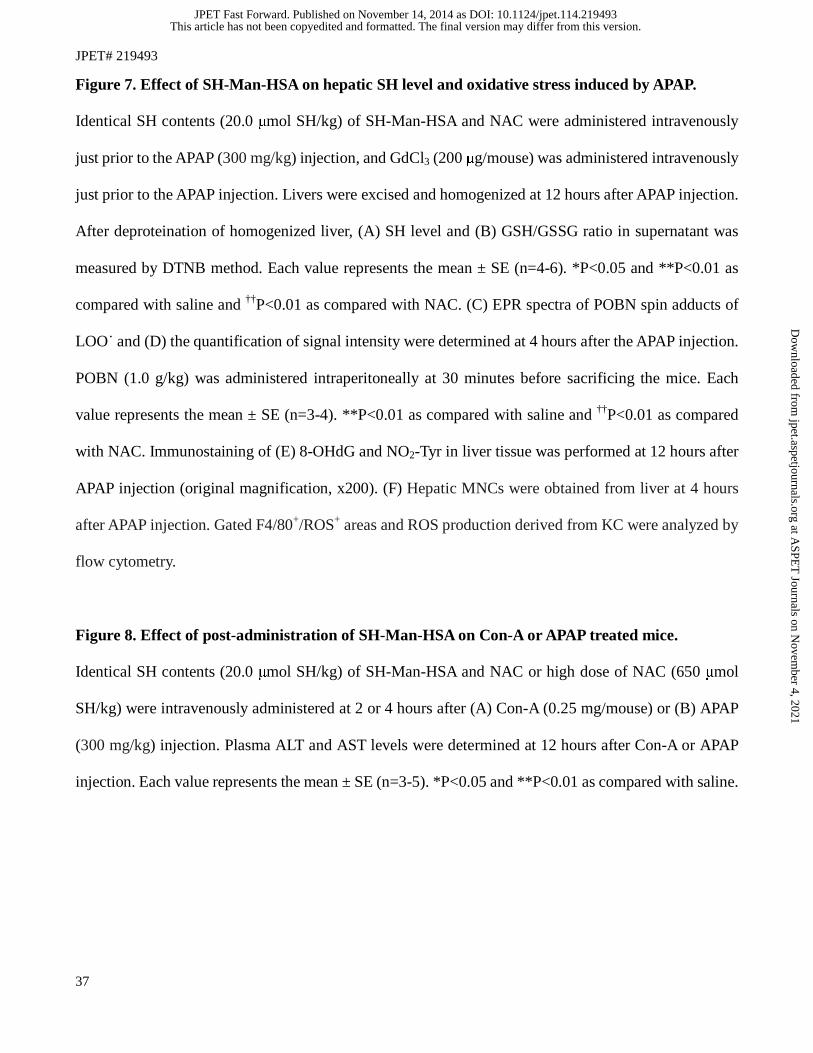
Figure 1. Radical-scavenging activity of the SH-Man-HSA.
(A) EPR spectra of ˙OH and (B) the quantification of signal intensity were determined using an EPR
method. The reaction solution contained 100 μM DTPA, 9 mM DMPO and 500 μM H2O2 in the absence
or presence of 75 μM HSA, Man-HSA or SH-Man-HSA in PBS (pH 7.4). Each value represents the mean
± SE (n=3). *P<0.05 and **P<0.01 as compared with control.
Figure 2. Pharmacokinetic analysis of the SH-Man-HSA.
(A) Plasma concentration curve and organ distribution of SH-Man-HSA after 10.0 nmol SH/kg of 111In
labeled SH-Man-HSA was injected in tail vein of mice. Each value represents the mean ± SE (n=3). (B)
Furthermore, SH-Man-HSA distribution in the liver was observed in liver section using fluorescence
image technique at 1 hour after the administration of bromobimane-labeled SH-Man-HSA (20.0 μmol
SH/kg) (original magnification, x400). 6.0 mg/mouse of mannan were administered at 30 minutes before
the administration of bromobimane-labeled SH-Man-HSA.
Figure 3. Evaluation of CD68+/CD206+ KC distribution of SH-Man-HSA.
The prepared bromobimane-labeled SH-Man-HSA (3.6 mg/mouse) was injected into the tail vein of mice.
At 1 and 12 hours after the administration of bromobimane-labeled SH-Man-HSA, the liver was removed,
covered with OCT compound and frozen at −80 °C. Fresh frozen sections (4 μm thickness) of the liver
were cut on a cryostat, collected on slides, and immediately dried. The sections were fixed with
phosphate-buffered 4% paraformaldehyde and then washed. After incubation with 1% Block Ace for 10
minutes, the slides were incubated overnight with an anti-HSA, CD68, CD206 antibody diluted 100, 100
or 200 times, respectively. (A-B) SH-Man-HSA distribution in subtypes of KC was observed in liver at 1
hour after the administration of bromobimane-labeled SH-Man-HSA (3.6 mg/mouse) (original
This article has not been copyedited and formatted. The final version may differ from this version.JPET Fast Forward. Published on November 14, 2014 as DOI: 10.1124/jpet.114.219493
at ASPE
T Journals on N
ovember 4, 2021
jpet.aspetjournals.orgD
ownloaded from
JPET# 219493
35
magnification, x1200) (C) Fluorescence images of anti-HSA antibody and bromobimane in CD68+ KC at
1 (left panel) or 12 hours (right panel) after the administration of bromobimane-labeled SH-Man-HSA
(3.6 mg/mouse) (original magnification, x1200). The fluorescence of bromobimane (blue) and the
anti-HSA antibody (red) merged cells were a purple color. The fluorescence of the anti-HSA antibody
and either anti-CD68 antibody or anti-CD206 antibody (green) merged cells were a yellow color. The
fluorescenses of bromobimane, anti-HSA antibody and either anti-CD68 antibody or anti-CD206
antibody merged cells were a white color. Insets: enlarged image details of white square marked typical
section.
Figure 4. Survival and hepato-protective effect of SH-Man-HSA on lethal and low dose of Con-A
treated mice.
(A) Identical SH contents (20.0 μmol SH/kg) of SH-Man-HSA and NAC were administered
intravenously just prior to lethal dose of Con-A (1.0 mg/mouse; i.v.) injection (n=10-15). **P<0.01 as
compared with saline and †P<0.05 as compared with NAC. (B) 5.0, 10.0 and 20.0 μmol SH/kg
SH-Man-HSA and 20.0 μmol SH/kg NAC were administered intravenously just prior to low dose of
Con-A (0.25 mg/mouse) injection, and GdCl3 (200 μg/mouse) was administered intravenously at 24
hours prior to the Con-A injection. Plasma ALT and AST levels were determined at 12 hours after Con-A
injection. Each value represents the mean ± SE (n=4-6). **P<0.01 as compared with saline and ††P<0.01
as compared with NAC. (C) HE and TUNEL staining (original magnification, x100 and x200,
respectively) of mice liver were performed at 12 hours after the Con-A injection. (D) 2.66 μmol SH/kg of
Man-HSA or 20.0 μmol SH/kg of SH-HSA was administered intravenously just prior to the Con-A
injection. Each value represents the mean ± SE (n=3). **P<0.01 as compared with saline.
Figure 5. Effect of SH-Man-HSA on hepatic SH level and oxidative stress induced by Con-A.
This article has not been copyedited and formatted. The final version may differ from this version.JPET Fast Forward. Published on November 14, 2014 as DOI: 10.1124/jpet.114.219493
at ASPE
T Journals on N
ovember 4, 2021
jpet.aspetjournals.orgD
ownloaded from

JPET# 219493
36
Identical SH contents (20.0 μmol SH/kg) of SH-Man-HSA and NAC were administered intravenously
just prior to Con-A (0.25 mg/mouse) injection, and GdCl3 (200 μg/mouse) was administered
intravenously at 24 hours before Con-A. Livers were excised and homogenized at 12 hours after the
Con-A injection. After deproteination of homogenized liver samples, (A) SH levels and (B) GSH/GSSG
ratios in the supernatant were measured by the DTNB method. Each value represents the mean ± SE
(n=4-6). *P<0.05 and **P<0.01 as compared with saline and ††P<0.01 as compared with NAC. (C) EPR
spectra of POBN spin adducts of LOO˙ and (D) the quantification of signal intensity was done at 4 hours
after Con-A injection. POBN (1.0 g/kg) was administered intraperitoneally at 30 minutes before mice
were sacrificed. Each value represents the mean ± SE (n=3-4). *P<0.05 and **P<0.01 as compared with
saline and ††P<0.01 as compared with NAC. Immunostaining of (E) 8-OHdG and NO2-Tyr in liver tissue
was performed at 12 hours after the Con-A injection (original magnification, x100). (F) MNCs were
obtained from liver at 4 hours after Con-A injection. Gated F4/80+/ROS+ areas and ROS production
derived from KC were analyzed by flow cytometry. (G) TNF and IFN-γ levels in plasma at 1 and 12
hours, respectively, after Con-A (0.25 mg/mouse) injection with or without SH-Man-HSA (20.0 μmol
SH/kg) administration (n=4). **P < 0.01 as compared with saline, Student's t test.
Figure 6. Hepato-protective effect of SH-Man-HSA on APAP treated mice.
5.0, 10.0 and 20.0 μmol SH/kg SH-Man-HSA and 20.0 μmol SH/kg NAC were administered
intravenously just prior to APAP (300 mg/kg) injection, and GdCl3 (200 μg/mouse) was administered
intravenously at 24 hours before APAP injection. (A) Plasma ALT and AST levels were determined at 12
hours after APAP injection. Each value represents the mean ± SE (n=4-6). **P<0.01 as compared with
saline and ††P<0.01 as compared with NAC. (B) HE and TUNEL staining (original magnification, x100
and x200, respectively) of mice liver tissue were performed at 12 hours after APAP injection.
This article has not been copyedited and formatted. The final version may differ from this version.JPET Fast Forward. Published on November 14, 2014 as DOI: 10.1124/jpet.114.219493
at ASPE
T Journals on N
ovember 4, 2021
jpet.aspetjournals.orgD
ownloaded from
JPET# 219493
37
Figure 7. Effect of SH-Man-HSA on hepatic SH level and oxidative stress induced by APAP.
Identical SH contents (20.0 μmol SH/kg) of SH-Man-HSA and NAC were administered intravenously
just prior to the APAP (300 mg/kg) injection, and GdCl3 (200 μg/mouse) was administered intravenously
just prior to the APAP injection. Livers were excised and homogenized at 12 hours after APAP injection.
After deproteination of homogenized liver, (A) SH level and (B) GSH/GSSG ratio in supernatant was
measured by DTNB method. Each value represents the mean ± SE (n=4-6). *P<0.05 and **P<0.01 as
compared with saline and ††P<0.01 as compared with NAC. (C) EPR spectra of POBN spin adducts of
LOO˙ and (D) the quantification of signal intensity were determined at 4 hours after the APAP injection.
POBN (1.0 g/kg) was administered intraperitoneally at 30 minutes before sacrificing the mice. Each
value represents the mean ± SE (n=3-4). **P<0.01 as compared with saline and ††P<0.01 as compared
with NAC. Immunostaining of (E) 8-OHdG and NO2-Tyr in liver tissue was performed at 12 hours after
APAP injection (original magnification, x200). (F) Hepatic MNCs were obtained from liver at 4 hours
after APAP injection. Gated F4/80+/ROS+ areas and ROS production derived from KC were analyzed by
flow cytometry.
Figure 8. Effect of post-administration of SH-Man-HSA on Con-A or APAP treated mice.
Identical SH contents (20.0 μmol SH/kg) of SH-Man-HSA and NAC or high dose of NAC (650 μmol
SH/kg) were intravenously administered at 2 or 4 hours after (A) Con-A (0.25 mg/mouse) or (B) APAP
(300 mg/kg) injection. Plasma ALT and AST levels were determined at 12 hours after Con-A or APAP
injection. Each value represents the mean ± SE (n=3-5). *P<0.05 and **P<0.01 as compared with saline.
This article has not been copyedited and formatted. The final version may differ from this version.JPET Fast Forward. Published on November 14, 2014 as DOI: 10.1124/jpet.114.219493
at ASPE
T Journals on N
ovember 4, 2021
jpet.aspetjournals.orgD
ownloaded from
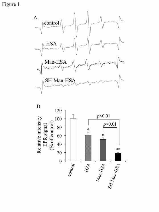
This article has not been copyedited and formatted. The final version may differ from this version.JPET Fast Forward. Published on November 14, 2014 as DOI: 10.1124/jpet.114.219493
at ASPE
T Journals on N
ovember 4, 2021
jpet.aspetjournals.orgD
ownloaded from

This article has not been copyedited and formatted. The final version may differ from this version.JPET Fast Forward. Published on November 14, 2014 as DOI: 10.1124/jpet.114.219493
at ASPE
T Journals on N
ovember 4, 2021
jpet.aspetjournals.orgD
ownloaded from
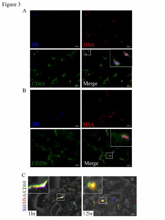
This article has not been copyedited and formatted. The final version may differ from this version.JPET Fast Forward. Published on November 14, 2014 as DOI: 10.1124/jpet.114.219493
at ASPE
T Journals on N
ovember 4, 2021
jpet.aspetjournals.orgD
ownloaded from

This article has not been copyedited and formatted. The final version may differ from this version.JPET Fast Forward. Published on November 14, 2014 as DOI: 10.1124/jpet.114.219493
at ASPE
T Journals on N
ovember 4, 2021
jpet.aspetjournals.orgD
ownloaded from

This article has not been copyedited and formatted. The final version may differ from this version.JPET Fast Forward. Published on November 14, 2014 as DOI: 10.1124/jpet.114.219493
at ASPE
T Journals on N
ovember 4, 2021
jpet.aspetjournals.orgD
ownloaded from

This article has not been copyedited and formatted. The final version may differ from this version.JPET Fast Forward. Published on November 14, 2014 as DOI: 10.1124/jpet.114.219493
at ASPE
T Journals on N
ovember 4, 2021
jpet.aspetjournals.orgD
ownloaded from
This article has not been copyedited and formatted. The final version may differ from this version.JPET Fast Forward. Published on November 14, 2014 as DOI: 10.1124/jpet.114.219493
at ASPE
T Journals on N
ovember 4, 2021
jpet.aspetjournals.orgD
ownloaded from
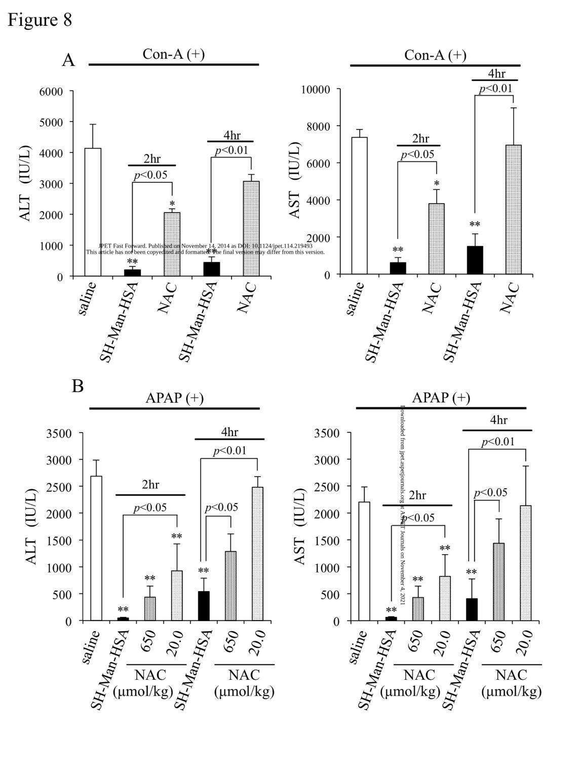
This article has not been copyedited and formatted. The final version may differ from this version.JPET Fast Forward. Published on November 14, 2014 as DOI: 10.1124/jpet.114.219493
at ASPE
T Journals on N
ovember 4, 2021
jpet.aspetjournals.orgD
ownloaded from