ki - ginmu.naramed-u.ac.jp
TRANSCRIPT
NIRS study in adu 1tADHD
Reduced Prefrontal Hemodynamic Response in Adult Attention-Deficit/Hyperactivity
Disorder as Measured by Nea r-Infrared Spectroscopy
Shotaro Ueda ,l MD; Toyosaku Ota ,l MD, PhD; Junzo Iida ,2 MD, PhD; Kazuhiko
Yamamuro ,l MD, PhD; Hiroki Yoshino ,l MD, PhD; Naoko Kishimoto ,l PhD; and
Toshifumi Kishimoto ,l MD, PhD
1 Department of Psychiatry , N ara Medic a:l U niversity , N ara , J apan
2Faculty ofNursing , Nara Medical University , Nara , Japan
Corresponding author: Shotaro Ueda , Department of Psychiatry , Nara Medical
University , 840 Shijocho , Kashihara , Nara 634 ・8522 ,Japan
Tel: +81 ・744 ・22・3051
Fax:+81 ・744 ・22・3854
Email: [email protected]
Regular Article , 5,733 words , 3 figures , 3 tables
Field: neuroimaging
Running title: NIRS study in adu 1tADHD
1

NIRS study in adult ADHD
Abstract
Aim: Recent developments in nea r-infrared spectroscopy have enabled non-inva sive
clarification of brain functions in psychiatric disorders. In pediatric
att ention-deficit/hyperactivity disorder , reduced prefront al h emod yn amic responses
have been observed with nea r-infrared spectroscopy repeatedly. How ever, there are few
studi es of adult attention-deficit/hyperactivity disorder by multi-channel nea r-infrared
spectroscopy. Therefore , in this study , we used multi-channel nea r-infrared spectro scopy
to examine the characteristics of prefrontal hemodynamic response s during the Stroop
colo r-word task in adult attention-deficit/hyperactivity disorder patients and in age- and
sex-matched control subjects.
Methods: Twelve treatment-naive adult s with attention-deficit/hyperactivity disor der
and 12 age- and sex-matched healthy control subject s participated in the present study
after giv ing cons ent. We used 24-channel near infrar ed spectroscopy to measure th e
oxyhemoglobin changes at the frontallobes of participants during the Stroop colo r-word
task. We compared the oxyhemoglobin changes between adult s wit h
attention-deficit/hyperactivity disorder and control subjects by ttests with Bonferroni
correction.
Results: Durin g the Stroop colo r-word task , the oxyhemoglobin ch anges observed in the
attention-deficit/hyperactivity disorder group were significantly smaller than those in
the con trol group in ch annels 11, 16, 18,21, 22, 23 and 24, correspond to the prefro ntal
2

NIRS study in adult ADHD
cortex. At channels 16, 21, 23 and 24 of the attention-deficit/hyperactivity disorder
group , there were negative correlations between the symptomatic severity and the
oxy- Hb changes.
Conclusion: The present study suggests that adults with attention-deficit/hyperactivity
disorder have reduced prefrontal hemodynamic response as measured by nea r-infrared
spectroscopy.
Keywords: adult attention-deficit/hyperactivity disorder , functional neuroimaging study ,
nea r-infrared spectroscopy , prefrontal hemodynamic response , Stroop colo r-word task
3

NIRS study in adu 1tADHD
Introduction
Attention-deficit/hyperactivity disorder (ADHD) is a common neurodevelopmental
disorder in children and adolescents with high pervasiveness into adulthood. The
prevalence of ADHD in adulthood is estimated at approximately 4% [1, 2]. The
symptoms are often severe and may cause serious difficulties in the daily life of affected
individuals [3]. Given the great morbidity associated with the disorder , including
persistent neuropsychological impairments [4], determining the underlying
neurobiology of ADHD is of great importance. Recent reviews of data from neuroimaging ,
neuropsychological , genetic , and neurochemical studies have generally implicated
frontostriatal network abnormalities as the likely cause of ADHD. However , recent
findings have suggested that , for some individuals , ADHD may not arise until
adolescence or adulthood and may therefore be associated with different risk factors and
outcomes than childhood ADHD [5, 6].
Studies have shown that ADHD is characterised by multiple functional and structural
neural network abnormalities including most prominently fronto ・striatal ,but also
fronto-parieto 圃temporal ,fronto-cerebellar and even fronto"limbic networks . Evidence
from longitudinal structural imaging studies has shown that ADHD is characterised by
a delay in structural brain maturation. In a meta -analysis offunctional imaging studies
in children and adults , Hart et al [7] found ADHD-related hypoactivation in the right
inf erior frontal cortex , supplementary motor area , anterior cingulate cortex , and
4

NIRS study in adu1t ADHD
striato 廿la1amic areas when pooling studies on inhibition , and found ADHD-re1ated
hypoactivation in the right dorso1atera1 prefronta1 cortex , posterior basa1 ganglia , and
tha1amic and parieta1 regions when focusing on attention tasks. In another
meta-ana1ysis focused on timing tasks , including studies in both chi1dren and adu1ts ,
Hart et a1 [8] found ADHD-re1ated hypoactivation in the cerebellar vermis , 1eft inferior
prefronta1 cortex and insu1a , and 1eft supramargina1 gyrus extending into 1eft superior
tempora1 and postcentra1 gyri. Lee et a1 [9] reported that chi1dren with clinica1 ADHD
show reduced orbitofronta1 regiona1 cerebra1 b100d flow on sing1e photon emission
computed tomography (SPECT) scans. Functiona1 magnetic resonance imaging (岱1RI)
also shows dysfunction in the prefrontal cortex in children with ADHD [10 , 11]. Other
work shows hypop erfusion in orbitofronta1 regions by SPECT scans of both chi1dren and
adu1ts with clinica1 ADHD [12 ・14]. Studies of adult ADHD patients using
positron-emission tomography (PET) have found glucose metabo1ism reduction in the
premotor cortex and the superior prefronta1 cortex [15]. Additionally , one study of adult
ADHD patients using PET showed significant1y increased dopamine active transporter
binding in the right caudate [16].
However , functiona1 brain imaging methodo1ogies , such as PET , SPECT , and fMRI
hav e the disadvantage of requiring 1arge apparatuses , which precludes their use in a
bedside setting for diagnostic and treatment purposes. Additionally , these functiona1
brain imaging m ethodo1ogies do not offer high tempora1 reso1ution. By contrast ,
5
NIRS study in adult ADHD
multi-channel nea r-infrared spectroscopy (NIRS) systems have recently been developed
to allow non-invasive and bedside functional mapping of the cerebral cortex , with high
temporal resolution [17-19].
Multi-channel NIRS enables the noninvasive detection of neural activity near the
surface of the brain using nea r-infrared light [20 , 21]. It measures alterations in
oxygenated hemoglobin (oxy- Hb) and deoxygenated hemoglobin (deoxy- Hb)
concentrations in micro-blood vessels on the brain surface_ Local increases in the
concentration of oxy-Hb and decreases in the concentration of deoxy-Hb are indicators of
cortical activity [21 , 22]. In addition , changes in the concentration of oxy-Hb have been
associated with changes in regional cerebral blood volume , using a combination of PET
and NIRS measur ements [23 ,24]. NIRS is a neuroimaging modality that is particularly
suitable for use in psychiatric patients for several reasons [25]. First , the subject can be
examined in a natural sitting position , without any surrounding distract ion. Second , the
cost is much lower than other neuroimaging modalities. Third , because NIRS is
relatively insensitive to motion artifact , it can be applied to experiments that might
cause some motion of the subjects such as vocalization. Fourth , the setup is very easy.
Fifth , the high temporal resolution of NIRS is useful in characterizing the time course of
prefrontal activity of psychiatric disorders [26 , 27]. Accordingly , NIRS has been used to
assess brain functions in many psychiatric disorders , including schizophrenia ,
depression , bipolar disorder , obsessive-compulsive disord er, post traumatic stress
6

NIRS study in adult ADHD
disorder , dementia , pervasive developmental disord ers, and ADHD [25 ・35].
In pediatric ADHD , reduced prefrontal hemodynamic response s have been observed
with NIRS. Negoro et al [35] examined reduced prefrontal hemod ynamic respon se in
ADHD children as measur ed by NIRS and determin ed cerebral hem odynami c ch anges
in response to the Stroop colorword task in 20 children with ADHD and 20 health y age-
and sex-matched controls . They showed that the oxy -Hb change s in the ADHD group
were significantly smaller than those in the control group in the inferior prefrontal
cortex during performance ofthe Stroop colo r-word task .
The Stroop color 幽word task is one of the most commonly used tools for determining
attentional problems. It is also a test of executive function and working memor y. The
Stroop colo r-word task m easures m ainl y a person's selecti ve attention and an effect of int erference.
Previous review and meta-analysis revealed abnormal Stroop interference inADHD [36].
Thus , it is signi ficant to examin e the cha racteri stics of prefron tal h emod yn amic
responses in adults with ADHD using NIRS during the Stroop colo r-word task. In
addition to these reason s, w e used the Stroop colo r-word task for the followin g reas ons.
First , the inferior frontal 白Trus has been described as one of the regions most strongly
related to Stroop interference [37]. Second , in the NIRS study using the same task,
Okada et al [29] conclud ed that th e word reading task and incongru ent colo r naming
task produ ced suitable prefrontal brain activation in healthy adult s.
Base d on previou s studi es which show ed dysfun ction of the prefrontal cor tex by other
7
NIRS study in adu 1tADHD
neuroimaging modalities , we could predict that adu 1ts with ADHD had reduced
prefrontal hemodynamic responses. However , there are few studies by multi "channel
NIRS to examine prefrontal hemodynamic responses in adult ADHD. In the pre sent
study , we hypothesized that adults with ADHD would have reduced prefront al
hemodynamic responses as measured by NIRS. Therefore , in this study , we used
multi"channel NIRS machines to examine the characteristics of prefrontal
hemodynamic responses during the Stroop color 圃word task in adult ADHD patients and
in age" and sex"matched control subjects .
Methods
Parti cipants
Twelve subjects (4 males and 8 females) , aged 21"47 years and diagnosed with ADHD
according to the Diagnostic and Statistical Manual of Mental Disor ders Fifth Edition
(DSM"5) [38] , were compared with 12 age" , sex ・, and intelligence quotient (IQ)"matched
healthy control subjects (4 males and 8 females) , aged 20・46years (Table 1).
The subjects with ADHD , who had no history of previous psychiatric disorder
treatment , consulted one of the experienced psychiatrists at the Department of
Psychiatry of Nara Medical Univ ers ity with a chief complaint of attention deficit ,
hyperactivity , or impulsiveness. They underwent a standard clinical assessment
comprising a psychiatric evaluation , a standard diagnostic inter view, and the takin g of a
8

NIRS study in adultADHD
medical history by the experienced psychiatrist. Two experienced psychiatrists
confirmed the diagnosis ofADHD in accordance with the DSM-5 [38] , and confirmed the
presence of ADHD symptoms prior to the age of 12. Intellectuallevel was assessed with
the Wechsler Adult Intelligence Scale-Third Edition by the psychologist , and patients
whose full-scale IQ (FIQ) scores were below 70 were excluded. In addition , we excluded
patients who presented a comorbid psychiatric disorder defined by the DSM-5 , a
neurodevelopmental disorder (including autism spectrum disorder) , a neurological
disorder , a head injury , a serious medical condition or a history of substance
abuse/dependence; two patients with autism spectrum disorder and two patients with
bipolar disorder were excluded. Finally , 12 subjects with ADHD , who had no previous
medication , were enrolled in this study.
Healthy control subjects were recruited via local print advertising. They also
underwent a standard clinical assessment comprising a psychiatric evaluation , a
standard diagnostic interview , and the taking of a medical history by the experienced
psychiatrist. Intellectual level was assessed with the Wechsler Adult Intelligence
Scale-Third Edition by the psychologist. Finally , 12 healthy control subjects , who did
not have confirmed ADHD and who had no current or past history of psychiatric or
neurological disorder , were enrolled in the present study as well.
All subjects were right-handed and Japanese. This study was approved by the
Institutional Review Board at the Nara Medical University. Written informed consent
9
NIRS study in adult ADHD
was obtained from all subjects before the study.
Assessment of ADHD symptoms
The Conners' Adult ADHD Rating Scale-Investigator Rated (CAARS-Inv): Screening
Version was used to evaluate ADHD symptoms in the participant s. The CAARS- Inv:
Screening Version is a 30・item ,investigato r-rated scale that measures the severity of
ADHD symptoms. Each item is measured on a fou r-point scale ranging from 0 (not at all ,
never) to 3 (very much , very frequently) , and separate scores can be derived for the
Inattention subscale (9 items) , the Hyperactivity/lmpulsivity subscale (9 items) , and the
ADHD Index subscale (12 items). The CAARS-Inv total score is the sum of the
Inatt ention subscale score and the H yperactivity/lmpulsivity subs cale score , with
possible scores ranging from 0 to 54.
ADHD subjects und erwent assessment on the CAARS-Inv (Table 1) on the same day
as the NIRS element of the study. As shown in Table 1, the mean CAARS- Inv total score
was 28.42 (SD , 8.11) , ranging from 16 to 48.
Stroop Colo r-Word Task
The traditional Stroop task comprises a word read ing task , an incongru ent color naming
task and a color naming tas k. Here , however , we reproduced the Stroop task according
to an alternative method that has been previously describe d [39]. The Stroop color 圃word
10

NIRS study in adu 1tADHD
task consisted of two pages: each page had 100 items in five co1umns of 20 items each
and the page size was 210 x 297 mm. On the first page , the words RED , GREEN , and
BLUE were printed in b1ack ink. On the second page , the words RED , GREEN , and
BLUE were printed in red , green , or b1ue ink , with the limitation that the word meaning
and ink co1or cou1d not match. The item s on both page s were random1 y distributed , with
the exception that no item cou1d appear direct1y after the same item within a co1umn .
Before the task , the examiners instructed the participants as follows: “This is to test
how quick1y you can read the words on the first page , and say the co1ors of the words on
the second page. After we say 'begin ,' p1ease read the words in the co1umns , starting at
the top 1eft , and say the words/colors as quickly as you can. After you finish reading the
word s in the first co1umn , go on to the next co1umn , and so on. After you have read the
words on the first page for 45 s, we will turn the page. P1ease rep eat this procedure for
the seco nd page プ
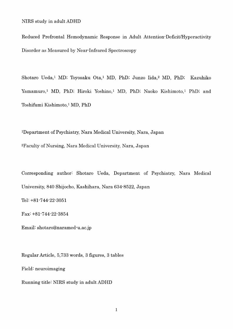
The entire Stroop co1o r-word task sequence con sisted of three cycles of 45・s spent
reading the first page , follow ed by 45-s spent reading the seco nd page (the co1or-word
task). The task ended with 45-s spent reading the first page , which we designated as the
baseline task (Fig. 1 (C)) . W e rec orded the number of correct answers in each cycle.
Examiners who were blind to the diagnoses of the participants adm ini stere d the Stroop
co1o r-word task.
The Stroop task used in this study was different to the traditiona1 Stroop tas k. We
11

NIRS study in adultADHD
excluded the color naming task (part of the traditional Stroop task) because we neede d
only two tasks (baseline task and activation task) for our NIRS study. At the NIRS study ,
it has been shown previously that sui table brain activation in health y adults is elicited
by using the word reading task and incongruent color naming task [29 , 40] , suggesting
that this was an appropriate approach for the present study.
NIRS Measurements
Increased oxy-Hb and decreased deoxy-Hb , as measured by NIRS , have been shown to
reflect cortical activation. In animal studies , because the direction of change in
deoxy- Hb is determined by the degree of changes in venous blood oxygenation and
volum e, oxy-Hb is the most sensitive indicator ofregional cerebral blood flow [41]. Thus ,
we determined to focus on changes in oxy- Hb. We measured oxy- Hb using a 24-channel
NIRS machine (Hitachi ETG 同4000 ,Hitachi M edical Corporation , Tokyo , Japan). We
measured the absorption of two wavelengths of nea r-infrared light (760 and 840 nm).
We analyzed the optical data bas ed on the modified Bee r-Lambelt Law [42] as
previously described [17]. This method enabled us to calculate signals reflecting the
oxy-Hb , deoxy-Hb , and total-Hb signal changes . The scale ofthe hemoglobin quantit y is
mmol X mm, which means that all concentration chang es dep end on the path length of
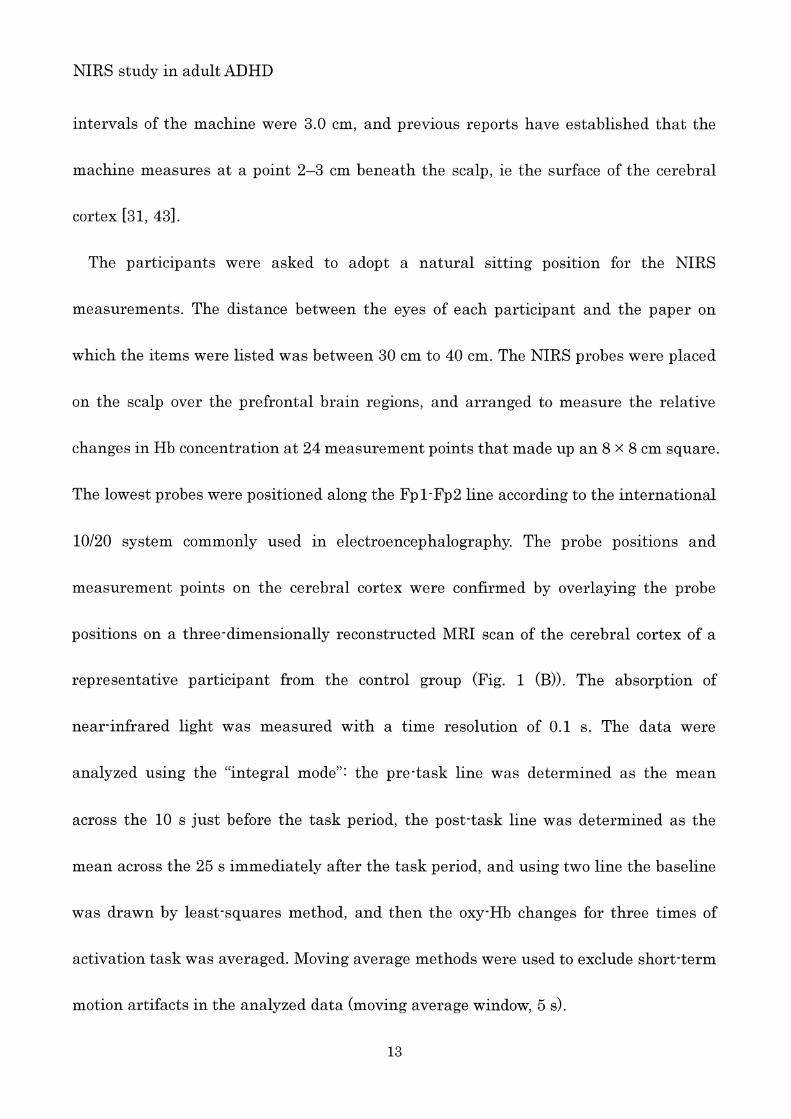
the nea r-infrared light. The recording channels resided in the optical path in the brain
between neighbouring pairs of emitters and detec tors (Fig. 1 (A)). The inte r-probe
12
NIRS study in adult ADHD
intervals of the machine were 3.0 cm, and previous reports have established that the
machine measures at a point 2-3 cm beneath the scalp , ie the surface of the cerebral
cortex [31 , 43].
The participants were asked to adopt a natural sitting position for the NIRS
measurements. The distance between the eyes of each participant and the paper on
which the items were listed was between 30 cm to 40 cm. The NIRS probes were placed
on the scalp over the prefrontal brain regions , and arranged to measure the relative
changes in Hb concentration at 24 measurement points that made up an 8 x 8 cm square.
The lowest probes were positioned along the Fp 1-Fp2 line according to the international
10/20 system commonly used in electroencephalography. The probe positions and
measurement points on the cerebral cortex were confirmed by overlaying the probe
positions on a three-dimensionally reconstructed MRI scan of the cerebral cortex of a
representative participant from the control group (Fig . 1 (B)) . The absorption of
nea r-infrared light was measured with a time resolution of 0.1 s. The data were
analyzed using the “integral mode": the pre-task line was determined as the mean
across the 10 s just before the task period , the post-task line was determined as the
mean across the 25 s immediately after the task period , and using two line the baseline
was drawn by least-squares method , and then the oxy
activation task was averaged. Moving average methods were used to exclude short-term
motion artifacts in the analyzed data (moving average window , 5 s).
13
NIRS study in adult ADHD
We attempted to exclude motion artifacts by closely monitoring artifact-evoking body
movements , such as neck movements , biting , and blinking (identified as being the most
infl. uential in a preliminary artifact-evoking study) , and by instructing the participants
to avoid these movements during the NIRS measurements . Examiners were blind to
diagnoses of the participants.
Statistical Analyses
Oxy- Hb changes were compared between each of the two groups with Student 's ttests
using the grand average waveforms every 0.1 s in each channe l. This analysis enabled
more detailed comparison of oxy- Hb changes along the time course of the tas k. Data
analyses were conducted using MATLAB 6.5.2 (Mathworks , Natick , MA, USA) and Topo
Signal Processing type-G version 2.05 (Hitachi Medical Corporation , Tokyo , Japan).
OT-A4 version 1.63 K (Hitachi Medical Corporation , Tokyo , Japan) was used for the
overlap display of the grand average waveforms in both groups in Fig. 2 and was also
used to calculate mean oxy
かtests ,a Bonferroni correction for multiple comparisons was applied. PASW Statistics
18.0 J for Windows (SPSS , Tokyo , Japan) was used for statistical analysis.
Results
D emographic data
14
NIRS study in adult ADHD
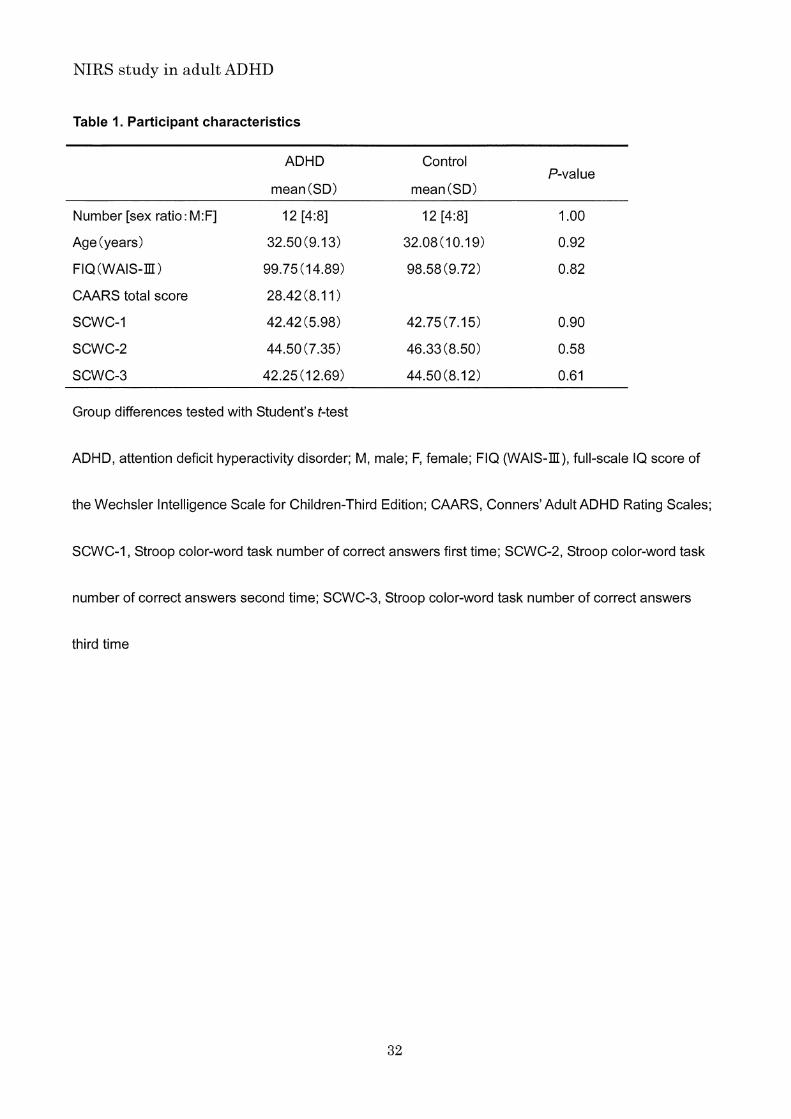
Demographic and clinical data are shown in Table 1. Age , sex and FIQ did not differ
significantly across patients with adult ADHD and healthy controls (t=O.11 , df=22 ,
P=0.92; chi-square=O.OO , df=l , P=l.OO; t=0.23 , df=22 , P二0.82). The mean CAARS-Inv
total score of adult ADHD subjects was 28.42 (SD , 8.11; range , 16 to 48). Ther e were no
significant differences in the SCWC-1 , SCWC-2 , and SCWC-3 scores between the two
groups (t=-0.12 , df=22 , P=0 .90; t=-0.57 , df=22 , P=0.58; t=-0.52 ヲ df=22 ,P=0.61).
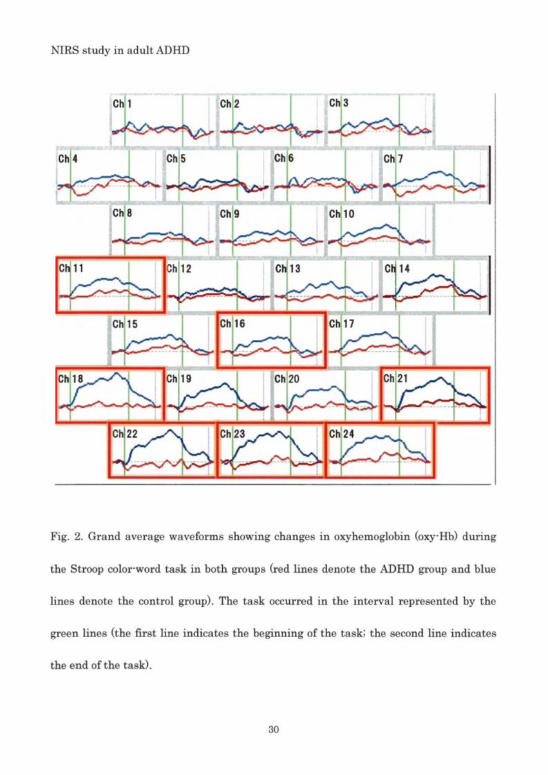
NIRS data ofthe subjects during the Stroop colo r-word task
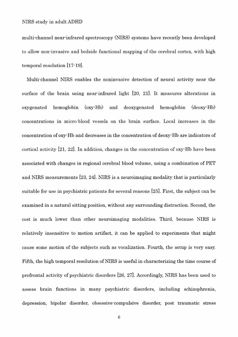
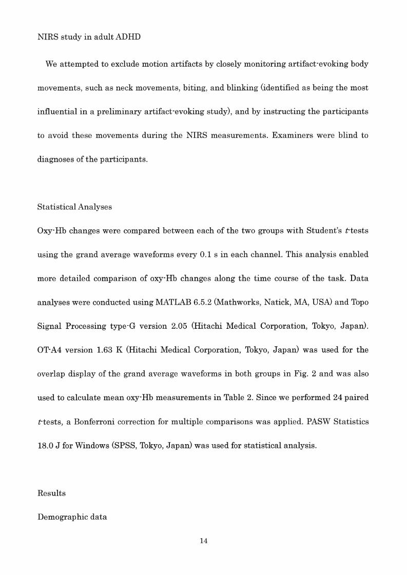
The grand average waveforms of oxy- Hb concentration changes during the Stroop
color-word task in both groups can be seen in Fig. 2. The grand average waveform s of
oxy -Hb concentr ation change in the control group incr ease d during the task period ,
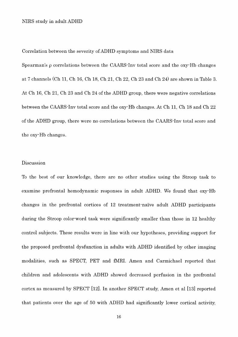
while those of the ADHD group did not change wel l. The difference of mean oxy-Hb
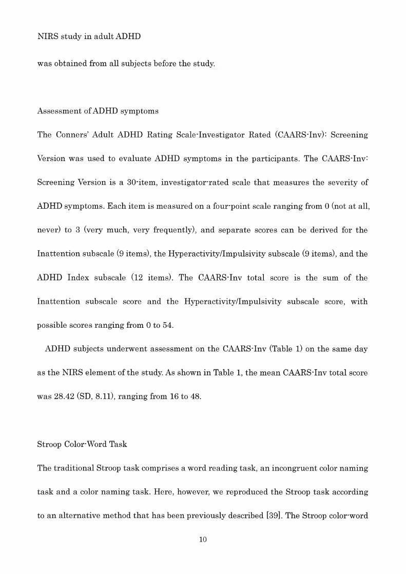
meas urements between task and post-task periods in the 24 channels can be seen in
Table 2. Group differences were tested with Bonferroni correction. Between task and
post 司task periods , the mean oxy- Hb difference of the control group was signific ant ly
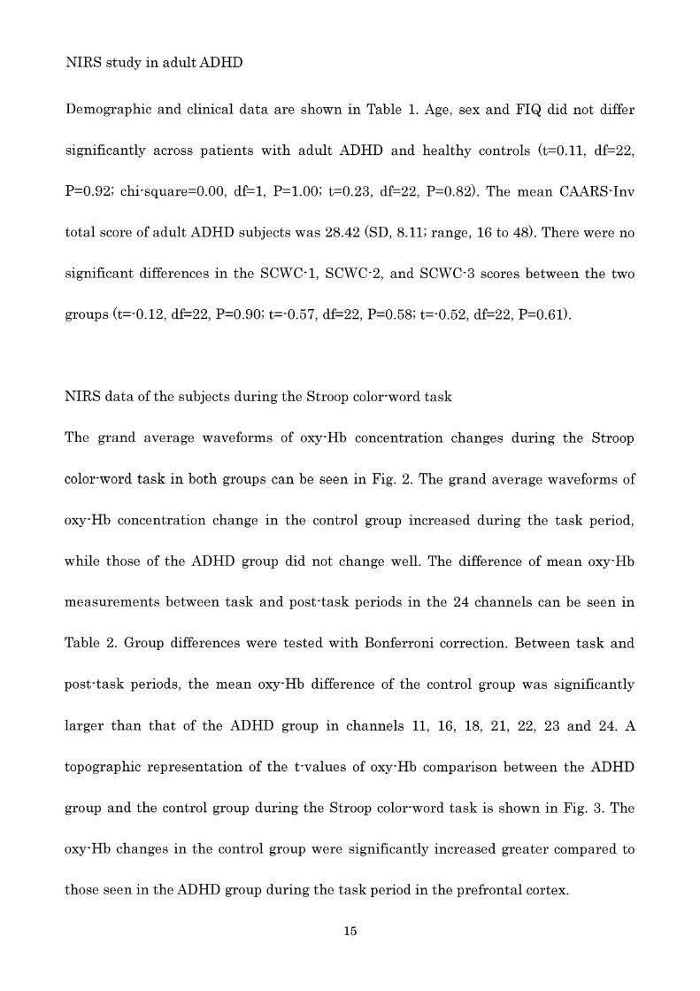
larger than that of the ADHD group in channels 11, 16, 18, 21, 22, 23 and 24. A
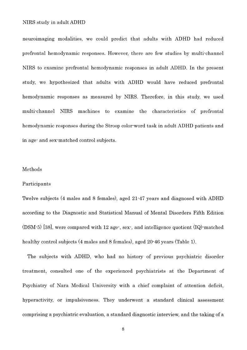
topographic representation of the t-values of oxy- Hb comparison between the ADHD
group and the control group durin g the Stroop colo r-word task is shown in Fig . 3. The
oxy- Hb changes in the control group were significantly increased greater compared to
tho se seen in the ADHD group during the task period in the prefrontal cortex .
15
NIRS study in adultADHD
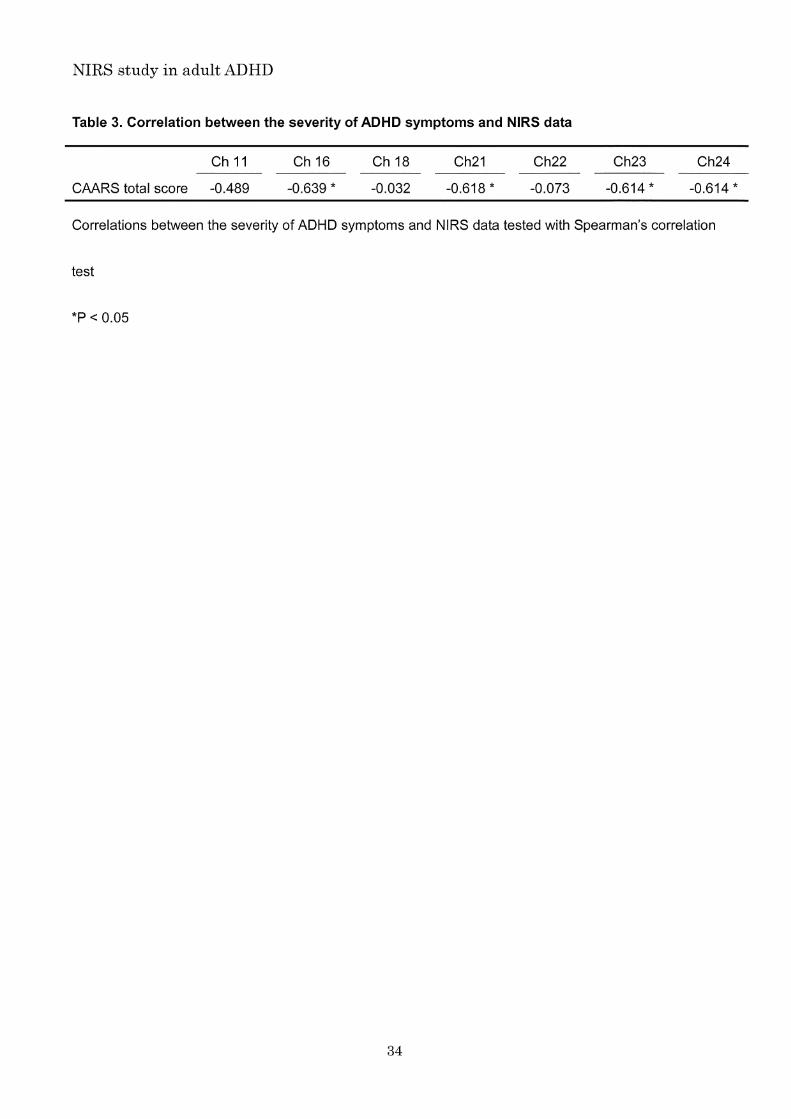
Correlation between the severity of ADHD symptoms and NIRS data
Spearman's p correlations between the CAARS- Inv total score and the oxy- Hb changes
at 7 channels (Ch 11, Ch 16, Ch 18, Ch 21, Ch 22, Ch 23 and Ch 24) are shown in Table 3_
At Ch 16, Ch 21, Ch 23 and Ch 24 of the ADHD group , there were negative correlations
between the CAARS- Inv total score and the oxy- Hb changes. At Ch 11, Ch 18 and Ch 22
of the ADHD group , there were no correlations between the CAARS- Inv total score and
the oxy-Hb changes.
Discussion
To the best of our knowledge , there are no other studies using the Stroop task to
examine prefrontal hemodynamic responses in adult ADHD. 羽w厄efound that oxy
changes in the prefrontal cortices of 12 treatment ヤ-na厄Ive adult ADHD participants
during the Stroop colo r-word task were significantly smaller than those in 12 healthy
control subjects. These results were in line with our hypotheses , providing support for
the proposed prefrontal dysfunction in adults with ADHD identified by other imaging
modalities , such as SPECT , PET and fMRI. Amen and Carmichael reported that
children and adolescents with ADHD showed decreased perfusion in the prefrontal
cortex as measured by SPECT [12]. In another SPECT study , Amen et al [13] reported
that patients over the age of 50 with ADHD had significantly lower cortical activity ,
16
NIRS study in adultADHD
particularly in the frontal pole cortex , orbitofrontal cortex , and parietal cortex. Studies
of adult ADHD patients using PET have suggested glucose metabolism reduction in the
premotor cortex and the superior prefrontal cortex [15] , and relative lack oftask-rela ted
frontal activations during performance of a working memory task [44]. Evid ence from
岱lRI studies suggests that there are frontal dysfunctions localized to ventr 吐ateral and
dorsolateral prefrontal cortex , as well as in the dorsal anterior cingulate cort ex, during
performance of a Stroop task by adult patients with ADHD [45 , 46]. A meta-analysis of
fMRI studies of adult ADHD revealed that almost all (97%) hypoactivated voxels were
located in a frontoparietal network [47]. In adult ADHD , reduced prefrontal
hemodynamic responses have been measured by NIRS [48 , 49]. Schecklmann et al [49]
found reduced bilateral activation of the inferior frontal cortex in adults with ADHD
compared with healthy adults as measured by NIRS during a working memory and
response inhibition tas k. In this study , additionally , there wer e negative correlations
between the CAARS-Inv total score and the oxy-Hb changes at channels 16, 21, 23, and
24 of the ADHD group , ie lower brain activity was associated with severity of ADHD
symptoms. Multi-channel NIRS systems may therefore be very useful tools to assess the
symptoms ofADHD , as well as frontal function.
At channels 11, 16, 18, 21, 22, 23, and 24, adults with ADHD showed significantly
smaller oxy- Hb changes than those seen in the healthy controls in the present study.
Nego ro et al [35] used NIRS to examine reduc ed prefrontal h emod ynamic respon ses in
17

NIRS study in adult ADHD
pediatric ADHD during the Stroop colo r-word task , which is the same task used in this
study. They reported a lower increase of the oxy 圃Hb changes at channels 8, 18, 19, 21,
and 22 in pediatric ADHD compared to control subjects. These findings are in line with
our own: the oxy-Hb changes in the ADHD group were significantly smaller than those
in the control group in the inferior prefrontal cortex , which has been described as one of
the regions that is most strongly related to Stroop interference [37].
Potential limitations of the present study should be taken into consideration. First ,
NIRS has disadvantages compared to other modalities [18]: for instance , it enables
measurement of Hb concentration changes only as relative values , not as absolute
values. We used the Stroop colo r-word task that had a clear baseline task to overcome
these potential problems. Additionally , we measured Hb concentration changes from the
activation task to the baseline task and performed the task three times to average
pot ential accidental changes and prevent the participants from becoming tired. The
grand average waveforms of oxy-Hb concentration changes in the ADHD group do not
show a regional cerebral blood f10w decrease during the activation task or a difference
between the blood f10ws during the baseline and activation tasks. Second , spatial
resolution for detecting hemodynamic responses from the scalp surface using NIRS is
lower than those for 品1RI ,SPECT and PET . However , abnormal prefrontal
hemodynamic responses in individuals with ADHD are certainly detectible by NIRS.
Third , several studies have shown that superficial h emodynamic changes such as skin
18
NIRS study in adult ADHD
blood flow can affect the prefrontal NIRS hemoglobin signals [50 , 51]. The pre sent
findings could be skin blood flow. However , Sato et al [52] conducted simultaneous NIRS ,
fMRI, and laser Doppler flowmeter measurements to determine whether prefrontal
NIRS hemoglobin signal s reflect cortical activity rath er than superficial eff ects. They
concluded that NIRS can be used to measure hemod ynamic signal s originating from
prefrontal cortex activation. Fourth , the sample size was small , although the 12 adults
with ADHD were treatment-naive and none of them had comorbid psychiatric ,
neurodevelopmental or neurological disorder. Future research is needed , with larger
sample sizes. Fifth , the range of their age was wide. However , it was thought that the
influence was small because there were no correlations between the age and the oxy -Hb
changes , and between the age and the performan ce of the Stroop colo r-word task. Sixth ,
adults with ADHD , who were confirmed the presence of ADHD symptoms prior to age 12
years, participat ed in the pres ent study , althou gh ther e is the lat e-onset adult ADHD
that do not have ADHD symptoms prior to age 12 years. In the future study , it is needed
to compare the prefrontal hemodynamic respons e betw een the present adult ADHD and
the late-onset adult ADHD.
Conclusion
To the best of our knowledge , this is the first NIRS study using the Stroop task to
examin e prefront al h emodyn amic respon se in adul t ADHD. W e found that the oxy-Hb
19
N1RS study in adult ADHD
changes in the ADHD group were significantly smaller than tho se in the control group
in the inferior prefrontal cortex. We also found that smaller oxy-Hb change s were
associated with severer ADHD symptom . Multi 同channel N1RS syst em appears to be a
ver y useful tools for assessing the symptom of ADHD as well as frontal function , as it
enables non-invasive functional mapping of the cerebral cortex and has much shorter
m easur ement times (about 5 min) compared with other functional brain imaging
methodologies .
Acknow ledgments
W e wish to thank the participants for their valuabl e involv em ent with the study. The
authors would also like to thank the Hitachi Medical Corporation for the ETG-4000
equipm ent and the skill ed technic al and m ethodical suppo rt.
Disclo sure Stat em ent
All the authors declare that they have no conflicts of interest.
Author Contributi ons
SU was involved in the collection of the data and wrote the first draft of the manu script.
TO, J1, KY, HY, NK and TK sup ervi sed th e entire proj ect, w ere critically involved in the
20

N1RS study in adultADHD
design , and contributed to the editing of the final manuscript. All authors have read and
approved the final manuscript.
References
1. de Graaf R, Kessler RC, Fayyad J et al. The prevalence and effects of adult
attention-deficit/hyperactivity disorder (ADHD) on the performance of workers:
results from the WHO World Mental Health Survey 1nitiative. Occup. Environ. Med.
2008;65:835 ・842.
2. de Zwaan M, Gruss B, Muller A et al. The estimated prevalence and correlates of
adult ADHD in a German community sample. Eur. Arch. Psychia tJァα'in. Nθurosc 1.
2012;262:79 ・86.
3. Halmoy A, Fasmer OB, Gillberg C, Haavik J. Occupational outcome in adult ADHD:
impact of symptom profile , comorbid psychiatric problems , and treatment: a
cross-sectional study of 414 clinically diagnosed adult ADHD patients. J Att θ'n.
Disor d. 2009;13:175 ・187.
4. Seidman LJ, Biederman J, Weber W, Hatch M, Faraone Sv. Neuropsychological
function in adults with attention-deficit hyperactivity disorder. Bio l. Psychiatry
1998;44:260 ・268.
5. Moffitt TE, Houts R, Ash erson P et al. 1s Adult ADHD a Childhood-Onset
21
NIRS study in adu 1tADHD
Neurodevelopmental Disorder? Evidence From a Fou r-Decade Longitudinal Cohort
Study.Am. よPsychIatry 2015;172:967 圃977.
6. Agnew-Blais JC, Polanczyk Gv, Danese A, Wertz J, Moffitt TE, Arseneault L.
Evaluation of the Persistence , Remission , and Emergenc e of
Attention-Deficit/Hyperactivity Disorder in Young Adulthood. ~七匹4 PsychIatry
2016;73:713 ・720.
7. Hart H, Radua J, Nakao T, Mataix -Cols D, Rubia K. Meta-analysis of functional
magnetic resonance imaging studies of inhibition and attention in
attention 圃deficit/hyperactivity disorder: exploring tas k-specific , stimulant medication ,
and age effects. ~七\iAPsychiatry 2013 ;70:185 ・198.
8. H art H, Radua J, Mataix-Cols D, Rubia K. Meta -anal ysis offMRI studies oftiming in
attention-deficit hyperactivity disorder (ADHD). .Mθuros cI BIobeha v RθV
2012;36 :2248 ・2256.
9. Lee JS, Kim BN, Kang E et al. Regional cerebral blood f10w in children with attention
deficit hyperactivity disorder: comparison before and after methylphenidate
treatment. Hum. BraIn Mapp. 2005;24 :157 同164.
10. R ubia K, Cubillo A, Woolley J, Brammer MJ, Smith A. Disorde r-specific dysfunctions
in pati ents with attention-deficit /hyperactivity disord er compared to pati ents with
obsessive'compulsive disorder during interference inhibition and attention allocation.
Hum. BraIn Mapp. 201 1;32:601 画611.
22
NIRS study in adu 1tADHD
11. Rubia K, Halari R, Cubillo A et al. Methylphenidate normalizes fronto ・striatal
underactivation during interference inhibition in medication-naive boys with
attention-deficit hyperactivity disorder. Neuropsychopharmacology 201 1;36:
1575 ・1586 .
12. Amen DG, Carmichael BD. High-re solution brain SPECT imaging in ADHD. Ann.
α'in. Psychiatry 1997;9 :81・86.
13. Amen DG, Hanks C, Prunella J. Preliminary evidence differentiating ADHD using
brain SPECT imaging in older patients. J Psychoactive Drugs 2008;40:139 ・146.
14. Amen DG, Trujillo M, Newberg A et al. Brain SPECT imaging in complex psychiatric
cases: an evidence-based , underutiliz ed too l. Opθn Neuroimaging J 2011;5:40-48.
15. Zametkin AJ, Nordahl TE, Gross M et al. Cerebral glucose metabolism in adults
with hyperactivity of childhood onset. N Eng l. J Med. 1990;323:1361 ・1366 .
16. Spencer TJ, Bieder man J, Madras BK et al. Further evidence of dop ami ne
transporter dysregulation in ADHD: a controlled PET imaging study using a1tropane.
Bio l. Psychiatry 2007;62:1059 ・106 1.
17. Maki A, Yamashita Y, Ito Y, Watanabe E, Mayanagi Y, Koizumi H. Spatial and
temporal analysis of human motor activity using non-invasive NIR topography. Mθd.
Phys. 1995;22:1997 ・2005.
18. Yamashita Y, Maki A, Koizumi H . Nea r-infrared topographic mea surement system:
Imaging of absorbers localiz ed in a scattering medi um. Rθv. Sci. Instrum.
23
NIRS study in adult ADHD
1996;67:730 ・732.
19. Koizumi H, Yamashita Y, Maki A et al. Highe r-order brain function analysis by
trans-cranial dynamic nea r-infrared spectroscopy imaging. J Biom θd. Opt.
1999;4:403 ・413.
20. Boas DA, Dale AM, Franceschini MA. Diffuse optical imaging of brain activation:
approaches to optimizing image sensitivity , resolution , and accuracy. Neuroimage
2004;23(Suppl1) :8275-288.
21. Strangman G, Boas DA, Sutton JP. Non-invasive neuroimaging using nea r-infrared
light. Bio 1. Psychiatr y. 2002;52:679-693.
22. Obrig H, Villringer A . Beyond the visible-imaging the human brain with light. J
Cθ'reb. Blood Flow Metab. 2003;23:1-18.
23. Ohmae E, Ouchi y, Oda M et al. Cerebral hemodynamics evaluation by nea r-infrared
time-resolved spectroscopy: correlation with simultaneous positron emission
tomography measurements. Nθur01mag ,θ2006;29:697-705.
24. Villringer K, Minoshima S, Hock C et al. Assessment of local brain activation. A
sim ultaneous PET and nea r-infrared spectroscopy study. Adv. Exp. Mθd. Bio 1.
1997;413:149 -153.
25. Matsuo K, Kato T, Taneichi K et al. Activation of the prefrontal cortex to
trauma-related stimuli measured by nea r-infrared spectroscopy in posttraumatic
stress disorder due to terrorism. Psychophysiology 2003;40:492-500.
24
NIRS study in adult ADHD
26. Kameyama M, Fukuda M, Yamagishi Y et al. Frontal lobe function in bipolar
disorder: a multichannel nea r-infrared spectroscopy study. Nθur01magl θ
2006;29:172 ・184.
27. Suto T, Fukuda M, Ito M, Uehara T, Mikuni M. Multichannel nea r-infrared
spectroscopy in depression and schizophrenia: cognitive brain activation study. Bio l.
Psychiatry 2004;55:501 ・511.
28. Ota T, Iida J, Sawada M et al. Reduced prefrontal hemodynamic response in
pediatric obsessive-compulsive disorder as measured by nea r-infrared spectroscopy.
Child Psychiatry Hum. Dθv. 2013;44:265 圃277.
29. Okada K, Ota T, Iida J et al. Lower prefrontal activity in adults with
obsessive-compulsive disorder as measured by nea r-infrared spectroscopy. Prog.
Neuropsychopharmacol. Bio l. Psychiatry2013 ;43:7-13.
30. Fallgatter AJ, Roesler M, Sitzmann L, Heidrich A, Mueller TJ, Strik WK. Loss of
functional hemispheric asymmetry in Alzheimer's dementia assessed with
nea r-infrared spectroscopy. Brain Rθs. Cogn. Brain Rθs. 1997;6:67 ・72.
31. Hock C, Villringer K, Mulle r-Spahn F et al. Decrease in parietal cerebral hemoglobin
oxygenation during performance of a verbal f1uency task in patients with Alzheimer's
disease monitored by means of nea r-infrared spectroscopy (NIRS) ーcorrelation with
simultaneous rCBF-PET measurements. Brain Res. 1997;755:293 ・303.
32. Kubota Y, Toichi M, Shimizu M et al. Prefrontal activation during verbal f1uency
25
NIRS study in adult ADHD
tests in schizophrenia--a nea r-infrared spectroscopy (NIRS) study. Schizoph r. Rθs.
2005;77:65 ・73.
33. Kuwabara H, Kasai K, Takizawa R et al. Decreased prefrontal activation during
letter fluency task in adults with pervasive developmental disorders : a near-infrared
spectroscopy study. Beha v. Brain Res. 2006;172:272 ・277.
34. Shinba T, Nagano M, Kariya N et al. Nea r-infrared spectroscopy analysis of frontal
lobe dysfunction in schizophrenia. Bio J. Psychiatl ア2004;55: 154 ・164.
35. Negoro H, Sawada M, Iida J, Ota T, Tanaka S, Kishimoto T. Prefrontal dysfunction
in attention-deficit/hyperactivity disorder as measured by nea r-infrared spectroscopy.
Child Psychiatry Hum. De収 2010;41:193 ・203.
36. Lansbergen MM, Kenemans JL, van Engeland H. Stroop interference and
attention -deficit/hyperactivity disorder: a review and meta -analysis. Nθuropsychology.
2007;21 :251 ・262.
37. Laird AR, McMillan KM, Lancaster JL et al. A comparison of label-based review and
ALE meta-analysis in the Stroop task. Hum. Brain Mapp. 2005;25:6-2 1.
38. American Psychiatric Association. Diagnostic and Statistical Manual of Mθ'ntal
Disorders ,βfth edition. American PsychiatricAssociation , Arlington , VA, 2013.
39. Goldman CJ. A group version of the Stroop Color and Word Test. J PIθ'rs. Ass θss.
1975;39:386 ・388.
40. Yamamuro K, Kimoto S, Iida J et al. Reduced Prefrontal Cortex H emodynamic
26
NIRS study in adultADHD
Response in Adults with Methamphetamine Induced Psycho sis: Relevance for
Impulsivity. PLoS Onθ2016; 11 :eO 152373.
41. Hoshi Y, Kobayashi N, Tamura M. Interpretation of nea r-infrared spectroscopy
signals: A study with a newly deve lop ed perfused rat brain mode l. J App l. Physio l.
200 1;90:1657 ・1662.
42. Cope M, Delpy DT, Reynolds EO, Wray S, Wyatt J, van der Zee P. Methods of
quantitating cerebral near infrared spectroscopy data. Adv Exp Mθd Biol
1988;222:183 ・189.
43. Toronov V, Webb A, Choi JH et al. Investigation of human brain hemodynamics by
simultaneous nea r-infrared spectroscopy and functional magnetic resonance imaging.
Med. Phys. 2001;28:521 ・527.
44. Schweitzer JB, Faber TL, Grafton ST, Tune LE, Hoffman JM, Kilts CD. Alterations
in the functional anatomy of working memory in adult attention-deficitlhyperactivity
disorder. Am. J Psychiatry 2000;157:278 ・280.
45. Bush G, Frazier JA, Rauch SL et al. Anterior cingulate cortex dysfunction in
attention-deficit/hyperactivity disorder revealed by 品1RI and the counting stroop.
Bio 1. Psychiatry 1999;45:1542-1552.
46. Banich MT, Burgess GC, Depue BE et al. The neural basis of sustained and
transient attentional control in young adults with ADHD . Neuropsychologia
2009;4 7:3095-3104.
27
NIRS study in adultADHD
47. Cortese S, Kelly C, Chabernaud C et al. Toward systems neuroscience of ADHD: a
meta-analysis of 55 fMRI studies. Am. J. Psychiatry2012;169:1038 ・1055.
48. Ehlis AC, Bahne CG, Jacob CP, Herrmann MJ, Fallgatter AJ. Reduced lateral
prefrontal activation in adult patients with attention-deficit/hyperactivity disorder
(ADHD) during a working memory task: a functional nea r-infrared spectroscopy
(fN IRS) study. J. Psychiat r. R θs.2008;42:1060 ・1067.
49. Schecklmann M, Ehlis AC, Plichta MM et al. Working memory and response
inhibition as one integral phenotype of adult ADHD? A behavioral and imaging
correlational investigation. J. Aaθ'fl. Disor d. 2013;17:470-482.
50. Takahashi T, Takikawa Y, Kawagoe R, Shibuya S, Iwano T, Kitazawa S. Influence of
skin blood flow on nea r-infrared spectroscopy signals measured on the forehead
during a verbal fluency tas k. N θur01magl θ2011;57:991 ・1002.
51. Kirilina E, J elzow A, Heine A et al. The physiological origin of task 市voked systemic
artefacts in functional near infrared spectroscopy. Nθuroimage 2012;61:70 ・81.
52. Sato H, Yahata N, Funane T et al. A NIRS- fMRI investigation of prefrontal cortex
activity during a working memory task. N θur01mag ,θ2013;83:158 ・173.
28

NIRS study in adult ADHD
(A) (8)
、
Emitler Detector
固 Channel
、 ‘ , ,. ‘ ,clh , , . ‘ 、
455 905 1355 1805 2255 2705 3155 ba5eline 455
activation 455
ba5eline 455
activation 455
ba5eline 455
activation 455
ba5eline 455
baseline tョskあか (RED) みどり (GREEN) あお (BLUE)
aIctivation task あか(BLUE) みどり (RED) あお(GREEN)
Fig. 1. Location of the 24 channels in the nearinfrared spectroscopy instrument. (A)
Arrangement of emitters and detectors according to the definition of each channe l. (B)
Corresponding anatomical site of each channe l. (C) Timeline of stimulus presentations.
The baseline task is the word reading tas k. The activation is the incongruent color
naming tas k.
29
NIRS study in adult ADHD
Fig. 2. Grand average waveforms showing changes in oxyhemoglobin (oxy- Hb) during
the Stroop colo r-word task in both groups (red lines denote the ADHD group and blue
lines denote the control group). The task occurred in the interval represented by the
green lines (the first line indicates the beginning of the task; the second line indicates
the end of the task).
30

NIRS study in adult ADHD
pre-task
1 5
task start
10 15 25 35 45
凶s住e nd
55
1%
5%
5%
1%
post-task
65 75 (sec)
Fig . 3. Topographic representation of t values corresponding to the difference in
oxyhemoglobin (oxy-Hb) between the control group and the
attention-deficit/hyperactivity disorder (ADHD) group during the Stroop colo r-word task.
The t values of oxy- Hb for the control and ADHD groups are presented as a topographic
map along the time course of the task (from top to bottom). The red , green , and blue
areas in the maps indicate positive , zero , and negative t values , with 土2.8 and 土2.1 for
1 % and 5% statistical significanc e lev els, resp ecti vely.
31
NIRS study in adult ADHD
Table 1. Participant characteristics
ADHD
mean (SD)
Number [sex ratio: M:F] 12 [4:8]
Age(years) 32.50(9.13)
FIO(WAIS-m) 99.75(14.89)
CAARS total score 28.42 (8.11)
SCWC-1 42.42 (5.98)
SCWC ・2 44.50 (7.35)
SCWC-3 42.25(12.69)
Group differences tested with Studen t's トtest
Control
mean(SD )
12 [4:8]
32.08(10.19 )
98.58 (9.72)
42.75(7.15 )
46.33(8.50)
44.50 (8.12 )
P-value
1.00
0.92
0.82
0.90
0.58
0.61
ADHD , attention deficit hyperactivity disorder; M, male; F, female; FIO (WAIS-m) , full-scale 10 score of
the Wechsler Intelligence Scale for Children-Third Edition; CAARS , Conners' Adult ADHD Rating Scales;
SCWC ・1,Stroop color-word task number of correct answers first time; SCWC-2 , Stroop color-word task
number of correct answers second time; SCWC-3 , Stroop color-word task number of correct answers
third time
32

NIRS study in adult ADHD
Table 2. Mean difference in oxyhemoglobin (oxy-Hb) measurements between the task and
post 圃task periods in 24 Channels
AOHO (mMmm) Control (mMmm) Student's Bonferroni
Mean SO Mean SO t-test correctlon
Ch 1 -0.0028 0 .0576 0.0171 0.0451 NS NS
Ch 2 0.0052 0.0527 0.0179 0.0579 NS NS
Ch 3 0.0058 0.0445 0.0417 0.0882 NS NS
Ch 4 0.0027 0 .0615 0.0470 0.0479 NS NS
Ch 5 -0.0088 0 .0440 0 .0186 0 .0601 NS NS
Ch 6 0.0047 0 .0452 0.0289 0.0931 NS NS
Ch 7 -0.0234 0 .0525 0.0533 0.0601 夫 NS
Ch 8 -0.0016 0 .0426 0 .0344 0.0630 NS NS
Ch 9 ー0.0052 0 .0373 0.0425 0 .0550 * NS
Ch 10 0.0027 0 .0260 0 .0540 0 .0627 * NS
Ch 11 0.0128 0 .0410 0.0814 0.0398 9て *犬
Ch 12 -0.0020 0 .0372 0.0304 0 .0499 NS NS
Ch 13 -0.0139 0.0476 0 .0515 0 .0552 * NS
Ch14 0.0205 0 .0320 0 .0901 0.0747 * NS
Ch 15 0.0123 0 .0298 0.0650 0.0496 4疋 NS
Ch 16 -0.0035 0 .0304 0.0575 0.0 400 4ミ *女
Ch 17 0.0148 0.0222 0 .0828 0.0574 * NS
Ch 18 0.0084 0.0643 0.1382 0.0997 * **
Ch 19 -0.0003 0 .0556 0.0819 0.0650 * NS
Ch 20 ー0.0019 0 .0472 0.0789 0 .0951 * NS
Ch 21 0.0196 0 .0557 0.1213 0.0774 * **
Ch 22 ー0.0212 0 .0510 0 .1228 0 .0588 * 女*
Ch 23 -0.0046 0 .0433 0.1314 0 .0751 9巳 **
Ch 24 0.0228 0 .0571 0.1259 0 .0653 * **
Group differences were tested with Studen t's t-tests and Bonferroni correction for multiple comparisons .
会P< 0.05; **P < Bonferron トcorrected P
33
NIRS study in adult ADHD
Table 3. Correlation between the severity of ADHD symptoms and NIRS data
Ch 11
CAARS total score 圃0.489
Ch16
・0.639 女
Ch18
-0‘032
Ch21
-0.618 *
Ch22
-0.073
Ch23
・0.614 女
Ch24
-0.614 *
Correlations between the severity of ADHD symptoms and NIRS data tested with Spearman 's cor relation
test
*P < 0.05
34