liposomes
TRANSCRIPT
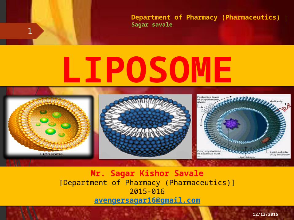
LIPOSOME1
Department of Pharmacy (Pharmaceutics) | Sagar savale
Mr. Sagar Kishor Savale[Department of Pharmacy (Pharmaceutics)]
12/13/2015
Contents1. Liposomes
2. Structural components of liposome's
3. Formation of liposomes
4. Theory of Liposomes
5. Advantages of Liposomes
6. Disadvantages
7. Importance of Liposomes in drug Delivery System
8. Mechanism Of Liposome Formation And Subsequent Processing To Generate Types Of Liposomes
9. Classification of liposomes
10. Conventional liposome preparation methods
11. Methods of Liposome Preparation
12. Stability of Liposomes
13. Liposomes in drug delivery
14. Characterization of liposomes
15. Application Of Liposomes
16. References
12/13/2015
2

3
12/13/2015

12/13/2015
4

12/13/2015
5

12/13/2015
6

12/13/2015
7

12/13/2015
8

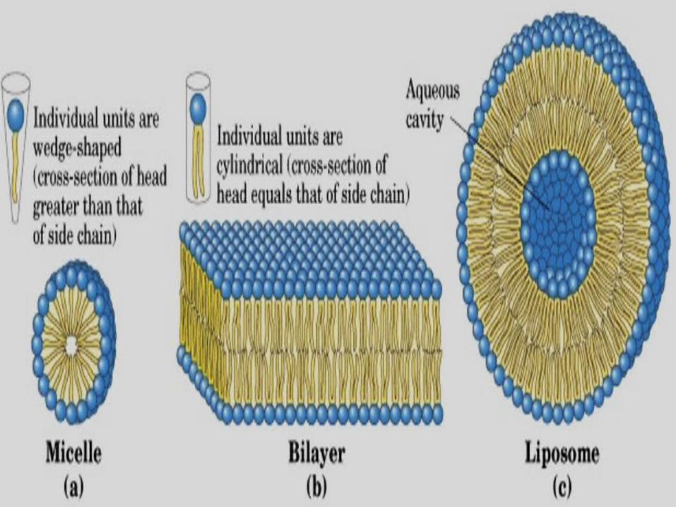
1. Liposome Liposomes are concentric bilayered vesicles in which an aqueous core is entirely enclosed
by a membranous lipid bilayer mainly composed of natural or synthetic phospholipids.
Liposomes are spherical microscopic vesicles consisting phospholipids bilayers which
enclose aqueous compartments.
The size of a liposome ranges from some 20 nm up to several micrometers.
Liposomes were first produced in England in 1961 by Alec D. Bangham, who was
studying phospholipids and blood clotting. Small unilamellar vesicles (SUV), 25 to 100 nm in size that consist of a single bilayer Large unilamellar vesicle (LUV), 100 to 500 nm in size that consist of a single bilayer Multilamellar vesicle (MLV), 200 nm to several microns, that consist of two or more
concentric bilayer
12/13/2015
9

12/13/2015
10

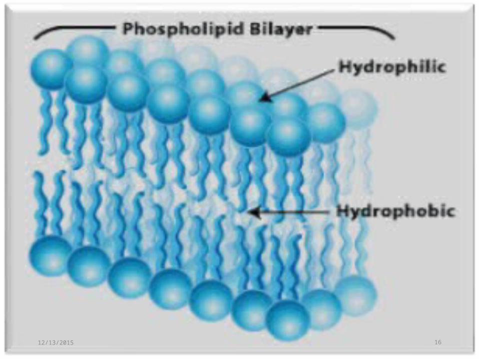
Liposome The lipid molecules are usually phospholipids- amphipathic moieties with a hydrophilic
head group and two hydrophobic tails.
On addition of excess water, such lipidic moieties spontaneously originate to give the most
thermodynamically stable conformation.
In which polar head groups face outwards into the aqueous medium, and the lipidic chains
turns inwards to avoid the water phase, giving rise to double layer or bilayer lamellar
structures.
12/13/2015
11

12/13/2015
12
12/13/2015
13

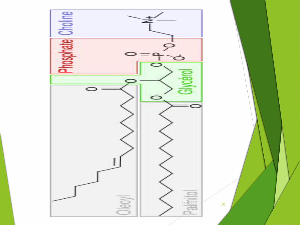
2. Structural components of liposome's
Ther are two main components of Liposomes system they are Phospholipid and cholesterol.
2.1 Phospholipids Phosphatidylcholine. Amphipathic molecule Hydrophobic tail- 2 fatty acid chain containing 10-24 carbon atoms and 0-6
double bond in each chain Hydrophilic polar head- Phosphoric acid bound to water soluble moleculeSelf organize in ordered supramolecular structure when confronted (meet face to face) with solvent
12/13/2015
14

12/13/2015
15
Polar Head Groups Three carbon glycerol
12/13/2015 16

12/13/2015 17

12/13/2015 19

12/13/2015
20
2.2 Flow chart of Lecithin extraction process

12/13/2015

12/13/2015 22
12/13/2015
23
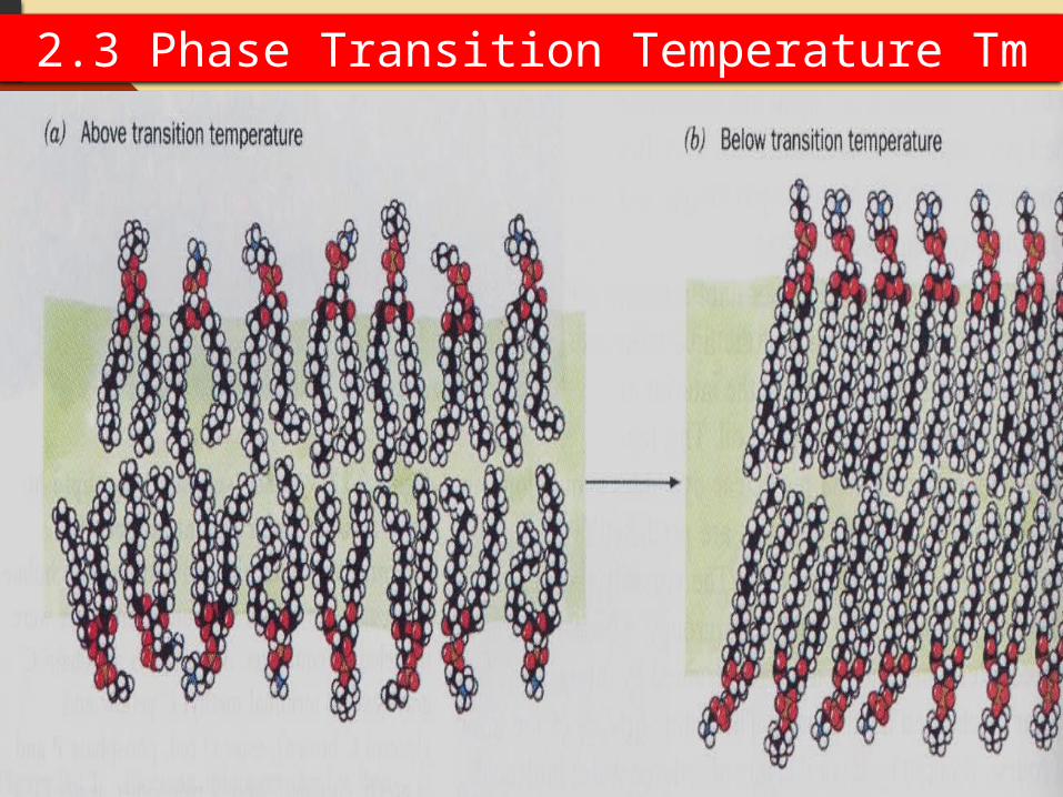
2.3 Phase Transition Temperature Tm

12/13/2015
24

12/13/2015
25
2.4 Membrane permeability

12/13/2015
26 2.5 Some other conventional used phospholipids
• Naturally occurring phospholipids
- PC: phosphatidylcholine
- PE: Phosphatidylethanolamine
- PS: Phosphatidylserine
• Synthetic phospholipids
- DOPC: Dioleoylphosphatidylcholine
- DSPC: Ditsearoylphosphatidylcholine
- DOPE: Dioleoylphosphatidylethanolamine
- DSPE: Ditsearoylphosphatidylethanolamine

12/13/2015
272.6 Cholesterol
Cholesterol by itself does not form bilayer structure.
Cholesterol act as fluidity buffer
After intercalation with phospholipid molecules alter the freedom of motion of carbon molecules in the acyl chain
Restricts the transformations of trans to gauche conformations
Cholesterol incorporation increases the separation between choline head group & eliminates normal electrostatic & hydrogen bonding interactions.
its rigid steroid ring system which interferes with motion of fatty acid tails, stabilizes the lipid bilayer and decrease the leakage of encapsulated drug

12/13/2015
28

12/13/2015
292.7 Non Structural Components

12/13/2015
30 3. Formation of liposomesSurfactants self assemble in water to make micelles and a variety of lipotropic liquid crystalline phases. Liposomes are generally formed from 2 phase mixtures of a lamellar phase with water. Depending on temperature, the lamellar phase can either be in the molten state (La phase) or solid, “gel” state
(Lb phase). Transition temperature = Tc.
Liposomes are formed from aqueous dispersions the “molten” La phase.
Surfactant molecular shape/interactions mainly determines aggregate geometry.
Critical packing factor = v/aolc (unit less), where:
v = molecular volume of surfactant chain
ao = area per surfactant head
lc = length of surfactant chain

12/13/2015
31
12/13/2015
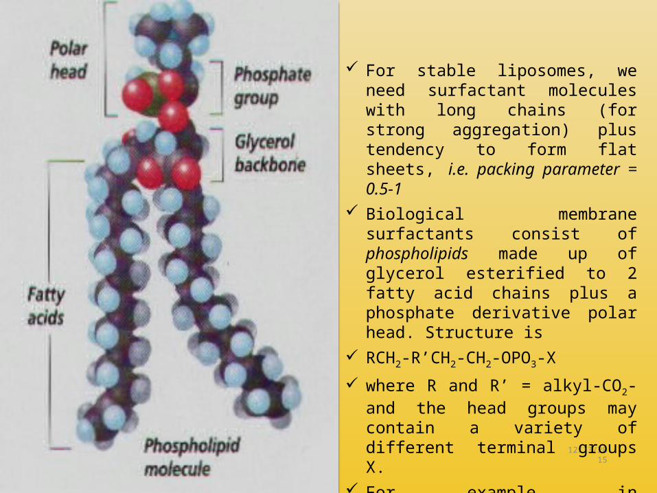
32 For stable liposomes, we need surfactant molecules with long chains (for strong aggregation) plus tendency to form flat sheets, i.e. packing parameter = 0.5-1
Biological membrane surfactants consist of phospholipids made up of glycerol esterified to 2 fatty acid chains plus a phosphate derivative polar head. Structure is
RCH2-R’CH2-CH2-OPO3-X
where R and R’ = alkyl-CO2- and the head groups may contain a variety of different terminal groups X.
For example, in phosphatidylcholine, X = -CH2CH2-N(CH3)3
+

4. Theory of Liposomes
1. The budding theory2. The bilayer phospholipids theory
12/13/2015
33
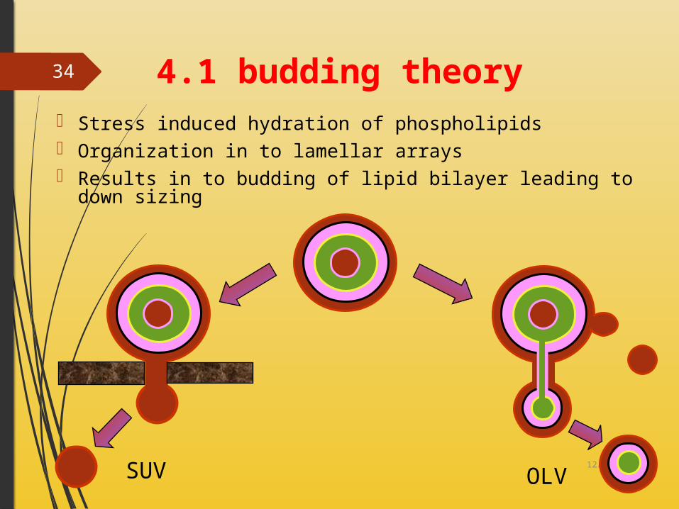
4.1 budding theory Stress induced hydration of phospholipids Organization in to lamellar arrays Results in to budding of lipid bilayer leading to down sizing
12/13/2015
34
SUV OLV

4.2 bilayer phospholipids theory Liposomes are formed when thin lipid films are hydrated The hydrated lipid sheets detach during agitation and self-close to form large,
Multilamellar vesicles (LMV)
12/13/2015
35

5. Advantages of Liposomes Provides selective passive targeting to tumor tissues
Increased efficacy & therapeutic index
Increased stability via encapsulation
Reduction in toxicity of the encapsulated agent
Site avoidance effect
Improved pharmacokinetic effects
Flexibility to couple with site specific ligands to achieve active targeting
Variety of Drugs Given In Low Dose As Encapsulated For Stability
Minimum Effective Concentration And Therapeutic Index
Low Toxicity Due To Reduced Exposure To Sensitive Tissues
Minimum ADR/No Side Effects
Possible Formulation- suspension, emulsion, gel, Cream, lotion, Aerosol, reconstituted Vesicles 12/13/2015
36

6. Disadvantages
Physical/ Chemical Stability
Very High Production Cost
Drug Leakage/ Entrapment/ Drug Fusion
Sterilization
Short Biological Activity / T ½
Oxidation of Bilayer …Lipids And Low Solubility
Overcoming Resistance
Extensive Clinical And Laboratory Research To A Certain Long Circulating Liposomes
Repeated Iv Administration Problems12/13/2015
37
7. Importance of Liposomes in drug Delivery System
Pharm kinetics - efficacy and toxicity
A. Changes the absorbance and bio distribution
B. Deliver drug in desired form
C. Multidrug resistance Protection
A. Decrease harmful side effectsB. Change where drug accumulates in the body
C. Protects drug Release
A. -Affect the time in which the drug is released
B. -Prolong time -increase duration of action and decrease administration
12/13/2015
38

8. Mechanism Of Liposome Formation And Subsequent Processing To Generate Types Of Liposomes
Phospholipids are amphipathic molecules having hydrophobic tail & a hydrophilic or polar head
The hydrophilic & hydrophobic domains within the molecular geometry of amphiphilic lipids orient & self organize in ordered supramolecular structure when confronted with solvents
Cholesterol have modulatory effect on the bilayer membrane (acts as fluidity buffer) Below phase transition it tends to make the membrane less ordered while above the
transition it tends to make the membrane more ordered.
12/13/2015
39

12/13/2015
40

12/13/2015 41
8.1 Mechanism Of Liposome
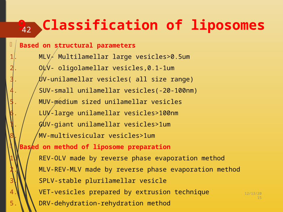
9. Classification of liposomes Based on structural parameters
1. MLV- Multilamellar large vesicles>0.5um
2. OLV- oligolamellar vesicles,0.1-1um
3. UV-unilamellar vesicles( all size range)
4. SUV-small unilamellar vesicles(-20-100nm)
5. MUV-medium sized unilamellar vesicles
6. LUV-large unilamellar vesicles>100nm
7. GUV-giant unilamellar vesicles>1um
8. MV-multivesicular vesicles>1um Based on method of liposome preparation
1. REV-OLV made by reverse phase evaporation method
2. MLV-REV-MLV made by reverse phase evaporation method
3. SPLV-stable plurilamellar vesicle
4. VET-vesicles prepared by extrusion technique
5. DRV-dehydration-rehydration method12/13/2015
42

12/13/2015
43
Based on composition & applications
1. Conventional liposomes-neutral or negatively charged phospholipids& chol.2. PH sensitive liposomes- phospholipid such as PE or DOPE3. Immuno liposomes-CL with attached monoclonal antibody4. Cationic liposomes-cationic lipids with DOPE5. Fusogenic liposomes-Reconstituted Sendai virus envelops

12/13/2015
44 9.1 Types of vesicles on bases of lamella

12/13/2015
45
Based on structural parametersMLV
Multilamellar Large
vesicles(>0.5 um)
OLV oligolamellar
vesicles(>0.1-1.0
um)
UV Unilam
ellarVesicle
s
MVVMultivesicularvesicles
(> 1.0 UM)
MUV
GUV>1um
SUV20-
100nmLUV>100n
m

12/13/2015
46
Based on method
of preparati
on
REV, SUV made by
reverse phase evaporation
method
SPLVStable
plurilamenar vesicles
FATMLVFrozen & thawed
MLV
VETVesicles prepared
by extrusion
tech.

12/13/2015
47
Based on
composition &
application
convential
fusogenic
pH sensitive
cationic
Long circula
tory
immuno

12/13/2015 48
Passive loading techniq
ue
Active/remote loading techniq
ue
Loading of the entrapped agents before/ during the manufacture procedure.Certain types of compounds with ionizable groups & those with both lipid & water solubility can be Introduced into liposomes after the formation of intact vesicles.
MethodS of Liposome Preparation
21

12/13/2015 49
10. Conventional liposome preparation methods
PhospholipidsCholesterol Antioxidant
Lipid component compounding Lipid solvent
Pyrogen Ultra filteryes
No Filter
Solvent removal
Drug ,Salt Antioxidant Buffer WFI
Filter
HydrationSolvent recovery
Extrusion Down sizing
Free drug removal
Prefilter
Sterile filter
Vial filling
Free drug recovery
Aseptic processing Lyophollization Seal / package

12/13/2015
50

11. Methods of Liposome Preparation PASSIVE LOADING TECHNIQUES 1. Mechanical Dispersion method
2. Solvent Dispersion method
3. Detergent Solubilization method
Mechanical dispersion methods of passive loading Technique begin with a lipid solution in organic solvent & end up with lipid dispersion in water
Various components are combined by co-dissolving the lipids in organic solvent which is then removed by film deposition under vacuum.
After solvent removal the solid lipid mixture is hydrated using aqueous buffer.
The lipids spontaneously swell & hydrate to form liposomes
The post hydration treatments include vortexing, sonication, freeze thawing & high pressure extrusion.12/13/2015
51

12/13/2015
52

12/13/2015
53
Post Hydration vortexing, sonication, freeze thawing & high pressure extrusion
LiposomeLipid spontaneously swell & Hydrate
Solid lipid mixture is hydrated by using aqueous buffer
Film deposition
Remove organic solvent under vacuum
Lipid dissolve in organic solvent/co-solvent
11.1 MECHANICAL DISPERSION METHODS

12/13/2015
54 11.2 ProliposomesTo increase the surface area of dried lipid film and to facilitate continuous hydration and
lipid is dried over the finally divided particulate support i.e.- NaCl, Sorbitol, or other polysaccharides. These dried lipid coated particulates are called as Proliposomes
Proliposomes form dispersion of MLVs on addition of water, where support is rapidly dissolved and lipid film hydrate to form MLVs
Methods overcome the stability problem and entrapment efficiency doesn’t matter when formation of stable liposome.

12/13/2015
55

12/13/2015
56 11.3 Sonication MethodProbe Sonicator: is employed for dispersions, which require high energy in a small volume (e.g., high concentration of lipids, or a viscous aqueous phase) Disadvantage- lipid degradation due to high energy and sonication tips release titanium particles into liposome dispersionBath Sonicator: The bath is more suitable for large volumes of diluted lipids. Method: Placing a test tube containing the dispersion in a bath sonicator and sonicating for 5-10min(1,00,000g) which yield a slightly hazy transparent solution. Using centrifugation to yield a clear SUV dispersion.

12/13/2015
57

12/13/2015
58 11.4 French pressure cell liposomes
The ultrasonic radiation degrades the lipids, other sensitive compounds, macromolecules for this extrusion of preformed larger liposomes in a French press under very high pressure is done This tech. yields unit or oligo lamellar liposomes of size (30-80nm in dia.)Includes high cost of press that consists of electric hydraulic press & pressure cellLiposomes prepared by this method are less likely to suffer from structural defects & instabilities as observed in sonicated vesicles.

12/13/2015
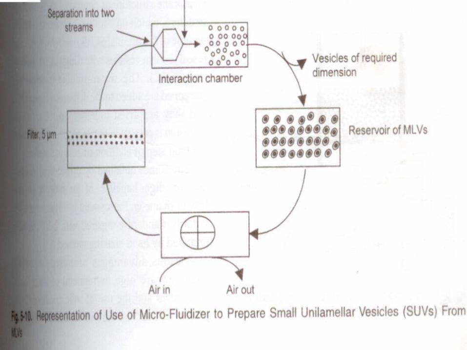
59 11.5 Micro Emulsification Liposomes(MEL)
“Micro Fluidizer” is used to prepare small MLVs from Concentrated lipid dispersion. The lipids can introduced into fluidizers, either as a dispersion of large MLVs or as a slurry
of anhydrated lipids in organic medium. Micro fluidizer pumps the fluid at very high pressure(10,000psi, 600-700 bar) through a
5um orifice. Then it is forced along defined micro channels, which direct two streams of fluid to collide
together at right angles at a very high velocity, thereby affecting an efficient transfer of energy.
The fluid collected can be recycled through the pump and interaction chamber until vesicles of
the spherical dimension are obtained. After a single pass, the size of vesicles is reduced to a size 0.1 and 0.2um in diameter.
12/13/2015
60

12/13/2015
61

12/13/2015
62 11.6 Vesicles Prepared By Extrusion Techniques (Vets)
It is used to process LUVs as well as MLVs.
Liposomes prepared by this tech. are called as membrane filter extrusion liposomes.
The 30% capture volume can be obtained using high lipid conc. The trapped volume in this process is 1-2 litre /mole of lipids.
It is due to their ease of production, readily selectable vesicle diameter, batch to batch reproducibility & freedom from solvent or surfactant contamination is possible

12/13/2015
63

12/13/2015
64 11.7 Freeze Thaw Sonication Method (FTS)
The method is based on freezing of a unilamellar dispersion & then thawing at room temp for 15 min.
Thus the process ruptures & refuses SUVs during which the solute equilibrates between inside & outside & liposomes themselves fuse & increase in size.
Entrapment volume can be upto 30% of the total vol. of dispersion. Sucrose, divalent metal ions & high ionic strength salt solutions can not be entrapped efficiently

12/13/2015
65

12/13/2015
66 11.8 Dried-reconstituted Vesicles
Liposomes obtained by this method are usually “uni or oligo lamellar” of the order of 1.0um or less in diameter.
SUVs in aqueous phase SUVs with solutes to be entrapped Freeze dried membrane Solutes in uni lamellar vesicles Solutes in uni or oligo lamellar vesicles.
FST method DRV method Rehydration Film stacks dispersion Aqueous phase Thawing Sonication (15-30 sec)

12/13/2015
67 11.9 Solvent dispersion Method
Liposome
Formation of monolayer and bilayer of phospholipid
Excess addition of aqueous phase
Lipid dissolve in organic solvent

12/13/2015
68

12/13/2015
69 11.10 Detergent solubilization methods
Formation of micelles (Liposome)
By addition optimized concentration of detergent
Phospholipid brought into intimate contact with aqueous phase

12/13/2015
70 11.11 Active Loading Techniques
Weak amphipathic bases accumulate in the aqueous phase of lipid vesicles in response to a difference in pH between the inside and outside of the liposomes (pHin & pHout)
Two steps process generates this pH imbalance and active (remote) loading. Vesicles are prepared in low pH solution, thus generating low pH within the liposomal
interiors, followed by addition of the base to extra liposomal medium. Basic compounds, carrying amino groups are relatively lipophilic at high pH and
hydrophilic at low pH. In two chambered aqueous system separated by membrane liposomes, accumulation occurs
at the low pH side, under dynamic equilibrium conditions. Thus the un protonated form of basic drug can diffuse through the bilayer The exchange of external medium by gel chromatography with neutral solution Weak base doxorubicin, Adriamycin and vincristine which co-exist in aqueous solutions in
neutral and charged forms have been successfully loaded into preformed liposomes via the pH gradient method.

12/13/2015 71
LIPID FILM HYDRATION
BY HAND SHAKING,FREEZE DRYING OR NON HAND SHAKINGMICRO
EMULSIFICATIONSONICATIONFRENCH PRESSURE
CELLMEMBRANE
EXTRUSONDRIED
RECONSTITUTED
VESICLES
ETHANOL INJECTION
ETHER INJECTION DOUBLE
EMULSION REVERSE PHASE VAPOURATION
VESICLESSTABLE PLURI
LAMELLER VESICLES
DETERGENT REMOVAL
FORM MIXED MICELLES BY DIALYSISCHROMATIGRALPYDIFFUSION VESICLES LIKE….
RECONSTITUTED &
SANDAI VIRUS ENVELOPE
Methods of liposome preparation
Passive loading techniques
Active loading techniques
Mechanical dispersion methods
Solvent dispersion methods
Detergent removal technique
22

12/13/2015
72
Method VesiclesMechanical methods
Vortex or hand shaking of phospholipid dispersions MLV
Extrusion through polycarbonate filters at low or medium pressure OLV, LUV
Extrusion through a French press cell “Micro fluidizer” technique Mainly SUV
High-pressure homogenization Mainly SUVUltrasonic irritation SUV of minimal sizeBubbling of gas BSV
Methods based on replacement of organic solvent(s) by aqueous mediaRemoval of organic solvent(s) MLV, OLV, SUV
Use of water-immiscible solvents: ether and petroleum MLV, OLV, LUV
Ethanol injection method LUVEther infusion (solvent vaporization) LUV, OLV, MLVReverse-phase evaporation
Methods based on detergent removalGel exclusion chromatography SUV“Slow” dialysis LUV, OLV, MLVFast dilution LUV, OLVOther related techniques MLV, OLV, LUV, SUV

12/13/2015
73 11.12 Rapid solvent exchange vesicles (RSEVs) Lipid mixture is transferred between pure solvent & a pure aq.environment. Organic sol. of lipids through orifice of syringe under vacuum into a tube containing
aqueous buffer. The tube is mounted on vortexed. It manifest high entrapment volumes

12/13/2015
74 11.13 Reverse phase evaporation method

12/13/2015
75 11.14 Purification of LiposomesGel Filtration Column Chromatography

12/13/2015
76

12/13/2015
77 12. Stability of Liposomes
Chemical degradation Physical degradation Prevention of chemical degradation Prevention of physical degradation
The liposomes are stable system having protection against physical, chemical and biological degradation.

12/13/2015
78 13. Liposomes in drug delivery
• Protect the encapsulated drug from metabolic degradation
• Increase the half-life of drug
• Reduce the systemic toxicity of drugs
• Could be used as sustained release vehicles
• It is possible to target them to selected tissues or cell
• Biodegradable and biocompatible

12/13/2015
79

12/13/2015
80 13.1 Stealth or PEG-Liposomes

12/13/2015
81

12/13/2015
82 13.2 Liposomes in tumor therapy
Targeting strategies using liposomes Natural targeting of conventional liposomes (passive vectorization) Use of long circulatory (stealth liposomes) Use of ligand mediated targeting (active targeting) The use of anti-receptor antibodies on the tumour vascular endothelium Use of stealth liposomes & ligands mediated targeting in combination
Drug Target disease
Status Product
Doxorubicin Kaposi's sarcoma
Approved SEQUUS
Daunosome Breast cancer Approved NeXstar,USANystatin Systemic fungal
infectionsPhase II Aronex, USA
Amikacin Serious bacterial infections
Phase II NeXstar,USA
Vincristin Solid tumours Preclinical dev. NeXstar,USA

12/13/2015
83 13.3 Liposomes in gene therapy
Recombinant DNA tech., studies of gene function & gene therapy all depend on delivery of nucleic acids( genetic material) into cells in vitro & in vivo.
Gene can be viral (adenovirus, retrovirus) & non viral( liposomes & lipid based systems, polymers & peptides)
Type of vectors Advantages DisadvantagesViral vectors(Adenovirus, retrovirus & adeno-associated virus)
Relatively high transfection efficiency
Immunogenicity, presence of contaminants & safetyVector restricted size limitation for recombinant gene
Non viral vectors(liposomes/lipid based systems, polymers & peptides)
Favorable, pharmaceutical issue-GMP, stability, costPlasmid independent structureLow immunogenicityOpportunity for chemical/physical manipulation
low transfection efficiency

12/13/2015
84
Liposomes in gene therapy

12/13/2015
85
PH sensitive liposomes
The PH sensitive liposomes have been reported as plasmid expression vectors for the cytosolic delivery of DNA.
PH sensitive immunoliposomes
PH sensitive liposomes have been developed to release their contents in response to an acid machinery within endosomal system following receptor mediated endocytosis of the immunological targeting ligand
Fusogenic liposomes & Virosomes
They fuse & merge with cell membranes & directly introduce molecules (entrapped or anchored) into cytoplasm & avoiding route followed by conventional liposomes. Fusion can be mediated by PEG, glycerol & Polyvinyl alcohol or by reconstituted fusogenic viral membrane based liposomes are termed as Virosomes

12/13/2015
86 13.4 Liposomal vaccines
New vaccines that are based on recombinant protein subunits & synthetic peptide antigens are usually non-immunogenic, hence need of immunopotentiation is realized.
The first liposome based vaccine (against hepatitis A) that has been licensed for use in human is an IRIV vaccine which are spherical, unilamellar vesicles with a diameter of 150nm.
IRIVs are prepared by detergent removal of influenza surface glycoproteins & a mixture of natural & synthetic phospholipids containing 70% egg yolk phosphatidylcholine,20 % synthetic PE & 10 % envelop phospholipids originating from H1N1 influenza virus.

12/13/2015
87 13.5 Liposomes as a carrier of Immunomodulation
The main purpose is to activate macrophages & render them tumouricidal. They acquire ability to recognize & destroy neoplastic cells both in vitro & in vivo.
Liposomes in Immunodiagnosis
1. LILA assays (liposome immune lysis assay) has been implicated in the detection of serum components such as carcinoembryonic antigen,C-reactive protein & other serum protein which serve as diagnostic tools for cancer
2 . LILA sandwich method has been used to detect many important antigens in serum, which are useful indicators of various abnormalities
12/13/2015
88 13.6 Liposomes in Dermatology and Cosmetology
Similar to biological membrane they can navigate water soluble & lipophilic substances in different phases.
They mimic the lipid composition & structure of human skin, which enables them to penetrate the epidermal barrier.
Liposomes are biodegradable & nontoxic, thus avoiding local/systemic side or toxic effects.
Moisturizing & restoring action of constitutive lipids.
Liposomes may act as localized drug depots in skin resulting in sustained release of drug, thus improving therapeutic index of drug at target site while reducing toxicity profile to minimum.
Cosmetic creams, e.g. Alpha Lipoic Acid Cream

12/13/2015
89 13.7 Liposomes as Radiopharmaceutical & radio diagnostic
carriers
Liposomes loaded with contrast agents are suitable for contrast agents are substances which are able to absorb certain types of signal much stronger
than surrounding tissue
Radio diagnostic application include liver & spleen imaging, tumor imaging, imaging cardiovascular pathologies, visualization of inflammation & infected sites, brain imaging, visualization of bone marrow
The RES avoidance of contrast agents can be achieved by using targeted liposomes like immunoliposomes
12/13/2015
90 13.8 Liposomes as Red cells substitutes & artificial RBCs
Synthetic & semisynthetic blood substitutes includes recombinant hemoglobin, glutaraldehyde cross linked hemoglobin, hemoglobin encapsulated liposomes.
Liposome encapsulated hemoglobin products are being investigated as artificial RBCs.
Researchers reported completely synthetic amphiphilic heme derivative (lipid heme) & incorporated them into the hydrophobic center of the bilayer membrane of the phospholipid vesicles, which has excellent oxygen carrying & transporting abilities.

12/13/2015 91
LIPOSOMES THERAPY DRUGS /USEAIDS Azidotymidine
Cancer Cisplatin,Taxol,Doxorubicin Malaria Primaquine, Chloroquine, Artemisinin
Gramicidin A
lung Isoniazid, Rifampicin, Budesonide
Infectious Diseases e.g. skin Amphotericin, Antimony, Pentamidine
DRUGS Antibiotics, Antifungal Disinfectant, Immunosuppressive agents
Dermatology and Cosmetology Local anesthetic e.g. Lidocaine and Benzocaine, Gentamycin, Cefazolin
immunological (Vaccine) Adjuvant
Hepatitis A rabies virus, Measles virus, influenza virus Herpes virus, HIV-1 and Vesicular stomatitis
DIEBETIS INSULIN / Hypoglycemic
Radiodiagnostic Carriers γ-scintigraphy, Magnetic resonance (MR), Computer tomography (CT) and Ultrasonography (US) of tumors

12/13/2015
92 14. Characterization of liposomes
Liposomes
Size Number of lamellae
Charge Stability
Preparation Raw materials
Protection
Sizing method
Hydration methods
Degree of saturation
Head group
Presence of sterols
Protecting
agents
Characterized by
Determined by
Classified by

Characterization of liposomes There are three main types of Characterization technique of liposomes
1. Physical Characterization1. Vesicles size/shape/morphology
2. Surface -charge/electrical potential
3. Phase bahaviour/ lamellarity
4. Drug release
5. % capture /free drug
12/13/2015
93

12/13/2015
94
2. Chemical Characterization1. Phospholipids /lipid concentration2. Drug concentration 3. PH / Osmolality4. Antioxidant degradation5. Phospholipids / cholesterols 6. peroxidation/oxidation/hydrolysis

12/13/2015
95
3. Biological Characterization1. Sterility2. Pyrogenisity3. Animal toxicity4. Plasma Stability
12/13/2015 96
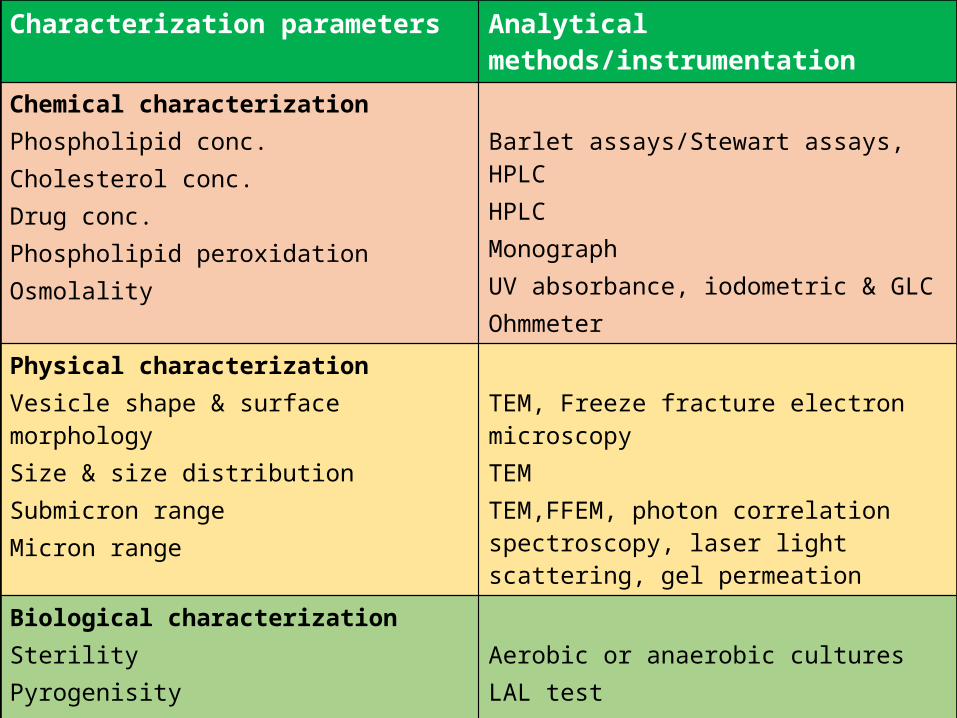
Characterization parameters Analytical methods/instrumentation
Chemical characterizationPhospholipid conc.Cholesterol conc.Drug conc.Phospholipid peroxidationOsmolality
Barlet assays/Stewart assays, HPLCHPLCMonographUV absorbance, iodometric & GLCOhmmeter
Physical characterizationVesicle shape & surface morphologySize & size distributionSubmicron rangeMicron range
TEM, Freeze fracture electron microscopyTEMTEM,FFEM, photon correlation spectroscopy, laser light scattering, gel permeation
Biological characterizationSterilityPyrogenisityAnimal toxicity
Aerobic or anaerobic culturesLAL testMonitoring survival rates, histology & pathology

14.1 Physical Characterization
Vesicle shape & lamellarity & Vesicle size & size distribution Microscopic techniques Optical Microscopy - Determination of gross size distribution of large vesicles
preparations such as MLVs & Morphological structure of liposome. various tech. include light microscopy, fluorescent microscopy, electron
microscopy, laser light scattering, field flow fractionation, gel permeation & gel exclusion, Zetasizer.
12/13/2015
97

Electron Microscopic Techniques Freez Fracture Electron Microscopy
Negative Stain Electron Microscopy
Transmission Electron Microscopy
Scanning Electron Microscopy
Cryo-Electron Microscopy
Laser Light Scattering Techniques
Fluorescence Electron Microscopy
Confocal Laser Light Scanning Microscopy
12/13/2015
98

Freez Fracture Electron Microscopy
The freeze-fracture/freeze etch technique starts with rapid freezing of a cell. Then the frozen cells are cleaved along a fracture plane. This fracture plane is in between the leaflets of the lipid bilayer , The two fractured sections are then coated with heavy metal (etched) and a replica is made of their surfaces. This replica is then viewed in an electron microscope.
12/13/2015
99

Negative Stain Electron Microscopy Negative stain electron microscopy visualizes electron transparent liposomes as bright
areas against a dark background. Negative stains used in the TEM analysis is ammonium molybdate.
12/13/2015
100

Transmission Electron Microscopy Transmission electron microscopy (TEM) is a microscopy technique whereby a beam of
electrons is transmitted through an ultra thin specimen, interacting with the specimen as it passes through. An image is formed from the interaction of the electrons transmitted through the specimen; the image is magnified and focused onto an imaging device, such as a fluorescent screen, on a layer of photographic film, or to be detected by a sensor such as a CCD camera.
12/13/2015
101
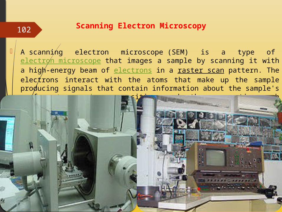
Scanning Electron Microscopy
A scanning electron microscope (SEM) is a type of electron microscope that images a sample by scanning it with a high-energy beam of electrons in a raster scan pattern. The electrons interact with the atoms that make up the sample producing signals that contain information about the sample's surface topography, composition, and other properties such as electrical conductivity.
12/13/2015
102
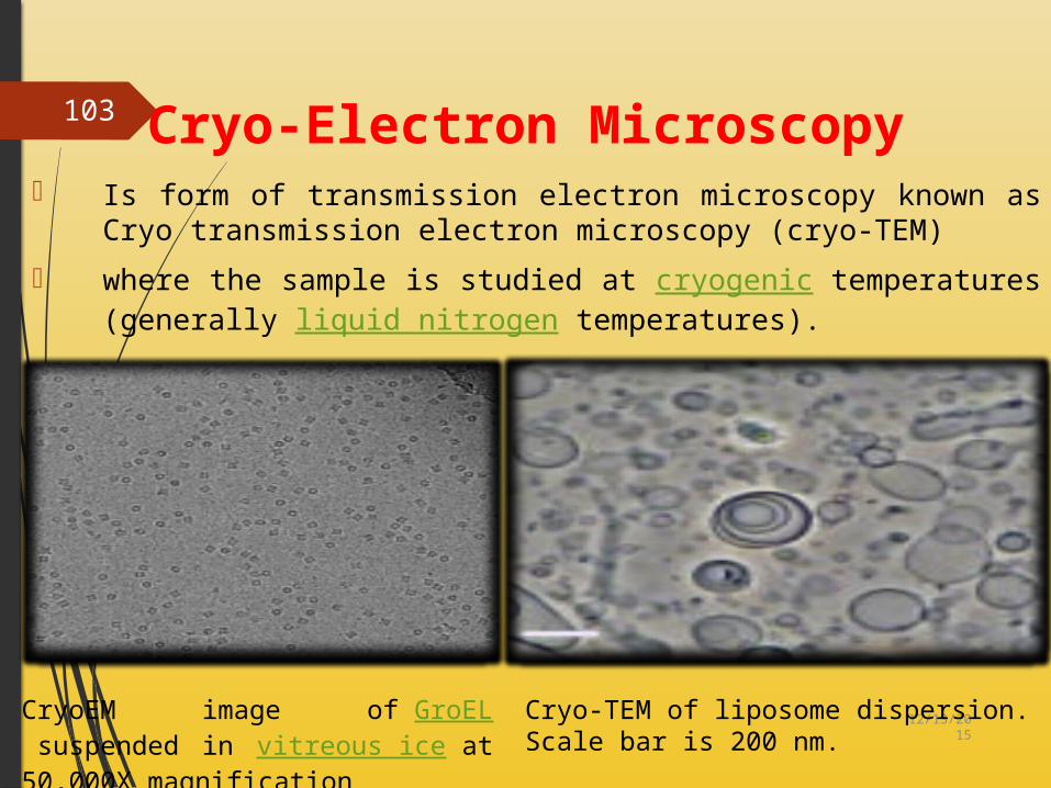
Cryo-Electron Microscopy Is form of transmission electron microscopy known as Cryo transmission
electron microscopy (cryo-TEM) where the sample is studied at cryogenic temperatures (generally
liquid nitrogen temperatures).
12/13/2015
103
CryoEM image of GroEL suspended in vitreous ice at 50,000X magnification
Cryo-TEM of liposome dispersion. Scale bar is 200 nm.

Laser Light Scattering Technique
12/13/2015
104

Fluorescence Electron Microscopy
The "fluorescence microscope" refers to any microscope that uses fluorescence to generate an image.
12/13/2015
105

Confocal Laser Light Scanning Microscopy
Technique for obtaining high-resolution optical images with depth selectivity & use for
Penetration and Permeation Studies.
Confocal microscopy is an optical imaging technique used to increase optical resolution and
contrast of a micrograph by using point illumination and a spatial pinhole to eliminate out-
of-focus light in specimens that are thicker than the focal plane. It enables the reconstruction
of three-dimensional structures from the obtained images.
12/13/2015
106

Zetasizer Zeta potential is an important and useful indicator of particle surface charge, which can be
used to predict and control the stability.
In general, particles could be dispersed stably when the absolute value of zeta potential was above 30mV due to the electric repulsion between particles
12/13/2015
107

Gel permeationPreferably used for the size distribution determination of liposomes
UltracentrifugeUsed for size distribution of liposomes
12/13/2015
108

Encapsulation efficiency - Determines % of the aq. Phase & hence % of water soluble drug which is entrapped & expressed as % entrapment/mg lipid.
Trapped volume - The internal or trapped volume is the aqueous entrapped volume per unit quantity of lipid & expressed as µ l/ µ mol or µ l/mg of total lipid. Radioactive markers are used to determine the internal volume.
Vesicle fusion measurements - It has been studied in case of cationic liposomes, PH sensitive liposomes. fusion has been monitored using a fluorescence resonance energy transfer (RET) between two lipid analogues originally placed in separate vesicle population that measures intermixing of membrane lipids
12/13/2015
109

12/13/2015
110
Phase response & transitional behavior
Lipid bilayers can exists in a low temperature solid ordered phase & above certain temp in a fluid disordered phase. Phase behavior of liposomal membrane determines prop. such as permeability, fusion, aggregation & protein binding Thermodynamic methods:-In differential scanning micro calorimeter, the heat required by liposomes to maintain a steady upward rise in temp is plotted as a function of temperature
Elasticity Measurement of Liposomes
Extrusion Method
Liposomal formulations were extruded through filter membrane (pore diameter 50 nm), using a stainless steel filter holder having 25-mm diameter, by applying a pressure of 2.5 bar. The quantity of vesicle suspension, extruded in 5 minutes was measured.
Skin Permeation Study
Franz Diffusion Cell
12/13/2015
111

14.2 Chemical Characterization Phospholipid conc. is determined in terms of lipid phosphorus content using Barlet
assay/Stewart assay or TLC Cholesterol conc. is determined using Ferric perchlorate method/Cholesterol
oxidase assay Lysolecithin:-which is one of the major product of hydrolysis is estimated using
densitometry Phospholipid peroxidation is determined by UV absorbance, iodometric, GLC
technique. Phospholipid hydrolysis is determined using HPLC & TLC Cholesterol auto oxidation can be determined by HPLC & TLC
12/13/2015
112

14.3 Biological Characterization
SterilityAerobic or anaerobic cultures
PyrogenisityLAL test
Animal toxicityMonitoring survival rates, histology & pathology
Plasma StabilityCytotoxicity Assay, HPLC Assay
12/13/2015
113

14.4 Stability after systemic administration
Two most frequently encountered biological events that the administered liposomal system undergoes are phagocytosis or antigen presentation via the macrophages of the RES system
Opsonins which are proteinaceous components of serum adsorb onto the surface of liposomes thus making these exogenous materials more palatable & conductive to phagocytes
High density lipoprotein removes phospholipid molecules from bilayered vesicular systems
The molecular origin of these interactions are mostly long range electrostatic, Vander waals & short range hydrophobic interactions of particulate surface with macromolecules in the serum
12/13/2015
114

14.5 Liposome-cell interaction
12/13/2015
115

12/13/2015
116 14.6 Stability of LiposomesStoring the vesicles at 4°C ± 0.5°C. Vesicle size, zeta potential, and entrapment efficiency of the vesicles was measured after 180 days.
The stability in vitro which covers the stability aspects prior to the administration of the formulation & with regard to the stability of the constitutive lipids.
The stability in vivo which covers the stability aspects once the formulation is administered via various routes to the biological fluids. It includes stability aspects in blood if administered by systemic route or in gastrointestinal tract if administered by oral or per oral routes.
Stability in vitro:- method of formulation, nature of amphiphilic & encapsulated drug, manipulate membrane fluidity/rigidity & permeability characteristics.
Storage temp. of these dispersions must be defined & controlled Liposomal phospholipids can undergo degradation such as oxidation & hydrolysis
12/13/2015
117
Lipid oxidation & Peroxidation
Lipid peroxidation measurement is based on disappearance of unsaturated fatty acids or appearance of conjugated dienes.
It can be prevented by minimizing use of unsaturated lipids, use of oxygen, argon or nitrogen environment, use of antioxidant such as Alpha tocopherols or BHT or use of light resistant containers for storage of liposomal preparations
Lipid hydrolysis
It leads to Lysolecithin formation The inclusion of charged molecule in the bilayer shifts the electrophoretic mobility & makes it positive with addition of stearylamine or negative with dicetyl phosphate thus prevents liposomal fusion/swelling or aggregation
Long term & Accelerated stability
High temp. testing(>250C) is universally used for heterogeneous products. Various laboratories store their products at temp ranging from 40C to 500 C.

12/13/2015
118
1.Liposomes as drug/protein delivery vehiclesControlled & sustained drug release in situEnhanced drug solubilizationAltered pharmacokinetics & bio distributionEnzyme replacement therapy & liposomal storage disorders
2.Liposomes in antimicrobial, antifungal & antiviral therapyLiposomal drugsLiposomal biological response modifiers
3.Liposomes in tumor therapyCarrier of small cytotoxic moleculesVehicle for macromolecules as genes
4.Liposomes in gene deliveryGene & antisense therapyGenetic vaccination
5.Liposomes in immunologyImmunoadjuvant
15. Application Of Liposomes

12/13/2015
119
ImmunomodulationImmunodiagnosis6. Liposomes as artificial blood surrogates7.Liposomes as radiopharmaceutical & radio diagnostic carriers8.Liposomes in cosmetics & dermatology9.Liposomes in enzyme immobilization & bioreactor technology

12/13/2015
120 16. References
• Jain N.K., Controlled & Novel Drug Delivery, CBS Publications, New Delhi • Jain N.K., Advances in Controlled & Novel Drug Delivery, CBS Publications, New Delhi. • Vyas S.P. and Khar R.K., Controlled drug delivery- Concepts & Advances, Vallabh
Prakashan, New Delhi. • Vyas S.P. and Khar R.K., Targeted & Controlled drug delivery- Novel Career System, CBS
Publications, New Delhi. • Chien Y, Novel Drug Delivery System, Mercel Decker Publications.• Lee & Robinson, Controlled Drug Delivery, Second Edition, Mercel Decker Publications.• Swarbrick J and Boylon J.C., Encyclopedia of Pharmaceutical Technology, Vol. 1-3, Mercel
Decker Inc.• Allen, Theresa M. "Liposomal Drug Formulations: Rationale for Development and What
We Can Expect for the Future." Drugs 56: 747-756, 1998.• Allen, Theresa M. "Long-circulating (serially stabilized) liposome for targeted drug
delivery." Tips 15: 214-219, 1994.• Vesicular drug delivery system by R.S.R. Murthy.

12/13/2015
121
• Allen, Theresa M. "Liposomal Drug Formulations: Rationale for Development and What We Can Expect for the Future." Drugs 56: 747-756, 1998.
• Allen, Theresa M. "Long-circulating (sterically stabilized) liposomes for targeted drug delivery."TiPs 15: 214-219, 1994.
• Allen, Theresa M. "Opportunities in Drug Delivery." Drugs 54 Suppl. 4: 8-14, 1997 Janknegt, Robert. "Liposomal and Lipid Formulations of Amphotericin B." Clinical Pharmacokinetics.23(4): 279-291, 1992.
• Kim, Anna et al. "Pharmacodynamics of insulin in polyethylene glycol-coated liposomes."International Journal of Pharmaceutics. 180: 75-81, 1999.
• Quilitz, Rod. "Oncology Pharmacotherapy: The Use of Lipid Formulations of Amphotericin B in Cancer
• Patients." Cancer Control.5:439-449, 1998. • Ranade, Vasant V. "Drug Delivery Systems: Site-Specific Drug Delivery Using
Liposomes as Carriers." • Pharmacology. 29: 685-694, 1989. • Storm, Gert and Daan J.A. Crommelin. "Liposomes:quo vadi?" PSTT 1: 19-31, 1998. • Taylor, KMG and JM Newton. "Liposomes as a vecicle for drug delivery." British Journal
of Hospital • Medicine. 51: 55-59, 1994

12/13/2015
122
• Navone, NM, et al. p53 mutations in prostate cancer bone metastases suggest that selected p53 mutants in th eprimary site define foci with metastatic potential. J Urol 161(1):304-8, 1/99.
• [email protected] www.prostatematters.com 1999• Pienta, K., Goodson, J., & Esper, P. Epidemiology of Prostate Cancer: Molecular and
Environmental Clues. http://www.cancer.med.umich.edu/prostcan/articles/clues.html• Smith, J, et al. Major Susceptibility Locus for Prostate Cancer on Chromosome 1 Suggested
by a Genome-Wide Search. Science 274: 1371-4, 11/22/96• Veterinary Genetics Laboratory, School of Veterinary Medicine University of California,
Davis. Microsatellites. http://www.vgl.ucdavis.edu/service/canine/micros.htm 12/30.97• Wolf, G. University Hospital Charite Institute of Pathology. http://amba.charite.de/cgh
1/15/99• Xu, J., et al. Evidence for a prostate cancer susceptibility locus on the X chromosome.
Nature Genet 20: 175-179, 1998.

12/13/2015
123