mechanism and stoichiometry of 2,2-diphenyl-1-picrylhydrazyl radical scavenging by glutathione and...
TRANSCRIPT
Bioorganic Chemistry 37 (2009) 126–132
Contents lists available at ScienceDirect
Bioorganic Chemistry
journal homepage: www.elsevier .com/locate /bioorg
Mechanism and stoichiometry of 2,2-diphenyl-1-picrylhydrazyl radicalscavenging by glutathione and its novel a-glutamyl derivative
Säde Viirlaid b, Riina Mahlapuu a, Kalle Kilk a, Aleksei Kuznetsov b, Ursel Soomets a, Jaak Järv b,*
a Institute of Biochemistry, Medical Faculty, University of Tartu, The Centre of Excellence for Translational Medicine, 19 Ravila St, 50411 Tartu, Estoniab Institute of Chemistry, University of Tartu, 2 Jakobi St, 51014 Tartu, Estonia
a r t i c l e i n f o
Article history:Received 17 February 2009Available online 12 May 2009
Keywords:Antioxidant activityGlutathioneGSH a-glutamyl analogDPPHj
Kinetic mechanismScavenging stoichiometry
0045-2068/$ - see front matter � 2009 Elsevier Inc. Adoi:10.1016/j.bioorg.2009.05.001
Abbreviations: DPPHj, 2,2-diphenyl-1-picrylhydrazyl-L-cysteinylglycine; a-GSH, a-L-glutamyl-L-cysteinamyl-L-serinylglycine; BDE, bond dissociation energy
* Corresponding author. Fax: +372 7375247.E-mail address: [email protected] (J. Järv).
a b s t r a c t
Kinetic mechanism and stoichiometry of scavenging the 2,2-diphenyl-1-picrylhydrazyl radical by gluta-thione and its novel analog, containing a-glutamyl residue in place of the c-glutamyl moiety, were stud-ied using different ratios of reagents. At low concentrations of the peptides, the process was described asa bimolecular reaction obeying the stoichiometric ratio 1:1. However, at excess of peptides the formationof a non-covalent complex between the reagents was discovered and characterized by dissociation con-stants K = 0.61 mM for glutathione and K = 0.27 mM for the glutathione a-glutamyl analog, respectively.The complex formation was followed by a reaction step that was characterized by the similar rate con-stant k = 0.02 s�1 for both peptides. Thus, the apparently different antioxidant activity of these two pep-tides, observed under common assay conditions, was determined by differences in the formation of thisnon-covalent complex.
� 2009 Elsevier Inc. All rights reserved.
1. Introduction
The thiol group of glutathione (GSH, Scheme 1) is the key intra-cellular scavenger of free radicals in biological systems, maintain-ing redox homeostasis for proper functioning of cellular processes[1]. This has generated a great interest in the design and pharma-cological applications of GSH analogs as possible drugs and bioac-tive compounds [2]. In parallel, the same fact has also initiatedinvestigations into the reaction mechanism of these compoundsin non-biological model systems, often used for in vitro evaluationof their antioxidant properties. These data are clearly valuable forunderstanding the structure–activity relationship for antioxidantsand elucidating the chemical mechanism behind their bioactivity.Moreover, linking the results with bioactivity data is generally be-lieved to be a promising reference material for design studies sincethe in vitro assays are fast and less expensive if used for prelimin-ary screening of drug candidates [3].
Previous studies have shown that alterations of the GSH struc-ture at different positions of this mini-peptide may have a stronginfluence on the activity of these congeners, revealing also higherantioxidant and cell protecting effects [4]. Furthermore, a signifi-cant increase in hydroxyl radical scavenging activity has been
ll rights reserved.
yl radical; GSH, c-L-glutam-ylglycine; a-GOH, a-L-glut-.
demonstrated with GSH analogs belonging to a series of noveltetrapeptidic GSH analogs called UPF peptides [5]. In these com-pounds, the c-glutamyl moiety of GSH has been replaced by thea-glutamyl group, corresponding to the ‘‘normal” peptide structure[5]. However, as several parts of the peptide structure were alteredin UPF peptides, it was impossible to assess the specific effect ofthe c to a replacement in the tripeptide structure. Therefore,the a-congener of GSH was subsequently synthesized (a-GSH,Scheme 1) and its reaction with the stable 2,2-diphenyl-1-picrylhydrazyl radical (DPPHj, Scheme 1) was investigated in thepresent paper.
The stable radical DPPHj was discovered by Goldschmidt andRenn in 1922 [6], and later it was introduced for the chemical assayof antioxidant properties by Blois [7]. Currently, this radical has be-come a popular tool in the antioxidant assays of synthetic and nat-ural compounds and their mixtures [8,9]. DPPHj has a strongabsorption maximum at k = 517 nm, characterized with the molarextinction coefficient 9660 and this value decreases to 1640 if theodd electron of the stable radical becomes paired [8,10]. In sum-mary, this process can be presented by the following equilibriumbetween DPPHj and the radical scavenging peptide (GSH or a-GSH):
DPPH� þ Peptide-SH ¡ DPPH-Hþ Peptide-S�: ð1Þ
In detail, the reaction (1) may proceed through different mech-anisms. Firstly, it may involve the one-step abstraction of thehydrogen atom (Hj) from the radical scavenging reagent and its
NHNH
O
O
SHO
OHNH2
O OH
NHNH
O
O
SHO
OH
OHO
NH2
GSH α−GSH
NN
NO2
NO2
O2N NHNH
O
O
OHO
OH
OHO
NH2
DPPH• α-GOH
Scheme 1.
S. Viirlaid et al. / Bioorganic Chemistry 37 (2009) 126–132 127
direct transfer to DPPHj, named as the hydrogen atom transfer(HAT) mechanism [10,11]. Secondly, the concerted transfer of elec-tron and proton may occur between the reagents [12–15]. The se-quence of these events varies in different reaction mechanisms,depending on the reagent structure and the reaction medium,especially if protic and aprotic solvents are compared [12–15].All these mechanisms, however, lead to the formation of the sameproduct, the quenched radical DPPH-H as defined by Eq. (1).
Our preliminary experiments with the a-glutamyl analog ofGSH (a-GSH) revealed that the DPPHj quenching rate for this deriv-ative exceeded the rate at which GSH reacted with this radical un-der the same assay conditions. The different behavior of GSH anda-GSH in this assay was rather surprising, as the alteration of theposition of the glutamyl residue in this peptide should not influ-ence the reactivity of the thiol group of cysteine. For example,the energy of the homolytic dissociation of the S–H bond (BDE)was found to be close for the free cystein (373 kJ/mol, 298 K) andfor the same amino acid in peptide structure (367 kJ/mol, the sametemperature) [16]. These values also coincide with the theoreticaland experimental BDE values for the S–H bond in various alkan-ethiols, ranging from 366 to 371 kJ/mol [17]. Based on the data,it has been recognized that the homolytic dissociation energy ofa typical S–H bond attached to aliphatic structures does not varymuch [17]. Therefore, it was intriguing to find the reasons whyGSH and its a-glutamyl derivative a-GSH still react with DPPHj
at different rates. To verify the role of the thiol group in these reac-tions, the serine analog of a-glutamyl GSH was synthesized (a-GOH, see Scheme 1) and its antioxidant activity was assayed underthe same conditions.
The mechanism of DPPHj scavenging by GSH and a-GSH wasstudied kinetically and the experiments established that the reac-tion involved the formation of a reversible complex between thereactants, followed by a reaction step where the scavenging ofDPPHj took place. Resolving of this reaction mechanism identifiedthe different equilibria of the reversible complex formation step asthe main reason of apparently different antioxidant properties ofGSH and a-GSH in this assay system.
2. Materials and methods
2.1. Chemicals and peptide synthesis
Fmoc–Gly–Wang resin, Fmoc-protected amino acids, 1-hydroxybenzotriazole (HOBt) and 2-(1H-benzotriazole-1-yl)-1,1,3,3-tetramethyluronium tetrafluoroborate (TBTU) were pur-chased from Novabiochem, Switzerland; N,N-dimethylformamide(DMF), dichloromethane (DCM), N,N-diisopropylethylamine (DIEA)and acetonitrile from BDH Laboratory Supplies, England; trifluoro-acetic acid (TFA) and triisopropylsilane (TIS) from Fluka, Switzer-land; 1,2-ethanedithiol (EDT), glutathione and 2,2-diphenyl-1-picrylhydrazyl (DPPHj) from Sigma-Aldrich, Germany.
The glutathione analogs were synthesized manually usingFmoc-chemistry starting from commercial Fmoc–Gly–Wang resin.Coupling of amino acid residues was carried out in a stepwise man-ner using the standard TBTU and HOBt activation protocol [18]. Thepeptides were removed from the resin and simultaneously depro-tected with TFA in the presence of scavengers, 2% water (v/v), 2%EDT (v/v) and 2.5%TIS (v/v) for 90 min at room temperature. Thepurity of the synthetic peptides was >99% as demonstrated byHPLC on an analytical Nucleosil 120-3 C18 reversed-phase column(0.4 cm � 10 cm). The molecular masses of the peptides weredetermined by a MALDI–TOF mass spectrometry (Voyager DEPro, Applied Biosystems) and the calculated values were obtainedin each case.
2.2. DPPHj scavenging assay
Scavenging of DPPHj was monitored spectrophotometrically asdescribed previously [5]. Briefly, 0.25 ml of GSH or a-GSH solution(within the concentration range of 5–2000 lM) in citric acid–so-dium citrate buffer (20 mM, pH 3.9) was added to 0.25 ml of DPPHj
solution in 95% ethanol. Freshly distilled solvents were used.Changes in the absorbance at 517 nm, occurring due to scavengingthe free radical, were monitored in 1 cm thermostated quartz cellsat 25 �C (UV–VIS spectrophotometer Unicam UV300, ThermoSpec-
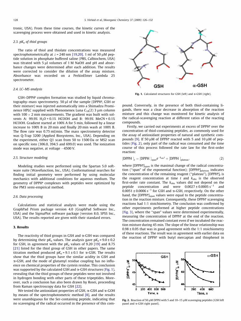
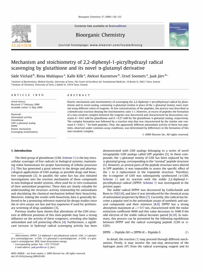
Fig. 1. Calculated structures for GSH (left) and a-GSH (right).
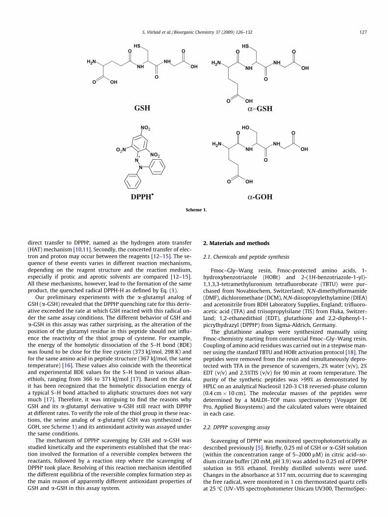
Fig. 2. Reaction of 50 lM DPPH with 5 and 10–15 lM scavenging peptides (GSH leftpanel and a-GSH right panel).
128 S. Viirlaid et al. / Bioorganic Chemistry 37 (2009) 126–132
tronic, USA). From these time courses, the kinetic curves of thescavenging process were obtained and used in kinetic analysis.
2.3. pKa of thiol groups
The ratio of thiol and thiolate concentrations was measuredspectrophotometrically at k = 240 nm [19,20]. 1 ml of 50 lM pep-tide solution in phosphate buffered saline (PBS, Calbiochem, USA)was titrated with 5 ll volumes of 1 M NaOH and pH and absor-bance changes were determined after each addition. The resultswere corrected to consider the dilution of the assay mixture.Absorbance was recorded on a PerkinElmer Lambda 25spectrometer.
2.4. LC–MS analysis
GSH–DPPHj complex formation was studied by liquid chroma-tography–mass spectrometry. 50 ll of the sample (DPPHj, GSH ortheir mixture) was injected automatically into a Shimadzu Promi-nence HPLC supplied with Phenomenex Luna C18(2) 3 lm columnwith 100 � 2 mm measurements. The gradient was built with sol-vents A: 99.9% H2O + 0.1% HCOOH and B: 99.9% MeCN + 0.1%HCOOH. Gradient started at 100% A for 5 min, followed by a linearincrease to 100% B in 20 min and finally 20 min wash at 100% B.The flow rate was 0.75 ml/min. The mass spectrometry detectorwas Q-Trap 3200 (Applied Biosystems, Inc., USA). Depending onthe experiment, either Q1 scan from 50 to 1500 Da or MS2 scanon specific ions (306.0, 394.5 and 699.0) was used. The ionizationmode was negative, at voltage �4500 V.
2.5. Structure modeling
Modeling studies were performed using the Spartan 5.0 soft-ware suite (Wavefunction, Inc., USA). Conformational searches forfinding initial geometry were performed by using molecularmechanics with additional conditions for aqueous medium. Thegeometry of DPPHj complexes with peptides were optimized bythe PM3 semi-empirical method.
2.6. Data processing
Calculations and statistical analysis were made using theGraphPad Prism package version 4.0 (GraphPad Software Inc.,USA) and the SigmaPlot software package (version 8.0, SPSS Inc.,USA). The results reported are given with their standard errors.
3. Results
The reactivity of thiol groups in GSH and a-GSH was comparedby determining their pKa values. The analysis gave pKa = 9.0 ± 0.2for GSH, in agreement with the pKa values of 9.20 [19] and 8.75[21] listed for the thiol group of GSH in other papers. The sametitration method produced pKa = 9.1 ± 0.1 for a-GSH. The resultsshow that the thiol groups have the similar acidity in GSH anda-GSH, and the mode of glutamyl residue coupling has no influ-ence on chemical properties of the cystein residue. This conclusionwas supported by the calculated GSH and a-GSH structures (Fig. 1),revealing that the thiol groups of these peptides were not involvedin hydrogen bonding with other parts of these tripeptides. More-over, such a conclusion has also been drawn by Rosei, proceedingfrom Raman spectroscopy data for GSH [22].
We tested the antioxidant properties of GSH, a-GSH and a-GOHby means of the spectrophotometric method [8], and the resultswere unambiguous for the Ser-containing peptide, indicating thatno scavenging of the radical occurred in the presence of this com-
pound. Conversely, in the presence of both thiol-containing li-gands, there was a clear decrease in absorption of the reactionmixture and this change was monitored for kinetic analysis ofthe radical-scavenging reaction at different ratios of the reactingcompounds.
Firstly, we carried out experiments at excess of DPPHj over theconcentration of thiol-containing peptides, as commonly used forthe assay of antioxidant properties of natural and synthetic com-pounds [9]. If 50 lM of DPPHj reacted with 5 and 10 lM of pep-tides (Fig. 2), only part of the radical was consumed and the timecourse of this process followed the rate law for the first-orderreaction:
½DPPH��t ¼ ½DPPH��spane�kobst þ ½DPPH��plateau; ð2Þ
where [DPPHj]span is the maximal change of the radical concentra-tion (‘‘span” of the exponential function); [DPPHj]plateau indicatesthe concentration of the remaining reagent (‘‘plateau”), [DPPHj]t isthe reagent concentration at time t and kobs is the observedfirst-order rate constant. The kobs values did not depend on thepeptide concentration and were 0.0027 ± 0.0005 s�1 and0.0051 ± 0.0006 s�1 for GSH and a-GSH, respectively. On the otherhand, the [DPPHj]span values were equal to the peptide concentra-tion in the reaction mixture. Consequently, these DPPHj scavengingreactions had 1:1 stoichiometry. The conclusion was confirmed byother experiments performed at higher peptide concentrations(Fig. 3), where the ‘‘span” values were determined experimentally,measuring the concentration of DPPHj at the end of the reaction.The concentration remained constant even if we incubated the reac-tion mixture during 45 min. The slope of the linear relationship was0.98 ± 0.05 that was in good agreement with the 1:1 stoichiometryof these reactions. The result was in agreement with earlier data onthe reaction of DPPHj with butyl mercaptan and thiophenol in
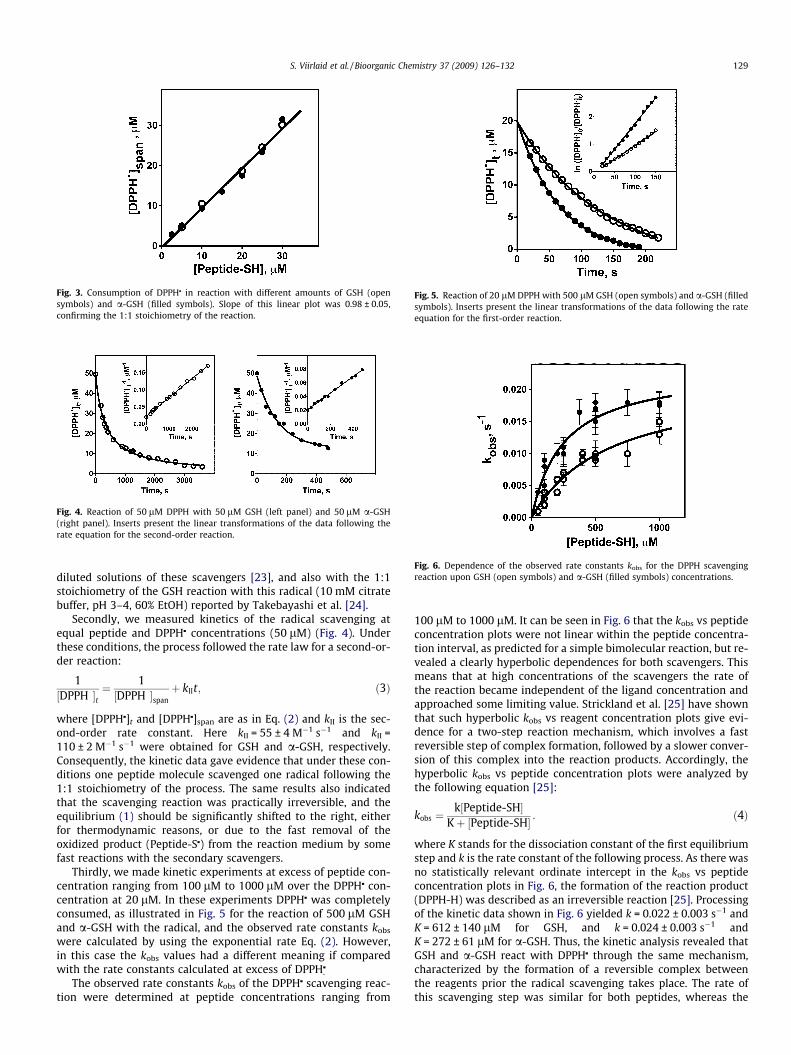
Fig. 3. Consumption of DPPHj in reaction with different amounts of GSH (opensymbols) and a-GSH (filled symbols). Slope of this linear plot was 0.98 ± 0.05,confirming the 1:1 stoichiometry of the reaction.
Fig. 4. Reaction of 50 lM DPPH with 50 lM GSH (left panel) and 50 lM a-GSH(right panel). Inserts present the linear transformations of the data following therate equation for the second-order reaction.
Fig. 5. Reaction of 20 lM DPPH with 500 lM GSH (open symbols) and a-GSH (filledsymbols). Inserts present the linear transformations of the data following the rateequation for the first-order reaction.
Fig. 6. Dependence of the observed rate constants kobs for the DPPH scavengingreaction upon GSH (open symbols) and a-GSH (filled symbols) concentrations.
S. Viirlaid et al. / Bioorganic Chemistry 37 (2009) 126–132 129
diluted solutions of these scavengers [23], and also with the 1:1stoichiometry of the GSH reaction with this radical (10 mM citratebuffer, pH 3–4, 60% EtOH) reported by Takebayashi et al. [24].
Secondly, we measured kinetics of the radical scavenging atequal peptide and DPPHj concentrations (50 lM) (Fig. 4). Underthese conditions, the process followed the rate law for a second-or-der reaction:
1½DPPH��t
¼ 1½DPPH��span
þ kIIt; ð3Þ
where [DPPHj]t and [DPPHj]span are as in Eq. (2) and kII is the sec-ond-order rate constant. Here kII = 55 ± 4 M�1 s�1 and kII =110 ± 2 M�1 s�1 were obtained for GSH and a-GSH, respectively.Consequently, the kinetic data gave evidence that under these con-ditions one peptide molecule scavenged one radical following the1:1 stoichiometry of the process. The same results also indicatedthat the scavenging reaction was practically irreversible, and theequilibrium (1) should be significantly shifted to the right, eitherfor thermodynamic reasons, or due to the fast removal of theoxidized product (Peptide-Sj) from the reaction medium by somefast reactions with the secondary scavengers.
Thirdly, we made kinetic experiments at excess of peptide con-centration ranging from 100 lM to 1000 lM over the DPPHj con-centration at 20 lM. In these experiments DPPHj was completelyconsumed, as illustrated in Fig. 5 for the reaction of 500 lM GSHand a-GSH with the radical, and the observed rate constants kobs
were calculated by using the exponential rate Eq. (2). However,in this case the kobs values had a different meaning if comparedwith the rate constants calculated at excess of DPPHj
.
The observed rate constants kobs of the DPPHj scavenging reac-tion were determined at peptide concentrations ranging from
100 lM to 1000 lM. It can be seen in Fig. 6 that the kobs vs peptideconcentration plots were not linear within the peptide concentra-tion interval, as predicted for a simple bimolecular reaction, but re-vealed a clearly hyperbolic dependences for both scavengers. Thismeans that at high concentrations of the scavengers the rate ofthe reaction became independent of the ligand concentration andapproached some limiting value. Strickland et al. [25] have shownthat such hyperbolic kobs vs reagent concentration plots give evi-dence for a two-step reaction mechanism, which involves a fastreversible step of complex formation, followed by a slower conver-sion of this complex into the reaction products. Accordingly, thehyperbolic kobs vs peptide concentration plots were analyzed bythe following equation [25]:
kobs ¼k½Peptide-SH�
Kþ ½Peptide-SH� : ð4Þ
where K stands for the dissociation constant of the first equilibriumstep and k is the rate constant of the following process. As there wasno statistically relevant ordinate intercept in the kobs vs peptideconcentration plots in Fig. 6, the formation of the reaction product(DPPH-H) was described as an irreversible reaction [25]. Processingof the kinetic data shown in Fig. 6 yielded k = 0.022 ± 0.003 s�1 andK = 612 ± 140 lM for GSH, and k = 0.024 ± 0.003 s�1 andK = 272 ± 61 lM for a-GSH. Thus, the kinetic analysis revealed thatGSH and a-GSH react with DPPHj through the same mechanism,characterized by the formation of a reversible complex betweenthe reagents prior the radical scavenging takes place. The rate ofthis scavenging step was similar for both peptides, whereas the
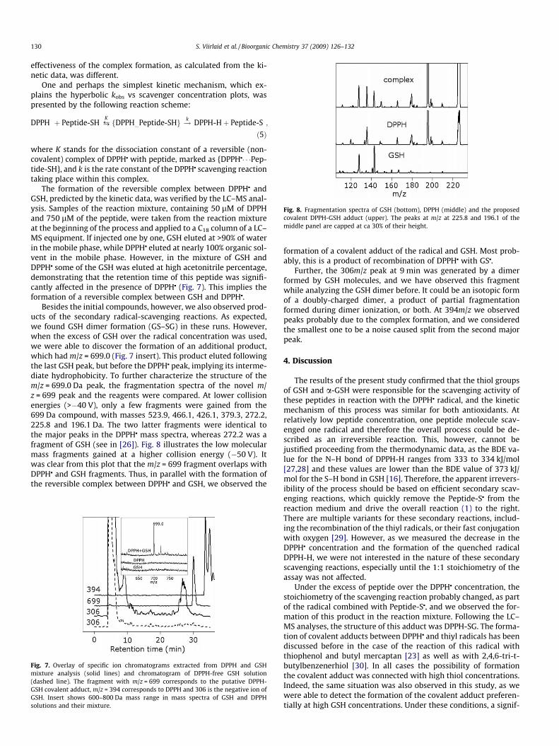
Fig. 8. Fragmentation spectra of GSH (bottom), DPPH (middle) and the proposedcovalent DPPH-GSH adduct (upper). The peaks at m/z at 225.8 and 196.1 of themiddle panel are capped at ca 30% of their height.
130 S. Viirlaid et al. / Bioorganic Chemistry 37 (2009) 126–132
effectiveness of the complex formation, as calculated from the ki-netic data, was different.
One and perhaps the simplest kinetic mechanism, which ex-plains the hyperbolic kobs vs scavenger concentration plots, waspresented by the following reaction scheme:
DPPH� þ Peptide-SH ¡KfDPPH�...Peptide-SHg !k DPPH-Hþ Peptide-S�;
ð5Þ
where K stands for the dissociation constant of a reversible (non-covalent) complex of DPPHj with peptide, marked as {DPPHj� � �Pep-tide-SH}, and k is the rate constant of the DPPHj scavenging reactiontaking place within this complex.
The formation of the reversible complex between DPPHj andGSH, predicted by the kinetic data, was verified by the LC–MS anal-ysis. Samples of the reaction mixture, containing 50 lM of DPPHand 750 lM of the peptide, were taken from the reaction mixtureat the beginning of the process and applied to a C18 column of a LC–MS equipment. If injected one by one, GSH eluted at >90% of waterin the mobile phase, while DPPHj eluted at nearly 100% organic sol-vent in the mobile phase. However, in the mixture of GSH andDPPHj some of the GSH was eluted at high acetonitrile percentage,demonstrating that the retention time of this peptide was signifi-cantly affected in the presence of DPPHj (Fig. 7). This implies theformation of a reversible complex between GSH and DPPHj.
Besides the initial compounds, however, we also observed prod-ucts of the secondary radical-scavenging reactions. As expected,we found GSH dimer formation (GS–SG) in these runs. However,when the excess of GSH over the radical concentration was used,we were able to discover the formation of an additional product,which had m/z = 699.0 (Fig. 7 insert). This product eluted followingthe last GSH peak, but before the DPPHj peak, implying its interme-diate hydrophobicity. To further characterize the structure of them/z = 699.0 Da peak, the fragmentation spectra of the novel m/z = 699 peak and the reagents were compared. At lower collisionenergies (>�40 V), only a few fragments were gained from the699 Da compound, with masses 523.9, 466.1, 426.1, 379.3, 272.2,225.8 and 196.1 Da. The two latter fragments were identical tothe major peaks in the DPPHj mass spectra, whereas 272.2 was afragment of GSH (see in [26]). Fig. 8 illustrates the low molecularmass fragments gained at a higher collision energy (�50 V). Itwas clear from this plot that the m/z = 699 fragment overlaps withDPPHj and GSH fragments. Thus, in parallel with the formation ofthe reversible complex between DPPHj and GSH, we observed the
Fig. 7. Overlay of specific ion chromatograms extracted from DPPH and GSHmixture analysis (solid lines) and chromatogram of DPPH-free GSH solution(dashed line). The fragment with m/z = 699 corresponds to the putative DPPH-GSH covalent adduct, m/z = 394 corresponds to DPPH and 306 is the negative ion ofGSH. Insert shows 600–800 Da mass range in mass spectra of GSH and DPPHsolutions and their mixture.
formation of a covalent adduct of the radical and GSH. Most prob-ably, this is a product of recombination of DPPHj with GSj.
Further, the 306m/z peak at 9 min was generated by a dimerformed by GSH molecules, and we have observed this fragmentwhile analyzing the GSH dimer before. It could be an isotopic formof a doubly-charged dimer, a product of partial fragmentationformed during dimer ionization, or both. At 394m/z we observedpeaks probably due to the complex formation, and we consideredthe smallest one to be a noise caused split from the second majorpeak.
4. Discussion
The results of the present study confirmed that the thiol groupsof GSH and a-GSH were responsible for the scavenging activity ofthese peptides in reaction with the DPPHj radical, and the kineticmechanism of this process was similar for both antioxidants. Atrelatively low peptide concentration, one peptide molecule scav-enged one radical and therefore the overall process could be de-scribed as an irreversible reaction. This, however, cannot bejustified proceeding from the thermodynamic data, as the BDE va-lue for the N–H bond of DPPH-H ranges from 333 to 334 kJ/mol[27,28] and these values are lower than the BDE value of 373 kJ/mol for the S–H bond in GSH [16]. Therefore, the apparent irrevers-ibility of the process should be based on efficient secondary scav-enging reactions, which quickly remove the Peptide-Sj from thereaction medium and drive the overall reaction (1) to the right.There are multiple variants for these secondary reactions, includ-ing the recombination of the thiyl radicals, or their fast conjugationwith oxygen [29]. However, as we measured the decrease in theDPPHj concentration and the formation of the quenched radicalDPPH-H, we were not interested in the nature of these secondaryscavenging reactions, especially until the 1:1 stoichiometry of theassay was not affected.
Under the excess of peptide over the DPPHj concentration, thestoichiometry of the scavenging reaction probably changed, as partof the radical combined with Peptide-Sj, and we observed the for-mation of this product in the reaction mixture. Following the LC–MS analyses, the structure of this adduct was DPPH-SG. The forma-tion of covalent adducts between DPPHj and thiyl radicals has beendiscussed before in the case of the reaction of this radical withthiophenol and butyl mercaptan [23] as well as with 2,4,6-tri-t-butylbenzenerhiol [30]. In all cases the possibility of formationthe covalent adduct was connected with high thiol concentrations.Indeed, the same situation was also observed in this study, as wewere able to detect the formation of the covalent adduct preferen-tially at high GSH concentrations. Under these conditions, a signif-
S. Viirlaid et al. / Bioorganic Chemistry 37 (2009) 126–132 131
icant part of DPPHj was involved in the formation of the reversiblecomplex with the peptide, as revealed by kinetic data, andconfirmed by an HPLC analysis of the reaction mixture. Therefore,we suggested that the formation of the reversible complex mightsupport the reaction of recombination of these two radicals, butwas obviously not the necessary requirement of this process.
Kinetic evidence for the formation of the reversible complex be-tween DPPHj and the peptides was obtained from the hyperbolickobs vs reagent concentration plots shown in Fig. 6 for both GSHand a-GSH. Following the results of the present kinetic analysis,we calculated kinetic parameters K and k for the studied peptides.Comparison of these parameters revealed that the ability of GSHand a-GSH to scavenge DPPHj was similar, as the reaction step ofthe scavenging process was characterized by the rate constantsk = 0.022 ± 0.003 s�1 and k = 0.024 ± 0.003 s�1, respectively. In con-trast, variation in positioning of the glutamyl residue in the peptidestructure changed the stability of the non-covalent complexes,characterized by K = 0.61 mM and K = 0.27 mM for GSH and a-GSH, respectively. This indicated that the apparently different anti-oxidant activity of these peptides in model experiments withDPPHj is connected with the step of complex formation. Therefore,direct comparison of the reaction rate at low peptide concentra-tions did not characterize the genuine reactivity of the correspond-ing SH groups. It was obviously a somewhat surprising result thatmay have vast implication on the meaning of the results of thispopular assay of antioxidant properties of various compounds.
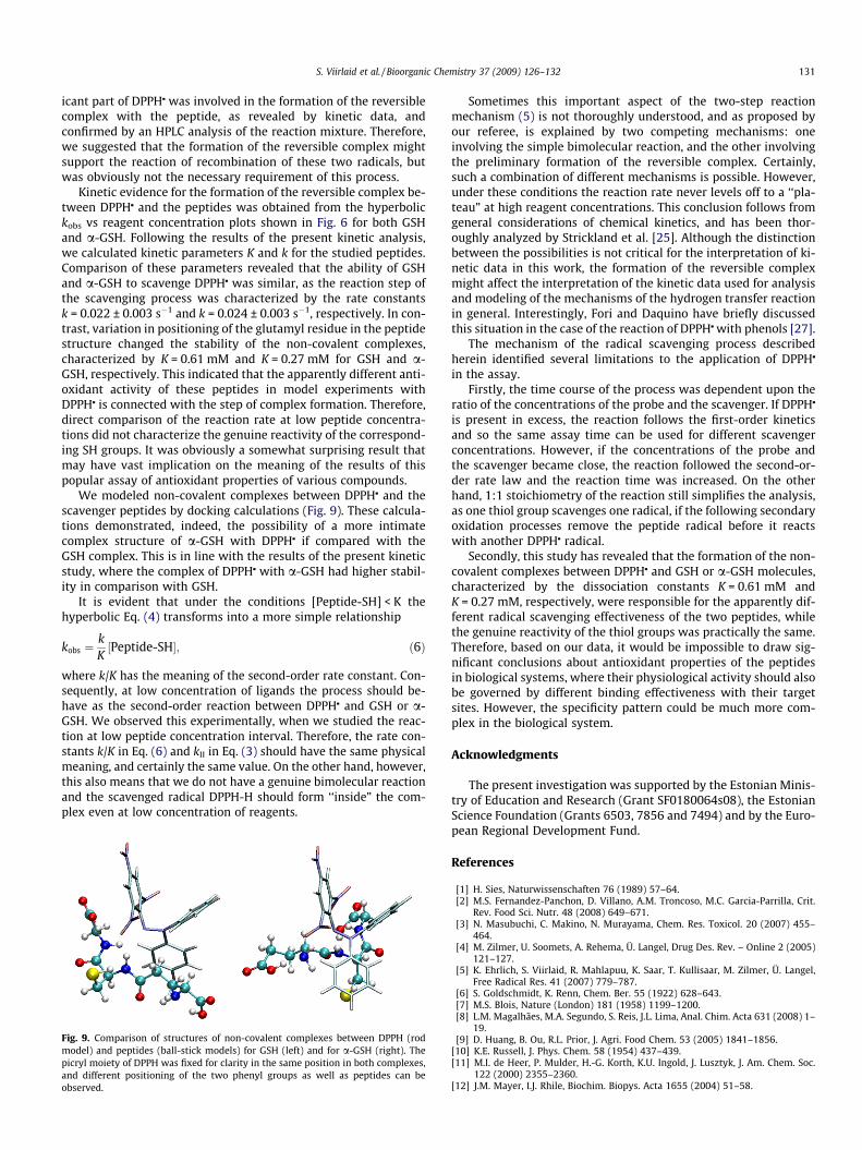
We modeled non-covalent complexes between DPPHj and thescavenger peptides by docking calculations (Fig. 9). These calcula-tions demonstrated, indeed, the possibility of a more intimatecomplex structure of a-GSH with DPPHj if compared with theGSH complex. This is in line with the results of the present kineticstudy, where the complex of DPPHj with a-GSH had higher stabil-ity in comparison with GSH.
It is evident that under the conditions [Peptide-SH] < K thehyperbolic Eq. (4) transforms into a more simple relationship
kobs ¼kK½Peptide-SH�; ð6Þ
where k/K has the meaning of the second-order rate constant. Con-sequently, at low concentration of ligands the process should be-have as the second-order reaction between DPPHj and GSH or a-GSH. We observed this experimentally, when we studied the reac-tion at low peptide concentration interval. Therefore, the rate con-stants k/K in Eq. (6) and kII in Eq. (3) should have the same physicalmeaning, and certainly the same value. On the other hand, however,this also means that we do not have a genuine bimolecular reactionand the scavenged radical DPPH-H should form ‘‘inside” the com-plex even at low concentration of reagents.
Fig. 9. Comparison of structures of non-covalent complexes between DPPH (rodmodel) and peptides (ball-stick models) for GSH (left) and for a-GSH (right). Thepicryl moiety of DPPH was fixed for clarity in the same position in both complexes,and different positioning of the two phenyl groups as well as peptides can beobserved.
Sometimes this important aspect of the two-step reactionmechanism (5) is not thoroughly understood, and as proposed byour referee, is explained by two competing mechanisms: oneinvolving the simple bimolecular reaction, and the other involvingthe preliminary formation of the reversible complex. Certainly,such a combination of different mechanisms is possible. However,under these conditions the reaction rate never levels off to a ‘‘pla-teau” at high reagent concentrations. This conclusion follows fromgeneral considerations of chemical kinetics, and has been thor-oughly analyzed by Strickland et al. [25]. Although the distinctionbetween the possibilities is not critical for the interpretation of ki-netic data in this work, the formation of the reversible complexmight affect the interpretation of the kinetic data used for analysisand modeling of the mechanisms of the hydrogen transfer reactionin general. Interestingly, Fori and Daquino have briefly discussedthis situation in the case of the reaction of DPPHj with phenols [27].
The mechanism of the radical scavenging process describedherein identified several limitations to the application of DPPHj
in the assay.Firstly, the time course of the process was dependent upon the
ratio of the concentrations of the probe and the scavenger. If DPPHj
is present in excess, the reaction follows the first-order kineticsand so the same assay time can be used for different scavengerconcentrations. However, if the concentrations of the probe andthe scavenger became close, the reaction followed the second-or-der rate law and the reaction time was increased. On the otherhand, 1:1 stoichiometry of the reaction still simplifies the analysis,as one thiol group scavenges one radical, if the following secondaryoxidation processes remove the peptide radical before it reactswith another DPPHj radical.
Secondly, this study has revealed that the formation of the non-covalent complexes between DPPHj and GSH or a-GSH molecules,characterized by the dissociation constants K = 0.61 mM andK = 0.27 mM, respectively, were responsible for the apparently dif-ferent radical scavenging effectiveness of the two peptides, whilethe genuine reactivity of the thiol groups was practically the same.Therefore, based on our data, it would be impossible to draw sig-nificant conclusions about antioxidant properties of the peptidesin biological systems, where their physiological activity should alsobe governed by different binding effectiveness with their targetsites. However, the specificity pattern could be much more com-plex in the biological system.
Acknowledgments
The present investigation was supported by the Estonian Minis-try of Education and Research (Grant SF0180064s08), the EstonianScience Foundation (Grants 6503, 7856 and 7494) and by the Euro-pean Regional Development Fund.
References
[1] H. Sies, Naturwissenschaften 76 (1989) 57–64.[2] M.S. Fernandez-Panchon, D. Villano, A.M. Troncoso, M.C. Garcia-Parrilla, Crit.
Rev. Food Sci. Nutr. 48 (2008) 649–671.[3] N. Masubuchi, C. Makino, N. Murayama, Chem. Res. Toxicol. 20 (2007) 455–
464.[4] M. Zilmer, U. Soomets, A. Rehema, Ü. Langel, Drug Des. Rev. – Online 2 (2005)
121–127.[5] K. Ehrlich, S. Viirlaid, R. Mahlapuu, K. Saar, T. Kullisaar, M. Zilmer, Ü. Langel,
Free Radical Res. 41 (2007) 779–787.[6] S. Goldschmidt, K. Renn, Chem. Ber. 55 (1922) 628–643.[7] M.S. Blois, Nature (London) 181 (1958) 1199–1200.[8] L.M. Magalhães, M.A. Segundo, S. Reis, J.L. Lima, Anal. Chim. Acta 631 (2008) 1–
19.[9] D. Huang, B. Ou, R.L. Prior, J. Agri. Food Chem. 53 (2005) 1841–1856.
[10] K.E. Russell, J. Phys. Chem. 58 (1954) 437–439.[11] M.I. de Heer, P. Mulder, H.-G. Korth, K.U. Ingold, J. Lusztyk, J. Am. Chem. Soc.
122 (2000) 2355–2360.[12] J.M. Mayer, I.J. Rhile, Biochim. Biopys. Acta 1655 (2004) 51–58.

132 S. Viirlaid et al. / Bioorganic Chemistry 37 (2009) 126–132
[13] G. Litwinenko, K.U. Ingold, J. Org. Chem. 70 (2005) 8982–8990.[14] E. Baciocchi, A. Calcagni, O. Lanzalunga, J. Org. Chem. 73 (2008) 4110–4115.[15] M.C. Foti, C. Daquino, I.D. Mackie, G.A. Dilabio, K.U. Ingold, J. Org. Chem. 73
(2008) 9270–9282.[16] A. Rauk, D. Yu, D.A. Armstrong, J. Am. Chem. Soc. 120 (1998) 8848–8855.[17] P.C. do Couto, B.J.C. Cabral, J.A.M. Simões, Chem. Phys. Lett. 421 (2006) 504–
507.[18] U. Soomets, M. Zilmer, Ü. Langel, Meth. Mol. Biol. 298 (2005) 241–257.[19] R.E. Benesch, R. Benesh, J. Am. Chem. Soc. 77 (1955) 5877–5881.[20] T. Kortemme, T.E. Creighton, J. Mol. Biol. 253 (1995) 799–812.[21] J. Bjerrum, G. Schwarzenbach, L.G. Sillen, Stability Constants, Part I, Organic
Ligands, The Chemical Society, London, 1957.
[22] M.A. Rosei, Cell Mol. Life Sci. 35 (1979) 1178–1179.[23] A.G. Brook, R.J. Anderson, J.T. Van Patot, Can. J. Chem. 36 (1958) 159–166.[24] J. Takebayashi, A. Tai, E. Gohda, I. Yamamoto, Biol. Pharm. Bull. 4 (2006) 766–
771.[25] S. Strickland, G. Palmer, V. Massey, J. Biol. Chem. 250 (1975) 4048–4052.[26] C.M. Dieckhaus, C.L. Fernández-Metzler, R. King, P.H. Krolikowski, T.A. Baillie,
Chem. Res. Toxicol. 4 (2005) 630–638.[27] M.C. Foti, C. Daquino, Chem. Commun. (Camb) 39 (2006) 3252–3254.[28] L.R. Mahoney, G.D. Mendenhall, K.U. Ingold, J. Am. Chem. Soc. 95 (1973) 8610–
8614.[29] P. Eyer, Environ. Health Perspect. 102 (S6) (1994) 123–132.[30] J. Flood, K.E. Russell, Can. J. Chem. 53 (1975) 1123–1128.