multiple intracranial tumors in philadelphia chromosome-positive acute lymphoblastic leukemia
TRANSCRIPT
IMAGES IN HEMATOLOGY
Multiple intracranial tumors in Philadelphiachromosome-positive acute lymphoblastic leukemia
Naoki Hatakeyama • Tsukasa Hori • Masaki Yamamoto •
Natsuko Inazawa • Keita Igarashi • Hiroyuki Tsutsumi •
Nobuhiro Suzuki
Received: 1 October 2012 / Revised: 22 January 2013 / Accepted: 22 January 2013 / Published online: 24 March 2013
� The Japanese Society of Hematology 2013
Keywords Acute lymphoblastic leukemia � Intracranial
tumors � Philadelphia chromosome
A three-year-old female presented with prolonged fever
and petechiae. Her initial complete blood workup showed a
white blood cell (WBC) count of 159 9 109/L with 92 %
lymphoblasts, hemoglobin of 9.3 g/dL, and a platelet count
of 22 9 109/L. Marked elevations of C-reactive protein
(CRP) and lactate dehydrogenase were identified.
The immunophenotype of the blast cells was CD101,
CD191, HLA-DR?, smIg-, and smIg j=k�, which is
compatible with a diagnosis of precursor B cell lympho-
blastic leukemia (pre-B ALL). CD66c expression was also
detected, so we confirmed the bcr/abl rearrangement by
fluorescence in situ hybridization analysis, and used
transcriptase-polymerase chain reaction analysis to detect
the minor bcr/abl fusion gene transcript. Given these
results, the patient was diagnosed with Philadelphia chro-
mosome-positive acute lymphoblastic leukemia (Ph?
ALL). By conventional cytogenetic analysis of bone mar-
row and peripheral blood cells at the onset, abnormalities
of 9p-q? and 22q- were suggested, but the complete
karyotype of this case could not be confirmed at the time;
however, the karyotype was later revealed at her first relapse to
be 46, XX, dup(1)(q25q32), add(1)(q32), del(3)(q2?),
der(9)t(9;22)(q34;q11)del(9)(p13), del(14)(13).
As she presented with severe hyperleukocytosis, she
received intravenous hyperhydration and prednisone at low
doses for the purpose of cytoreduction until the beginning
of remission induction therapy. A broad-spectrum anti-
bacterial agent and fluconazole were also administered
intravenously against her suspected systemic infection.
However, her blood culture was negative, and her serum
b-D glucan was not elevated.
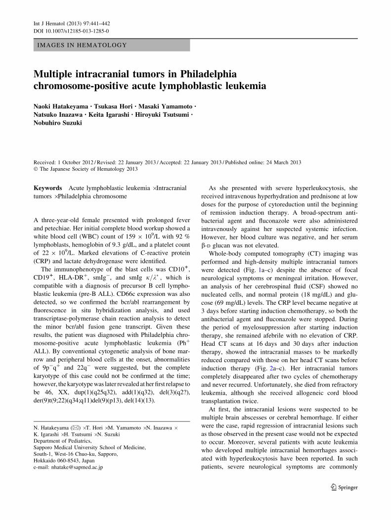
Whole-body computed tomography (CT) imaging was
performed and high-density multiple intracranial tumors
were detected (Fig. 1a–c) despite the absence of focal
neurological symptoms or meningeal irritation. However,
an analysis of her cerebrospinal fluid (CSF) showed no
nucleated cells, and normal protein (18 mg/dL) and glu-
cose (69 mg/dL) levels. The CRP level became negative at
3 days before starting induction chemotherapy, so both the
antibacterial agent and fluconazole were stopped. During
the period of myelosuppression after starting induction
therapy, she remained afebrile with no elevation of CRP.
Head CT scans at 16 days and 30 days after induction
therapy, showed the intracranial masses to be markedly
reduced compared with those on her head CT scans before
induction therapy (Fig. 2a–c). Her intracranial tumors
completely disappeared after two cycles of chemotherapy
and never recurred. Unfortunately, she died from refractory
leukemia, although she received allogeneic cord blood
transplantation twice.
At first, the intracranial lesions were suspected to be
multiple brain abscesses or cerebral hemorrhage. If either
were the case, rapid regression of intracranial lesions such
as those observed in the present case would not be expected
to occur. Moreover, several patients with acute leukemia
who developed multiple intracranial hemorrhages associ-
ated with hyperleukocytosis have been reported. In such
patients, severe neurological symptoms are commonly
N. Hatakeyama (&) � T. Hori � M. Yamamoto � N. Inazawa �K. Igarashi � H. Tsutsumi � N. Suzuki
Department of Pediatrics,
Sapporo Medical University School of Medicine,
South-1, West-16 Chuo-ku, Sapporo,
Hokkaido 060-8543, Japan
e-mail: [email protected]
123
Int J Hematol (2013) 97:441–442
DOI 10.1007/s12185-013-1285-0

observed and fatal outcomes are not uncommon if adequate
and aggressive therapies, including neurosurgery treat-
ments, are not received as rapidly as possible. For these
reasons, the multiple intracranial tumors in our case were
regarded as originating from Ph? ALL cells.
Extramedullary tumors occur less commonly in pre-B
ALL, and those in intracranial sites are extremely rare. To
our knowledge, other than the present case, there has only
been one reported case of multiple intracranial tumors at
diagnosis of pre-B ALL [1]. Interestingly, both of these
two cases were diagnosed with Ph? ALL. We suggest that
due to certain biological properties, Ph? ALL cells may
tend to invade into the brain parenchyma and develop
multiple intracranial tumors.
Reference
1. Cuvelier GD, Vitali AM, Ford JC, et al. Multiple intracranial
tumors in Philadelphia chromosome positive acute lymphoblastic
leukemia: successful treatment following aggressive supportive
care, early cranial radiation, high dose chemotherapy and imatinib.
Pediatr Blood Cancer. 2008;51:135–7.
Fig. 1 Non-contrast computed tomography of head. Multiple intracranial tumors were demonstrated (a–c)
Fig. 2 Computed tomography images at the same lesion before
induction therapy (a), at day 16 (b) and at day 30 (c) after the
beginning of induction therapy. The intracranial tumor was markedly
and rapidly reduced
442 N. Hatakeyama et al.
123