dspace.jaist.ac.jpof chemistry, biology, and physics. taking the large research field of...
TRANSCRIPT

Japan Advanced Institute of Science and Technology
JAIST Repositoryhttps://dspace.jaist.ac.jp/
Titleα-シクロデキストリンの包接錯体からなる分解性ポリ
ロタキサンの分子設計
Author(s) 辛, 昊俊
Citation
Issue Date 2014-03
Type Thesis or Dissertation
Text version ETD
URL http://hdl.handle.net/10119/12094
Rights
DescriptionSupervisor:金子 達雄, マテリアルサイエンス研究科
, 博士

Molecular Design of Degradable Polyrotaxane Composed of α-Cyclodextrin Inclusion
Complexes
HOJOON SHIN
Japan Advanced Institute of Science and Technology

Doctoral Dissertation
Molecular Design of Degradable Polyrotaxane Composed of α-Cyclodextrin Inclusion
Complexes
HOJOON SHIN
Supervisor : Associate Professor Tatsuo Kaneko
School of Materials Science
Japan Advanced Institute of Science and Technology
March 2014

II
ABSTRACT
Polyrotaxanes, demonstrated as a molecular necklace composed of a polymeric chain and cyclic compounds,
are interesting in terms of their structural features. A mechanism of biodegradation based on the dissociation of
supramolecular structure triggered by cleavage of end groups in a cleavable polyrotaxane can give various novel
designs of biodegradable polymers. By utilizing these merits of supramolecular structure, we designed and
prepared polyrotaxane hydrogel based on water-soluble hydrolyzable polyrotaxane and polyamide rotaxane
composed of photo-cleavable 4,4’-diacetoamido-α-truxillic acid.
In chapter 2, a hydrolyzable polyrotaxane composed of an ester-containing poly(ethylene glycol) chain and α-
cyclodextrins was prepared and showed gradual degradation into its water-soluble components in aqueous
conditions, based on the dissociation of the polyrotaxane triggered by the hydrolysis of the ester groups.
In chapter 3, we designed and prepared various hydrolyzable polyrotaxane hydrogel composed of α-
cyclodextrins, poly(ethylene glycol) with Mw 3K and 20K as back bone chain, and two types of crosslinker
(liner and multi-arm PEG). One of the degradable polyrotaxanes was crosslinked between the internal rings with
linear PEG chain and the other was crosslinked between the terminals with multi-arm PEG chain. Every
hydrogels had good water content (>90%) and hydrophilic surface. The internally-crosslinked gels had higher
compressive stress and initial modulus, while the terminally-crosslinked gels showed higher ultimate strain.
These results imply that terminally-crosslinked gels were rigid and the terminally-crosslinked gels were flexible.
In stress relaxation test, the internally-crosslinked gels showed unique viscoelastic behavior, in which was
similar to that of topological gel. We investigated the gradual degradation of the hydrogels in terms of changes
in mass and storage modulus of the gels. The internally-crosslinked gels degraded and disappeared in a relatively
shorter period than the terminally-crosslinked gels. The terminally-crosslinked gels survived for weeks with
gradual degradation that allowed us to apply the degrading gels to a preliminary cell adhesion test. In the cell
adhesion test, the number, size, and morphology of NIH 3T3 cells was changed by degradation of hydrogels and
change of surface chemical characteristics due to cleavage of carboxyl residue introduced to hydroxyl group in
CD at the same time.
In chapter 4, we introduced a unique chemical structure, polyrotaxane, to give structural rigidity to polymer
back bones using a necklace-like structure and cinnamic acid as a photo-reactive monomer to prepare a new
polyamide. By utilizing this unique concept, we designed aliphatic-aromatic polyamides containing the rotaxane
structure composed of 4,4’-diacetoamido-α-truxillic acid, poly(ethylene glycol) bisamine (PEGBA) and
methylated-α-cyclodextrin (Me-α-CD), and observed their thermo-mechanical performance and degradation
behavior under UV-irradiation. The thermal degradation temperature of the polyamides was enhanced by
inclusion complex formation with Me-α-CD, and the polyamide polyrotaxane was degraded by UV-irradiation
via the photocleavage of the cyclobutane ring in 4,4’-diacetoamido-α-truxillic acid.
Keywords : hydrogel, supramolecular structure, hydrolyzable polyrotaxane, polyamide, photocleavage

III
TABLE OF CONTENTS
Chapter 1. General Introduction 1
1-1. Supramolecular chemistry 2
1-1-1. Supramolecular chemistry 2
1-1-2. Host-guest chemistry 5
1-2. Polyrotaxanes 7
1-2-1. Cyclodextrin (CD) 7
1-2-2. Backgrounds and basic characteristics 10
1-2-3. Water-soluble polyrotaxane 15
1-2-4. Biodegradable polyrotaxane 17
1-2-5. Multivalent interaction 19
1-2-6. Hydrogel based on polyrotaxane. 21
1-3. Aim and outline of this dissertation 23
Reference 24
Chapter 2. Preparation of Hydrolyzable polyrotaxane Containing Ester
Linkages
28
2-1. Introduction 29
2-2. Experimental section 31
2-2-1. Materials 31
2-2-2. Preparation of polyrotaxane 1 and 4 31
2-2-3. Hydrolysis of polyrotaxane 1, 4, and pseudopolyrotaxane 2 37

IV
2-3. Results and discussion 38
2-4. Conclusion 43
Reference and notes 44
Supporting information 46
Chapter 3. Molecular Design of Degradable Polyrotaxane Hydrogels for
Tissue Engineering
50
3-1. Introduction 51
3-2. Experimental section 55
3-2-1. Materials 55
3-2-2. Preparation of polyrotaxanes hydrogels I-IV 56
3-2-3. Measurement 68
3-2-3-1. water content 68
3-2-3-2. Static contact angle using air-bubble (in wet condition) 68
3-2-3-3. Compression test 69
3-2-3-4. Stress relaxation test 69
3-2-3-5. Curve fitting of stress relaxation curve 70
3-2-3-6. Changes in mass and storage modules upon degradation 71
3-2-3-7. Quantification of the amount of fibronectin adhered on the
hydrogel
72
3-2-3-8. Quantification of the amount and size of cells adhered on the
hydrogel upon degradation
72
3-3. Results and discussion 74

V
3-3-1. Design and preparation of ester-containing polyrotaxanes 1-4
for gelation
74
3-3-2. Design and preparation of degradable hydrogels I-IV 78
3-3-3. Mechanical properties of degradable hydrogels I-IV 81
3-3-4. Degradation behavior of hydrogels I-IV 86
3-3-5. Cell adhesion on hydrogels I-IV 88
3-4. Conclusion 94
Reference 95
Chapter 4. Synthesis of Photo-degradable Polyamide Rotaxanes from
Cinnamate Photodimer and α-Cyclodextrin
99
4-1. Introduction 100
4-2. Experimental section 102
4-2-1. Materials 102
4-2-2. Synthesis of polymer 103
4-2-3. Measurement 107
4-2-4. UV degradation 107
4-3. Results and discussion 108
4-3-1. Synthesis and Characterization of Polyamides 4 108
4-3-2. Thermal properties of 4 113
4-3-3. UV-degradation 116
4-4. Conclusion 119
Reference 120

VI
Chapter 5. General Conclusion 122
Appendices 126
Acknowledgement 128
Minor-Research Theme 130

Chapter 1 General Introduction

1. General Introduction
2
1 General Introduction
1-1. Supramolecular chemistry
1-1-1. Supramolecular chemistry1-3
Supramolecular chemistry is concerned with the study of the basic feature of molecular
nitration and with their implementation in designed non-natural systems. In this chapter,
introductory descriptions are presented on the basis of reviews and books.
Figure 1. Comparison between the scope of molecular and supramolecular chemistry
according to Lehn.

1. General Introduction
3
For more than150 years, molecular chemistry has developed a vast array of highly
sophisticated and powerful methods for the construction of more complex molecular structure
by the making or breaking of covalent bonds with atoms in a controlled and precise fashion.
The time has come to do the same for non-covalent intermolecular forces. Just as there is a
field of molecular chemistry based on the covalent bond, and there is a field of
supramolecular chemistry, the chemistry of molecular assembles and of the intermolecular
bond. It is “chemistry beyond the molecule,” whose objects are supramolecular entities,
supramolecules processing feature as well defined as those of molecules themselves. This
kind of definitions is highlighted in Figure 1, which illustrated the relationship between
molecular and supramolecular chemistry in terms of both structures and function.
Such labels, while helpful, are by their nature noncomprehensive and there are many
exceptions if these definitions are taken too literally. The problem may be linked to the
definition of organometallic chemistry as ‘the chemistry of compounds with metal-to-carbon
bonds’. This immediately rules out Wilkinson’s compound, RhCl(PPh3)3, for example, which
is one of the most important industrial catalysts for organometallic transformations known in
the filed. Indeed, it is often the objectives and thought processes of the chemist undertaking
the work, as much as the work itself, which determine its field. The rapid expansion in
supramolecular chemistry over the past 15 years has resulted in an enormous diversity of

1. General Introduction
4
chemical systems, both designed and accidentally stumbled upon, which may lay some claim,
either in concept, origin or nature, to being supramolecular. In particular, workers in the filed
of supramolecular photochemistry have chosen to adopt a rather different definition of a
supramolecular compound as a group of molecular components that contribute properties that
each component processes individually to the whole assembly (covalent or noncovalent).
Thus an entirely covalent molecule comprising, for example, a chromophore (light-absorbing
moiety), spacer and redox centre might be thought of as supramolecular because the
chromophore and redox centre are able to absorb light, or change oxidation state, whether
they form part of the supramolecule or not. Similarly, much recent work has focused on the
development of self-assemble using a variety of interactions, some of which are clearly
noncovalent (e.g. hydrogen bonds) and some of which process a significant covalent
compound (e.g. metal-ligand interactions).
Supramolecular chemistry is an interdisciplinary field of science that covers chemical,
physical, and biological features by means of intermolecular (non-covalent) binding
interactions. Its roots extend into synthetic procedures for molecular construction, into
biological process that all start with substrate binding and recognition, and into the
mechanical properties of solids. A major feature is the range of perspectives offered by the
cross fertilization of supramolecular chemical research owing to its location at the intersection

1. General Introduction
5
of chemistry, biology, and physics. Taking the large research field of supramolecular
chemistry into account, it can be called a supramolecular science. Such wide horizons are a
challenge and a stimulus to the creative imagination of the chemist.
1-1-2. Host-guest chemistry
In supramolecular chemistry, host-guest chemistry describes complexes that are composed
of two or more molecules or ions held together in unique structural relationships by hydrogen
bonding or by ion pairing or by van der Waals force other than those of full covalent bonds.
The host component is defined as an organic molecule or ion whose binding sites converge in
the complex. In 1967, Pederson reported the synthesis of crown ether (Figure 2).4 The crown
ether is a cyclic oligoether and it can selectively form an inclusion complex with a cationic
ion in relation to its ring size. The cyclic molecule is called the host molecules. The guest
component is defined as any molecule or ion whose binding sites diverge in the complex.
Many artificial cyclic molecules have been researched and there artificial host molecules
were designed to interact with the guest molecule by the electrostatic interaction, π-π
interaction and charge-transfer interaction.5

1. General Introduction
6
O
O
O
O
O
O
O
O
O
O
O
O
(a) (b)
Figure 2. Schematic diagram of crown ether derivatives.4

1. General Introduction
7
1-2. Polyrotaxanes
1-2-1. Cyclodextrin (CD)6
Cyclodextrins, as they are known today, were called "cellulosine" when first described by A.
Villiers in 1891. Soon after, F. Schardinger identified the three naturally occurring
cyclodextrins -α, -β, and -γ. These compounds were therefore referred to as "Schardinger
sugars". For 25 years, between 1911 and 1935, Pringsheim in Germany was the leading
researcher in this area, demonstrating that cyclodextrins formed stable aqueous complexes
with many other chemicals. By the mid 1970's, each of the natural cyclodextrins had been
structurally and chemically characterized and many more complexes had been studied. Since
the 1970s, extensive work has been conducted by Szejtli and others exploring encapsulation
by cyclodextrins and their derivatives for industrial and pharmacologic applications.
Typical cyclodextrins are constituted by 6-8 glucopyranoside units (Figure 3), can be
topologically represented as toroids with the larger and the smaller openings of the toroid
exposing to the solvent secondary and primary hydroxyl groups respectively. Because of this
arrangement, the interior of the toroids is not hydrophobic, but considerably less hydrophilic
than the aqueous environment and thus able to host other hydrophobic molecules. In contrast,

1. General Introduction
8
the exterior is sufficiently hydrophilic to impart cyclodextrins (or their complexes) water
solubility. The characteristics of the CDs are summarized in Table 1.
Figure 3. Schematic diagram of α-, β-, γ-CDs
The formation of the inclusion compounds greatly modifies the physical and chemical
properties of the guest molecule, mostly in terms of water solubility. This is the reason why
cyclodextrins have attracted much interest in many fields, especially pharmaceutical
applications: because inclusion compounds of cyclodextrins with hydrophobic molecules are
able to penetrate body tissues, these can be used to release biologically active compounds
under specific conditions. In most cases the mechanism of controlled degradation of such
complexes is based on pH change of water solutions, leading to the cleavage of hydrogen or
ionic bonds between the host and the guest molecules. Alternative means for the disruption of
the complexes take advantage of heating or action of enzymes able to cleave α-1,4 linkages
between glucose monomers.

1. General Introduction
9
Table 1. Characteristics of α-, β-, γ-CDs
α β γ
No. of glucose units 6 7 8
Molecular weight 972 1135 1297
Solubility in water. g/L 145 18.5 232
Cavity diameter, Å 4.7-5.3 6.0-6.5 7.5-8.3
Height of torus, Å 7.9±0.1 7.9±0.1 7.9±0.1
Diameter of outer periphery, Å 14.6±0.4 15.4±0.4 17.5±0.4
Approx volume of cavity, Å3 174 262 427
Crystal water, wt% 10.2 13.2-14.5 8.1-17.7
Diffusion constant at 40℃ 3.44 3.22 3.00

1. General Introduction
10
1-2-2. Backgrounds and basic characteristics
Supramolecular assemblies of cyclic molecules and linear molecules were demonstrated in
1967.7 The name is the rotaxane comes from the Latin words for wheel and axle. Schill and
his co-workers developed a synthetic procedure strategy.7-9 They reported that cyclic space of
24 skeletal C, O or N atoms were requested in order to provide a cavity size enough to be
threaded onto a polymer chain 8) and the relationship between ring size, linear molecular
length, blocking size of terminal molecules, and the threading efficiency.9 The application of
host-guest chemistry to the synthesis of rotaxane and catenanes was studied by Sauvage et al.
and Stoddart et al..10,11
Polyrotaxanes are polymers with a novel molecular architecture where cyclic molecules are
threaded onto the polymer chain. The bulky end-groups of polyrotaxane capped linear
polymer and prevent the escape of cyclic molecules from the assembly by dethreading. There
are in principle many subclasses of polyrotaxanes which differ in the nature and location of
the covalent and physical linkages. Since polyrotaxanes are a novel class of polymer
molecular topology it is anticipated that their properties are distinct from other polymeric
architectures. The distinguishing feature of polyrotaxanes is the potential for lateral and
translational motion of the cyclic molecules relative to the linear chain that penetrates it.

1. General Introduction
11
In 1976, Ogata and co-workers studied the formation of poly(amide / rotaxanes) using the
reaction of β-cyclodextrin complexation of α, ω-diaminohydrocabons with diacid chlorides.12
This paper was the first one related to synthesis and characterization of polyrotaxane. They
called this polymer to “tunnel polymer”. Tunnel polymer showed specific viscosity. The
polyrotaxane were soluble in dipolar aprotic solvent while the parent polymers were not.
Further, the polyrotaxane did not show melting points, in contrast to the parent backbone
polymer. These characteristics were not taken into account in the case of low molecular
weight rotaxanes.
Wenz and co-workers reported inclusion complexation consisting α-CD and
poly(iminooligomethylene)s.13-15 Inclusion complex carried out by Wenz et al. was water
soluble in acidic condition. Hydrophobic interaction between cyclodextrin and
oligomethylene spacer was considered to play an important role in inclusion complex.
Harada and his co-workers have reported the first different type of pseudo- and
polyrotaxane based on complex formation between CDs and PEG16-23 and then they have
reported preparation of pseudo- and polyrotaxane with CDs and the various polymers, such as
poly(isobutylene),20 poly(ε-caporolactone) (PCL),21 poly(dimethyl siloxane).22 In these
researches, they reported that the size of CD cavity and the cross-section area of the polymer
chain were related to the interaction between CD cavity and the polymer chain (Figure 4).

1. General Introduction
12
Figure 4. Crystal structure of inclusion complexes of β-CDs with poly(trimethylene oxide).
Intramolecular hydrogen bonds are shown as dotted lines. Solvent molecules which form
direct hydrogen bonds with β-CDs are as open circles with hydrogen bonds.
They confirmed that 6-cynnamoyl β-CD forms a cyclic daisy-chain pseudopolyrotaxane
and prepared a cyclic tri[2]-rotaxane (daisy-chain necklace) by attaching bulky stopper.24
They reported the formation of daisy-chain pseudopolyrotaxane from the bi-functional host
and guest molecules (Figure 5).25

1. General Introduction
13
Figure 5. Supramolecular polymer consisting for bi-functional host and guest molecules.25
Some researchers have reported that inclusion complexation between ABA triblock
copolymer and α-, β-, and γ-CDs, respectively. Tonelli and coworker prepared α- and γ-CD
complexes using PEG-PCL-PEG ABA triblock copolymer in order to research the behavior of
isolated and segregated polymer chain. They showed that sided-by-side chains could reside in
the γ-CD channel as only a single chain can be incorporated inside the α-CD cavity.26
Yui and his coworkers prepared stimuli-responsive polyrotaxane composed of α-, and β-
CDs and ABA triblock copolymers.27-29 Temperature-sensitive polyrotaxane27,28 was designed
by complexation between β-CDs and PEG-PPG-PEG triblock. It showed that majority of β-
CDs on PEG-PPG- PEG triblock copolymer moved toward the PPG segment with increasing

1. General Introduction
14
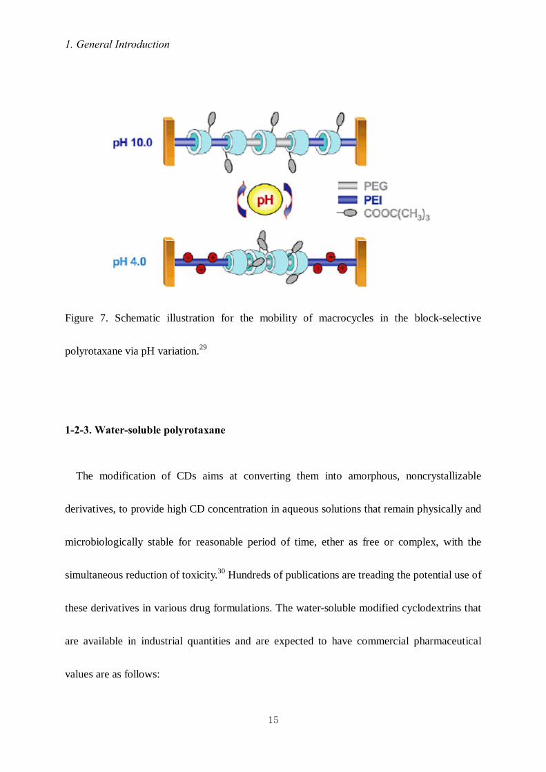
temperature although some β-CDs might reside on the PEG segment (Figure 6). pH-sensitive
polyrotaxane29 was prepared by utilizing α-CDs and PEI-PEG-PEI triblock copolymer. As
shown in Figure 7, α-CDs in the water-soluble polyrotaxane showed pH-dependent movement
along PEI and PEG block.
Figure 6. Schematic diagram of stimuli-responsive polyrotaxane composed of β- CDs and
PEG-PPG-PEG triblock copolymer.27
1. General Introduction
15
Figure 7. Schematic illustration for the mobility of macrocycles in the block-selective
polyrotaxane via pH variation.29
1-2-3. Water-soluble polyrotaxane
The modification of CDs aims at converting them into amorphous, noncrystallizable
derivatives, to provide high CD concentration in aqueous solutions that remain physically and
microbiologically stable for reasonable period of time, ether as free or complex, with the
simultaneous reduction of toxicity.30 Hundreds of publications are treading the potential use of
these derivatives in various drug formulations. The water-soluble modified cyclodextrins that
are available in industrial quantities and are expected to have commercial pharmaceutical
values are as follows:

1. General Introduction
16
(1) Methylated derivatives of β-CD31
(2) 2-hydroxypropylated β- and γ-CD32
(3) Branched CDs (glucosyl- and maltosyl-β-CDs)
(4) Acetylated β- and γ-CD
(5) Sulfated CDs33
Utilizing previous methods, some researchers have prepared water-soluble polyrotaxane by
modifying CDs.
Yui and coworkers prepared some types of water-soluble polyrotaxane. They increased the
solubility of polyrotaxane in aqueous solution by elimination the hydrogen bonding between
the α-CDs by introducing tert-butoxy groups (Figure 8).29 A carboxyethylester-polyrotaxane,
introducing carboxyethylester (CEE) to OH-group of CDs, was prepared as a novel calcium
chelating polymer in the field of oral drug delivery (Figure 9).34,35
Figure 8. Butoxy-PRx-Tyr composed of PEI-PEG-PEI copolymer and α-CDs.29

1. General Introduction
17
Figure 9. Carboxyethylester-polyrotxane.34
1-2-4. Biodegradable polyrotaxane
Since the later half of 1990s, Yui et al. have demonstrated the feasibility of the
biodegradable polyrotaxanes, in which a number of α-CDs are threaded onto a linear chain
capped with bulky end-groups via biodegradable linkages.36-40 They reported the enzymatic
degradable polyrotaxane as gene delivery system and hydrolyzable polyrotaxane as polymer
scaffold in tissue engineering by using bio-cleavable groups (Figure 10).
Biodegradable polyrotaxane has some attractive characteristics comparing to other

1. General Introduction
18
biodegradable polymers, such as PGA, PLAG, PLLA and PCL. Biodegradable polyrotaxane
was composed of α-CDs, backbone polymer (such as PEG), and end-capping materials via
bio-cleavable groups such as SS and ester linkage. By cleavage of SS or ester linkage,
supramolecular structure is dissociated and then polyrotaxane degraded perfectly. The other
characteristics involve; (Ⅰ) high crystallinity due to intermolecular hydrogel bonds between
hydroxyl groups of CDs; and (Ⅱ) the position of cleavable groups. In the case of PLA, the
high crystallinity is based on well-arranged packing of the repeating units of ester groups.
This indicates that the crystallinity region involves almost all ester groups, which reduce
water intrusion. Moreover, cleavage of many ester-groups generates acidic byproduct. On the
other hands, the ester or SS groups of polyrotaxane are independent of the high crystalline
region. It is expected that the supramolecular dissociation may be achieved via terminal group
hydrolysis due to water intrusion in polyrotaxane and stable on pH condition due to a little
acidic byproduct.

1. General Introduction
19
Figure 10. Characteristic image of cleavable polyrotaxane. The supramolecular structure can
be dissociated by the terminal cleavage of capping groups by means of external stimuli.40
1-2-5. Multivalent interaction
The valency of a particle a small molecules, oligosaccharide, protein, nucleic acid, lipid or
aggregate of these molecules; a membrane or organelle; a virus, bacterium, or cell is the
number of separate connections of the same kind that it can form with other particles through
ligand -receptor interactions. The idea that many biological systems interact through multiple
simultaneous molecular contacts is familiar; it has, however, become of interactions involving
multiple protein and ligands has begun to be unraveled. The possibility that multiple

1. General Introduction
20
simultaneous interactions have unique collective properties that are qualitatively different
from properties displayed by their constituents, which interact monovalently, suggests new
strategies for the design of drugs and research reagents for biochemistry and biology.
Enhancing or blocking collective or multivalent interactions may benefit from strategies
fundamentally different from those used in monovalent molecular interactions.41
Ooya and Yui et al. have reported that ligand-introduced polyrotaxane enhance multivalent
interaction with complement binding protein due to both many lingnds and high mobility of
ligand-introduced α-CDs (Figure 11).35,42 They investigated how α-CDs and ligand mobility
in ligand-polyrotaxane conjugates affect the multivalent interaction with a binding protein.
Maltose and Concanavalin A (Con A) were selected as a ligand and a binding protein,
respectively, because Con A recognizes maltose. When the threading percentage of α-CDs
against the threading percentage was 38%, the highest mobility of maltosyl groups is
observed. And the maltose-polyrotaxane conjugate with a highly mobile nature maintains
water cluster structure. From these results, the combination of multiple copies of ligands and
their supramolecular mobility along the mechanically locked structure should contribute to
significant enhancement of the multivalent interaction due to a reduction of the special
mismatches of binding

1. General Introduction
21
Figure 11. The effect of “mobile” motion of the cyclic compounds in polyrotaxanes on
binding receptor proteins in a multivalent manner: Image of binging/dissociating equilibrium
a) between a ligand–polyrotaxane conjugate and receptor sites and b) between a ligand–
immobilized-polymer and receptor sites.40
1-2-6. Hydrogel based on polyrotaxane.
Some researchers have reported polyrotaxane gel based on supramolecular network.43,44
Supramolecular network is a network crosslinked by supramolecular structure.
Yui et al. prepared a polyrotaxane hydrogel crosslinked by PEG between CDs in the
polyrotaxane (Figure 12 (a)).43 This polyrotaxane hydrogel was consisted of hydrolyzable

1. General Introduction
22
polyrotaxane with hydrolyzable bulky end groups. Polyrotaxane dissociated in a few days by
the hydrolysis of end groups. However, it takes several months to degrade the polyrotaxane
hydrogel. They reported that degradation time could be controlled the molecular weigh of the
crosslinker.
Ito et al. prepared polyrotaxane gel by crosslinking the CDs in polyrotaxane (Figure 12
(b)).44 This hydrogel was called “topological gel”. This hydrogel was composed of large
molecular weight of back bone polymer in polyrotaxane. And the number of CDs in the
polyrotaxane is a little. Thus the cyclic compound can move along the polymer chain. In this
gel, polymer chains are neither covalently cross-linked like chemical gel, nor do they interact
like physical gel.
(a) (b)
Figure 12. Schematic diagram of the various polyrotaxane gels (a)43 and (b)44

1. General Introduction
23
1-3. Aim and outline of this dissertation
The thesis deals with design, synthesis and characterization of various degradable
polyrotaxanes; such as hydrolyzable hydrogel based on hydrolyzable polyrotaxane and
polyamide rotaxane composed of photo-cleavable monomer. The objectives of this thesis are
as follows;
I. To design and prepare hydrolyzable polyrotaxane containing ester-linkage.
II. To design and prepare two types of water-soluble hydrolyzable hydrogel containing
different crosslinking system. One is crosslinked by linear PEG between OH-groups of
CDs and the other is crosslinked by multi-arm PEG between terminal groups in
polyrotaxane.
III. To observe the characteristics of hydrolyzable polyrotaxane hydrogels, such as
mechanical properties, water content, degradation, and cell adhesion properties.
IV. To observe the thermal-mechanical performance and UV-degradation behavior of
photo-reactive polyrotaxane composed of aliphatic-aromatic polyamides as back bone
chain.
This dissertation consists of five chapters. Chapter 2 describes the preparation of

1. General Introduction
24
hydrolyzable polyrotaxanes containing ester-linkage in terminal group. Chapter 3 deals with
the preparation of water-soluble polyrotaxane containing ester-linkage in end-capping
molecules and two types of hydrogels with different crosslinking systems, respectively. These
developed structures of polyrotaxane would affect the gel formation and properties of
hydrogels. Chapter 4 shows synthesis of polyrotaxane composed of aliphatic-aromatic
polyamides and their thermo-mechanical performances and UV degradation behavior.
.
Reference
1. Steed, J. W.; Atwood, J. L. Supramolecular Chemistry, John Wiley & Sons, Ltd: Chichester,
2000, 2.
2. Lehn, J. -M. Science, 1993, 260, 1762.
3. Lehn, J. -M. Supramolecular chemistry, VCH, Weinhein, 1995.
4. Pederson, C. J. J. Am. Chem. Soc., 1967, 89, 7017.
5. Odashima, K.; Koga, K. Comprehensive supramolecular Chemistry, Elsevier Science, New
York, 1996, Vol.2, p143.
6. Szejtli, J. Chem. Rev. 1998, 98, 1743.

1. General Introduction
25
7. Lipatov, Y. S.; Lipatova, T. E.; Kosyanchuk, L. F. Adv. Polym. Sci., 1989, 88, 49.
8. Harrison, I. T.; Harrison, S.; J. Am. Chem. Soc., 1967, 89, 5723.
9. Agam, G.; Gravier, D.; Zikha, A. J. Am. Chem. Soc., 1976, 98, 5206.
10. Dietrich-Buchecker, C. O.; Sauvage J. –P. Chem. Rev., 1987, 87, 795.
11. Anelli, P. L.; Ashoton, P. R.; Ballardini, R.; Balzani, V.; Delgado, M.; Gandolfi, M. T.;
Goodnow, T. M.; Kaifer, A. E.; Philp, D.; Pietraszkiewicz, M.; Prodi, L.; Reddington, M.
V.; Slawin, A. M. Z.; Spencer, N.; Stoddart, J. F.; Vicent, C.; Williams, D. J. J. Am. Chem.
Soc. 1992. 114, 193.
12. Ogata, N.; Sanui, K.; Wada, J. J. Polym. Sci., Polym. Lett. Ed. 1976, 14, 459.
13. Wens, G.; Keller, B, Angew. Chem., Int. Ed. Engl., 1992, 31, 197.
14. Wens, G.; Keller, B, Macromol. Symp., 1994, 87, 11.
15. Meier, L. P.; Heule, M.; Caseri, W. R.; Shelden, R. A.; Suter, U. W. Macromolecules, 1996,
29, 718.
16. Harada, A.; Kamachi, M. Macromolecules, 1990, 23, 2821.
17. Harada, A.; Li, J.; Kamachi, M. Nature, 1992, 356, 325.

1. General Introduction
26
18. Harada, A.; Li, J.; Kamachi, M. Macromolecules, 1993, 26, 5698.
19. Harada, A.; Okada, M.; Li, J.; Kamachi, M. Macromolecules, 1995, 28, 8406.
20. Harada, A.; Suzuki, S.; Okada, M.; Kamachi, M. Macromolecules, 1996, 29, 5611.
21. Kawaguchi, Y.; Nishiyama, T.; Okada, M.; Kamachi, M.; Harada, A. Macromolecules,
2000, 33, 4472.
22. Okumura, H.; Okada, M.; Kawauchi, Y.; Harada, A. Macromolecules, 2000, 33, 4297.
23. Kamitori, S.; Matsukaka, O.; Kondo, S.; Muraoka, S.; Okuyama, K.; Noguchi, K.; Okada,
M.; Harada, A. Macromolecules, 2000, 33, 1500.
24. Hoshino, T.; Miyauchi, M.; Kawaguchi, Y,; Yamaguchi, H.; Harada, A. J. Am. Chem. Soc.
2000, 122, 9876.
25. Ogami, K.; Hoshino, T.; Miyauchi, M.; Kawaguchi, Y,; Harada, A. Polym. Prep. Jpn, 2001,
50, 1471.
26. Lu, J.; Shin, I. D.; Nojima, S.; Tonelli, A. E. Polymer, 2000, 41, 5871
27. Fujita, H.; Ooya, T.; Yui, N. Macromolecules, 1999, 32, 2534.
28. Fujita, H.; Ooya, T.; Yui, N. Macromol. Chem. Phys. 1999, 200, 706.
29. Choi, H. S.; Lee, S. C.; Yamamoto, K.; Yui, N. Macromolecules, 2005, 38, 9878.
30. Szente, L.; Szejtli, J. Adv. Drug Deliv. Rev., 1999, 36, 17.
31. Takeo, K.; Kuge, T. Starch / Starke, 1976, 287, 226.

1. General Introduction
27
32. Pitha, J,; Szabo, L.; Fales, H. Carbohydr. Res., 1987, 168, 191.
33. Folkman, J.; Weisz, P.; Joullie, M. Scinece, 1989, 243, 1490.
34. Ooya, T.; Eguchi, M.; Ozaki, A.; Yui, N. Int. J. Pharmaceutics, 2002, 242, 47.
35. Ooya, T.; Eguchi, M.; Yui, N. J. Am. Chem. Soc., 2003, 125, 13016.
36. Watanabe, J.; Ooya, T.; Yui, N. J. Biomater. Sci. Polym. Ed., 1999, 10, 1275.
37. Watanabe, J.; Ooya, T.; Yui, N. J. Chem. Lett., 1998, 1031
38. Ooya, T.; Yui, N. J. Control. Release, 1999, 58, 251.
39. Yamashita, A.; Yui, N.; Ooya, T.; Kano, A.; Maruyama, A.; Akita, A.; Kogure, K.;
Harashima, H. Nature Protocols, 2006, 1, 2861.
40. Yui, N.; Ooya, T. Chem. Eur. J. 2006, 12, 6730.
41. Mammen, M.; Choi, S. K.; Whitesides, G. N. Angew. Chem. Int. Ed. 1998. 37, 2755.
42. Ooya, T.; Utsunomiya, H.; Eguchi, M.; Yui, N. Bioconjug. Chem. 2005, 16, 62.
43. Ichi, T.; Watanabe, J.; Ooya, T.; Yui, N. Biomacromolecules, 2001, 2, 204.
44. Okumura, Y.; Ito, K. Adv. Mater., 2001, 13, 485.

Chapter 2 Preparation of Hydrolyzable polyrotaxane Containing Ester Linkages
Published in Chemistry Letters, 37, 2008, pp988-989

2. Preparation of Hydrolyzable Polyrotaxane Containing Ester Linkages
29
2 Preparation of Hydrolyzable Polyrotaxane
Containing Ester Linkages
2-1. Introduction
In the last several decades, biodegradable polymers have been studied as implantable
materials for cell growth and tissue engineering.1 These materials must satisfy various
requirements such as sufficient mechanical strength, non-toxicity, and bioinertness before and
after degradation for clinical use in a living body. In order to design and construct
biodegradable materials,2 some attention should be paid to the fact that the material has to
have the potential to degrade perfectly in appropriate conditions, and that undesirable
decomposition has to be avoided during the preparation and purification. It is of course
necessary to purify the materials aimed at a clinical use in a living body. In this context, our
approach for the design of biodegradable materials based on the structural features of
polyrotaxanes, and especially on their dissociation, is promising. Stimuli-responsive
biodegradable polyrotaxanes as shown in Scheme 1 would provide quite a new model as a
mode of biodegradation.3 Degradation based on the dissociation of polyrotaxanes, that is, the
transformation of the supramolecular materials with a high molecular weight into water-

2. Preparation of Hydrolyzable Polyrotaxane Containing Ester Linkages
30
soluble and bioinert components would be favorable in terms of effective degradation, low
toxicity, and biocompatibility in a living body.
A polyrotaxane composed of a PEG chain containing ester groups at both ends and α-
cyclodextrins (α-CDs) was designed and prepared as a candidate for biodegradable polymers,
in which the ester group(s) would be expected to hydrolyze to trigger the following
dissociation in response to pH. Thus, a successful preparation of the ester-containing
polyrotaxane in spite of the potential of degradation4 and its hydrolysis behavior were
demonstrated.
Scheme 1. Biodegradation based on dissociation of polyrotaxane triggered by stimuli-
responsive cleavage of biodegradable linkages.

2. Preparation of Hydrolyzable Polyrotaxane Containing Ester Linkages
31
2-2. Experimental section
2-2-1. Materials
Polyethylene Glycol 4,000 (averaged molecular weight; approximate 3,000), Tetraborate
pH standard solution adjusted to pH 9.18 (028-03205), and Phosphate pH standard solution
adjusted to pH 6.86 (025-03195) were purchased from Wako Pure Chemical Industries, Ltd.
2-2-2. Preparation of polyrotaxane 1 and 4
Scheme 2. Preparation of hydrolyzable polyrotaxane 1 and chemical structure of ester-free
polyrotaxane 4

2. Preparation of Hydrolyzable Polyrotaxane Containing Ester Linkages
32
Preparation of 3
To a solution of Z-phenylalaninol (3.0 g, 11 mmol) in pyridine (36 mL) were added
succinic anhydride (1.5 g, 15 mmol) and 4-dimethylaminopyridine (0.65 g, 5.3 mmol). The
mixture was stirred at room temperature for 18 hrs. After evaporation of the solvent in vacuo,
the residue was diluted with AcOEt and 1N HCl aq., and extracted with AcOEt. The organic
layer was washed with water, brine, and dried over MgSO4. The crude product was purified
by column chromatography on silica gel (AcOEt / CH2Cl2, 2:3) to give 3 (3.0 g) as a white
solid in 74% yield.
Data of 3
mp 126-127 °C; 1H NMR (300 MHz, DMSO-d6) δ/ppm 12.5-12.0 (1H, br. s, -(C=O)OH ),
7.44 - 7.12 (10H, m, ArH ), 4.96 (2H, s, -OCH2Ph ), 4.12-3.98 (1H, m, -CH(CH2Ph)NH- ),
3.98-3.82 (2H, m, -OCHHCH(CH2Ph)NH-), 2.81 (1H, dd, J = 4.2, 13.2 Hz), 2.67 (1H, dd, J
= 8.4, 13.2 Hz), 2.54-2.43 (4H, br, -CH2CH2-); 13C NMR (75 MHz, DMSO-d6) δ/ppm 173.6
(-C(=O)OH), 172.1 (-CH2C(=O)OCH2-), 155.9 (-NC(=O)O-), 138.4, 137.4, 129.3, 128.5,
128.4, 127.9, 127.7, 126.4 (CAr×8), 65.5, 65.3 (-OCH2-, -OCH2-), 51.5 (-CH(CH2Ph)NH-),
36.8 (-CH(CH2Ph)NH-), 28.9, 28.8 (-CH2CH2-); IR (KBr) 3246 (-(C=O)NH-), 1741 (-
CH2(C=O)OCH2-), 1715 (-(C=O)OH), 1662 (-N(C=O)O-) cm-1; ESI-MS m/z 386 ([M+H]+,
BP); ESI-HR-MS Calcd. for C21H23NO6Na 408.1418, Found 408.1410.

2. Preparation of Hydrolyzable Polyrotaxane Containing Ester Linkages
33
Preparation of PEG bis[2-(N-tert-buthoxycarbonyl)-aminoethylcarbamate] 6
To a solution of N,N’-carbonyldiimidazole (21.0 g, 130 mmol) in CH2Cl2 (130 mL) was
added a solution of PEG (20.0 g, 6.7 mmol) in CH2Cl2 (70 mL) at room temperature, and then
the mixture was stirred for 60 hrs. After quenching the reaction by addition of water (150 mL),
the organic layer was separated, and dried over MgSO4. After removal of a solid by filtration,
the filtrate was evaporated to some extent. Addition of Et2O into the remaining solution gave
PEG bis(N-imidazolecarboxylate) (21.0g) as a white solid in 99% yield. 1H NMR (300 MHz,
CDCl3) δ/ppm 8.168 (2H, s, -(C=O)NCHN-), 7.451 (2H, s, -(C=O)NCH=CHN-), 7.076 (2H, s,
-(C=O)NCH=CHN-), 4.60-4.55 (4H, m, -PEG-OCH2CH2O(C=O)-), 3.87-3.81 (4H, m, -
OCH2CH2O(C=O)-), 3.92-3.38 (256H, m, -OCH2CH2O-).
To a solution of N-(tert-buthoxycarbonyl)ethylenediamine1 (0.18 g, 1.2 mmol) in CH2Cl2 (4
mL) was added a solution of PEG bis(N-imidazolecarboxylate) (0.57 g, 0.19 mmol) in CH2Cl2
(6 mL) over 30 min by using an additional funnel. After stirring at room temperature for 24
hrs, the mixture was poured into water, and then separated. The organic layer was washed
with brine, and dried over MgSO4. The crude product was purified by reprecipitation from
CH2Cl2/Et2O to give 6 (0.49 g) as a white solid in 79% yield.
Data of 6
mp 41-43 °C; 1H NMR (300 MHz, CDCl3) δ/ppm 5.36-5.26 (2H, br. s, -O(C=O)NHCH2-),

2. Preparation of Hydrolyzable Polyrotaxane Containing Ester Linkages
34
5.09-4.95 (2H, br. s, -HN(C=O)O-), 4.21 (4H, t, J = 4.4 Hz, -(C=O)OCH2-), 3.91-3.82 (272H,
m, -OCH2CH2O-), 3.33-3.16 (8H, br. s, -NHCH2CH2NH-), 1.44 (18H, s, -(C=O)OC(CH3)3);
13C NMR (75 MHz, CDCl3) δ/ppm 156.2, 156.7 (-(C=O)NH-), 79.2 (-OC(CH3)3), 76.6-70.4
(-CH2CH2O-), 69.4 (-CH2CH2O-CH2CH2O(C=O)N-), 63.9 (-CH2CH2O-CH2CH2O(C=O)N-),
28.3 (-OC(CH3)3).; IR (KBr) 3339 (NH), 2885 (CH), 1709 (N(C=O)O) cm-1.
Preparation of PEG bis(2-aminoethylcarbamate) 5
A solution of 6 (14g, 4.2 mmol) in TFA/CH2Cl2 (10 mL/40 mL) was stirred at room
temperature for 13 hrs. After removal of the volatiles by evaporation, the residue was
dissolved in CH2Cl2 and 0.5 N NaOH aq.. The organic layer was separated, and dried over
MgSO4. The crude product was purified by reprecipitation from CH2Cl2/Et2O to give 5 (11 g)
in 84% yield as a white solid.
Data of 5
mp 47-49 °C; 1H NMR (300 MHz, CDCl3) δ/ppm 5.33-5.20 (2H, br. s, -(C=O)NHCH2-), 4.22
(4H, t, J = 4.5 Hz, -(C=O)OCH2-), 3.91-3.83 (272H, m, -OCH2CH2O-), 3.23 (4H, dt, J = 5.7,
6.0 Hz, -CH2(C=O)NHCH2-), 2.82 (4H, t, J = 5.7 Hz, -CH2NH2); 13C NMR (75 MHz, CDCl3)
δ/ppm 156.6 (-(C=O)NH-), 71.8-70.5 (-CH2CH2-), 69.5 (-(C=O)OCH2CH2-), 63.9 (-
(C=O)OCH2CH2-), 41.7, 43.7 (-CH2CH2NH2); IR (KBr) 3379 (NH), 2885 (CH), 1714

2. Preparation of Hydrolyzable Polyrotaxane Containing Ester Linkages
35
(N(C=O)O) cm-1.
Preparation of pseudopolyrotaxane 2
To a solution saturated with α-CD (8.7 g, 9.0 mmol) in water (60 mL) was added 5 (0.78 g,
0.25 mmol) at room temperature. After stirring for 24 hrs, centrifugation of the mixture gave
an inclusion complex (4.2 g) as a white paste, followed by freeze-drying. The ratio of α-CD to
PEG in the inclusion complex was determined by 1H NMR in D2O containing approximate
1wt% NaOD to be ca. 24/1.
Preparation of ester-containing polyrotaxane 1
To a solution of 3 (2.0 g, 5.2 mmol) and N-hydroxysuccinimide (0.65 g, 5.7 mmol) in THF
(12 mL) was added a solution of N,N’-dicyclohexylcarbodiimide (1.0 g, 4.9 mmol) in THF
(7.8 mL) at 0 °C over 30 min.2 The mixture was stirred at room temperature for 20 hrs. After
removal of precipitates by filtration, the filtrate was added into cold hexane to give
succinimidyl succinate (2.3 g) as a white solid in 92%.
To a suspension of 2 (10.0 g containing 0.99 g, 0.31 mmol of 5) in DMF (30 mL) was
added the succinimidyl succinate (3.15 g, 6.2 mmol), and the mixture was stirred for 60 hrs at
room temperature. After dilution with DMF (80 mL), centrifugation of the reaction mixture

2. Preparation of Hydrolyzable Polyrotaxane Containing Ester Linkages
36
gave a white solid. The crude product was washed with acetone, and dried in vacuo. The solid
was dissolved in DMSO, and precipitated in acidic water and then dialyzed against acidic
water, DMSO for 2 days with a cellulose tubing (MWCO = 3,500) and then freeze-dried to
give 1 (1.85 g) as a white powder in 32% yield. The number of CD molecules in 1 was
calculated to be ca. 18.
Data of 1
mp > 278 °C (decomp.); 1H NMR (300MHz, DMSO-d6) δ/ppm 7.41-7.12 (br., Ar-H), 5.89-
5.60 (br. s, OH2, CD), 5.60-5.35 (br. s, OH3, CD), 5.01-4.64 (br. s, H1, CD), 4.64-4.30 (br. s,
OH6, CD), 3.99-3.68 (br. s, H3, H5, H6, H6’, CD), 3.60-3.42 (br. s, -CH2-, PEG), 3.43-3.27 (br.
m, H4, H2, CD) (Figure 1a).; 1H NMR (300MHz, D2O containing approximate 1wt% NaOD)
δ/ppm 7.34-7.14 (m, Ar-H), 4.84 (105H, d, J = 3.3 Hz), 3.86-3.63 (m, 420H, H3, H5, H6, H6’,
CD), 3.55 (s, 272H, -CH2-, PEG), 3.44-3.26 (m, 120H, H4, H2, CD), 3.19-3.03 (m, -
NCH2CH2N-), 2.31-2.27 (br. s, -C(=O)CH2CH2C(=O)-); IR (KBr) 3357 (OH, CD), 2923 (CH,
CD), 1697 (C=O), 1647 (OH, CD) cm-1.
Preparation of ester-free polyrotaxane 4
To a suspension of 2 (1.6 g containing 0.19 g, 0.060 mmol of 5) in DMF (10 mL) was
added Z-phenylalanine succinimidyl ester (0.95 g, 2.4 mmol), and the mixture was stirred for

2. Preparation of Hydrolyzable Polyrotaxane Containing Ester Linkages
37
72 hrs at room temperature. After dilution with DMF, centrifugation of the reaction mixture
gave a white solid. The crude product was washed with acetone, and dried in vacuo. The solid
was dissolved in DMSO, and precipitated in acidic water and then dialyzed against water,
DMSO with a cellulose tubing (MWCO = 3,500) and then freeze-dried to give 4 (0.61 g) as a
white powder in 51% yield. The number of CD molecules in 4 was calculated to be ca. 17.
Data of 4
mp > 277 °C (decomp.); 1H NMR (300MHz, D2O containing approximate 1wt% NaOD)
δ/ppm 7.36-7.05 (m., Ar-H), 5.0-4.8 (br. m, H1, CD), 4.0-3.6 (br. m, H3, H5, H6, H6’, CD), 3.6-
3.5 (br. s, -CH2-, PEG), 3.5-3.2 (br. m, H2, H4, CD) (Figure S3b).; 1H NMR (300MHz,
DMSO-d6) δ/ppm 7.36-7.18 (br. m, Ar-H), 5.90-5.59 (br. s, OH2, CD), 5.59-5.33 (br. s, OH3,
CD), 4.88-4.71 (br. s, H1, CD), 4.57-4.32 (br. s, OH6, CD), 4.05-2.94 (br. s, H2-6,6’, CD), 3.51
(br. s, -CH2-, PEG) (Figure S3c).; IR (KBr) 3366 (OH, CD), 2925 (CH, CD), 1700 (C=O),
1648 (OH, CD) cm-1.
2-2-3. Hydrolysis of polyrotaxane 1, 4, and pseudopolyrotaxane 2
To a tetraborate/phosphate pH standard solution adjusted to pH 9.18/6.86 (2 mL) was
added respectively 1.5 mg of polyrotaxane 1 which had been freeze-dried until just before the

2. Preparation of Hydrolyzable Polyrotaxane Containing Ester Linkages
38
use. The suspension was applied to the transmittance measurement at 500 nm with stirring at
room temperature. Each of pseudopolyrotaxane 2 and ester-free polyrotaxane 4 was examined
in the same manner as that for 1.
2-3. Results and discussion
The hydrolyzable polyrotaxane 1 was prepared by capping a pseudopolyrotaxane 2 with an
ester-containing bulky N-benzyloxycarbonyl (Z-) phenylalanine-based succinic acid
derivative 3 in DMF as shown in Scheme 2. The pseudopolyrotaxane 2 was obtained by
mixing a PEG chain 5 attached to amino groups at both ends with α-CD in water, followed by
lyophilization according to a method reported by Harada et al.5 The bulky capping molecule 3
was derived from a commercially available Z-phenylalaninol by treatment with succinic
anhydride in pyridine containing a catalytic dimethylaminopyridine, and then employed as a
succinimidyl succinate just before the capping reaction by condensation with 2.
Reprecipitation and dialysis using DMSO and acidic water (pH 3.2)9 allowed 1 to be isolated
without decomposition, which was confirmed by 1H NMR and GPC measurements. The ester-
free polyrotaxane 4 was also prepared as a reference by capping the pseudopolyrotaxane 2
with Z-phenylalanine in a similar manner to that used for 1.

2. Preparation of Hydrolyzable Polyrotaxane Containing Ester Linkages
39
According to Figure 1a, notable broadening signals were observed for CD protons in 1,
which is characteristic of CD based polyrotaxanes in DMSO-d6.5 Aromatic protons assigned
to capping moieties in 1 were also detected. Figure 1b shows sharp signals as is observed for
common pseudopolyrotaxanes without any terminal bulky groups implying the dissociation of
2 in DMSO-d6 into the respective components α-CD and PEG. These observations clearly
indicate that the polyrotaxane 1 was successfully prepared and isolated in pure form (Figure
1-3).6 This was also confirmed by GPC measurements for 1, 2, and 4 eluted with DMSO, in
which a shorter retention time (40 min) for the polyrotaxanes 1 and 4 was observed than for
the α-CD (50 min) accompanied with the dissociation of pseudopolyrotaxane 2 in DMSO
(Figure 1).
Figure 1. GPC profiles for (a) polyrotaxane 1, (b) pseudopolyrotaxane 2, (c) α-CD, and (d)
polyrotaxane 4 (DMSO, flow rate = 0.4 mL/min, RI).

2. Preparation of Hydrolyzable Polyrotaxane Containing Ester Linkages
40
Figure 2. 1H NMR spectra (300 MHz) of (a) polyrotaxane 1 and (b) pseudopolyrotaxane 2 in
DMSO-d6 at room temperature.
Figure 3. 1H NMR spectra (300 MHz) of (a) polyrotaxane 1, (b) polyrotaxane 4 in D2O
containing approximate 1wt% NaOD and (c) polyrotaxane 4 in DMSO-d6 at room
temperature.

2. Preparation of Hydrolyzable Polyrotaxane Containing Ester Linkages
41
The hydrolysis property of the ester-containing polyrotaxane 1 was investigated by
monitoring the time change in the transmittance by comparison with that for the
pseudopolyrotaxane 2 and the ester-free polyrotaxane 4. It is difficult to estimate exactly the
amount of cleaved ester linkages from the change in transmittance because the cleavage of
both ester linkages is not required for triggering the dissociation. Each sample was suspended
in a tetraborate pH standard solution adjusted to a pH of 9.18. A gradual increase in the
transmittance at 500 nm from the baseline for 1 indicates qualitatively7 that the water-
insoluble 1 was gradually reduced by hydrolysis to produce water-soluble component(s),
whereas an instantaneous change from a turbid suspension to a clear solution for 2 showed the
dissociation of the inclusion complex. Any change in the transmittance for the ester-free
polyrotaxane 4 was not found during the observation (Figure 4a). In order to obtain further
information on water-soluble component(s) in each suspension, several clear upper portions
were collected at an arbitrary time for GPC measurements. The signals detected by RI for
each portion collected from the suspension of 1 and 2 had the same retention time, indicating
that at least one of the water-soluble components is α-CD (Figure 5). The concentration of the
PEG component in the portion was too low to be detected by RI on GPC measurements.8 On
the other hand, the portions collected from the suspension of 4 did not contain any water-
soluble components, which means that it was intact under the conditions in accordance with

2. Preparation of Hydrolyzable Polyrotaxane Containing Ester Linkages
42
the result of the transmittance measurements. These observations demonstrate that the ester-
containing polyrotaxane 1 was hydrolyzed to produce its water-soluble components.
Hydrolysis under almost neutral conditions (pH 6.86) was also examined through
transmittance measurement (Figure 4b). It then took much more time for the ester-containing
polyrotaxane 1 to initiate hydrolysis than under basic conditions (pH 9.18), to reach only 10%
even after 13 days.
Figure 4. Continuous changes in transmittance of suspensions for 1 (thin line), 2 (bold line),
and 4 (dashed line) at (a) pH 9.18 and 1 (thin line), and 2 (bold line) at (b) pH 6.86 at room
temperature.

2. Preparation of Hydrolyzable Polyrotaxane Containing Ester Linkages
43
Figure 5. GPC profiles for clear upper portions collected from suspensions of a) 1, b) 2 and c)
4 at 0 h, and e) 1, f) 2 and g) 4 at 24 h in water adjusted to pH 9.18 (tetraborate pH standard
solution adjusted to pH 9.18, flow rate = 1 mL/min, RI).
2-4. Conclusion
In conclusion, we demonstrated the design and preparation of the hydrolyzable
polyrotaxane 1 composed of an ester-containing PEG chain and α-CDs. The hydrolysis of 1
into its water-soluble components under aqueous conditions adjusted to both a basic and
almost neutral pH was confirmed by monitoring continuous changes in transmittance and by
GPC measurements of clear upper portions collected from suspensions. We are now studying
the design of hydrogels based on hydrolysable polyrotaxanes. It is easily conceivable that the

2. Preparation of Hydrolyzable Polyrotaxane Containing Ester Linkages
44
dissociation of polyrotaxanes in hydrogel form into its components upon degradation of labile
linkage(s) such as the ester group will be suitable for biocompatible materials such as
scaffolds for tissue engineering.
References and Notes
1. a) Langer, R.; Tirrel, D. A. Nature, 2004, 428, 487. b) Rezwan, K.; Chen, Q. Z.; Blaker, J.
J.; Boccaccini, A. R. Biomaterials, 2006, 27, 3413. c) Furth, M. E.; Atala, A.; Dyke, M. E.
V. Biomaterials, 2007, 28, 5068.
2. Aliphatic polyesters such as poly(lactic acid), and poly-(glycolic acid) have been widely
investigated to develop biomaterials which undergo hydrolysis for degradation. Recent
reviews: a) Tokiwa, Y.; Jarerat, A.; Biotechnol. Lett., 2004, 26, 771. b) Tsuji, H. Macromol.
Biosci., 2005, 5, 569. c) Tokiwa, Y.; Calabia, B. P.; Appl. Microbiol. Biotechnol., 2006, 72,
244. d) Chitkara, D.; Shikanov, A.; Kumar, N.; Domb, A. J. Macromol. Biosci., 2006, 6,
977.
3. Yui, N.; Ooya, T. Chem.- Eur. J., 2006, 12, 6730.
4. A labile bond such as ester linkage was introduced prospectively into a capping agent not a
CD-based pseudopolyrotaxane5 which is commonly prepared in water. Another

2. Preparation of Hydrolyzable Polyrotaxane Containing Ester Linkages
45
estercontaining polyrotaxane 1’ had been designed in previous reports such as the following
a) and b), however, the preparation and isolation of desired polyrotaxane 1’ could not be
achieved sufficiently due to the lack of attention to the potential of degradation. a)
Watanabe, J.; Ooya, T.; Yui, N. Chem. Lett., 1998, 1031. b) Watanabe, J.; Ooya, T.; Yui, N.
J. Biomater. Sci. Polym. Ed., 1999, 10, 1275.
5. Harada, A.; Li, J.; Kamachi, M. Nature, 1992, 356, 325.
6. The number of threading α-CD molecules in the obtained polyrotaxane 1 was calculated
from the ratio of peak integrations for both C(1) protons in CD and methylene protons in
PEG in the NMR spectrum measured in D2O containing approximately 1wt% NaOD to be
ca. 18 (See Figure 3).
7. A quantitative understanding for the dissociation of polyrotaxane is difficult because some
inclusion complexes can be soluble in water when the number of threading α-CD
molecules is small.
8. A PEG component in the clear upper portion was detectable by TLC on silica gel
significantly. Also, after evaporation of the portion, the remaining solid was suspended in
CH2Cl2. It was confirmed by 1H NMR that the CH2Cl2 layer contained the PEG component.
9. See the supporting information.

2. Preparation of Hydrolyzable Polyrotaxane Containing Ester Linkages
46
Supporting Information
Preparation of polyrotaxane 1’ containing ester-linkage in back-bone polymer
In order to obtain another ester-containing polyrotaxane,1 analogous PEG chain 5’ attached
to amino groups at both ends is also available as shown in Scheme S1, however, it should be
dealt with much more attention in water. Preparation of an inclusion complex 2’ by mixing
the PEG chain 5’ and α-CD in water adjusted to pH 5-10 can be competitive to decomposition
due to hydrolysis and/or aminolysis. It was found that the above competition upon inclusion
complexation was avoided by the adjustment of the solution to pH 3.2, in which the amine
groups was thought to be fully-protonated (Figure S1, Table S1). The following capping
reaction of the pseudopolyrotaxane 2’ with Z-phenylalanine succinimidyl ester 2 in DMF was
allowed to proceed by the gradual addition of triethylamine to neutralize. Eventually
reprecipitation from DMSO/water gave the ester-containing polyrotaxane 1’ (yield 45%).

2. Preparation of Hydrolyzable Polyrotaxane Containing Ester Linkages
47
Scheme S1. Preparation of polyrotaxane 1’. Reagents and conditions; a) succinic anhydride,
toluene (77%); b) N-hydroxysuccinimide, DCC, THF (82%); c) (N-tert-
buthoxycarbonyl)ethylenediamine, CH2Cl2 (86%); d) TFA, CH2Cl2 (92%); e) α-CD, water
(pH = 3.2); f) N-hydroxysuccinimide, DCC, THF; g) Et3N, DMF (45%).

2. Preparation of Hydrolyzable Polyrotaxane Containing Ester Linkages
48
Figure S1. 1H NMR spectra (300 MHz) of an ester-containing PEG bis(amine) 5’ dissolved
for 24 hrs in D2O adjusted to a) an acidic (pH 3.2), b) a neutral (pH 7.0), and c) a basic (pH
10.0) conditions, respectively.
Table S1. Changes in survival rate of the ester group in 5’ and pH in respective aqueous
conditions.
Time (h) 0 0.5 24
acidic 1.0 (3.2) > 0.9 > 0.9 (3.2)
Survival rate (pH) neutral 1.0 (7.0) 0.8 0.3 (6.8)
basic 1.0 (10.0) 0 0 (8.5)

2. Preparation of Hydrolyzable Polyrotaxane Containing Ester Linkages
49
Reference
1. a) Watanabe, J.; Ooya, T.; Yui, N. Chem. Lett. 1998, 1031. b) Watanabe, J.; Ooya, T.; Yui,
N.; J. Biomater. Sci. Polym. Edn., 1999, 10, 1275, where the preparation and isolation of
desired polyrotaxane 1’ could not be achieved sufficiently due to the lack of attention to the
degradation of intermediate 5’ which underwent hydrolysis in water without any careful
adjustment of pH. And also it was difficult to confirm whether the pseudopolyrotaxane 1’
composed of the labile intermediate 5’ and α-CD still contained ester linkage(s) or not
before the capping reaction.

Chapter 3 Molecular Design of Degradable Polyrotaxane Hydrogels for Tissue Engineering

3. Molecular Design of Degradable Polyrotaxane Hydrogels for Tissue Engineering
51
3 Molecular Design of Degradable
Polyrotaxane Hydrogels for Tissue
Engineering
3-1. Introduction
During the past decade, many researchers have developed and investigated synthetic
degradable biomaterials for drug delivery application, using micro- / nanoparticular system,
and implant application, such as scaffold, for tissue engineering of various organs and tissue.1-
4 Poly(lactic acid) (PLA), poly(glycolic acid) (PGA), and their copolymer poly(lactic-co-
glycolic acid) (PLGA), polycaprolactone (PCL), polydioxanone (PDS), and poly(trimethylene
carbonate) were the degradable polymers to be utilized to various surgical implantable
materials and drug delivery service.2,5,6 PLGA has been shown the various rate of degradation
speed depending on a variety of parameters including the GA/LA ratio and molecular weight.
Especially, the popularity of PLGA is attributed in their approval by FDA for use in human.6
Polyrotaxane is a molecule in which a number of cyclic molecules are threaded on a long-
chained polymer capped with a bulky group at both terminals,7,8 and a promising candidate to
design degradable materials thanks to a supramolecular feature of leading to dissociation upon

3. Molecular Design of Degradable Polyrotaxane Hydrogels for Tissue Engineering
52
cleavage of a bond in the main chain in response to external stimuli.9,10 In addition to the
degradable feature, chemical and physical properties of a polyrotaxane molecule are
modulated by lots of structural parameters such as residual appendage to the cyclic
component, molecular weight of the main chain, and so on.11-13 Materials based on
polyrotaxanes14 as well as a polyrotaxane molecule itself are fascinating because further
modulation is available, for instance, on the construction of materials by crosslinking, giving
a chance to select a crosslinking manner as a parameter. In fact, the slide-ring gel, which has a
network structure crosslinked at the cyclic component of a polyrotaxane forming a movable
crosslink point, should be representative of polyrotaxane-based materials.15 We envisaged that
not only materials property but also degrading behavior would vary on the crosslinking
method if the materials are based on a degradable polyrotaxane. Thus we designed two types
of degradable polyrotaxane hydrogels with different crosslink method. One of the two has
crosslink points at the cyclic component, and the other has crosslink points at the terminals.
Both were based on a degradable polyrotaxane composed of modified α-cyclodextrin (α-CD),
poly(ethylene glycol) (PEG), and Z-phenylalanine and Z-tyrosine as capping moieties
containing ester linkages, and were crosslinked with a ditopic or tetratopic crosslinker
(Scheme 1). The polyrotaxanes in the crosslinked materials would degrade on the cleavage of
ester linkages and result in degradation of the materials (Figure 1). During the degradation,

3. Molecular Design of Degradable Polyrotaxane Hydrogels for Tissue Engineering
53
the crosslinked materials would undergo modulation of chemical and physical properties. The
degradable materials, of course, should be obtained with avoiding undesired decompositions
during the preparation. Here we demonstrated a successful preparation of degradable
polyrotaxane hydrogels, and degradation behavior of the crosslinked materials by monitoring
changes in mass and storage modulus during the duration. For some hydrogels with relatively
longer periods of duration, we attempted a preliminary cell adhesion test on the degrading
materials. Details of the above mentioned demonstration are described below.

3. Molecular Design of Degradable Polyrotaxane Hydrogels for Tissue Engineering
54
Scheme 1. Chemical structures of degradable polyrotaxanes 1-4 and crosslinkers 7 and 8.
Figure 1. Schematic illustration for degradation of hydrogels based on degradable
polyrotaxanes triggered by ester cleavage.

3. Molecular Design of Degradable Polyrotaxane Hydrogels for Tissue Engineering
55
3-2. Experimental section
3-2-1. Materials
Poly(ethylene glycol) 4000 (PEG, average molecular weight 3000) and poly(ethylene
glycol) 20000 (Mr; 16000-24000) were purchased from Wako Pure Chemicals Industries, Ltd.
(Osaka, Japan) and Fluka Chemie AG (Buchs, Switzerland), and were used as starting
materials for preparing PEG bis(2-aminoethylcarbamate)s (PEGBAs 93k9 and 920k). Capping
molecules 5 and 6 were derived from Z-phenylalanine and Z-tyrosine. Ditopic and tetratopic
crosslinkers 7 and 8 were prepared according to literature.16,17 Abbreviations for some
reagents (Scheme 2 and 3) are as follows: CDI (carbonyldiimidazole), HOBt (1-
hydroxybenzotriazole), DCC (N,N'-dicyclohexylcarbodiimide), HOSu (N-
hydroxysuccinimide), DMT-MM (4-(4,6-dimethoxy-l,3,5-triazin-2-yl)-4-
methylmorpholinium chloride).

3. Molecular Design of Degradable Polyrotaxane Hydrogels for Tissue Engineering
56
3-2-2. Preparation of polyrotaxanes hydrogels I-IV

3. Molecular Design of Degradable Polyrotaxane Hydrogels for Tissue Engineering
57
Scheme 2. Preparation of polyrotaxane hydrogels I – IV ; Reagents. (a) CDI, CH2Cl2 (85 %
for PEG3k, 99 % for PEG20k); (b) N-Boc-ethylenediamine, CH2Cl2 (87 % for PEG3k, 87 % for
3. Molecular Design of Degradable Polyrotaxane Hydrogels for Tissue Engineering
58
PEG20k); (c) TFA, CH2Cl2 (89 % for PEG3k, 95 % for PEG20k); (d) α-CD, water, (93k : 91 %;
920k : 79 %); (e) Propargyl bromide, K2CO3, 2-butanoe (85 %); (f) NaBH4, THF (84 %); (g)
Succinic anhydride, DMAP, pyridine (6a : 88 %; 5a : 91%); (h) HOBt, DCC, THF (97%); (i)
LiOH, THF, water (90%); (j) HOSu, DCC, THF (6b : 87%; 5a : 79%; 5b : 70%); (k) TsCl,
Et3N, CH2Cl2 (14 : 78%; 16 : 87%); (l) NaN3, EtOH (76%); (m) Potassium phthalimide, DMF
(90%); (n) H2NNH2, EtOH (91%); (o) DMF (1a'3k : 36%; 1b'3k : 50%, 2a'3k: 46%; 2b'3k : 36%;
3a'20k : 54%; 4a'20k : 42%); (p) Succinic anhydride, pyridine (1a3k : 74%; 1b3k : 82%; 2a3k :
68%; 2b3k : 87%; 3a20k : 68%; 4a20k : 79%); (q) 8, CuSO4, (+)-ascorbic acid, 01.M phosphate
buffer solution (pH = 7.0) containing 1v/v% DMSO; (r) 7, DMT-MM,39,40 01.M phosphate
buffer solution (pH = 7.0).
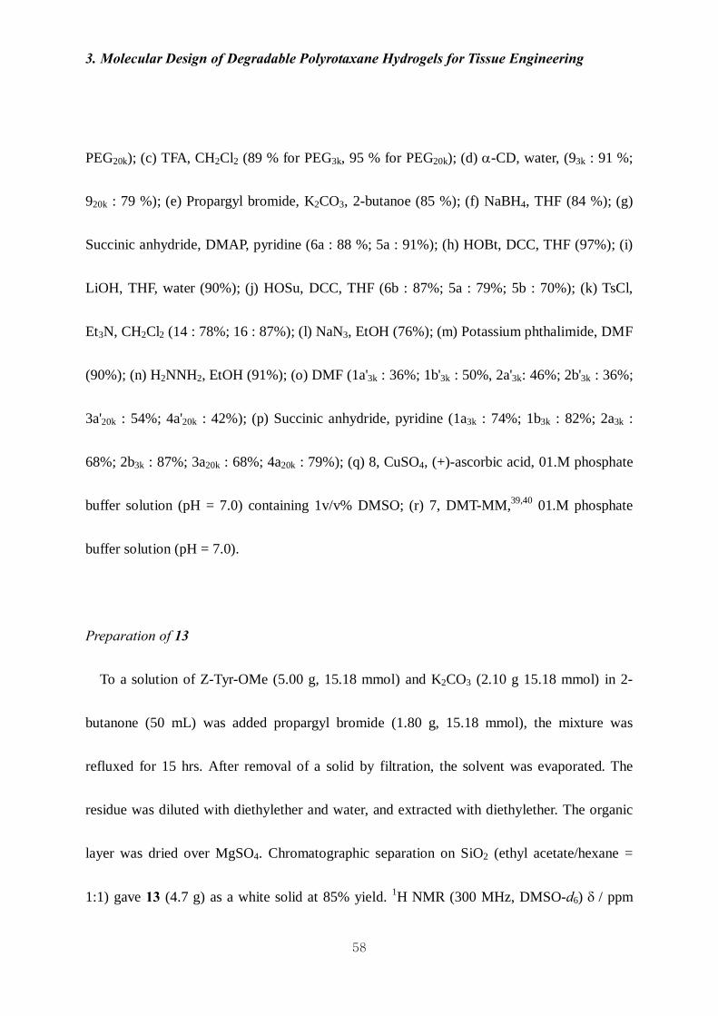
Preparation of 13
To a solution of Z-Tyr-OMe (5.00 g, 15.18 mmol) and K2CO3 (2.10 g 15.18 mmol) in 2-
butanone (50 mL) was added propargyl bromide (1.80 g, 15.18 mmol), the mixture was
refluxed for 15 hrs. After removal of a solid by filtration, the solvent was evaporated. The
residue was diluted with diethylether and water, and extracted with diethylether. The organic
layer was dried over MgSO4. Chromatographic separation on SiO2 (ethyl acetate/hexane =
1:1) gave 13 (4.7 g) as a white solid at 85% yield. 1H NMR (300 MHz, DMSO-d6) δ / ppm

3. Molecular Design of Degradable Polyrotaxane Hydrogels for Tissue Engineering
59
7.77 (1H, d, J=9 Hz), 7.36-7.20 (5H, m), 7.13 (2H, d, J = 8.7 Hz), 6.84 (2H, d, J = 8.4 Hz),
4.93 (2H, s), 4,71 (2H, d, J = 2.7 Hz), 4.43-4.10 (1H, m), 3.58 (3H, s), 3.29 (1H, s), 2.93 (1H,
dd, J = 9, 19.2 Hz), 2.76 (1H, dd, J = 3.3, 24 Hz).
Preparation of 11
To an ice-cooled solution of 13 (10.41 g, 28.34 mmol) in THF/MeOH (1:1, 210 mL) was
added NaBH4 (2.14 g, 56.68 mmol), the mixture was stirred for 40 min at room temperature.
To the reaction mixture was added 1% HCl aq. (200 mL) at 0 °C, the solution was further
stirred for 1 hr at the temperature. After removal of the solvent by evaporation, the residue
was diluted with ethyl acetate and water, and extracted with ethyl acetate. The organic layer
was dried over MgSO4. Chromatographic separation on SiO2 (ethyl acetate/hexane = 2:1)
gave 11 (7.69 g) as a white solid at 84% yield. 1H NMR (300 MHz, CDCl3) δ / ppm 7.39-7.25
(5H, m), 7.10 (2H, d, J = 8.1 Hz), 6.88 (2H, d, J = 8.1 Hz), 5.11 (1H, d, J = 7.8 Hz), 5.05 (2H,
s), 4.64 (2H, d, J = 2.4 Hz), 3.87 (1H, s), 3.63 (1H, dd, J = 7.8, 14.4 Hz), 3.52 (1H, dd, J =
6.3, 15 Hz), 2.77 (2H, d, J = 7.2 Hz), 2.50 (1H, ddd, J = 1.5, 2.4, 5.7 Hz).
Preparation of 6a
To a solution of 11 (6.51 g, 19.39 mmol) in pyridine (65 mL) were added succinic

3. Molecular Design of Degradable Polyrotaxane Hydrogels for Tissue Engineering
60
anhydride (1.98 g, 19.78 mmol) and 4-(dimethylamino)pyridine (DMAP, 2.41 g, 19.78 mmol),
the mixture was stirred for 36 hrs at room temperature. After removal of the solvent by
evaporation, the residue was diluted with ethyl acetate and 0.5N HCl aq., and extracted with
ethyl acetate. The organic layer was washed with DW, brine, and then dried over MgSO4.
After removal of a solid by filtration, the filtrate was evaporated and then purified by column
chromatography on SiO2 (ethyl acetate : CH2Cl2 = 2 : 1) to give an acid (7.56 g) as a white
solid at 88% yield. 1H NMR (300 MHz, CDCl3) δ / ppm 7.39-7.27 (5H, m), 7.07 (2H, d, J =
6.3 Hz), 6.87 (2H, d, J = 8.1 Hz), 5.05 (2H, s), 4.94 (1H, d, J = 8.1 Hz), 4.63 (2H, d, J = 2.4
Hz), 4.10 (2H, d, J = 8.4 Hz), 4.07-3.87 (1H, m), 2.76 (2H, t, J = 4.8 Hz), 2.70-2.51 (4H, br),
2.48 (1H, t, J = 2.1 Hz).
To a solution of the acid (3.93 g, 8.93 mmol) and 1-hydroxybenzotriazole (HOBT, 1.38 g,
9.02 mmol) in THF (53 mL) was added N,N-dicyclohexylcarbodiimide (DCC, 1.86 g, 9.02
mmol), the mixture was stirred for 24 hrs at room temperature. After removal of a solid by
filtration, the filtrate was concentrated, dissolved into THF / acetone (3 : 1), and precipitated
into cold hexane to give 6a (4.37 g) as a white solid at 97% yield. 1H NMR (300 MHz,
CDCl3) δ / ppm 8.31 (1H, d, J = 8.1 Hz), 7.97 (1H, d, J = 8.4 Hz), 7.69 (1H, t, J = 7.5 Hz),
7.53 (1H, t, J = 7.5 Hz), 7.36-7.27 (5H, m), 7.11 (2H, d, J = 8.1 Hz), 6.88 (2H, d, J = 8.4 Hz),

3. Molecular Design of Degradable Polyrotaxane Hydrogels for Tissue Engineering
61
5.10 (2H, d, J = 8.4 Hz), 5.05 (1H, s), 4.65 (2H, d, J = 2.1 Hz), 4.26-3.93 (3H, m), 3.43 (2H,
dd, J = 1.2, 12.6 Hz), 2.88-2.73 (4H, m), 2.52 (1H, t, J = 2.4 Hz).
Preparation of 12
A mixture of 13 (5.14 g, 13.99 mmol) and LiOH (1.34 g 55.96 mmol) was dissolved in
THF / DW (1:1, 51 mL), and stirred for 22 hrs at room temperature, and then concentrated.
The residue was dissolved in 0.5M NaHCO3 aq, and extracted with ethyl acetate. The organic
layer was dried over MgSO4. The crude product was purified by reprecipitation from
CH2Cl2/hexane to give 12 (4.46 g) as a white solid at 90% yield. 1H NMR (300 MHz, DMSO-
d6) δ / ppm 7.39-7.26 (5H, m), 7.06 (2H, d, J = 7.8 Hz), 6.78 (2H, d, J = 9 Hz), 4.94 (2H, dd,
J = 12.6, 12.9 Hz), 4.70 (2H, d, J = 2.4 Hz), 3.52 (1H, t, J = 2.4 Hz), 3.05 (1H, d, J = 11.4 Hz),
2.81 (1H, dd, J = 6, 21 Hz).
Preparation of 6b
To a solution of 12 (2.70 g, 8.09 mmol) and HOSu (0.93 g, 8.09 mmol) in THF (20 mL)
was added a solution of DCC (1.67 g, 8.09 mmol), the mixture was stirred for 24 hrs at room
temperature. The reaction mixture was cooled, filtered, and the filtrate was precipitated into
cold hexane to give 6b (3.19 g) as a white solid at 87% yield. 1H NMR (300 MHz, CDCl3) δ /

3. Molecular Design of Degradable Polyrotaxane Hydrogels for Tissue Engineering
62
ppm 7.41-7.28 (5H, m), 7.19 (2H, d, J = 8.7 Hz), 6.90 (2H, d, J = 9 Hz), 5.10 (2H, d, J = 6.9
Hz), 5.06-4.95 (1H, m) 4.66 (2H, d, J = 2.4 Hz), 4.62-4.48 (1H, br), 3.31-3.13 (2H, m), 2.85
(4H, s), 2.51 (1H, t, J = 2.4 Hz).
Preparation of 1417
To a solution of pentaerythritol ethoxylate (2.01 g, 2.54 mmol) and Et3N (1.34 mL, 10.26
mmol) in CH2Cl2 (20 mL) was added p-toluenesulfonyl chloride (TsCl, 1.96 g, 10.26 mmol),
and the mixture was stirred for 48 hrs at room temperature, and then 1N HCl aq. or water was
added. The organic layer was separated, and dried over MgSO4. Chromatographic separation
on SiO2 (MeOH/CH2Cl2 = 1:9) gave 14 (2.91 g) as a colorless oil at 78% yield. 1H NMR (300
MHz, CDCl3) δ / ppm 7.79 (8H, J = 8.1 Hz). 7.34 (8H, d, J = 8.1 Hz), 4.15 (8H, t, J = 4.8 Hz),
3.68 (8H, t, J = 4.8 Hz), 3.65-3.32 (68H, m), 2.44 (12H, s).
Preparation of 817
A mixture of 14 (9.00 g, 6.36 mmol), NaN3 (3.31 g, 50.9 mmol), and EtOH (120 mL) was
refluxed for 18 hrs. After removal of a solid by filtration, the filtrate was evaporated, and
dissolved in CH2Cl2. The solution was washed with water, and dried over MgSO4. The
organic layer was dried to give 8 (4.35 g) as a colorless oil at 76% yield. 1H NMR (300 MHz,

3. Molecular Design of Degradable Polyrotaxane Hydrogels for Tissue Engineering
63
CDCl3) δ / ppm 3.66-3.36 (76H, m), 3.34 (8H, t, J = 4.8 Hz); 13C NMR (75 MHz, CDCl3)
δ/ppm 77.1, 77.0, 96.9, 76.4, 57.1
Preparation of 1616
To a solution of tetraethylene glycol (12.40 g, 61.98 mmol) and Et3N (34.6 mL, 247.92
mmol) in CH2Cl2 (300 mL) was added TsCl (47.26 g, 247.92 mmol), and the mixture was
stirred for 5 hrs at room temperature, and then DW was added. The organic layer was
separated, and dried over MgSO4. Chromatographic separation on SiO2 (MeOH/CH2Cl2 =
5:95) gave 16 (22.53 g) as a colorless oil at 87% yield. 1H NMR (300 MHz, CDCl3) δ / ppm
7.78 (4H, d, J = 8.4 Hz), 7.34 (4H, d, J = 8.1 Hz), 4.15 (4H, t, J = 4.2 Hz), 3.66 (4H, t, J = 5.1
Hz), 3.55 (8H, s), 2.43 (6H, s); 13C NMR (75 MHz, CDCl3) δ/ppm 145, 133.1, 130, 128.1,
70.8, 69.5, 68.8, 21.8.
Preparation of 1516
A mixture of 16 (13.99 g, 27.83 mmol), potassium phthalimide (30.93 g, 166.98 mmol),
and DMF (280 mL) was stirred at 60 °C for 6 hrs, and the solvent was removed by
evaporation. The residue was dissolved in CH2Cl2, washed with 0.5N NaHCO3 aq., DW and
brine, and dried over MgSO4. The organic layer was concentrated and purified by column

3. Molecular Design of Degradable Polyrotaxane Hydrogels for Tissue Engineering
64
chromatography (MeOH/CH2Cl2 = 5:95) to give 15 (11.45 g) as a colorless oil at 90% yield.
1H NMR (300 MHz, CDCl3) δ / ppm 7.80 (4H, dd, J = 2.4, 8.7 Hz), 7.67 (4H, dd, J = 2.7, 8.4
Hz), 3.84 (4H, t, J = 6 Hz), 3.67 (4H, t, J = 6.3 Hz), 3.51 (8H, m); 13C NMR (75 MHz,
CDCl3) δ/ppm 168.5, 134.6, 132.5, 123.9, 70.9, 70.4, 68.2, 37.6.
Preparation of 716
A mixture of 15 (4.00 g, 8.84 mmol), hydrazine hydrate (9.28 g, 88.4 mmol), and EtOH
(200 mL) was refluxed for 12 hrs, and concentrated. The resiue was dissolved in CH2Cl2, the
resulting precipitates were removed by filtration. The filtrate was extracted with DW and
concentrated to give 7 (1.55 g) as a curde oil at 91% yield. 1H NMR (300 MHz, D2O) δ / ppm
3.52 (8H, s), 3.39 (4H, t, J = 5.4 Hz), 2.61 (4H, t, J = 5.4 Hz); 13C NMR (75 MHz, CDCl3)
δ/ppm 72.7, 69.9, 69.6, 41.1.
Preparation of pseudopolyrotaxanes 93k9
/20k
A mixture of α-CD (17.00 mmol), PEG-BA3k/20k (0.47 mmol for 3k and 0.075mmol for
20k), and water ([α-CD] = 149 mM) was stirred for 24 hrs at room temperature. The resulting
precipitates were collected by centrifugation, and washed with water, and then freeze-dried to
give pseudopolyrotaxanes 93k/20k as a white solid at 91% yield for 3k and 79% yield for 20k.

3. Molecular Design of Degradable Polyrotaxane Hydrogels for Tissue Engineering
65
The ratio of α-CD to PEG-BA3k/20k (l) in 93k/20k was determined to be 33 / 166 by 1H NMR
measured in D2O containing 1wt% NaOD.
A typical spectral data is as follows (93k); 1H NMR (300 MHz, D2O, signal for H2O was set
as 4.7 ppm) δ / ppm 4.90 (198H, d, J = 3.3 Hz), 3.89-3.63 (792H, m), 3.58 (272H, s), 3.50-
3.35 (396H, m).
General procedure for the preparation of precursor polyrotaxanes 1'-4'
A mixture of pseudopolyrotaxane 93k/20k (0.31 mmol), 5/6 (6.2 mmol), and DMF ([9] = 10
mM for 3k and 1.5 mM for 20k) was stirred at room temperature for 3 days. The suspension
was collected by centrifugation, and washed with DMF and acetone. The resulting solid was
dissolved in DMSO, and the solution was added to water adjusted to a pH of 3.2 to give a
solid. The solid was dialyzed in water (pH 3.2) for 3 days, and the resulting solid was
collected by centrifugation, and freeze-dried to give polyrotaxanes 1'-4' as a white solid at 36-
54% yield. The number of α-CD molecules (m) in the polyrotaxane was determined by 1H
NMR measured in D2O containing 1wt% NaOD.
A typical spectral data is as follows (1a'3k); 1H NMR (300 MHz, D2O containing 1wt%
NaOD, signal for H2O was set as 4.7 ppm) δ / ppm 7.36-7.26 (10H, m), 7.15 (4H, d, J = 7.8
3. Molecular Design of Degradable Polyrotaxane Hydrogels for Tissue Engineering
66
Hz), 6.87 (4H, d, J = 7.8 Hz), 4.82 (108H, d, J = 3 Hz), 3.84-3.62 (432H, m), 3.55 (272H, s),
3.42-3.25 (216H, m).
General procedure for the preparation of polyrotaxanes 1-4
To a solution of polyrotaxane 1'-4' (0.10 mmol) in pyridine ([1'/2'] = 2 mM and [3'/4'] =
0.37 mM) was added succinic anhydride (18 equiv. of α-CD), the mixture was stirred at room
temperature for 24 hrs, and then poured into diethylether to give a solid. The solid was
collected by filtration, and then dialyzed in water (pH 3.2) for 3days. The resulting solid was
collected by centrifugation, and freeze-dried to give polyrotaxanes 1-4 as a white solid in 68-
87% yield. The number of carboxyl groups (x) appended to an α-CD molecule was
determined by 1H NMR measured in D2O containing 1wt% NaOD.
A typical spectral data is as follows (1a3k); 1H NMR δ / ppm (300 MHz, D2O containing
1wt% NaOD, signal for H2O was set as 4.7 ppm) 7.34-7.24 (10H, m), 7.17 (4H, d, J = 9 Hz),
6.90 (4H, d, J = 9 Hz), 4.86 (108H, d, J = 3 Hz), 3.87-3.65 (432H, m), 3.56 (272H, s), 3.46-
3.51 (216H, m), 2.26 (432H, s).
Preparation of hydrogels IA3k, IB3k and IIIA20k for the internal crosslink

3. Molecular Design of Degradable Polyrotaxane Hydrogels for Tissue Engineering
67
To a 0.1M phosphate buffer solution (PBS) (pH 7.0, 11 mL for 1a/b or 50 mL for 3a) of
1a/b (0.015 mmol) or 3a (0.030 mmol) were added DMT-MM (2.16 mmol) and 7 (0.54
mmol). Portions of the mixture were poured into a Teflon mold (diameter / thickness 15 mm /
10 mm for a compressive stress-strain test; 10 mm / 5 mm for a degradation test), which was
set in a shaking apparatus (60 rpm) with maintaining at 37 °C for 24 hrs. The resulting gel
was rinsed by immersion in DMSO and mixture of 0.1M PBS (pH 7.0) for periods of 2 and 5
days, respectively. The time when the purification was completed is defined as t = 0 in the
degradation. The dry weight of a hydrogel was measured after dialysis in acidic water (pH
3.2) for 2 days to remove salts.
Preparation of hydrogels IIA3k, IIB3k and IVA20k for the terminal crosslink
To a 0.1M PBS (pH 7.0) of 2a/b (0.015 mmol) or 4a (0.015 mmol) (11 mL for 2a/b or 50
mL for 4a) were added a solution of 8 (0.0075 mmol) in DMSO (0.06 mL for 2a/b or 4a) and
a 0.1M PBS (pH 7.0) of CuSO4·5H2O (0.012 mmol) and ascorbic acid (0.012 mmol) (0.3 mL
for 2a/b or 4a) in a Teflon mold (diameter / thickness 15 mm / 10 mm for a compressive
stress-strain test; 10 mm / 5 mm for a degradation test). The mixture was set in a shaking
apparatus (60 rpm) with maintaining at 37 °C for 24 hrs. The resulting gel was rinsed by
immersion in DMSO and 0.1M PBS (pH 7.0) for periods of 1 and 5 days, respectively. The
3. Molecular Design of Degradable Polyrotaxane Hydrogels for Tissue Engineering
68
time when the purification was completed is defined as t = 0 in the degradation test and cell
adhesion test. The dry weight of a hydrogel was measured after dialysis in acidic water (pH
3.2) for 2 days to remove salts.
3-2-3. Measurement
3-2-3-1. Water content
The pre-weight (Wd) dry hydrogels were immersed into a 0.1M PBS (pH 7.0) and left to
reach equilibrium swelling for 48hrs at 37 °C. The swollen hydrogels were removed from
PBS, whipped with filter paper gently, and weighted as soon as possible (Ws). Water content
(%) was defined according to the following equation: Water content (%) = (Ws − Wd) / Ws ×
100, where Wd and Ws represent the dry and swollen mass of samples.
3-2-3-2. Static contact angle using air-bubble (in wet condition)42
Contact angles (θ) were measured by using the static captive bubble method. Hydrogels
(t=0) attached to slide-glass were inverted in the pure water at a room temperature. Air bubble
was injected (5-10 ㎕) from a syringe onto the hydrogels surface water. At least 10

3. Molecular Design of Degradable Polyrotaxane Hydrogels for Tissue Engineering
69
measurement of bubble were averaged to obtain angle (θ') on the surface of hydrogels.
Contact angles were calculated by 180 - θ'.
3-2-3-3. Compression test
The mechanical property of the hydrogels was characterized in terms of a compressive
stress-strain measurement through a uni-axial and unconfined compression test using
Microload System (R & B Co. Ltd., Daejeon, Korea). The unconfined compression test was
performed in a 0.1 M PBS (pH 7.0) at 37 °C to maintain the sample hydration. Prior to the
compression test, the samples were preconditioned for 20 cycles at 3% strain, put in the fluid
bath, and then loaded by an upper jig fitted with a 1 kg load cell. After confirming their full
swelling, the samples were compressed at a rate of 1 mm/min until their fracture. Load -
displacement data were recorded at 2 Hz and converted into the strain-stress curves.
3-2-3-4. Stress relaxation test
IB3k and IIB3k were immersed in 0.1M PBS (pH 7.0) in a chamber at 37℃ and cyclic
compressive load at a rate of 3% strain / sec (20 times) applied as precondition. After samples
swelled fully in buffer, compressive load applied at a rate of 100% strain / min and held at 10,
20, 30 and 40%.

3. Molecular Design of Degradable Polyrotaxane Hydrogels for Tissue Engineering
70
3-2-3-5. Curve fitting of stress relaxation curve 41
The governing standard linear solid (SLS) equation is displayed in equation (1) and solved
for the case of a constant strain ε0, yielding a mathematical description of stress relaxation. In
order to adjust model descriptors to experiment data (time-stress curve), Equation (4) was
calculated.
Standard linear solid (SLS) model
[ ] ετ
εστ
σ 121
1 EdtdEE
dtd
++=+
(1)
τ
εσ /
210
)()( teEEtGt −+==
(2)
τεεσ /2010)( teEEt −+= (3)
τσ /)( tBeAt −+= (4)
2Eητ =

3. Molecular Design of Degradable Polyrotaxane Hydrogels for Tissue Engineering
71
where A, B, τ are constant determined by suing experimental data. Experimental data
were used to determine constant A, B, and τ from equation (4). Linear regression was
performed using SPSS to generate model function for each sample. High Pearson’s
correlation (R > 0.95) between the modeled responses and experimental data supports the
choice of viscoelastic theory and particularly the SLS model to describe stress relaxation
behavior.
3-2-3-6. Changes in mass and storage modules upon degradation
Degradation of the hydrogels was examined at 37 °C in a 0.1M PBS (pH 7.0). The
samples were immersed into a 50 mL of PBS, which was renewed every two weeks.
Degrading samples were harvested, freeze-dried for 2 days, and then weighed (Md).
Harvested samples were gently wiped with tissue paper to remove attached water droplet and
weighed (Ws). Mass loss (%) was defined according to the following equation:
Mass loss (%) = [1 − (M0 − Md) / M0] ×100
where Mo and Md represent the dry mass of the samples before and after the degradation
test, respectively.
The storage modulus of the hydrogels was determined through a dynamic viscoelastic
measurement using Rheosol-G3000 (UBM Co., Ltd, Kyoto, Japan) with parallel plate

3. Molecular Design of Degradable Polyrotaxane Hydrogels for Tissue Engineering
72
geometry. The samples were fully swollen in a 0.1 M PBS (pH 7.0) at 37 °C and subjected to
a condition of frequency sweep with the range of 0.07-10 Hz at 37 °C.
3-2-3-7. Quantification of the amount of fibronectin adhered on the hydrogel
Three hydrogel samples were immersed in a 0.1M PBS (pH 7.4) at room temperature for 3
hrs after the duration time was 0, 2 or 4 weeks for IIA3k and IIB3k, and 0, 3 or 7 days for
IVA20k. After the 3-hour-immersion, each sample was then immersed in a human fibronectin
(FN) solution (5 µg/mL in PBS, Sigma-aldrich, Inc., MO, USA) at 37 °C for 1 hr to coat, and
which was gently washed with PBS. The FN-coated hydrogels were further immersed in 1%
SDS solution and sonicated for 30 minutes to detach excess of FN on the surface of FN-
coated gels. The quantity of FN adhered on each surface of the hydrogels was determined
using a Micro BCATM protein assay kit (Thermo Fisher Scientific Inc., Waltham, MA, USA)
by a spectrophotometer (570 nm) (Multiskan FC, Thermo Fisher Scientific Inc., Waltham,
MA, USA).
3-2-3-8. Quantification of the amount and size of cells adhered on the hydrogel
Three types of hydrogels (IIA3k., IIB3k and IVA20k) were immersed in a serum-free
Dulbecco’s modified Eagle’s medium (DMEM) containing penicillin and streptomycin at

3. Molecular Design of Degradable Polyrotaxane Hydrogels for Tissue Engineering
73
room temperature for 3 hrs as pre-wetting when the duration time was 0, 2 or 4 weeks for
IIA3k and IIB3k, and 0, 3 or 7 days for IVA20k. After the immersion, each sample was then
immersed in a FN solution (5 µg/mL/ in PBS) at 37 °C for 1 hr to be coated, and which was
placed into a stainless steel ring to prevent loss of cells during cell seeding. NIH 3T3 cells
were seeded on upper side of the hydrogels at a density of 1.1 × 10⁵ cells/cm2 and
cultivated in DMEM containing 10% fetal bovine serum (FBS) for 12 hrs under a humidified
5% CO2 atmosphere at 37 °C. After removing the medium, cells on the hydrogel surface were
stained with Cell-Tracker™ Green fluorescent probe (Molecular Probes, Inc., Oregon, USA)
in the serum-free medium at 37 °C for 45 minutes, and then loosely adherent cells were
removed by washing with PBS. Cells were fixed with 3.7% formaldehyde in PBS. Cell
morphology on hydrogels was visualized and imaged by an Olympus FluoView™ FV1000
confocal scanning laser microscope (Olympus Optical, Tokyo, Japan). The number and size
of cells were quantified by using Image J software (National Institutes of Health, Maryland,
USA) from the image of confocal microscope.

3. Molecular Design of Degradable Polyrotaxane Hydrogels for Tissue Engineering
74
3-3. Results and discussion
3-3-1. Design and preparation of ester-containing polyrotaxanes 1-4 for gelation
Preparation of degradable polyrotaxane hydrogels consists of the following three parts:
preparation of precursor polyrotaxanes (1'-4'), solubilization of 1'-4' in water (1-4), and
gelation of 1-4 (I-IV). The precursor polyrotaxanes 1a'9 and 2a' were successfully prepared
by endcapping of a pseudpolyrotaxane (α-CDl·PEGBA 93k (l = 33)) with ester-containing
capping molecules 5a and 6a, respectively (Scheme 2), because preinstalled ester linkages in
the main chain undergo undesired hydrolysis during preparation of intermediates9 and caused
very difficulty to prepare pure polyrotaxanes well and very low yield.18,19 The ester-
containing capping molecules 5a and 6a were derived from Z-phenylalanine for the internal
crosslink and Z-tyrosine for the terminal crosslink, respectively (Scheme 2), according to our
previous report.9 The precursor polyrotaxanes 1a' and 2a' were characterized by gel
permeation chromatography (Figure 2) and NMR spectroscopy (Figure 3), showing that 1a'
and 2a' were purified enough to determine the number of α-CD in the polyrotaxane (m = 18
for 1a' and 2a'). The precursor polyrotaxanes 1a' and 2a' were so insoluble in water20-25 that a
carboxylic residue was attached to α-CD in 1a' and 2a' to attain water solubility (x = 108 for
3. Molecular Design of Degradable Polyrotaxane Hydrogels for Tissue Engineering
75
1a and 2a). The internal crosslink utilized the carboxyl group in a condensation reaction of 1a
with a ditopic amine 7 upon galation of 1a to give a hydrogel IA3k (Scheme 3). The terminal
crosslink was achieved through a 1,3-dipolar addition reaction26,27 between an alkyne tethered
to the tyrosine in 2a and a tetratopic azide 8 upon gelation of 2a to give a hydrogel IIA3k
(Scheme 3). Reference polyrotaxanes 1b and 2b, which do not have any ester linkages in the
main chain, were prepared using similar end-capping reactions of α-CDl·93k (l = 33) with
ester-not-containing capping molecules 5b and 6b, and were crosslinked to give hydrogels
IB3k (internal) and IIB3k (terminal), respectively (Scheme 2). Degradable polyrotaxanes with
higher molecular weight 3a and 4a (average n = 454, m = 88, x = 528 for 3a and 4a) were also
obtained by the similar method employing a pseudopolyrotaxane (see supporting information)
α-CDl·PEGBA 920k (l = 166) and capping molecules 5a and 6a, and were crosslinked to give
hydrogels IIIA20k (internal) and IVA20k (terminal) (Scheme 3).

3. Molecular Design of Degradable Polyrotaxane Hydrogels for Tissue Engineering
76
Figure 2. GPC profiles of (a) 93k, (b) 1a'3k, and (c) 1b'3k, eluted with DMSO containing 0.1M
NaNO3 with 0.3mL/min flow rate at 313K, detected by RI. (Column : TSKgel G-5000HHR +
TSKgel G-3000HHR).
Figure 3. 1H NMR spectra (300MHz) (a) 1a'3k, and (b) 2a'3k measured in D2O containing
1wt% NaOD.

3. Molecular Design of Degradable Polyrotaxane Hydrogels for Tissue Engineering
77
Scheme 3. Gelation of polyrotaxanes, (a) the internal crosslink and (b) the terminal crosslink.

3. Molecular Design of Degradable Polyrotaxane Hydrogels for Tissue Engineering
78
3-3-2. Design and preparation of degradable hydrogels I-IV
The internal crosslink was carried out for gelation of 1 through a condensation reaction
with the ditopic amine 7 in PBS adjusted to a pH of 7. Although we know the ester linkages in
these polyrotaxanes are more stable against the hydrolysis in lower-pH-solutions,9 in such
solutions the carboxylic residue did not act as the solubilizing group due to neutralization and
the amine decreased reactivity due to protonation. Four equivalents of the amine to one
molecule of α-CD in 1 were aimed to crosslink the polyrotaxanes intermolecularly, and
73/82% weight of the total amount of 1a/b and 7 was found in a solid obtained by drying after
purification. For the polyrotaxane 3 with higher molecular weight, the similar treatment
resulted in a hydrogel that gave a solid with 74% weight of the total amount of 3 and 7 by
drying after purification. Crosslink reactions at the terminals of 2 or 4 with the tetratopic azide
8 were also performed in PBS adjusted to pH 7, in which equi-molar amounts of the alkyne
and azide groups were feed, and gave hydrogels (81/87% weight for 2a/b and 8, and 76%
weight for 4 and 8 in respective solids after purification and drying). Details of gelation
conditions are summarized in Table 1.
Water contents and contact angle of hydrogels were summarized in Table 2. Hydrogel I-IV
composed of water-soluble polyrotaxane showed very high water content (> 90%). Water

3. Molecular Design of Degradable Polyrotaxane Hydrogels for Tissue Engineering
79
content of hydrogel II was superior to that of hydrogel I and hydrogel III and IV composed
of PEGBA 20K as back bone chain also showed higher water content than that of I and II
composed of PEGBA 3K. These results represent that low crosslink density by means of long
back bone chain or fewer crosslink points formed large cavity in gel network, in which could
absorb more water in gel network.
We measured static contact angle utilizing air-bubble because surface of hydrogel absorbs
drop-water easily. Contact angles of hydrogel I-IV was 21 - 43°and this results represented
that surface of polyrotaxane hydrogel I-IV had hydrophilicity, in which might have an good
influence on adhesion of cell and protein on hydrogel surface.

3. Molecular Design of Degradable Polyrotaxane Hydrogels for Tissue Engineering
80
Table 1. Gelation conditions.
PRx Crosslinker
CuSO4 ∙5H2O
Ascorbic acid
DMT-MM 0.1M
phosphate buffer Hydrogel
Comp. Eq. mol
Comp. Eq. mol Eq. mol Eq. mol mM
IA3k 1a3k 1 7 36 144 1.36
IB3k 1b3k 1 7 36 144 1.36
IIA3k 2a3k 1 8 0.5 0.8 1.36
IIB3k 2b3k 1 8 0.5 0.8 1.36
IIIA20k 3a20k 1 7 176 704 0.3
IVA20k 4a20k 1 8 0.5 0.8 0.3
Table 2. Water content and static contact angle.
Hydrogels Water content (%) Contact angle (°)
IA3k 90 32
IB3k 92 43
IIA3k 95 21
IIB3k 95 34
IIIA20k 96 36
IVA20k 97 20

3. Molecular Design of Degradable Polyrotaxane Hydrogels for Tissue Engineering
81
3-3-3. Mechanical properties of degradable hydrogels I-IV
We carried out compressive test and stress relaxation test in order to observe mechanical
stability and viscoelastic characteristics of hydrogels I-IV. Different cross-linking system
allowed outstanding different mechanical properties.
Comparing the profiles of compressive stress-strain curves for the internally-crosslinked
hydrogels IA3k and IB3k indicated that degradation of IA3k had already proceeded to some
extent during the purification (Figure 4). This was also the case for the terminally-crosslinked
hydrogels IIA3k and IIB3k (Figure 4). Degradation of the hydrogel IA3k with the internal
crosslink would be triggered by dual cleavage of two ester linkages embedded in the main
chain and the residual succinic moiety at α-CD (Figure 1). The hydrogel IIA3k with the
terminal crosslink would degrade only by cleavage of the ester linkage in the main chain, and
the residual ester groups in 2a would have no responsibility for the degradation at all (Figure
1). Internally-crosslinked hydrogels IA3k, IB3k and IIIA20k showed higher ultimate
compressive stress and initial modulus than those of terminally-crosslinked hydrogels IIA3k
IIB3k and IVA20k. On the contrary to this, ultimate strain of terminally-crosslinked hydrogels
was superior to internally-crosslinked hydrogels (Figure 4 and Table 3). These results
represented that dense gel network in internally-crosslinked hydrogel allowed higher

3. Molecular Design of Degradable Polyrotaxane Hydrogels for Tissue Engineering
82
mechanical stability and reduced flexibility of gel structure comparing with the terminally-
crosslinked hydrogels.
0
5
10
15
20
25
0.0 0.2 0.4 0.6 0.8Strain
Stre
ss (K
Pa)
Figure 4. Stress - strain curves of internal crosslink hydrogels and terminal crosslink
hydrogels. ◇ : IA3k, □ : IB3k, △ : IIIA20k, ◆ : IIA3k, ■ : IIB3k, ▲ : IVA20k
Table 3. Results of compressive test.
Ultimate
compressive strain
Ultimate compressive stress (kPa)
Initial modulus (kPa)
Modulus before fracture (kPa)
IA3k 0.30±0.01 5.77±0.17 4.60±0.42 46.40±3.56
IB3k 0.31±0.01 11.69±0.56 8.58±1.02 89.78±10.73
IIA3k 0.57±0.04 5.23±0.39 0.79±0.19 36.20±5.04
IIB3k 0.57±0.01 10.97±0.29 2.34±0.59 90.26±5.64
IIIA20k 0.49±0.01 21.79±1.31 4.70±0.76 182.81±12.22
IVA20k 0.70±0.03 1.18±0.04 0.69±0.09 3.82±0.29

3. Molecular Design of Degradable Polyrotaxane Hydrogels for Tissue Engineering
83
Results of stress relaxation of hydrogel I-IV were represented to Figure 5 and Table 4. In
stress relaxation test, internally-crosslinked hydrogel showed very unique phenomenon.
Equilibrium stress of internally-crosslinked hydrogel was lower that of terminally-crosslinked
hydrogels although instantaneous stress of internally-crosslinked hydrogels was higher than
that of terminally-crosslinked hydrogel. Relative relaxation and relaxation time of internally-
crosslinked hydrogel were higher and faster than those of terminally-crosslinked hydrogel,
respectively. This phenomenon represents that network structure of internally-crosslinked
hydrogel could reduce stress transferred to inside via polymer chain. This unique viscoelastic
behavior was very similar to that of topological gel reported by Kohzo Ito and his co-
works.12,13,15 Topological gel composed of PEG chain with large molecular weigh and α-CD
was topologically interlocked by figure-of-eight cross-links. These cross-links could be
movable on the back bone chain freely to equalize the tension of the threaded polymer chains
like pulleys. Therefore, stress might be equalized in the gel.15 The fast relaxation time of
internally-crosslinked hydrogel might result from equalization of internal stress via movable
crosslink point in gel network.

3. Molecular Design of Degradable Polyrotaxane Hydrogels for Tissue Engineering
84
0
1
2
3
4
5
0 10 20 30 40 50 60Time (h)
Stre
ss (k
Pa)
Figure 5. Stress relaxation curves of IB3k and IIB3k at various strain. ◇ : IB3k (10%), □ :
IB3k (20%), ● : IIB3k (10%),◆ : IIB3k (20%), ■ : IIB3k (30%), ▲ : IIB3k (40%).

3. Molecular Design of Degradable Polyrotaxane Hydrogels for Tissue Engineering
85
Table 4. Stress relaxation of IA3k - IVA20k.
Strain (%)
Instantaneous stress (kPa)
Equilibrium stress (kPa)
Relaxed stress (kPa)
Relative relaxation
(%)
Relaxation time (h) *
10 0.84 0.37 0.47 56 1.3 IA3k 20 2.08 0.57 1.51 72 1.7
10 0.23 0.17 0.07 29 4.4 20 0.40 0.24 0.16 39 10.8 30 0.68 0.35 0.33 49 14.0
IIA3k
40 1.28 0.79 0.49 38 23.9 10 0.69 0.17 0.51 75 3.6
IB3k 20 3.98 0.40 3.57 90 10.3 10 0.43 0.34 0.09 20 11.4 20 0.65 0.46 0.19 30 24.4 30 1.72 0.72 1.00 58 40.0
IIB3k
40 2.76 1.53 1.24 45 40.3 10 - - - - - 20 0.55 0.40 0.15 27 9.4 30 3.15 0.33 2.82 90 22.7
IIIA20k
40 8.94 0.38 8.57 96 28.1 IVA20k - - - - - -
* Calculated by SLS equation (1)

3. Molecular Design of Degradable Polyrotaxane Hydrogels for Tissue Engineering
86
3-3-4. Degradation behavior of hydrogels I-IV
Degradation was investigated by monitoring changes in mass and storage modulus of the
hydrogels immersed in a 0.1M PBS at 37 °C. Hydrogels IIB3k, which have no ester linkages
in the back bone chain but have a number of ester linkages in the residue of the polyrotaxanes,
showed 30% mass loss due to cleavage of the residual esters (Figure 6). During the 30% mass
loss, the terminally-crosslinked IIB3k had maintained the storage modulus (~103 Pa), while
the storage modulus for the internally-crosslinked IB3k was reduced gradually (Figure 7)
because gel network was cleaved by cleavage of crosslink point connected with CDs in
polyrotaxane. Interesting point is that both hydrogels maintained good water content (>90%)
even though the polyrotaxanes lost the water-solubilizing group on CD. We found a
remarkable difference in the rate of degradation for the internal (IA3k and IIIA20k) and
terminal (IIA3k and IVA20k) systems. The former showed a relatively rapid mass loss (50%
loss, t = 5 and 7 days for IA3k and IIIA20k) and the latter showed a gradual mass loss over a
longer period (50% loss, t = 26 and 12 days for IIA3k and IVA20k) (Figure 6). Continuous
monitoring of the storage modulus of the hydrogels IA3k, IIA3k, IIIA20k, and IVA20k showed
gradual decreases for both systems. In the internal-crosslink system, relatively larger amounts
of α-CD and cross-linker molecules feed in a gelation reaction of the polyrotaxane and the

3. Molecular Design of Degradable Polyrotaxane Hydrogels for Tissue Engineering
87
crosslinker led to higher storage modulus for IIIA20k than for IA3k over the duration (Table 1
and Figure 7). In the terminal-crosslink system, a relatively lower ratio of the terminals in a
polyrotaxane allowed the storage modulus of IVA20k to reach faster <10 Pa than IIA3k (35
days for IIA3k and 21 days for IVA20k) (Table 1 and Figure 7). Differences in erosion should
come from the crosslinking method. The longer period of erosion for the terminally-
crosslinked gels would be suitable for a preliminary cell adhesion test, described below.
Figure 6. Changes in mass of hydrogels IA3k (◇), IB3k (□), IIA3k (◆), IIB3k (■), IIIA20k
(△), and IVA20k (▲).

3. Molecular Design of Degradable Polyrotaxane Hydrogels for Tissue Engineering
88
Figure 7. Changes in storage modulus of hydrogels IA3k (◇), IB3k (□), IIA3k (◆), IIB3
k (■), IIIA20k (△), and IVA20k (▲), measured at a frequency of 1 Hz.
3-3-5. Cell adhesion on hydrogels I-IV
Cell adhesion on a hydrogel surface is influenced by many factors, such as chemical and
mechanical characteristics of a polymer surface,28-30 cell adhesion proteins,31-34 and so on.
Some researchers have reported that mechanical stability and protein adhesion on a surface
play important roles in cell adhesion at early stages of cell culture.35-36 Fibronectin (FN), one
component of extracellular matrices, is important for cell adhesion and growth by means of
binding to integrin α5β1 via its cell binding domain.32,33 A preliminary test for cell adhesion
onto the terminally-crosslinked hydrogels IIA/B3k and IVA20k on degradation was carried out

3. Molecular Design of Degradable Polyrotaxane Hydrogels for Tissue Engineering
89
using NHI3T3 cells in terms of the number, size, and morphology of adhered cells. Prior to
seeding, we coated FN on surface of hydrogel and measured amount of adherent FN (Figure
8). Amount of FN adhered on IIB3k was higher than other hydrogels and amount of FN
decreased and then increased at 14 and 28 days, respectively, while amount of FN adhered on
IIA3k decreased. This result might imply that stability of hydrogel played an important role in
protein adhesion on hydrogel surface. We also focused on increase of FN amount adhered on
IIB3k at 28 days. Amount of FN on IIB3k increased at 28 days although mechanical stability
of IIB3k did not change (Figure 7 and 8). Most of carboxyl residue in of IIB3k were cleaved at
28 days (Figure 6), in which might cause change of chemical condition of hydrogel surface
and have influence on FN adhesion on hydrogels. Lee et al. reported that the ability of FN to
enhance cellular attachment was dependent on the conditions of its adsorption to the surface
with different functional group (OH, COOH, NH2 and CH3)43, in which could explain the
change of FN amount adhered on hydrogel II and IV due to chemical condition of surface.
Some researchers have reported that cell attachment and spreading depended on
compatibility with surface and carboxylic acid groups on surface had a negative effect on cell
adhesion.37,38 In this research, we observed different cell morphology on each hydrogel during
degradation. In IIB3k, cell spreading and increase of cell adhesion were observed continuously
during degradation. We assumed that loss of carboxyl residue and stable substratum enhanced

3. Molecular Design of Degradable Polyrotaxane Hydrogels for Tissue Engineering
90
cell adhesion and spreading. However, IIA3k and IV20k showed different phenomenon. Cell
spreading and increase of cell adhesion in IIA3k at 14 days was observed, but spherical cell
and decrease of cell number and size were observed at 28 days. This phenomenon explains
that positive influence by means of loss of carboxyl residue and negative influence owing to
collapse of hydrogel might affect cell adhesion and spreading in IIA3k at the same time. So
that reason, in this cellular experiment, change of chemical characteristics by decreasing
carboxyl residue upregulated cell biocompatibility on surface of hydrogels at early
degradation. However, exceeding collapse of hydrogel structure by means of degradation had
more negative influence on cell adhesion and spreading. As same reason, the cellular behavior
on IV20k, showing continuous decreasing cell adhesion and spherical cell morphology, also
came from very fast degradation of hydrogels. This phenomenon implies that cell morphology
adhered on polyrotaxane hydrogel was affected by two parameters, change of surface
chemical condition by means of loss of carboxyl residue and mechanical stability of
hydrogels at the same time (Figure 10).

3. Molecular Design of Degradable Polyrotaxane Hydrogels for Tissue Engineering
91
Figure 8. Amount of FN adhered on a surface of hydrogels IIA3k (◇), IIB3k (□), and IVA20k
(△).

3. Molecular Design of Degradable Polyrotaxane Hydrogels for Tissue Engineering
92
Figure 9. (a) Number and (b) size of adhered cells on a surface of FN-coated hydrogels IIA3k
(◇), IIB3k (□), and IVA20k (△).

3. Molecular Design of Degradable Polyrotaxane Hydrogels for Tissue Engineering
93
Figure 10. Confocal microscopy images of cells adhered on surface of hydrogels during
degradation (scale bar : 100 ㎛).

3. Molecular Design of Degradable Polyrotaxane Hydrogels for Tissue Engineering
94
3-4. Conclusion
We prepared series of polyrotaxanes containing ester linkages in the main chain to trigger
the supramolecular dissociation in water by means of hydrolysis of the esters. A modified
procedure successfully reduced a chance to cleave unfortunately such labile linkages in the
course of preparation using an ester-free pseudopolyrotaxane and ester-containing capping
molecules. Crosslinking of the hydrolyzable polyrotaxanes was carried out in two ways, one
was a crosslinking between intermolecular cyclic components (internal crosslink) and the
other was a crosslinking between the terminals of the polyrotaxanes (terminal crosslink).
Hydrogel I-IV showed high water content (>90%) and hydrophilic surface. The internally-
crosslinked gels had higher compressive stress and initial modulus, while the terminally-
crosslinked gels showed higher ultimate strain. Viscoelastic behavior of internally-crosslinked
hydrogels was very different from that of terminally-crosslinked hydrogels and similar to that
of topological gel. The terminally-crosslinked hydrogels were more stable during degradation
than the internally-crosslinked hydrogels in PBS. These degradations accompanied decrease
in the storage modulus, and showed various degradation profiles, which were modulated by
the crosslinking manners and molecular weights of the polyrotaxanes. A preliminary cell
adhesion test using NIH3T3 cells onto the terminally-crosslinked hydrogels demonstrated

3. Molecular Design of Degradable Polyrotaxane Hydrogels for Tissue Engineering
95
cellular responses by means of decrease in storage modulus and change of surface chemical
characteristics of hydrogels. These results could provide a possibility to develop
polyrotaxane-based hydrogel systems as biodegradable materials for tissue engineering, such
as implantable scaffold and drug delivery system, since polyrotaxanes are allowed to be
designed uniquely by modulating lots of structural parameters, such as various crosslinking
methods, combinations of polyrotaxane component, and molecular weight of the backbone
polymer.
Acknowledgments. We thank Professor Jung-woog Shin (Inje University, Gimhae, Korea)
for his help and comments in the measurement of mechanical properties.
Reference
1. Langer, R.; Vacanti, J. P. Science, 1993, 260, 920.
2. Passl, W. J. Prog. Polym. Sci., 1989, 14, 629.
3. Barrows, T. H. Clin. Mater., 1986, 1, 233.
4. Nuttelman, C. R.; Henry, S. M.; Anseth, K. S. Biomaterials, 2002, 23, 3617.
5. Vainionpaa, S.; Rokkanen, P.; Tormala, P. Prog. Polym. Sci., 1989, 14, 69.

3. Molecular Design of Degradable Polyrotaxane Hydrogels for Tissue Engineering
96
6. Nair, L. S.; Laurencin, C. T. Prog. Polym. Sci., 2007, 32, 762.
7. Harada, A.; Li, J.; Kamachi, M. Nature, 1992, 356, 325.
8. Wenz, G.; Han, B.; Müller, A. Chem. Rev., 2006, 106, 782.
9. Katoono, R.; Fukuda, S.; Shin, H.; Yui, N. Chem. Lett., 2008, 37, 988.
10. Ooya, T.; Yui, N. J. Control. Release., 1999, 58, 251.
11. Arai, T.; Hayashi, M.; Takagi, N.; Takata, T. Macromolecules, 2009, 42, 1881.
12. Araki, J.; Ito, K. J. Polym. Sci.: Part A. Polym. Chem., 2006, 44, 6312.
13. Kakaoka, T.; Kidowaki, M.; Zhao, C.; Minamikawa, H.; Shimizu, T.; Ito, K. J. Phys.
Chem. B, 2006, 110, 24377.
14. Loethen, S.; Kim, J.; Thompson D. H. Polym. Rev., 2007,47, 383.
15. Okumura, Y.; Ito, K. Adv. Mater., 2001, 13, 485.
16. Numata, M.; Koumoto, K.; Mizu, M.; Sakurai, K.; Shinkai, S. Org. Biomol. Chem., 2005,
3, 2255.
17. Kobayashi, Y.; Katoono, R.; Yamaguchi, M.; Yui, N. Polym. J., 2011, 43, 893.
18. Watanabe, J.; Ooya, T.; Yui, N. Chem. Lett., 1998, 1031.

3. Molecular Design of Degradable Polyrotaxane Hydrogels for Tissue Engineering
97
19. Watanabe, J.; Ooya, T.; Yui, N. J. Biomater. Sci. Polym. Edn., 1999, 10, 1275.
20. Harada, A.; Kamachi, M. Macromolecules, 1990, 23, 2821.
21. Harada, A.; Li, J.; Kamachi, M. Macromolecules, 1993, 26, 5698.
22. Harada, A.; Kamachi, M. J. Chem. Soc., Chem. Commun., 1990, 1322.
23. Harada, A.; Okada, M.; Li, L.; Kamachi, M. Macromolecules, 1995, 28, 8406.
24. Harada, A.; Li, J.; Suzuki, S.; Kamachi, M. Macromolecules, 1993, 26, 5267.
25. Harada, A.; Li, J.; Kamachi, M. Nature, 1992, 356, 325.
26. Rostovtsev, V. V.; Green, L. G.; Fokin, V. V.; Sharpless, K. B. Angew. Chem. Int. Ed., 2002,
41, 2596.
27. Rodionov, V. O.; Fokin, V. V.; Finn, M. G. Angew. Chem. Int. Ed., 2005, 44, 2210.
28. Keselowsky, B. G.; Collard, D. M.; Garcia, A. J. J. Biomed. Mater. Res. A, 2003, 66,
247.
29. Engler, A. J.; Sen, S.; Sweeney, H. L.; Discher, D. E. Cell, 2006, 25, 677.
30. Gunn, J. W.; Turner, S. D.; Mann, B. K. J. Biomed. Mater. Res. A, 2005, 72, 91.
31. Boudreau, N. J.; Jones, P. L. Biochem. J., 1999, 339, 481.

3. Molecular Design of Degradable Polyrotaxane Hydrogels for Tissue Engineering
98
32. Wittmer, C. R.; Phelps, J. A.; Saltzman, W. M.; Vam Tassel, P. R. Biomaterials, 2007, 28,
851.
33. Garcia, A. J.; Boettiger, D. Biomaterials, 1999, 20, 2427.
34. Mao, Y.; Schwarzbauer, J. E. Matrix Biol., 2005, 24, 389.
35. Schmedlen, R. H.; Masters, K. S.; West, J. L. Biomaterials, 2002, 23, 4325.
36. Huang, S.; Ingber, D. E. Nat. Cell Biol., 1999, 1, 131.
37. Lee, J. H.; Lee, J. W.; Khang, G.; Lee, H. B. Biomaterials, 1997, 18, 351.
38. Ma, Z.; Gao, C.; Gong, Y.; Shen, J. Biomaterials, 2003, 24, 3725.
39. Kunishima, M.; Kawachi, C.; Iwasaki, F.; Terao, K.; Tani, S. Tetrahedron Lett., 1999, 40,
5327.
40. Kunishima, M.; Kawachi, C.; Morita, J.; Terao, K.; Iwasaki, F.; Tani, S. Tetrahedron, 1999,
55, 13159.
41. Shazly, T. M.; Artzi, N.; Boehning, F,; Edelman, E. R. Biomaterials, 2008, 29, 4584.
42. Dahlgren, C.; Elwing, H.; Magnusson, K.E. Colloids Surf., 1986, 17, 295.
43. Lee, M.H.; Ducheyne, P.; Lynch, L.; Boetiger, D.; Composto R.J Biomaterials, 2006, 27,
1907.

Chapter 4 Synthesis of Photo-degradable Polyamide Rotaxanes from Cinnamate Photodimer and α-Cyclodextrin

4. Synthesis of Photo-degradable Polyamide Rotaxanes from Cinnamate Photodimer and α-Cyclodextrin
100
4 Synthesis of Photo-degradable Polyamide
Rotaxanes from Cinnamate Photodimer
and α-Cyclodextrin
4-1. Introduction
Molecular design to give rigidity to a polymer back bone is an important concept to
increase the thermal stability of polymers, as many researchers have designed aromatic high-
performance polymers.1 Here, we introduced a unique chemical structure to give structural
rigidity to a polymer back bone not by using an aromatic component, but a necklace-like
structure. Supramolecules have been studied as promising candidates for the construction of
novel molecular architectures. Polyrotaxanes, which can be visualized as a molecular
necklace composed of a polymeric chain and cyclic compounds, are interesting in terms of
their structural features.2-4 Cyclic compounds in a polyrotaxane structure are allowed to slide
and rotate along a polymeric chain since the components are mechanically interlocked each
other. Stimuli-responsive polyrotaxanes permit the design of various novel and smart
polymeric materials, such as biosensors5, hydrogels6, scaffolds for tissue engineering7, and
carriers for drug delivery release8,9. Moreover, some researchers have reported that a number

4. Synthesis of Photo-degradable Polyamide Rotaxanes from Cinnamate Photodimer and α-Cyclodextrin
101
of CDs threaded onto the back bone enabled the regulation of thermo-mechanical
performance by means of flexibility and crystalline structural changes.10,11
Cinnamic acid has been utilized as a photo-reactive monomer which showed
photoreactions such as [2+2] cycloaddition12 and trans / cis isomerization13,14 to control the
mechanical performance15-17 and nanostructures18 in the corresponding photo-polymers.
Cinnamic acid formed a photodimer of truxillic acid after irradiation with ultraviolet (UV)
light at a wavelength range above 260 nm, and the cyclobutane ring of truxillic acid is
reversible to the original cinnamic acid under deep-UV light with a wavelength range below
260 nm.12,19
By utilizing the unique concept described above, we designed aliphatic-aromatic polyamides
containing the rotaxane structure composed of 4,4’-diacetoamido-α-truxillic acid,
poly(ethylene glycol) bisamine (PEGBA) and methylated-α-cyclodextrin (Me-α-CD). We
focused on observing the thermo-mechanical performance of these new polyamides and their
degradation behavior via the photocleavage of 4,4’-diacetoamido-α-truxillic acid under UV
irradiation.

4. Synthesis of Photo-degradable Polyamide Rotaxanes from Cinnamate Photodimer and α-Cyclodextrin
102
4-2. Experimental section
4-2-1. Materials
4-aminocinnamic acid (4-ACA) as photo-monomer was purchased from Tokyo Chemical
Industry Co., Ltd. (Tokyo, Japan). Poly (ethylene glycol) bis (3-aminopropyl) terminated
(PEGBA, molecular weight 1500) and anhydrous dimethylformamide (DMF) were purchased
from Sigma-Aldrich Inc. (MI, USA). α-cyclodextrin (α-CD) was purchased from Wako Pure
Chemicals Industries Ltd. (Osaka. Japan). Sodium hydride and iodomethane for preparation
of Me-α-CD were purchased from Tokyo Chemical Industry Co. Ltd. (Tokyo, Japan).
Triphenylphosphite (TPP) and anhydrous pyridine as condensation agents were purchased
from Junsei Chemical Co., Ltd. (Tokyo, Japan) and Wako Pure Chemicals Industries Ltd.
(Osaka. Japan). Anhydrous dimethylacetamide (DMAc), hexane, methanol, acetic anhydride,
and dichloromethane were purchased from Kanto Chemical Co., Ltd. (Tokyo, Japan).
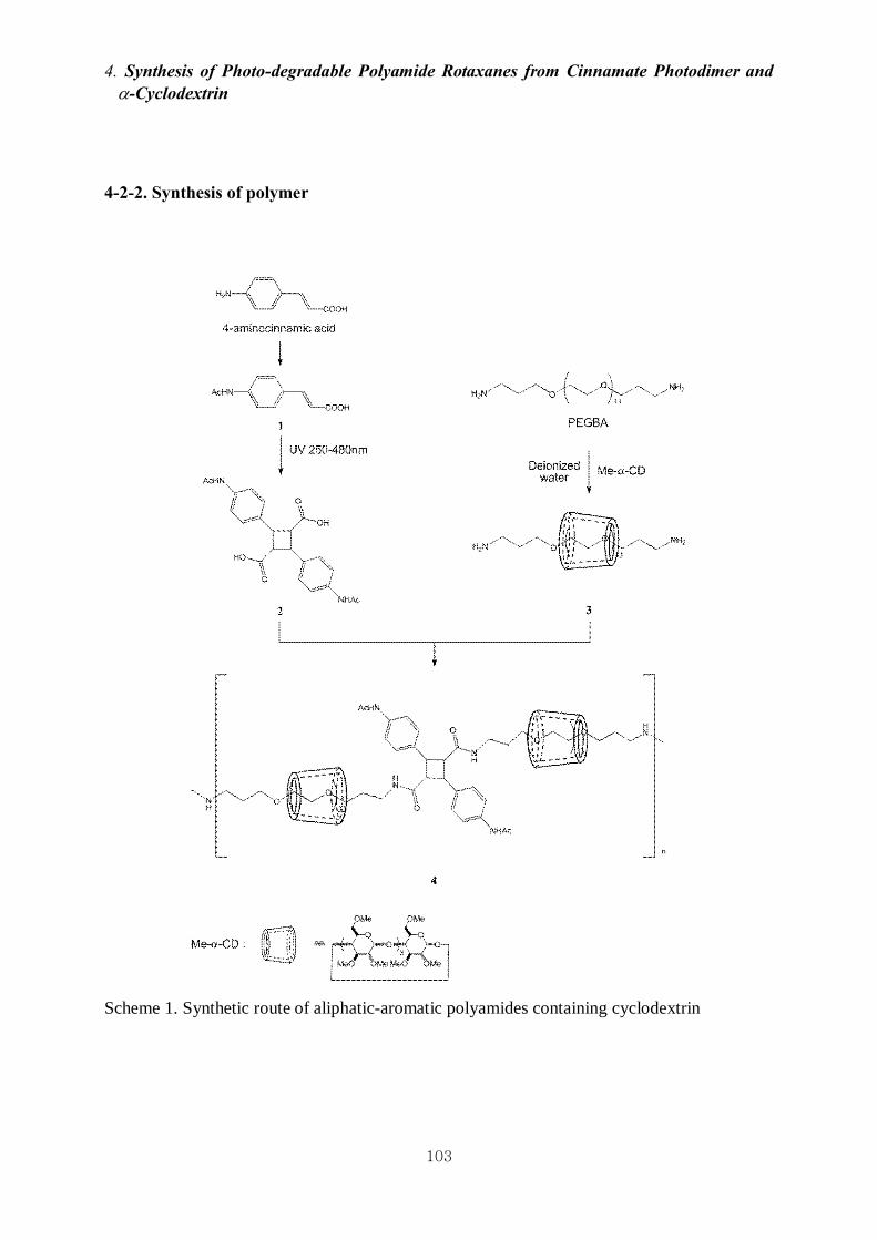
4. Synthesis of Photo-degradable Polyamide Rotaxanes from Cinnamate Photodimer and α-Cyclodextrin
103
4-2-2. Synthesis of polymer
Scheme 1. Synthetic route of aliphatic-aromatic polyamides containing cyclodextrin

4. Synthesis of Photo-degradable Polyamide Rotaxanes from Cinnamate Photodimer and α-Cyclodextrin
104
Preparation of N-acetyl-4-aminocinnamic acid 1
4-aminocinnamic acid (4-ACA; 2.47 g, 15.13 mmol) was dissolved in 30 ml of methanol,
and acetic anhydride (5.40 g, 52.98 mmol) was then added dropwise with slow stirring under
a nitrogen atmosphere at room temperature. After the complete addition of the acetic
anhydride, the mixture was stirred for 23 hrs. The precipitate was filtered, washed with
methanol, and then dried under reduced pressure to give 1 as a yellow powder at 79 % yield.
1H NMR (400 MHz, DMSO-d6) δ / ppm 12.63 - 12.02 (1H, br, s), 10.13 (1H, s), 7.61 (4H, s),
7.52 (1H, d, J = 16 Hz), 6.40 (1H, d, J = 16 Hz), 2.05 (3H, s); 13C NMR (100 MHz, DMSO-
d6) δ / ppm 168, 167, 143, 141, 128, 118, 117, 24; MALDI-TOF [M+Na]+ m / z : Calcd. for
C11H11NO3Na 228.07, Found 228.0.
Preparation of 4,4’-diacetoamido-α-truxillic acid 2
The photochemical [2+2] cycloaddition of the cinnamoyl group in 1 was utilized to form
the cyclobutane structure. 1 (1.00 g, 4.87 mmol) was dispersed in hexane (10 ml), and then
the photoreaction was carried out with UV irradiation using a mercury lamp (250-480 nm,
light intensity: 70 mW / cm2)13 with stirring for 24 hrs. After UV-irradiation, the product was
washed with methanol, and then dried under reduced pressure to give 2 as a yellow powder at
95% yield. 1H NMR (400 MHz, DMSO-d6) δ / ppm 12.4 - 11.8 (2H, br, s), 9.91 (2H, s), 7.51

4. Synthesis of Photo-degradable Polyamide Rotaxanes from Cinnamate Photodimer and α-Cyclodextrin
105
(4H, d, J = 8 Hz), 7.26 (4H, d, J = 8 Hz), 4.20 (2H, dd, J = 4, 20 Hz), 3.74 (2H, dd, J = 4, 20
Hz), 2.03 (6H, s); 13C NMR (100 MHz, DMSO-d6) δ / ppm 172, 168, 137, 133, 127, 118, 46,
40, 23; MALDI-TOF [M+Na]+ m / z : Calcd. for C22H22N2O6Na 433.14, Found 433.2.
Preparation of methylated α-CD (Me-α-CD) 20-22
α-CD (1.01 g, 1.03 mmol) in anhydrous DMF (40 ml) was treated with sodium hydride
(0.62 g, 25.92 mmol, 1.4 equiv to the OH groups of CD) for 2 hrs under a nitrogen
atmosphere at room temperature. Next, iodomethane (3.68 g, 25.92 mmol) was added
dropwise to the solution with stirring for 24 hrs at room temperature. Methanol was then
added to decompose the excess hydride, and the solvent was evaporated and dried under
reduced pressure for 36 hrs. After washing with hexane, the product was dissolved in
dichloromethane (100 ml), and then washed with deionized water, concentrated, and dried
under reduced pressure. The crude product was purified by recrystallization in hexane. The
residual oil in the product was removed by hexane and then dried to give Me-α-CD as a white
powder at 43 % yield. 1H NMR (400 MHz, DMSO-d6) δ / ppm 4.95 (6H, d, J = 4 Hz), 3.80 -
3.68 (12H, m), 3.52 (6H, d, J = 9.6 Hz), 3.47 (18H, s), 3.41 (6H, t, J = 8.8 Hz), 3.36 (18H, s),
3.21 (18H, s), 3.01 (6H, dd, J = 12, 4 Hz); 13C NMR (100 MHz, DMSO-d6) δ / ppm 98, 81.7,
81.5, 81, 71, 70, 60, 58, 57; MALDI-TOF [M+Na]+ m / z : Calcd. for C54H96O30Na 1247.59,

4. Synthesis of Photo-degradable Polyamide Rotaxanes from Cinnamate Photodimer and α-Cyclodextrin
106
Found 1247.7.
Synthesis of 4
The inclusion complex (pseudopolyrotaxane) 3 of PEGBA and Me-α-CD was formed in
deionized water at various molar compositions ([Me-α-CD] / [PEGBA] = 0, 1, 3, 6, 13).
PEGBA and Me-α-CD were dissolved in deionized water (140 mg/mL) at 60 ℃ for 24 hrs,
and then freeze-dried. 3 and 2 were dissolved in anhydrous dimethylacetamide (DMAc,
133mM) and then triphenylphosphite (TPP, 5 equimolar to PEGBA) and anhydrous pyridine
(5 equimolar to PEGBA) as condensation agents23 were added to the mother solution. The
reaction mixtures ([Me-α-CD] / [PEGBA] = 0, 1, 3, 6, 13) were heated in an oil bath until
100 ℃ at a heating rate of 10 ℃ / 10 min under a nitrogen atmosphere with stirring for 1, 2,
3, 4 and 5 days, respectively. After cooling, DMSO (10 ml) was added into the reaction
solution and then dialyzed with a dialysis membrane (MWCO: 25 kDa) in methanol for 5
days. The products were dried under reduced pressure at 80 ℃ for 24 hrs to give 4. 1H NMR
(400 MHz, DMSO-d6) δ / ppm 10.02 - 9.74 (2H, m), 7.66 (2H, s), 7.46 (4H, d J = 8.4), 7.14
(4H, d J = 8.4), 4.95 (0.82H, s). 4.16 (2H, t, J = 8.8 Hz), 3.66 (2H, t, J = 8.8 Hz), 3.61 - 3.38
(132H, m), 3.35 (2.46H, s), 3.32 (4H, t, J = 2.8 Hz), 3.22 (2.46H, s), 3.02 (4H, t, J = 5.6 Hz),
1.99 (6H, s), 1.22 (4H, s)

4. Synthesis of Photo-degradable Polyamide Rotaxanes from Cinnamate Photodimer and α-Cyclodextrin
107
4-2-3. Measurement
1H NMR and 13C NMR spectra were recorded on a Burker DRX 500 NMR instrument
(400.13 MHz for 1H NMR) using DMSO-d6 (internal reference 2.50 ppm) as a solvent at
room temperature. The molecular weight of 4 was determined using gel permeation
chromatography (Shodex GPC-101 with a connection column system of 803 and 807). The
chromatogram was calibrated with polystyrene standards. Dimethylformamide was utilized as
the eluent at a flow rate of 0.5 mL / min. The thermal properties were analyzed by differential
scanning calorimetry (DSC; EXSTAR 6100, Seiko Instruments Inc.) and thermogravimetric
analysis (TGA; SSC/5200 SII, Seiko Instruments Inc.). The DSC thermograms covered a
temperature range from -100 to 250 ℃ at a scanning rate of 10 ℃ / min under a nitrogen
atmosphere. The TGA was carried out at a heating rate of 10 ℃ / min under a nitrogen
atmosphere.
4-2-4. UV degradation
4e dissolved in DMSO was irradiated with a UV lamp (light intensity: 2.5 mW / cm2, λ =
254 nm; xenon light source, MAX-303, Asahi Spectra Inc.) in the open air for 6 days. The

4. Synthesis of Photo-degradable Polyamide Rotaxanes from Cinnamate Photodimer and α-Cyclodextrin
108
degradation of 4e with increasing UV exposure time was observed by 1H NMR and GPC
analysis.
4-3. Results and discussion
4-3-1. Synthesis and Characterization of Polyamides 4
The synthesis protocol for the aliphatic-aromatic polyamides is shown in Scheme 1. 4-
aminocinnamic acid (4-ACA) was utilized as a base compound in order to prepare
photodimer 2. The amine group of 4-ACA was protected by acetic anhydride to inhibit side
reactions during the polymerization. 2 was prepared by using photonic [2+2] cycloaddition
between the vinylene groups of cinnamic acid via UV irradiation in the solid state. The [2+2]
cycloaddition was confirmed by 1H NMR analysis (Figure 1). The new signal from the
cyclobutane ring was observed at 4.20 and 3.74 ppm, and the signal from the aromatic group
in 1 was shifted from 7.61 ppm to 7.51 and 7.26 ppm. All of the hydroxyl groups in α-CD
were methylated by iodomethane in order to prevent the reaction of the CD hydroxyls with
the 2 carboxyls during the polyamide polymerization. In addition, the CD methylation should
prevent hydrogen bonding between the hydroxyl groups in CD after rotaxanation. The

4. Synthesis of Photo-degradable Polyamide Rotaxanes from Cinnamate Photodimer and α-Cyclodextrin
109
inter/intra-molecular interactions were enhanced by the structural effects of polyrotaxane to
form crystalline complexes and to affect their thermo-mechanical properties.
Polymerizations of the photodimer 2 with diaminated pseudopolyrotaxane 3 were carried out
via polyamide reactions using triphenylphosphite/pyridine as condensation agents. In this
synthetic stage, we focused on controlling the CD composition in the inclusion complex and
removing free Me-α-CDs liberating from the PEG backbone. In order to control the
composition of the inclusion complex, polymerizations were performed while changing the
CD concentration from 133 mM to 1.73 M in solvent, i.e. the amount of Me-α-CDs in 3
increased from 0.067 mmol to 0.871 mmol. As a result, we synthesized 4 with various CD
compositions (Table 1). The structure of 4 was confirmed by NMR analyses. In the 1H NMR
spectrum, the signals from the PEG-BA, 2 and C (1) H in Me-α-CD were detected, and the
molar ratio of each component was summarized in Table 1. The signal from the amide bond
between PEG-BA and 2 also appeared at 7.66 ppm.

4. Synthesis of Photo-degradable Polyamide Rotaxanes from Cinnamate Photodimer and α-Cyclodextrin
110
Figure 1. 1H NMR spectra of: (a) Me-α-CD, (b) 2, and (c) 4 (400 MHz, DMDO-d6)

4. Synthesis of Photo-degradable Polyamide Rotaxanes from Cinnamate Photodimer and α-Cyclodextrin
111
Table1. Basic characterization of aliphatic-aromatic polyamide 4 containing cyclodextrin
Molar composition
[Me-α-CD] / [PEGBA] Name
Feed Found a
Mw b (g / mol) PI Yield (%) c
4a 0 0 (-) 2.64 × 10⁴ 1.21 42
4b 1 0.018 (1.8%) 2.82 × 10⁴ 1.18 46
4c 3 0.030 (1.0%) 1.91 × 10⁴ 1.21 49
4d 6 0.064 (1.1%) 1.97 × 10⁴ 1.18 31
4e 13 0.137 (1.1%) 1.74 × 10⁴ 1.18 47
a Determined by 1H NMR analysis. The rotaxanation degree of CD to the amount of CD used
in the feed is shown in parenthesis; b Determined by GPC analysis; c Based on the amount of
PEGBA and 2

4. Synthesis of Photo-degradable Polyamide Rotaxanes from Cinnamate Photodimer and α-Cyclodextrin
112
The rotaxanation and purification were also confirmed by GPC, in which just one peak
around an elution time of 37-38 min for 4a and 4e was observed (Figure 2). A mixture of 4a
and Me-α-CD whose composition was adjusted to be the same as 4e was applied to the GPC
in order to confirm the removal of the Me-α-CD from 4e. Chromatogram (a) showed two
independent peaks around 37.0 and 44.5 min. Since the peaks at 37.0 min were clearly
assigned to polyamide 4a, since the shape and elution time were almost the same as the peak
in chromatogram (b), the other peak at 44.5 nm could be assigned to the Me-α-CD. As shown
in Figure 2, chromatogram (c) showed no signal assignable to free Me-α-CD. Chromatogram
(c) showed no peak around 44.5 nm, indicating the removal of the free Me-α-CD to obtain the
pure inclusion complex 4e. When we tried to form the inclusion complex by a direct reaction
of photodimer 2 with PEG-BA in the presence of Me-α-CD, the 1H NMR spectrum of the
resulting polymer showed a negligibly low signal for Me-α-CD. This result suggests that the
accidental rotaxanation in DMAc barely occurred, and the very low rotaxanation degree
ranging from 1.0-1.8 % shown in Table 1 means that the speed of CD dissociation from the
PEG was much faster than the capping rate of 3 with 2. As a consequence, these results
indicated that the inclusion complex of Me-α-CD with the polyamide chain was successfully
formed during the polymerization of 2 with 3, and that the pure product could be isolated by
the present method.

4. Synthesis of Photo-degradable Polyamide Rotaxanes from Cinnamate Photodimer and α-Cyclodextrin
113
Figure 2. Representative GPC chromatograms of (a) a mixture of 4a and Me-α-CD, (b) 4a,
and (c) 4e (DMF, 0.5 mL/min, 40℃)
4-3-2. Thermal properties of 4
The thermo-mechanical performance of 4 was measured by DSC (Figure 3) and TGA
(Figure 4) analyses. As depicted in Figure 3, the DSC curves of 4a - 4e displayed a similar
curve pattern. The Tg of each polyamide was almost the same (-54℃). The Tm also showed no
outstanding changes, although the Tc showed a slight increase and decrease (Table 2). Upon
the increased amount of Me-α-CD threaded onto the back bone chain, the intensity of both the
exothermic peak of crystallization and the endothermic peak of melting decreased with an
increasing CD amount. This phenomenon means that the presence of CD made polymer
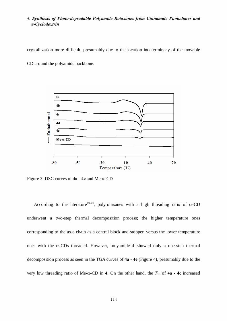
4. Synthesis of Photo-degradable Polyamide Rotaxanes from Cinnamate Photodimer and α-Cyclodextrin
114
crystallization more difficult, presumably due to the location indeterminacy of the movable
CD around the polyamide backbone.
Figure 3. DSC curves of 4a - 4e and Me-α-CD
According to the literature10,24, polyrotaxanes with a high threading ratio of α-CD
underwent a two-step thermal decomposition process; the higher temperature ones
corresponding to the axle chain as a central block and stopper, versus the lower temperature
ones with the α-CDs threaded. However, polyamide 4 showed only a one-step thermal
decomposition process as seen in the TGA curves of 4a - 4e (Figure 4), presumably due to the
very low threading ratio of Me-α-CD in 4. On the other hand, the T10 of 4a - 4c increased

4. Synthesis of Photo-degradable Polyamide Rotaxanes from Cinnamate Photodimer and α-Cyclodextrin
115
gradually with an increase in the amount of CD in 4, whereas the T10 of 4d and 4e decreased
and approached the T10 of Me-α-CD. The T50 also showed a similar trend with the T10 (Table
2). As a result, the T10 of the polymer increased after the threading of a very small amount of
CD (0.018 and 0.030) to the chain backbone, owing to the chains being rendered rigid by
rotaxanation.
Figure 4. TGA curves of 4a - 4e and Me-α-CD

4. Synthesis of Photo-degradable Polyamide Rotaxanes from Cinnamate Photodimer and α-Cyclodextrin
116
Table 2. DSC / TGA analysis of polyamides and Me-α-CD
Name T10 /℃ T50 /℃ Tg /℃
Tc /℃
(△Hc /Jg-1)
Tm /℃
(△Hm /Jg-1)
4a 357 401 -54 7 (-20.2) 30 (19.2)
4b 367 406 -54 11 (-14.6) 29 (17.1)
4c 373 408 -54 12 (-10.8) 28 (10.1)
4d 356 398 -53 14 (-5.31) 26 (6.6)
4e 346 396 -54 9 (-1.92) 27 (2.18)
Me-α-CD 346 373 70 127 218
4-3-3. UV-degradation
Figure 5 depicts the changes of the 1H NMR spectra of 4e before and after UV irradiation.
Yang et al. reported that the cyclobutane structure of α-truxillic acid dimethyl ester, which
was formed by [2+2] cycloaddition with UV irradiation (λ > 260 nm), was photocleaved into
two cinnamic acids by means of UV irradiation (λ < 260 nm).19 We performed a
photocleavage test of 4e using a xenon-lamp (2.5 mW / cm2) (Scheme 2). After illumination
to the 4e solution in DMSO (20 mg / mL) for 6 days, 1H NMR and GPC analysis were carried

4. Synthesis of Photo-degradable Polyamide Rotaxanes from Cinnamate Photodimer and α-Cyclodextrin
117
out to confirm the cleavage of 4e. From the NMR analysis (Figure 5 (b)), two peaks at 6.50
and 7.35 ppm appeared after UV-irradiation for 6 days. The two peaks corresponded with
those of the vinylene groups of 4-acetoamidocinnamamide conjugated with PEG-BA (Figure
5 (a)). At the same time, the integration intensity of the cyclobutane peak at 4.16 ppm
decreased obviously. These results support the photocleavage of the cyclobutane ring of 4e by
UV-irradiation. The photocleavage of polyamide 4e was also confirmed by GPC analysis
(Figure 6). With an increasing time of UV exposure, the peak of 4e was shifted to a higher
elution time, and the molecular weight of 4e also decreased from 1.74 × 10⁴ to 5.28 × 10³
g / mol.
Scheme 2. Degradation of 4e with UV exposure

4. Synthesis of Photo-degradable Polyamide Rotaxanes from Cinnamate Photodimer and α-Cyclodextrin
118
Figure 5. 1H NMR spectra of: (a) 4-acetoamidocinnamamide conjugated with PEG-BA, (b) 4e
after UV exposure for 6 days, and (c) 4e (400 MHz, DMDO-d6)
Figure 6. GPC chromatograms of 4e (a) before and (b) after UV exposure for 6 days (DMF,
0.5 mL/min, 40℃)

4. Synthesis of Photo-degradable Polyamide Rotaxanes from Cinnamate Photodimer and α-Cyclodextrin
119
4-4. Conclusion
Polyamide rotaxanes were prepared by a polycondensation of 4,4’-acetoamido-α-truxillic
acid with diaminated pseudorotaxanes from an inclusion complex of polyethyleneglycol
bisamine with methylated-α-cyclodextrin. The thermal degradation temperature of the
polyamides was enhanced by inclusion complex formation with methylated-α-cyclodextrin,
even when a very small amount of methylated-α-cyclodextrin was threaded into the polymer
backbone. The polyamide polyrotaxane with methylated-α-cyclodextrin was degraded by
UV-irradiation (λ < 260 nm) via the photocleavage of the cyclobutane ring in 4,4’-
diacetoamido-α-truxillic acid to 4-aminocinnamic acid.
Acknowledgement: The researches were financially supported by ALCA project (5100270)
of JST.

4. Synthesis of Photo-degradable Polyamide Rotaxanes from Cinnamate Photodimer and α-Cyclodextrin
120
Reference
1. Kaneko, T.; Tran, H.T.; Matsusaki, M.; Akashi, M. Chem. Mater., 2006, 18, 6220.
2. Harada, A.; Kamachi, M. J. Chem. Soc. Chem. Commun., 1990, 19, 1322.
3. Harada, A.; Li, J.; Kamachi, M. Nature, 1992, 356, 325.
4. Wenz, G.; Han, B.H.; Müller, A. Chem. Rev., 2006, 106, 782.
5. Ooya, T.; Eguchi, M.; Yui, N. J. Am. Chem. Soc., 2003, 125, 13016.
6. Okumura, Y.; Ito, K. Adv. Mater., 2001, 13, 485.
7. Lee, W.K.; Ichi, T.; Ooya, T.; Yamamoto, T.; Katoh, M.; Yui, N. J. Biomed. Mater. Res. A,
2003, 67, 1087.
8. Yamashita, A.; Yui, N.; Ooya, T.; Kano, A.; Maruyama, A.; Akita, A.; Kogure, K.;
Harashima, H. Nat. Protoc., 2007, 1, 2861.
9. Ooya, T.; Yui, N. J. Control. Release, 1999, 58, 251.
10. Yamaguchi, I,; Miya, K.; Osakada, K.; Yamamoto, T. Polym. Bull., 2000, 44, 247.
11. Tong, X.; Hou, D.; Zhang, X.; Ye, L.; Zhang, A.; Feng, Z. Chinese J. Polym. Sci., 2008,
26, 723.
12. Lendlein, A.; Jiang, H.; Jünger, O.; Langer, R. Nature, 2005, 434, 879.
13. Yasaki, K.; Suzuki, T.; Yazawa, K.; Kaneko, D.; Kaneko, T. J. Polym. Sci. A1, 2011, 48,

4. Synthesis of Photo-degradable Polyamide Rotaxanes from Cinnamate Photodimer and α-Cyclodextrin
121
1112.
14. Pattabiraman, M.; Kaanumalle, L.S.; Natarajan, A.; Ramamurthy, V. Langmuir, 2006, 22,
7605.
15. Kaneko, T.; Thi, T.H.; Shi, D.J.; Akashi, M. Nat. Mater., 2006, 5, 966.
16. Kaneko, T.; Kaneko, D.; Wang, S. Plant Biotechnol., 2010, 27, 243.
17. Wang, S.; Tateyama, S.; Kaneko, D.; Ohki, S.; Kaneko, T. Polym. Degrad. Stab., 2011, 96,
2048.
18. Shi, D.J.; Matsuaski, M.; Kaneko, T.; Akashi, M. Macromolecules, 2008, 41, 8167.
19. Yang, H.; Jia, L.; Wang, Z.; Di-Cicco, A.; Levy, D.; Keller, P. Macromolecules, 2011, 44,
159.
20. Guieu, S.; Sollogoub, M. J. Org. Chem., 2008, 73, 2819.
21. Takeo, K.; Mitoh, H.; Uemura, K. Carbohydr. Res., 1989, 187, 203.
22. Arai, T.; Hayashi, M.; Takagi, N.; Takata, T. Macromolecules, 2009, 42, 1881.
23. Yamashita, Y.; Chujo, Y.; Kobayashi, H.; Kawakami, Y. Polym. Bull., 1981, 5, 361.
24. Tong, X.; Zhang, X.; Ye, L.; Zhang, A.; Feng, Z. Soft Matter., 2009, 5, 1848.

General Conclusion

5. General Conclusion
123
5 General Conclusion
Every chapter was summarized by described below.
In chapter 2, we demonstrated the design and preparation of the hydrolyzable polyrotaxane
composed of an ester-containing PEG chain and α-CDs. The hydrolysis of polyrotaxane into
its water-soluble components under aqueous conditions adjusted to both a basic and almost
neutral pH was confirmed by monitoring continuous changes in transmittance and by GPC
measurements of clear upper portions collected from suspensions.
In chapter 3, we prepared series of polyrotaxanes containing ester linkages in the main
chain to trigger the supramolecular dissociation in water by means of hydrolysis of the esters.
Crosslinking of the hydrolyzable polyrotaxanes was carried out in two ways, one was a
crosslinking between intermolecular cyclic components (internal crosslink) and the other was
a crosslinking between the terminals of the polyrotaxanes (terminal crosslink). Hydrogel I-IV
showed high water content and hydrophilic surface. The internally-crosslinked gels had
higher compressive stress and initial modulus, while the terminally-crosslinked gels showed
higher ultimate strain. Viscoelastic behavior of internally-crosslinked hydrogels was very

5. General Conclusion
124
different from that of terminally-crosslinked hydrogels and similar to that of topological gel.
The terminally-crosslinked hydrogels were more stable during degradation than the
internally-crosslinked hydrogels in PBS. These degradations accompanied decrease in the
storage modulus, and showed various degradation profiles, which were modulated by the
crosslinking manners and molecular weights of the polyrotaxanes. A preliminary cell
adhesion test using NIH3T3 cells onto the terminally-crosslinked hydrogels demonstrated
cellular responses by means of decrease in storage modulus and change of surface chemical
characteristics of hydrogels. These results could provide a possibility to develop
polyrotaxane-based hydrogel systems as biodegradable materials for tissue engineering, such
as implantable scaffold and drug delivery system, since polyrotaxanes are allowed to be
designed uniquely by modulating lots of structural parameters, such as various crosslinking
methods, combinations of polyrotaxane component, and molecular weight of the back bone
polymer.
In chapter 4, polyamide rotaxanes were prepared by a polycondensation of 4,4’-
acetoamido-α-truxillic acid with diaminated pseudorotaxanes from an inclusion complex of
polyethyleneglycol bisamine with methylated-α-cyclodextrin. The thermal degradation
temperature of the polyamides was enhanced by inclusion complex formation with
methylated-α-cyclodextrin, even when a very small amount of methylated-α-cyclodextrin

5. General Conclusion
125
was threaded into the polymer backbone. The polyamide polyrotaxane with methylated-α-
cyclodextrin was degraded by UV-irradiation (λ < 260 nm) via the photocleavage of the
cyclobutane ring in 4,4’- diacetoamido-α-truxillic acid to 4-aminocinnamic acid.

Achievement
126
ACHIEVEMENT
Articles
1. Hojoon SHIN, Seiji TATEYAMA, and Tatsuo KANEKO ; Synthesis of Photo-degradable
Polyamide Rotaxanes with Cinnamate Photodimer and Cyclodextrin. Journal of Materials
Life Society, Accepted.
2. Ryo KATOONO, Shin-ichiroh FUKUDA, Hojoon SHIN, and Nobuhiko YUI ; Preparation
of hydrolyzable polyrotaxane containing ester linkages and its degradation behavior.
Chemistry Letters. Vol. 37 (2008), No. 9, pp.988-989
Presentations
1. Hojoon SHIN, Seiji TATEYAMA, and Tatsuo KANEKO, Synthesis of bio-based aliphatic-
aromatic polyamides containing cyclodextrin via solid inclusion complexes. ICBP, Seoul,
2013.
2. 辛昊俊, 上遠野亮, Jung-woog SHIN, 由井伸彦, エステル含有ポリロタキサン架
橋ヒドロゲルの加水分解過程における特性の解析. 第39回医用高分子シンポジウ
ム, Tokyo, 2010.
3. Hojoon SHIN, Ryo KATOONO and Nobuhiko YUI ; Preparation of polyrotaxane
hydrogels based on water-soluble hydrolyzable polyrotaxane. 2nd AMBC, Singapore, 2009.

Achievement
127
4. Hojoon SHIN, Ryo KATOONO, and Nobuhiko YUI ; Preparation of supramolecular
hydrogels composed of hydrolyzable polyrotaxanes and their mechanical properties and
degradation behavior. 第58回高分子学会年次大会, Kobe, 2009.
5. Hojoon SHIN, Shin-ichiroh FUKUDA, Ryo KATOONO and Nobuhiko YUI ; Preparation
of hydrogels based on hydrolyzable polyrotaxane. MACRO 2008, Taipai, 2008.
6. Hojoon SHIN, Ryo KATOONO and Nobuhiko YUI ; Preparation of supramolecular
hydrogels based on biodegradable polyrotaxanes cross-linked by multi-armed PEG.
第57回高分子学会年次大会, Yokohama, 2008.
7. Hojoon SHIN, Shin-ichiroh FUKUDA, Ryo KATOONO and Nobuhiko YUI ; Preparation
of supramolecular hydrogels composed of hydrolyzable polyrotaxane and their
mechanical properties. 第57回高分子討論会, Osaka, 2008.

Acknowledgment
128
ACKNOWLEDGMENT
The author’s supreme appreciation is owed on Prof. Tatsuo Kaneko who is my supervisor,
for his guidance, dedicated support and continuous motivation during research and the writing
of this dissertation. When I suffered from serious mental pain, his advice gave me metal
vitality and courage to overcome every trouble.
The advice and support of the other member of his committee, namely, Prof. Ebitani, Prof.
Matsumi, Prof. Miura, and Prof. Yamaguchi also gratefully acknowledged.
I would like to express my gratitude to Research Assistant Dr. Seiji Tateyama for his
supports laid a smooth way for my research. Through rewarding discussions, I have gained
much more knowledge in experimental technique. At the same time, I would like to express
my appreciation to Kaneko Lab members. They supported my experiment and living truly in
JAIST for a long time.
When I came to Japan for the first time in order to challenge doctor degree, Prof. Yui gave
me good chance of studying abroad and to challenge new research filed. He supported my
early school life and taught seriousness of scientific research filed. Also, Assistant Prof.
Katoono, he taught many things to me about chemical experiment and basic attitude as
chemist. I would like to thank two Professors and all Yui Lab members.

Acknowledgment
129
For my Minor-research theme and advice on technique of cellular experiment, I thank Prof.
Jung-woog Shin (Inje University) and Prof. Masayuki Yamaguchi. I also would like to thank
many Lab members in Prof. Shin and Prof. Yamaguchi Lab.
I would like to thank Dr. Yamaoka, Dr. Kakinoki, and Dr. Yamashita in Department of Bio
medical Engineering, National Cerebral and Cardiovascular Center. They supported and
advised my cellular experiment using hydrogels truly. Thanks to their help, I was able to
endure and finish hard experiment in Osaka for a long time.
Finally, I would like to express my thanks to Korean society members in JAIST.
Hojoon Shin
March, 2014

Minor-Research Theme
130
Minor-Research Theme
Quantitative Measurement of Cell Adhesive Force between
Cell and PLGA Surface
HOJOON SHIN
Supervised by Professor Jung-Woog Shin, Ph.D, 1
and Professor Masayuki Yamaguchi, Ph.D 2
1Collage of Biomedical Science & Engineering, Inje University (Korea) 2School of Materials Science, Japan Advanced Institute of Science & Technology (Japan)
School of Materials Science
Japan Advanced Institute of Science and Technology
March 2009

Minor-Research Theme
131
ABSTRACT
Adhesion of an anchorage dependent cell to extracellular matrix is a fundamental and
important process in physiological cell behavior such as survival, proliferation, differentiation,
activation and movement. Qualitative and quantitative measurements have been adopted to
measure adhesive force. In this study, we utilized a micropipette as a cantilever beam to
measure quantitative cell adhesive force. The adhesive force was calculated by using the
relationship between the deflection and force experienced by the pipette when detaching a cell
from polymer surface. BMSC was seeded on PLGA-coated glass surface and cultured for 4hrs
and 12hrs. The shape and dimension of pipette was measured by using image analysis
software and the pipette was reconstructed with finite element analysis program and the
amount of deflection measured was imposed in the program. The average attachment forces
of the cells on PLGA at 4 hrs and 12 hrs were 0. 13 ± 0.015 nN and 0.42±0.035 nN,
respectively. Evaluating cell attachment more precisely by measuring the detachment force
quantitatively could allow finding of optimal condition of parameters related to surface
condition.
Keywords : Quantitative measurements, Adhesive force, BMSC, PLGA, Cell adhesive force