osteologia - archeo.uw.edu.pl · określanie wieku • szwy czaszkowe 17 left coronal (endocr.)...
TRANSCRIPT
Osteologia
Określanie wieku i płci

Określanie wieku• wiek zębowy

Określanie wieku• wiek zębowy

Określanie wieku• wiek zębowy
19.113.98.513.512.712.48.97.9—————Root apex half closedA ½
17.512.37.012.211.211.08.37.33.12.03.11.751.5Root completeRc
16.411.46.111.210.29.97.76.7—————Root three-quartersR¾
——————7.26.2—————Root two-thirdsR 2/3
15.610.65.510.19.38.86.65.6—————Root halfR ½
14.89.84.98.67.86.95.8——————Root one-quarterR ¼
14.18.74.1——————————Root cleft presentRcl
13.27.63.27.36.45.2———————Root initiatedRi
12.46.82.56.65.64.4——0.70.40.70.20.15Crown completeCrc
11.86.11.95.84.93.4———————Crown three-quartersCr ¾
11.35.41.35.04.12.5———————Crown one halfCr ½
10.64.90.84.53.31.7———————Crown outline completeCoc
10.04.30.43.92.61.0———————Cusp coalescenceCeo
9.53.80.13.22.10.6———————Cusp initiationCi
M3M2M1P4P3CI2I1dm2dm1dcdi2di1A. MALES
Określanie wieku• wiek zębowy
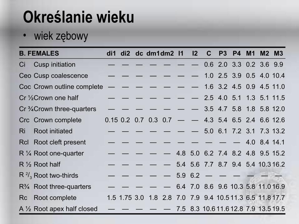
19.513.57.912.811.610.68.37.5—————Root apex half closedA ½
17.711.86.511.310.59.47.97.02.81.83.01.751.5Root completeRc
16.911.05.810.39.68.67.06.4—————Root three-quartersR¾
——————6.25.9—————Root two-thirdsR 2/3
16.210.35.49.48.77.75.65.4—————Root halfR ½
15.29.54.88.27.46.25.04.8—————Root one-quarterR ¼
14.18.44.0——————————Root cleft presentRcl
13.27.33.17.26.15.0———————Root initiatedRi
12.66.62.46.55.44.3——0.70.30.70.20.15Crown completeCrc
12.05.81.85.84.73.5———————Crown three-quartersCr ¾
11.55.11.35.14.02.5———————Crown one halfCr ½
11.04.50.94.53.21.6———————Crown outline completeCoc
10.44.00.53.92.51.0———————Cusp coalescenceCeo
9.93.60.23.32.00.6———————Cusp initiationCi
M3M2M1P4P3CI2I1dm2dm1dcdi2di1B. FEMALES
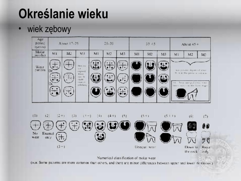
Określanie wieku• wiek zębowy

Określanie wieku• wiek zębowy
12-1812-18
16-2016-20
16-2016-20
18-2218-22
20-2420-24
12-1812-18
16-2016-20
16-2016-20
18-2218-22
20-2420-24
20-3020-30
30-3530-35
35-4035-40
40-5040-50
20-3020-30
30-3530-35
35-4035-40
40-4540-45
45-5545-55
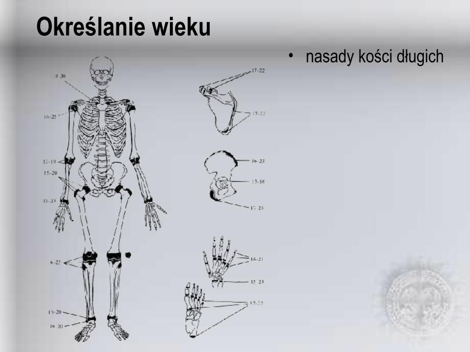
Określanie wieku• nasady kości długich
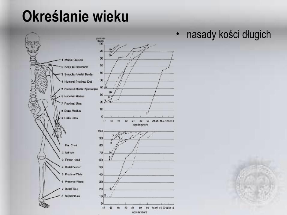
Określanie wieku• nasady kości długich
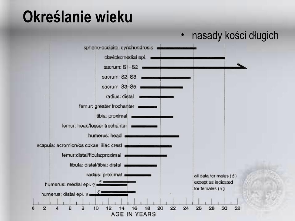
Określanie wieku• nasady kości długich
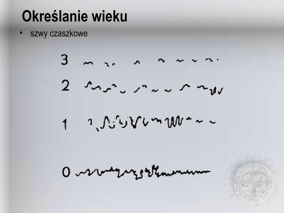
Określanie wieku• szwy czaszkowe

Określanie wieku• szwy czaszkowe

Określanie wieku• szwy czaszkowe
Score indicated portionLeft Coronal (endocr.)17
Score indicated portionLeft Lambdoidal (endocr.)16
Entire sagittal suture endocraniallySagittal (endocr.)15
Score entire lengthTransverse Palatine14
Score entire lengthPosterior Median Palatine13
Score entire length on paired maxillae between incisive foramen and palatine boneAnterior Median Palatine12
Incisive suture separating maxilla and premaxillaIncisive Suture11
On left sphenotemporal suture 2 cm below junction with parietalSuperior Sphenotemporal10
Intersection between left sphenotemporal suture and line between articular tubercles of the temporomandibular jointInferior Sphenotemporal9
Midpoint of left sphenofrontal sutureSphenofrontal8
Usually where parietosphenoid suture meets the frontalPterion7
Midpoint of left coronal sutureMidcoronal6
At bregmaBregma5
One-third the distance from bregma to lambdaAnterior Sagittal4
At obelionObelion3
Intersection of sagittal and lambdoidalLambda2
Midpoint of L. lambdoid sutureMidlambdoid1
DescriptionSite Name

Określanie wieku• szwy czaszkowe
——2112.651.519-2010.548.816-1812.645.212-159.139.47-117.834.73-69.630.51-2——0
Standard DeviationMean AgeComposite
Score
——158.556.211-14
12.551.99-108.945.57-8
10.743.4610.041.13-56.236.228.332.01——0
Standard DeviationMean AgeComposite
Score
punkty 1-7punkty 1-7punkty 6-10punkty 6-10
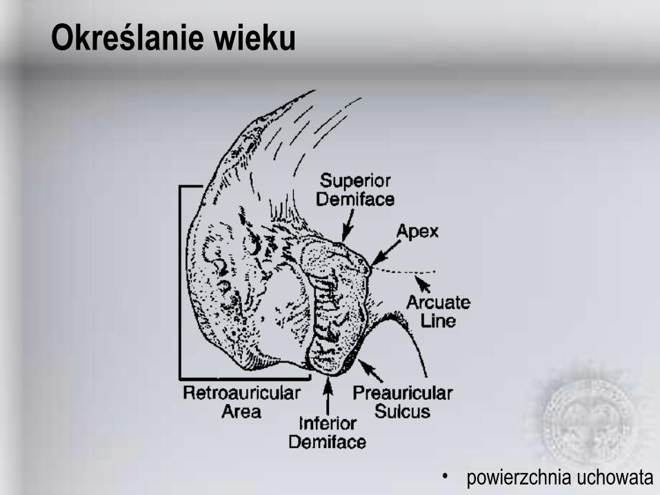
Określanie wieku
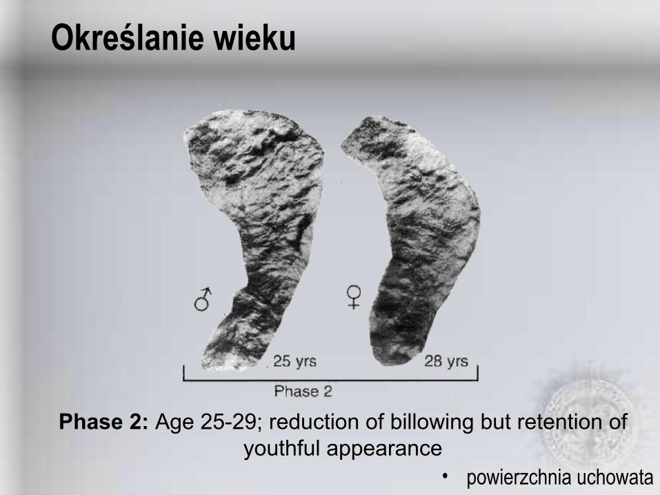
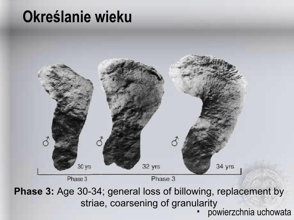
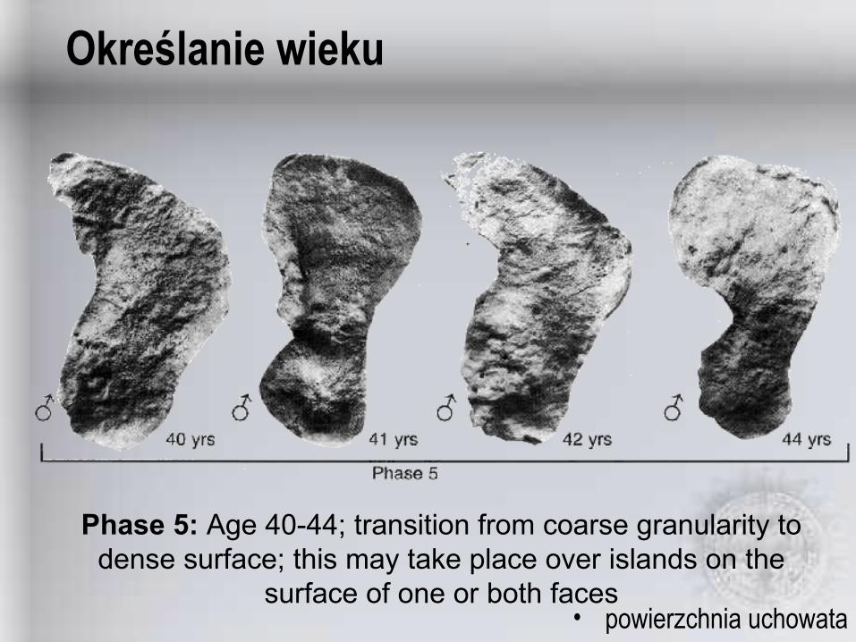
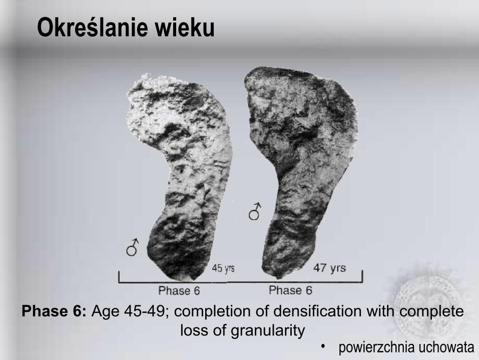
• powierzchnia uchowata

Określanie wiekuModal changes to the auricular surface with age. Phases described by Lovejoy et al. (1985b) as follows:
– Phase 1: Age 20-24; billowing and very fine granularity– Phase 2: Age 25-29; reduction of billowing but retention of youthful
appearance– Phase 3: Age 30-34; general loss of billowing, replacement by striae,
coarsening of granularity– Phase 4: Age 35-39; uniform coarse granularity– Phase 5: Age 40-44; transition from coarse granularity to dense surface; this
may take place over islands on the surface of one or both faces– Phase 6: Age 45-49; completion of densification with complete
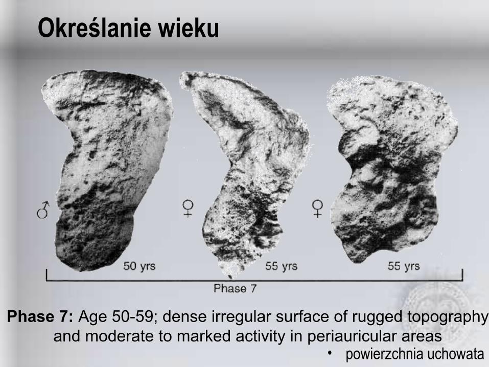
loss of granularity– Phase 7: Age 50-59; dense irregular surface of rugged topography and
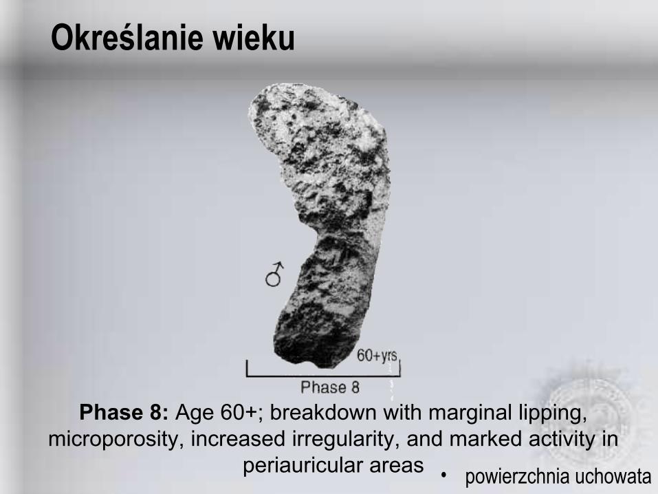
moderate to marked activity in periauricular areas– Phase 8: Age 60+; breakdown with marginal lipping, microporosity, increased
irregularity, and marked activity in periauricular areas

Określanie wieku
• powierzchnia uchowataPhase 1: Age 20-24; billowing and very fine granularity
Określanie wieku
• powierzchnia uchowata
Phase 2: Age 25-29; reduction of billowing but retention of youthful appearance
Określanie wieku
• powierzchnia uchowata
Phase 3: Age 30-34; general loss of billowing, replacement by striae, coarsening of granularity

Określanie wieku
• powierzchnia uchowataPhase 4: Age 35-39; uniform coarse granularity
Określanie wieku
• powierzchnia uchowata
Phase 5: Age 40-44; transition from coarse granularity to dense surface; this may take place over islands on the
surface of one or both faces
Określanie wieku
• powierzchnia uchowata
Phase 6: Age 45-49; completion of densification with complete loss of granularity
Określanie wieku
• powierzchnia uchowata
Phase 7: Age 50-59; dense irregular surface of rugged topography and moderate to marked activity in periauricular areas
Określanie wieku
• powierzchnia uchowata
Phase 8: Age 60+; breakdown with marginal lipping, microporosity, increased irregularity, and marked activity in
periauricular areas
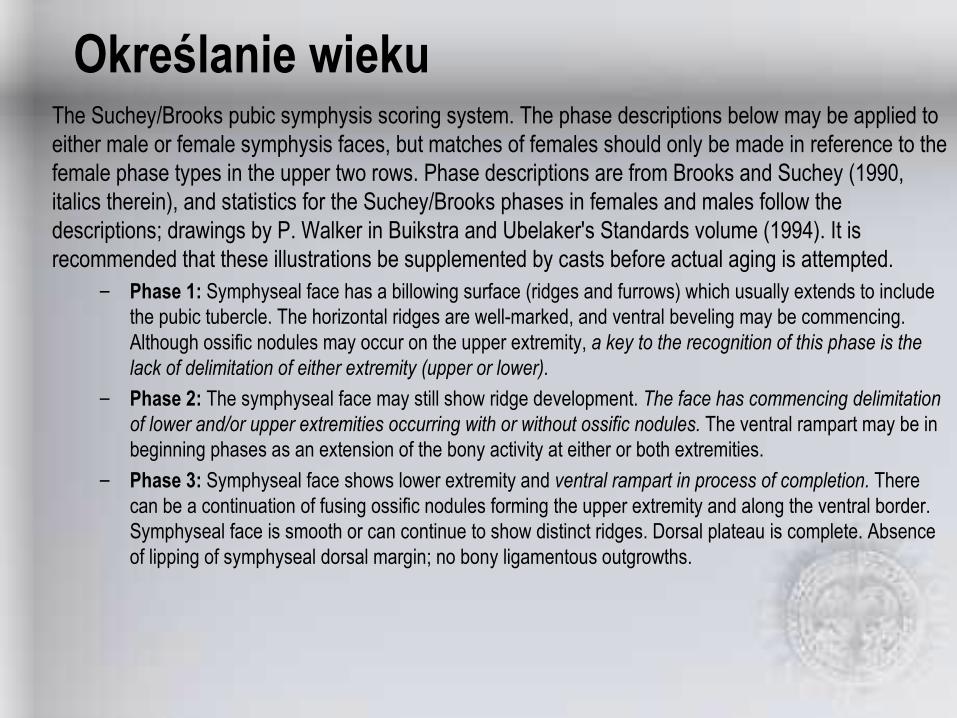
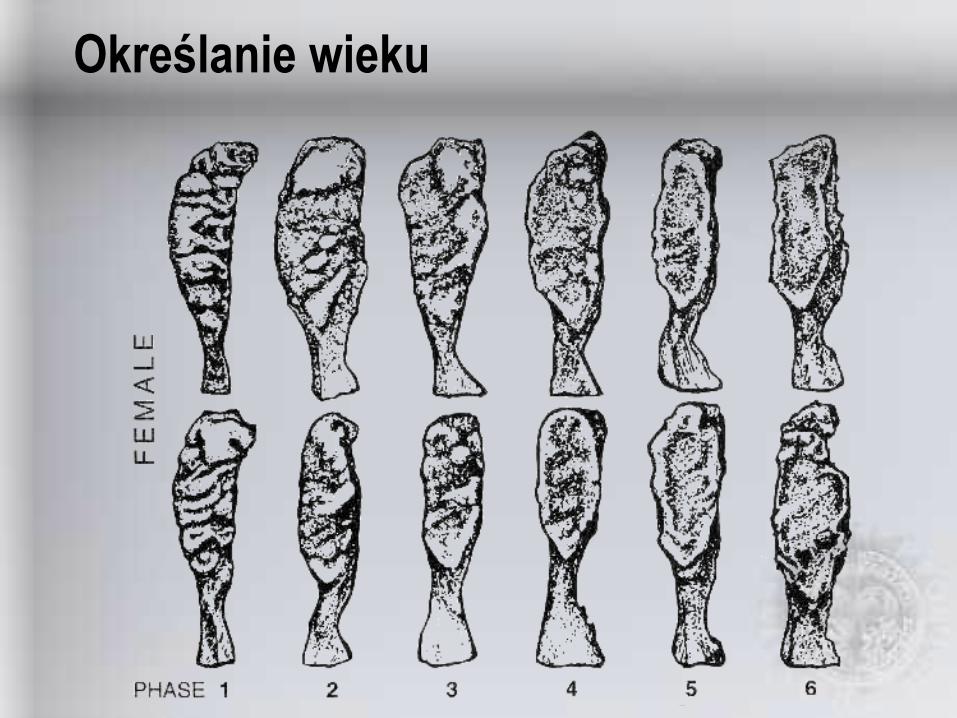
Określanie wiekuThe Suchey/Brooks pubic symphysis scoring system. The phase descriptions below may be applied to either male or female symphysis faces, but matches of females should only be made in reference to the female phase types in the upper two rows. Phase descriptions are from Brooks and Suchey (1990, italics therein), and statistics for the Suchey/Brooks phases in females and males follow the descriptions; drawings by P. Walker in Buikstra and Ubelaker's Standards volume (1994). It is recommended that these illustrations be supplemented by casts before actual aging is attempted.
– Phase 1: Symphyseal face has a billowing surface (ridges and furrows) which usually extends to include the pubic tubercle. The horizontal ridges are well-marked, and ventral beveling may be commencing. Although ossific nodules may occur on the upper extremity, a key to the recognition of this phase is the lack of delimitation of either extremity (upper or lower).
– Phase 2: The symphyseal face may still show ridge development. The face has commencing delimitation of lower and/or upper extremities occurring with or without ossific nodules. The ventral rampart may be in beginning phases as an extension of the bony activity at either or both extremities.
– Phase 3: Symphyseal face shows lower extremity and ventral rampart in process of completion. There can be a continuation of fusing ossific nodules forming the upper extremity and along the ventral border. Symphyseal face is smooth or can continue to show distinct ridges. Dorsal plateau is complete. Absence of lipping of symphyseal dorsal margin; no bony ligamentous outgrowths.

Określanie wieku– Phase 4: Symphyseal face is generally fine grained although remnants of the old ridge and furrow system
may still remain. Usually the oval outline is complete at this stage, but a hiatus can occur in upper ventral rim. Pubic tubercle is fully separated from the symphyseal face by definition of upper extremity. The symphyseal face may have a distinct rim. Ventrally, bony ligamentous outgrowths may occur on inferior portion of pubic bone adjacent to symphyseal face. If any lipping occurs, it will be slight and located on the dorsal border.
– Phase 5: Symphyseal face is completely rimmed with some slight depression of the face itself, relative to the rim. Moderate lipping is usually found on the dorsal border with more prominent ligamentous outgrowths on the ventral border. There is little or no rim erosion. Breakdown may occur on superior ventral border.
– Phase 6: Symphyseal face may show ongoing depression as rim erodes. Ventral ligamentous attachments are marked. In many individuals the pubic tubercle appears as a separate bony knob. The face may be pitted or porous, giving an appearance of disfigurement with the ongoing process of erratic ossification. Crenulations may occur. The shape of the face is often irregular at this stage.

Określanie wieku
34-8612.261.242-8712.460.06
27-6610.445.625-8314.648.15
23-579.435.226-7010.938.24
21-466.528.721-538.130.73
19-343.623.419-404.925.02
15-232.118.515-242.619.41
95% RangeStandard Dev.Mean95% RangeStandard Dev.Mean
Male (n = 739)Female (n =273)Phase
Określanie wieku

Określanie wieku
Określanie wieku
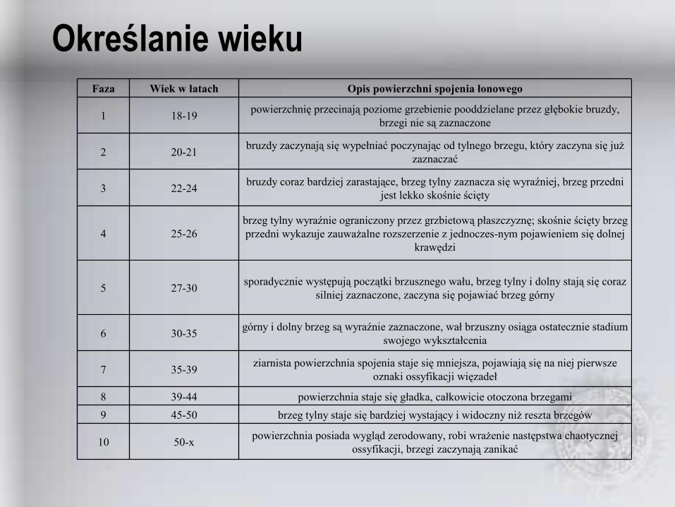
powierzchnia posiada wygląd zerodowany, robi wrażenie następstwa chaotycznej ossyfikacji, brzegi zaczynają zanikać50-x10
brzeg tylny staje się bardziej wystający i widoczny niż reszta brzegów45-509powierzchnia staje się gładka, całkowicie otoczona brzegami39-448
ziarnista powierzchnia spojenia staje się mniejsza, pojawiają się na niej pierwsze oznaki ossyfikacji więzadeł35-397
górny i dolny brzeg są wyraźnie zaznaczone, wał brzuszny osiąga ostatecznie stadium swojego wykształcenia30-356
sporadycznie występują początki brzusznego wału, brzeg tylny i dolny stają się coraz silniej zaznaczone, zaczyna się pojawiać brzeg górny27-305
brzeg tylny wyraźnie ograniczony przez grzbietową płaszczyznę; skośnie ścięty brzeg przedni wykazuje zauważalne rozszerzenie z jednoczesnym pojawieniem się dolnej
krawędzi25-264
bruzdy coraz bardziej zarastające, brzeg tylny zaznacza się wyraźniej, brzeg przedni jest lekko skośnie ścięty22-243
bruzdy zaczynają się wypełniać poczynając od tylnego brzegu, który zaczyna się już zaznaczać20-212
powierzchnię przecinają poziome grzebienie pooddzielane przez głębokie bruzdy, brzegi nie są zaznaczone18-191
Opis powierzchni spojenia łonowegoWiek w latachFaza
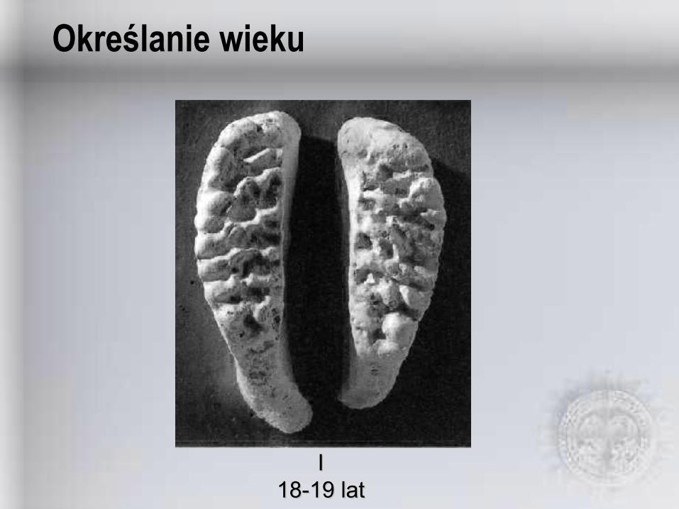
Określanie wieku
II18-19 lat18-19 lat
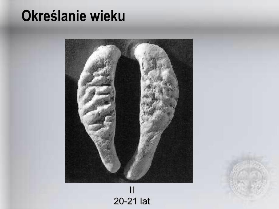
Określanie wieku
IIII20-21 lat20-21 lat
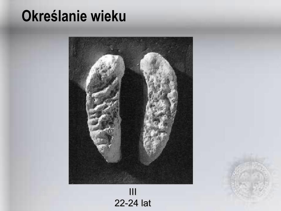
Określanie wieku
IIIIII22-24 lat22-24 lat

Określanie wieku
IVIV25-26 lat25-26 lat

Określanie wieku
VV27-30 lat27-30 lat
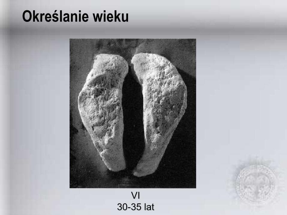
Określanie wieku
VIVI30-35 lat30-35 lat

Określanie wieku
VIIVII35-39 lat35-39 lat

Określanie wieku
VIIIVIII39-44 lat39-44 lat
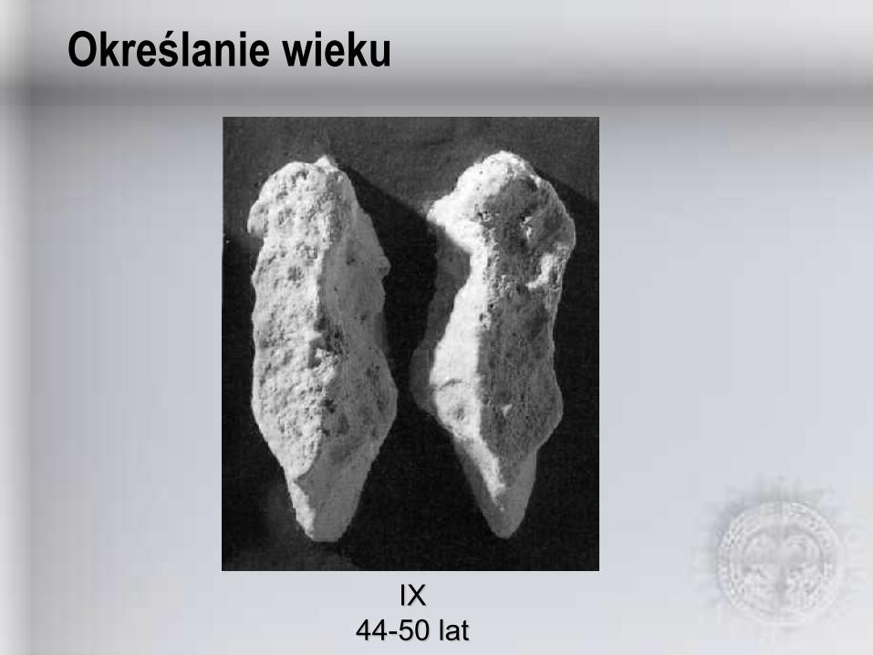
Określanie wieku
IXIX44-50 lat44-50 lat
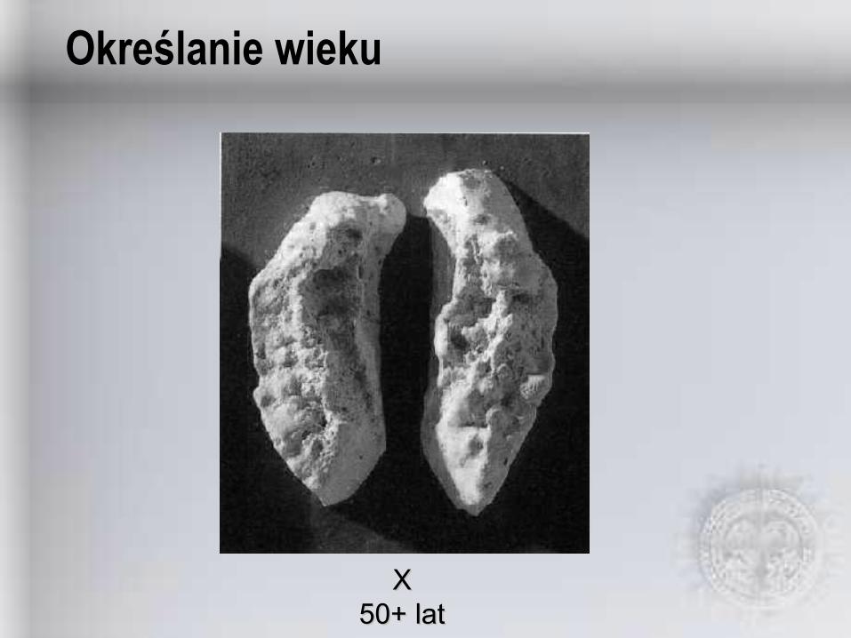
Określanie wieku
XX50+ lat50+ lat

Określanie wieku
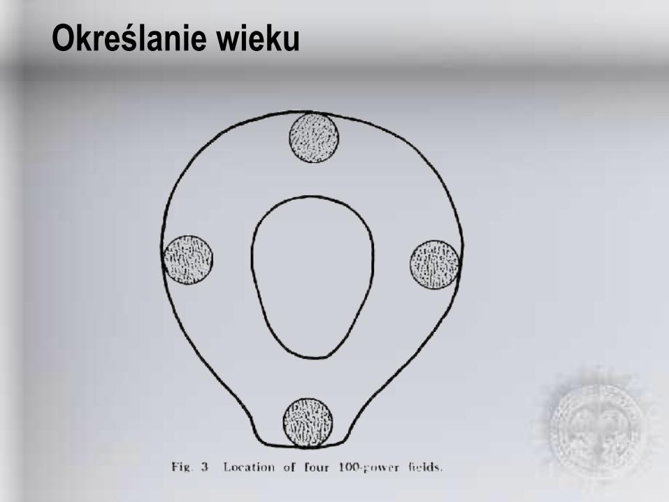
Określanie wieku
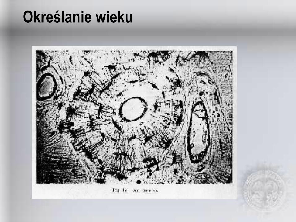
Określanie wieku
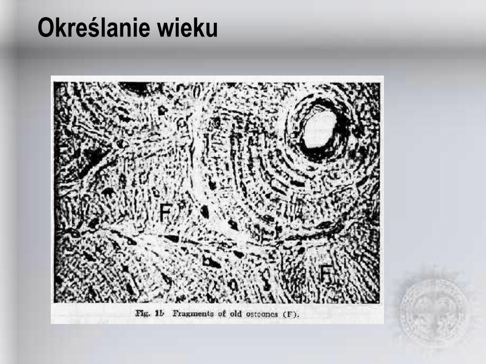
Określanie wieku
Określanie wieku
Określanie wieku
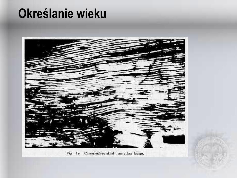
Określanie wieku
Określanie wieku

Określanie wieku
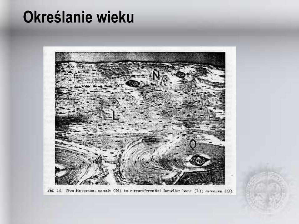
Określanie wieku
Określanie wieku

Określanie wieku