pcr-based investigation of the presence of herpesvirus in ... · herpesvirus in the peripheral...
TRANSCRIPT

PCR-based investigation of the presence of
herpesvirus in the peripheral vestibular
system in cats and dogs
Birgit Parzefall

Aus dem Institut für Tierpathologie
Lehrstuhl für Allgemeine Pathologie und Neuropathologie
(Vorstand: Prof. Dr. Wolfgang Schmahl)
der Tierärztlichen Fakultät der Ludwig-Maximilians-Universität München
Arbeit angefertigt unter Leitung von Dr. med. vet. K. Matiasek
PCR-based investigation of the presence of
herpesvirus in the peripheral vestibular system
in cats and dogs
Inaugural-Dissertation
zur Erlangung der tiermedizinischen Doktorwürde
der Tierärztlichen Fakultät der Ludwig-Maximilians-Universität München
von Birgit Parzefall
aus Regensburg
München, 2010

Gedruckt mit Genehmigung der Tierärztlichen Fakultät
der Ludwig-Maximilians-Universität München
Dekan:
Univ.-Prof. Dr. Braun
Berichterstatter:
Univ.-Prof. Dr. Schmahl
Korreferent/en: Univ.-Prof. Dr. Sutter
Priv.-Doz. Dr. Werckenthin
Priv.-Doz. Dr. Fischer
Univ.-Prof. Dr. Köstlin
Tag der Promotion: 13. Februar 2010

For my mother and my sister

Table of contents
page
1. Introduction 1
2. Scientific background 2
2.1 Alpha-herpesviruses 2
2.1.1 General view 2
2.1.1.1 Architecture of the herpesviruses 2
2.1.1.2 Herpesvirus taxonomy 3
2.1.1.3 Alpha-herpesvirus characteristics 4
2.1.1.4 Latency 5
2.1.1.5 Herpesvirus reactivation 6
2.1.1.6 Antiviral therapy 6
2.1.2 Survey on FHV-1 and CHV-1 8
2.1.2.1 FHV-1 8
2.1.2.1.1 Taxonomy and prevalence 8
2.1.2.1.2 Epidemiology and pathogenesis 8
2.1.2.1.3 Clinical signs 9
2.1.2.1.4 Vaccination 9
2.1.2.1.5 Diagnosis 10
2.1.2.2 CHV-1 11
2.1.2.2.1 Taxonomy and prevalence 11
2.1.2.2.2 Epidemiology and pathogenesis 11
2.1.2.2.3 Clinical signs 12
2.1.2.2.4 Vaccination 12
2.1.2.2.5 Diagnosis 13
2.2 Functional neuroanatomy of the vestibular system 14
2.2.1 Anatomy 14
2.2.2 Physiology 16
2.2.3 Nervous pathways 18
2.3 Vestibular dysfunction 19
2.4 Processing of the canine and feline vestibular labyrinth 20

3. Own scientific experiments 21
3.1 “A rapid approach to ultrastructural evaluation and DNA
analysis of the vestibular labyrinth and ganglion in dogs and
cats”
21
Abstract 23
1. Introduction 24
2. Materials and methods 25
2.1 Animals and tissues 25
2.2 Preparation of the vestibular labyrinth 25
2.2.1 Gross preparation 25
2.2.2 Micropreparation 27
2.3 Sample processing for histology 30
2.4 Transmission electron microscopy 31
2.5 Polymerase chain reaction 31
3. Results 33
3.1 Preparation 33
3.2 Histology 34
3.3 Transmission electron microscopy 35
3.4 Integrity and amount of isolated DNA 37
4. Discussion 38
References 39
3.2 “Evidence of feline herpesvirus-1 DNA in the vestibular ganglion
of domestic cats”
44
Abstract 46
Body of manuscript 47
References 51
3.3 “Naturally-occuring canine herpesvirus (CHV-1) infection of the
vestibular labyrinth and ganglion in dogs”
53
Abstract 55
Introduction 56
Material and methods 56
Results 57
Discussion 60

Conclusions 62
References 63
4. Discussion 65
4.1 General aspects of the study 65
4.2 New method for feline and canine temporal bone processing 65
4.3 Experimental design of the study 68
4.4 Field versus vaccine virus 70
4.5 Type of virus infection 71
4.6 Distribution of FHV-1/CHV-1 infection 72
4.7 Potential routes of virus infection 73
4.8 Clinical relevance of vestibular herpesvirus infection 74
4.9 Conclusions and future prospects 75
5. Summary 77
6. Zusammenfassung 79
7. References 81
Acknowledgments 107
Please note that the consecutive numbering of the figures and tables only applies to
the scientific background. In papers 1 to 3 the layout of the text and the numbering of
the figures and tables is in accordance with the editorial guidelines of the respective
journals.

Abbreviations
bp base pair
CHV-1 Canine herpesvirus 1
CN cranial nerve
DNA deoxyribonucleic acid
EDTA ethylene diamine tetraacetic acid
ELMI electron microscopy
FCV Feline calicivirus
FHV-1 Feline herpesvirus 1
HSV-1 Herpes simplex virus type 1
IFN interferon
IHC immunohistochemistry
ISH in-situ hybridization
kbp kilo base pairs
LATs latency-associated transcripts
LMU Ludwig-Maximilians-Universität München
MLV modified live virus vaccine
N. nervus
NLV non-live virus vaccine
PCR polymerase chain reaction
RNA ribonucleic acid
RT-PCR reverse-transcription PCR
TG trigeminal ganglion
VG vestibular ganglion
VL vestibular labyrinth

- 1 -
1. Introduction
The vestibular labyrinth is the organ for sensation of equilibrium. Being part of the
inner ear, it is located in the caudodorsal aspect of the temporal bone 83; 151. The
peripheral compartment of the vestibular system consists of the vestibular labyrinth,
the vestibular portion of the vestibulocochlear nerve and the vestibular ganglion 49; 144;
212. Disruption of any part of this signal chain may cause peripheral vestibular
dysfunction.
In humans, morphological alterations of vestibular inner ear structures suggestive of
herpesvirus infections were demonstrated in patients suffering from various
vestibular diseases 68; 70-72. Moreover, herpesvirus infections in the human vestibular
system have been detected by using different molecular tools 9; 10; 61; 210; 230. Members
of the herpesvirus family display a marked neurotropism and have the ability to
establish lifelong latency in the nervous system 21; 55; 64. Distress and
immunosuppression may cause virus reactivation and replication with subsequent
host cell damage, potentially leading to clinical deficits 21; 64; 107. Suchlike reactivation
within the vestibular ganglion is discussed to cause various recurrent human
vestibulopathies 61; 65; 68.
Even though, vestibular diseases in cats and dogs are common 144; 212 and
herpesvirus infections show a high prevalence in these species 50; 75, vestibular inner
ear structures have not been investigated for a possible herpesvirus infection so far.
Comparable to humans 140, vestibular inner ear structures in cats 116 and dogs 36; 41
are very difficult to access and technical processing is challenging and time
consuming. Therefore, only few morphological 6; 39; 62; 103; 143; 175; 187 and
immunohistochemical 41 investigations have been performed so far while molecular
analyses of the vestibular labyrinth and ganglion in cats and dogs have not been
reported.
The aim of the present study was to evaluate, if vestibular inner ear structures of
dogs and cats can be infected by herpesviruses. For this purpose, first a method for
preparation of vestibular inner ear samples from cats and dogs had to be
established, allowing for subsequent performance of PCR-based analyses.
- 2 -
2. Scientific background
2.1 Alpha-herpesviruses
2.1.1 General view
2.1.1.1 Architecture of the herpesviruses
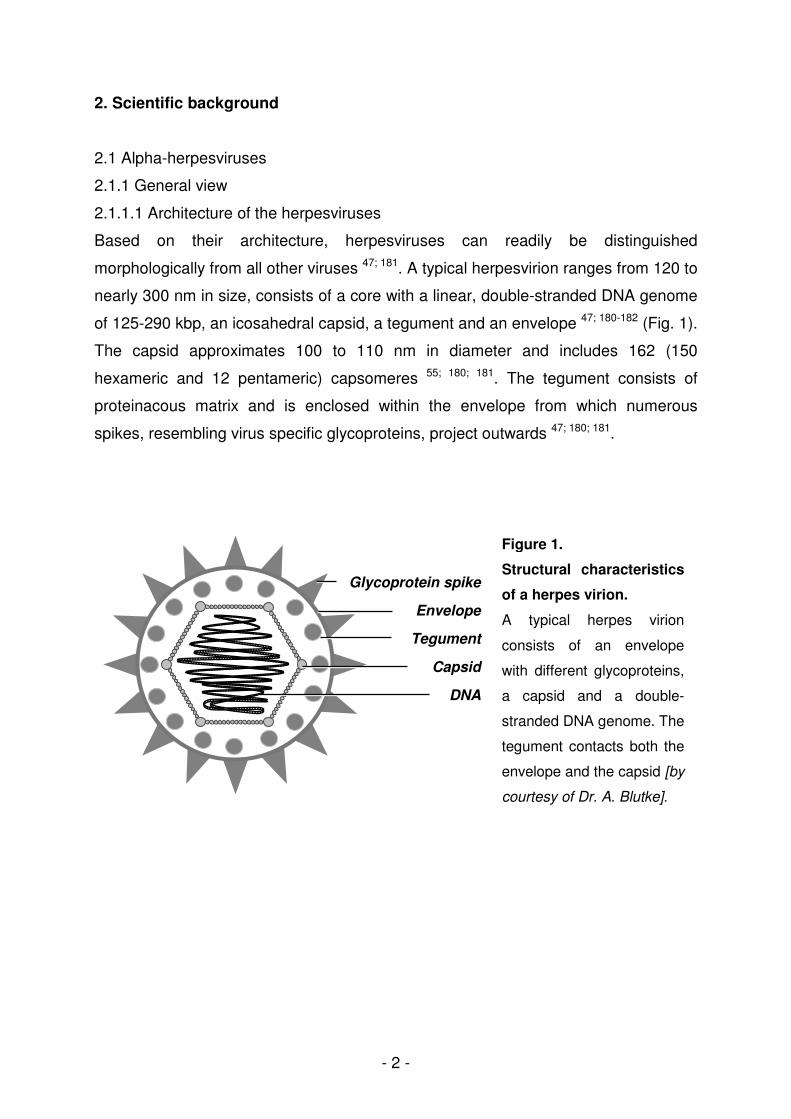
Based on their architecture, herpesviruses can readily be distinguished
morphologically from all other viruses 47; 181. A typical herpesvirion ranges from 120 to
nearly 300 nm in size, consists of a core with a linear, double-stranded DNA genome
of 125-290 kbp, an icosahedral capsid, a tegument and an envelope 47; 180-182 (Fig. 1).
The capsid approximates 100 to 110 nm in diameter and includes 162 (150
hexameric and 12 pentameric) capsomeres 55; 180; 181. The tegument consists of
proteinacous matrix and is enclosed within the envelope from which numerous
spikes, resembling virus specific glycoproteins, project outwards 47; 180; 181.
Glycoprotein spike
Envelope
Tegument
Capsid
DNA
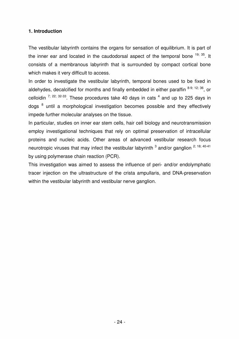
Figure 1.
Structural characteristics
of a herpes virion.
A typical herpes virion
consists of an envelope
with different glycoproteins,
a capsid and a double-
stranded DNA genome. The
tegument contacts both the
envelope and the capsid [by
courtesy of Dr. A. Blutke].

- 3 -
2.1.1.2 Herpesvirus taxonomy
According to the latest update by the International Committee on Taxonomy of
Viruses, the former family Herpesviridae consists of three families, which constitute
the new order Herpesvirales 47. The family Herpesviridae includes mammal, avian
and reptile viruses whereas the new families Allo- and Malacoherpesviridae comprise
fish and frog, and a bivalve virus, respectively 47 (Table 1).
By means of biologic properties and genetic background, the family Herpesviridae is
grouped into three subfamilies (α, β, γ) which contain several genera 47; 108; 180; 181
(Table 2). Since feline and canine herpesvirus are alpha-herpesviruses 47, this
subfamily will be discussed in the following.
Table 1: Current taxonomy of the order Herpesvirales.
[Data according to Davison et al. (2009), Arch Virol 154:171-177]. Numbers given in brackets
indicate yet unassigned species and viruses in the subfamily and family.
Subfamily
Species
Order Herpesvirales
-herpesvirinae
(1)
α
34 (2)
β
11 (3)
γ
31 (3) 1
Virus (35) (12)
Family Herpes Alloherpes Malacoherpes
Genus 4 4 4 1 1
1
-viridae
- 4 -
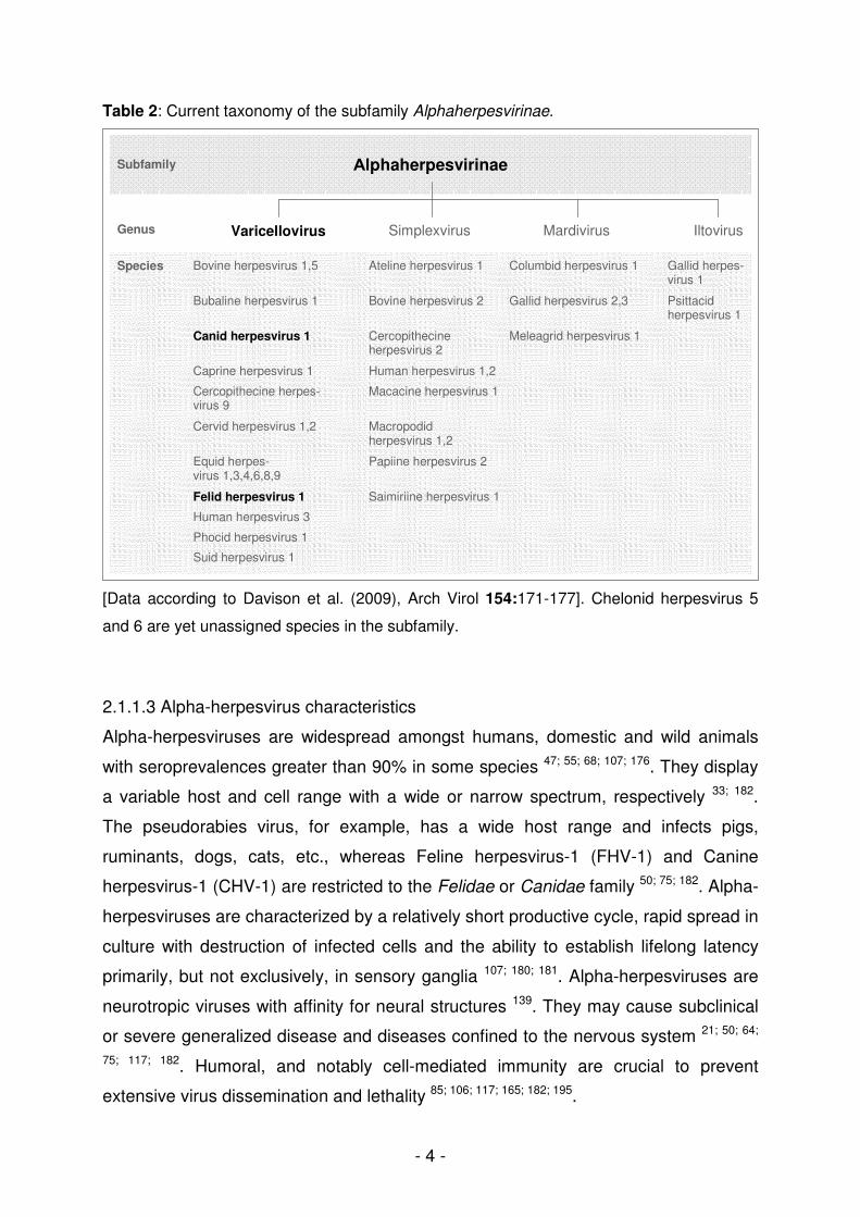
Table 2: Current taxonomy of the subfamily Alphaherpesvirinae.
[Data according to Davison et al. (2009), Arch Virol 154:171-177]. Chelonid herpesvirus 5
and 6 are yet unassigned species in the subfamily.
2.1.1.3 Alpha-herpesvirus characteristics
Alpha-herpesviruses are widespread amongst humans, domestic and wild animals
with seroprevalences greater than 90% in some species 47; 55; 68; 107; 176. They display
a variable host and cell range with a wide or narrow spectrum, respectively 33; 182.
The pseudorabies virus, for example, has a wide host range and infects pigs,
ruminants, dogs, cats, etc., whereas Feline herpesvirus-1 (FHV-1) and Canine
herpesvirus-1 (CHV-1) are restricted to the Felidae or Canidae family 50; 75; 182. Alpha-
herpesviruses are characterized by a relatively short productive cycle, rapid spread in
culture with destruction of infected cells and the ability to establish lifelong latency
primarily, but not exclusively, in sensory ganglia 107; 180; 181. Alpha-herpesviruses are
neurotropic viruses with affinity for neural structures 139. They may cause subclinical
or severe generalized disease and diseases confined to the nervous system 21; 50; 64;
75; 117; 182. Humoral, and notably cell-mediated immunity are crucial to prevent
extensive virus dissemination and lethality 85; 106; 117; 165; 182; 195.
Genus Mardivirus Iltovirus Varicellovirus Simplexvirus
Αlphaherpesvirinae Subfamily
Human herpesvirus 3
Phocid herpesvirus 1
Suid herpesvirus 1
Species Columbid herpesvirus 1 Gallid herpes- virus 1
Bovine herpesvirus 1,5 Ateline herpesvirus 1
Gallid herpesvirus 2,3 Bubaline herpesvirus 1 Bovine herpesvirus 2 Psittacid herpesvirus 1
Meleagrid herpesvirus 1 Canid herpesvirus 1 Cercopithecine herpesvirus 2
Caprine herpesvirus 1 Human herpesvirus 1,2
Cercopithecine herpes-virus 9
Macacine herpesvirus 1
Cervid herpesvirus 1,2 Macropodid herpesvirus 1,2
Felid herpesvirus 1 Saimiriine herpesvirus 1
Equid herpes- virus 1,3,4,6,8,9
Papiine herpesvirus 2
- 5 -
A variety of murine models, thus, demonstrated the importance of various T-cells in
the clearance of infectious virus from peripheral and neural sites 84; 85; 106; 194; 195.
After primary infection of the mucosal epithelium, virus may enter nerve terminals
therein and translocate to sensory ganglia at a rate of ~1.8 mm/h using the
retrograde axonal transport 21; 35; 64; 106; 142; 195; 210. A productive cycle with lysis of
infected ganglion cells does not necessarily occur; instead, latency may follow
infection 21; 150.
2.1.1.4 Latency
Latency is described as a reversible non-productive infection of a cell by a
replication-competent virus which can be reactivated following different types of
distress 21; 64; 107. During latency, viral DNA exists in an unintegrated and circular form
primarily within the nerve cell nuclei 21; 33; 107; 155. While Herpes simplex virus type-1
(HSV-1) 21; 33; 196, FHV-1 155 and CHV-1 142 have been demonstrated in peripheral
ganglion cells, Varicella-zoster virus genome was also detected in satellite cells
during latency 21; 33; 120; 230.
Estimates both in humans and animals propose that 0.01%-10% of ganglion cells
may be latently infected, with 10-1000 copies of virus genome per neuron 21; 33; 60; 142;
155; 193; 196. The number of infected ganglion cells and the copies of virus genome
therein has been suggested to correlate with the probability and the frequency of
reactivation 21.
During latency, transcription seems to be restricted to latency-associated transcripts
(LATs) which therefore provide a molecular marker for latency 21; 107; 213. Detection of
viral DNA via polymerase chain reaction (PCR) does not permit a differentiation
between infectious virus and latent infection 21. Demonstration of LATs, using
reverse-transcription PCR (RT-PCR) 21; 210; 213 or in-situ hybridization (ISH) 21; 155; 210,
however, indicate true latency. Consequently, even low-level persistent infection may
be distinguished from latent infection 21.
Moreover, latent virus may be detected with explant and co-cultivation techniques 21;
77; 146, whereas common virus isolation 11; 24; 136; 204 and immunohistochemistry (IHC)
24; 136; 142, for detection of viral antigen, can be used for identification of infectious
virus.

- 6 -
2.1.1.5 Herpesvirus reactivation
Once latency has been established, reactivation of latent virus may occur following
different types of distress or immunosuppression 21; 75. Reactivated virus travels from
its site of latency back to its entrance zone resulting in further virus replication,
shedding and transmission to susceptible hosts, as well as destruction of infected
cells with possible recrudescence of clinical signs 21; 64; 75; 182.
Herpesvirus reactivation in humans is implicated in a great variety of diseases
including recurrent vestibulopathies, (e.g. vestibular neuritis 65; 68; 72, benign
paroxysmal positional vertigo 65; 68; 71, Ménière’s disease 65; 68; 70; 220, cochleovestibular
signs in Ramsay-Hunt-Syndrome 61; 130; 156), herpes simplex labialis 182, conjunctivitis
182, keratitis 182, shingles 33; 182, Bell’s palsy 69; 189, trigeminal neuralgia 182 and
encephalitis 32; 33. FHV-1 reactivation from the trigeminal ganglion is thought to cause
conjunctivitis, keratitis and rhinitis in cats 75; 211 and CHV-1 reactivation corneal ulcers
in dogs 125.
2.1.1.6 Antiviral therapy
Antiviral therapy against alpha-herpesviruses has been used in humans for some
decades now. Even though clinical signs and virus shedding is reduced and
recrudescence of clinical signs may be suppressed, neither the establishment of
latency can be prevented with early antiviral therapy nor eradication of herpesvirus
genome from latently infected cells will be achieved with today’s antiviral compounds
55; 211.
To date, the key protein target for antiviral therapy against alpha-herpesviruses is
mainly the virus encoded DNA-polymerase 55; 99. During latency, however, the virus
does not express this gene. Thus conventional nucleoside analogues do not affect
latent virus 55. Development of successful antiviral therapies in human medicine is
largely based on experimental work, using laboratory animals as in-vivo infection
models. However, our domestic animals had little benefit from these experiments so
far 55.
In cats, different nucleoside analogues (e.g. trifluridine, idoxuridine, ganciclovir,
acyclovir, penciclovir, cidofovir, valaciclovir and famciclovir), feline IFN-ω, human
IFN-α, L-lysine and lactoferrin have been proposed for the treatment of FHV-1-
associated ocular disease 57; 75; 100; 137; 211. While in vitro efficacy has mostly been
established, in vivo trials are still missing for the majority of these drugs 211.

- 7 -
Besides, systemic application of nucleoside analogues in cats has been problematic
so far in terms of poor in-vivo-efficacy against FHV-1, poor bioavailability or toxic side
effects such as bone marrow suppression, renal and liver necrosis 5; 55; 57.
The efficacy of lactoferrin has been tested in-vitro, and it is thought to protect against
CHV-1 infection in-vivo. Apart from lactoferrin, there is no other antiviral therapy,
which has been tested in dogs 55; 208.

- 8 -
2.1.2 Survey on FHV-1 and CHV-1
2.1.2.1 FHV-1
2.1.2.1.1 Taxonomy and prevalence
FHV-1 is a member of the Varicellovirus genus of the herpesvirus subfamily
Alphaherpesvirinae and is closely related, genetically as well as antigenically, to
CHV-1 47; 75; 186. FHV-1 infects felids and it is widespread amongst cat populations 76;
171; 199. All isolates of FHV-1 belong to one single serotype and are genetically fairly
homogenous 75; 97; 231.
2.1.2.1.2 Epidemiology and pathogenesis
Virus is shed in ocular, nasal and oral secretions and is mainly transmitted by close
contact with an infected cat 18; 81; 171; 199. Since virus is short-lived in the environment
and is inactivated by common disinfectants, indirect transmission is not thought to
play a major role in virus transmission 75; 171; 199 .
Nasal, oral and conjunctival mucous membranes are natural sites for infection,
whereas vaginal and transplacental infections have only been shown experimentally
19; 75; 96. After infection, virus replication primarily takes place in the mucosae of the
nasal septum, the turbinates, nasopharynx and tonsils, however, the conjunctivae,
mandibular lymph nodes and upper trachea are often involved, too 75; 80. Viremia and
generalized disease may occur in neonatal kittens or in debilitated animals.
Nevertheless it appears to be rare, since viral replication preferentially occurs at
lower body temperatures like in the respiratory tract 75; 197.
After infection, more than 80% of the diseased and recovered cats become latent
virus carriers 79; 80; 221. FHV-1 is thought to be transported retrograde from peripheral
infectious sites to neurones of the trigeminal ganglion (TG) 155; 223. Even though viral
DNA has been detected in a wide range of tissues, true latency has only been
suggested for the TG so far 77; 146; 155; 213.
Viral reactivation with or without recrudescence of clinical signs may occur
spontaneously or within a three-week period following a stressful event such as re-
housing, pregnancy, lactation or corticosteroid treatment, thus, transmitting virus to
cats in a new environment or from the queen to their kittens 75; 78; 79; 134.

- 9 -
2.1.2.1.3 Clinical signs
Infection with FHV-1 causes severe upper respiratory tract disease in susceptible
animals 44; 75. Early clinical signs of disease may include depression, marked
sneezing, inappetence, pyrexia and excessive salivation with drooling 44; 75.
Conjunctivitis, corneal ulcers, serous to mucopurulent ocular and nasal discharge as
well as dyspnoea and coughing in severely affected cats may follow 44; 75; 171. Oral
ulceration may occur with FHV-1 infection, but is more commonly caused by feline
calicivirus (FCV) 170; 171. Generalized infection and primary viral pneumonia are
occasionally encountered especially in young or debilitated animals, whereas
neurological signs as a sequel to infection are rare 75; 76; 82.
Although in-utero transmission, abortion and congenitally-infected kittens have been
reported after experimental FHV-1 infection of pregnant queens, reproductive
disease does not seem to be clinically relevant under natural conditions 75; 76; 218.
Moreover, FHV-1 is thought to be involved in different recurrent and/or chronic
conditions, such as conjunctivitis, ulcerative and stromal keratitis, corneal
sequestrae, keratoconjunctivitis sicca 5; 134; 145; 147-149; 200; 201, ulcerative dermatitis,
gingivostomatitis and rhinitis 88; 95; 174.
2.1.2.1.4 Vaccination
Vaccination against FHV-1 has been used for a number of years to control disease 17;
161. FHV-1 vaccines are used to reduce the severity of disease, however, they neither
prevent infection nor the development of a carrier state 119; 199. There are modified
live (MLV) and non-live (adjuvanted inactivated) virus vaccines (NLV), both invariably
given in association with FCV 75; 122; 199. MLV and NLV are usually administered
parenterally and intranasal MLV are also licensed in some countries 75; 122; 199. FHV-1
vaccines are generally safe, however, transient signs like fever, sneezing, nasal and
ocular discharge, conjunctivitis and ulceration of the nasal philtrum may occur in
some cats 75; 122; 178. Intranasally applied MLV can become latent 75; 221, whereas the
situation with parenteral MLV is still unclear 75; 203.
Despite possible respiratory signs, intranasal MLV vaccine is advocated for use in
young kittens and in conjunction with parenteral vaccine on entry into shelters since it
induces rapid protection 75; 122. On the contrary, conventional MLV inadvertently
administered via the oro-nasal route induces disease 75; 119.
- 10 -
After primary immunization and the first annual booster, annual vaccination is
generally recommended in the EU and triannually in the USA, however, with
individual risk-benefit assessment for a particular animal 48; 75; 211.
2.1.2.1.5 Diagnosis
The diagnosis of primary FHV-1 infection in kittens is commonly based on
characteristic clinical signs, whereas chronic or recurrent conditions in older cats
possibly related to FHV-1 infection are more difficult to interpret 134; 199. Since direct
and indirect diagnostic tests only aid in the diagnosis of FHV-1 associated disease
due to the occurrence of healthy carriers and the widespread use of FHV-1 vaccines,
test results should be critically questioned and always evaluated in conjunction with
clinical signs 17; 24; 75; 199; 204.
Serum antibody tests are positive in most cats with varying titers as a consequence
of the widespread use of vaccines and possible previous infection and hence are not
significant 136; 199. Given that there are no commercially available marker FHV-1
vaccines, positive PCR results for example could imply vaccine or field-virus 75; 135;
203; 221. Furthermore, the detection of viral DNA could resemble FHV-1 as a causative
agent of the disease, a consequential or coincidental finding 24; 134; 135. Negative
results of direct diagnostic test, on the other hand, either demonstrate the absence of
FHV-1 or the limitations of some tests. Additionally, under chronic conditions, viral
antigens may be bound by secreted antibodies which impede the binding of the
primary IHC antibody and, therefore, may lead to false negative results 134.

- 11 -
2.1.2.2 CHV-1
2.1.2.2.1 Taxonomy and prevalence
CHV-1 belongs to the Varicellovirus genus of the alpha-herpesvirus subfamily and
infects canids 47; 50. Amongst dogs CHV-1 is distributed worldwide with
seroprevalences ranging as high as 94% 1; 16; 118; 138; 152; 176; 179; 184; 206.
2.1.2.2.2 Epidemiology and pathogenesis
Since CHV-1 is quickly inactivated in the environment, direct transmission via
oronasal and veneral secretions are the main routes of infection 43; 50; 158; 183. In
contrast to FHV-1, in utero-transmission and reproductive disorder is common and
poses a severe problem with high economic losses to breeding units 75; 90; 92; 101; 172;
185. Infection of pregnant bitches with CHV-1 may cause abortion, resorption or
mumification of the fetus, stillbirth or weak pups which seem normal at parturition but
will die within a few days 90-92; 168; 185. Dams develop protective immunity after
infection, and pass maternal antibodies on to the litter thus protecting the following
generation of pups from fatal CHV-1 related disease 50; 101. Neonatal pups may also
be infected with CHV-1 during passage through the birth canal or via infected
oronasal secretions of other dogs 43; 129.
Several studies demonstrate that virus grows best at temperatures between 35°C
and 37°C, resembling the normal body temperature of pups during their first two
weeks of life, whereas virus formation is largely depressed at the average
temperature of 39°C of adult dogs 11; 27; 28; 101; 132. The inability of neonates to
adequately regulate their body temperature and their naïve immune system benefits
viral growth and favors severe virus replication and viremia 28; 101; 165. Thus, initial
replication of CHV-1 in the oropharyngeal mucosa is followed by hematogenous virus
dissemination to the liver, kidneys, lymphatic tissues, lungs and CNS 43; 101; 226. If
maternally derived antibodies are missing litter mortality may reach 100% in neonatal
puppies, whereas puppies older than two weeks of age usually remain asymptomatic
after CHV-1 infection 50; 101.
Infection with CHV-1 is thought to be followed by a latent carrier state 26; 50; 142; 157; 158.
Latency of different alpha-herpesviruses has been subjected to advanced research
for many years, whereas data on CHV-1 latency is still rudimentary 22; 32; 73; 84; 87; 150;
155; 193; 196; 209; 213; 229.

- 12 -
Burr and coworkers investigated twelve key sites associated with latency for other
herpesviruses and detected CHV-1 DNA in nine out of twelve dogs with no history of
exposure or illness from CHV-1, using PCR 26. Tissues most commonly affected
included lumbo-sacral ganglia, tonsils, parotid salivary glands and liver 26. Miyoshi
and coworkers aimed to determine sites of latency in convalescent dogs that had
been experimentally infected with CHV-1 by the intravaginal, intranasal and/or
intravenous inoculation route using PCR, ISH, and IHC 142. PCR showed that TG and
retropharngeal lymph nodes were most frequently infected with CHV-1 and ISH
demonstrated virus genome in the nuclei of ganglion cells and in lymphocytes 142.
Antigen on the other hand could not be detected in any tissue sections via IHC 142.
Even though the studies of Burr and Miyoshi suggest CHV-1 latency in neural and
non-neural tissues of dogs, molecular markers are still missing to confirm true
latency. Latent carriers are epidemiologically important, since they may experience
episodic viral reactivation with subsequent viral shedding with or without
recrudescence of clinical signs, thus, transmitting virus to susceptible hosts 50; 157; 158.
2.1.2.2.3 Clinical signs
Based on the biology of CHV-1, clinical manifestation of an infection mainly concerns
puppies. Fatal CHV-1 infection in neonates may cause vocalization, anorexia,
dyspnea, abdominal pain, incoordination, soft feces, serous/hemorrhagic nasal
discharge and petechial hemorrhage on the mucous membranes 30; 43; 50. Puppies
older than two weeks of age at the time of infection present with mild upper
respiratory disease whereas CHV-1 infection in adult dogs is usually subclinical 7; 27;
76. Nevertheless, CHV-1 is further associated with reproductive disorders, genital
lesions, respiratory disease, and different ocular diseases such as corneal ulcers and
retinal dysplasia in adult dogs 3; 50; 52; 89; 91; 94; 109; 125; 168; 172; 185.
2.1.2.2.4 Vaccination
Since 2001, there is a commercially available subunit vaccine (Eurican Herpes 205®,
Merial, France) for active immunization of pregnant dams in order to prevent fatal
hemorrhagic disease in neonatal puppies which is authorized in the European Union
by the European Medicines Agency (EMEA).
- 13 -
Detailed information on this vaccine is available under:
http://www.emea.europa.eu/vetdocs/PDFs/EPAR/euricanherpes/V-059-PI-de.pdf and
http://www.emea.europa.eu/vetdocs/vets/Epar/euricanherpes/euricanherpes.htm.
The vaccine contains antigen of the CHV-1 glycoprotein gene B and is first
administered subcutaneously either during the dam’s season or within seven to ten
days after mating. Since the virus is a poor immunogen and antibody titers decrease
rapidly, a second injection one to two weeks before birth is necessary for optimal
protection of the puppies 26; 169; 183; 185. Thus, puppies with passive immunity derived
from neutralizing antibodies in the colostrum of vaccinated dams were protected,
whereas 62% of the puppies from unvaccinated dams died of fatal hemorrhagic
disease when challenged with a virulent strain of CHV-1 169.
2.1.2.2.5 Diagnosis
Diagnosis of neonatal CHV-1 infection may be based on distinct pathologic findings
and include multifocal necrosis and hemorrhage in most organs (lungs, liver, brain,
kidneys, and intestines), meningoencephalitis, intranuclear inclusion bodies (Cowdry
Type A), and enlarged lymph nodes and spleen 30; 166; 225-227. Even though PCR, ISH,
IHC, electron microscopy (ELMI) and virus isolation may strengthen the diagnosis in
puppies with classical hemorrhagic disease, the interpretation of test results in adult
dogs is more challenging, since CHV-1 has been detected in a variety of tissues from
healthy and diseased adult dogs 26; 29; 52; 111; 142; 190; 202. Thus, positive test results
must be interpreted in conjunction with clinical signs and carefully in terms of their
significance. Future research on transcriptional level hopefully will aid in identifying
the importance of these findings. Detection of antibody titers has been used in
determining seroprevalences in dog populations and the presence of CHV-1 in
kennels 50; 118; 138; 152; 176; 179; 184; 206. Assessment of individual antibody titers, however,
is difficult. Thus, a positive result may indicate CHV-1 infection or vaccination in
breeding bitches, whereas a negative test could indicate the absence of CHV-1
infection, an early phase of infection or previous infection with a decline of antibody
titres below detection limits 118; 185. Even though titer height in paired samples gives
information about current CHV-1 infection, it does not necessarily correlate with the
possible presence or absence of reproductive disorders 183; 185.

- 14 -
2.2 Functional neuroanatomy of the vestibular system
2.2.1 Anatomy
The inner ear comprises two different special sense organs, namely the cochlea
within the rostroventral and the vestibular labyrinth in the caudodorsal aspect of the
temporal bone 83; 151 (Fig. 2). The cochlea detects sound waves transmitted to the
inner ear thus serving as the organ for hearing, whereas the vestibular labyrinth is
responsible for sensation of equilibrium 34; 86; 167.
Figure 2. Location of the membranous labyrinth within a dog’s skull. The asterisk
indicates the sectional plane through the skull and therewith the position of the membranous
labyrinth. [by courtesy of Dr. A. Blutke]
The inner ear is composed of a membranous labyrinth filled with endolymph,
surrounded by perilymph, and enclosed within the compact cortical bone of the
osseous labyrinth 83; 86; 151 (Fig. 3).
The endolymph, produced by the stria vascularis of the cochlea and by vestibular
dark cells, is high on potassium and low on sodium ions and resembles intracellular
fluid, whereas the perilymph has a contrary electrolyte composition and is similar to
extracellular or cerebrospinal fluid 110; 114; 124; 198. This converse electrolyte
composition of endo- and perilymphatic fluid thus causes an electrochemical gradient
essential for impulse generation within the labyrinth 86; 167.
Temporal bone
External auditory canal
**
Cochlea
Vestibular labyrinth Ampulla
Semicircular duct
- 15 -
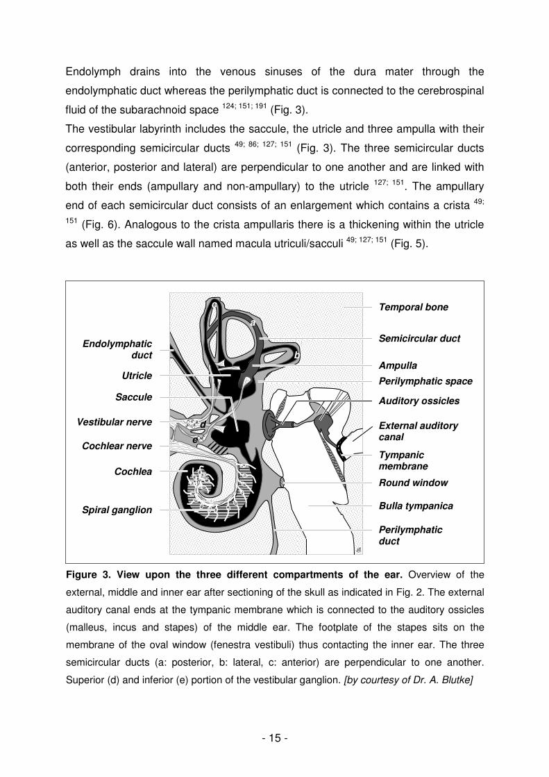
Endolymph drains into the venous sinuses of the dura mater through the
endolymphatic duct whereas the perilymphatic duct is connected to the cerebrospinal
fluid of the subarachnoid space 124; 151; 191 (Fig. 3).
The vestibular labyrinth includes the saccule, the utricle and three ampulla with their
corresponding semicircular ducts 49; 86; 127; 151 (Fig. 3). The three semicircular ducts
(anterior, posterior and lateral) are perpendicular to one another and are linked with
both their ends (ampullary and non-ampullary) to the utricle 127; 151. The ampullary
end of each semicircular duct consists of an enlargement which contains a crista 49;
151 (Fig. 6). Analogous to the crista ampullaris there is a thickening within the utricle
as well as the saccule wall named macula utriculi/sacculi 49; 127; 151 (Fig. 5).
a
b
c
dd
ee
Utricle
Saccule
Cochlear nerve
Vestibular nerve
Cochlea
Spiral ganglion
Endolymphatic duct
Temporal bone
Auditory ossicles
Tympanic membrane
External auditory canal
Bulla tympanica
Round window
Semicircular duct
Ampulla
Perilymphatic duct
Perilymphatic space
Figure 3. View upon the three different compartments of the ear. Overview of the
external, middle and inner ear after sectioning of the skull as indicated in Fig. 2. The external
auditory canal ends at the tympanic membrane which is connected to the auditory ossicles
(malleus, incus and stapes) of the middle ear. The footplate of the stapes sits on the
membrane of the oval window (fenestra vestibuli) thus contacting the inner ear. The three
semicircular ducts (a: posterior, b: lateral, c: anterior) are perpendicular to one another.
Superior (d) and inferior (e) portion of the vestibular ganglion. [by courtesy of Dr. A. Blutke]
- 16 -
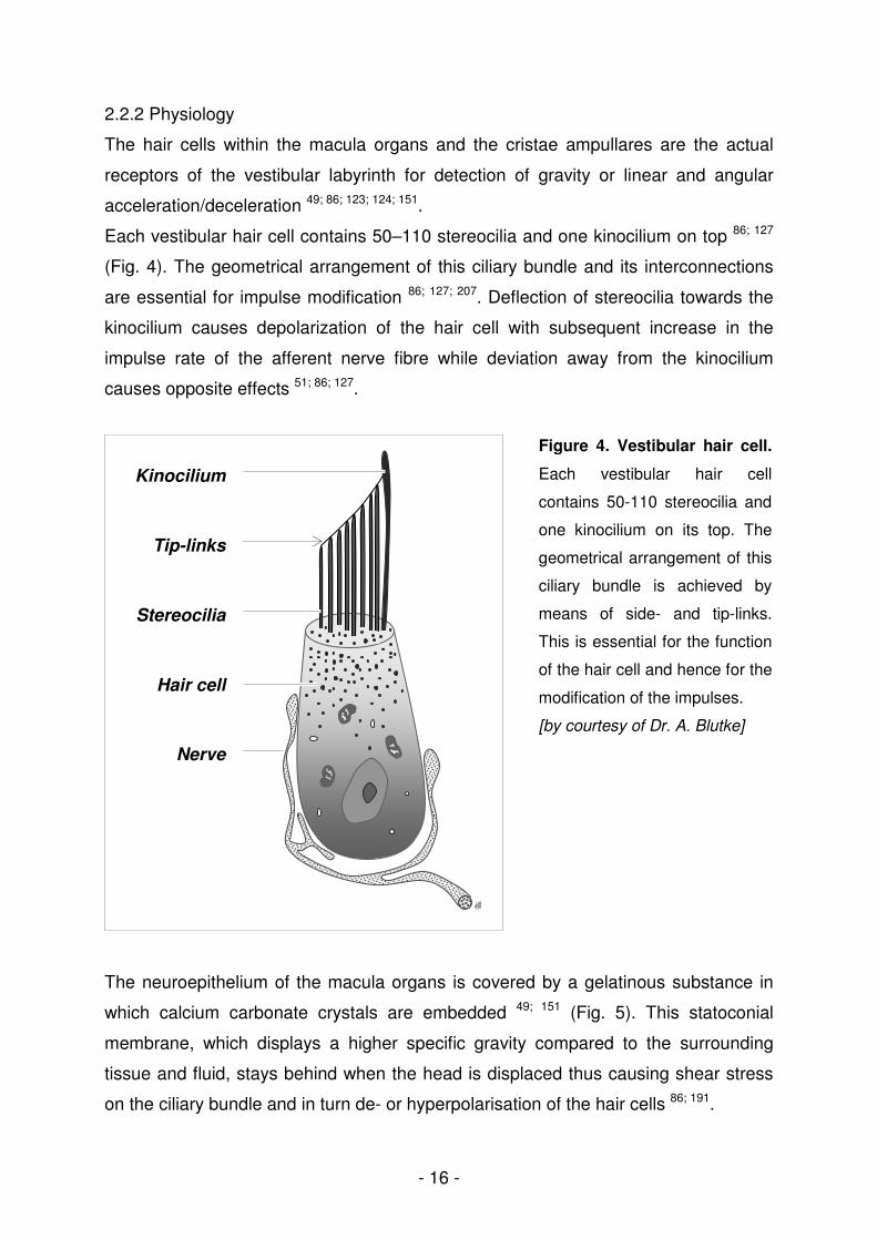
2.2.2 Physiology
The hair cells within the macula organs and the cristae ampullares are the actual
receptors of the vestibular labyrinth for detection of gravity or linear and angular
acceleration/deceleration 49; 86; 123; 124; 151.
Each vestibular hair cell contains 50–110 stereocilia and one kinocilium on top 86; 127
(Fig. 4). The geometrical arrangement of this ciliary bundle and its interconnections
are essential for impulse modification 86; 127; 207. Deflection of stereocilia towards the
kinocilium causes depolarization of the hair cell with subsequent increase in the
impulse rate of the afferent nerve fibre while deviation away from the kinocilium
causes opposite effects 51; 86; 127.
The neuroepithelium of the macula organs is covered by a gelatinous substance in
which calcium carbonate crystals are embedded 49; 151 (Fig. 5). This statoconial
membrane, which displays a higher specific gravity compared to the surrounding
tissue and fluid, stays behind when the head is displaced thus causing shear stress
on the ciliary bundle and in turn de- or hyperpolarisation of the hair cells 86; 191.
Nerve
Kinocilium
Stereocilia
Tip-links
Hair cell
Figure 4. Vestibular hair cell.
Each vestibular hair cell
contains 50-110 stereocilia and
one kinocilium on its top. The
geometrical arrangement of this
ciliary bundle is achieved by
means of side- and tip-links.
This is essential for the function
of the hair cell and hence for the
modification of the impulses.
[by courtesy of Dr. A. Blutke]

- 17 -
Since the macula organs within the utricle and saccule are perpendicular to one
another and the hair cells therein are orientated in all directions 127, any head
displacement, linear acceleration/deceleration or the constant force of gravity may be
detected 86; 191; 212.
Figure 5. Macula utriculi/sacculi. There is a thickening in the utricle and saccule wall called
the macula uriculi/sacculi. Each macula consists of a neuroepithelium with hair and
supporting cells, covered by a proteinaceous mass with calcium carbonate crystals, named
the otolith or statoconial membrane. [by courtesy of Dr. A. Blutke]
Sensory cells of the crista again are embedded in a gelatinous mass called cupula
which extends from the neuroepithelial surface to the ampulla roof 49; 151 (Fig. 6).
Rotation of the head thus causes deflection of the cupula and therefore shear stress
on the ciliary bundle, due to the inertia of the endolymph 86; 191; 212. Since hair cells
within the crista ampullaris are morphologically and functionally polarized, all the
kinocilia within a crista are either directed towards the semicircular duct or towards
the utricle 127. Opposite polarization of hair cells in the ampulles of the right and left
ear as well as different orientation within the three ampulles enables the detection of
angular (rotational) acceleration/deceleration 86; 127; 191; 212.
Otoliths
Hair cell
Supporting cell
Nerve fiber
Otolith membrane
Macula

- 18 -
Figure 6. Crista ampullaris. Each semicircular duct has an enlargement on the utricle side
called an ampulla with a crista ampullaris. The crista contains the neuroepithelium, which is
covered by the gelatinous cupula reaching up to the ampulla roof.
[by courtesy of Dr. A. Blutke]
2.2.3 Nervous pathways
Generated impulses are transmitted from the N. utricularis, N. saccularis and Nn.
ampullares to the superior and inferior division of the vestibular ganglion (VG) 151.
Vestibular fibres from the VG then join the cochlear nerve, and along with the facial
nerve, exit the temporal bone within a common dura sheath 151. The
vestibulocochlear nerve (cranial nerve (CN) VIII) travels to the rhombencephalon
where it divides into a radix vestibularis and cochlearis before entering the rostral
medulla 151. Fibres of the vestibular division synapse to the four vestibular nuclei
(rostral, medial, lateral and caudal) and some directly enter the cerebellum 49; 86; 115;
123; 144; 151; 191. The vestibular nuclei are connected to multiple parts of the nervous
system (cerebellum, spinal cord, motor nuclei of CN III, IV, VI, reticular formation,
thalamus, cortex and vestibular labyrinth) in order to maintain equilibrium 34; 49; 67; 123;
124; 144; 151; 191. An appropriate relationship of eye, trunk, limb and head position during
posture and locomotion is achieved by means of vestibulospinal and vestibuloocular
reflexes 63; 67; 123; 124; 212.
Nerve
Hair cells
Cupula Crista ampullaris
Ampulla

- 19 -
2.3 Vestibular dysfunction
The vestibular system consists of a peripheral (vestibular labyrinth within the inner
ear, vestibular portion of CN VIII) and a central component (vestibular nuclei,
vestibulocerebellum, pathways) 49; 144; 212.
Vestibular system disorders, which are relatively common, both in man and in
domestic mammals, cause varying degrees of loss of equilibrium manifesting as
vestibular ataxia, falling, circling, rolling, head tilt, and nystagmus 49; 115; 124; 144; 191; 212.
Vestibular signs may be unilateral or bilateral. Distinction of central from peripheral
vestibular disease is achieved by recognition of clinical signs caused by the
dysfunction of other systems located in the brainstem or cerebellum 34; 49; 144; 212; 214.
While neoplasia and infection/inflammation are the most common causes of central
vestibular disease in cats and dogs 144; 212, otitis media/interna and idiopathic
vestibular disease are the major etiologies for peripheral 144; 191; 192; 212 vestibular
disorders. Besides, thiamine deficiency 123; 191; 217, metronidazole intoxication 123; 212;
217, head trauma 191; 217, hypothyroidism 93; 219, anomalies 144, cerebrovascular 144 and
degenerative diseases 144; 191 may cause central vestibular dysfunction. Tumors of or
involving the middle/inner ear 123; 191; 212; 217, head trauma resulting in a fractured
temporal bone or fractured bulla tympanica 123; 191; 217, hypothyroidism 105; 212; 219,
congenital diseases 14; 58; 123; 126; 191; 217, and application of ototoxic drugs 123; 212; 217
may induce peripheral vestibular disease.
Apart from vestibular disorders with known etiology, idiopathic vestibular diseases
play a major role in cats and dogs. Idiopathic vestibular disease affects geriatric dogs
and cats at all ages and manifests as a peracute onset of severe peripheral
vestibular dysfunction resolving without therapy within two weeks 25; 123; 144; 191; 212.
The etiology of the disease is still unknown and diagnosis is largely based on the
exclusion of other causes for vestibular disease and the resolution of clinical signs 25;
49; 123; 144; 191; 212. Even though herpesvirus infections are widespread amongst cats 75
and dogs 50 and infection of vestibular inner ear structures by FHV-1 or CHV-1 may
be a potential cause of idiopathic vestibular disease, such investigations have not
been reported yet. In humans on the other hand, the possible association of
vestibular herpesvirus infections with vestibulopathies has been subject of research
for many years. Thus, a variety of herpesviruses have been located in different
compartments of the vestibular system 8-10; 61; 230 and are discussed to cause various
recurrent vestibulopathies 65; 159; 173.

- 20 -
2.4 Processing of the canine and feline vestibular labyrinth
Due to the inaccessibility and the complex anatomy of the membranous labyrinth,
technical processing of these structures for further investigations is very challenging
and tedious and, therefore, currently not a routine procedure in cats and dogs 46; 49;
214. Established standard protocols, e.g. for histological investigation, include long-
lasting fixation and decalcification of temporal bones 36. Depending on the chemicals
and temporal bone size, decalcification following fixation, takes approximately 40
days in cats 13 and up to 225 days in dogs 36.
Consequently, only few reports about morphological 6; 39; 62; 103; 143; 175; 187 and
immunohistochemical 41 investigation of canine and feline vestibular inner ear
structures exist.
Molecular analyses, as PCR-based investigations, performed on isolated structures
of the vestibular inner ear, have not been reported in cats and dogs so far. This might
probably be due to the absence of protocols suitable for molecular analyses following
standard vestibular inner ear preparation procedures.

- 21 -
3. Own scientific experiments
3.1 A rapid approach to ultrastructural evaluation and DNA analysis of
the vestibular labyrinth and ganglion in dogs and cats
Birgit Parzefall, Wolfgang Schmahl, Andreas Blutke, Kerstin Baiker,
Kaspar Matiasek
Journal of Neuroscience Methods (2009) 177: 217-224

- 22 -
Journal of Neuroscience Methods (2009) 177: 217-224
A rapid approach to ultrastructural evaluation and DNA
analysis of the vestibular labyrinth and ganglion in dogs
and cats*
Birgit Parzefalla, Wolfgang Schmahla, Andreas Blutkea,
Kerstin Baikera, Kaspar Matiaseka,b,**
a Institute of Veterinary Pathology, Ludwig-Maximilians University, Veterinärstraße 13,
80539 Munich, Germany
b Neuropathology Laboratory, Diagnostic Laboratory Services, The Animal Health
Trust, Newmarket, UK
**Corresponding author at: Neuropathology Laboratory, Diagnostic Services, The
Animal Health Trust, Lanwades Park, Kentford, Newmarket, CB8 7UU, Suffolk, UK.
Tel.: + 44 1638750659; fax: + 44 1638 555643.
E-mail address: [email protected]
Figures: 13
Tables: 2
* Parts of this study were presented as a poster presentation at the 21
st annual symposium of the
European Society of Veterinary Neurology (ESVN) in Rhodes, Greece (2008) and as an oral presentation at the 52
nd annual meeting of the Fachgruppe Pathologie of the Deutsche
Veterinärmedizinische Gesellschaft (DVG) in Fulda, Germany (2009). The abstract has been published in the Deutsche Tierärztliche Wochenschrift: Parzefall, B., Schmahl, W., Blutke, A., Baiker, K., Matiasek, K., 2009. Peri-/endolymphatisches Tracing zur bimodalen Untersuchung des vestibulären Labyrinths und Ganglions von Hund und Katze. Dtsch Tierarztl Wochenschr. 116 (7), 279.
- 23 -
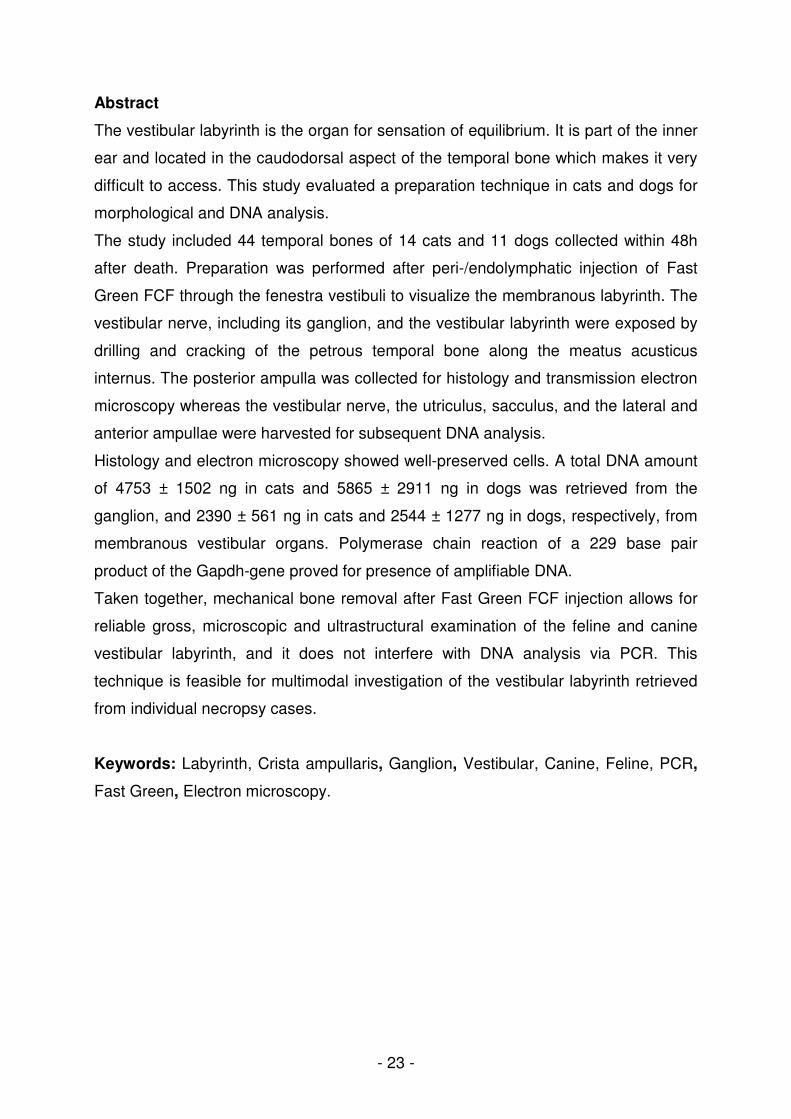
Abstract
The vestibular labyrinth is the organ for sensation of equilibrium. It is part of the inner
ear and located in the caudodorsal aspect of the temporal bone which makes it very
difficult to access. This study evaluated a preparation technique in cats and dogs for
morphological and DNA analysis.
The study included 44 temporal bones of 14 cats and 11 dogs collected within 48h
after death. Preparation was performed after peri-/endolymphatic injection of Fast
Green FCF through the fenestra vestibuli to visualize the membranous labyrinth. The
vestibular nerve, including its ganglion, and the vestibular labyrinth were exposed by
drilling and cracking of the petrous temporal bone along the meatus acusticus
internus. The posterior ampulla was collected for histology and transmission electron
microscopy whereas the vestibular nerve, the utriculus, sacculus, and the lateral and
anterior ampullae were harvested for subsequent DNA analysis.
Histology and electron microscopy showed well-preserved cells. A total DNA amount
of 4753 ± 1502 ng in cats and 5865 ± 2911 ng in dogs was retrieved from the
ganglion, and 2390 ± 561 ng in cats and 2544 ± 1277 ng in dogs, respectively, from
membranous vestibular organs. Polymerase chain reaction of a 229 base pair
product of the Gapdh-gene proved for presence of amplifiable DNA.
Taken together, mechanical bone removal after Fast Green FCF injection allows for
reliable gross, microscopic and ultrastructural examination of the feline and canine
vestibular labyrinth, and it does not interfere with DNA analysis via PCR. This
technique is feasible for multimodal investigation of the vestibular labyrinth retrieved
from individual necropsy cases.
Keywords: Labyrinth, Crista ampullaris, Ganglion, Vestibular, Canine, Feline, PCR,
Fast Green, Electron microscopy.
- 24 -
1. Introduction
The vestibular labyrinth contains the organs for sensation of equilibrium. It is part of
the inner ear and located in the caudodorsal aspect of the temporal bone 19; 35. It
consists of a membranous labyrinth that is surrounded by compact cortical bone
which makes it very difficult to access.
In order to investigate the vestibular labyrinth, temporal bones used to be fixed in
aldehydes, decalcified for months and finally embedded in either paraffin 8-9; 12; 36, or
celloidin 7; 22; 32-33. These procedures take 40 days in cats 4 and up to 225 days in
dogs 8 until a morphological investigation becomes possible and they effectively
impede further molecular analyses on the tissue.
In particular, studies on inner ear stem cells, hair cell biology and neurotransmission
employ investigational techniques that rely on optimal preservation of intracellular
proteins and nucleic acids. Other areas of advanced vestibular research focus
neurotropic viruses that may infect the vestibular labyrinth 3 and/or ganglion 2; 18; 40-41
by using polymerase chain reaction (PCR).
This investigation was aimed to assess the influence of peri- and/or endolymphatic
tracer injection on the ultrastructure of the crista ampullaris, and DNA-preservation
within the vestibular labyrinth and vestibular nerve ganglion.

- 25 -
2. Materials and methods
2.1 Animals and tissues
This study enrolled 44 temporal bones from 14 cats and 11 dogs (Table 1), presented
for routine post-mortem examination. All samples were processed within 48 hours
after death.
Table 1: Numbers, ages and gender of investigated animals.
Age Gender
Number
of animals
Number of investigated
temporal bones Adult
> 1 year
Juvenile
< 1 year
Female Male
Cats
14
24
12
2
5
9
Dogs 11 20 8 3 5 6
2.2 Preparation of the vestibular labyrinth
2.2.1 Gross preparation
On necropsy, the head was obtained after removal of skin and neck muscles with
subsequent exarticulation in the atlanto-occipital joint. The skin, the two pinnae and
muscles were removed from the ventral part of the head to expose the bony part of
the external auditory canal and the ventral surface of the bulla tympanica. In the next
step, the skull was split sagitally in the midline with a band saw. After removal of the
brain from the caudal cranial fossa, the yellow-white tinged temporal bone, which is
slightly paler than the surrounding bone became visible (Fig. 1). The bulla tympanica
was opened from the ventrolateral aspect (Fig. 2). The auditory ossicles, the M.
tensor tympani and all bulla parts were removed in order to expose the fenestra
cochleae and fenestra vestibuli (Fig. 3).

- 26 -
Figure 1: Topographic anatomy of the
cat’s temporal bone (TB) visible after
calvarial removal. The cutting lines for
preparation are indicated. Scale bar =
0.5 cm.
Figure 2: Ventral view on the opened
bullae tympanicae (BT) of an unsplit cat’s
skull which are divided into a
rostromedial and a caudolateral portion
(dashed lines) through an incomplete
bony septum (dotted lines).
FM = foramen magnum, FC = fenestra
cochleae, vB = ventral BT.
Scale bar = 1 cm.
Figure 3: Lateroventral view on the TB
after removal of the bony parts of the BT.
The fenestra vestibuli and cochleae are
the most important landmarks for
orientation during the preparation
procedure.

- 27 -
2.2.2 Micropreparation
All the following preparations were performed using a dissection microscope (Stemi
DV4, Carl Zeiss AG, Jena, Germany) with the temporal bone placed in a Petri dish
filled with Aqua bidest. in order to allow preparation through perpetual removal of
bone graft and avoidance of exsiccation and extensive heat production during the
burr procedure (see below).
Throughout all further steps of preparation, the fenestra cochleae and vestibuli were
the most important landmarks (Fig. 4).
Figure 4: Closer view on the left TB.
The asterisk is placed on the fenestra
cochleae. The fenestra vestibuli
(surrounded by the dashed line) still
contains the footplate of stapes sitting on
its membrane. The facial nerve (Nf;
dotted line) emerges from the facial canal
and, over a short distance, is exposed to
the lumen of the middle ear cavity.
cd = caudodorsal, rv = rostroventral.
Scale bar = 0.5 cm.
The caudodorsal aspect of the petrous temporal bone (PTB) was directed towards,
the rostroventral apex of the cochlea positioned away from the right-handed
preparator. For the left PTB bone, the medial side of the PTB, containing the meatus
acusticus internus, was oriented to the right, while the lateral aspect was directed to
the left, with the fenestra vestibuli on top of the fenestra cochleae. For the right PTB
the medial side was positioned to the left, while the lateral part was on the right side,
respectively.

- 28 -
The membrane of the fenestra cochleae was fenestrated with a sterile 23 gauge
needle and between 0.05 ml and 0.1 ml of 1% Fast Green FCF 1; 13 were peri-
/endolymphatically injected into the vestibulum through the pierced membrane of the
fenestra vestibuli (Fig. 5). Thereby the dye merged with the peri/endolymphatic fluid
and visualized the almost transparent membranous labyrinth. The injection was
performed slowly, over a period of at least 3 s until the dark-green discoloration of the
cochlear parts gleamed through the overlying bone.
Figure 5: Peri-/endolymphatic
injection of the dye. In order to
visualize the membranous vestibular
labyrinth Fast Green FCF is injected
via the fenestra vestibuli after
puncture of the membrane of the
fenestra cochleae for pressure relief.
rv = rostroventral, cd = caudodorsal.
To gain access to the ampullae, the facial canal was opened by a burr (GG 12,
Proxxon, Niersbach, Germany) and the facial nerve was removed. Its rostral aspect,
however was spared, in order to guarantee an intact superior division of the
vestibular nerve (own unpublished observation) that anastomoses with the facial
nerve 17; 35.
At first, the lateral ampulla was identified, followed by the anterior ampulla. From
there, the course of the lateral semicircular canal was followed to the posterior
ampulle, that is located caudolateral to the fenestra cochleae. For unambiguous
identification of the ampullae, the bony labyrinth surrounding the ampullae and the
beginning of the corresponding semicircular ducts was carefully drilled, leaving
nothing but a very thin bone lamella intact (Fig. 6). Thereafter, the PTB was split
along the internal auditory canal (Fig. 7) into two parts containing the rostroventral
cochlea and the caudodorsal vestibular labyrinth (Fig. 8), respectively.

- 29 -
Figure 6: Left feline TB after stepwise
removal of the superficial bone laminae
overlying the vestibular labyrinth.
Through injection of Fast Green FCF the
posterior semicircular canal (SC) and
ampulla (AP) became clearly visible.
Asterisk = fenestra cochleae.
Scale bar = 0.5 cm.
Figure 7: Further preparation of the TB.
In order to expose the ampullae and the
beginning of the semicircular ducts the
TB is drilled with a burr followed by
cracking (plane indicated through dotted
lines) along the meatus acusticus
internus.
Figure 8: View of the vestibular labyrinth.
After cracking of the petrous temporal
bone, the vestibular labyrinth which
includes the utriculus, sacculus, ampulla
anterior (a), posterior (b) and lateralis (c)
become visible in the osseous
vestibulum. The vestibular nerve (VN)
with its ganglion superior and inferior can
now be harvested.
VN
- 30 -
The vestibular nerve, including the superior and inferior vestibular ganglia, was
carefully separated from the cochlear and facial nerve by microforceps, and collected
for further sample processing.
In order to remove the utriculus, the sacculus, the ampullae and semicircular ducts,
the thin bone layer covering the ampullae and semicircular ducts was removed. In
close proximity to the posterior ampulla, the utriculus, the crus commune (resembling
the non-ampullary aspect of the posterior and anterior semicircular ducts) as well as
the non-ampullary region of the lateral semicircular duct and the ampullary part of the
posterior semicircular duct were transsected with microscissors. Then, the posterior
ampulla was harvested for histotechnical processing, while the utriculus, the sacculus
and the lateral and anterior ampulla were collected for DNA isolation.
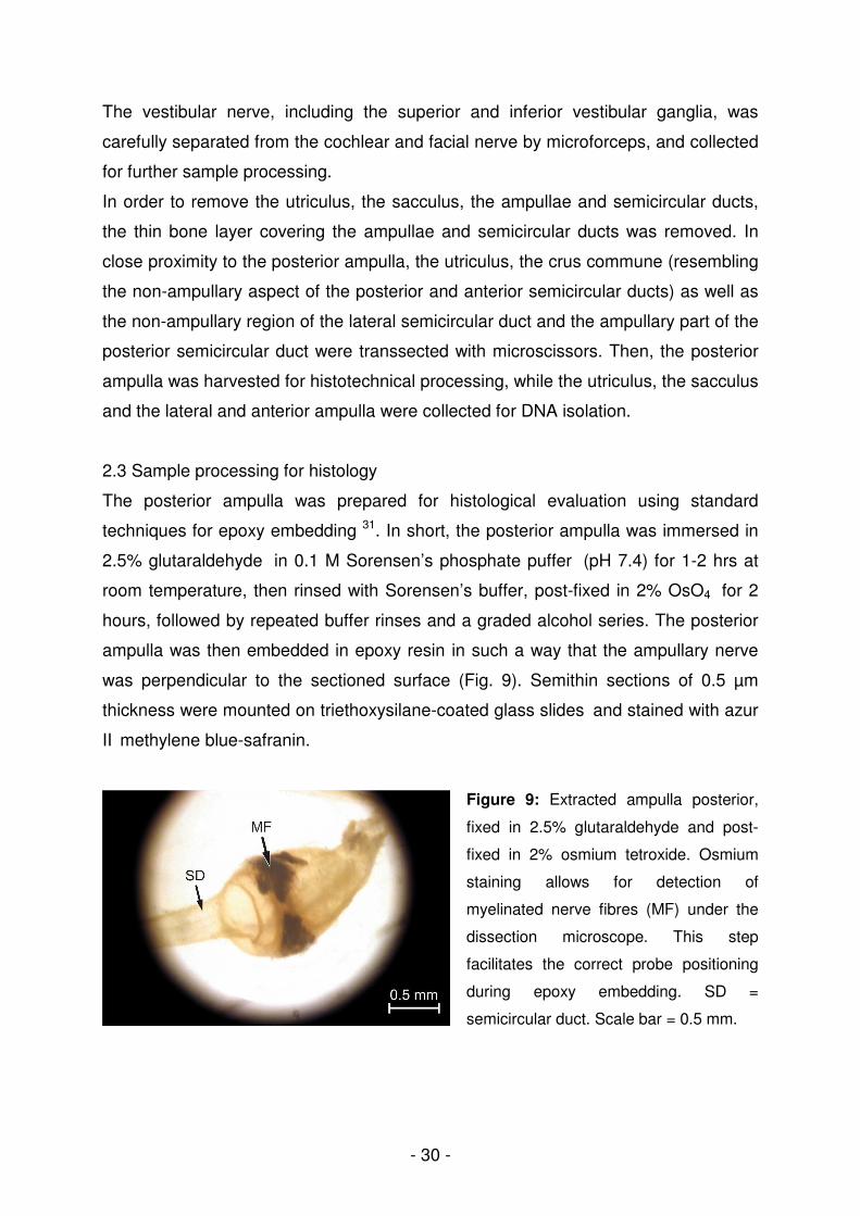
2.3 Sample processing for histology
The posterior ampulla was prepared for histological evaluation using standard
techniques for epoxy embedding 31. In short, the posterior ampulla was immersed in
2.5% glutaraldehyde in 0.1 M Sorensen’s phosphate puffer (pH 7.4) for 1-2 hrs at
room temperature, then rinsed with Sorensen’s buffer, post-fixed in 2% OsO4 for 2
hours, followed by repeated buffer rinses and a graded alcohol series. The posterior
ampulla was then embedded in epoxy resin in such a way that the ampullary nerve
was perpendicular to the sectioned surface (Fig. 9). Semithin sections of 0.5 µm
thickness were mounted on triethoxysilane-coated glass slides and stained with azur
II methylene blue-safranin.
Figure 9: Extracted ampulla posterior,
fixed in 2.5% glutaraldehyde and post-
fixed in 2% osmium tetroxide. Osmium
staining allows for detection of
myelinated nerve fibres (MF) under the
dissection microscope. This step
facilitates the correct probe positioning
during epoxy embedding. SD =
semicircular duct. Scale bar = 0.5 mm.
- 31 -
2.4 Transmission electron microscopy
Ultrathin sections (~70 nm) of the crista ampullaris posterior were mounted on
formvar-chloroform covered copper grids, stained with uranyl acetate and lead
citrate, and examined with a transmission electron microscope (EM 10, Carl Zeiss
AG, Jena, Germany). Cells were identified according to the listed criteria (Table 2).
2.5 Polymerase chain reaction
Superior/inferior vestibular ganglion, utriculus, sacculus, anterior and lateral ampullae
and their corresponding semicircular ducts were placed in two sterile 1.5 ml tubes,
snap frozen in liquid nitrogen, and stored at -80°C. DNA extraction was performed
with a commercially available kit (QIAmp DNA Micro-Kit, Qiagen, Hilden, Germany),
according to the manufacturer’s description, followed by spectrophotometric
measurement (NanoDrop 1000, peQLab Biotechnologie GmbH, Erlangen, Germany)
of DNA content.
To confirm the presence of amplifiable DNA in the nucleic acid preparations,
published primers were used to amplify a 229 base pair segment of the
glyceraldehyde-3-phosphate dehydrogenase gene (Gapdh) 20; 23. Reaction products
were electrophoresed through a 2% agarose gel with 0.5 x TAE buffer and stained
with ethidium bromide.

- 32 -
Table 2: Ultrastructural cell characteristics.
Cell type Shape Cytoplasm Membrane
specifications
Cell-to-cell
connections
Other
Haircell I
(HC I)
Flask
shaped
-Nucleus located in
the bulbous area
-Most organelles located
supranuclear (endoplasmatic
reticulum, Golgi complex,
ribosomes, vesicles,
mitochondria)
-Cuticular plate
20-100 Sc,
1 kinocilium, Mv
TJ, AJ Chalice-like
nerve ending
Haircell II
(HC II)
Irregular,
cylindrical
-Nucleus varies through the
middle one-third of the cell
-Organelles appear more
evenly distributed than in HC I
20-100 Sc,
1 kinocilium, Mv
TJ, AJ Bouton-
shaped
nerve ending
Supporting
cell (SC)
Tall -Run from the epithelial surface
down to the basal lamina
-Nucleus located in the lower
part of the cell near the basal
lamina and below the nuclei of
the HCs
-Reticular lamina
-Upper half to two-thirds of the
SC is filled with large, densely
packed vesicles (mitochondria,
Golgi complexes, lysosomes,
free ribosomes)
Mv TJ, AJ, GJ
Transitional
cells (TCs)
Columnar-
cuboidal
-Nucleus more central than in
SC
Mv GJ, AJ, JC
Dark cells
(DCs)
Tall-
cuboidal-
squamous
-Central located lobulated
nucleus
-Extensive interdigitation of the
cytoplasm in the basal portion
of the cell
-Most organelles located in the
upper two-thirds of the cell
-Nucleus and cytoplasm stain
darkly
Few Mv TJ, AJ Degenerating
otoconia on
cell surface,
subepithelial
melanocytes
Planum
semilunatum
(PSL) cells
Columnar-
cuboidal
-Nucleus circular-oval, basal or
centrally located
Few Mv GJ, AJ, JC Subepithelial
reticular layer
Epithelium Squamous -Nucleus circular, centrally
located
TJ, AJ
Cell characteristics are extracted from specific literature 15; 21; 24-29; 38-39. Mv = mikrovilli, TJ =
tight junction, AJ = adherens junction, GJ = gap junction, JC = illdefined junctional complex.
- 33 -
3. Results
3.1 Preparation
In all included cases, peri-/endolymphatic injection of Fast Green FCF resulted in a
discoloration of the membranous labyrinth and, hence, facilitated identification and
preparation of the cavitary inner ear compartments. Preparation of both vestibular
labyrinths and removal for further processing was achieved within 2 hours in cats and
within 3 hours in dogs. There was no obvious difference in gross tissue preservation
or its feasibility for preparation amongst specimens collected within 48 hours after
death, whereas older samples, collected the same way, (not included in the study)
became decomposed which impedes a clear identification of the vestibular nerve and
a proper removal of the membranous vestibular parts. The position of the ampullae in
relation to the fenestra vestibuli and cochleae, and their depth within the PTB was
found to be individually variable. Ampulles in cats and and small dogs displayed a
more superficial localization than in middle- and large-sized dogs and did not vary as
much in position. In general, the anterior and lateral ampullae are easier to locate
and isolate than the posterior ampulla. Preparation of feline temporal bones does not
raise any special difficulties, whereas preparation of canine temporal bones is far
more challenging due to the great variety of ampulla location and the increased
fragility of the PTB in this species.

- 34 -
3.2 Histology
Mechanical removal of the bone enabled routine soft tissue processing without
decalcification. Correct orientation of the specimens in the embedding procedure is
crucial to obtain the same section plane. Some ampullae were displaced within the
first hour of the embedding process and had to be put back into the right position. All
histological sections of the ampulla revealed the crista ampullaris (Fig. 10), the
epithelial lining of the ampulla, and sometimes parts of the semicircular duct. The
same section plane was obtained throughout all samples, whereas the cutting level,
with regard to the neighboring dark cell (DC) and planum semilunatum zones,
sometimes differed and, therefore, slightly changed the histological picture. All
sections revealed the stroma of the crista including myelinated nerve fibres and blood
vessels as well as the sensorineural epithelium consisting of hair- and supporting
cells. Transitional cells and DCs were obtained in deeper and planum semilunatum
(PSL) cells in more shallow sections. The injection of Fast Green FCF did not
interfere with the staining.
Figure 10: Histological appearance
of the crista ampullaris posterior on
semithin sections.
a = hair cell, b = transitional zone,
c = stereocilia of the hair cells
projecting into the lumen (Lu) of the
ampulla, d = squamous ampulla
epithel, e = myelinated fibres of the
vestibular nerve (ampullaris).
Scale bar = 60 µm.
- 35 -
3.3 Transmission electron microscopy
Due to adequate preservation, all cells listed in Table 2 were readily identified on the
basis of their ultrastructural characteristics. Numerous hair cells (HCs) type I (Fig.
11A) were seen along the crista ampullaris, whereas hair cells type II were only
occasionally detected. HCs were in close contact with supporting cells (SCs) and
displayed tight and adherens junctions (Fig. 11A). Stereocilia (Sc) were implanted in
the cuticular plate (Fig. 11B) and their spatial relationship to the kinocilium was not
always clearly discernible. However, the single kinocilium was found in some HCs.
Cytoplasmic organelles were clearly visible. For many instances, and throughout all
cells, the cisternal system appeared dilated. The nuclei of SCs were basally placed
and their apical part was in close contact to the HCs where it displayed the typical
reticular laminae (Fig. 11A). Cytoplasmic protrusions in the apical part of the SCs
were frequently noticed. Some transitional cells (TCs) (Fig. 11C) were found at the
base of the crista. They were contiguous with dark cells DCs. TCs were columnar
with the nucleus located in the lower part of the cell. Their junctional complexes were
clearly visible. DCs revealed darkly stained nuclei and also a rather electron-dense
cytoplasm. Nuclei were lobulated and the cytoplasm showed characteristic basal
interdigitations. Degenerating otoconia were often detected, confined to the DC
surface. PSL were sometimes visible, depending on the cutting level. If present, PSL
were found to be cuboidal with round central nuclei. Squamous epithelium was
detected in the semicircular duct. The stroma of the crista ampullaris was well
preserved showing myelinated nerve fibres (Fig. 11D) and blood vessels.

- 36 -
Figure 11: Well-preserved ultramorphology of the crista ampullaris posterior. A, type I hair
cell with nerve calyx (arrows), showing numerous mitochondria. Neighboring supporting cells
(SCs) with reticular lamina (asterisk) and desmosomes (arrowheads). B, apical part of a type
I hair cell with stereocilia (Sc) implanted in the cuticular plate (CP), with numerous
mitochondria in the infracuticular region. C, transitional cells (TCs). Junctional complexes are
indicated by small arrows. D, myelinated nerve fibre with well-preserved compacted myelin
(My), axon (Ax) and axoplasmic mitochondria (Mi). Nc = nucleus.
Scale bar: A = 1.4 µm, B = 1.1 µm, C = 2.0 µm, and D = 0.6 µm.

- 37 -
3.4 Integrity and amount of isolated DNA
DNA could be extracted from all samples with a mean amount of 4753 ng (±1502) for
the feline and 5865 ng (±2911) for the canine vestibular ganglion.
The average amount of isolated DNA from the utriculus/sacculus, the lateral and
anterior ampullae with their corresponding semicircular ducts were 2390 ng (±561) in
cats and 2544 ng (±1277) in dogs, respectively (Fig. 12).
The integrity of DNA of all samples was confirmed by amplification of a 229 base pair
product of Gapdh (Fig. 13).
Figure 12: Box and whisker plot
displaying the total amount of isolated
DNA in nanogram (ng).
F = feline, C = canine,
A/U = ampulla/utriculus,
G = vestibular ganglion.
Figure 13: Amplification of the
housekeeping gene Gapdh by PCR.
M: Fragment size marker. Visible
marker bands indicate fragment sizes
of 300, 250 and 200 base pairs from
top to bottom. Ø: spacing lane.
NTC: no template control. DNA samples
(approx. 100 ng) from cats’ (F-) and
dogs’ (C-) ampullae (A/U) and
vestibular ganglia (G). All templates
show a specific PCR-product of 229
base pairs length (arrow).

- 38 -
4. Discussion
Previous studies performed in human cadavers have employed physical preparation
techniques to collect the vestibular labyrinth for further DNA analysis 3. To date,
studies of the canine and feline inner ear have concentrated on morphological 6; 16; 30;
34 and histochemical 10-11 investigations while the preparation of the vestibular
labyrinth for further DNA analysis has not been described. This study employed a
histologic dye 1; 13 in order to visualize the membranous labyrinth which facilitates the
orientation for the preparator. This is the first report that proves that Fast Green FCF
is feasible for bimodal analyses at ultrastructural and DNA level. This method is
reasonably fast and, therefore, advantageous compared to anatomical standard
techniques which require time-consuming decalcification steps. The published
standard procedures also have a negative effect on the integrity of DNA in terms of
cross-linking and degradation which interferes with PCR analysis 5; 14; 37.
In addition, stepwise preparation of the vestibular labyrinth under the dissection
microscope also allows for thorough gross examination which would be impossible if
the entire temporal bone is fixed, decalcified and embedded 33. Even though
autolysis in the membranous labyrinth starts early and can cause difficulties in
cadaver studies, the 48-h time frame still enables the preparator to perform the
described investigations.
Taking the methodological aspects of this study into account, gross, microscopic and
ultrastructural examination of the feline and canine vestibular labyrinth, as well as
performance of PCR, e.g. for epidemiologic studies, can conveniently be performed
with the described protocol herewith, even if just one single temporal bone is
available.
Acknowledgments
The authors thank the Institute for Medical Microbiology, Infectious and Epidemic
Diseases, Ludwig-Maximilians University, Munich, for sharing the spectrophotometer.
This study was supported by the Nachlassstiftung Dr. Kurtze.

- 39 -
References
1. Adams, M.E., Hurd, E.A., Beyer, L.A., Swiderski, D.L., Raphael, Y., Martin, D.M.,
2007. Defects in vestibular sensory epithelia and innervation in mice with loss of
Chd7 function: implications for human CHARGE syndrome. J Comp Neurol 504, 519-
532.
2. Arbusow, V., Schulz, P., Strupp, M., Dieterich, M., von Reinhardstoettner, A.,
Rauch, E., Brandt, T., 1999. Distribution of herpes simplex virus type 1 in human
geniculate and vestibular ganglia: implications for vestibular neuritis. Ann Neurol 46,
416-419.
3. Arbusow, V., Theil, D., Strupp, M., Mascolo, A., Brandt, T., 2001. HSV-1 not only in
human vestibular ganglia but also in the vestibular labyrinth. Audiology & Neuro-
Otology 6, 259-262.
4. Balogh, K., Jr., Nomura, Y., 1964. A technique for the demonstration of
acetylcholinesterase activity in the inner ear after decalcification with EDTA. J
Histochem Cytochem 12, 931-933.
5. Ben-Ezra, J., Johnson, D.A., Rossi, J., Cook, N., Wu, A., 1991. Effect of fixation on
the amplification of nucleic acids from paraffin-embedded material by the polymerase
chain reaction. J Histochem Cytochem 39, 351-354.
6. Branis, M., Burda, H., 1985. Inner ear structure in the deaf and normally hearing
Dalmatian dog. J Comp Pathol 95, 295-299.
7. Chole, R.A., Charpied, G.L., 1981. Preparation of small temporal bones for high-
resolution light microscopy. Arch Otolaryngol 107, 610-612.
8. Coppens, A.G., Gilbert-Gregory, S., Steinberg, S.A., Heizmann, C., Poncelet, L.,
2005. Inner ear histopathology in "nervous Pointer dogs" with severe hearing loss.
Hear Res 200, 51-62.

- 40 -
9. Coppens, A.G., Kiss, R., Heizmann, C.W., Deltenre, P., Poncelet, L., 2001. An
original inner ear neuroepithelial degeneration in a deaf Rottweiler puppy. Hear Res
161, 65-71.
10. Coppens, A.G., Kiss, R., Heizmann, C.W., Schafer, B.W., Poncelet, L., 2001.
Immunolocalization of the calcium binding S100A1, S100A5 and S100A6 proteins in
the dog cochlea during postnatal development. Brain Res Dev Brain Res 126, 191-
199.
11. Coppens, A.G., Resibois, A., Poncelet, L., 2000. Immunolocalization of calbindin
D28k and calretinin in the dog cochlea during postnatal development. Hear Res 145,
101-110.
12. Coppens, A.G., Steinberg, S.A., Poncelet, L., 2003. Inner ear morphology in a
bilaterally deaf Dogo Argentino pup. J Comp Pathol 128, 67-70.
13. Curthoys, I.S., Markham, C.H., Curthoys, E.J., 1977. Semicircular duct and
ampulla dimensions in cat, guinea pig and man. J Morphol 151, 17-34.
14. Falconi, M., Teti, G., Zago, M., Pelotti, S., Gobbi, P., Breschi, L., Mazzotti, G.,
2007. Effect of fixative on chromatin structure and DNA detection. Microsc Res Tech
70, 599-606.
15. Friedmann, I., Ballantyne, J., 1984. Ultrastructural atlas of the inner ear.
Butterworths, London.
16. Gacek, R.R., 1961. The macula neglecta in the feline species. J Comp Neurol
116, 317-323.
17. Gacek, R.R., Rasmussen, G.L., 1961. Fiber analysis of the statoacoustic nerve of
guinea pig, cat, and monkey. Anatomical Record 139, 455-463.
18. Gartner, M., Bossart, W., Linder, T., 2008. Herpes virus and Meniere's disease.
ORL J Otorhinolaryngol Relat Spec 70, 28-31.

- 41 -
19. Getty, R., Foust, H.L., Presley, E.T., Miller, M.E., 1956. Macroscopic anatomy of
the ear of the dog. Am J Vet Res 17, 364-375.
20. Gröne, A., Weckmann, M.T., Capen, C.C., Rosol, T.J., 1996. Canine
glyceraldehyde-3-phosphate dehydrogenase complementary DNA: polymerase chain
reaction amplification, cloning, partial sequence analysis, and use as loading control
in ribonuclease protection assays. Am J Vet Res 57, 254-257.
21. Hackett, L., Davies, D., Helyer, R., Kennedy, H., Kros, C., Lawlor, P., Rivolta,
M.N., Holley, M., 2002. E-cadherin and the differentiation of mammalian vestibular
hair cells. Exp Cell Res 278, 19-30.
22. Hall, K.L., Pitts, D.R., Anne, S., Semaan, M.T., Alagramam, K.N., Megerian, C.A.,
2007. Optimization of ribonucleic acid detection from archival Guinea pig temporal
bone specimens. Otol Neurotol 28, 116-123.
23. Hayashiya, S., Tani, K., Morimoto, M., Hayashi, T., Hayasaki, M., Nomura, T.,
Une, S., Nakaichi, M., Taura, Y., 2002. Expression of T helper 1 and T helper 2
cytokine mRNAs in freshly isolated peripheral blood mononuclear cells from dogs
with atopic dermatitis. J Vet Med A Physiol Pathol Clin Med 49, 27-31.
24. Hunter-Duvar, I.M., 1983. An electron microscopic study of the vestibular sensory
epithelium. Acta Otolaryngol 95, 494-507.
25. Igarashi, Y., 1989. Submicroscopic study of the vestibular dark cell area in human
fetuses. Acta Otolaryngol 107, 29-38.
26. Kawamata, S., Harada, Y., Tagashira, N., 1986. Electron-microscopic study of
the vestibular dark cells in the crista ampullaris of the guinea pig. Acta Otolaryngol
102, 168-174.
- 42 -
27. Kikuchi, T., Adams, J.C., Paul, D.L., Kimura, R.S., 1994. Gap junction systems in
the rat vestibular labyrinth: immunohistochemical and ultrastructural analysis. Acta
Otolaryngol 114, 520-528.
28. Kim, T.S., Nakagawa, T., Kitajiri, S., Endo, T., Takebayashi, S., Iguchi, F., Kita,
T., Tamura, T., Ito, J., 2005. Disruption and restoration of cell-cell junctions in mouse
vestibular epithelia following aminoglycoside treatment. Hear Res 205, 201-209.
29. Kimura, R., Lundquist, P.G., Wersaell, J., 1964. Secretory epithelial linings in the
ampullae of the Guinea Pig labyrinth. Acta Otolaryngol 57, 517-530.
30. Kirchner, F.R., Toledo, P.S., Holdcraft, J., 1969. Fixation in vivo by preassessed
intralabyrinthine perfusion in cats' inner ears. Laryngoscope 79, 857-867.
31. Matiasek, K., Gais, P., Rodenacker, K., Jutting, U., Tanck, J.J., Schmahl, W.,
2008. Stereological characteristics of the equine accessory nerve. Anat Histol
Embryol 37, 205-213.
32. McIntire, C.L., 1968. Rapid processing of cat temporal bones. Arch Otolaryngol
88, 258-263.
33. Michaels, L., Wells, M., Frohlich, A., 1983. A new technique for the study of
temporal bone pathology. Clin Otolaryngol Allied Sci 8, 77-85.
34. Mount, R.J., Harrison, R.V., 1987. Scanning electron microscopic observations of
the canine inner ear. Scanning Microsc 1, 1167-1174.
35. Nickel, R., Schummer, A., Seiferle, E. (Eds.), 1992. The anatomy of the domestic
animals (Lehrbuch der Anatomie der Haustiere: Nervensystem, Sinnesorgane,
endokrine Drüsen), Vol. IV. Paul Parey, Berlin.
36. Nilsson, M., Hellstrom, S., Albiin, N., 1991. Decalcification by perfusion. A new
method for rapid softening of temporal bones. Histol Histopathol 6, 415-420.

- 43 -
37. Ohara, Y., Honma, M., Iwasaki, Y., 1992. Sensitivity of the polymerase chain
reaction for detecting human T-cell leukemia virus type I sequences in paraffin-
embedded tissue. Effect of unbuffered formalin fixation. J Virol Methods 37, 83-88.
38. Scarfone, E., Ulfendahl, M., Lofstrand, P., Flock, A., 1991. Light- and electron
microscopy of isolated vestibular hair cells from the guinea pig. Cell Tissue Res 266,
51-58.
39. ten Cate, W.J., Rarey, K.E., 1992. Planum semilunatum of the rat: new light and
electron microscopy observations. Ann Otol Rhinol Laryngol 101, 601-606.
40. Theil, D., Arbusow, V., Derfuss, T., Strupp, M., Pfeiffer, M., Mascolo, A., Brandt,
T., 2001. Prevalence of HSV-1 LAT in human trigeminal, geniculate, and vestibular
ganglia and its implication for cranial nerve syndromes. Brain Pathology 11, 408-413.
41. Vrabec, J.T., 2003. Herpes simplex virus and Meniere's disease. Laryngoscope
113, 1431-1438.

- 44 -
3.2 Evidence of feline herpesvirus-1 DNA in the vestibular ganglion of
domestic cats
Birgit Parzefall, Wolfgang Schmahl, Andrea Fischer, Andreas Blutke,
Uwe Truyen, Kaspar Matiasek
The Veterinary Journal (2009) [Epub ahead of print]

- 45 -
The Veterinary Journal
Evidence of feline herpesvirus-1 DNA in the vestibular
ganglion of domestic cats*
Birgit Parzefalla, Wolfgang Schmahla, Andrea Fischerb,
Andreas Blutkea, Uwe Truyenc, K. Matiaseka,b,**
a Chairs of General Pathology and Neuropathology, Institute of Veterinary Pathology,
Ludwig-Maximilians University, Munich, Germany
b Section of Neurology, Department of Small Animal Medicine, Ludwig-Maximilians
University, Munich, Germany
c Institute of Animal Hygiene and Veterinary Public Health, University of Leipzig,
Leipzig, Germany
d Neuropathology Laboratory, Diagnostic Laboratory Services, The Animal Health
Trust, Newmarket, UK
**Corresponding author at: Neuropathology Laboratory, Diagnostic Services, The
Animal Health Trust, Lanwades Park, Kentford, Newmarket, CB8 7 UU, Suffolk, UK.
Tel.: + 44 1638750659; fax: + 44 1638 555643.
E-mail address: [email protected]
Tables: 2
* Parts of this study were presented as a poster presentation at the 21
st annual symposium of the
European Society of Veterinary Neurology (ESVN) in Rhodes, Greece (2008) and as an oral presentation at the 52
nd annual meeting of the Fachgruppe Pathologie of the Deutsche
Veterinärmedizinische Gesellschaft (DVG) in Fulda, Germany (2009). The abstracts have been published in the Journal of Veterinary Internal Medicine and in the Deutsche Tierärztliche Wochenschrift: Parzefall, B., Schmahl, W., Fischer, A., Matiasek, K., 2009. Feline herpesvirus-1 in the vestibular ganglia of german domestic cats. J Vet Intern Med 23, 409. Parzefall, B., Schmahl, W., Fischer, A., Truyen, U., Matiasek, K., 2009. Untersuchung zum Vorkommen von FHV-1 DNA im vestibulären Labyrinth und Ganglion von Hauskatzen. Dtsch Tierarztl Wochenschr. 116 (9), 341.

- 46 -
Abstract
In humans, herpes simplex virus type-1 has recently been detected in the vestibular
ganglion (VG) and labyrinth (VL) and may be associated with vestibular signs. Feline
herpesvirus-1 (FHV-1) is widespread amongst cat populations and affects many
different tissues. The aim of this pilot study was to investigate the presence of FHV-1
DNA in the VG and VL of randomly selected domestic cats using PCR. FHV-1 DNA
was detected in the VG of 14% of the cats. There was no detectable FHV-1 DNA in
the VL of any cat. None of the infected cats had vestibular signs related to the VG
infection.
Keywords: FHV-1, Feline herpesvirus, Vestibular ganglion, Labyrinth, PCR.

- 47 -
Body of manuscript
Feline herpesvirus-1 (FHV-1) is a widespread alpha-herpesvirus causing acute upper
respiratory tract and ocular disease in cats. More than 80% of cats become latent
carriers after infection despite a specific immune response. The primary site of
latency is considered to be the trigeminal ganglion (TG). Reactivation and shedding
of virus occur spontaneously or after stressful stimuli 3. Herpes simplex virus type-1
(HSV-1) has been detected in the vestibular ganglion (VG) and labyrinth (VL) in
humans. Reactivation of HSV-1 in the VG is assumed to cause vestibular neuritis,
and benign paroxysmal positional vertigo could be a sequel of viral labyrinthitis 1. To
date, none of these structures has been investigated for the presence of FHV-1 in
healthy or diseased cats. This pilot study was launched to screen for FHV-1 DNA in
the VG and VL of randomly selected domestic cats using PCR.
Tissue specimens were obtained from 50 cats (31 males, 19 females) presented to
the Department of Small Animal Medicine, Ludwig-Maximilians University, Munich. All
cats underwent post-mortem examination between 2007 and 2008. Their age ranged
from 4 weeks to 17 years (mean ± standard deviation [SD]: 6.82 ± 5.39 years).
Altogether, the VL and the VG of 97 temporal bones, and 60 TG were harvested as
described elsewhere 5. All samples were collected within 48 h after death, snap
frozen in liquid nitrogen, and stored at -80 °C. DNA extraction was performed with a
commercial kit (DNA Micro Kit, Qiagen) followed by spectrophotometric
measurement of total DNA content (NanoDrop 1000, peQLab). The presence of
amplifiable DNA was confirmed by PCR of a 229 bp segment of the Gapdh gene 4.
For PCR-based detection of FHV-1, 0.3 µg of total DNA was used to amplify a 383
bp fragment of the FHV-1-thymidine-kinase-gene 6. DNA extracted from FHV-1
infected cultured cells was used as positive and water as negative target template in
each PCR run. All clinical and pathological records were screened for neurological
and non-neurological diseases and the cat owners were questioned about the
vaccination status.
A mean DNA content of 2.8 µg (± 0.9) was obtained from the VL, 4.5 µg (± 1.1) from
the VG, and 4.1 µg (± 1.2) from the extracted portion of the TG. FHV-1 DNA was
detected in the VG of 14% of the cats with a unilateral distribution in six and bilateral
affection in one of the cats. Thirty-two per cent of the examined cats harbored FHV-1
DNA in the TG.

- 48 -
All bilaterally investigated TGs were affected on both sides (Table 1). All available TG
from VG-infected animals were positive for FHV-1. Twenty-two per cent of the TG
positive temporal bones also exhibited FHV-1 in the VG. No FHV-1 DNA was
detected in the VL of any cat. FHV-1 positive cats showed no sex predilection and
ranged from 2.5 to 16 years of age (mean ± SD: 9.29 ± 4.64). All infected cats
presented with systemic underlying diseases. The vaccination status is depicted in
Table 2. The presence of viral DNA in the VG was not accompanied by related
vestibular deficits.
Table 1: FHV-1 infected tissues.
Cat
number VG left VG right TG left TG right VL left VL right
8 + ― n/a n/a ― ―
13 + ― n/a n/a ― ―
16 ― ― + + ― ―
19 ― ― + + ― ―
21 + + + n/a ― ―
24 ― + + n/a ― ―
27 + ― + + ― ―
29 ― + + + ― ―
35 ― ― + + ― ―
37 ― ― + + ― ―
40 + ― + + ― ―
48 ― ― + + ― ―
TG: trigeminal ganglion; VG: vestibular ganglion; VL: vestibular labyrinth; n/a: not available.

- 49 -
Table 2: Summary of vaccination status of investigated cats.
Vaccination status
Investigated cats
Cats with infected
VG
Cats with infected
TG
Yes 33 7 9
No 11 0 1
Data not available 6 0 0
Total 50 7 10
VG: vestibular ganglion; TG: trigeminal ganglion.
To date, the presence of FHV-1 in the VG of domestic cats has not been documented
in literature. In the present study, we identified FHV-1 in 14% of randomly collected
cats; this incidence ranged slightly below infection rates that we and others have
observed in the TG. Notably, in all cats for which the complete data set was
obtainable, infection of VG was accompanied by involvement of the TG. On the
contrary, 78% of temporal bones with positive TG did not involve the VG. Bilateral TG
affection is common after experimental infection via relevant natural routes, namely
conjunctival sacs and external nares. Except for one cat, all cats harboring FHV-1
DNA were vaccinated. In the unvaccinated cat and the cat vaccinated with non-live-
vaccine an intraganglionic presence of field-virus can be postulated, whereas the
virus type in the other cats remains unclear. Cats vaccinated with modified-live-
vaccine (MLV) could either exhibit FHV-1 positive ganglia due to infection with field-
virus, exposition to vaccine-virus or superinfection. Intranasally applied MLV has
been shown to establish latency in the TG 10. MLV applied subcutaneously is less
virulent because of the higher core temperature at the injection site, compared to the
upper respiratory airways, that interferes with viral replication. The absence of marker
FHV-1 vaccines, however, makes any further discrimination between vaccine and
field-virus in these cats impossible 7. Since infected VG were associated with infected
TG but not vice versa, a primary involvement of the TG with subsequent infection of
the VG seems probable. In humans, HSV-1 is thought to ascend within the lingual
nerve from the primary site of infection to the trigeminal and geniculate ganglion.

- 50 -
Viral reactivation is then followed by infection of the VG via faciovestibular
anastomosis 8. This anastomosis has also been described in dogs and cats 2, but it
seems to be rather inconsistent (unpublished data). Anterograde spread from the TG
to the trigeminal brain stem nuclei with subsequent infection of the vestibular nuclei,
via trigeminovestibular projections, and the associated ganglion is the most likely
infection route.
Whether the FHV-1 infection of VG neurons is of latent type or lytic is unknown.
Detection of latency-associated transcripts (LATs) or the absence of other viral gene
transcripts may provide evidence for latency, whereas the absence of LATs and the
presence of other viral gene transcripts could indicate a productive infection 9. All of
the FHV-1 positive cats may have been immunosuppressed due to underlying
systemic diseases so that reactivation of a latent infection seems plausible. In order
to elucidate the impact of FHV-1 infection on the vestibular function, further cats with
vestibular signs need to be investigated. In-situ hybridization or RT-PCR for detection
of LATs would be helpful to determine the type of infection. Furthermore,
quantification of infected and/or damaged neurones in the VG would be necessary to
correlate an infection with the degree of possible vestibular impairment.
In conclusion, we detected FHV-1 DNA in the VG of 14% of the cats with a unilateral
distribution in six and bilateral affection in one of the cats. There was no detectable
FHV-1 DNA in the VL of any cat. Infected VG were associated with infected TG but
not vice versa. None of the infected cats had vestibular signs related to the VG
infection.
Conflict of interest statement
None of the authors of this paper has a financial or personal relationship with other
people or organizations that could inappropriately influence or bias the content of the
paper.
Acknowledgements
The authors thank the Institute for Medical Microbiology, Infectious and Epidemic
Diseases, Ludwig Maximilians University, Munich, for the PCR positive-control
sample and for sharing the spectrophotometer. We are grateful to Sue Murphy for her
critical review of this manuscript. This study was supported by the Nachlassstiftung
Dr. Kurtze.
- 51 -
References
1. Arbusow, V., Theil, D., Strupp, M., Mascolo, A., Brandt, T., 2001. HSV-1 not only in
human vestibular ganglia but also in the vestibular labyrinth. Audiol Neurootol 6, 259-
262.
2. Gacek, R.R., Rasmussen, G.L., 1961. Fiber analysis of the statoacoustic nerve of
guinea pig, cat, and monkey. Anat Rec 139, 455-463.
3. Gaskell, R., Dawson, S., Radford, A., Thiry, E., 2007. Feline herpesvirus. Vet Res
38, 337-354.
4. Gröne, A., Weckmann, M.T., Capen, C.C., Rosol, T.J., 1996. Canine
glyceraldehyde-3-phosphate dehydrogenase complementary DNA: polymerase chain
reaction amplification, cloning, partial sequence analysis, and use as loading control
in ribonuclease protection assays. Am J Vet Res 57, 254-257.
5. Parzefall, B., Schmahl, W., Blutke, A., Baiker, K., Matiasek, K., 2009. A rapid
approach to ultrastructural evaluation and DNA analysis of the vestibular labyrinth
and ganglion in dogs and cats. J Neurosci Methods 177, 217-224.
6. Reubel, G.H., Ramos, R.A., Hickman, M.A., Rimstad, E., Hoffmann, D.E.,
Pedersen, N.C., 1993. Detection of active and latent feline herpesvirus 1 infections
using the polymerase chain reaction. Arch Virol 132, 409-420.
7. Sussman, M.D., Maes, R.K., Kruger, J.M., 1997. Vaccination of cats for feline
rhinotracheitis results in a quantitative reduction of virulent feline herpesvirus-1
latency load after challenge. Virology 228, 379-382.
8. Theil, D., Arbusow, V., Derfuss, T., Strupp, M., Pfeiffer, M., Mascolo, A., Brandt, T.,
2001. Prevalence of HSV-1 LAT in human trigeminal, geniculate, and vestibular
ganglia and its implication for cranial nerve syndromes. Brain Pathol 11, 408-413.

- 52 -
9. Townsend, W.M., Stiles, J., Guptill-Yoran, L., Krohne, S.G., 2004. Development of
a reverse transcriptase-polymerase chain reaction assay to detect feline herpesvirus-
1 latency-associated transcripts in the trigeminal ganglia and corneas of cats that did
not have clinical signs of ocular disease. Am J Vet Res 65, 314-319.
10. Weigler, B.J., Guy, J.S., Nasisse, M.P., Hancock, S.I., Sherry, B., 1997. Effect of
a live attenuated intranasal vaccine on latency and shedding of feline herpesvirus 1
in domestic cats. Arch Virol 142, 2389-2400.
- 53 -
3.2 Naturally-occuring canine herpesvirus (CHV-1) infection of the
vestibular labyrinth and ganglion in dogs
Birgit Parzefall, Andrea Fischer, Andreas Blutke, Wolfgang Schmahl,
Kaspar Matiasek
In revision at The Veterinary Journal

- 54 -
In revision at The Veterinary Journal
Naturally-occurring canine herpesvirus (CHV-1) infection of
the vestibular labyrinth and ganglion in dogs
Birgit Parzefalla, Andrea Fischerb, Andreas Blutkec, Wolfgang
Schmahla, Kaspar Matiasekd,*
a Chair of General Pathology and Neuropathology, Institute of Veterinary Pathology,
Department of Veterinary Clinical Sciences, Ludwig-Maximilians University, Munich,
Germany
b Section of Neurology, Clinic of Small Animal Medicine, Department of Veterinary
Clinical Sciences, Ludwig-Maximilians University, Munich, Germany
c Chair of General Pathology & Pathological Anatomy, Institute of Veterinary
Pathology, Department of Veterinary Clinical Sciences, Ludwig-Maximilians
University, Munich, Germany
d Neuropathology Laboratory, Diagnostic Laboratory Services, The Animal Health
Trust, Newmarket, UK
* Corresponding author: Tel: +44-1638-750659; fax: +44-1638-555643.
E-mail address: [email protected] (Kaspar Matiasek)
Figures: 1
Tables: 1

- 55 -
Abstract
In humans, the involvement of herpesviruses in vestibular diseases has been
subjected to research for many years whereas detailed information in companion
animal species is restricted to cats. This study was conducted to assess the
prevalence of canine herpesvirus 1 (CHV-1) in the canine vestibular labyrinth (VL)
and vestibular ganglion (VG) by PCR. Field-virus was detected in the VL of 17% and
in the VG of 19% of 52 dogs.
In two of five dogs with vestibular dysfunction, the possible contribution of the CHV-1
infection could not be excluded. Distribution of CHV-1 infected tissues in dogs
differed significantly from FHV-1 infected tissues in cats thereby indicating different
infection routes. As in the other investigated species, the causative role of
herpesviruses in canine vestibular diseases remains to be further elucidated.
Keywords: Vestibular ganglion, herpesvirus, canine, labyrinth, infection

- 56 -
Introduction
Canine herpesvirus 1 (CHV-1) is a worldwide distributed alpha-herpesvirus, which
causes fatal hemorrhagic disease in newborn puppies and mild upper airway and
genital tract infections in adult dogs 4. Like other alpha-herpesviruses, sensory
ganglia as the trigeminal and lumbosacral ganglia are considered a site of latency 9.
In humans, various herpesviruses have been detected in the vestibular nuclei, the
vestibular labyrinth (VL) and ganglion (VG) and in the endolymphatic sac 1; 5; 15.
Reactivation of latent virus is thought to cause inflammation and vestibular
dysfunction in Ramsay Hunt syndrome, benign paroxysmal positional vertigo,
vestibular neuritis and Ménière’s disease 6; 10. In a previous study, we identified feline
herpesvirus-1 (FHV-1) DNA in the VG of cats. Within the investigated population, the
infection could not be related to vestibular signs 12. In order to shed light on the
epidemiology of vestibular CHV-1 infection in dogs and its possible clinical
implications, we screened the VG and VL of randomly selected dogs for the presence
of virus DNA using polymerase chain reaction (PCR).
Material and methods
Tissue specimens were obtained from 52 dogs (26 males and 26 females; various
breeds) that underwent post-mortem examination at the University of Munich in 2007
and 2008. Their age ranged from 3 days to 15 years (mean ± standard deviation
[SD]: 5.68 ± 4.53 years).
Altogether, the VL and the VG of 100 temporal bones and 65 trigeminal ganglia (TG)
were harvested as described earlier 11. All samples were collected within 48 h after
death, snap frozen in liquid nitrogen, and stored at -80°C until further investigation.
DNA extraction was performed using two commercial kits (DNA Micro Kit, Blood and
Tissue Kit, Qiagen) followed by spectrophotometric measurement of total DNA
content (Nano-Drop 1000, peQLab). The presence of amplifiable DNA was confirmed
by PCR of a 229 base pair (bp) segment of the canine GAPDH gene 7. For PCR-
based detection of CHV-1, 0.3 µg of total DNA was used to amplify a 450 bp
fragment of the CHV-1 glycoprotein gene B 13. DNA extracted from CHV-1 infected
cultured cells was used as positive control and water as no template control in each
PCR run.
- 57 -
All clinical and pathological records were screened for neurological and non-
neurological diseases and the pet owners and/or referring veterinarians were
questioned about the vaccination status. In accordance with the literature, vestibular
signs were classified as peripheral, central or paradox 3.
Results
CHV-1 DNA was detected in the VL of nine dogs (17%) with a unilateral affection in
three and bilateral involvement in six animals. Ten dogs (19%) harbored CHV-1 DNA
in the VG with a unilateral and bilateral affection in any five animals. Four out of
thirty-three dogs with available TG (12%) showed bilateral infection of the TG (Table
1). Distribution of CHV-1 infected tissues in dogs with a complete data set (14) is
depicted in Fig. 1. CHV-1 positive dogs showed no sex predilection and ranged from
17 days to 12 years of age (mean ± SD: 4.65 ± 3.84). None of the infected dogs had
been vaccinated against CHV-1.
A 20 day old pup, which revealed CHV-1 DNA in the VL, VG, and TG, had presented
with acute fatal CHV-1 infection. Histology revealed multifocal necrosis in several
organs and a variety of other tissues such as lung, liver, and spleen proved positive
for CHV-1 (data not shown). All the other dogs suffered from systemic diseases of
non-CHV origin.
A vestibular dysfunction was reported in five animals. Two CHV-1 negative dogs
exhibited a central unilateral vestibular disease. Another dog showed central
vestibular signs caused by a meningioma and associated brain edema. This dog had
a contralaterally infected VG. Two other dogs had presented with a paradox
vestibular syndrome due to an ependymoma at the cerebellopontine angle and an
asymmetric descending (caudal) transtentorial herniation caused by a pituitary
macroadenoma, respectively. CHV-1 DNA was detected in both VL in the former and
in one VG in the latter dog. None of the examined dogs had presented with
peripheral vestibular signs.

- 58 -
Figure 1: Schematic drawing of the distribution of CHV-1 infected tissues in dogs (n=14) with
a complete data set. A, bilateral affection; B, unilateral affection of examined tissues;
TG, trigeminal ganglion; VG, vestibular ganglion; VL, vestibular labyrinth.
Legend to Table 1 (next page):
d, days; w, weeks; y, years; n/a, not available; L, left; R, right.

- 59 -
Table 1: Spatial distribution of CHV-1 infected tissues.
Dog breed
Age
Sex
CHV-1 associated
disease
Vestibular
disease
Side
VL
VG
TG
L + + ― Yorkshire Terrier 9 y ♀ ― ―
R ― ― ―
L + + + Mongrel 20 d ♂ yes ―
R + + +
L + + + Labrador cross 6 y ♂ ― ―
R + + +
L + + ― Munsterlander 2 y ♀ ― ―
R + + ―
L + ― ― Maltese cross 6.5 y ♀ ― ―
R + ― ―
L + ― ― Samoyede 11 w ♂ ― ―
R ― ― ―
L ― + ― Puli 4 y ♂ ― yes
R ― ― ―
L + + ― Mongrel 5 y ♂ ― ―
R + + ―
L ― ― ― Miniature Pinscher 1 y ♀ ― ―
R ― + ―
L ― ― ― Bernese
Mountain Dog 2 y ♀ ― ―
R + ― ―
L + ― ― Labrador Retriever 9 y ♀ ― yes
R + ― ―
L ― + ― Poodle 9 y ♂ ― yes
R ― ― ―
L ― ― + Pekinese cross 12 y ♀ ― ―
R ― ― +
L ― ― + Dobermann
Pinscher 7 y ♂ ― ―
R ― ― +
L ― ― n/a Norfolk Terrier 17 d ♀ ― ―
R ― + n/a
L ― + n/a Beagle 1.5 y ♀ ― ―
R ― + n/a

- 60 -
Discussion
Clinically silent inner ear infections by CHV-1 affected nearly 20% of the randomly
selected dogs investigated in this study. In humans, the involvement of herpesviruses
in vestibular diseases has been subjected to research for many years. In veterinary
medicine detailed information regarding herpesvirus infection of the vestibular system
in companion animal species is restricted to cats 12.
This study was initated in order to elaborate base data on the prevalence of CHV-1 in
canine vestibular compartments as a pilot to future investigations regarding the
possible association with vestibular dysfunction in this species. An infection of the
canine VG was identified in 19% of tested individuals which ranges slightly higher
than what we have observed in cats (14%) 12. In contrast to the latter, herpesvirus
DNA was also found in the vestibular labyrinth of 17% of the dogs.
The involvement of this receptor zone for sensation of equilibrium could possibly
interfere with mechanoelectrical transduction and signal transmission if the epithelial
cells, the calyx or bouton like nerve endings resemble the target structures, as
previously shown in hamsters experimentally infected with Herpes simplex virus type-
1 (HSV-1) 2.
Concerning the clinical relevance of the vestibular CHV-1 infection, 11 of 14 positive
dogs did not show vestibular dysfunction and vestibular signs in one of the remaining
dogs was not related to the infection but an intracranial neoplasia. In another two
dogs suffering from intracranial neoplasia the contribution of VL or VG infection to
vestibular deficits is unlikely but cannot reliably be ruled out. A similarly low evidence
of clinical significance was previously observed in cats 12.
Apart from the viremic puppy, the clinical data suggest either latent or infectious virus
with subliminal impairment of functional structures. In humans, latent infection of the
VG and other cranial nerve ganglia by members of the Herpesviridae have been
demonstrated 14. Distress can stimulate virus reactivation and replication at any time
with subsequent host cell damage and associated deficits. Reactivated virus may
spread from other cranial nerve ganglia to the VG or it is reactivated within the VG
causing neuronal and axonal degeneration in loco. From there, virus travels via
vestibular nerve branches and possibly via perilymph to and within the membranous
labyrinth 2; 6; 10.

- 61 -
Omission of CHV-1 vaccination identifies the virus detected in our dogs as field-virus.
In contrast to the results of human studies, CHV-1 DNA was found in the VL in 4 out
of 14 dogs without concurrent VG infection, which could either reflect separate
primary infection of the VL or a virus translocation from the VG-based cell body to the
axon terminal.
Regarding the route of infection, it is noteworthy that in cats VG infection was always
accompanied by an infected TG 12, whereas a simultaneous infection of the TG in
dogs does not appear to be mandatory. In humans, an ascending infection of the TG
and geniculate ganglion via the lingual nerve, with successive spread to the VG and
VL via facio-vestibular anastomosis has been discussed 14. Similarly in dogs, the
oronasal mucosa resembles the most important entry site, followed by local
replication and infection of the trigeminal nerve branches 9. Although our results do
not necessarily support a preceding infection of the TG, it needs to be considered
that the VG, in contrast to other cranial nerve ganglia such as the geniculate ganglion
or the TG has no direct connection with sensory nerve endings of the body surfaces
5. Thus, spread from the TG to trigeminal brain stem nuclei with subsequent infection
of the vestibular nuclei, via trigemino-vestibular projections, followed by centrifugal
virus dissemination to the VG and/or VL would be a rather likely route. Moreover, an
infection of the VG and/or VL via facio-vestibular anastomosis needs to be
considered.
Apart from the neurogenic route, the VL can also be infected from the meninges via
endolymphatic duct and cochlear aquaeduct, which resemble the drainage pathways
of endolymph and perilymph into the intracranial venous sinuses and the
subarachnoid space, respectively 8. The latter has been demonstrated in HSV-1
infected hamsters. In accordance with these experiments, hematogenic infection of
the VL appears unlikely, while, the TG can also be infected secondary to viremia 2.
- 62 -
Conclusions
The results of this study provide the essential basis for future investigation regarding
the potential role of herpesvirus infection in vestibular diseases in dogs. Since most
of the dogs with VL and/or VG infection lacked related vestibular deficits, the
contribution of CHV-1 infection to vestibular disease remains momentarily unclear.
Future studies using in-situ techniques will determine the exact cellular targets of
CHV-1 and clarify if neurophysiologically relevant structures are affected and/or
damaged. Besides, dogs with clinically unambiguously diagnosed peripheral
vestibular disease should be examined to further elucidate the significance of the
presence of CHV-1 in vestibular compartments. In addition, clinical trials with antiviral
treatment in dogs suffering from vestibular disease may help to resolve open
questions since application of antiviral drugs achieved control of vertigo in 90% of
human patients with vestibular neuritis and Ménière’s disease and in almost 70% of
patients with benign paroxysmal vertigo 6.
Conflict of interest statement
None of the authors of this paper has a financial or personal relationship with other
people or organizations that could inappropriately influence or bias the content of the
paper.
Acknowledgements
The authors thank the staff members of the Institute for Medical Microbiology,
Infectious and Epidemic Diseases, Ludwig Maximilians University, Munich, for the
positive control and for sharing their technical equipment. Birgit Parzefall was
supported by the Nachlassstiftung Dr. Kurtze.

- 63 -
References
1. Arbusow, V., Theil, D., Strupp, M., Mascolo, A., Brandt, T., 2001. HSV-1 not only in
human vestibular ganglia but also in the vestibular labyrinth. Audiology & Neuro-
Otology 6, 259-262.
2. Davis, L.E., Johnson, R.T., 1976. Experimental viral infections of the inner ear. I.
Acute infections of the newborn hamster labyrinth. Lab Invest 34, 349-356.
3. de Lahunta, A., Glass, E., 2009. Vestibular system: special proprioception. In,
Veterinary neuroanatomy and clinical neurology Saunders Elsevier, St. Louis, pp.
319-347.
4. Decaro, N., Martella, V., Buonavoglia, C., 2008. Canine adenoviruses and
herpesvirus. Vet Clin North Am Small Anim Pract 38, 799-814, viii.
5. Furuta, Y., Takasu, T., Suzuki, S., Fukuda, S., Inuyama, Y., Nagashima, K., 1997.
Detection of latent varicella-zoster virus infection in human vestibular and spiral
ganglia. J Med Virol 51, 214-216.
6. Gacek, R.R., 2008. Evidence for a viral neuropathy in recurrent vertigo. ORL J
Otorhinolaryngol Relat Spec 70, 6-14; discussion 14-15.
7. Gröne, A., Weckmann, M.T., Capen, C.C., Rosol, T.J., 1996. Canine
glyceraldehyde-3-phosphate dehydrogenase complementary DNA: polymerase chain
reaction amplification, cloning, partial sequence analysis, and use as loading control
in ribonuclease protection assays. Am J Vet Res 57, 254-257.
8. LeCouteur, R.A., Vernau, K.M., 1999. Feline vestibular disorders. Part I: anatomy
and clinical signs. J Feline Med Surg 1, 71-80.

- 64 -
9. Miyoshi, M., Ishii, Y., Takiguchi, M., Takada, A., Yasuda, J., Hashimoto, A.,
Okazaki, K., Kida, H., 1999. Detection of canine herpesvirus DNA in the ganglionic
neurons and the lymph node lymphocytes of latently infected dogs. J Vet Med Sci 61,
375-379.
10. Ohtani, F., Furuta, Y., Aizawa, H., Fukuda, S., 2006. Varicella-zoster virus load
and cochleovestibular symptoms in Ramsay Hunt syndrome. Ann Otol Rhinol
Laryngol 115, 233-238.
11. Parzefall, B., Schmahl, W., Blutke, A., Baiker, K., Matiasek, K., 2009. A rapid
approach to ultrastructural evaluation and DNA analysis of the vestibular labyrinth
and ganglion in dogs and cats. J Neurosci Methods 177, 217-224.
12. Parzefall, B., Schmahl, W., Fischer, A., Blutke, A., Truyen, U., Matiasek, K., 2009.
Evidence of feline herpesvirus-1 DNA in the vestibular ganglion of domestic cats.
Vet J [article in press].
13. Ronsse, V., Verstegen, J., Thiry, E., Onclin, K., Aeberle, C., Brunet, S., Poulet,
H., 2005. Canine herpesvirus-1 (CHV-1): clinical, serological and virological patterns
in breeding colonies. Theriogenology 64, 61-74.
14. Theil, D., Arbusow, V., Derfuss, T., Strupp, M., Pfeiffer, M., Mascolo, A., Brandt,
T., 2001. Prevalence of HSV-1 LAT in human trigeminal, geniculate, and vestibular
ganglia and its implication for cranial nerve syndromes. Brain Pathology 11, 408-413.
15. Yazawa, Y., Suzuki, M., Hanamitsu, M., Kimura, H., Tooyama, I., 2003. Detection
of viral DNA in the endolymphatic sac in Meniere's disease by in situ hybridization.
ORL J Otorhinolaryngol Relat Spec 65, 162-168.

- 65 -
4. Discussion
4.1 General aspects of the study
In humans, members of the Herpesviridae have been detected in different
compartments of the vestibular system 10; 61; 230 and are associated with various
vestibular diseases 65. In cats and dogs, however, herpesvirus infections of vestibular
inner ear structures have not been a subject of investigation so far, although
vestibular diseases are common 49; 144 and herpesvirus infections show a high
prevalence in these species 50; 75.
To date, studies of canine and feline vestibular inner ear structures have
concentrated on morphological 36; 39; 62; 143 and immunohistochemical 41
investigations. Reports on molecular analyses of the canine and feline vestibular
labyrinth (VL) and the vestibular ganglion (VG), however, do not exist in the literature.
Conventional temporal bone processing, which includes long-lasting decalcification
following fixation procedures 36, is very time-consuming and has negative effects on
nucleic acid preservation 15; 54; 154. The first aim of the present study, therefore, was to
establish a suitable and fast method allowing for both morphological and molecular
evaluation of the VL and VG harvested from post-mortem samples in cats and dogs.
4.2 New method for feline and canine temporal bone processing
This study, which included 44 temporal bones of 14 cats and eleven dogs presented
for post-mortem examination, successfully demonstrated that the described
anatomical preparation after Fast Green FCF tracing is a fast method applicable for
both DNA analysis and morphological evaluation of vestibular inner ear structures.
However, distinct practical skills and the ability of three-dimensional imagination are
absolutely necessary for applying this preparation technique. Even though, the basic
approach may be achieved within some weeks, preparation remains a great
challenge and affords maximal concentration and fingertip feel due to the anatomical
variations amongst and between different species.
The foramen vestibuli and the foramen cochleae can easily be located within the
temporal bone. These structures serve as landmarks for identification of the three
vestibular ampullae. The lateral and anterior ampulla, which are right next to each
other, were located caudo-medial to the foramen vestibuli and varied in position
within a radius of approximately 1-2 mm.

- 66 -
The posterior ampulla was situated caudal to the foramen cochlea with similar
position variations. In particular, the position of the ampullae in relation to the
fenestra vestibuli and cochleae, and their location within the petrous temporal bone
was found to be individually variable. Ampulles in cats and small dogs were more
superficially localized than in middle- and large-sized dogs and did not vary as much
in position. In general, the anterior and lateral ampullae are easier to locate, prepare
and to harvest than the posterior ampulla. Preparation of feline vestibular inner ear
structures does not raise any special difficulties, whereas preparation of canine
temporal bones is far more challenging due to the great variety of ampulla location
and an increased fragility of the temporal bone if compared to the compact texture in
cats. As mentioned above, standard temporal bone processing includes long-lasting
fixation and decalcification which further delays vestibular inner ear investigations for
several months 36; 37. Using a dissection microscope and the dye Fast Green FCF,
removal of the VL and VG from the left and right inner ear was achieved within 2 h in
cats and within 3 h in dogs. In addition, stepwise preparation also allows for thorough
gross examination 140 which would be impossible if the entire temporal bone was
fixed, decalcified and embedded in accordance to previous protocols.
Peri-/endolymphatic injection of Fast Green FCF resulted in an intense staining of the
membranous labyrinth and hence facilitated identification and preparation of these
vestibular inner ear structures.
Fast Green FCF is a sea green triarylmethane food dye, that is also used in scientific
research 141; 215. In the past, it has already been applied for visualization of the
semicircular ducts and ampulles of the vestibular labyrinth 2; 45. In addition, Fast
Green FCF is an acidophilic dye used in histology 20; 141 and electrophoresis for
protein staining 131; 141.
The results obtained in this study demonstrate that the application of Fast Green FCF
does not impair tissue morphology on light and electron-microscopic level.
Furthermore, there was no evidence of a potential interaction of Fast Green FCF with
genomic or viral DNA in our study, possibly impeding downstream DNA-analyses.
Chemicals necessary for conventional vestibular inner ear processing include
fixatives and decalcifiers that might exert negative effects on the integrity of DNA.
Common fixatives used on vestibulocochlear inner ear specimens are aldehydes
(e.g. form- 23; 39; 42, para- 37; 38; 40 and glutaraldehyde 39; 143; 187) and to a lesser extent
alcohols 39.
- 67 -
Aldehydes have been reported to cause cross-linking and degradation of DNA and
therefore negatively influence DNA integrity in a time-dependant manner 15; 54.
Alcohol-based fixatives on the other hand act as a good preserving agent of nucleic
acids and are, therefore, much more suitable for subsequent DNA-analyses 15; 54; 102.
However, alcohols are rarely used as fixatives for vestibular inner ear structures 39.
Strong decalcifiers such as formic, nitric or hydrochloric-based acids allow rapid
decalcification, however, they impede further DNA analyses due to degradation of
DNA 4; 133; 177; 224. Ethylene diamine tetraacetic acid (EDTA) has been reported to be
superior to strong decalcifiers in terms of molecular investigations, but relatively slow-
acting 4; 177; 224. Using EDTA, decalcification of osseous samples on the scale of feline
or canine temporal bones takes a long time.
In this investigation, spectrophotometric measurements confirmed sufficient amounts
and purity of isolated genomic DNA from our vestibular inner ear samples. The
preserved integrity of isolated DNA was demonstrated by PCR of a fragment of the
housekeeping gene GAPDH (229 bp) and the target genes [FHV-1 (383 bp) and
CHV-1 (450 bp)].
Even though autolysis in the membranous labyrinth starts early and may raise
difficulties in cadaver studies, the 48-h time frame still allowed for DNA-analysis,
gross, microscopic, and ultrastructural evaluation.
Taken together, this novel method for investigation of vestibular inner ear samples is
advantageous compared to anatomical standard techniques, since the performance
of DNA-analysis has been achieved for the first time in addition to previously reported
morphological examinations.

- 68 -
4.3 Experimental design of the study
The present study was designed to address the question if the vestibular labyrinth
and ganglion of cats and dogs is a possible target for canine or feline herpesvirus
infections.
Herpesvirus infections in general can be more or less confidently diagnosed using a
broad range of diagnostic tools, including enzyme-linked immunosorbent assay 176;
183; 185; 228 or serum neutralization test 109; 176; 228 for the detection of antibodies or virus
isolation 91; 157; 158, IHC 24; 136, ISH 111; 112, PCR 52; 185; 223 and ELMI 125; 188; 202 for direct
virus evidence. These methods differ with respect to their specificity, sensitivity and,
therefore, their diagnostic value concerning a potential virus infection.
Seroprevalences in cats and dogs are usually very high due to the widespread use of
vaccines especially within the cat populations and the great distribution of FHV-1 and
CHV-1 in general 50; 75. Moreover, seroprevalences are not synonymous with tissue
infection and, therefore, detection of anti-herpesvirus-antibodies does not provide
sufficient evidence for a possible herpesvirus infection of a distinct tissue type. IHC
demonstrates virus antigen and thus proves tissue infection. However, IHC can only
detect infectious virus while latent virus is missed due to the lack of antigen during
latency 21. Additionally, in chronic conditions, viral antigens may be bound by
secreted antibodies which interferes with binding of the primary IHC antibody and,
therefore, may lead to false negative results 134.
Virus isolation procedures are often used in the diagnosis of herpesvirus infections
and serve as an excellent tool for the confirmation of infectious virus 21. However,
detection of latent virus, immature virus, which is not fully infective, or detection of
virus with damaged envelope due to improper sample handling, cannot be achieved
applying these methods 24. Diagnostic tools such as electron microscopy or ISH can
exactly localise virus particles, or viral DNA, respectively, within different cells and
provide additional information about possible morphological changes induced by
virus infection. However, ELMI is not capable of detecting a latent herpesvirus
infection and ISH is known to have a comparably low sensitivity for demonstrating
viral DNA in tissue sections 21. In addition, in a study designed as a large scale
screen of vestibular inner ear samples for the presence of herpesvirus infections,
proper application of these methods would afford unreasonably high technical efforts.

- 69 -
Since a qualitative detection of herpesvirus-DNA is representative for both infectious
and latent herpesvirus 21, the possible presence of FHV-1 and CHV-1 DNA in
vestibular inner ear structures, analysed in the present study, was performed by
PCR. Compared to other standard techniques commonly used in herpesvirus
diagnostics, PCR has previously been shown to be more sensitive and specific 24; 136;
211. Arbusow et al. 10 were the first to demonstrate HSV-1 DNA in the VL and VG of
human temporal bones via PCR. Since PCR allows for a rapid and repeatable
screening of large sample numbers, it was, therefore, designated as the method of
choice for the present experiments.
Former studies in humans have estimated that only 1%–10% of ganglion cells may
be infected with HSV-1 and that infected neurones are grouped in clusters 21; 68; 71; 72.
For the spatial distribution and exact cellular assignment of herpesvirus infected cells
within the human vestibular labyrinth, no information exists. This knowledge,
however, is essential for the understanding of the underlying pathobiology and,
therefore, it is absolutely necessary for estimating the impact of vestibular labyrinth
infection on vestibular function.
Assuming the possibility of a low share of herpesvirus infected cells and their
potential uneven distribution within the tissues to be investigated, each the complete
VL and VG were subjected to further DNA-analysis in the present study, in order to
exclude false negative results due to analysis of non-representative sample sizes.
Since PCR performed in our study exclusively provided qualitative information on the
presence of genomic or viral DNA sequences in a given sample, it did not determine
the viral load within the investigated samples or the transcriptional activity of the virus
genome. The answer of these further questions will remain subject of future
research. In order to address the question if the vestibular labyrinth and ganglion is a
possible target for canine and feline herpesvirus infections, PCR is the method of
choice.

- 70 -
4.4 Field versus vaccine virus
While omission of CHV-1 vaccination identifies the virus detected in the dogs as field-
virus, the situation in the investigated cats is not as clear. Except for two animals, all
cats harboring FHV-1 DNA had been vaccinated with a modified live virus vaccine
(MLV). One cat had been vaccinated with a none-live virus vaccine and one cat had
not been vaccinated at all. In these two cases, infection with field-virus can be
assumed. Due to the lack of commercially available marker vaccines, however,
differentiation between field-virus and MLV is impossible, using standard PCR
protocols for detection of FHV-1 75; 135; 203; 211. Weigler et al. 221 showed that frequency
and distribution of FHV-1 infected tissues for vaccine-only and vaccine-challenge
groups were identical thus indicating that MLV applied intranasally has the capability
of establishing latency. MLV applied subcutaneously, however, is less virulent
because of the higher core temperature at the injection site, compared to the upper
respiratory airways that interferes with viral replication 75; 119. While MLV applied
intranasally has the ability to become latent in the vaccinated cat, the biologic
properties of subcutaneously applied MLV concerning the possibility of latency are
still unclear. Thus, cats subcutaneously vaccinated with MLV could either exhibit
FHV-1 positive VG due to infection with field-virus, exposition to vaccine virus or
superinfection.
In the future, introduction of commercial marker vaccines and establishment of PCR
protocols for unambiguous identification of vaccine-virus will allow to distinguish
between field and vaccine-virus. The implementation of such marker vaccines is
nowadays mainly applied in productive livestock 59; 104; 216, e.g. for classical swine
fever vaccination 128. For cats, different promising FHV-1 marker vaccines have been
developed in the past 178; 231. Their application, however, has been restricted to
experimental research so far.
As soon as the genomes of various field-virus strains, and importantly, also of distinct
vaccine viruses are fully sequenced, and these sequences are made available in
public data bases, sequence analyses of eligible genes will probably enable a
distinction between different field-virus strains and known vaccine viruses.
Performance of sequence analyses on virus isolates from investigated sample
materials could then also be applied as a diagnostic tool.

- 71 -
4.5 Type of virus infection
In general, detection of viral DNA in the investigated tissues could either represent
infectious (primary, low-level persistent infection or reactivation of latent virus) or
latent virus 21. Depending on the age and immune status of the animal, primary
infection with FHV-1 or CHV-1 can either be subclinical or cause mild or severe
clinical signs 50; 75. Latent infections and low level persistent infections differ in terms
of transcriptional activity of the viral genome 21. During latency, only latency-
associated transcripts (LATs) are abundantly produced, and the transcription of other
viral genes is largely suppressed, whereas low-level persistent infection is
accompanied by a typical lytic virus cycle with expression of the entire set of viral
proteins 21; 107. Reactivation of latent virus may be induced by different types of
distress and causes a change from the latent to the productive/lytic virus cycle with
subsequent damage of infected cells 21; 65; 107. After reactivation, infectious virus may
travel from its site of latency (e.g. the neuron) to the periphery where further virus
replication particularly within mucosal sites takes place and virus is shed and
possibly transmitted to susceptible hosts 21; 75; 182. In humans, it has been proposed
that different strains of HSV-1 have the ability to either travel retrograde or
anterograde after reactivation from the VG thus determining to a large degree the
clinical expression of viral ganglionitis 68; 70; 72. While the McIntyre B strain of HSV-1
flows towards the periphery after reactivation from the VG and may cause
sensorineural hearing loss together with episodic vertigo (Ménière’s disease), the
H 129 strain is preferentially carried towards the brain and, therefore, may also cause
central signs 68; 70; 72. Virus reactivation in general may be but is not necessarily
accompanied by overt clinical signs 21. Whether the FHV-1 infection of the VG is a
latent or lytic type is unkown. Qualitative detection of LATs or the absence of other
viral gene transcripts may provide evidence for latency, whereas the absence of
LATs and the presence of other viral gene transcripts could indicate a productive/lytic
infection 213.
The degree and progress of post-mortem RNA degradation, and in particular,
degradation of distinct transcripts 12; 31 is variable, and, basically, depends on the
presence and activity of RNA degrading enzymes 56; 98 (RNAses), the temperature
121, the pH-value 31 and humidity 160, the post-mortem time-interval 31 and the type of
tissue 12; 56 and its pathological alterations 31.

- 72 -
Therefore, common procedures for RNA-detection generally require the use of fresh
sample materials 53; 56. The feasibility of detection and quantification of distinct RNAs
also depends on the applied RNA extraction method, as well as on specific
properties of the investigated transcripts themselves (transcript length, abundance,
sequence, and degree of degradation), and additionally differs with respect to the
applied analytical methods (as e.g. RT-PCR, real-time PCR, gene array, etc.) 12; 31; 56.
The acceptable timeframe for generation of sample materials for analyses of distinct
transcripts by a particular analytical method, has therefore in each case to be
estimated in preliminary experiments under defined conditions 12.
Since the described preparation technique permits recovery of vestibular inner ear
samples from both ears within 2 h in cats and 3 h in dogs, retrieval of these
structures immediately after death may allow for performance of RNA-based
analyses. Thus, the qualitative detection of viral transcripts could allow for distinction
of latent and lytic herpesvirus infections.
Since all of the FHV-1 positive cats in this study may have been immunosuppressed
due to underlying systemic diseases, reactivation of a latent infection seems
possible. The puppy suffering from fatal hemorrhagic disease displayed CHV-1 DNA
not only within vestibular inner ear structures, but also within the liver, spleen and
lung. Besides, multifocal necrosis was present in a variety of organs, thus indicating
viremia and the presence of infectious virus 30; 166; 226. Comparable to the cats,
reactivation of latent virus or low-level persistent infection in the remaining dogs
seems plausible, since they also suffered from systemic diseases.
4.6 Distribution of FHV-1/CHV-1 infection
FHV-1 DNA was detected in the VG of 14% of the cats with a unilateral distribution in
six and a bilateral affection in one of the cats. Thirty-two percent of examined cats
harboured FHV-1 DNA in the TG while no virus DNA could be found in the VL of any
cat. Notably, in all cats for which the complete data set was obtainable, infection of
the VG was accompanied by involvement of the TG. On the contrary, 78% of
temporal bones with positive TG did not involve the VG. Analogous to studies
performed in human cadavers, the percentage of infected TG in cats is higher than of
infected VG 60; 210. Since infected VG were always associated with infected TG (but
not vice versa), a primary involvement of the TG with subsequent infection of the VG
seems probable.

- 73 -
CHV-1 DNA was detected in the VL of nine dogs (17%) with a unilateral affection in
three and bilateral involvement in six animals. Ten dogs (19%) harbored CHV-1 DNA
in the VG with a unilateral and bilateral affection in any five animals. Four out of
thirty-three dogs with available TG (12%) showed a bilateral CHV-1 infection. Here,
peripheral vestibular system infection was not always accompanied by an infected
TG. Thus, only 22% of VL and/or VG infected temporal bones revealed a
simultaneous TG infection. The dogs in contrast to the cats also showed infection of
the VL. The involvement of this receptor zone for sensation of equilibrium could
possibly interfere with mechanoelectrical transduction and signal transmission if the
epithelial cells, the calyx or bouton like nerve endings resemble the target structures.
While VL infection was always accompanied by VG infection in a study of 21 human
temporal bones, simultaneous infection of the VG was not always observed in our
dogs 10. Besides, infectious rates of the TG in the investigated dogs were lower than
in the cats and even below the ones of VL or VG infection. Furthermore and opposed
to the cats, TG infection was not mandatory for peripheral vestibular system infection.
Hence, a CHV-1 infection of vestibular inner ear structures was only accompanied by
CHV-1 infection of the TG in 22%.
4.7 Potential routes of virus infection
On the basis of FHV-1 distribution pattern within the investigated tissues it can be
assumed that TG infection precedes VG infection in cats. Anterograde spread from
the TG to the trigeminal brain stem nuclei with subsequent infection of the vestibular
nuclei via trigemino-vestibular projections, and the associated ganglion seems to be
the most likely infection route in the cat. In the dog, distribution of CHV-1 infected
tissues was somewhat different compared to both cats and humans, and therefore
permits several routes of infection. In humans, an ascending infection to the TG and
geniculate ganglion via the lingual nerve with successive spread to the VG and VL
via facio-vestibular anastomosis has been discussed 9; 156; 189; 210. Similarly in dogs,
the oronasal mucosa resembles the most important entry site, followed by local
replication and infection of the trigeminal nerve branches 50; 142. Although our results
do not necessarily support a preceding infection of the TG, it should be considered
that the VG, in contrast to other cranial nerve ganglia such as the geniculate ganglion
or the TG, has no direct connection with sensory nerve endings of the body surfaces
61.

- 74 -
Thus, spread from the TG to trigeminal brain stem nuclei with subsequent infection of
the vestibular nuclei, via trigemino-vestibular projections, followed by centrifugal virus
dissemination to the VG and/or VL would be a rather likely route. Moreover, an
infection of the VG and/or VL via facio-vestibular anastomosis needs to be
considered.
Apart from the neurogenic route the VL can also be infected from the meninges via
endolymphatic duct and cochlear aquaeduct, which resemble the drainage pathways
of endolymph and perilymph into the intracranial venous sinuses and the
subarachnoid space, respectively 124. The latter has been demonstrated in HSV-1
infected hamsters 46. In accordance to these experiments, hematogenic infection of
the VL appears unlikely, while the TG can also be infected in the course of viremia 46.
4.8 Clinical relevance of vestibular herpesvirus infection
Concerning the clinical relevance of peripheral vestibular system infection with FHV-1
/CHV-1 in cats and dogs, most of the animals with an infected VG and/or VL did not
show vestibular deficits. Vestibular disease in the remaining animals with VG and/or
VL infection was attributed to other causes than herpesvirus infection. Thus, only two
of seven cats with infected VG revealed vestibular signs which resulted from otitis
media/interna in the one and a mass effect due to vasogenic brain edema in the
other cat. Analogous to the cats, 11 of 14 dogs with VL and/or VG infection did not
show vestibular dysfunction and vestibular signs in three dogs were related to an
intracranial neoplasia. Despite intensive research, it is still controversial if
herpesviruses in men are the causative agent for different vestibulopathies including
vestibular neuritis, Ménière’s disease, benign paroxysmal positional vertigo, etc.
since they have been detected in vestibular compartments of both healthy and
diseased humans 66; 74; 159; 173; 205; 222; 230. In summary, the present study demonstrated
the presence of FHV-1 and CHV-1 DNA in vestibular inner ear samples of cats and
dogs for the first time. Since infection of the VG and/or VL was not associated with
vestibular disease in the animals investigated in the present study, a possible
contribution of vestibular herpesvirus infection to vestibular dysfunction remains to be
further clarified. For investigation of potential subclinical alterations following
herpesvirus infections of vestibular inner ear structures, future studies could employ
methods as e.g. ISH or ELMI for identification of the infected cell types, and the
degree of potential morphological injury.
- 75 -
The identification of such cellular patterns of herpesvirus infections and subsequent
alterations in the vestibular system could then elucidate its possible functional
consequences. For example, vestibular hair cells of adult mammals only retain a
limited capacity of proliferation and regeneration 153; 162. Infection and possible injury
of these receptors might, therefore, directly result in hair cell loss and secondary
vestibular ganglion cell degeneration. Infection and damage of supporting cells,
which surround the hair cells 113, however, may not immediately cause subsequent
hair cell loss. Thus, infection and impairment of hair cells as compared to supporting
cells, could faster lead to vestibular dysfunction.
4.9 Conclusions and future prospects
We were the first to establish and publish a fast and successful method for retrieval
of vestibular inner ear samples in cats and dogs suitable for molecular analyses in
epidemiological studies. In the following, we were able to show for the first time that
FHV-1 and CHV-1, very much like human herpesviruses 10, infect the VL and/or VG
in cats and dogs. Thus, FHV-1 DNA could be demonstrated in the VG of 14% of 50
randomly selected cats while CHV-1 DNA has been detected in the VL of 17% and in
the VG of 19% of 52 examined dogs.
Our studies provide the essential basis for future investigations regarding the
potential role of herpesvirus infection in vestibular diseases in cats and dogs. In order
to estimate the relevance of herpesvirus infections of the vestibular system in the
pathogenesis of peripheral vestibular disorders, animals with clinically
unambiguously diagnosed peripheral vestibular disease should be examined. Here,
investigation of the peripheral vestibular system of cats and dogs with, and during
episodes of benign idiopathic vestibular disease would be of particular interest. This
approach will answer the question if the incidence of herpesvirus infections of the
vestibular system correlates with idiopathic peripheral vestibular disease. A major
limitating factor of these investigations, however, seems to be the retrieval of inner
ear samples from these animals, since vestibular signs usually resolve within two
weeks without therapy 144; 191; 212, and, therefore, it is rather unlikely that these
animals will be euthanized and donated for post-mortem examination.

- 76 -
Appliance of the methodological approaches described in the present study would
allow for resolving of important, yet unanswered questions. As mentioned above,
these future queries would e.g. concern the identification of cell types in the
vestibular system infected by herpesviruses, possible morphological alterations and
their quantification in order to estimate their impact on vestibular function.
Furthermore, studies based on the analyses of viral RNA- (LATs) and protein-
expression in fresh samples of infected vestibular tissue by means of ISH 155,
RT-PCR 213 and ICH 24; 142 would provide additional information on the respective
type of infection (latent or lytic) and their morphologic pendants.

- 77 -
5. Summary
PCR-based investigation of the presence of herpesvirus in the peripheral
vestibular system in cats and dogs
Peripheral vestibular diseases are a common cause of disequilibrium in both man
and domestic mammals. In humans, herpesvirus infections of the peripheral
vestibular system have been discussed as potential causes for various vestibular
disorders. Vestibular inner ear structures are difficult to access and their examination
is very time-consuming when using standard techniques. Up to now, only few
morphological and immunohistochemical studies have been performed in cats and
dogs, while molecular analyses have not been reported.
The aim of the present study was to evaluate if vestibular inner ear structures of dogs
and cats are infected by herpesvirus. Consequently, a fast and reliable method for
isolation of the vestibular labyrinth and ganglion had to be established. To ensure the
preservation of tissue morphology and to avoid DNA degradation by autolytic
processes, the inner ear preparation was performed within a 48-h post-mortem
timeframe. After gross preparation of the temporal bone, a suitable dye was injected
into the endo- and/or perilymphatic space for better visualization of the membranous
labyrinth, followed by gradual removal of cortical bone under a dissection
microscope. Retrieval of vestibular samples from both temporal bones per animal
was achieved within 2 h in cats and 3 h in dogs. Light- and transmission
electronmicroscopic investigations confirmed well-preserved vestibular cell
morphology. Genomic DNA was isolated from the remaining samples of the
vestibular labyrinth and from the vestibular ganglion, using standard protocols.
Spectrophotometric measurement of DNA content and polymerase chain reaction
(PCR) for amplification of housekeeping gene sequences (Gapdh) proved for
sufficient amounts and integrity of generated DNA samples. Using specific PCR’s,
Feline herpesvirus-1 (FHV-1) and Canine herpesvirus-1 (CHV-1) - DNA sequences
were detected in vestibular inner ear structures of cats and dogs for the first time.
Clinical and pathological records were screened for underlying diseases especially
with regard to vestibular dysfunction. Pet owners and veterinarians were questioned
about additional anamnestic data and the vaccination status of the respective
animals.

- 78 -
FHV-1 DNA was detected in the vestibular ganglion of 14% of the cats, whereas 17%
and 19% of the dogs showed CHV-1 DNA in the vestibular labyrinth and ganglion,
respectively. Besides, 32% and 12% of the cats and dogs displayed a bilateral
infection of the trigeminal ganglion. In cats, FHV-1 infection of the vestibular ganglion
was always accompanied by simultaneous involvement of the trigeminal ganglion,
indicating a primary infection of the trigeminal ganglion. In dogs, however, a CHV-1
infection of vestibular inner ear structures was only accompanied by CHV-1 infection
of the trigeminal ganglion in 22%. In 5 of 7 cats and in 11 of 14 dogs, vestibular
herpesvirus infection did not cause vestibular dysfunction, and vestibular signs in the
remaining animals were attributed to other causes than herpesvirus infections.
The results of these studies have been published in the Journal of Neuroscience
Methods 163 and in The Veterinary Journal 164. A manuscript entitled “Naturally-
occurring canine herpesvirus (CHV-1) infection of the vestibular labyrinth and
ganglion in dogs” is currently under revision at The Veterinary Journal.
The present studies, therefore, provide the essential basis for future examinations
regarding the potential role and relevance of herpesvirus infections for vestibular
diseases in cats and dogs. These investigations will identify the distinct cell types
within the vestibular system infected by infectious or latent herpesvirus and
quantitatively characterize the morphological alterations following infection.

- 79 -
6. Zusammenfassung
PCR-basierte Untersuchungen zum Vorkommen von Herpesvirusinfektionen
des peripheren vestibulären Systems von Hunden und Katzen
Krankheitsbedingte Alterationen des peripheren vestibulären Systems treten als
häufige Ursache von Gleichgewichtsstörungen bei Mensch und Tier in Erscheinung.
In der Humanmedizin werden Herpesvirusinfektionen als mögliche Ursache
unterschiedlicher vestibulärer Erkrankungsentitäten diskutiert. Die Entnahme
einzelner vestibulärer Innenohrstrukturen ist äußerst anspruchsvoll und
zeitaufwendig. Bei Hunden und Katzen wurden daher an diesem Sinnesorgan
bislang nur wenige morphologische und immunhistochemische Studien durchgeführt
während molekularbiologische Untersuchungen gänzlich fehlen.
Ziel der vorliegenden Arbeit war es, das mögliche Vorkommen von
Herpesvirusinfektionen peripherer vestibulärer Strukturen bei Hunden und Katzen zu
untersuchen. Hierzu wurde eine schnelle und sichere Präparationstechnik entwickelt.
Um einen möglichst hohen morphologischen Erhaltungsgrad als auch die Integrität
der DNA zu gewährleisten, wurden die Proben innerhalb eines Zeitrahmens von 48
Stunden post-mortem gewonnen. Zur besseren Darstellung des häutigen Labyrinths
im Felsenbein wurde ein geeigneter Farbstoff in den endo- und/oder
periplymphatischen Raum des Innenohres injiziert und der kompakte Knochen unter
einer Stereolupe abgetragen. Vestibuläre Strukturen beider Innenohren konnten so
bei Katzen binnen zwei und bei Hunden binnen drei Stunden gewonnen werden. Der
gute Erhaltungszustand der Proben wurde durch licht- und elektronenmikroskopische
Untersuchungen bestätigt. Genomische DNA wurde aus dem verbleibenden
vestibulären Probenmaterial mittels Standardverfahren isoliert. Spektro-
photometrische Messungen des DNA-Gehaltes sowie die Amplifikation eines
Fragments des GAPDH Gens mittels Polymerase Kettenreaktion (PCR) belegten
eine für weitere molekularbiologische Untersuchungen ausreichende Menge und
Integrität der extrahierten genomischen DNA.
Die Anwendung der beschriebenen Präparationstechnik gestattete es im Folgenden
erstmalig, das Vorkommen und die Prävalenz von Caninen (CHV-1) und Felinen
Herpesvirus-1 (FHV-1) Infektionen in vestibulären Strukturen nachzuweisen.
- 80 -
Die klinischen Untersuchungsbefunde und Sektionsberichte der jeweiligen Tiere
wurden mit besonderem Augenmerk auf Anzeichen für vestibuläre Alterationen
studiert. Weitere anamnestische Daten sowie der Impfstatus der Tiere wurden bei
den entsprechenden Tierbesitzern und behandelnden Haustierärzten erfragt.
Bei 14% der Katzen und 19% der Hunde wurde FHV-1 bzw. CHV-1 DNA in
wenigstens einem Vestibularganglion nachgewiesen. Im vestibulären Labyrinth
wurde CHV-1 DNA bei 17% der Hunde detektiert. Bei 12% der Hunde und 32% der
Katzen konnte eine bilaterale Herpesvirusinfektion beider Trigeminalganglien
nachgewiesen werden. Hinweisend auf einen möglichen Ausbreitungsweg der
Herpesvirusinfektion zeigten sämtliche Katzen, bei denen FHV-1 DNA im
Vestibularganglion detektiert wurde, ebenfalls eine Infektion des entsprechenden
Trigeminusganglions. Ein derartiges Verteilungsmuster war hingegen lediglich bei
22% der Hunde festzustellen. Bei 5 von 7 Katzen und 11 von 14 Hunden war die
nachgewiesene Herpesvirusinfektion peripherer vestibulärer Strukturen nicht mit
einer klinisch feststellbaren vestibulären Dysfunktion vergesellschaftet. Die
vestibulären Symptome der übrigen Tiere wurden auf andere Ursachen als eine
Herpesvirusinfektion des peripheren vestibulären Systems zurückgeführt.
Die Ergebnisse der vorliegenden Arbeit wurden in den Fachzeitschriften des Journal
of Neuroscience Methods 163 und The Veterinary Journal 164 veröffentlicht. Ein
weiteres Manuskript mit dem Titel „Naturally-occurring canine herpesvirus (CHV-1)
infection of the vestibular labyrinth and ganglion in dogs“ befindet sich derzeit beim
The Veterinary Journal in Revision. Die methodischen und inhaltlichen Ergebnisse
dieser Arbeit stellen somit die entscheidende Grundlage für weiterführende
Untersuchungen bezüglich der Pathogenese und Relevanz von
Herpesvirusinfektionen vestibulärer Innenohrstrukturen bei Hunden und Katzen dar.
Gegenstand der unmittelbar nachfolgenden Studien wird beispielsweise die
Identifikation der von infektiösen oder latenten Herpesvirusinfektionen betroffenen
Zelltypen des peripheren vestibulären Systems sowie die quantitative
Charakterisierung ihrer morphologischen Alterationen sein.

- 81 -
7. References
1. Ackermann, U., 1975. Seroepizootologische Untersuchungen über das
Vorkommen von Reo-, Adeno-, Herpes-, Parainfluenza- und Influenzaviren beim
Hund. Diss. med. vet., Ludwig-Maximilians-Universität München.
2. Adams, M.E., Hurd, E.A., Beyer, L.A., Swiderski, D.L., Raphael, Y., Martin, D.M.,
2007. Defects in vestibular sensory epithelia and innervation in mice with loss of
Chd7 function: implications for human CHARGE syndrome. J Comp Neurol 504, 519-
532.
3. Albert, D.M., Lahav, M., Carmichael, L.E., Percy, D.H., 1976. Canine herpes-
induced retinal dysplasia and associated ocular anomalies. Invest Ophthalmol 15,
267-278.
4. Alers, J.C., Krijtenburg, P.J., Vissers, K.J., van Dekken, H., 1999. Effect of bone
decalcification procedures on DNA in situ hybridization and comparative genomic
hybridization. EDTA is highly preferable to a routinely used acid decalcifier. J
Histochem Cytochem 47, 703-710.
5. Andrew, S.E., 2001. Ocular manifestations of feline herpesvirus. J Feline Med
Surg 3, 9-16.
6. Anniko, M., Fabiansson, E., Nilsson, O., 1977. Deafness in an old English
sheepdog. A case report. Arch Otorhinolaryngol 218, 1-7.
7. Appel, M.J., Menegus, M., Parsonson, I.M., Carmichael, L.E., 1969. Pathogenesis
of canine herpesvirus in specific-pathogen-free dogs: 5- to 12-week-old pups. Am J
Vet Res 30, 2067-2073.
8. Arbusow, V., Schulz, P., Strupp, M., Dieterich, M., von Reinhardstoettner, A.,
Rauch, E., Brandt, T., 1999. Distribution of herpes simplex virus type 1 in human
geniculate and vestibular ganglia: implications for vestibular neuritis. Ann Neurol 46,
416-419.

- 82 -
9. Arbusow, V., Strupp, M., Wasicky, R., Horn, A.K., Schulz, P., Brandt, T., 2000.
Detection of herpes simplex virus type 1 in human vestibular nuclei. Neurology 55,
880-882.
10. Arbusow, V., Theil, D., Strupp, M., Mascolo, A., Brandt, T., 2001. HSV-1 not only
in human vestibular ganglia but also in the vestibular labyrinth. Audiol Neurootol 6,
259-262.
11. Aurelian, L., 1968. Effect of environmental conditions on formation and release of
canine herpesvirus in infected canine kidney cells. Am J Vet Res 29, 1945-1952.
12. Bahar, B., Monahan, F.J., Moloney, A.P., Schmidt, O., Machugh, D.E., Sweeney,
T., 2007. Long-term stability of RNA in post-mortem bovine skeletal muscle, liver and
subcutaneous adipose tissues. BMC Mol Biol 8, 108.
13. Balogh, K., Jr., Nomura, Y., 1964. A technique for the demonstration of
acetylcholinesterase activity in the inner ear after decalcification with EDTA. J
Histochem Cytochem 12, 931-933.
14. Bedford, P.G., 1979. Congenital vestibular disease in the English cocker spaniel.
Vet Rec 105, 530-531.
15. Ben-Ezra, J., Johnson, D.A., Rossi, J., Cook, N., Wu, A., 1991. Effect of fixation
on the amplification of nucleic acids from paraffin-embedded material by the
polymerase chain reaction. J Histochem Cytochem 39, 351-354.
16. Bibrack, B., Schaudinn, W., 1976. Untersuchungen über das Vorkommen von
Herpesinfektionen bei Hunden in der Bundesrepublik Deutschland mit Hilfe eines
Neutralisations-Schnelltestes. Zbl. Vet. Med. B, 23, 384-390.
17. Binns, S.H., Dawson, S., Speakman, A.J., Cuevas, L.E., Hart, C.A., Gaskell, C.J.,
Morgan, K.L., Gaskell, R.M., 2000. A study of feline upper respiratory tract disease
with reference to prevalence and risk factors for infection with feline calicivirus and
feline herpesvirus. J Feline Med Surg 2, 123-133.
- 83 -
18. Bistner, S.I., Carlson, J.H., Shively, J.N., Scott, F.W., 1971. Ocular manifestations
of feline herpesvirus infection. J Am Vet Med Assoc 159, 1223-1237.
19. Bittle, J.L., Peckham, J.C., 1971. Genital infection induced by feline
rhinotracheitis virus and effects on newborn kittens. J Am Vet Med Assoc 158, Suppl
2:927-928.
20. Böck, P., 1989. Romeis "Mikroskopische Technik". Urban & Schwarzenberg,
München.
21. Borchers, K., Field, H.J., 2001. Neuronal latency in human and animal
herpesvirus infections. Curr Top Microbiol Immunol 253, 61-94.
22. Borchers, K., Wolfinger, U., Ludwig, H., 1999. Latency-associated transcripts of
equine herpesvirus type 4 in trigeminal ganglia of naturally infected horses. J Gen
Virol 80 (Pt 8), 2165-2171.
23. Branis, M., Burda, H., 1985. Inner ear structure in the deaf and normally hearing
Dalmatian dog. J Comp Pathol 95, 295-299.
24. Burgesser, K.M., Hotaling, S., Schiebel, A., Ashbaugh, S.E., Roberts, S.M.,
Collins, J.K., 1999. Comparison of PCR, virus isolation, and indirect fluorescent
antibody staining in the detection of naturally occurring feline herpesvirus infections.
J Vet Diagn Invest 11, 122-126.
25. Burke, E.E., Moise, N.S., de Lahunta, A., Erb, H.N., 1985. Review of idiopathic
feline vestibular syndrome in 75 cats. J Am Vet Med Assoc 187, 941-943.
26. Burr, P.D., Campbell, M.E., Nicolson, L., Onions, D.E., 1996. Detection of canine
herpesvirus 1 in a wide range of tissues using the polymerase chain reaction. Vet
Microbiol 53, 227-237.
27. Carmichael, L.E., 1970. Herpesvirus canis: aspects of pathogenesis and immune
response. J Am Vet Med Assoc 156, 1714-1721.

- 84 -
28. Carmichael, L.E., Barnes, F.D., Percy, D.H., 1969. Temperature as a factor in
resistance of young puppies to canine herpesvirus. J Infect Dis 120, 669-678.
29. Carmichael, L.E., Medic, B.L., 1978. Small-plaque variant of canine herpesvirus
with reduced pathogenicity for newborn pups. Infect Immun 20, 108-114.
30. Carmichael, L.E., Squire, R.A., Krook, L., 1965. Clinical and pathologic features
of a fatal viral disease of newborn pups. Am J Vet Res 26, 803-814.
31. Catts, V.S., Catts, S.V., Fernandez, H.R., Taylor, J.M., Coulson, E.J., Lutze-
Mann, L.H., 2005. A microarray study of post-mortem mRNA degradation in mouse
brain tissue. Brain Res Mol Brain Res 138, 164-177.
32. Clement, C., Popp, M.P., Bloom, D.C., Schultz, G., Liu, L., Neumann, D.M.,
Bhattacharjee, P.S., Hill, J.M., 2008. Microarray analysis of host gene expression for
comparison between naive and HSV-1 latent rabbit trigeminal ganglia. Mol Vis 14,
1209-1221.
33. Cohrs, R.J., Gilden, D.H., 2001. Human herpesvirus latency. Brain Pathol 11,
465-474.
34. Cook, L.B., 2004. Neurologic evaluation of the ear. Vet Clin North Am Small Anim
Pract 34, 425-435, vi.
35. Cook, M.L., Stevens, J.G., 1973. Pathogenesis of herpetic neuritis and
ganglionitis in mice: evidence for intra-axonal transport of infection. Infect Immun 7,
272-288.
36. Coppens, A.G., Gilbert-Gregory, S., Steinberg, S.A., Heizmann, C., Poncelet, L.,
2005. Inner ear histopathology in "nervous Pointer dogs" with severe hearing loss.
Hear Res 200, 51-62.

- 85 -
37. Coppens, A.G., Kiss, R., Heizmann, C.W., Deltenre, P., Poncelet, L., 2001. An
original inner ear neuroepithelial degeneration in a deaf Rottweiler puppy. Hear Res
161, 65-71.
38. Coppens, A.G., Kiss, R., Heizmann, C.W., Schafer, B.W., Poncelet, L., 2001.
Immunolocalization of the calcium binding S100A1, S100A5 and S100A6 proteins in
the dog cochlea during postnatal development. Brain Res Dev Brain Res 126, 191-
199.
39. Coppens, A.G., Resibois, A., Poncelet, L., 2000. Bilateral deafness in a maltese
terrier and a great pyrenean puppy: inner ear morphology. J Comp Pathol 122, 223-
228.
40. Coppens, A.G., Resibois, A., Poncelet, L., 2000. Immunolocalization of calbindin
D28k and calretinin in the dog cochlea during postnatal development. Hear Res 145,
101-110.
41. Coppens, A.G., Salmon, I., Heizmann, C.W., Poncelet, L., 2004. Dark-cell areas
in the dog vestibular endorgans: an immunohistochemical study. Histol Histopathol
19, 1227-1235.
42. Coppens, A.G., Steinberg, S.A., Poncelet, L., 2003. Inner ear morphology in a
bilaterally deaf Dogo Argentino pup. J Comp Pathol 128, 67-70.
43. Cornwell, J.C., Wright, N.G., 1969. Neonatal canine herpesvirus infection: a
review of present knowledge. Vet Rec 84, 2-6.
44. Crandell, R.A., 1973. Feline viral rhinotracheitis (FVR). Adv Vet Sci Comp Med
17, 201-224.
45. Curthoys, I.S., Markham, C.H., Curthoys, E.J., 1977. Semicircular duct and
ampulla dimensions in cat, guinea pig and man. J Morphol 151, 17-34.

- 86 -
46. Davis, L.E., Johnson, R.T., 1976. Experimental viral infections of the inner ear. I.
Acute infections of the newborn hamster labyrinth. Lab Invest 34, 349-356.
47. Davison, A.J., Eberle, R., Ehlers, B., Hayward, G.S., McGeoch, D.J., Minson,
A.C., Pellett, P.E., Roizman, B., Studdert, M.J., Thiry, E., 2009. The order
Herpesvirales. Arch Virol 154, 171-177.
48. Day, M.J., Horzinek, M.C., Schultz, R.D., 2007. Guidelines for the vaccination of
dogs and cats. Compiled by the Vaccination Guidelines Group (VGG) of the World
Small Animal Veterinary Association (WSAVA). J Small Anim Pract 48, 528-541.
49. de Lahunta, A., Glass, E., 2009. Vestibular system: special proprioception. In,
Veterinary neuroanatomy and clinical neurology. Saunders Elsevier, St. Louis, pp.
319-347.
50. Decaro, N., Martella, V., Buonavoglia, C., 2008. Canine adenoviruses and
herpesvirus. Vet Clin North Am Small Anim Pract 38, 799-814, viii.
51. Dohlman, G., 1960. [The mechanism of excitation of the hair cells.]. Acta
Otolaryngol 51, 439-442.
52. Erles, K., Dubovi, E.J., Brooks, H.W., Brownlie, J., 2004. Longitudinal study of
viruses associated with canine infectious respiratory disease. J Clin Microbiol 42,
4524-4529.
53. Fajardy, I., Moitrot, E., Vambergue, A., Vandersippe-Millot, M., Deruelle, P.,
Rousseaux, J., 2009. Time course analysis of RNA stability in human placenta. BMC
Mol Biol 10, 21.
54. Falconi, M., Teti, G., Zago, M., Pelotti, S., Gobbi, P., Breschi, L., Mazzotti, G.,
2007. Effect of fixative on chromatin structure and DNA detection. Microsc Res Tech
70, 599-606.

- 87 -
55. Field, H.J., Biswas, S., Mohammad, I.T., 2006. Herpesvirus latency and therapy--
from a veterinary perspective. Antiviral Res 71, 127-133.
56. Fleige, S., Pfaffl, M.W., 2006. RNA integrity and the effect on the real-time qRT-
PCR performance. Mol Aspects Med 27, 126-139.
57. Fontenelle, J.P., Powell, C.C., Veir, J.K., Radecki, S.V., Lappin, M.R., 2008.
Effect of topical ophthalmic application of cidofovir on experimentally induced primary
ocular feline herpesvirus-1 infection in cats. Am J Vet Res 69, 289-293.
58. Forbes, S., Cook, J.R., Jr., 1991. Congenital peripheral vestibular disease
attributed to lymphocytic labyrinthitis in two related litters of Doberman pinscher
pups. J Am Vet Med Assoc 198, 447-449.
59. Fowler, V.L., Paton, D.J., Rieder, E., Barnett, P.V., 2008. Chimeric foot-and-
mouth disease viruses: evaluation of their efficacy as potential marker vaccines in
cattle. Vaccine 26, 1982-1989.
60. Furuta, Y., Takasu, T., Fukuda, S., Inuyama, Y., Sato, K.C., Nagashima, K.,
1993. Latent herpes simplex virus type 1 in human vestibular ganglia. Acta
Otolaryngol Suppl 503, 85-89.
61. Furuta, Y., Takasu, T., Suzuki, S., Fukuda, S., Inuyama, Y., Nagashima, K.,
1997. Detection of latent varicella-zoster virus infection in human vestibular and
spiral ganglia. J Med Virol 51, 214-216.
62. Gacek, R.R., 1961. The macula neglecta in the feline species. J Comp Neurol
116, 317-323.
63. Gacek, R.R., 1971. Anatomical demonstration of the vestibulo-ocular projections
in the cat. Laryngoscope 81, 1559-1595.
64. Gacek, R.R., 2002. The biology of neurotropic viruses. Adv Otorhinolaryngol 60,
1-11.

- 88 -
65. Gacek, R.R., 2008. Evidence for a viral neuropathy in recurrent vertigo. ORL J
Otorhinolaryngol Relat Spec 70, 6-14; discussion 14-15.
66. Gacek, R.R., 2008. General comments on this issue. ORL J Otorhinolaryngol
Relat Spec 70, 71-72.
67. Gacek, R.R., 2008. A place principle for vertigo. Auris Nasus Larynx 35, 1-10.
68. Gacek, R.R., Gacek, M.R., 2002. A classification of recurrent vestibulopathy. Adv
Otorhinolaryngol 60, 89-104.
69. Gacek, R.R., Gacek, M.R., 2002. Meatal ganglionitis: a pathologic correlate in
idiopathic facial paralysis. Adv Otorhinolaryngol 60, 32-53.
70. Gacek, R.R., Gacek, M.R., 2002. Meniere's disease: a form of vestibular
ganglionitis. Adv Otorhinolaryngol 60, 67-79.
71. Gacek, R.R., Gacek, M.R., 2002. The pathology of benign paroxysmal positional
vertigo. Adv Otorhinolaryngol 60, 80-87.
72. Gacek, R.R., Gacek, M.R., 2002. Vestibular neuronitis: a viral neuropathy. Adv
Otorhinolaryngol 60, 54-66.
73. Garber, D.A., Schaffer, P.A., Knipe, D.M., 1997. A LAT-associated function
reduces productive-cycle gene expression during acute infection of murine sensory
neurons with herpes simplex virus type 1. J Virol 71, 5885-5893.
74. Gartner, M., Bossart, W., Linder, T., 2008. Herpes virus and Meniere's disease.
ORL J Otorhinolaryngol Relat Spec 70, 28-31.
75. Gaskell, R., Dawson, S., Radford, A., Thiry, E., 2007. Feline herpesvirus. Vet Res
38, 337-354.

- 89 -
76. Gaskell, R., Willoughby, K., 1999. Herpesviruses of carnivores. Vet Microbiol 69,
73-88.
77. Gaskell, R.M., Dennis, P.E., Goddard, L.E., Cocker, F.M., Wills, J.M., 1985.
Isolation of felid herpesvirus I from the trigeminal ganglia of latently infected cats. J
Gen Virol 66 (Pt 2), 391-394.
78. Gaskell, R.M., Povey, R.C., 1973. Re-excretion of feline viral rhinotracheitis virus
following corticosteroid treatment. Vet Rec 93, 204-205.
79. Gaskell, R.M., Povey, R.C., 1977. Experimental induction of feline viral
rhinotracheitis virus re-excretion in FVR-recovered cats. Vet Rec 100, 128-133.
80. Gaskell, R.M., Povey, R.C., 1979. Feline viral rhinotracheitis: sites of virus
replication and persistence in acutely and persistently infected cats. Res Vet Sci 27,
167-174.
81. Gaskell, R.M., Povey, R.C., 1982. Transmission of feline viral rhinotracheitis. Vet
Rec 111, 359-362.
82. Gaskell, R.M., Wardley, R.C., 1978. Feline viral respiratory disease: a review with
particular reference to its epizootiology and control. J Small Anim Pract 19, 1-16.
83. Getty, R., Foust, H.L., Presley, E.T., Miller, M.E., 1956. Macroscopic anatomy of
the ear of the dog. Am J Vet Res 17, 364-375.
84. Ghiasi, H., Perng, G., Nesburn, A.B., Wechsler, S.L., 1999. Either a CD4(+)or
CD8(+)T cell function is sufficient for clearance of infectious virus from trigeminal
ganglia and establishment of herpes simplex virus type 1 latency in mice. Microb
Pathog 27, 387-394.
85. Grubor-Bauk, B., Simmons, A., Mayrhofer, G., Speck, P.G., 2003. Impaired
clearance of herpes simplex virus type 1 from mice lacking CD1d or NKT cells
expressing the semivariant V alpha 14-J alpha 281 TCR. J Immunol 170, 1430-1434.

- 90 -
86. Guyton, A.C., Hall, J.E., 2006. Vestibular sensations and maintenance of
equilibrium. In, Textbook of medical physiology. Elsevier Saunders, Philadelphia, pp.
692-697.
87. Hamza, M.A., Higgins, D.M., Ruyechan, W.T., 2007. Two alphaherpesvirus
latency-associated gene products influence calcitonin gene-related peptide levels in
rat trigeminal neurons. Neurobiol Dis 25, 553-560.
88. Hargis, A.M., Ginn, P.E., 1999. Feline herpesvirus 1-associated facial and nasal
dermatitis and stomatitis in domestic cats. Vet Clin North Am Small Anim Pract 29,
1281-1290.
89. Hashimoto, A., Hirai, K., Fukushi, H., Fujimoto, Y., 1983. The vaginal lesions of a
bitch with a history of canine herpesvirus infection. Jpn. J. Vet. Sci. 45, 123-126.
90. Hashimoto, A., Hirai, K., Okada, K., Fujimoto, Y., 1979. Pathology of the placenta
and newborn pups with suspected intrauterine infection of canine herpesvirus. Am J
Vet Res 40, 1236-1240.
91. Hashimoto, A., Hirai, K., Suzuki, Y., Fujimoto, Y., 1983. Experimental
transplacental transmission of canine herpesvirus in pregnant bitches during the
second trimester of gestation. Am J Vet Res 44, 610-614.
92. Hashimoto, A., Hirai, K., Yamaguchi, T., Fujimoto, Y., 1982. Experimental
transplacental infection of pregnant dogs with canine herpesvirus. Am J Vet Res 43,
844-850.
93. Higgins, M.A., Rossmeisl, J.H., Jr., Panciera, D.L., 2006. Hypothyroid-associated
central vestibular disease in 10 dogs: 1999-2005. J Vet Intern Med 20, 1363-1369.
94. Hill, H., Mare, C.J., 1974. Genital disease in dogs caused by canine herpesvirus.
Am J Vet Res 35, 669-672.
- 91 -
95. Holland, J.L., Outerbridge, C.A., Affolter, V.K., Maggs, D.J., 2006. Detection of
feline herpesvirus 1 DNA in skin biopsy specimens from cats with or without
dermatitis. J Am Vet Med Assoc 229, 1442-1446.
96. Hoover, E.A., Griesemer, R.A., 1971. Pathogenicity of feline viral rhinotracheitis
virus and effect on germfree cats, growing bone, and the gravid uterus. J Am Vet
Med Assoc 158, Suppl 2:929+.
97. Horimoto, T., Limcumpao, J.A., Xuan, X., Ono, M., Maeda, K., Kawaguchi, Y.,
Kai, C., Takahashi, E., Mikami, T., 1992. Heterogeneity of feline herpesvirus type 1
strains. Arch Virol 126, 283-292.
98. Houseley, J., Tollervey, D., 2009. The many pathways of RNA degradation. Cell
136, 763-776.
99. Hussein, I.T., Field, H.J., 2008. Development of a quantitative real-time TaqMan
PCR assay for testing the susceptibility of feline herpesvirus-1 to antiviral
compounds. J Virol Methods 152, 85-90.
100. Hussein, I.T., Menashy, R.V., Field, H.J., 2008. Penciclovir is a potent inhibitor
of feline herpesvirus-1 with susceptibility determined at the level of virus-encoded
thymidine kinase. Antiviral Res 78, 268-274.
101. Huxsoll, D.L., Hemelt, I.E., 1970. Clinical observations of canine herpesvirus. J
Am Vet Med Assoc 156, 1706-1713.
102. Iesurum, A., Balbi, T., Vasapollo, D., Cicognani, A., Ghimenton, C., 2006.
Microwave processing and ethanol-based fixation in forensic pathology. Am J
Forensic Med Pathol 27, 178-182.
103. Igarashi, M., Alford, B.R., Cohn, A.M., Saito, R., Watanabe, T., 1972. Inner ear
anomalies in dogs. Ann Otol Rhinol Laryngol 81, 249-255.

- 92 -
104. Jadhao, S.J., Lee, C.W., Sylte, M., Suarez, D.L., 2009. Comparative efficacy of
North American and antigenically matched reverse genetics derived H5N9 DIVA
marker vaccines against highly pathogenic Asian H5N1 avian influenza viruses in
chickens. Vaccine.
105. Jaggy, A., Oliver, J.E., Ferguson, D.C., Mahaffey, E.A., Glaus, T., Jr., 1994.
Neurological manifestations of hypothyroidism: a retrospective study of 29 dogs. J
Vet Intern Med 8, 328-336.
106. Johnson, A.J., Chu, C.F., Milligan, G.N., 2008. Effector CD4+ T-cell involvement
in clearance of infectious herpes simplex virus type 1 from sensory ganglia and
spinal cords. J Virol 82, 9678-9688.
107. Jones, C., 1998. Alphaherpesvirus latency: its role in disease and survival of the
virus in nature. Adv Virus Res 51, 81-133.
108. Karlin, S., Mocarski, E.S., Schachtel, G.A., 1994. Molecular evolution of
herpesviruses: genomic and protein sequence comparisons. J Virol 68, 1886-1902.
109. Karpas, A., Garcia, F.G., Calvo, F., Cross, R.E., 1968. Experimental production
of canine tracheobronchitis (kennel cough) with canine herpesvirus isolated from
naturally infected dogs. Am J Vet Res 29, 1251-1257.
110. Kawamata, S., Harada, Y., Tagashira, N., 1986. Electron-microscopic study of
the vestibular dark cells in the crista ampullaris of the guinea pig. Acta Otolaryngol
102, 168-174.
111. Kim, O., 2004. Optimization of in situ hybridization assay using non-radioactive
DNA probes for the detection of canine herpesvirus (CHV) in paraffin-embedded
sections. J Vet Sci 5, 71-73.
112. Kim, O., Yi, S.J., 2003. Optimization of in situ hybridization protocols for
detection of feline herpesvirus 1. J Vet Med Sci 65, 1031-1032.

- 93 -
113. Kim, T.S., Nakagawa, T., Kitajiri, S., Endo, T., Takebayashi, S., Iguchi, F., Kita,
T., Tamura, T., Ito, J., 2005. Disruption and restoration of cell-cell junctions in mouse
vestibular epithelia following aminoglycoside treatment. Hear Res 205, 201-209.
114. Kimura, R.S., 1969. Distribution, structure, and function of dark cells in the
vestibular labyrinth. Ann Otol Rhinol Laryngol 78, 542-561.
115. King, A.S., 2005. Special senses. In, Physiological and clinical anatomy of the
domestic mammals, Vol. 1 Central nervous system. Blackwell Science, Oxford, pp.
100-114.
116. Kirchner, F.R., Toledo, P.S., Holdcraft, J., 1969. Fixation in vivo by preassessed
intralabyrinthine perfusion in cats' inner ears. Laryngoscope 79, 857-867.
117. Kleinschmidt-DeMasters, B.K., Gilden, D.H., 2001. The expanding spectrum of
herpesvirus infections of the nervous system. Brain Pathol 11, 440-451.
118. König, M., Neiseke, J., Thiel, H.-J., 2004. Verbreitung des caninen Herpesvirus
1 (CHV-1) in deutschen Hundezuchten. Tierärztl. Umschau 59, 559-565.
119. Kruger, J.M., Sussman, M.D., Maes, R.K., 1996. Glycoproteins gl and gE of
feline herpesvirus-1 are virulence genes: safety and efficacy of a gl-gE deletion
mutant in the natural host. Virology 220, 299-308.
120. Kuhweide, R., Van de Steene, V., Vlaminck, S., Casselman, J.W., 2002.
Ramsay Hunt syndrome: pathophysiology of cochleovestibular symptoms. J Laryngol
Otol 116, 844-848.
121. Kuliwaba, J.S., Fazzalari, N.L., Findlay, D.M., 2005. Stability of RNA isolated
from human trabecular bone at post-mortem and surgery. Biochim Biophys Acta
1740, 1-11.
- 94 -
122. Lappin, M.R., Sebring, R.W., Porter, M., Radecki, S.J., Veir, J., 2006. Effects of
a single dose of an intranasal feline herpesvirus 1, calicivirus, and panleukopenia
vaccine on clinical signs and virus shedding after challenge with virulent feline
herpesvirus 1. J Feline Med Surg 8, 158-163.
123. LeCouteur, R.A., 2003. Feline vestibular diseases--new developments. J Feline
Med Surg 5, 101-108.
124. LeCouteur, R.A., Vernau, K.M., 1999. Feline vestibular disorders. Part I:
anatomy and clinical signs. J Feline Med Surg 1, 71-80.
125. Ledbetter, E.C., Riis, R.C., Kern, T.J., Haley, N.J., Schatzberg, S.J., 2006.
Corneal ulceration associated with naturally occurring canine herpesvirus-1 infection
in two adult dogs. J Am Vet Med Assoc 229, 376-384.
126. Lee, M., 1983. Congenital vestibular disease in a German shepherd dog. Vet
Rec 113, 571.
127. Lindeman, H.H., 1969. Regional differences in structure of the vestibular
sensory regions. J Laryngol Otol 83, 1-17.
128. Liu, L., Hoffmann, B., Baule, C., Beer, M., Belak, S., Widen, F., 2009. Two real-
time RT-PCR assays of classical swine fever virus, developed for the genetic
differentiation of naturally infected from vaccinated wild boars. J Virol Methods 159,
131-133.
129. Love, D.N., Huxtable, C.R., 1976. Naturally-occurring neonatal canine
herpesvirus infection. Vet Rec 99, 501-503.
130. Lu, Y.C., Young, Y.H., 2003. Vertigo from herpes zoster oticus: superior or
inferior vestibular nerve origin? Laryngoscope 113, 307-311.

- 95 -
131. Luo, S., Wehr, N.B., Levine, R.L., 2006. Quantitation of protein on gels and blots
by infrared fluorescence of Coomassie blue and Fast Green. Anal Biochem 350, 233-
238.
132. Lust, G., Carmichael, L.E., 1971. Suppressed synthesis of viral DNA, protein,
and mature virions during replication of canine herpesvirus at elevated temperature.
J Infect Dis 124, 572-580.
133. Madden, V.J., Henson, M.M., 1997. Rapid decalcification of temporal bones with
preservation of ultrastructure. Hear Res 111, 76-84.
134. Maggs, D.J., 2005. Update on pathogenesis, diagnosis, and treatment of feline
herpesvirus type 1. Clin Tech Small Anim Pract 20, 94-101.
135. Maggs, D.J., Clarke, H.E., 2005. Relative sensitivity of polymerase chain
reaction assays used for detection of feline herpesvirus type 1 DNA in clinical
samples and commercial vaccines. Am J Vet Res 66, 1550-1555.
136. Maggs, D.J., Lappin, M.R., Reif, J.S., Collins, J.K., Carman, J., Dawson, D.A.,
Bruns, C., 1999. Evaluation of serologic and viral detection methods for diagnosing
feline herpesvirus-1 infection in cats with acute respiratory tract or chronic ocular
disease. J Am Vet Med Assoc 214, 502-507.
137. Malik, R., Lessels, N.S., Webb, S., Meek, M., Graham, P.G., Vitale, C., Norris,
J.M., Power, H., 2009. Treatment of feline herpesvirus-1 associated disease in cats
with famciclovir and related drugs. J Feline Med Surg 11, 40-48.
138. Manteufel, J., Günzel-Apel, A.-R., Truyen, U., 2008. Kanines Herpesvirus und
Canine Minute Virus: eine Querschnittsstudie unter Zuchthunden in Deutschland.
Tierärztl. Prax 36, 405-411.
139. Mettenleiter, T.C., 2003. Pathogenesis of neurotropic herpesviruses: role of viral
glycoproteins in neuroinvasion and transneuronal spread. Virus Res 92, 197-206.

- 96 -
140. Michaels, L., Wells, M., Frohlich, A., 1983. A new technique for the study of
temporal bone pathology. Clin Otolaryngol Allied Sci 8, 77-85.
141. Mittal, A., Kaur, D., Mittal, J., 2009. Batch and bulk removal of a triarylmethane
dye, Fast Green FCF, from wastewater by adsorption over waste materials. J Hazard
Mater 163, 568-577.
142. Miyoshi, M., Ishii, Y., Takiguchi, M., Takada, A., Yasuda, J., Hashimoto, A.,
Okazaki, K., Kida, H., 1999. Detection of canine herpesvirus DNA in the ganglionic
neurons and the lymph node lymphocytes of latently infected dogs. J Vet Med Sci 61,
375-379.
143. Mount, R.J., Harrison, R.V., 1987. Scanning electron microscopic observations
of the canine inner ear. Scanning Microsc 1, 1167-1174.
144. Munana, K.R., 2004. Head tilt and nystagmus. In: Platt, S.R., Olby, N. (Eds.),
BSAVA Manual of canine and feline neurology. British Small Animal Veterinary
Association, Gloucester, pp. 155-171.
145. Nasisse, M.P., 1990. Feline herpesvirus ocular disease. Vet Clin North Am
Small Anim Pract 20, 667-680.
146. Nasisse, M.P., Davis, B.J., Guy, J.S., Davidson, M.G., Sussman, W., 1992.
Isolation of feline herpesvirus 1 from the trigeminal ganglia of acutely and chronically
infected cats. J Vet Intern Med 6, 102-103.
147. Nasisse, M.P., English, R.V., Tompkins, M.B., Guy, J.S., Sussman, W., 1995.
Immunologic, histologic, and virologic features of herpesvirus-induced stromal
keratitis in cats. Am J Vet Res 56, 51-55.
148. Nasisse, M.P., Glover, T.L., Moore, C.P., Weigler, B.J., 1998. Detection of feline
herpesvirus 1 DNA in corneas of cats with eosinophilic keratitis or corneal
sequestration. Am J Vet Res 59, 856-858.

- 97 -
149. Nasisse, M.P., Guy, J.S., Davidson, M.G., Sussman, W.A., Fairley, N.M., 1989.
Experimental ocular herpesvirus infection in the cat. Sites of virus replication, clinical
features and effects of corticosteroid administration. Invest Ophthalmol Vis Sci 30,
1758-1768.
150. Nichol, P.F., Chang, J.Y., Johnson, E.M., Jr., Olivo, P.D., 1994. Infection of
sympathetic and sensory neurones with herpes simplex virus does not elicit a shut-
off of cellular protein synthesis: implications for viral latency and herpes vectors.
Neurobiol Dis 1, 83-94.
151. Nickel, R., Schummer, A., Seiferle, E., 2004. Sinnesorgane: Gleichgewichts-
und Gehörorgan. In, Lehrbuch der Anatomie der Haustiere, Vol. IV. Verlag Paul
Parey, Stuttgart, pp. 444-472.
152. Nothling, J.O., Hussy, D., Steckler, D., Ackermann, M., 2008. Seroprevalence of
canine herpesvirus in breeding kennels in the Gauteng Province of South Africa.
Theriogenology 69, 276-282.
153. Oesterle, E.C., Cunningham, D.E., Westrum, L.E., Rubel, E.W., 2003.
Ultrastructural analysis of [3H]thymidine-labeled cells in the rat utricular macula. J
Comp Neurol 463, 177-195.
154. Ohara, Y., Honma, M., Iwasaki, Y., 1992. Sensitivity of the polymerase chain
reaction for detecting human T-cell leukemia virus type I sequences in paraffin-
embedded tissue. Effect of unbuffered formalin fixation. J Virol Methods 37, 83-88.
155. Ohmura, Y., Ono, E., Matsuura, T., Kida, H., Shimizu, Y., 1993. Detection of
feline herpesvirus 1 transcripts in trigeminal ganglia of latently infected cats. Arch
Virol 129, 341-347.
156. Ohtani, F., Furuta, Y., Aizawa, H., Fukuda, S., 2006. Varicella-zoster virus load
and cochleovestibular symptoms in Ramsay Hunt syndrome. Ann Otol Rhinol
Laryngol 115, 233-238.
- 98 -
157. Okuda, Y., Hashimoto, A., Yamaguchi, T., Fukushi, H., Mori, S., Tani, M., Hirai,
K., Carmichael, L., 1993. Repeated canine herpesvirus (CHV) reactivation in dogs by
an immunosuppressive drug. Cornell Vet 83, 291-302.
158. Okuda, Y., Ishida, K., Hashimoto, A., Yamaguchi, T., Fukushi, H., Hirai, K.,
Carmichael, L.E., 1993. Virus reactivation in bitches with a medical history of
herpesvirus infection. Am J Vet Res 54, 551-554.
159. Oliveira, C.A., Sampaio, A.L., Bahmad, F.M., Jr., Araujo, M.F., 2008. Viral
etiology for inner ear diseases: proven, unproven, unlikely. ORL J Otorhinolaryngol
Relat Spec 70, 42-50; discussion 50-41.
160. Ordway, G.A., Szebeni, A., Duffourc, M.M., Dessus-Babus, S., Szebeni, K.,
2009. Gene expression analyses of neurons, astrocytes, and oligodendrocytes
isolated by laser capture microdissection from human brain: detrimental effects of
laboratory humidity. J Neurosci Res 87, 2430-2438.
161. Orr, C.M., Gaskell, C.J., Gaskell, R.M., 1978. Interaction of a combined feline
viral rhinotracheitis-feline calicivirus vaccine and the FVR carrier state. Vet Rec 103,
200-202.
162. Oshima, K., Senn, P., Heller, S., 2009. Isolation of sphere-forming stem cells
from the mouse inner ear. Methods Mol Biol 493, 141-162.
163. Parzefall, B., Schmahl, W., Blutke, A., Baiker, K., Matiasek, K., 2009. A rapid
approach to ultrastructural evaluation and DNA analysis of the vestibular labyrinth
and ganglion in dogs and cats. J Neurosci Methods 177, 217-224.
164. Parzefall, B., Schmahl, W., Fischer, A., Blutke, A., Truyen, U., Matiasek, K.,
2009. Evidence of feline herpesvirus-1 DNA in the vestibular ganglion of domestic
cats. Vet J [article in press].
165. Percy, D.H., 1970. Comments on canine herpesvirus: pathogenesis and
immune response. J Am Vet Med Assoc 156, 1721-1724.
- 99 -
166. Percy, D.H., Olander, H.J., Carmichael, L.E., 1968. Encephalitis in the newborn
pup due to a canine herpesvirus. Pathol Vet 5, 135-145.
167. Pierau, F.K., 2005. Gleichgewicht und Hören. In: Engelhardt, W.v., Breves, G.
(Eds.), Physiologie der Haustiere. Enke Verlag, Stuttgart, pp. 79-86.
168. Poste, G., King, N., 1971. Isolation of a herpesvirus from the canine genital
tract: association with infertility, abortion and stillbirths. Vet Rec 88, 229-233.
169. Poulet, H., Guigal, P.M., Soulier, M., Leroy, V., Fayet, G., Minke, J., Chappuis
Merial, G., 2001. Protection of puppies against canine herpesvirus by vaccination of
the dams. Vet Rec 148, 691-695.
170. Povey, R.C., 1976. Feline respiratory infections--a clinical review. Can Vet J 17,
93-100.
171. Povey, R.C., 1979. A review of feline viral rhinotracheitis (feline herpesvirus I
infection). Comp Immunol Microbiol Infect Dis 2, 373-387.
172. Pravieux, J.J., Poulet, H., Charreyre, C., Juillard, V., 2007. Protection of
newborn animals through maternal immunization. J Comp Pathol 137 Suppl 1, S32-
34.
173. Pyykko, I., Zou, J., 2008. Do viruses cause inner ear disturbances? ORL J
Otorhinolaryngol Relat Spec 70, 32-40; discussion 40-31.
174. Quimby, J.M., Elston, T., Hawley, J., Brewer, M., Miller, A., Lappin, M.R., 2008.
Evaluation of the association of Bartonella species, feline herpesvirus 1, feline
calicivirus, feline leukemia virus and feline immunodeficiency virus with chronic feline
gingivostomatitis. J Feline Med Surg 10, 66-72.
175. Raymond, J., Dememes, D., 1983. Efferent innervation of vestibular receptors in
the cat: a radioautographic visualization. Acta Otolaryngol 96, 413-419.

- 100 -
176. Reading, M.J., Field, H.J., 1998. A serological study of canine herpes virus-1
infection in the English dog population. Arch Virol 143, 1477-1488.
177. Reineke, T., Jenni, B., Abdou, M.T., Frigerio, S., Zubler, P., Moch, H., Tinguely,
M., 2006. Ultrasonic decalcification offers new perspectives for rapid FISH, DNA, and
RT-PCR analysis in bone marrow trephines. Am J Surg Pathol 30, 892-896.
178. Richter, M., Schudel, L., Tobler, K., Matheis, F., Vogtlin, A., Vanderplasschen,
A., Costes, B., Spiess, B., Ackermann, M., 2009. Clinical, virological, and
immunological parameters associated with superinfection of latently with FeHV-1
infected cats. Vet Microbiol.
179. Rijsewijk, F.A., Luiten, E.J., Daus, F.J., van der Heijden, R.W., van Oirschot,
J.T., 1999. Prevalence of antibodies against canine herpesvirus 1 in dogs in The
Netherlands in 1997-1998. Vet Microbiol 65, 1-7.
180. Roizman, B., Baines, J., 1991. The diversity and unity of Herpesviridae. Comp
Immunol Microbiol Infect Dis 14, 63-79.
181. Roizmann, B., Desrosiers, R.C., Fleckenstein, B., Lopez, C., Minson, A.C.,
Studdert, M.J., 1992. The family Herpesviridae: an update. The Herpesvirus Study
Group of the International Committee on Taxonomy of Viruses. Arch Virol 123, 425-
449.
182. Rolle, M., Mayr, A., 2007. Infektionen und Krankheiten durch Herpesviren. In,
Medizinische Mikrobiologie, Infektions- und Seuchenlehre. Enke Verlag, Stuttgart,
pp. 166-192.
183. Ronsse, V., Verstegen, J., Onclin, K., Farnir, F., Poulet, H., 2004. Risk factors
and reproductive disorders associated with canine herpesvirus-1 (CHV-1).
Theriogenology 61, 619-636.

- 101 -
184. Ronsse, V., Verstegen, J., Onclin, K., Guiot, A.L., Aeberle, C., Nauwynck, H.J.,
Poulet, H., 2002. Seroprevalence of canine herpesvirus-1 in the Belgian dog
population in 2000. Reprod Domest Anim 37, 299-304.
185. Ronsse, V., Verstegen, J., Thiry, E., Onclin, K., Aeberle, C., Brunet, S., Poulet,
H., 2005. Canine herpesvirus-1 (CHV-1): clinical, serological and virological patterns
in breeding colonies. Theriogenology 64, 61-74.
186. Rota, P.A., Maes, R.K., 1990. Homology between feline herpesvirus-1 and
canine herpesvirus. Arch Virol 115, 139-145.
187. Rouse, R.C., Johnsson, L.G., Wright, C.G., Hawkins, J.E., Jr., 1984. Abnormal
otoconia and calcification in the labyrinths of deaf Dalmatian dogs. Acta Otolaryngol
98, 61-71.
188. Schiefer, B., Ruckstuhl, B., Metzler, A., Shirley, I., 1978. [Occurrence of canine
herpesvirus infections in Switzerland]. Schweiz Arch Tierheilkd 120, 409-418.
189. Schulz, P., Arbusow, V., Strupp, M., Dieterich, M., Rauch, E., Brandt, T., 1998.
Highly variable distribution of HSV-1-specific DNA in human geniculate, vestibular
and spiral ganglia. Neurosci Lett 252, 139-142.
190. Schulze, C., Baumgärtner, W., 1998. Nested polymerase chain reaction and in
situ hybridization for diagnosis of canine herpesvirus infection in puppies. Vet Pathol
35, 209-217.
191. Schunk, K.L., 1988. Disorders of the vestibular system. Vet Clin North Am Small
Anim Pract 18, 641-665.
192. Schunk, K.L., Averill, D.R., Jr., 1983. Peripheral vestibular syndrome in the dog:
a review of 83 cases. J Am Vet Med Assoc 182, 1354-1357.

- 102 -
193. Simmons, A., Slobedman, B., Speck, P., Arthur, J., Efstathiou, S., 1992. Two
patterns of persistence of herpes simplex virus DNA sequences in the nervous
systems of latently infected mice. J Gen Virol 73 ( Pt 5), 1287-1291.
194. Simmons, A., Tscharke, D.C., 1992. Anti-CD8 impairs clearance of herpes
simplex virus from the nervous system: implications for the fate of virally infected
neurons. J Exp Med 175, 1337-1344.
195. Speck, P., Simmons, A., 1998. Precipitous clearance of herpes simplex virus
antigens from the peripheral nervous systems of experimentally infected C57BL/10
mice. J Gen Virol 79 ( Pt 3), 561-564.
196. Speck, P.G., Simmons, A., 1991. Divergent molecular pathways of productive
and latent infection with a virulent strain of herpes simplex virus type 1. J Virol 65,
4001-4005.
197. Spradbrow, P.B., Carlisle, C., Watt, D.A., 1971. The association of a
herpesvirus with generalised disease in a kitten. Vet Rec 89, 542-544.
198. Steel, K.P., Barkway, C., 1989. Another role for melanocytes: their importance
for normal stria vascularis development in the mammalian inner ear. Development
107, 453-463.
199. Stiles, J., 2003. Feline herpesvirus. Clin Tech Small Anim Pract 18, 178-185.
200. Stiles, J., McDermott, M., Bigsby, D., Willis, M., Martin, C., Roberts, W., Greene,
C., 1997. Use of nested polymerase chain reaction to identify feline herpesvirus in
ocular tissue from clinically normal cats and cats with corneal sequestra or
conjunctivitis. Am J Vet Res 58, 338-342.
201. Stiles, J., Pogranichniy, R., 2008. Detection of virulent feline herpesvirus-1 in
the corneas of clinically normal cats. J Feline Med Surg 10, 154-159.

- 103 -
202. Strandberg, J.D., Carmichael, L.E., 1965. Electron microscopy of a canine
herpesvirus. J Bacteriol 90, 1790-1792.
203. Sussman, M.D., Maes, R.K., Kruger, J.M., 1997. Vaccination of cats for feline
rhinotracheitis results in a quantitative reduction of virulent feline herpesvirus-1
latency load after challenge. Virology 228, 379-382.
204. Sykes, J.E., Browning, G.F., Anderson, G., Studdert, V.P., Smith, H.V., 1997.
Differential sensitivity of culture and the polymerase chain reaction for detection of
feline herpesvirus 1 in vaccinated and unvaccinated cats. Arch Virol 142, 65-74.
205. Takahash, K., Aono, T., Shichinohe, M., Tamura, M., Iwata, Y., Yamanishi, K.,
Shigeta, S., 2001. Herpesvirus DNA in peripheral blood mononuclear cells of some
patients with Meniere's disease. Microbiol Immunol 45, 635-638.
206. Takumi, A., Kusanagi, K., Tuchiya, K., Xuan, X.N., Azetaka, M., Takahashi, E.,
1990. Serodiagnosis of canine herpesvirus infection--development of an enzyme-
linked immunosorbent assay and its comparison with two improved methods of
serum neutralization test. Nippon Juigaku Zasshi 52, 241-250.
207. Takumida, M., 2001. Functional morphology of the crista ampullaris: with special
interests in sensory hairs and cupula: a review. Biol Sci Space 15, 356-358.
208. Tanaka, T., Nakatani, S., Xuan, X., Kumura, H., Igarashi, I., Shimazaki, K.,
2003. Antiviral activity of lactoferrin against canine herpesvirus. Antiviral Res 60, 193-
199.
209. Tenser, R.B., Edris, W.A., Hay, K.A., 1993. Neuronal control of herpes simplex
virus latency. Virology 195, 337-347.
210. Theil, D., Arbusow, V., Derfuss, T., Strupp, M., Pfeiffer, M., Mascolo, A., Brandt,
T., 2001. Prevalence of HSV-1 LAT in human trigeminal, geniculate, and vestibular
ganglia and its implication for cranial nerve syndromes. Brain Pathol 11, 408-413.

- 104 -
211. Thiry, E., Addie, D., Belak, S., Boucraut-Baralon, C., Egberink, H., Frymus, T.,
Gruffydd-Jones, T., Hartmann, K., Hosie, M.J., Lloret, A., Lutz, H., Marsilio, F.,
Pennisi, M.G., Radford, A.D., Truyen, U., Horzinek, M.C., 2009. Feline herpesvirus
infection ABCD guidelines on prevention and management. J Feline Med Surg 11,
547-555.
212. Thomas, W.B., 2000. Vestibular dysfunction. Vet Clin North Am Small Anim
Pract 30, 227-249, viii.
213. Townsend, W.M., Stiles, J., Guptill-Yoran, L., Krohne, S.G., 2004. Development
of a reverse transcriptase-polymerase chain reaction assay to detect feline
herpesvirus-1 latency-associated transcripts in the trigeminal ganglia and corneas of
cats that did not have clinical signs of ocular disease. Am J Vet Res 65, 314-319.
214. Troxel, M.T., Drobatz, K.J., Vite, C.H., 2005. Signs of neurologic dysfunction in
dogs with central versus peripheral vestibular disease. J Am Vet Med Assoc 227,
570-574.
215. van Hooft, J.A., 2002. Fast Green FCF (Food Green 3) inhibits synaptic activity
in rat hippocampal interneurons. Neurosci Lett 318, 163-165.
216. Vannie, P., Capua, I., Le Potier, M.F., Mackay, D.K., Muylkens, B., Parida, S.,
Paton, D.J., Thiry, E., 2007. Marker vaccines and the impact of their use on
diagnosis and prophylactic measures. Rev Sci Tech 26, 351-372.
217. Vernau, K.M., LeCouteur, R.A., 1999. Feline vestibular disorders. Part II:
diagnostic approach and differential diagnosis. J Feline Med Surg 1, 81-88.
218. Verstegen, J., Dhaliwal, G., Verstegen-Onclin, K., 2008. Canine and feline
pregnancy loss due to viral and non-infectious causes: A review. Theriogenology 70,
304-319.
219. Vitale, C.L., Olby, N.J., 2007. Neurologic dysfunction in hypothyroid,
hyperlipidemic Labrador Retrievers. J Vet Intern Med 21, 1316-1322.

- 105 -
220. Vrabec, J.T., 2003. Herpes simplex virus and Meniere's disease. Laryngoscope
113, 1431-1438.
221. Weigler, B.J., Guy, J.S., Nasisse, M.P., Hancock, S.I., Sherry, B., 1997. Effect
of a live attenuated intranasal vaccine on latency and shedding of feline herpesvirus
1 in domestic cats. Arch Virol 142, 2389-2400.
222. Welling, D.B., Miles, B.A., Western, L., Prior, T.W., 1997. Detection of viral DNA
in vestibular ganglia tissue from patients with Meniere's disease. Am J Otol 18, 734-
737.
223. Westermeyer, H.D., Thomasy, S.M., Kado-Fong, H., Maggs, D.J., 2009.
Assessment of viremia associated with experimental primary feline herpesvirus
infection or presumed herpetic recrudescence in cats. Am J Vet Res 70, 99-104.
224. Wickham, C.L., Sarsfield, P., Joyner, M.V., Jones, D.B., Ellard, S., Wilkins, B.,
2000. Formic acid decalcification of bone marrow trephines degrades DNA:
alternative use of EDTA allows the amplification and sequencing of relatively long
PCR products. Mol Pathol 53, 336.
225. Wright, N.G., Cornwell, H.J., 1968. Experimental herpes virus infection in young
puppies. Res Vet Sci 9, 295-299.
226. Wright, N.G., Cornwell, H.J., 1970. Further studies on experimental canine
herpesvirus infection in young puppies. Res Vet Sci 11, 221-226.
227. Wright, N.G., Cornwell, H.J., Thompson, H., Stewart, M., 1970. Canine
herpesvirus respiratory infection. Vet Rec 87, 108-109.
228. Xuan, X., Horimoto, T., Limcumpao, J.A., Tohya, Y., Takahashi, E., Mikami, T.,
1992. Glycoprotein-specific immune response in canine herpesvirus infection. Arch
Virol 122, 359-365.

- 106 -
229. Yang, L., Voytek, C.C., Margolis, T.P., 2000. Immunohistochemical analysis of
primary sensory neurons latently infected with herpes simplex virus type 1. J Virol 74,
209-217.
230. Yazawa, Y., Suzuki, M., Hanamitsu, M., Kimura, H., Tooyama, I., 2003.
Detection of viral DNA in the endolymphatic sac in Meniere's disease by in situ
hybridization. ORL J Otorhinolaryngol Relat Spec 65, 162-168.
231. Yokoyama, N., Maeda, K., Tohya, Y., Kawaguchi, Y., Shin, Y.S., Ono, M.,
Ishiguro, S., Fujikawa, Y., Mikami, T., 1996. Pathogenicity and vaccine efficacy of a
thymidine kinase-deficient mutant of feline herpesvirus type 1 in cats. Arch Virol 141,
481-494.
- 107 -
Acknowledgments
I thank Prof. Dr. Wolfgang Schmahl for giving me the opportunity to perform this
dissertation. I am invaluably grateful for his immense support and encouragement,
his confidence and humor. I also wish to express my great gratitude to Dr. Kaspar
Matiasek for his generous help and motivation, for strengthening my interest in
neuropathology and neurology, and the manifold opportunities he has given me
during this thesis.
Furthermore, I would like to thank all the members of the Institute of Veterinary
Pathology for their help, especially Sepp Grieser for his committed and consistent aid
in retrieval of the numerous ear samples. I thank Angela Siebert and Claudia Mair for
their excellent help in electron microscopy. I also thank Dr. Daniela Emrich for her
support and friendship. My special thanks go to Dr. Andreas Blutke for his
immeasurable, continuous and constructive support and his encouragement during
this study. In addition, I would like to thank the employees of the veterinary clinics of
the LMU as well as the clinicians from private practices for data acquisition. I thank
PD Dr. Andrea Fischer for her support and discussion of neurological cases.
Besides, I like to thank the Insitutes of Veterinary Virology and Physiology, the Gene-
Centre of the LMU, and the Helmholtz Center in Munich for technical support. I also
want to express my gratitude to the Dr. Karl Heinz Kurtze foundation for their
scholarship.
Finally, I thank my mother, my sister and my friends for having faith in me and for
their continuous loving and grand support.