ultrastructural analysis of an enterolith composed of...
TRANSCRIPT

Ultrastructural Analysis of an Enterolith Composed of Deoxycholic Acid
Masaya Iwamuroa,b*, Yuichi Miyashimac, Takahiro Yoshiokac, Toshihiro Muratac, Yoshio Miyabea, Yoshinari Kawaia, Haruo Uratad, Hidenori Shirahab,
Hiroyuki Okadae, and Kazuhide Yamamotob
Departments of aGastroenterology, and cSurgery, Onomichi Municipal Hospital, Onomichi, Hiroshima 722-8503, Japan, bDepartment of Gastroenterology and Hepatology, Okayama University Graduate School of Medicine,
Dentistry and Pharmaceutical Sciences, dCentral Research Laboratory, Okayama University Medical School, eDepartment of Endoscopy, Okayama University Hospital, Okayama 700-8558, Japan
A 67-year-old Japanese man underwent enterotomy because of enterolith ileus. Component analysis by infrared spectroscopy revealed that the enterolith was composed of a high concentration of deoxy-cholic acid. We further analyzed and compared the ultrastructure of the enterolith and a commer-cially available powdered form of deoxycholic acid by means of scanning electron microscopy and energy dispersive X-ray spectroscopy. Energy dispersive X-ray spectroscopy analysis revealed that the ratios of carbon and oxygen in the enterolith were equal to those in the deoxycholic acid powder. Scanning electron microscopy analysis showed rectangular prism-shaped particles on the surface of the enterolith. This structure was similar to that of the deoxycholic acid powder. The surgically removed enterolith had a twisted and coiled appearance. Possible mechanisms underlying the formation of this unique form are discussed.
Key words: enterolith, deoxycholic acid, scanning electron microscopy, infrared spectroscopy, energy dis-persive X-ray spectroscopy
n enterolith is a stone-like mass found in the intestinal tract. Enteroliths are subdivided into
two categories: false enteroliths and true enteroliths. False enteroliths are formed from indigestible sub-stances trapped in the intestinal tract, which include plant materials such as fibers, skins and seeds of vegetables and fruits (i.e., phytobezoars), ingested hair (i.e., trichobezoars), medications (i.e., pharma-cobezoars), gallstones, or other foreign objects [1, 2]. True enteroliths, on the other hand, are formed from precipitation of enteric contents. The majority
of true enteroliths are composed of bile acid or cal-cium [2-4]. Such precipitated materials are normally found in the intestinal juice as a soluble form. It has been speculated that enteroliths are formed when bowel stasis due to strictures or diverticula and the resulting acid-base imbalance and bacterial over-growth induce the precipitation of substances [5-11]. Recently, we treated a patient with enterolith ileus. Analyses by transmission electron microscopy, energy dispersive X-ray spectroscopy (EDX), and infrared spectroscopy revealed that the surgically removed enterolith was composed of a high concentra-
A
Acta Med. Okayama, 2014Vol. 68, No. 6, pp. 369ン374CopyrightⒸ 2014 by Okayama University Medical School.
Case Report http ://escholarship.lib.okayama-u.ac.jp/amo/
Received April 28, 2014 ; accepted August 5, 2014.*Corresponding author. Phone : +81ン86ン235ン7219; Fax : +81ン86ン225ン5991E-mail : [email protected] (M. Iwamuro)
Conflict of Interest Disclosures: No potential conflict of interest relevant to this article was reported.

tion of deoxycholic acid. It was noteworthy that the enterolith had a twisted and coiled shape. We consid-ered the possible mechanisms underlying this unique formation.
Case Report
A 67-year-old Japanese man presented at Onomichi Municipal Hospital with a two-day history of abdomi-nal pain. He had a history of surgical repair of a left inguinal hernia 20 years prior. The patient had no past history of ileus, subileus or any symptom of gastrointestinal obstruction. He had been taking no medications. His abdomen was distended but soft with left-sided tenderness. A blood test revealed a white cell count of 9.4×10⁹/L and CRP of 14.76mg/dL. A plain abdominal film demonstrated a typical image of ileus with formation of multiple niveaus. Plain abdominal CT scans showed distension of the small intestines and an irregularly shaped, complex mass composed of radiolucent material and internal gas (Fig. 1A, arrow). The mass was located on the proximal side of the obstructed site of the small bowel loop. A transnasal ileus tube was inserted, but the patientʼs symptoms were not relieved. A contrast study of the small bowel with meglumine amidotrizo-ate showed a horseshoe-shaped filling defect (Fig. 1B). Based on the radiologic features, a small bowel obstruction with a bezoar or an enterolith was sus-pected. Laparoscopic observation revealed a mass
lodged in the small bowel in his right upper abdomen (Fig. 2A, arrow), in addition to a linear adhesion of the intestines to the left lower abdominal wall. It was not possible to manually crush the impacted mass or move it to the distal side of the bowel. A 4-cm yellow concretion was removed by enterotomy (Fig. 2B-F). Written informed consent was obtained from the patient for publication of this case report and accom-panying images. Infrared spectroscopy analysis was performed to investigate the component (SRL Inc., Tokyo, Japan). The surgically removed mass was manually segmented with a surgical knife and air-dried. Infrared spectros-copy analysis of a fragment revealed a spectrum with more than 98オ similarity to that of deoxycholic acid. Consequently, a diagnosis of true enterolith composed of deoxycholic acid was made. We then used a scanning electron microscope to reveal the microstructure of the enterolith. The enterolith fragments were immersed for 18h in a mixture of 2オ paraformaldehyde and 2オ glutaralde-hyde in 0.1mol/l phosphate buffer solution (pH7.4). The samples were washed twice in phosphate buffer solution for 30min each time and postfixed for 90min in 2オ OsO4 in phosphate buffer. Samples were rinsed for 1h in phosphate buffer and dehydrated in an ascending ethanol series (50オ for 10min, 70オ for 10min, 90オ for 30min, 100オ for 30min, and 100オ for 30min) before 2h of drying with tert-butyl alcohol. The polished surface was coated with osmium using an
370 Acta Med. Okayama Vol. 68, No. 6Iwamuro et al.
A B
Fig. 1 Radiographic images of the patient. Plain abdominal CT scans showed distended small bowel loops and an irregularly-shaped mass (A, arrow). The distal side of the obstructed site was collapsed (A, arrowhead). A contrast study suggested a filling defect (B, arrow) in the small bowel.

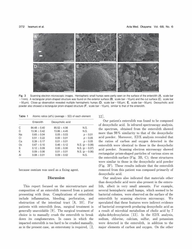
HPC-1S type osmium coater (Shinku Device Co., Ltd., Ibaraki, Japan) and examined closely with an S4800 scanning electron microscope (Hitachi, Tokyo, Japan). Images of the scanning electron microscopic obser-vation of the enterolith are shown in Fig. 3A-E. Hemispheric small humps were sporadically seen on the surface of the enterolith (Fig. 3A, arrows), but the surface was mainly composed of rectangular prism-shaped particles of various sizes (Fig. 3B, arrow). The rectangular prism-shaped structure was more clearly visible on the cut surface of the entero-lith (Fig. 3C). Close-up observation of the multiple hemispheric humps revealed that they were composed of numerous granules of approximately 1 micrometer in diameter (Fig. 3D, E). We speculated that these hemispheric humps were bacterial colonies and the rectangular prism-shaped structure was crystallized deoxycholic acid. We then analyzed a powdered form of deoxycholic acid commercially available as a reagent (Cat no. D2510; Sigma-Aldrich, St. Louis, MO, USA) by scanning electron microscope. The deoxycholic acid powder had a rectangular prism-shaped form (Fig. 3F) that was similar to that of the main component of the enterolith.
To investigate the elemental composition of the enterolith and deoxycholic acid powder, EDX analysis was performed by S4800 scanning electron micros-copy with an accelerating voltage of 25kV, and the EDX system EDAX Genesis APEX2 (AMETEK Inc., Paoli, PA, USA). The spectra of the EDX results were analyzed by Genesys software (AMETEK Inc.). The amount of each element and the atomic ratios (atオ) were quantified by the standardless EDAX ZAF quantification method. Hydrogen cannot be detected by this system. The atomic ratio of each element was measured at 3 different points on the surface of the enterolith and deoxycholic acid reagent, respectively. For comparisons, t-tests were performed using JMP 8.0.1 software (SAS Institute, Cary, NC, USA), and values of p<0.05 were considered significant. EDX analysis of the enterolith showed that the main constituents were carbon (88.48オ) and oxygen (13.36オ). This result was concordant with the molecular formula of deoxycholic acid, C24H40O4, and the ratios of carbon and oxygen were equal to those in the deoxycholic acid powder. Other elements detected in the enterolith are shown in Table 1. Osmium was detected in both the enterolith and deoxycholic acid powder. However, this was probably a contaminant,
371Ultrastructure of an EnterolithDecember 2014
D E F
A B C
Fig. 2 Images of the enterolith. Laparoscopic observation revealed a mass lodged in the small bowel in the right upper abdomen (A, arrow). The mass was impacted in the small bowel (B). A 4-cm yellow concretion was removed by enterotomy (C-E). The cut surface showed an air layer (F).
because osmium was used as a fixing agent.
Discussion
This report focused on the microstructure and composition of an enterolith removed from a patient presenting with ileus. Complications of enteroliths include inflammation, bleeding, perforation, and obstruction of the intestinal tract [8, 10]. For patients with enterolith ileus, surgical treatment is generally unavoidable [9]. The surgical treatment of choice is to manually crush the enterolith to break down its conglomerates. In cases in which the impacted enterolith is too hard to be crushed manually, as in the present case, an enterotomy is required, [2,
12]. Our patientʼs enterolith was found to be composed of deoxycholic acid. In infrared spectroscopy analysis, the spectrum, obtained from the enterolith showed more than 98オ similarity to that of the deoxycholic acid powder. Moreover, EDX analysis revealed that the ratios of carbon and oxygen detected in the enterolith were identical to those in the deoxycholic acid powder. Scanning electron microscopy showed rectangular prism-shaped particles of various sizes on the enterolith surface (Fig. 3B, C); these structures were similar to those in the deoxycholic acid powder (Fig. 3F). These results indicate that the enterolith removed from this patient was composed primarily of deoxycholic acid. Our analyses also indicated that materials other than deoxycholic acid were also present in the entero-lith, albeit in very small amounts. For example, several hemispheric small humps, which seemed to be bacterial colonies, were observed on the surface of the enterolith by scanning electron microscopy. We speculated that these features were indirect evidence of bacterial overgrowth producing deoxycholic acid as a result of microbial metabolism of cholic acid by 7 alpha-dehydroxylation [13]. In the EDX analysis, sodium, chlorine, calcium, sulfur, and potassium were detected in the enterolith, in addition to the major elements of carbon and oxygen. On the other
372 Acta Med. Okayama Vol. 68, No. 6Iwamuro et al.
Table 1 Atomic ratios (at%) (average±SD) of each element
Enterolith Deoxycholic acid
C 84.48±0.40 85.52±4.56 N.S.O 13.36±0.42 13.86±4.45 N.S.Na 0.65±0.04 0.05±0.03 p<0.01Cl 0.51±0.22 0.00±0.01 p<0.05Ca 0.39±0.17 0.01±0.01 p<0.05Os 0.67±0.10 0.46±0.12 N.S. (p=0.08)S 0.12±0.09 0.00±0.00 N.S. (p=0.07)K 0.09±0.06 0.01±0.01 N.S. (p=0.06)Al 0.08±0.01 0.09±0.02 N.S.
A C
E
B
D F
Fig. 3 Scanning electron microscopic images. Hemispheric small humps were partly seen on the surface of the enterolith (A, scale bar=1mm). A rectangular prism-shaped structure was found on the exterior surface (B, scale bar=10µm) and the cut surface (C, scale bar=50µm). Close-up observation revealed multiple hemispheric humps (D, scale bar=100µm; E, scale bar=50µm). Deoxycholic acid powder also showed a rectangular prism-shaped structure (F, scale bar=10µm), similar to that of the enterolith.

hand, such elements were not present in the deoxy-cholic acid powder, or present only in negligible amounts. These elements probably originated from intestinal juice or other substances contained in the intestinal tract, such as food debris or bacteria. Although the definite mechanism of formation of true enteroliths remains unknown [2], it has been postulated that enterolith formation may result from slowed gut transit, increased stasis of fecal matter in the small bowel, and the subsequent precipitation of enteric content. Such speculation about enterolith formation has been based on the fact that enteroliths arise in a variety of disease states where bowel stasis exists. For example, enteroliths have been reported in patients with bowel stricture due to tuberculosis or radiation, Meckelʼs diverticula, or Crohnʼs disease [2-11]. Thus, it is believed that bowel stasis and the subsequent acid-base imbalance and bacterial over-growth initiate the precipitation of substances, leading to enterolith formation. Other factors such as food content, oral medications, and enterobacterial flora might also affect the formation of enteroliths. However, no investigations have been conducted from this viewpoint to date, mainly because of the rarity of this disease entity. In general, enteroliths are spherical, ovoid, or polygonal in shape [1, 2, 8, 14]. In addition, enteroliths formed in the diverticula can be reniform [9, 15, 16]. The enterolith removed from the pres-ent patient, however, assumed none of these previ-ously reported shapes; it seemed to be twisted and coiled like knotted rope. The cut surface of the enterolith showed an air layer, suggesting that its shape was formed by being folded at least once (Fig. 2F). In regard to the mechanism of the enterolith formation, we speculated the following sequence, shown in Fig. 4. Stasis of the small bowel content may have been triggered by the post-surgical stricture of the small intestine due to the repair of the left inguinal hernia. As described above, stasis of the luminal contents causes bacterial overgrowth. Primary bile acids (i.e., cholic acid and chenodeoxy-cholic acid) are metabolized into secondary bile acids (i.e., deoxycholic acid and litocholic acid) by anaerobic bacteria, and these bile acids are relatively insoluble in the acidic milieu of the small bowel [9, 17, 18]. In the present patient, deoxycholic acid was probably produced in the stagnated bowel contents, which sub-
sequently precipitated into aggregates. The aggre-gates may then have incidentally passed through the bowel stricture, becoming twisted and coiled as they flowed into the small bowel lumen. This tangled mass would have increased in diameter and would finally have become impacted in the small bowel. The above-mentioned mechanism is strictly speculative, because it is impossible to confirm the development process of the enterolith. However, it appears to be supported by the following facts: i) no stricture or diverticula was observed during enterotomy in the small bowel where the enterolith was located, suggesting the enterolith had been formed at another site; ii) a con-trast study and CT scanning of the small bowel showed no diverticula, suggesting that the bowel sta-sis was not initiated by diverticula; iii) although post-surgical stricture due to the repair of the left inguinal hernia could not be clearly demonstrated by a contrast study, adhesions of the intestines to the left lower abdominal wall were observed by laparoscopy. In conclusion, we treated a patient with enterolith ileus. The surgically removed enterolith was com-
373Ultrastructure of an EnterolithDecember 2014
ab
c
Fig. 4 Schematic diagram of a possible mechanism of enterolith formation in this patient. Stasis of the small bowel content may have been caused by the post-surgical stricture of the small intes-tine due to the repair of the left inguinal hernia (arrowheads). The resulting bacterial overgrowth and the acidic milieu could have prompted the formation of the enterolith (a). As the enterolith passed through the bowel stricture (b), it may have become twisted and coiled as it flowed into the small bowel lumen, before finally becoming impacted in the small bowel (c).

posed of a high concentration of deoxycholic acid. This is a very rare disease entity and to our knowledge its ultrastructure has never been studied. This is thus the first study to analyze an enterolith using scanning electron microscopy, EDX, and infrared spectros-copy.
References
1. Jones MW, Koper B and Weatherhead WF: Crohnʼs disease with enterolith treated laparoscopically. JSLS (2005) 9: 339-341.
2. Monchal T, Hornez E, Bourgouin S, Sbardella F, Baudoin Y, Butin C, Salle E and Thouard H: Enterolith ileus due to jejunal diverticulosis. Am J Surg (2010) 199 :e45-47.
3. Gardiner KR and Maxwell RJ: Incidental enterolithiasis. Ulster Med J (1989) 58: 196-197.
4. Atwell JD and Pollock AV: Intestinal calculi. Br J Surg (1960) 47: 367-374.
5. Kirshtein B, Perry ZH, Klein J, Laufer L and Sion-Vardi N: Giant enterolith in ileal diverticulum following ileoplastic bladder augmen-tation. Int J Surg Case Rep (2013) 4: 385-387.
6. Tewari A, Weiden J and Johnson JO: Small-bowel obstruction associated with Crohnʼs enterolith. Emerg Radiol (2013) 20: 341-344.
7. Lai HC: Intestinal obstruction due to Meckelʼs enterolith. Pediatr Neonatol (2010) 51: 139-140.
8. Cartanese C, Campanella G, Milano E and Saccò M: Enterolith causing acute afferent loop syndrome after Billroth II gastrectomy:
a case report. G Chir (2013) 34: 164-166. 9. Liu CH, Huang KW, Mo YH and Yang PM: Enterolith ileus in a
patient with jejunal diverticulosis: sonographic findings. J Clin Ultrasound (2007) 35: 169-173.
10. Quazi MR, Mukhopadhyay M, Mallick NR, Khan D, Biswas N and Mondal MR: Enterolith containing uric Acid: an unusual cause of intestinal obstruction. Indian J Surg (2011) 73: 295-297.
11. Paige ML, Ghahremani GG and Brosnan JJ: Laminated radiopaque enteroliths: diagnostic clues to intestinal pathology. Am J Gastro-enterol (1987) 82: 432-437.
12. Hayee B, Khan HN, Al-Mishlab T and McPartlin JF: A case of enterolith small bowel obstruction and jejunal diverticulosis. World J Gastroenterol (2003) 9: 883-884.
13. Wells JE, Berr F, Thomas LA, Dowling RH and Hylemon PB: Isolation and characterization of cholic acid 7alpha-dehydroxylating fecal bacteria from cholesterol gallstone patients. J Hepatol (2000) 32: 4-10.
14. Miller M, Macdonald P and OʼBichere A: Gastrointestinal: Small bowel enterolith ileus. J Gastroenterol Hepatol (2001) 16: 697.
15. Fantl P, Rollo AJ and Strosberg H: Chemical analysis of an enter-olith. Gut (1965) 6: 384-386.
16. Pantongrag-Brown L, Levine MS, Buetow PC, Buck JL and Elsayed AM: Meckelʼs enteroliths: clinical, radiologic, and patho-logic findings. AJR Am J Roentgenol (1996) 167: 1447-1450.
17. Hofmann AF and Mysels KJ: Bile acid solubility and precipitation in vitro and in vivo: the role of conjugation, pH, and Ca2+ ions. J Lipid Res (1992) 33: 617-626.
18. Garnet DJ, Scalcione LR, Barkan A and Katz DS: Enterolith ileus: liberated large jejunal diverticulum enterolith causing small bowel obstruction in the setting of jejunal diverticulitis. Br J Radiol (2011) 84: e154-157.
374 Acta Med. Okayama Vol. 68, No. 6Iwamuro et al.